CroniconO P E N A C C E S S EC DENTAL SCIENCE
Research Article
Autotransplantation and Cryopreservation of Premolars. Tooth Loss Treatment in the Anterior Region
Hans Ulrik Paulsen1*, Jens Ove Andreasen2 and Ole Schwartz3
1Department of Orthodontics, Karolinska Institute, Stockholm, Sweden and Municipal Dental Health Service, Copenhagen, Denmark2Department of Maxillo-Facial Surgery, Copenhagen University Hospital, Copenhagen, Denmark3Department of Maxillo-Facial Surgery, Copenhagen University Hospital, Copenhagen, Denmark
*Corresponding Author: Hans Ulrik Paulsen, Department of Orthodontics, Karolinska Institute, Stockholm, Sweden and Municipal Dental Health Service, Copenhagen, Denmark.
Citation: Hans Ulrik Paulsen., et al. “Autotransplantation and Cryopreservation of Premolars. Tooth Loss Treatment in the Anterior Region”. EC Dental Science 12.2 (2017): 71-82.
Received: June 02, 2017; Published: July 05, 2017
Abstract
Avulsed and lost anterior teeth are common in young people. Using autotransplantation, it is possible to move problems in dental arches to regions where they are more easy to solve orthodontically. Transplantation of premolars with three-quarter root formation or full root formation with wide-open apical foramina provides the best prognosis for long-term survival. This article describes the use of autotransplantation and orthodontic treatment, together with cryopreservation, in connection with complicated trauma in the anterior region of an 8- year-old girl.
Keywords: Anterior Region; Autotransplantation; Cryopreservation; Tooth Loss
Introduction
Autotransplantation of teeth has become a predictable treatment approach for certain orthodontic conditions, namely aplasia of pre-molars and malformation or tooth loss of permanent incisors. In conventional orthodontics, tooth movements are usually limited to relatively small distances in sagittal, vertical, and transverse dimensions. Including tooth transplantation in the orthodontic equipment, tooth movement is no longer limited to short distances in a quadrant of the dental arch, but wider freedom is achieved to place the tooth exactly where needed, may it be the contralateral side in the dental arch or the opposing jaw. The periodontium is the key for bone induc-tion and bone modeling in the recipient area, and the tooth will normally erupt in a similar manner as a contralateral, non- transplanted tooth in its normal area.
However, transplantation of teeth is more traumatic to the pulp and periodontium than conventional orthodontics. Donor teeth with wide-open apices and a sing le root canal are recommended as grafts of choice for long- term survival. The surgical procedure is thus an essential key for the successful outcome of this treatment selection and outcome.
However, orthodontists are generally the most competent professionals to identify available donor teeth; because over all occlusal sta-tus must be assessed, the orthodontist should be considered a key person in planning, referring, and coordinating treatment that includes transplantation of teeth. With autotransplantation, it has become possible to move problems in the dental arches to regions where they are easier to solve orthodontically.
72
Autotransplantation and Cryopreservation of Premolars. Tooth Loss Treatment in the Anterior Region
Citation: Hans Ulrik Paulsen., et al. “Autotransplantation and Cryopreservation of Premolars. Tooth Loss Treatment in the Anterior Region”. EC Dental Science 12.2 (2017): 71-82.
The survival of transplanted premolars has been studied postoperatively for more than 30 years. A prerequisite for the use of this method is, however, a thorough knowledge of the prognosis. Thus, the stage of root development of transplants has been found to be of great importance. Transplantation at 3/4 to 4/4 root development of transplants with wide-open apices, a careful surgical technique pre-serving the periodontal ligament, and a light-force orthodontic tooth movement or tooth rotation carried out from 3 to 9 months after surgery are the most crucial factors for the survival of transplants and their continued development. Autotransplantation, if applied with knowledge of the predictive outcomes, is a method that supplements and enhances conventional orthodontic treatment [1-12].
This article describes the use of autotransplantation and orthodontic treatment, together with cryopreservation, in connection with a complicated trauma in the anterior region in an 8-year-old girl. Despite improved possibilities for treating trauma injuries in the anterior region, there are unfortunately still some types of trauma (e.g. exarticulation with replantation, intrusion and crown/root fractures), in which the permanent tooth is lost after treatment. This is due to root resorption.
The definitive treatment of such tooth loss includes an evaluation of the following treatment options:
• Prosthetic replacement in the form of a fixed partial denture
• Orthodontic closure of the diastema that has occurred
• Autotransplantation of a premolar to the anterior region followed by orthodontic closure of the donor area and morphologic reconstruction of the crown on the transplanted premolar.
Coordination of Surgical and Orthodontic Treatment
Many serious dental injuries are related to extreme maxillary overjet. Extraction of first premolars in connection with retraction of the anterior region of the maxillary jaw is therefore sometimes indicated. In that connection, the question of autotransplantation of premo-lars to the damaged area can arise. However, because of their root anatomy, these premolars are not suitable as donor teeth. The most suit- able teeth for transplantation are second premolars from the maxillary jaw and premolars from the mandibular jaw because of suitably shaped roots and a possibility of altering the shape of the crown. Furthermore, for transplantation of premolars to the anterior region, a complete analysis of the development of the patient’s teeth and jaws is required. Therefore, it is necessary to draw up an orthodontic treatment plan. The plan will depend on whether the patient has a malocclusion that requires treatment, as well as the dental injury. If the orthodontic treatment plan means that several premolars have to be extracted, there is a further treatment option - namely, freezing and storage of premolars in a tooth bank (cryopreservation) for possible later use.
In addition, an evaluation must be carried out to determine: (1) The type of premolar that best replaces a given incisor; (2) the timing of the treatment; (3) coordination of transplantation and orthodontic treatment; (4) restoration of the transplanted tooth; and (5) the possibility of storing premolars in a tooth bank for possible later use.
Treatment of a patient with replantation of three incisors
Description of the trauma
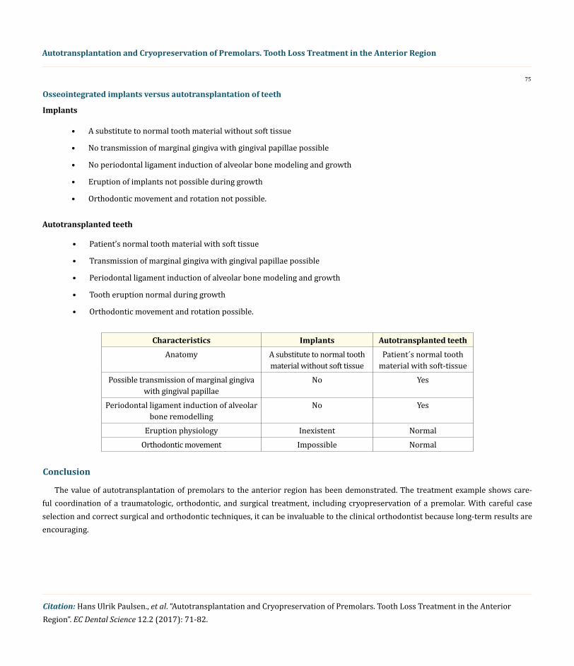
While playing in the schoolyard, an 8-year-old female child (Figure 1) fell and hit her face on a bench. This resulted in serious lacera-tion of about 1 cm of the mandibular jaw at the transition to the prolabia.
73
Autotransplantation and Cryopreservation of Premolars. Tooth Loss Treatment in the Anterior Region
Citation: Hans Ulrik Paulsen., et al. “Autotransplantation and Cryopreservation of Premolars. Tooth Loss Treatment in the Anterior Region”. EC Dental Science 12.2 (2017): 71-82.
Both maxillary central incisors and right mandibular central incisor were exarticulated with gingival lacerations in the traumatized regions, and the facial bone lamella was pushed in at the right maxillary central incisor region. Immediately after the fall, the teeth were gathered up from the asphalt and kept in a serviette for a few minutes after which they were immediately reset by the dentist at the school. On arrival at the hospital, it was found that the right maxillary central incisor was only partially re-implanted.
Primary trauma treatment and observation
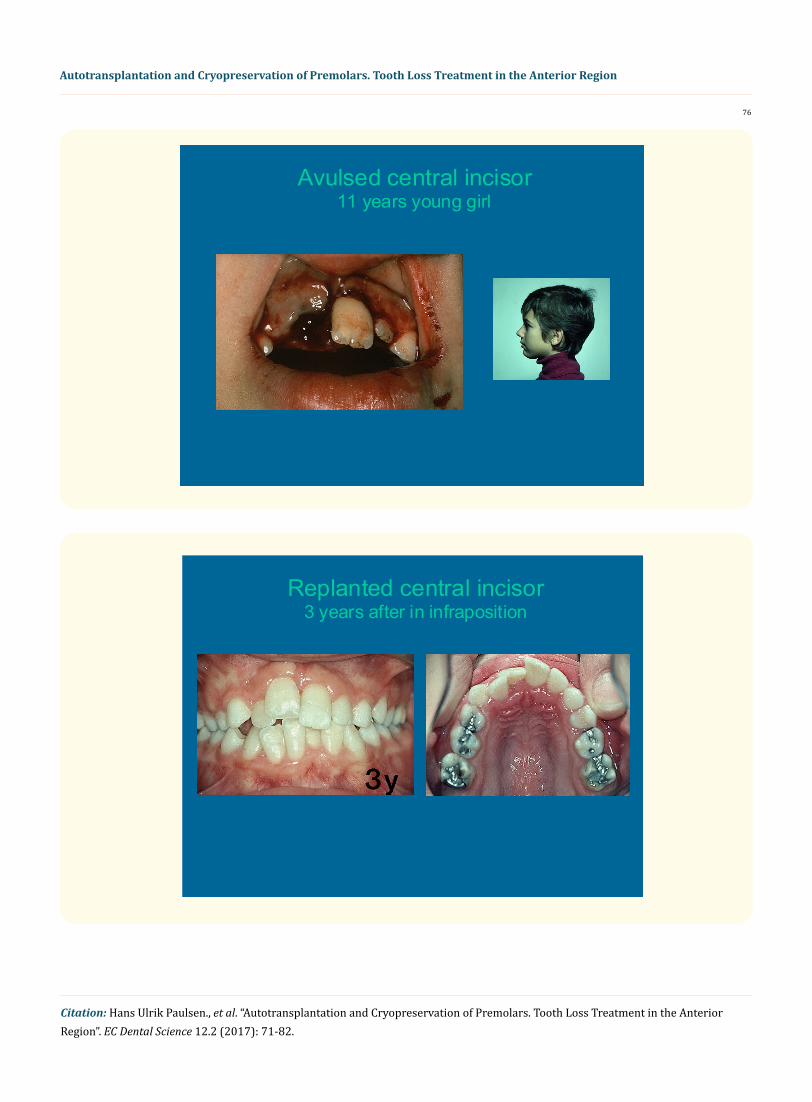
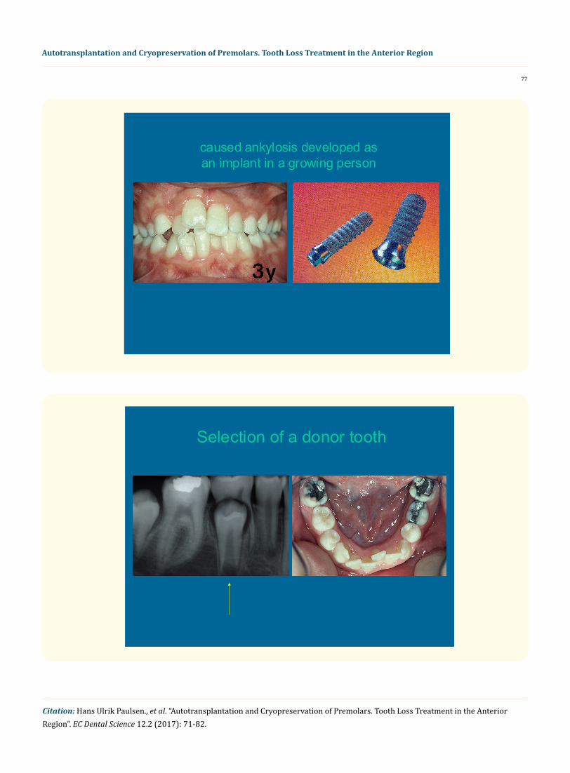
The right maxillary central incisor was removed from the alveolus about 90 min after the accident and was stored for 30 min in physi-ologic saline solution while the alveolar bone fracture was reset. The tooth was then replanted. All the damaged teeth were fixed with a scutaneous rail (A scutaneous rail is made of an elastic material. It is placed on the facial side of the traumatized and neighboring teeth. It allows the teeth to move during a fixation period, to prevent ankylosis). Two months after the trauma, ankylosis of the replanted teeth was suspected, particularly of the right maxillary central incisor, which was in slight infraposition. One year after the trauma, a combined radiographic and mobility examination showed that there was apparently no ankylosis of the left maxillary central incisor and the right mandibular central incisor. Pulp sensitivity was positive in all the replanted teeth. After 2 years, the right maxillary central incisor was clearly in infraposition. The right mandibular central incisor showed reparative-related root resorption in the middle of the distal root face and pulp obliteration.
Three years after the trauma, a coordinated orthodontic and surgical treatment plan was established because the infraposition of the right maxillary central incisor had increased (Figure 2) and (Figure 3). Due to the untreatable ankylosis, the patient was offered autotrans-plantation of a premolar to the right maxillary central incisor region.
Orthodontic examination
All the teeth had formed (except all third molars); the right maxillary central incisor was in pronounced infraposition and ankylosis, and the left maxillary canine showed mesial ectopic eruption. The first molars were poorly mineralized, with large restorations, particu-larly in the maxillary jaw. The maxillary overjet was caused by an enlarged inclination of the maxillary incisors, together with an enlarged sagittal jaw relation due to bimaxillary retrognathia. Both jaws were inclined posteriorly, particularly the mandible, resulting in an in-creased vertical jaw relationship. The maxillary jaw zone had increased to compensate a basal-based open bite. Furthermore, there was crowding of the anterior regions of the maxillary and mandibular jaws.
Treatment plan
Transplantation of the right mandibular second premolar (Figure 4) (alternatively, the left mandibular second premolar) was advised since extraction of the maxillary first molars would be part of the orthodontic treatment plan. The left mandibular second premolar would act as “reserve donor tooth,” either directly from the oral cavity or after an observation period during which the tooth was cryopreserved in Copenhagen University Hospital’s tooth bank. Extensive preliminary and subsequent orthodontic treatment had to be anticipated.
Treatment
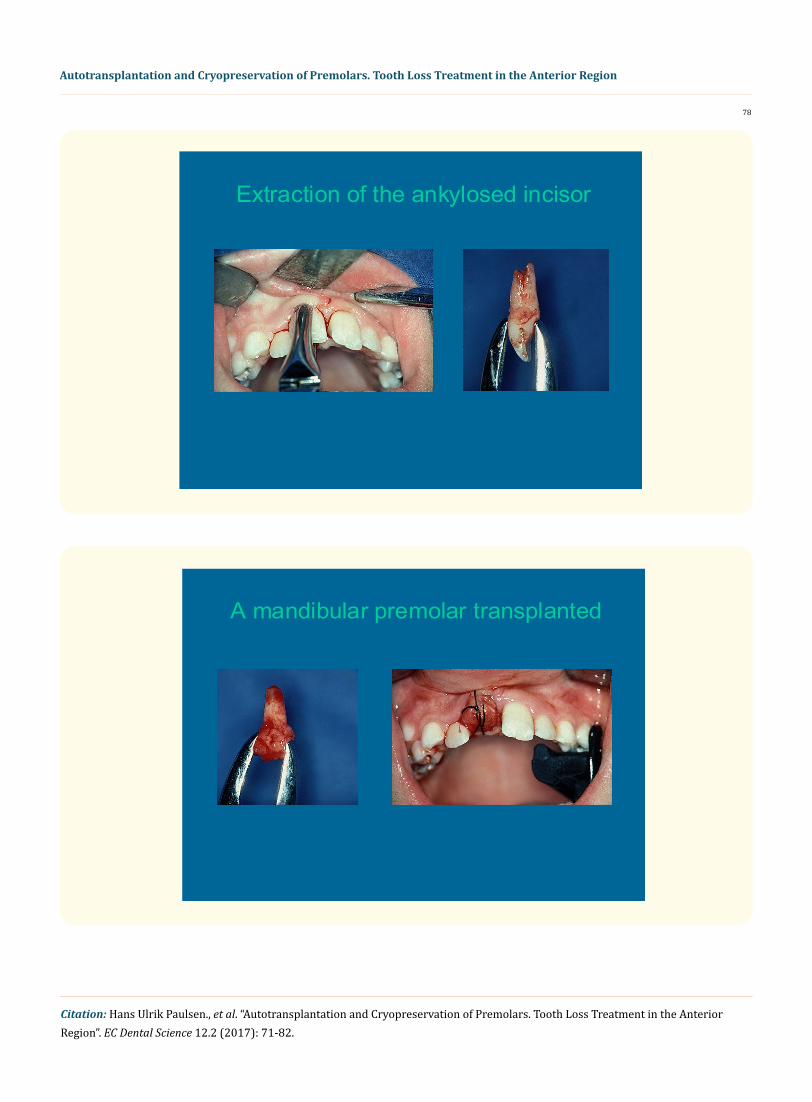
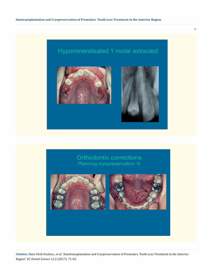
When the patient was 11 years 3 months of age, both mandibular second premolars were estimated to have reached 3/4 root length. To create room for transplantation of the right mandibular second premolar to the right maxillary central incisor region, an expansion plate was used for 2 months. At the time of transplantation, the right maxillary central incisor was extracted (Figure 5) after which the right mandibular second premolar was surgically removed and placed in the right maxillary central incisor region after expansion of the alveoli with a drill with internal cooling (Figure 6). Three months after the transplantation, electrometric sensitivity reaction of the pulp was positive for the new right maxillary central incisor. Ten months after autotransplantation, the maxillary first molars and the remain-ing temporary teeth were extracted to speed up eruption of the permanent teeth (Figure 7). At that time, incipient obliteration of the right
74
Autotransplantation and Cryopreservation of Premolars. Tooth Loss Treatment in the Anterior Region
Citation: Hans Ulrik Paulsen., et al. “Autotransplantation and Cryopreservation of Premolars. Tooth Loss Treatment in the Anterior Region”. EC Dental Science 12.2 (2017): 71-82.
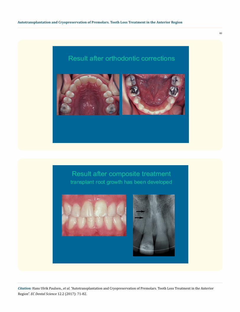
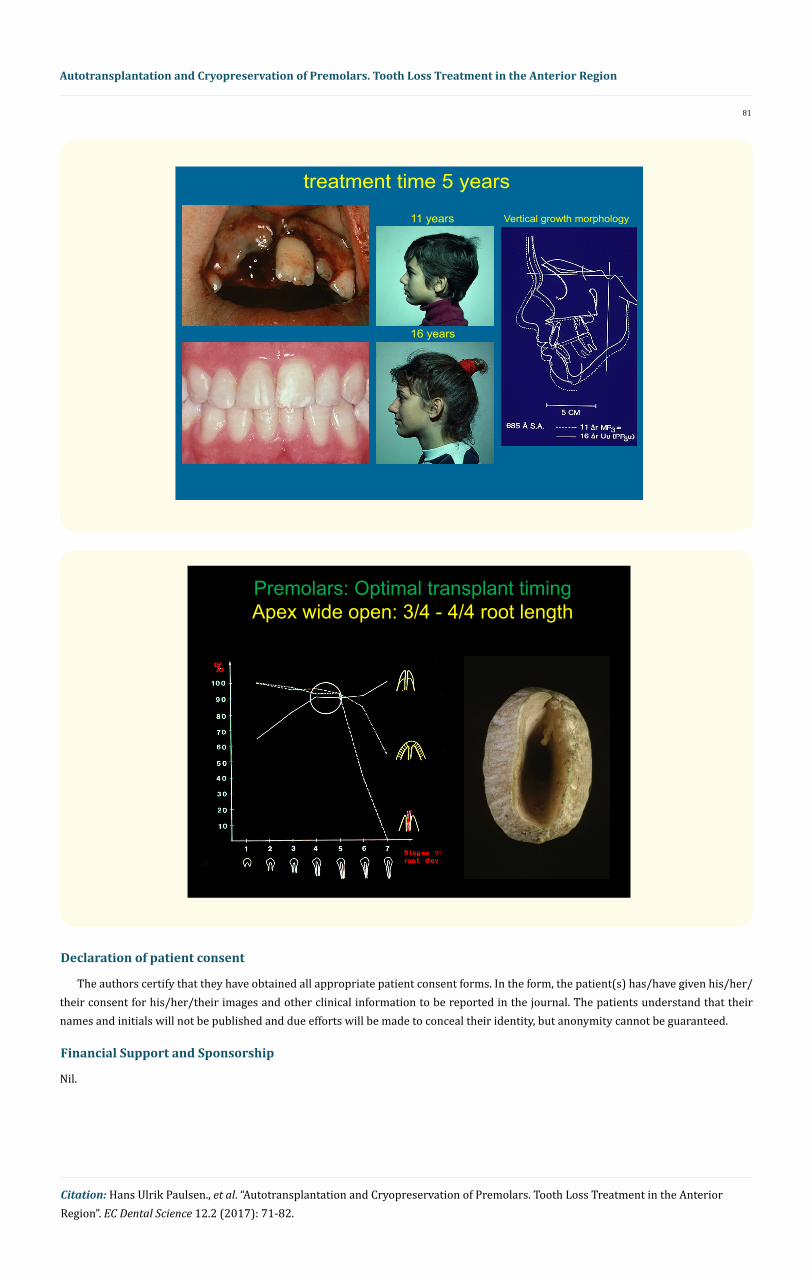
maxillary central incisor and incipient eruption of both maxillary second molars were found. Six months later, a fixed appliance was in-serted on both jaws. Two years after the transplantation, a radiograph of the new right maxillary central incisor showed total obliteration of the pulp formed before the transplantation, together with continued root growth of the transplant. In addition, the right mandibular central incisor had healed, with the distal root face almost remodeled, pulp obliterated, and the length of the root shorter than the ho-mologous incisor. Six months later, the transplant’s crown morphology was altered by selective grinding in the enamel and subsequent buildup with composite plastic. Three years after the transplantation, the prognosis for the transplant was considered so satisfactory that the alternative tooth, the left mandibular second premolar, could be extracted and cryopreserved for later use in case the new right maxil-lary central incisor or another tooth was later lost (Figure 8). A reverse headgear was placed for mesial shifting of the left first and second molars. Four years after the transplantation, when the patient was 16 years of age, the root of the new maxillary right central incisor was finally fully formed and a little longer than that of the maxillary left central incisor (Figure 9) and (Figure 10). All appliances were then removed and retainers were inserted and glued to the canines in both jaws.
Analysis of treatment
The pattern of growth was mainly vertical for both jaws. The alveolar prognathism was not reduced as a result of the extraction treat-ment (Figure 11).
Discussion
This case demonstrates the result of coordinated traumatologic, orthodontic, and surgical treatment, including the possibility of later use of a cryopreserved premolar in the event of the loss of the traumatized tooth [1,2]. The entire therapy took more than 7 years due to continued observation of the trauma, timing of the transplantation, guided eruption of maxillary jaw premolars and molars, orthodontic treatment, waiting for the premolar to develop to full root length for the tooth bank, and concluding orthodontic treatment.
Experimental and clinical research of autotransplantation of teeth in recent years has shown that it is now advantageous to include this method as an orthodontic treatment option [3-12]. When the method is used in the anterior region, it is important to conduct a thorough analysis and evaluation of the differences (esthetic, functional) and risks (durability, root resorption) resulting from intervention in the recipient region and also of the problems created in the donor region and how these can be solved in a traumatologic, orthodontic treat-ment plan [6,11-16]. The risks connected with the intervention relate mainly to the occurrence of pulp and periodontal complications in connection with the revascularization of these structures. A prospective study of the long-term prognosis for 370 autotransplanted premolars has shown that most healing complications could be predicted and avoided if the transplantation is performed at specific root stages. For example, (Figure 12) shows that root length, pulp healing (i.e., without partial or total pulp necrosis), healing of periodontal membrane (i.e., without root resorption), and achievement of expected root length are decisively related to the choice of donor tooth and its root development stage. In that connection, it seems obvious to transplant premolars at the 3/4 to full root development stage, assum-ing a fully open apex [stage 4], [stages 5], (Figure 12). In these stages, the transplantation is a predictable procedure that ensures optimum healing in more than 90% of cases [7-10].
75
Autotransplantation and Cryopreservation of Premolars. Tooth Loss Treatment in the Anterior Region
Citation: Hans Ulrik Paulsen., et al. “Autotransplantation and Cryopreservation of Premolars. Tooth Loss Treatment in the Anterior Region”. EC Dental Science 12.2 (2017): 71-82.
Osseointegrated implants versus autotransplantation of teeth
Implants
• A substitute to normal tooth material without soft tissue
• No transmission of marginal gingiva with gingival papillae possible
• No periodontal ligament induction of alveolar bone modeling and growth
• Eruption of implants not possible during growth
• Orthodontic movement and rotation not possible.
Autotransplanted teeth
• Patient’s normal tooth material with soft tissue
• Transmission of marginal gingiva with gingival papillae possible
• Periodontal ligament induction of alveolar bone modeling and growth
• Tooth eruption normal during growth
• Orthodontic movement and rotation possible.
Characteristics Implants Autotransplanted teethAnatomy A substitute to normal tooth
material without soft tissuePatient´s normal tooth
material with soft-tissuePossible transmission of marginal gingiva
with gingival papillaeNo Yes
Periodontal ligament induction of alveolar bone remodelling
No Yes
Eruption physiology Inexistent NormalOrthodontic movement Impossible Normal
Conclusion
The value of autotransplantation of premolars to the anterior region has been demonstrated. The treatment example shows care-ful coordination of a traumatologic, orthodontic, and surgical treatment, including cryopreservation of a premolar. With careful case selection and correct surgical and orthodontic techniques, it can be invaluable to the clinical orthodontist because long-term results are encouraging.
76
Autotransplantation and Cryopreservation of Premolars. Tooth Loss Treatment in the Anterior Region
Citation: Hans Ulrik Paulsen., et al. “Autotransplantation and Cryopreservation of Premolars. Tooth Loss Treatment in the Anterior Region”. EC Dental Science 12.2 (2017): 71-82.
Avulsed central incisor11 years young girl
Replanted central incisor3 years after in infraposition
77
Autotransplantation and Cryopreservation of Premolars. Tooth Loss Treatment in the Anterior Region
Citation: Hans Ulrik Paulsen., et al. “Autotransplantation and Cryopreservation of Premolars. Tooth Loss Treatment in the Anterior Region”. EC Dental Science 12.2 (2017): 71-82.
caused ankylosis developed asan implant in a growing person
Selection of a donor tooth
78
Autotransplantation and Cryopreservation of Premolars. Tooth Loss Treatment in the Anterior Region
Citation: Hans Ulrik Paulsen., et al. “Autotransplantation and Cryopreservation of Premolars. Tooth Loss Treatment in the Anterior Region”. EC Dental Science 12.2 (2017): 71-82.
Extraction of the ankylosed incisor
A mandibular premolar transplanted
79
Autotransplantation and Cryopreservation of Premolars. Tooth Loss Treatment in the Anterior Region
Citation: Hans Ulrik Paulsen., et al. “Autotransplantation and Cryopreservation of Premolars. Tooth Loss Treatment in the Anterior Region”. EC Dental Science 12.2 (2017): 71-82.
Hypomineralisated 1´molar extracted
Orthodontic correctionsPlanning cryopreservation -5
80
Autotransplantation and Cryopreservation of Premolars. Tooth Loss Treatment in the Anterior Region
Citation: Hans Ulrik Paulsen., et al. “Autotransplantation and Cryopreservation of Premolars. Tooth Loss Treatment in the Anterior Region”. EC Dental Science 12.2 (2017): 71-82.
Result after orthodontic corrections
Result after composite treatmenttransplant root growth has been developed
81
Autotransplantation and Cryopreservation of Premolars. Tooth Loss Treatment in the Anterior Region
Citation: Hans Ulrik Paulsen., et al. “Autotransplantation and Cryopreservation of Premolars. Tooth Loss Treatment in the Anterior Region”. EC Dental Science 12.2 (2017): 71-82.
Declaration of patient consent
The authors certify that they have obtained all appropriate patient consent forms. In the form, the patient(s) has/have given his/her/their consent for his/her/their images and other clinical information to be reported in the journal. The patients understand that their names and initials will not be published and due efforts will be made to conceal their identity, but anonymity cannot be guaranteed.
Financial Support and Sponsorship
Nil.
16 years
Vertical growth morphology11 years
treatment time 5 years
Premolars: Optimal transplant timingApex wide open: 3/4 - 4/4 root length
82
Autotransplantation and Cryopreservation of Premolars. Tooth Loss Treatment in the Anterior Region
Citation: Hans Ulrik Paulsen., et al. “Autotransplantation and Cryopreservation of Premolars. Tooth Loss Treatment in the Anterior Region”. EC Dental Science 12.2 (2017): 71-82.
Conflicts of Interest
There are no conflicts of interest.
Bibliography
1. Schwartz O. “Cryopreservation as long-term storage of teeth for transplantation or replantation”. International Journal of Oral and Maxillofacial Surgery 15.1 (1986): 30-32.
2. Schwartz O and Rank CP. “Autotransplantation of cryopreserved tooth in connection with orthodontic treatment”. American Journal of Orthodontics and Dentofacial Orthopedics 90.1 (1986): 67-72.
3. Kristerson L. “Autotransplantation of human premolars. A clinical and radiographic study of 100 teeth”. International Journal of Oral Surgery 14.2 (1985): 200-213.
4. Schwartz O., et al. “Autotransplantation of human teeth. A life-table analysis of prognostic factors”. International Journal of Oral Sur-gery 14.3 (1985): 245-258.
5. Schwartz O., et al. “Resorption of autotransplanted human teeth: A retrospective study of 291 transplantations over a period of 25 years”. International Endodontic Journal 18.2 (1985): 119-131.
6. Paulsen HU and Andreasen JO. “Autotransplantation in orthodontic treatment”. Nederlandse Vereniging Voor Orthodontische Studie. Studieweek (1985): 248-264.
7. Andreasen JO., et al. “A long-term study of 370 autotransplanted premolars. Part I. Surgical procedures and standardized techniques for monitoring healing”. European Journal of Orthodontics 12.1 (1990): 3-13.
8. Andreasen JO., et al. “A long-term study of 370 autotransplanted premolars. Part II. Tooth survival and pulp healing subsequent to transplantation”. European Journal of Orthodontics 12.1 (1990): 14-24.
9. Andreasen JO., et al. “A long-term study of 370 autotransplanted premolars. Part III. Periodontal healing subsequent to transplanta-tion”. European Journal of Orthodontics 12.1 (1990): 25-37.
10. Andreasen JO., et al. “A long-term study of 370 autotransplanted premolars. Part IV. Root development subsequent to transplanta-tion”. European Journal of Orthodontics 12.1 (1990): 38-50.
11. Paulsen HU., et al. “Pulp and periodontal healing, root development and root resorption subsequent to transplantation and orthodon-tic rotation: A long-term study of autotransplanted premolars”. American Journal of Orthodontics and Dentofacial Orthopedics 108.6 (1995): 630-640.
12. Paulsen HU and Andreasen JO. “Tooth eruption sub-sequent to transplantation. A longitudinal study of autotransplanted premolars”. European Journal of Orthodontics 20.1 (1998): 45-55.
Volume 12 Issue 2 July 2017© All rights reserved by Hans Ulrik Paulsen., et al.
Recommended