Copyright © 2011 American Heart Association.
Percutaneous Coronary Intervention Percutaneous Coronary Intervention
Sripal Bangalore, M.D., M.H.A.and
Deepak L. Bhatt, M.D., M.P.H., F.A.H.A
Copyright © 2011 American Heart Association.
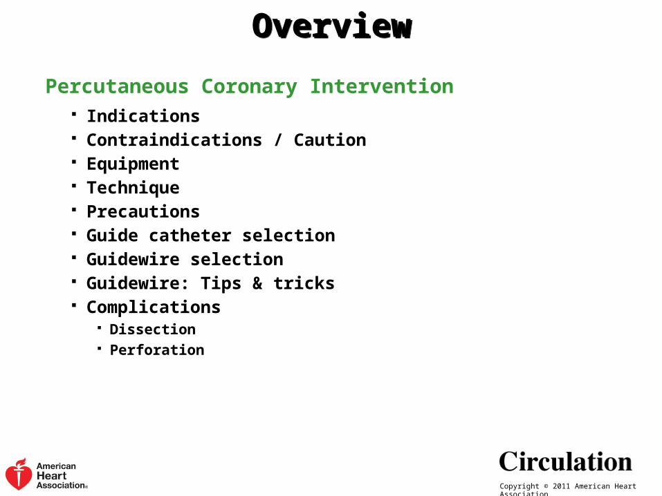
OverviewOverview
Percutaneous Coronary Intervention Indications Contraindications / Caution Equipment Technique Precautions Guide catheter selection Guidewire selection Guidewire: Tips & tricks Complications
Dissection Perforation
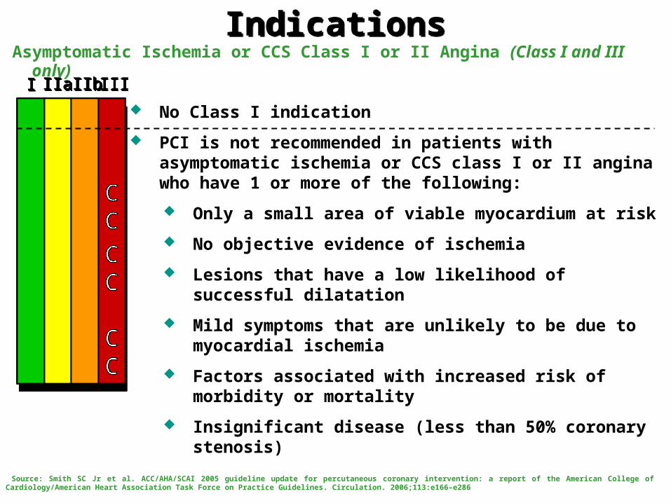
IndicationsIndicationsAsymptomatic Ischemia or CCS Class I or II Angina (Class I and III only)
III IIaIIaIIa IIbIIbIIb IIIIIIIIIIII IIaIIaIIa IIbIIbIIb IIIIIIIIIIII IIaIIaIIa IIbIIbIIb IIIIIIIIIIIaIIaIIa IIbIIbIIb IIIIIIIII
No Class I indication
PCI is not recommended in patients with asymptomatic ischemia or CCS class I or II angina who have 1 or more of the following:
Only a small area of viable myocardium at risk
No objective evidence of ischemia
Lesions that have a low likelihood of successful dilatation
Mild symptoms that are unlikely to be due to myocardial ischemia
Factors associated with increased risk of morbidity or mortality
Insignificant disease (less than 50% coronary stenosis)
Source: Smith SC Jr et al. ACC/AHA/SCAI 2005 guideline update for percutaneous coronary intervention: a report of the American College of Cardiology/American Heart Association Task Force on Practice Guidelines. Circulation. 2006;113:e166–e286
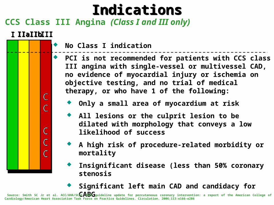
IndicationsIndicationsCCS Class III Angina (Class I and III only)
III IIaIIaIIa IIbIIbIIb IIIIIIIIIIII IIaIIaIIa IIbIIbIIb IIIIIIIIIIII IIaIIaIIa IIbIIbIIb IIIIIIIIIIIaIIaIIa IIbIIbIIb IIIIIIIII
No Class I indication
PCI is not recommended for patients with CCS class III angina with single-vessel or multivessel CAD, no evidence of myocardial injury or ischemia on objective testing, and no trial of medical therapy, or who have 1 of the following:
Only a small area of myocardium at risk
All lesions or the culprit lesion to be dilated with morphology that conveys a low likelihood of success
A high risk of procedure-related morbidity or mortality
Insignificant disease (less than 50% coronary stenosis
Significant left main CAD and candidacy for CABG
Source: Smith SC Jr et al. ACC/AHA/SCAI 2005 guideline update for percutaneous coronary intervention: a report of the American College of Cardiology/American Heart Association Task Force on Practice Guidelines. Circulation. 2006;113:e166–e286
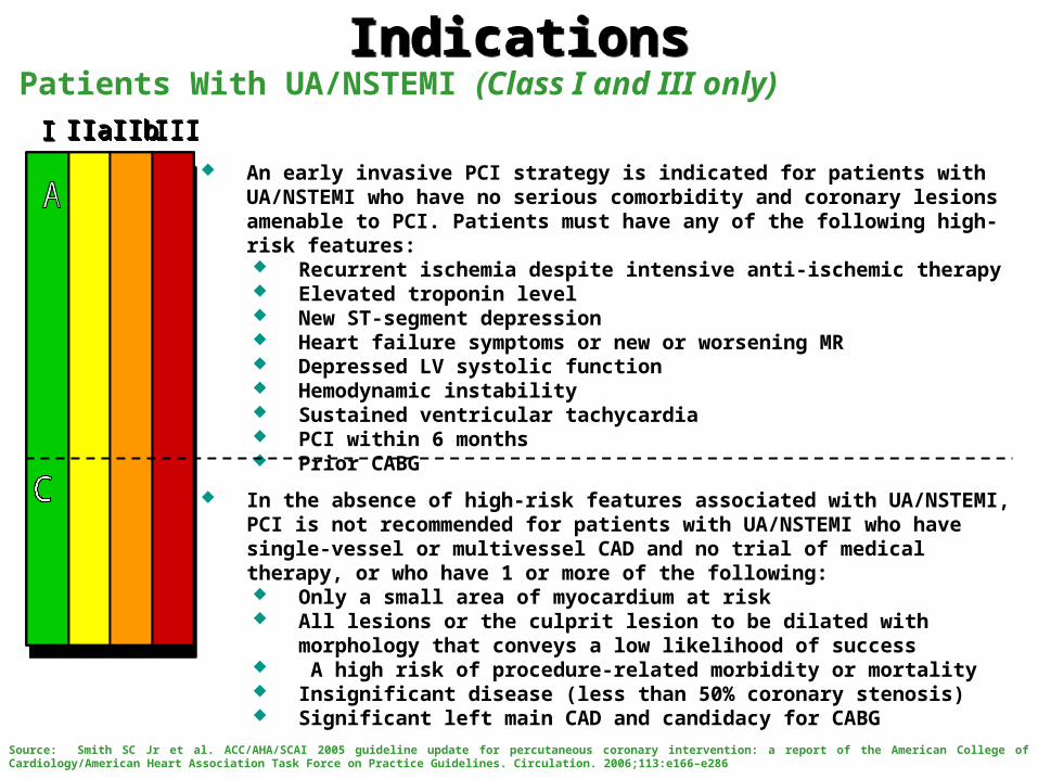
IndicationsIndicationsPatients With UA/NSTEMI (Class I and III only)
III IIaIIaIIa IIbIIbIIb IIIIIIIIIIII IIaIIaIIa IIbIIbIIb IIIIIIIIIIII IIaIIaIIa IIbIIbIIb IIIIIIIIIIIaIIaIIa IIbIIbIIb IIIIIIIII
An early invasive PCI strategy is indicated for patients with UA/NSTEMI who have no serious comorbidity and coronary lesions amenable to PCI. Patients must have any of the following high-risk features: Recurrent ischemia despite intensive anti-ischemic therapy Elevated troponin level New ST-segment depression Heart failure symptoms or new or worsening MR Depressed LV systolic function Hemodynamic instability Sustained ventricular tachycardia PCI within 6 months Prior CABG
In the absence of high-risk features associated with UA/NSTEMI, PCI is not recommended for patients with UA/NSTEMI who have single-vessel or multivessel CAD and no trial of medical therapy, or who have 1 or more of the following: Only a small area of myocardium at risk All lesions or the culprit lesion to be dilated with morphology that conveys a low
likelihood of success A high risk of procedure-related morbidity or mortality Insignificant disease (less than 50% coronary stenosis) Significant left main CAD and candidacy for CABG
Source: Smith SC Jr et al. ACC/AHA/SCAI 2005 guideline update for percutaneous coronary intervention: a report of the American College of Cardiology/American Heart Association Task Force on Practice Guidelines. Circulation. 2006;113:e166–e286
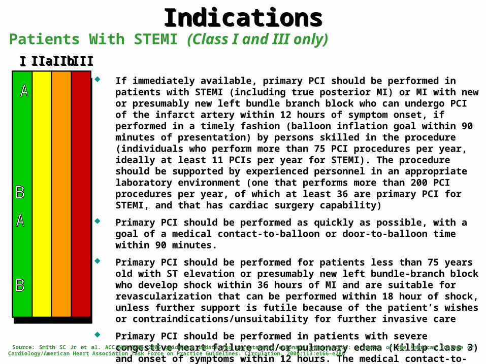
IndicationsIndicationsPatients With STEMI (Class I and III only)
III IIaIIaIIa IIbIIbIIb IIIIIIIIIIII IIaIIaIIa IIbIIbIIb IIIIIIIIIIII IIaIIaIIa IIbIIbIIb IIIIIIIIIIIaIIaIIa IIbIIbIIb IIIIIIIII
If immediately available, primary PCI should be performed in patients with STEMI (including true posterior MI) or MI with new or presumably new left bundle branch block who can undergo PCI of the infarct artery within 12 hours of symptom onset, if performed in a timely fashion (balloon inflation goal within 90 minutes of presentation) by persons skilled in the procedure (individuals who perform more than 75 PCI procedures per year, ideally at least 11 PCIs per year for STEMI). The procedure should be supported by experienced personnel in an appropriate laboratory environment (one that performs more than 200 PCI procedures per year, of which at least 36 are primary PCI for STEMI, and that has cardiac surgery capability)
Primary PCI should be performed as quickly as possible, with a goal of a medical contact-to-balloon or door-to-balloon time within 90 minutes.
Primary PCI should be performed for patients less than 75 years old with ST elevation or presumably new left bundle-branch block who develop shock within 36 hours of MI and are suitable for revascularization that can be performed within 18 hour of shock, unless further support is futile because of the patient’s wishes or contraindications/unsuitability for further invasive care
Primary PCI should be performed in patients with severe congestive heart failure and/or pulmonary edema (Killip class 3) and onset of symptoms within 12 hours. The medical contact-to-balloon or door-to balloon time should be as short as possible (i.e., goal within 90 minutes)
Source: Smith SC Jr et al. ACC/AHA/SCAI 2005 guideline update for percutaneous coronary intervention: a report of the American College of Cardiology/American Heart Association Task Force on Practice Guidelines. Circulation. 2006;113:e166–e286
IndicationsIndicationsPatients With STEMI (Class I and III only)
III IIaIIaIIa IIbIIbIIb IIIIIIIIIIII IIaIIaIIa IIbIIbIIb IIIIIIIIIIII IIaIIaIIa IIbIIbIIb IIIIIIIIIIIaIIaIIa IIbIIbIIb IIIIIIIII
Elective PCI should not be performed in a noninfarct-related artery at the time of primary PCI of the infarct related artery in patients without hemodynamic compromise
Primary PCI should not be performed in asymptomatic patients more than 12 hours after onset of STEMI who are hemodynamically and electrically stable
Source: Smith SC Jr et al. ACC/AHA/SCAI 2005 guideline update for percutaneous coronary intervention: a report of the American College of Cardiology/American Heart Association Task Force on Practice Guidelines. Circulation. 2006;113:e166–e286
IndicationsIndicationsPCI for Cardiogenic Shock (Class I and III only)
III IIaIIaIIa IIbIIbIIb IIIIIIIIIIII IIaIIaIIa IIbIIbIIb IIIIIIIIIIII IIaIIaIIa IIbIIbIIb IIIIIIIIIIIaIIaIIa IIbIIbIIb IIIIIIIII
Primary PCI is recommended for patients less than 75 years old with ST elevation or left bundle-branch block who develop shock within 36 hours of MI and are suitable for revascularization that can be performed within 18 hours of shock, unless further support is futile because of the patient’s wishes or contraindications/ unsuitability for further invasive care
Source: Scanlon PJ et al. ACC/AHA Guidelines for Coronary Angiography: Executive Summary and Recommendations. A Report of the American College of Cardiology/American Heart Association Task Force on Practice Guidelines (Committee on Coronary Angiography). Circulation 1999;99;2345-2357
IndicationsIndicationsPatients With Prior Coronary Bypass Surgery (Class I and III only)
III IIaIIaIIa IIbIIbIIb IIIIIIIIIIII IIaIIaIIa IIbIIbIIb IIIIIIIIIIII IIaIIaIIa IIbIIbIIb IIIIIIIIIIIaIIaIIa IIbIIbIIb IIIIIIIII
When technically feasible, PCI should be performed in patients with early ischemia (usually within 30 days) after CABG
It is recommended that distal embolic protection devices be used when technically feasible in patients undergoing PCI to saphenous vein grafts
PCI is not recommended in patients with prior CABG for chronic total vein graft occlusions
PCI is not recommended in patients who have multiple target lesions with prior CABG and who have multivessel disease, failure of multiple SVGs, and impaired LV function unless repeat CABG poses excessive risk due to severe comorbid conditions
Source: Smith SC Jr et al. ACC/AHA/SCAI 2005 guideline update for percutaneous coronary intervention: a report of the American College of Cardiology/American Heart Association Task Force on Practice Guidelines. Circulation. 2006;113:e166–e286

Copyright © 2011 American Heart Association.
There are no absolute contraindications to PCI.
Relative contraindications include:
Coagulopathy (Radial approach can be attempted based on urgency)
Decompensated congestive heart failure
Uncontrolled Hypertension
Pregnancy
Inability for patient cooperation
Active infection
Renal Failure
Contrast medium allergy
ContraindicationsContraindications
Copyright © 2011 American Heart Association.
Conscious sedation using a narcotic and a benzodiazepine
Vascular access: femoral (described in the section on vascular access and closure devices), radial, or brachial
Antiplatelet therapy with aspirin and a thienopyridine (clopidogrel or prasugrel)
Antithrombotic therapy with either unfractionated heparin, low molecular weight heparin, or bivalirudin
Glycoprotein receptor IIB/IIIA inhibitors can be used based on the procedure
Flush the selected guiding catheter (connected to a Y-port) with saline to ensure an air free system
Once arterial access is obtained (as described in the section on vascular access and closure devices) a guiding catheter of appropriate size and configuration is advanced over a 0.035 or 0.038 inch guidewire
Equipment & TechniqueEquipment & Technique
Copyright © 2011 American Heart Association.
Once in the ascending aorta, the guidewire is removed, the catheter allowed to bleed back to remove any thrombus or atherosclerotic debris
The catheter is then connected to a manifold assembly connected to a pressure transducer for continuous central pressure monitoring
The guiding catheter is flushed to ensure an air free system
The guiding catheter should then be filled with 3-4 cc of contrast and advanced to engage the coronary ostium, in the LAO projection
After ensuring that there is no ventricularization or damping of the pressure, 2 to 3 cc of contrast should be injected to confirm the position of the catheter in the coronary ostium
After adequate antithrombotic agents have been given, a 0.014 inch guidewire is advanced through the guide catheter into the coronary artery and across the lesion
At this stage, in the setting of thrombotic STEMI lesions, a manual aspiration catheter can be used to aspirate thrombus
TechniqueTechnique

Copyright © 2011 American Heart Association.
For SVG interventions, an embolic protection device should be deployed prior to any angioplasty or stenting if feasible (as described in the section on EPDs)
An appropriate size compliant balloon may now be advanced over this guidewire to the region of the stenosis and the balloon inflated with saline/contrast to pre-dilate the lesion
The balloon is then removed
Pre-dilatation should be avoided in SVG grafts (if possible) to prevent distal embolization
Once adequate pre-dilatation is performed, a stent of suitable type, size, and length is taken and is flushed to ensure an air-free system
This is advanced over the guidewire, across the lesion and deployed by inflating the balloon mounted stent to appropriate pressures
The stent balloon is now removed and coronary angiography performed to ensure no complications and to assess for adequate stent expansion
TechniqueTechnique
Copyright © 2011 American Heart Association.
Some laboratories believe in routine post-dilatation of all stents (except SVG interventions) to ensure adequate strut expansion
If desired, this is accomplished by using a non-compliant balloon of appropriate size and length
The balloon is removed and coronary angiography performed in two orthogonal views assessing the following:
Stent and the proximal and distal edge to ensure no dissections or perforation
The distal wire site to ensure no perforation
The ostium to ensure no dissection caused by the guiding catheter
The guidewire is then removed and angiography is performed to confirm adequate stent deployment and no complications as described above
The guiding catheter should then be removed and intravenous antithrombotic therapy stopped unless further continuation is required due to heavy thrombus burden
TechniqueTechnique
Copyright © 2011 American Heart Association.
Depending on the support provided by the guide catheter, they can be divided into the following 3 types:
Standard guide catheters which do not provide any additional support. Examples: JL4, JR4, etc.
Support guide catheters - these rest in the sinus of valsalva and provide more support than the standard guide catheters. Examples: AL, AR, etc.
Extra support guide catheters - these provide extra support from the back wall of the aorta and are especially useful in situations needing extra support such as tortuous or calcified lesions, chronic total occlusions, etc. Examples: EBU, XB, etc.
Coronary stenting can be performed using 4 to 6Fr guide catheters
Larger guide catheters (7-8Fr) are needed for more complex procedures- bifurcation stenting, rotational atherectomy, to provide extra support for chronic total occlusions or tortuous and calcified lesions
Guide Catheter SelectionGuide Catheter Selection

Copyright © 2011 American Heart Association.
Coronary guidewires are usually 0.010-0.018in diameter with the most commonly used being 0.014in made of stainless steel or nitinol
Length: standard length -175 to 190 cm; Exchange length- 270-400 cm Guidewires are classified:
Based on coating (increasing order of lubricity and decreasing order of tactile feedback) No coating Hydrophobic Hydrophilic - becomes gel when wet and reduces friction Polymer cover with hydrophilic coating
Based on support Soft Moderate support Extra support - For chronic total occlusions Super extra support - For chronic total occlusions
Coronary Guidewire SelectionCoronary Guidewire Selection
Copyright © 2011 American Heart Association.
Always start with the least traumatic wire (‘work horse’ wire)
Always advance the wire under fluoroscopic guidance
Never allow the guidewire tip to buckle (more prone to dissection)
Always ensure that the wire tip is free
If on wire advancement, frequent PVC’s are noted, wire tip might have perforated and may be irritating the myocardium. Withdraw the wire.
If wire gets trapped and difficult to retrieve (especially in calcified arteries) even with gentle traction, use a low profile balloon or small caliber catheter and advance until hinge point and withdraw both as a unit
For stenting over a bifurcation with a wire protecting the branch vessel, always deploy the stent at low pressures, withdraw the branch wire, re-cross through the stent strut and post-dilate to high pressures.
If wire tip breaks off and embolizes: Attempt to retrieve it using a snare If attempts fail, tip may be plastered against the wall using a stent
Coronary Guidewire: Tips & TricksCoronary Guidewire: Tips & Tricks
Copyright © 2011 American Heart Association.
Apart from the complications listed under diagnostic coronary angiography, those related to PCI include
Abrupt closure and dissections
Perforation
Intramural hematoma
Side branch occlusion
Distal embolization
Stroke
Non-fatal MI
Death
Emergent CABG
Others: ventricular arrhythmia, acute renal failure, radiation injury
ComplicationsComplications
Copyright © 2011 American Heart Association.
Usually due to balloon dilatation injury, during guidewire passage, or trauma due to the guiding catheter
Types:
Type A — Luminal haziness
Type B — Linear dissection
Type C — Extraluminal contrast staining
Type D — Spiral dissection
Type E — Dissection with reduced flow
Type F — Dissection with total occlusion
Treatment depends on the type of dissection and the TIMI flow of the involved vessel. Though stenting is routinely used to treat dissections, some dissections (Type A, B) can be left alone
Complications: DissectionsComplications: Dissections
Copyright © 2011 American Heart Association.
Usually due to guidewire exit, over-aggressive balloon dilation or stent deployment, and rarely due to cutting balloon angioplasty or during rotational atherectomy
Types:
Class I - intramural crater without extravasation
Class II - pericardial or myocardial blushing (staining)
Class III - perforation ≥1 mm in diameter with contrast streaming or cavity spilling
Treatment depends on the type of perforation. For severe perforations the following steps should be considered
Stop anticoagulation and consider reversal
Inflate balloon or a perfusion balloon to tamponade the site of perforation
Contra-lateral arterial access with an 8F sheath
Complications: PerforationComplications: Perforation
Copyright © 2011 American Heart Association.
Severe perforation treatment (continued):
If patient is hemodynamically unstable, perform pericardiocentesis immediately
Drain blood and autotransfuse into a central vein
Deflate the coronary balloon periodically to see if the extravasation stops. If not, reinflate the balloon to low pressures
Using contralateral access and an 8F guiding catheter, recross the site of perforation using a second wire (partially deflating the balloon when you are ready to cross the site)
Load a suitable size PTFE coated stent onto the guidewire, deflate the first balloon, and advance the stent to the site of perforation and deploy the stent. This should seal the perforation site
Complications: PerforationComplications: Perforation
Recommended