Biological and Pharmaceutical Bulletin Advance Publication by J-STAGE
DOI:10.1248/bpb.b19-01052
Ⓒ 2020 The Pharmaceutical Society of Japan
Advance Publication
August 8, 2020
Construction of genomic library and high-throughput screening of Pseudomonas
aeruginosa novel antigens for potential vaccines
Wanting Xu1, Lei Li
1, Xiaobin Wen
1, Qun Liu
1, Yan Liu
1, Xingyong Wang
2,3*,
Langhuan Lei2,3
, Qiushan Chen2,3
, Li Liu1,*
1 The second affiliated hospital of Chengdu, Chengdu, 610017, China.
2 Ministry of Education Key Laboratory of Child Development and Disorders, Chongqing,
400014, China.
3 Chongqing International Science and Technology Cooperation Center for Child
Development and Disorders, Chongqing, 400014, China.
Dr. Xingyong Wang: [email protected]
Dr. Li Liu: [email protected]

Biological and Pharmaceutical Bulletin Advance Publication
Abstract
Hospital-acquired infections with Pseudomonas aeruginosa have become a great challenge in
caring for critically ill and immunocompromised patients. The cause of high mortality is the
presence of multi-drug-resistant (MDR) strains, which confers a pressing need for vaccines.
Although vaccines against P. aeruginosa have been in development for more than several
decades, there is no vaccine for patients at present. In this study, we purified genomic DNA of
P. aeruginosa from sera of patients affected, constructed genome-wide library with random
recombinants, and screened candidate protein antigens by evaluating their protective effects
in vivo. After 13-round of screening, 115 reactive recombinants were obtained, among which
13 antigens showed strong immunoreactivity (more than 10% reaction to PcrV, a
well-characterized V-antigen of P. aeruginosa). These 13 antigens were: PpiA, PtsP, OprP,
CAZ10_34235, HmuU_2, PcaK, CarAd, RecG, YjiR_5, LigD, KinB, RtcA, and PscF. In vivo
studies showed that vaccination with PscF protected against lethal P. aeruginosa challenge,
and decreased lung inflammation and injury. A genomic library of P. aeruginosa could be
constructed in this way for the first time, which could not only screen candidate antigens but
also in a high-throughput way. PscF was considered as an ideal promising vaccine candidate
for combating P. aeruginosa infection and was supported for further evaluation of its safety
and efficacy.
Keywords
Genome-wide library, antigen screening, Pseudomonas aeruginosa

Biological and Pharmaceutical Bulletin Advance Publication
1. Introduction
P. aeruginosa (PA) is a kind of Gram-negative bacteria, which is widely existed in a
natural environment and is one of the most formidable opportunistic pathogens in the clinic
[1]. The prevalence of P. aeruginosa carriage is around 15% in hospitalized patients in the
Intensive Care Unit (ICU) [2]. In the Pediatric Intensive Care Unit (PICU), P. aeruginosa
infections even account for 55% bacterial infection in children [3]. Furthermore, over the last
15 years, the nosocomial infection caused by P. aeruginosa is associated with prolonging
hospital stay, high expense as well as complications [4-7]. Anti-Pseudomonas aeruginosa
agents that can be used to control P. aeruginosa infections at present are extremely limited in
clinical practice because P. aeruginosa is known to utilize their high levels of intrinsic and
acquired resistance mechanisms to counter most antibiotics [8-10]. The MDR phenotype
could be mediated by a wide array of mechanisms include multidrug efflux systems, enzyme
production, outer membrane protein (porin) loss, and target mutations, as well as the
formation of biofilms [11, 12]. The discovery and development of novel therapeutic strategies
against P. aeruginosa infections are urgently demanded and gained more and more attention.
Our historical experience fighting against pathogenic microorganisms indicates that
vaccines are one of the effective weapons to prevent and control them [13-16]. Hence, the
successful development of the P. aeruginosa vaccine will not only reduce the incidence of
infectious diseases, lessen the indiscriminately use of antibiotics, but also reduce the severity
of antibiotic resistance. In the past 40 years, numerous vaccines have been developed against
P. aeruginosa infection, and protective antigens used in these studies included
lipopolysaccharide (LPS), polysaccharide, polysaccharide conjugates, extracellular protein,
outer membrane protein (OMP), flagella, type 3 secretion system (T3SS), IC 43[17], as well
as pili [18]. Several vaccines have entered phase II and III clinical trials, but there is no
vaccine against P. aeruginosa authorized for immunization in humans so far [19, 20]. Failure
of these antigens was mainly attributed to multiple pathways utilized by P. aeruginosa to
cause infection and frequent variations in its genome. In contrast to some other bacterial
genomes, whose size reflects gene duplication rather than genetic diversity, the P. aeruginosa
genome has a large size of encoding almost 6,000 genes and contains numerous and distinct
Biological and Pharmaceutical Bulletin Advance Publication
gene families, which are predicted to encode outer membrane proteins, transport systems and
enzymes [21, 22]. The diversity of P. aeruginosa strains and genome makes it extraordinarily
difficult for selecting conversed antigens. As a consequence, the complexity and diversity of
the genetic components lead to the difficulty of antigen screening by the evaluation of an
individual protein.
In this study, we constructed a genome-wide library from a clinical strain P. aeruginosa
strain XN-1 that was isolated from a severely infected patient in Southwest Hospital in China.
And, this library was subjected to 13 rounds of screening by using an enzyme-linked
immunosorbent assay (ELISA), in which serum of convalescent patients with P. aeruginosa
infection was used as a primary antibody. In total, we obtained 115 reactive recombinants,
among which 13 antigens showed strong immunoreactivity (more than 10% reaction to PcrV,
a well-characterized V-antigen of PA), specifically PpiA, PtsP, OprP, CAZ10_34235,
HmuU_2, PcaK, CarAd, RecG, YjiR_5, LigD, KinB, RtcA, and PscF. Vaccination with PcrV
effectively protected mice from P. aeruginosa. To screen out strong reactive antigens, novel
antigens’ immunogenicity and their effects of protective rates were evaluated by experiments
on BALB/c mice. Novel candidate antigens of P. aeruginosa were screened out in this way,
which would lay a firm basis for the development of its vaccine.
2. Materials and methods
Ethics Statement
In this study, all animal care and use were performed according to the rules of Animal
Ethics Procedures and regulations of the People’s Republic of China. All animal
experiments in this study were approved by the Animal Ethical and Experimental
Committee of Chongqing International Science and Technology Cooperation Center for
Child Development and Disorders. All surgeries were conducted under the circumstance
of sodium pentobarbital anesthesia, and all efforts were engaged to minimize suffering.
Bacterial strains
Biological and Pharmaceutical Bulletin Advance Publication
The P. aeruginosa strain named XN-1, strain number CCTCC M 2015730, was isolated
from a severely infected patient in Southwest Hospital in China, whose serotype was detected
by Mei serotyping kit (Mei assay, Meiji Seika).
Animals
Eight-twelve week-old female BALB/c mice (weight at 18.0-22.0 grams) were
purchased from Experimental Animal Center of Chongqing Medical University, under the
circumstances of specific pathogen-free (SPF) conditions. Female New Zealand white rabbits
(weight at 2.0-2.2 kilograms) were provided by TenXin Company (Chongqing, China).
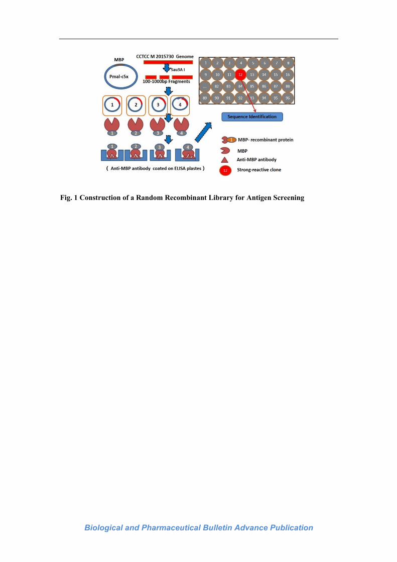
High-throughput screening of P. aeruginosa novel antigens
The anti- Maltose Binding Protein (anti-MBP)antibody was coated on ELISA plates to
capture MBP fusion protein expressed by random recombinants. Moreover, the serum of
patients who were infected with P. aeruginosa was used as a primary antibody to detect
reactivity. After that, the encoding sequence of strong reactive antigens was tested by genetic
sequencing, and their genetic information was obtained by sequence alignment on the BLAST
website (Fig. 1). Moreover, the relative reactivities of antigens were ranked according to their
titers of ELISA.
Construction of the genomic library
Genome DNA of P. aeruginosa XN-1 was extracted by using Wizard@
Genomic DNA
Purification kit (Promega) following the protocol and was digested with a restriction enzyme
named Sau3A I (’GATC) to get random fragments with the size of between 100 bp and 1000
bp. Then, these digested genomic DNA fragments were inserted to the plasmid of vector
pMal-c5x digested by restriction enzyme named BamH I (G’GATCC), followed by
dephosphorylation with Shrimp Alkaline Phosphatase (Takara, Bio. China) (Supplemental Fig.
1), to construct randomly recombinant plasmids. After purification, the fragments of the XN-1
genome were ligated to the pMal-c5x vector. Moreover, the reaction mixture was transformed
into E.coli X-Blue Competent cells (Huayueyang Biotech Company, China), which were used
to construct a genome-wide library by collecting colonies grown on antibiotic plates not only
Biological and Pharmaceutical Bulletin Advance Publication
massively, but also simultaneously. And all these recombinant plasmids were conformed
respectively by restriction endonuclease digestion and DNA sequencing.
Identification of random recombinants
To verify the success of the construction of P. aeruginosa antigen library, three clones
were selected randomly from the same plate, which was named as clone A, B, and C. After
digestion with enzyme BamH I, both clone A and clone C released a fragment of 1000 bp,
indicating that genome fragments of P. aeruginosa XN-1 were successfully inserted into
clone A and clone C, while clone B was considered as a negative (Supplemental Fig. 2).
Recombinant protein expressions
There were 2392 colonies in all picked from a relatively fresh plate (<4 weeks) and grown
at 37℃ in 20mL Luria-Bertani medium (Leagene Biotechnology, China) containing 100 μg/
mL of Ampicillin antibiotics. The recombinant proteins were induced by adding 1.0 mM
Isopropyl β-D-Thiogalactoside (IPTG) at 37°C for 15 hours. Then, these proteins were
purified by Maltose Binding Protein (MBP) resin using affinity chromatography, and
Bicinchoninic acid (BCA) from Applygen Technologies Inc.(Beijing, China)was used to
measure their concentrations.
Evaluations of genomic libraries and protein expressions of inserted fragments
A single colony PCR assay was used to initially determine the presence of DNA
fragments and their size distribution in the genomic libraries, which were selected from the
same plate after X-Blue competent cell transformation (Supplemental Fig. 3a). The inserted
fragments of samples NO.1, NO.2, and NO.3 were about 1000 bp, 250 bp, and 100 bp
respectively, but samples NO.4 and NO.5 did not have any fragment inserted, while sample
NO.6 was used as a positive control. Results of PCR indicated that inserted fragments had a
broad size distribution, and P. aeruginosa genomic library libraries were constructed with
acceptable complexity and representation. To evaluate their expressions, random
recombinants were selected from different screening batches and numbered as 1.1, 4.2, 4.19,
5.13, 5.15, and 5.18 respectively. After induced with IPTG, SDS-PAGE was used to detect

Biological and Pharmaceutical Bulletin Advance Publication
protein expressions. As the results indicated, pMal-c5x vector and pMal-c5x-PcrV expressed
protein with expected sizes after IPTG induction. Moreover, samples NO.1.1, NO. 5.18, NO.
5.15, and NO. 4.2 were observed to have protein bands at 55 kD、50 kD、45 kD and 50 kD
respectively, which suggested that these recombinants could express MBP fusion proteins
correctly. However, samples NO. 5.13 and NO. 4.19 did not, with no obvious protein band
and not expressing MBP fusion protein (Supplemental Fig. 3b). Preparations of rabbit
anti-MBP IgG antibody
The maltose-binding proteins (MBP) were produced by pMal-c5x/X-Blue transformed in
E. coli. After induced by 0.4 mM IPTG, MBP was purified by amylose affinity resin (NEB
company). After mixing by 50 μL with Freund Adjuvant (Sigma), 200μL of MBP-
recombinant protein was injected with the final concentration of 0.5 mg/rabbit on days 1st,
14th, and 21
st respectively. On the 28
th day, blood was collected by cardiac puncture under
anesthesia on the rabbits. Serum was obtained by allowing blood to stand for 1 hour at 37°C
followed by centrifugation with 8000 rpm for 10 min at 4℃ to remove the clotted material.
Anti-MBP IgG antibody was obtained by affinity purification with Protein A.
Screening strong reactive antigens
The rabbit anti-MBP IgG antibody was diluted with coating buffer (50mM of
carbonate/bicarbonate buffer, pH 9.6) to the final concentration of 10 μg/mL, and was coated
on the wells of microtiter plates at 37°C for 4 hours. After washing with sterile phosphate
tween buffer (PBST) four times, 100 μL supernatant of recombinant proteins was added into
each well and incubated for 1 hour at 37℃. Then strips were washed with PBST for four
times, a convalescent mixed serum of P. aeruginosa infection patients was used as the
primary antibody, and goat anti-human IgG was used as a secondary antibody for ELISA
detection. As a result, the relative reactivity of each random recombinant compared to PcrV
was calculated by the formula, Relative Reactivity %=(ODX-ODMBP)/(ODPcrV-ODMBP)×100%.
In this formula, ODX is the value of random recombinants in OD600nm, while ODMBP is a
negative control. The mean of log2 titers were used to express antibody express (ns = no

Biological and Pharmaceutical Bulletin Advance Publication
significance.) Besides, we also use one-way ANOVA to analyze multiple comparisons
between different groups.
Preparations of recombinant strong reactive antigens
We used PCR to amplify inserted DNA fragments of 13 antigens respectively, to obtain
the full-length sequences of the inserted gene fragments by using respective primers shown in
Supplemental Tab. S1. Meanwhile, all the amplified conditions of candidate antigens were
shown in Supplemental Tab. S2, and the amplification system was in Supplemental Tab. S3.
Then these genes were cloned into pMal-c5x vector to express MBP-antigen fusion protein.
The full-length of DNA sequences were then ligated to vector pMal-c5x, and transformed into
E. coli. After induction with 0.4 mM IPTG, 115 in all recombinants strong reactive antigens
were obtained, and 13 strong reactive candidate antigens of P. aeruginosa were obtained by
DNA sequencing and purified by amylose affinity resin. Then these 13 antigens plus MBP,
Al(OH)3 control group consisted of fifteen groups for the first, second-round animal
experiment.
Levels of antibody IgG detected in mice immunized with strong reactive antigens
150 female Balb/c mice in total were divided into 15 groups, which were matched for
sex, age, and weight for the first round, and another 150 Balb/c mice were also matched for
the second round under the same condition. Purified candidate antigens were mixed with
Al(OH)3 adjuvant and emulsified at 4 ℃ for 4 hours. Each emulsified antigen was injected
into 10 Balb/c mice intraperitoneally with the concentration of 50 μg/ mouse on the 1st
day,
the 14th day, and the 21
st day respectively, with MBP and Al(OH)3 injection on mice as a
negative control and an adjuvant control. After the final injection on the 7th day, 500 μL tail
venous blood was collected from mice for ELISA assay. In ELISA results, the cut-off value
of specific IgG antibody was calculated by the formula, cut-off value%= mean of the control
group [MBP, Al(OH)3] ×2.1×100. And in the second round, another 150 Balb/c mice in these
15 groups were also gone through the same immunity process and were calculated their
cut-off value%. Thus candidate antigens were ranked according to their mean cut-off values.
Biological and Pharmaceutical Bulletin Advance Publication
Evaluations of the protective effect of strong reactive antigens
On the 10th day after the final immunization, all vaccine mice were challenged with 1%
pentobarbital sodium as an anesthetic. After that, a volume of 20μL P. aeruginosa XN-1, with
a lethal dose of 2×10^9
CFU/mL, was used for tracheal intubation on mice. Moreover,
activities and survival rates of mice were carefully recorded every 12 hours for seven
consecutive days, and the protection rates of these reactive antigens were calculated, while the
top five of them were listed.
3.Statistical analysis
The data was presented as mean ±Standard Deviation(SD) or mean ±Standard Error of
Mean (SEM). The scores were recorded in a blind way. Kaplan-Meier survival curves were
used to analyze survival data. To calculate P values, non-parametric Mann-Whitney test,
log-rank test, Student’s t test, one-way ANOVA with Bonferroni correction were used to
depend on sample distribution and variation as mentioned in figure legends (SPSS statistics
18.0 and GraphPad Prism 6.0). Significant difference was accepted at P<0.05.
4.Results
Screening of P. aeruginosa strong reactive antigens
Twelve convalescent serums of P. aeruginosa infected patients were collected from the
Southwest Hospital and mixed in equal volumes. Through 13 cycles of screening, a total of
2392 random recombinants with DNA fragments were selected, 115 reactive recombinants
were obtained by ELISA testing, among which 13 antigens displayed stronger reactivity than
the control MBP group (Fig. 2). These reactive antigens were as follows, PpiA, PtsP, OprP,
CAZ10_34235, HmuU_2, PcaK, CarAd, RecG, YjiR_5, LigD, KinB, RtcA, PscF, compared
to MBP as control (P <0.0001). Their bioinformatics information including sample numbers,
gene, protein name, subcellular localization, amino acid sequence inserted, and length of
amino acid insertion fragments was listed in Tab. 1.
Evaluations of protective effects of candidate antigens

Biological and Pharmaceutical Bulletin Advance Publication
After IPTG induction, MBP-antigen recombinant proteins were purified with MBP tag.
Firstly, the purity of thirteen antigens was more than 80% detected by SDS-PAGE, which
could be made use of the following animal experiments (Supplemental Fig. 4a, Supplemental
Fig. 4b). Further, tilters of antigen-specific IgG antibodies in rat serum were detected by
ELISA (Fig. 2). Results indicate that these antigens could induce the antigen-specific IgG
antibody in vivo, and their tilters were significantly different from the IgG antibody induced
by the MBP tag. These results strongly indicated that PscF had the highest tilter among them
with its strongest immunogenicity, besides the antibody tilters of PpiA, PtsP, OprP, PecG,
and LigD were the next, suggesting them relative strong immunogenicity.
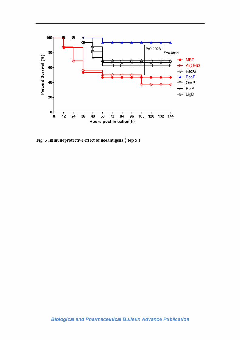
Novel antigens immunization provided protective effects in mice challenged with P.
aeruginosa XN-1
A significant increase in survival, as a function of time postinfection, was observed in
mice that received the recombinant vaccines compared to mice that received either PBS or
vector alone. As shown in Fig. 3, PscF was the most effective, providing host-protection for
18 of 20 (90%) of the immunized mice. Immunization with LigD, RecG yielded lesser,
though still significant degrees of survival, with 16/20 (80%) the mice surviving the infection.
In contrast, only 8/20 (40%) of control mice treated with MBP vector and 6/20 (30%) of mice
immunized with Al(OH)3 adjuvant survived after infection. The highest protective rates were
PscF, LigD, RecG, OprP, and PtsP. Among them, antigen PscF has the best protective rate
which reaches 90%, and the second protective rate was 80% indicating antigens LigD and
RecG, while the third protective rate was 60% with antigens OprP and PtsP respectively,
compared to 40% with MBP on mice as a negative control, with 30% Al(OH)3 as an adjuvant
control alone. Moreover, the survival rates of PscF groups were significantly higher than that
of Al(OH)3 group ( P PscF-Al(OH)3 =0.0014) and MBP group ( P PscF-MBP =0.0028). However,
there was no difference between PscF, LigD, RecG, OprP, and PtsP group ( P PscF- LigD=0.1063,
P PscF- RecG =0.1063, P PscF- OprP =0.0533, P PscF- PtsP=0.0533) .
Biological and Pharmaceutical Bulletin Advance Publication
5. Discussion
P. aeruginosa is a gram-negative bacterium that causes widely severe infection in
critically ill patients [23]. For the moment, it is one of the highest infective, opportunistic
pathogens in ICU ward and ventilator-associated pneumonia (VAP) [24, 25]. In common
VAP, P. aeruginosa attributes to lethality as 13.5% even with sufficient antibiotic treating.
Nevertheless, in MDR strains combined VAP, even with adequate antibiotic treatment, P.
aeruginosa inducing mortality has risen to 41.9% [26]. And this phenomenon may be closely
relative to powerful virulence factors, such as Type III Secretion System (TTSS) [26, 27],
which leads to serious infections in patients with low immunity [28]. Besides, T3SS
extracellular components were mainly composed of PcrG, PcrV, PcrH, PopB, and PopD[29],
and PcrV was one of the essential virulence factors of it [30]. In this study, we have
constructed a genome-wide library from a clinic strain P. aeruginosa XN-1 and acquired
several reactive recombinants and candidate antigens. However, we do not cover the entire
genome of P. aeruginosa for the present, which needs a larger size of the sample to get more
recombinant plasmids by this high-throughput screening method.
As a key virulence factor, PcrV plays an important role in the pathogenesis of P.
aeruginosa and is an excellent candidate antigen for the P. aeruginosa vaccine [31-33]. For
instance, the recombinant DNA vaccine pIRES-toxAm-pcrV was a candidate vaccine for the
prevention and control of P. aeruginosa infection in the treatment of PA-induced lung
infections [34]. In the study conducted by Ali SO et al., MEDI3902 exhibited a desired
antibacterial activity by inhibition of PcrV, promoting opsonophagocytic killing (OPK)
activity, and targeting inhibiting P. aeruginosa host cell attachment [35]. In another study
conducted by Milla Carlos E. et al., recombinant anti-PA- PcrV antibody Fab’ fragment
inhibited the function of TTSS, which at the same didn’t activate immune cells and
inflammation [36]. Very recently, in yet another study (for Aguilera-Herce Julia et al.), an
attenuated strain of S. enterica was an eligible carrier for delivering P. aeruginosa antigen
PcrV to design a vaccine to fight against its infection [37]. As a result, PcrV could be used as
a diagnostic marker for P. aeruginosa infection in serological experiments. All factors
Biological and Pharmaceutical Bulletin Advance Publication
considered, we have used PcrV as a positive control in the reactions of antigen evaluation in
this study, with MBP as a negative control, Al(OH)3 as an adjuvant control.
Moreover, PscF was considered as the candidate antigen of P. aeruginosa vaccine for
the following reasons. First of all, PscF was located on the main component of the T3SS
needle complex of P. aeruginosa [38] and played an important role in regulating NF-κB and
AP-1 pro-inflammatory cytokine pathway [39]. Secondly, PscF promoted mature immune
response of caspase-1 and IL-1β and enabled PscG and PscE to exist stably in P. aeruginosa
cells before polymerization [40, 41], which could be recognized as a target for P. aeruginosa
vaccine. Our animal experiment results indicated that PscF had the highest IgG antibody titer
and the best immuno-protective effects in vivo among 13 candidate antigens whose survival
rate rose to 90%, compared to 40% in MBP negative control, 30% in Al(OH)3 adjuvant
control.
Above all, these results indicate that PscF might be an ideal antigen for P. aeruginosa
vaccine. The whole genome-wide library from a clinical strain P. aeruginosa strain XN-1 was
constructed successfully in this study. Based on this library, thirteen strong reactive antigens
were successfully screened immunologically. Meanwhile,
Finally, PscF was identified as a potential candidate protein antigen of P. aeruginosa,
as a result of the best immunogenicity and the highest survival rate among animal
experiments, which would lay a solid foundation for the development of novel protein
vaccines of P. aeruginose.
However, our genomic library of P. aeruginosa did not list detailed statistical data of
neo-antigens’ functions, which would be a limitation of our study. Furthermore, we would
like to apply this high-throughput method for screening antigens among other P. aeruginosa
clinic isolations, combined with HPLC to measure molecular weight and endotoxin levels
before immunization for the next.
Acknowledgment
The authors would like to acknowledge Chongqing Medical University for Female
Balb/c mice; Southwest Hospital for P. aeruginosa strain (XN-1) and convalescent serums of
Biological and Pharmaceutical Bulletin Advance Publication
P. aeruginosa infected patients; Wangxing Yong for construction of the library; Langhuan
Lei for tracheal intubation in mice; Qiushan Chen for ELISA assay; Li Liu, Lei Li, Xiaobin
Wen for revising discussions; and Qun Liu, Yan Liu for editing the manuscript.
Conflict of interest
The authors declare no conflict of interest.
Supplementary Materials
The online version of this article contains supplementary materials.

Biological and Pharmaceutical Bulletin Advance Publication
References:
1. Moradali, M.F., S. Ghods, and B.H. Rehm, Pseudomonas aeruginosa Lifestyle: A
Paradigm for Adaptation, Survival, and Persistence. Front Cell Infect Microbiol,
2017. 7: p. 39.
2. Hoang, S., Georget A, Asselineau J, Risk factors for colonization and infection by
Pseudomonas aeruginosa in patients hospitalized in intensive care units in
France.Plos One. Vol. 13. 2018. e0193300.
3. Murni, I., Duke Trevor, Daley Andrew J, Antibiotic resistance and mortality in
children with nosomial bloodstream infection in a teaching hospital in
indonesia.2016. 47(5): p. 983-93.
4. Tacconelli, E., Smith G, Hieke K, Epidemiology, medical outcomes, and costs of
catheter-related bloodstream infections in intensive care units of four European
countries: literature- and registry-based estimates. 2009. 72(2): p. 97-103.
5. Sánchez-Velázquez, L.D., S.P.D.L. Rosales, and M.S.R.J.A.o.M.R. Frausto, The
Burden of Nosocomial Infection in the Intensive Care Unit: Effects on Organ Failure,
Mortality and Costs. A Nested Case-Control Study. 2006. 37(3): p. 370-375.
6. Laupland, K.B., Lee H, Gregson DB., Cost of intensive care unit-acquired
bloodstream infections. 2006. 63(2): p. 124-132.
7. Pittet, D., D. Tarara, and R.P. Wenzel, Nosocomial Bloodstream Infection in
Critically III Patients: Excess Length of Stay, Extra Costs, and Attributable Mortality.
JAMA, 1994. 271(20): p. 1598-1601.
8. Liu, S., Wang M.,Zheng L., Antimicrobial Resistance Profiles of Nosocomial
Pathogens in Regional China: A Brief Report from Two Tertiary Hospitals in China.
2018. 24: p. 8602-8607.
9. Rees, V., Yadav R., Rogers KE., Meropenem Combined with Ciprofloxacin Combats
Hypermutable Pseudomonas aeruginosa from Respiratory Infections of Cystic
Fibrosis Patients. 2018. 62(11).
10. Bassetti, M., Vena A., Russo A., Rational approach in the management of
Pseudomonas aeruginosa infections. 2018. 31(6): p. 578-586.

Biological and Pharmaceutical Bulletin Advance Publication
11. Poole, K., Pseudomonas aeruginosa: resistance to the max. Front Microbiol, 2011. 2:
p. 65.
12. Morita, Y., Tomida J., and Kawamura Y., Resistance and Response to
Anti-Pseudomonas Agents and Biocides, in Pseudomonas: Volume 7: New Aspects of
Pseudomonas Biology, J.-L. Ramos, J.B. Goldberg, and A. Filloux, Editors. 2015,
Springer Netherlands: Dordrecht. p. 173-187.
13. Zhou, X., Santosuosso, M., McCormick S., Recent advances in the development of
adenovirus- and poxvirus-vectored tuberculosis vaccines. 2005. 5(5): p. -.
14. Merakou, C., M.M. Schaefers, and G.P. Priebe, Progress Toward the Elusive
Pseudomonas aeruginosa Vaccine. Surg Infect (Larchmt), 2018. 19(8): p. 757-768.
15. Rashid, M.I., Naz, A., Ali, A, Andleeb S, Prediction of vaccine candidates against
Pseudomonas aeruginosa: An integrated genomic and proteomics approach. 2017.
109(3–4): p. 274-283.
16. Lucero, Y., R. Vidal, and G.M. O'Ryan, Norovirus vaccines under development.
Vaccine, 2018. 36(36): p. 5435-5441.
17. Adlbrecht, C., Wurm, R.,Depuydt, P, Efficacy, immunogenicity, and safety of IC43
recombinant Pseudomonas aeruginosa vaccine in mechanically ventilated intensive
care patients-a randomized clinical trial. Crit Care, 2020. 24(1): p. 74.
18. Hoggarth, A., Weaver, A., Pu, Q, Mechanistic research holds promise for bacterial
vaccines and phage therapies for Pseudomonas aeruginosa. Drug Des Devel Ther,
2019. 13: p. 909-924.
19. Priebe, G. and J.J.E.R.V. Goldberg, Vaccines for Pseudomonas aeruginosa: a long
and winding road. 2014. 13(4): p. 507-19.
20. Worgall, S., 40 years on: have we finally got a vaccine for Pseudomonas aeruginosa?
Future Microbiol, 2012. 7(12): p. 1333-5.
21. Silby, M.W., Winstanley C., Godfrey SAC, Pseudomonas genomes: diverse and
adaptable. FEMS Microbiol Rev, 2011. 35(4): p. 652-80.
22. Winsor, G.L., Griffiths EJ., Lo, R., Enhanced annotations and features for comparing
thousands of Pseudomonas genomes in the Pseudomonas genome database. Nucleic
Acids Res, 2016. 44(D1): p. D646-53.

Biological and Pharmaceutical Bulletin Advance Publication
23. Williams, B.J., J. Dehnbostel, and T.S. Blackwell, Pseudomonas aeruginosa: host
defense in lung diseases. Respirology, 2010. 15(7): p. 1037-56.
24. Nguyen, L., Garcia J., Gruenberg K., Multidrug-Resistant Pseudomonas Infections:
Hard to Treat, But Hope on the Horizon? Curr Infect Dis Rep, 2018. 20(8): p. 23.
25. Bassi, G.L., Ventilator-associated pneumonia. Semin Respir Crit Care Med, 2014.
35(4): p. 469-81.
26. Micek, S., Wunderink, RG., Kollef, MH., Chen, C., An international multicenter
retrospective study of Pseudomonas aeruginosa nosocomial pneumonia: impact of
multidrug resistance. 2015. 19: p. 219.
27. Wang, J., J. Wang, and L.H. Zhang, Immunological blocking of spermidine-mediated
host-pathogen communication provides effective control against Pseudomonas
aeruginosa infection. Microb Biotechnol, 2018.
28. Kloth, C., The Role of Pseudomonas aeruginosa ExoY in an Acute Mouse Lung
Infection Model. Toxins (Basel), 2018. 10(5).
29. Sato, H., Schirmer B., Munder, A., Stelzer, T., Modified needle-tip PcrV proteins
reveal distinct phenotypes relevant to the control of type III secretion and
intoxication by Pseudomonas aeruginosa. PLoS One, 2011. 6(3): p. e18356.
30. Sawa, T., Ito, E., Nguyen, VH.,Haight, M., Anti-PcrV antibody strategies against
virulent Pseudomonas aeruginosa. Hum Vaccin Immunother, 2014. 10(10): p.
2843-52.
31. Kinoshita, M., Kato, H., Yasumoto H., The prophylactic effects of human IgG derived
from sera containing high anti-PcrV titers against pneumonia-causing Pseudomonas
aeruginosa. 2016. 12(11): p. 2833-2846.
32. Naito, Y., Hamaoka, S.,Kinoshita M., The protective effects of nasal PcrV-CpG
oligonucleotide vaccination against Pseudomonas aeruginosa pneumonia. Microbiol
Immunol, 2018. 62(12): p. 774-785.
33. Yang, F., Gu, J., Yang, L., Gao C., Protective Efficacy of the Trivalent
Pseudomonas aeruginosa Vaccine Candidate PcrV-OprI-Hcp1 in Murine Pneumonia
and Burn Models. 2017. 7(1): p. 3957.

Biological and Pharmaceutical Bulletin Advance Publication
34. Jiang, M., Yao J., and G.J.P.O. Feng, Protective effect of DNA vaccine encoding
pseudomonas exotoxin A and PcrV against acute pulmonary P. aeruginosa Infection.
2014. 9(5): p. e96609.
35. Ali, S.O., Yu, XQ., Robbie, GJ., Phase 1 study of MEDI3902, an investigational
anti-Pseudomonas aeruginosa PcrV, and Psl bispecific human monoclonal antibody,
in healthy adults. Clin Microbiol Infect, 2019. 25(5): p. 629 e1-629 e6.
36. Milla, C.E., Anti-PcrV antibody in cystic fibrosis: a novel approach targeting
Pseudomonas aeruginosa airway infection. Pediatr Pulmonol, 2014. 49(7): p. 650-8.
37. Aguilera-Herce, J., García-Quintanilla, M., A Live Salmonella Vaccine Delivering
PcrV through the Type III Secretion System Protects against Pseudomonas
aeruginosa. mSphere, 2019. 4(2).
38. Pastor, A., Chabert, J., Louwagie M., PscF is a major component of the Pseudomonas
aeruginosa type III secretion needle. 2005. 253(1): p. 95-101.
39. Grandjean, T., Boucher, A., The human NAIP-NLRC4-inflammasome senses the
Pseudomonas aeruginosa T3SS inner-rod protein. 2017. 29(8): p. 377-384.
40. Plé, S., Job, V., Dessen A., Cochaperone interactions in export of the type III needle
component PscF of Pseudomonas aeruginosa. 2010. 192(14): p. 3801-8.
41. Quinaud, M., Plé, S., Structure of the heterotrimeric complex that regulates type III
secretion needle formation. 2007. 104(19): p. 7803-8.

Biological and Pharmaceutical Bulletin Advance Publication
Table 1 Basic information list of Antigens identified
Sample
Numbers
Gene Protein Name
Subcellular
localization
Amino acid
Sequence
Inserted
Length of whole
antigen (AA)
4.19 ppiA Peptidyl-prolyl cis-trans isomerase D cytoplasm
Ser549---Trp 621 622
1.8 ptsP
Phosphoenolpyruvate-protein
phosphotransferase PtsP
cytoplasm Arg164---Gln221 760
5.13 oprP
Phosphate-starvation-inducible E
( PA38182)
cell outer
membrane
Val37---Ile75 78
5.5 CAZ10_34235 Very short patch repair endonuclease nuclear Ile150---Pro170 170
5.23 hmuU_2 Iron ABC transporter permease
plasma
membrane
Ser233---Thr539 540
5.18 pcaK MFS transporter
cell inner
membrane
Ser130---Met383 384
5.15 carAd Ferredoxin
plasmid
pCAR1
Ala44---Ile105 108
4.78 recG ATP-dependent helicase cytoplasm Cys122---Ile135 398
5.25 yjiR_5 GntR family transcriptional regulator
cell inner
membrane
Pro130---Pro343 343
5.26 ligD DNA polymerase I cytoplasm Gly34…Ala893 913
4.49 kinB Two-component sensor
cell inner
membrane
Val274…Pro268 449
1.22 rtcA Adenylate cyclase cytoplasm Ser140---Ser202 379
4.67 pscF TonB-dependent receptor secreted Phe126—Ala249 654
Biological and Pharmaceutical Bulletin Advance Publication
Fig. 1 Construction of a Random Recombinant Library for Antigen Screening

Biological and Pharmaceutical Bulletin Advance Publication
Fig. 2 Immunogenicity of candidate antigens
Biological and Pharmaceutical Bulletin Advance Publication
0 12 24 36 48 60 72 84 96 108 120 132 1440
20
40
60
80
100
Al(OH)3
RecG
PscF
OprP
PtsP
MBP
LigD
P=0.0028P=0.0014
Hours post infection(h)
Perc
en
t S
urv
ival (%
)
Fig. 3 Immunoprotective effect of neoantigens(top 5)
Recommended