CONSERVATIVE TREATMENT OF PLANTAR FASCIITIS WITH DORSIFLEXION
NIGHT SPLINTS AND MEDIAL ARCH SUPPORTS: A PROSPECTIVE
RANDOMIZED STUDY
University of Pittsburgh
by
Ahmad H. Alghadir
BS in Physical Therapy, King Saud University, 1998
MS in Physical Therapy, University of Pittsburgh, 2002
2006
Submitted to the Graduate Faculty of the
School of Health and Rehabilitation Sciences
in partial fulfillment of the requirements for the degree of
PhD in Rehabilitation
SCHOOL OF HEALTH AND REHABILITATION SCIENCES
UNIVERSITY OF PITTSBURGH
Ahmad H. Alghadir
It was defended on
Dissertation Director
James Irrgang, PhD, PT, ATC, Associate Professor, Department of Physical Therapy
November 6, 2006
and approved by
Anthony Delitto, PhD, PT, FAPTA, Associate Professor, Department of Physical Therapy
Dane Wukich, MD, Assistant Professor, Department of Orthopaedic Surgery
Ray Burdett, PhD, PT, CPed, Associate Professor, Department of Physical Therapy
This dissertation was presented
by
ii

Copyright © by Ahmad H. Alghadir
2006
iii
CONSERVATIVE TREATMENT OF PLANTAR FASCIITIS WITH DORSIFLEXION
NIGHT SPLINTS AND MEDIAL ARCH SUPPORTS: A PROSPECTIVE
RANDOMIZED STUDY
Ahmad H. Alghadir, MS, PhD, PT
University of Pittsburgh, 2006
Background: Plantar fasciitis is an overuse injury causing inflammation at the origin of the
plantar fascia and is characterized by plantar heel pain that is provoked by taking the first few
steps in the morning and by prolonged standing. Dorsiflexion night splints are used to address
early morning pain by preventing contracture of the plantar fascia and Achilles tendon overnight.
Medial arch supports, on the other hand, address the end of the day pain by preventing
overstretch of the plantar fascia during prolonged weight bearing. Therefore, both night splints
and arch supports may be necessary to treat plantar fasciitis as they complement each other by
both controlling nocturnal contracture of the plantar fascia and Achilles tendon and reducing
stresses imposed on the plantar fascia during the day, respectively. Hypotheses: We
hypothesized that the night splint and arch support together would be more effective in the
treatment of plantar fasciitis than a night splint or arch support alone in terms of increasing the
range of pain-free passive ankle dorsiflexion, relieving heel tenderness and pain, and reducing
disability imposed by the heel pain/plantar fasciitis. A secondary hypothesis of this study was
that those with less passive dorsiflexion of the ankle would benefit from a night splint more than
those with greater passive dorsiflexion of the ankle and those with a lower medial longitudinal
arch would benefit from an arch support more than those with a higher medial longitudinal arch
in terms of the previously mentioned outcome measures. Methodology: Subjects of this study
were randomly assigned to one of three treatment groups. Group I was treated with night splints,
iv
group II with arch supports, and group III with a combination of night splints and arch supports.
Range of motion was measured with a goniometer; heel tenderness was measured with a pressure
algometer; and pain and disability were measured by the Foot Function Index before and after
six weeks of treatment. Results: Ninety patients with plantar fasciitis (23 men and 67 women)
were enrolled in the study, 30 in each group. Demographic, compliance and baseline evaluation
data showed no significant differences between the groups. Analysis of the post-intervention
evaluation data demonstrated significant differences between group I and III and group II and III,
but not between group I and II, for all outcome measures. The range of pain-free passive ankle
joint dorsiflexion and medial longitudinal arch height were not useful predictors of the success of
treatment with a night splint and arch support for all outcome measures. Discussion: Using night
splints and arch supports together may speed time to recovery by accelerating the healing
process. Limitations of the study include observer’s bias, subjects’ bias, and short follow-up
period. Conclusion: It was concluded that a night splint and arch support together may be more
effective in the treatment of plantar fasciitis than either a night splint or arch support alone.
Patients with plantar fasciitis who have less passive dorsiflexion of the ankle joint do not benefit
from a night splint more than those with greater passive dorsiflexion of the ankle joint. Patients
with plantar fasciitis who have a lower medial longitudinal arch do not benefit from an arch
support more than those with a higher medial longitudinal arch.
v

TABLE OF CONTENTS
PREFACE................................................................................................................................... xiv
1.0 INTRODUCTION........................................................................................................ 1
2.0 SPECIFIC AIMS/RESEARCH QUESTIONS/HYPOTHESES.............................. 3
2.1 SPECIFIC AIM/RESEARCH QUESTION/HYPOTHESIS 1........................ 3
2.1.1 Specific aim 1................................................................................................. 3
2.1.2 Research question 1 ...................................................................................... 3
2.1.3 Hypothesis 1................................................................................................... 4
2.2 SPECIFIC AIM/RESEARCH QUESTION/HYPOTHESIS 2........................ 4
2.2.1 Specific aim 2................................................................................................. 4
2.2.2 Research question 2 ...................................................................................... 4
2.2.3 Hypothesis 2................................................................................................... 5
2.3 SPECIFIC AIM/RESEARCH QUESTION/HYPOTHESIS 3........................ 5
2.3.1 Specific aim 3................................................................................................. 5
2.3.2 Research question 3 ...................................................................................... 5
2.3.3 Hypothesis 3................................................................................................... 6
3.0 REVIEW OF THE LITERATURE/STATEMENT OF PROBLEM...................... 7
3.1 ANATOMY .......................................................................................................... 7
3.2 PATHOMECHANICS ........................................................................................ 8
3.3 ETIOLOGY........................................................................................................ 10
3.4 DIAGNOSIS....................................................................................................... 13
3.5 DIFFERENTIAL DIAGNOSIS........................................................................ 16
3.6 CONSERVATIVE TREATMENT .................................................................. 18
3.6.1 Reduce pain and inflammation.................................................................. 19
3.6.2 Reduce tissue stress..................................................................................... 21
vi
3.6.3 Restore muscle strength and flexibility..................................................... 29
3.7 SURGICAL INTERVENTION........................................................................ 31
3.8 SIGNIFICANCE................................................................................................ 32
4.0 RESEARCH DESIGN AND METHODS ................................................................ 34
4.1 STUDY DESIGN ............................................................................................... 34
4.2 INCLUSION/EXCLUSION CRITERIA......................................................... 35
4.3 RECRUITMENT PROCEDURES .................................................................. 36
4.4 STUDY PROTOCOL........................................................................................ 37
4.5 MEASUREMENTS/INSTRUMENTATION.................................................. 41
4.6 DATA MANAGEMENT................................................................................... 46
4.7 DATA ANALYSIS............................................................................................. 46
4.7.1 Power analysis ............................................................................................. 46
4.7.2 Description of statistical procedures ......................................................... 47
4.7.2.1 Analysis of specific aim 1: To examine whether there will be any
difference between the efficacy of three different treatment regimens: (1)
dorsiflexion night splints; (2) medial arch supports; and (3) dorsiflexion
night splints and medial arch supports together, in the management of
plantar fasciitis in terms of: (1) the range of pain-free passive ankle joint
dorsiflexion; (2) plantar heel tenderness; (3) plantar heel pain; and (4)
disability imposed by the heel pain/plantar fasciitis....................................... 47
4.7.2.2 Analysis of specific aim 2: To investigate whether patients with
plantar fasciitis who have less passive dorsiflexion of the ankle joint will
benefit from a dorsiflexion night splint more than those with greater passive
dorsiflexion of the ankle joint in terms of: (1) the range of pain-free passive
ankle joint dorsiflexion; (2) plantar heel tenderness; (3) plantar heel pain;
and (4) disability imposed by the heel pain/plantar fasciitis.......................... 48
4.7.2.3 Analysis of specific aim 3: To investigate whether patients with
plantar fasciitis who have a lower medial longitudinal arch will benefit from
a medial arch support more than those with a higher medial longitudinal
arch in terms of: (1) the range of pain-free passive ankle joint dorsiflexion;
vii
(2) plantar heel tenderness; (3) plantar heel pain; and (4) disability imposed
by the heel pain/plantar fasciitis....................................................................... 49
5.0 RESULTS ................................................................................................................... 50
5.1 PROFILE OF PROSPECTIVE RANDOMIZED STUDY............................ 50
5.2 DESCRIPTION OF DEMOGRAPHIC, COMPLIANCE, BASELINE AND
POST-INTERVENTION EVALUATION DATA........................................................... 51
5.2.1 Subjects’ characteristics (Table 1) ............................................................ 52
5.2.2 Compliance (Table 2).................................................................................. 53
5.2.3 Scores of outcome measures at baseline (Table 3) ................................... 54
5.2.4 Scores of outcome measures at six weeks (Table 4) ................................. 55
5.2.5 Change in scores of outcome measures (Table 5) .................................... 56
5.3 ASSESSMENT OF STATISTICAL ASSUMPTIONS................................... 56
5.3.1 Normality test of demographic, compliance, baseline and post-
intervention evaluation data ..................................................................................... 57
5.3.1.1 Normality test of subjects’ characteristics ....................................... 58
5.3.1.2 Normality test of compliance ............................................................. 59
5.3.1.3 Normality test of outcome measures at baseline.............................. 60
5.3.1.4 Normality test of outcome measures at six weeks............................ 60
5.3.2 Test of homogeneity of variances of demographic, compliance, baseline
and post-intervention evaluation data...................................................................... 61
5.3.2.1 Test of homogeneity of variances of subjects’ characteristics ........ 62
5.3.2.2 Test of homogeneity of variances of compliance.............................. 63
5.3.2.3 Test of homogeneity of variances of outcome measures at baseline
64
5.3.2.4 Test of homogeneity of variances of outcome measures at six weeks
64
5.4 COMPARING DEMOGRAPHIC, COMPLIANCE, AND BASELINE
EVALUATION DATA....................................................................................................... 65
5.4.1 Comparing means of subjects’ characteristics......................................... 66
5.4.2 Comparing means of compliance .............................................................. 67
5.4.3 Comparing means of outcome measures at baseline ............................... 68
viii
5.5 RESULTS OF SPECIFIC AIM 1..................................................................... 68
5.5.1 Comparing means of outcome measures at six weeks ............................. 69
5.5.2 Post-hoc analyses of outcome measures at six weeks............................... 70
5.6 RESULTS OF SPECIFIC AIM 2..................................................................... 71
5.6.1 Regression of group I outcome measures on baseline passive ROM of
ankle dorsiflexion with straight knee ....................................................................... 71
5.6.2 Regression of group I outcome measures on baseline passive ROM of
ankle dorsiflexion with bent knee ............................................................................. 72
5.6.3 Regression of group I outcome measures on difference between baseline
passive ROM of ankle dorsiflexion with straight and bent knee ........................... 73
5.6.4 Regression of group I outcome measures on change in scores of passive
ROM of ankle dorsiflexion with straight knee ........................................................ 74
5.6.5 Regression of group I outcome measures on change in scores of passive
ROM of ankle dorsiflexion with bent knee.............................................................. 75
5.7 RESULTS OF SPECIFIC AIM 3..................................................................... 76
5.7.1 Regression of group II outcome measures on arch index ....................... 76
5.7.2 Regression of group II outcome measures on navicular drop ................ 77
6.0 DISCUSSION ............................................................................................................. 79
6.1 DISCUSSION OF SPECIFIC AIM 1 .............................................................. 79
6.1.1 Limitations of prospective randomized study .......................................... 83
6.1.1.1 Observer’s bias.................................................................................... 84
6.1.1.2 Subjects’ bias....................................................................................... 85
6.1.1.3 Short follow-up period ....................................................................... 85
6.2 DISCUSSION OF SPECIFIC AIM 2 AND 3.................................................. 86
7.0 CONCLUSION........................................................................................................... 91
APPENDIX A.............................................................................................................................. 93
APPENDIX B .............................................................................................................................. 94
APPENDIX C.............................................................................................................................. 99
APPENDIX D............................................................................................................................ 100
APPENDIX E ............................................................................................................................ 101
APPENDIX F ............................................................................................................................ 111
ix

APPENDIX G............................................................................................................................ 116
APPENDIX H............................................................................................................................ 117
BIBLIOGRAPHY..................................................................................................................... 121
x
LIST OF TABLES
Table 1: Subjects' characteristics .................................................................................................. 52
Table 2: Compliance ..................................................................................................................... 53
Table 3: Scores of outcome measures at baseline......................................................................... 54
Table 4: Scores of outcome measures at six weeks ...................................................................... 55
Table 5: Change in scores of outcome measures .......................................................................... 56
Table 6: Normality test of subjects’ characteristics...................................................................... 58
Table 7: Normality test of compliance.......................................................................................... 59
Table 8: Normality test of outcome measures at baseline ............................................................ 60
Table 9: Normality test of outcome measures at six weeks.......................................................... 61
Table 10: Test of homogeneity of variances of subjects’ characteristics ..................................... 62
Table 11: Test of homogeneity of variances of compliance ......................................................... 63
Table 12: Test of homogeneity of variances of outcome measures at baseline............................ 64
Table 13: Test of homogeneity of variances of outcome measures at six weeks ......................... 65
Table 14: Comparing means of subjects’ characteristics.............................................................. 66
Table 15: Comparing means of compliance ................................................................................. 67
Table 16: Comparing means of outcome measures at baseline .................................................... 68
Table 17: Comparing means of outcome measures at six weeks.................................................. 69
Table 18: Post-hoc analyses of outcome measures at six weeks .................................................. 70
Table 19: Regression of group I outcome measures on baseline passive ROM of ankle
dorsiflexion with straight knee...................................................................................................... 72
Table 20: Regression of group I outcome measures on baseline passive ROM of ankle
dorsiflexion with bent knee........................................................................................................... 73
Table 21: Regression of group I outcome measures on difference between baseline passive ROM
of ankle dorsiflexion with straight and bent knee......................................................................... 74
xi
Table 22: Regression of group I outcome measures on change in scores of passive ROM of ankle
dorsiflexion with straight knee...................................................................................................... 75
Table 23: Regression of group I outcome measures on change in scores of passive ROM of ankle
dorsiflexion with bent knee........................................................................................................... 76
Table 24: Regression of group II outcome measures on arch index............................................. 77
Table 25: Regression of group II outcome measures on navicular drop ...................................... 78
xii
LIST OF FIGURES
Figure 1: Study design .................................................................................................................. 35
Figure 2: Dorsiflexion night splint................................................................................................ 39
Figure 3: Medial arch support....................................................................................................... 40
Figure 4: Profile of prospective randomized study....................................................................... 51
Figure 5: Scatter plots of group I compliance data points ............................................................ 83
Figure 6: Scatter plots of group I range of pain-free passive ankle joint dorsiflexion data points88
Figure 7: Scatter plots of group II medial longitudinal arch height data points ........................... 89
xiii

PREFACE
The opportunity to live in Pittsburgh and to study at the School of Health and Rehabilitation
Sciences of the University of Pittsburgh has been one of the most exciting and enriching
experiences of my life, that I will treasure its memories forever. I have been so lucky to be
surrounded by a group of worldwide respected and highly prestigious pioneers in the field of
physical therapy who have facilitated my academic and professional growth and molded me into
a better scientist and person.
I would like to foremost give thanks to my academic advisor and thesis committee chair,
Dr. James Irrgang, whose guidance and support through each stage of this dissertation has been
exemplary. His comments and contributions have been thought provoking and extremely
important to the integrity of this study. His leadership and dedication to the field of research has
been an inspiration to me throughout this project. I sincerely appreciate his hard work, editorial
expertise, careful scrutiny, statistical consulting, valuable inputs, and constructive critiques. I am
very grateful for all of his dedication to this study from the beginning phases of brainstorms
through the final stages of this manuscript. He taught me that big accomplishments are not
supposed to be easy by forcing me to try and try again and never give up. He is a great
educational mentor and extraordinary person. I am forever in debt to him, and I consider him a
treasure to learn from.
xiv
In addition, with my deepest gratitude, I would like to thank the other members of my
thesis committee. Dr. Anthony Delitto always held out a helping hand especially during the most
difficult times. As an exceptional facilitator, he showed me how to clear obstacles during the
journey. His expertise, assistance, and support have played an instrumental role in the genesis of
this study. Words will probably not express my appreciation for all he has done. Without him I
surely would not have survived and, most importantly, I consider him a life-long friend. I wish
him the best of luck and look forward to future collaborations.
I also would like to convey my sincere gratitude to Dr. Dane Wukich for his generosity in
lending his expertise, knowledge, and professional advice, and for his aid in subject recruitment.
His wise counsel, valuable direction, and academic guidance have enhanced the quality of this
dissertation. I would like to thank him for his commitment of time and interest while serving as a
member of my thesis committee. He has demonstrated a level of professionalism that I have
aspired to emulate and supported me in my professional growth.
Last, but not least, I would like to extend my appreciation to Dr. Ray Burdett whose
expert suggestions, excellent insights, and scholarly critiques have enormously contributed to the
success of this research. I deeply thank him for his willingness to share his talent and wisdom
while serving as a member of my thesis committee, and for believing that my study had merit.
Without him I would not have been able to go this far.
Additionally, I owe a debt of gratitude to Dr. David Stone, Dr. Patrick Burns, and Dr.
Tanya Hagen for their assistance in subject recruitment. I also cannot forget to mention the true
driving force behind this study, all the participants, who have volunteered their time and energy
to make this possible.
xv
To thank everybody is a hard task because many people have been involved in this study
in one way or another. Therefore, to avoid the risk of forgetting names, I would like to thank
everyone who has contributed to the completion of this project. I also thank all my colleagues
and friends for taking me in and making Pittsburgh my home away from home.
Moreover, with great pleasure and appreciation, I reserve my greatest debt of sincere
gratitude to the one who shared every moment of this long process, my beloved wife Lama
Shahwan. Although the PhD will have my name on it, she deserves it more. It was a total team
effort that required an extreme degree of tolerance on her part. I cannot thank her enough for all
the patience and understanding she has provided over the past five years, and for her willingness
to live this experience with me even during the most difficult moments. She has devoted her time
and effort to provide me with the foundation of prodding, support, and love to accomplish my
dream. I would never have achieved this without her help. With her, I would also like to extend
my deepest thanks to my cheerful children, Talal and Faisal, who have been an endless source of
joy and strength during my graduate studies and, for them, I dedicate this dissertation.
Furthermore, very special thanks go to my parents. Although they live thousands of miles
away, their encouragement and prayers, is what got me to the finish line. They have never
stopped telling me that I could do it, and without their persistent motivation, I would not be who
I am today. The significance that these people hold in my life will never be expressed well
enough in words.
Finally, as this work could not have been accomplished without extramural funding, I
wish to acknowledge the gracious funding from the Saudi Arabian Cultural Mission.
xvi
1.0 INTRODUCTION
Plantar fasciitis is a common pathological condition affecting the hindfoot, and can often be a
challenge for clinicians to successfully treat.1,2 It is an overuse injury causing inflammation at the
origin of the plantar fascia and surrounding perifascial structures, such as the calcaneal
periosteum.3-6 It is the most common clinical problem that causes inferomedial heel pain in
adults.3,7-10 Lapidus and Guidotti, in 1965, found that the number of patients in their foot clinic
with plantar fasciitis was greater than those with any other recorded foot lesion.11 It is estimated
that more than two million people receive treatment for plantar fasciitis in the United States each
year.12
This overuse syndrome has been recognized for almost two hundred years.8,10 In 1812,
Wood described this condition, which has been referred to by various synonyms, including
plantar fasciitis, heel pain syndrome, subcalcaneal pain syndrome, calcaneodynia, subcalcaneal
bursitis, calcaneal periostitis, neuritis, heel spur syndrome, subcalcaneal spur syndrome, stone
bruise, medial arch sprain, runner’s heel, jogger’s heel, and policeman’s heel.2,8,9,12-18 This
confusion in terminology reflects the poor understanding of the etiology of the plantar
fasciitis.13,19
Successful treatment of plantar fasciitis usually requires a combination of treatment
modalities, rather than administering only one treatment at a time.4,14,19,20 Although many authors
agree that mechanical treatment should be considered a cornerstone of any plan of treatment,
1
some debate remains regarding the most effective form of mechanical intervention.14,20-22 The
aim of mechanical treatment modalities is to reduce the load and stress applied to inflamed
plantar fascia during activity to a tolerable level. These modalities may include foot orthoses,
foot taping, footwear, night splints, rest, and walking casts.1,22,23
Plantar fasciitis is typically characterized by pain in the inferior heel region, which is
aggravated by weight bearing after a long period of non-weight bearing and by prolonged weight
bearing.1,4,9,14,19,21,24,25 Night splints have been proven to be effective in relieving the pain
associated with the first step in the morning by preventing nocturnal contracture of the plantar
fascia and Achilles tendon.4,5,7,15,19,23,24,26-32 On the other hand, arch supports have been found to
relieve the end of the day pain by supporting the medial longitudinal arch and, thus, preventing
overstretch of plantar fascia during prolonged weight bearing.1,7,21,24,33-37 Therefore, the
combination of both night splints and arch supports may be a more effective treatment for plantar
fasciitis than either of these interventions alone because together these interventions would
control nocturnal contracture of the plantar fascia and Achilles tendon and reduce stress imposed
on the plantar fascia during the day. Although the isolated effectiveness of night splints and arch
supports in relieving symptoms of plantar fasciitis is well-established in the literature, no
previous study, to the best of our knowledge, has been conducted to evaluate the combined effect
of these treatment modalities.
2

2.0 SPECIFIC AIMS/RESEARCH QUESTIONS/HYPOTHESES
2.1 SPECIFIC AIM/RESEARCH QUESTION/HYPOTHESIS 1
2.1.1 Specific aim 1
To examine whether there will be any difference between the efficacy of three different
treatment regimens: (1) dorsiflexion night splints; (2) medial arch supports; and (3) dorsiflexion
night splints and medial arch supports together, in the management of plantar fasciitis in terms
of: (1) the range of pain-free passive ankle joint dorsiflexion; (2) plantar heel tenderness; (3)
plantar heel pain; and (4) disability imposed by the heel pain/plantar fasciitis.
2.1.2 Research question 1
In patients with plantar fasciitis, will a dorsiflexion night splint and medial arch support together,
compared to a dorsiflexion night splint or medial arch support each by itself, increase the range
of pain-free passive ankle dorsiflexion, relieve heel tenderness and pain, and reduce disability
imposed by the heel pain/plantar fasciitis?
3
2.1.3 Hypothesis 1
A dorsiflexion night splint and medial arch support together will be more effective in the
treatment of plantar fasciitis than a dorsiflexion night splint or medial arch support each by itself
in terms of increasing the range of pain-free passive ankle dorsiflexion, relieving heel tenderness
and pain, and reducing disability imposed by the heel pain/plantar fasciitis because, together,
they address both the early morning pain and the end of the day pain, respectively.
2.2 SPECIFIC AIM/RESEARCH QUESTION/HYPOTHESIS 2
2.2.1 Specific aim 2
To investigate whether patients with plantar fasciitis who have less passive dorsiflexion of the
ankle joint will benefit from a dorsiflexion night splint more than those with greater passive
dorsiflexion of the ankle joint in terms of: (1) the range of pain-free passive ankle joint
dorsiflexion; (2) plantar heel tenderness; (3) plantar heel pain; and (4) disability imposed by the
heel pain/plantar fasciitis.
2.2.2 Research question 2
In patients with plantar fasciitis, will the range of pain-free passive ankle joint dorsiflexion be a
useful predictor of the success of treatment with a dorsiflexion night splint in terms of increasing
4
the range of pain-free passive ankle dorsiflexion, relieving heel tenderness and pain, and
reducing disability imposed by the heel pain/plantar fasciitis?
2.2.3 Hypothesis 2
Patients with plantar fasciitis who have less passive dorsiflexion of the ankle joint will benefit
from a dorsiflexion night splint more than those with greater passive dorsiflexion of the ankle
joint in terms of increasing the range of pain-free passive ankle dorsiflexion, relieving heel
tenderness and pain, and reducing disability imposed by the heel pain/plantar fasciitis.
2.3 SPECIFIC AIM/RESEARCH QUESTION/HYPOTHESIS 3
2.3.1 Specific aim 3
To investigate whether patients with plantar fasciitis who have a lower medial longitudinal arch
will benefit from a medial arch support more than those with a higher medial longitudinal arch in
terms of: (1) the range of pain-free passive ankle joint dorsiflexion; (2) plantar heel tenderness;
(3) plantar heel pain; and (4) disability imposed by the heel pain/plantar fasciitis.
2.3.2 Research question 3
In patients with plantar fasciitis, will medial longitudinal arch height be a useful predictor of the
success of treatment with a medial arch support in terms of increasing the range of pain-free
5
passive ankle dorsiflexion, relieving heel tenderness and pain, and reducing disability imposed
by the heel pain/plantar fasciitis?
2.3.3 Hypothesis 3
Patients with plantar fasciitis who have a lower medial longitudinal arch will benefit from a
medial arch support more than those with a higher medial longitudinal arch in terms of
increasing the range of pain-free passive ankle dorsiflexion, relieving heel tenderness and pain,
and reducing disability imposed by the heel pain/plantar fasciitis.
6
3.0 REVIEW OF THE LITERATURE/STATEMENT OF PROBLEM
3.1 ANATOMY
The plantar fascia is an extremely strong structure composed of a thin multi-layered fibrous
aponeurosis.3,7,19,38-40 The fascia divides into medial, central and lateral components. The central
portion is the most dominant and the usual site of pathologic disorders.3,38 It originates on the
plantar surface of the posteromedial calcaneal tuberosity and runs forward to form the medial
longitudinal arch.3,7,15,19,28,38-41 Distally, five tracts are formed with superficial and deep
components.3,19 The superficial portion anchors the skin, providing support from shear forces.3
The deep portion of the plantar fascia attaches to the plantar plates of the metatarsophalangeal
joints and the bases of the proximal phalanges of the toes by connections to the flexor tendon
sheaths.3,19,38-40 The medial component is the fascial covering of the abductor hallucis. The
lateral component originates from the lateral margin of the medial calcaneal tubercle. It may be
rudimentary or a fully developed fascial structure with distal bands to the plantar plates of the
metatarsophalangeal joints of the fourth and fifth toes.3
The medial process of the calcaneal tubercle serves as the point of origin of the abductor
hallucis, flexor digitorum brevis and abductor digiti minimi muscles.3,15,41 The plantar fascia is
innervated by the medial calcaneal nerve, a branch of the posterior tibial nerve.28 The posterior
tibial nerve bifurcates into the medial and lateral plantar nerves, which course deep to the
7
abductor hallucis muscle. The lateral plantar nerve gives off the nerve to the abductor digiti
minimi before coursing deep to the abductor hallucis muscle. The nerve to the abductor digiti
minimi travels adjacent to the medial calcaneal tubercle in close proximity to the plantar fascia
and the fascia of the abductor hallucis where it may be compressed.13,41 A variety of bursae are
present in the foot. A subcutaneous plantar calcaneal bursa is a perifascial structure often
involved with plantar fasciitis.3
Histologically, the extracellular matrix within the plantar fascia is comprised of
collagenous and elastic fibers. The elastic fibers are present in longitudinal strands and in wavy,
bundled networks. These elastic fibers may alter orientation from wavy to straight under
increasing amount of acute and chronic loading, leading to stiffening of the fascia.38
3.2 PATHOMECHANICS
The function of the plantar fascia is to support the medial longitudinal arch during static and
dynamic loading of the foot, and to provide midfoot stability. It also assists the heel pad in
dynamic shock absorption.7,9,19,25,38,39,42-45 Just after heel strike during the first half of the stance
phase of the gait cycle, the tibia turns inward and the foot pronates to allow flattening of the foot.
This stretches the plantar fascia. The flattening of the medial longitudinal arch allows the foot to
accommodate to irregularities in the walking surface and also to absorb shock.3,19
The plantar fascia functions through the windlass mechanism to limit the flattening of the
foot and to elevate and stabilize the medial longitudinal arch. This occurs when the toes are
dorsiflexed, passively pulling the plantar fascia under the metatarsal heads. Thus, each time the
8

foot passes from heel rise to toe off in the stance phase of the gait cycle, the plantar fascia is
placed under increased tension.3,5,21,23,45
Mechanistically, Hicks appears to be the first to describe the windlass mechanism by
which passive dorsiflexion of the toes causes the medial longitudinal arch to rise, the hindfoot to
supinate, the leg to externally rotate, and the plantar fascia to become more tense than when the
foot and toes are in neutral. He stated that the plantar fascia acts as a cable that is wound around
the metatarsal head, which acts as a drum, with the proximal phalanx acting as a handle to
provide the winding.45
The plantar fascia is prone to repetitive injury at the posterior insertion due to its role in
maintaining the medial longitudinal arch and through the stress placed on it by the shock
absorbency function of the heel.16,40 If there is a predisposing or aggravating factor, the repetitive
traction placed on the plantar fascia during walking or running may lead to micro- and macro-
tears, which induce a reparative inflammatory response.1-6,10,12-17,19,21,25,39,44,46,47 The healing
response is then interrupted by the continued stress produced by weight bearing, resulting in
chronic degenerative changes.5,6,12,21,40
Histologically, these changes include collagen necrosis, angiofibroblastic hyperplasia,
chondroid metaplasia and matrix calcification.3,4,6,9,13,17,21,24,25,39,48 A single histologic study of
specimens obtained from cases with inflamed plantar fascia revealed mucinoid degeneration or
fibrous degeneration in 34 of 35 specimens.2 Pathologically, prolonged inflammatory changes in
the tissue are seen initially as edema, and are seen later as thickening of the plantar
fascia.21,25,38,40 In one study, the dorsoplantar thickness of the plantar fascia was 3 mm in normal
subjects and 15 mm in patients with plantar fasciitis.19
9

Indeed, the specific pathologic features responsible for any patient’s symptoms are not
well understood.38 However, it is suggested that the normally resilient fascia becomes stiffened
and prone to reinjury, thus setting up a vicious circle of persistent pain.4 In addition, thickening
of the plantar fascia, decreased vascularity, peritendinous inflammation, and alteration of
nocioceptor physiology all may play roles in the onset and persistence of the heel pain.38
3.3 ETIOLOGY
Despite its familiarity to physicians, the exact etiology of plantar fasciitis remains
obscure.8,9,13,14,19 The variety of treatments noted in the literature attests to the uncertainty of the
etiology and pathogenesis of plantar fasciitis.24 Snook and Chrisman wrote, “It is reasonably
certain that a condition which has so many different theories of etiology and treatment does not
have valid proof of any one cause.”49 This thinking has been reiterated by other authors.11,24
Several factors may contribute to the development of plantar fasciitis. The underlying
factors that have been said to precipitate the condition can be divided into anatomical,
biomechanical, and environmental factors.1,3,14,21 Anatomical factors include low arch or pes
planus, high arch or pes cavus, sudden gain in body weight or obesity, unequal leg length, and fat
pad atrophy.3-5,7,8,11,13,16-19,21,24,28,40,47,50-53 Biomechanical factors include tight Achilles tendon or
equinus, weak plantar flexor muscles, weak intrinsic musculature, excessive subtalar joint
pronation, and externally rotated lower extremity.3-5,7,11,13,16,18,19,47,50-53 Environmental factors
include trauma, an increase in activity, unyielding surfaces, going barefoot, improper or
excessively worn footwear, occupation involving prolonged weight bearing, and inadequate
10
stretching.1,4-6,11-13,16,18,19,21,24,28,40,46,52,53 In most cases, a combination of these factors leads to the
development of plantar fasciitis.1,6,21
Many authors have noted that specific anatomic foot configurations are associated with
the development of plantar fasciitis.3,47,51 Pes planus with excessive pronation is the most
common mechanical cause of structural strain on the plantar fascia resulting in plantar
fasciitis.1,3,28 Between 81 and 86% of individuals with symptoms consistent with plantar fasciitis
have been classified on examination as having pes planus with excessive pronation.1 The
theoretical basis for this finding is the increased tension placed on the plantar fascia as a result of
a lower arch during standing and walking.7,13,19,42,54 In addition, increased pronation results in
decreased stability of the hindfoot, which produces additional stress on the origin of the central
band of the plantar fascia and may ultimately lead to plantar fasciitis.1,15,28
Excessive pronation results in an inability of the foot to supinate from mid to terminal
stance.3,51 Consequently, little load is conveyed through the lateral portion of the midfoot and
normal loading forces are inadequately supported by the bones and ligaments. The vertical
impulse is thus shifted away from the midfoot, and secondary structures, such as the plantar
fascia, must assume a greater load.6 Mann and Inman confirmed this by noting that heel
pronation increased the tension along the medial aspect of the heel.54
It has been reported that most cases of plantar fasciitis are the result of different factors
that cause abnormal pronation.15 These include leg length discrepancy, ankle equinus, excessive
tibial torsion, worn shoes, loose heel counters, inadequate arch support, and tight shoebox
construction.3,15,28,46,50 However, research studies have not demonstrated that foot pronation is a
primary factor in the cause of plantar fasciitis.1
11
The cavus foot is also commonly associated with the occurrence of plantar fasciitis.6 It
has been suggested that the intrinsically tight plantar fascia develops fasciitis secondary to its
inability to dissipate force during stance phase.3,13,16,19,47 The result is similar to the stretching of
a bowstring with increased tension generated within the fascia.3,6 Notably, a cavus foot by itself,
without concurrent fasciitis, has been shown to load the midfoot to a lesser extent, and the
forefoot to a greater extent than in the normal foot. The shifting of the vertical impulse to the
forefoot and, more particularly, away from the midfoot is certainly consistent with the theory of
intrinsically tight fascia.6 While some authors have noted an association between pes cavus and
plantar fasciitis, another study of 323 patients (364 feet) with plantar fasciitis could find no
causal relationship.11
A tight Achilles tendon is found in 78% of patients with plantar fasciitis.4,13,16,17,24,39 It
limits ankle joint dorsiflexion, which increases the load on the intrinsic muscles of the foot and
results in abnormal compensatory pronation of the subtalar joint as ankle dorsiflexion progresses
during the stance phase of gait.3,16,19,46,47,55
The externally rotated lower extremity resulting from excessive femoral or tibial torsion
is another significant pathomechanical factor for plantar fasciitis. The stance foot is not capable
of supination from mid to terminal stance, and instead pronation occurs, because the medial
portion of the midfoot assumes a greater load.3
Obesity occurs in 40% of men and 90% of women with plantar fasciitis, compared to
20% of both men and women without plantar fasciitis.13,19,56 Hill and Cutting found a statistically
significant correlation between plantar fasciitis and increased body weight, and concluded that
increased body weight is an associated factor in many patients with plantar fasciitis.52 This
12

finding is consistent with other studies reporting a strong correlation between obesity and the
incidence and severity of plantar fasciitis.10,12,15,24,53
Overuse, rather than anatomy, is the most common cause of plantar fasciitis in athletes. A
history of an increase in weight bearing activities is common, especially those involving running,
which causes micro-trauma to the plantar fascia and exceeds the body’s capacity to recover.7
One study found a significant correlation between activity level and plantar fasciitis.
Specifically, the plantar fasciitis group was more active than the control group.10
Most patients with plantar fasciitis work on hard floors. Indeed, there is an association
between plantar fasciitis and the type of floor on which individuals work.10 Other associations
have been proposed, such as occupations involving prolonged weight bearing, wearing shoes
with poor cushioning or inadequate arch support, and walking barefoot. With the exception of
prolonged weight bearing, these associations have not been substantiated.19,53
3.4 DIAGNOSIS
Even in this age of modern technology, the diagnosis of plantar fasciitis is based mainly on the
patient history and physical examination.5,15,19 A detailed history will often provide enough
information to make the diagnosis of plantar fasciitis, and physical examination will confirm it.
A complete description of the pain is essential.4 Further investigations, such as radiographs,
electrophysiological studies, and blood tests, are used only to rule out other disorders that cause
inferior heel pain.19
The most common symptom associated with plantar fasciitis is pain and discomfort in the
inferior heel region, which is aggravated on weight bearing after a period of non-weight
13

bearing.1,4,9,14,19,21,24 Patients will often note that they have excruciating pain when arising from
bed in the morning. This is typical of plantar fasciitis because the foot tends to remain in an
equinus position during the night and the fascial tissues contract. In the morning, putting weight
on the foot puts the plantar fascia under tension, aggravating the pain. The pain may become so
incapacitating that the patient limps to the bathroom or hobbles around with the heel off the
ground. However, the acute discomfort will slowly subside during the next 30 to 45
minutes.1,4,7,9,14,15,19,21,24,38,42 If the patient has a long commute to work, he/she can also report that
his/her heel was not painful during the commute but that the pain commenced immediately as
he/she attempted to weight bear again on the involved extremity.1 Once at work, depending on
whether the patient’s job requires sitting or extended periods of weight bearing throughout the
day, he/she might be able to undertake various activities for 3 to 4 hours before the return of
his/her heel pain.1,4,7,21,24,38,42 The duration of activity before the onset of heel pain can serve as
an excellent indicator of the degree of irritability of the involved tissues.1 In general, the pain is
brought on by weight bearing activities, such as standing, walking, jogging, or running, and
relieved with rest.1,4,9,19,25
The source of pain is believed to be inflammation of the plantar fascia that results from
excessive tension.3,5 In its acute stage, the discomfort most often is localized to the origin of the
medial and central bands of the plantar fascia at the medial tubercle of the calcaneus and is
characterized as a sharp or knife-like intermittent pain. However, patients who present with
chronic complaints indicate that the pain may become dull or achy and constant, and the
discomfort may progress distally along the entire course of the central band in the region of the
medial longitudinal arch.1,3,5,14,15,19,21,24
14
The pain is usually insidious.4,5,9,15,19 It is not unusual for a patient to endure the
symptoms and try to relive them with home remedies for many years before seeking medical
treatment. Acute trauma is not common; however, further questioning may indicate a recent
increase in either the amount or intensity of physical activity or a change of shoe wear before the
onset of the symptoms.1,4,15,24
The condition is usually not completely disabling; however, patients frequently report
limitations in their routine daily activities.24,26 Using the Physical Activity sub-scales of the
Health Status Questionnaire Short Form 36, a recent study showed that, on average, physical
activity of patients with plantar fasciitis was inferior to that of patients with diabetes and
equivalent to that of patients with acute sciatica.26
Physical examination of patients with plantar fasciitis most often yields few objective
findings.26 Careful palpation is required in the physical examination to determine the exact
location of the patient’s discomfort and to ensure a correct diagnosis of plantar fasciitis.1 On
deep palpation, the patient usually has localized tenderness at the anteromedial aspect of the heel
with no significant pain on compression of the calcaneus from a medial to a lateral direction;
firm finger pressure is often necessary to localize the point of maximum tenderness.4,5,7,15,19,27
The patient may also have tenderness along the entire plantar fascia. Passive dorsiflexion of the
toes or ankle stretches the fascia, reproducing the pain of weight bearing, and facilitates
palpation of the plantar fascia.4,5,7 The pain may also be exacerbated by having the patient stand
on the tips of the toes.7 Tightness of the Achilles tendon, as noted by limited ankle dorsiflexion
with the knee in extension, is usually found in patients with this condition.4,13,16,17,24,39 Although
localized swelling is usually absent, nodules or thickening of the plantar fascia may be noted
when the condition is chronic.3-5
15
The clinical diagnosis of plantar fasciitis is relatively easy; however, when patients
present with atypical or chronic symptoms, differential diagnostic testing may provide useful
information.4,38 In a recent study, both ultrasonography and bone scintigraphy confirmed the
clinical diagnosis in a total of 25 of 27 heels, highlighting the accuracy of clinical diagnosis. This
suggests that clinical examination is sufficient to establish the initial diagnosis of plantar fasciitis
and that the diagnostic role of ultrasonography and scintigraphy should be limited to the
evaluation of persistent heel pain in order to rule out rare, alternative pathologies.40
Ultrasonography and bone scintigraphy are equally effective in the diagnosis of plantar
fasciitis.40 Ultrasound examination may show increased thickness of the plantar fascia and
appearance of inflammatory changes.6,19 On the other hand, bone scintigraphy confirms plantar
fasciitis by uptake at the origin of the fascia.4,16 MRI is rarely indicated but may show thickening
and inflammation of the medial bundle of the plantar fascia.19,27 Radiographically, a heel spur on
the inferior surface of the calcaneus frequently is evident but is not considered pathognomonic of
the disorder.38 In addition, standard weight bearing radiographs demonstrate the biomechanical
character of the hindfoot and forefoot; however, they usually serve only as an aid to confirm the
clinical diagnosis.15
3.5 DIFFERENTIAL DIAGNOSIS
Plantar fasciitis is often called “heel spur syndrome,” although this terminology is somewhat of a
misnomer because 15 to 25% of the general population without symptoms have heel spurs and
half of patients with plantar fasciitis do not.1,2,4,7,19,29 A heel spur is a bony osteophyte located at
the medial process of the calcaneal tubercle.1,2,7 The greater pull of the plantar fascia was thought
16
to lead to periosteal hemorrhage and inflammatory reaction, and to laying down of new bone and
heel spur formation, but the heel spur is more often associated with the flexor digitorum brevis
muscle than the plantar fascia.2,16,17,19,24,28,29,39,47,48 The spur has no diagnostic value and should
not be considered the cause of symptoms.1-4,19,29
Differential diagnosis includes rupture of the plantar fascia, inflammatory rheumatologic
conditions, tumors, nerve entrapment, tarsal tunnel syndrome, stress fracture of the calcaneus, fat
pad atrophy, subcalcaneal bursitis and calcaneal periostitis.2,8,11,16,39,50,52 Acute heel or arch pain
suggests rupture of the plantar fascia, especially following athletic activity. Bilateral symptoms
could represent a manifestation of an inflammatory disorder.4,5,15,24 The etiology in younger
patients, particularly when the symptoms are bilateral and are unresponsive to the usual
conservative modalities, may be juvenile or adult rheumatoid arthritis, spondylitis, or Reiter’s
syndrome.3,8,13,15-17,19,24,40 The older patient with bilateral plantar fasciitis may have gout or
osteomalacia.3,15 Nocturnal pain should raise the suspicion of several causes of heel pain such as
inflammatory disorders, tumors, and neuropathic pain including nerve entrapment and tarsal
tunnel syndrome.15,19,38 Heel pain was recently reported to involve the nerve to abductor digiti
minimi, which supplies a motor branch to the abductor digiti minimi and sensory branches to the
periosteum and plantar fascia. In 20% of the cases of inferior heel pain, the pain may be caused
by this nerve being trapped, or affected by inflammation of the plantar fascia.2,4,15,19,42
Tenderness on mediolateral compression of the heel (squeeze test) should lead to a suspicion of a
stress fracture of the calcaneus. Tenderness in the center of the posterior part of the heel may be
due to atrophy of the heel pad, subcalcaneal bursitis or calcaneal periostitis.4,13,15,19
Differential diagnostic testing is indicated in cases of atypical plantar fasciitis, in patients
with heel pain that is suspicious for other causes or in patients who are not responding to
17
appropriate treatment.4,7,38 Standard weight bearing radiographs in the lateral and anteroposterior
projection are usually taken to rule out rheumatoid arthritis in the calcaneus, tumors, a stress
fracture of the calcaneus, or erosions due to subcalcaneal bursitis. Positive percussion (Tinel’s
sign) on the medial aspect of the heel should lead to a suspicion of entrapment of the nerve to
abductor digiti minimi or a tarsal tunnel syndrome.4,5,15,19 Electrophysiological studies may be
performed to confirm the nerve entrapment and tarsal tunnel syndrome.4,5,9,13,15,19,38 A full blood
count and erythrocyte sedimentation rate (ESR) are recommended in patients with bilateral
disease or an atypical clinical picture to rule out inflammatory disorders.4,5,13,15,19
3.6 CONSERVATIVE TREATMENT
Despite the lack of understanding of the causes of plantar fasciitis, most authors agree that it is a
self-limiting condition in the vast majority of cases and that surgery is not the treatment of
choice.2,7,8,10,19,24,27,38,57 Approximately 95% of those with plantar fasciitis will have resolution of
their symptoms in six to eighteen months.2,4,5,7,8,10,40,57 Although the natural history may be
associated with symptomatic improvement in the absence of any intervention, most patients have
sufficient pain and incapacitation that they eventually seek medical evaluation and treatment.38
The mainstay of treatment for acute and chronic plantar fasciitis remains non-operative because
conservative techniques are successful in over 90% of patients.2,4,8-14,23,24,27,29,30,32,38,48,58-60
However, there is no consensus about which treatments are the best or the most cost-effective,
and there is inconsistency in the treatments provided by various practitioners.10,12,14,27,38
The success of conservative care for the treatment of patients with plantar fasciitis
requires a combination of treatment modalities.4,14,19,20 Such modalities should address the
18
inflammatory component that causes the discomfort and the biomechanical factors that produce
the disorder.1,5,7,15,50 Patient education is imperative. Patients must understand the etiology of
their pain, including the biomechanical factors that caused their symptoms.4,15 In addition, it is
important, but difficult, to make the patient understand that treatment consists of several methods
and that a total, not a fragmented, effort is necessary.4,14,19
Non-surgical management for the treatment of the symptoms and discomfort associated
with plantar fasciitis can be classified into three broad categories: reducing pain and
inflammation; reducing tissue stress to a tolerable level; and restoring muscle strength and
flexibility of involved tissues.1,5
3.6.1 Reduce pain and inflammation
Anti-inflammatory medications are frequently used to reduce pain and assist the natural healing
process of the involved tissues.1 Non-steroidal anti-inflammatory drugs (NSAIDs) and
corticosteroid injections into the region of pain are the two most commonly prescribed
medications used in the treatment of plantar fasciitis.1,3,7,9,46,58 The use of such medications is
based on the premise that plantar fasciitis is an inflammatory disorder.38 Oral NSAIDs provide
pain relief and are useful in temporarily decreasing the inflammation, but without correction or
modification of the structural changes within the plantar fascia that are manifested as marked
thickening on the MRI scan, the inflammation can readily recur.4,13,29,38
Corticosteroid injection remains a popular treatment method in most studies.24 If other
measures fail, a corticosteroid injection near the plantar fascia origin may provide adequate pain
relief.4,5,19 Despite its common use, there is minimal evidence for its effectiveness. One
randomized controlled trial found corticosteroid injection had a success rate of 70% or better,
19
and a second randomized trial indicated that corticosteroid injection relieved symptoms for four
weeks.61-63 Corticosteroid injections are not without complications. Potential risks of multiple
corticosteroid injections include osteomyelitis of the calcaneus, loss of cushioning through
atrophy of the fat pad beneath the calcaneus, collagen degeneration and calcification, and
weakness and rupture of the plantar fascia. In addition, corticosteroid injections are often
followed by a recurrence of symptoms.4,5,9,14-16,19,28,38,39,64,65
In addition to medications, a variety of physical agents, including iontophoresis,
phonophoresis, ultrasound, cryotherapy, and hydrotherapy, have been described as effective in
the management of plantar fasciitis.1,3,5,7,13,15,16,44 Although all these modalities have been
recommended for the management of pain and inflammation, no studies have been conducted on
patients with plantar fasciitis to determine their actual effectiveness.1,58
Because of the recognized risks and delayed healing often associated with surgery,
alternative non-operative therapeutic methods have been assessed. Particularly in Europe, since
1992, the use of shock waves for various musculoskeletal conditions has been investigated. This
includes use of shock waves to treat chronic conditions such as calcific tendonitis of the
shoulder, tennis elbow, and plantar fasciitis.38,66,67 Shock waves used to treat musculoskeletal
conditions are comparable with those currently in widespread clinical use for the fragmentation
of renal and ureteral stones.38 Although several studies found shock waves to be a safe and
effective therapy for chronic plantar fasciitis, the exact mechanism of action of this modality is
unclear.38,66,67
20
3.6.2 Reduce tissue stress
The most common interventions to reduce tissue stress to a tolerable level include foot orthoses,
strapping the foot with adhesive tape, and footwear. The primary reason for the selection of these
interventions has been the suggested association between foot pronation and the development of
plantar fasciitis.1,22 Foot orthoses, foot taping, and footwear have thus been used to reduce the
amount of foot pronation and redistribute load to the lateral portion of the foot during activity
and, thus, decrease the stresses applied to inflamed tissues.1,3,5-7,15,16,22,24,28,68 Other mechanical
treatment modalities include night splints, rest, and walking casts.23 Although many authors have
stated that mechanical therapy is important in treating plantar fasciitis, some debate remains
regarding the most effective form of mechanical treatment.14,20-22
Orthotic devices are the mainstay of ongoing conservative treatment for patients with
plantar fasciitis.15 The three most commonly used orthoses are over-the-counter arch supports,
custom orthotics, and heel pads. Over-the-counter arch supports may be useful in patients with
acute plantar fasciitis and mild pes planus. They are especially useful in the treatment of
adolescents whose rapid foot growth may require a new pair of arch supports once or more per
season.7,44 The support provided by over-the-counter arch supports is highly variable and
depends on the material used to make the support.7 Various rigid, semi-rigid, and soft arch
supports are available commercially. Rigid plastic arch supports rarely alleviate the symptoms
and often aggravate the heel pain.13,19 Arch supports made of softer materials provide cushioning
by reducing the shock when walking by up to 42%.19 In general, patients should try to find the
most dense material that is soft enough to be comfortable to walk on.7
Custom orthotics are usually designed to control biomechanical risk factors such as pes
planus, pes cavus, valgus heel alignment, and discrepancies in leg length.7 For patients with
21
plantar fasciitis, the most common prescription is for semi-rigid orthotics that support the
longitudinal arch, take some of the weight bearing load away from the plantar surface of the
calcaneus, and absorb weight bearing stresses.4,7,15,19 Two important characteristics for successful
treatment of plantar fasciitis with custom orthotics are the need to control pronation and
metatarsal head motion, especially of the first metatarsal head.3,7 However, only few patients
with plantar fasciitis require custom orthotics.4,12 The main disadvantage of custom orthotics is
the cost, which is frequently not covered by health insurance.7
Campbell and Inman, in 1974, were the first authors to describe success with mechanical
therapy using arch supports. They treated 33 patients with University of California Biomechanics
Laboratory (UC-BL) inserts and retrospectively reported a 94% success rate.37 In 1985, O’Brien
and Martin performed a retrospective telephone survey of 41 patients with plantar fasciitis.
Excellent and good results were recorded for 96.7% of the patients, most of whom received
multiple therapies. Subjectively, the patients stated that orthoses were the most successful
treatment modality.36 Recently, Kogler and colleagues reported that foot orthoses designed to
provide total contact to the plantar surface of the foot in combination with proper footwear
significantly decreased the strain on the plantar fascia during weight bearing.34 It was suggested
that the primary role of footwear and foot orthoses in the treatment of plantar fasciitis is not
controlling foot motion but rather providing total contact and, thus, support of the plantar
structures of the foot to reduce stress.1 In another study, orthotics were cited by 27% of patients
as the best treatment.24
Several studies have demonstrated that soft over-the-counter foot orthoses are just as
effective as custom-made foot orthoses.33,35 The reason for this may be that over-the-counter
orthotics provide total contact with the plantar surface of the foot.1 Another study conducted by
22

Martin and associates found that over-the-counter arch supports, custom-made orthoses, and
dorsiflexion night splints were all equally effective as initial treatments for plantar fasciitis.21
Pfeffer et al., in a recent multi-center prospective study of 236 patients, compared five treatment
modalities: stretching alone; a silicon heel cup; a rubber heel cup; a felt pad; and a custom-made
orthosis. The silicon insert was the most shock absorbent; followed by the rubber insert, felt
insert, and plastic orthosis. They concluded that, when used in conjunction with a stretching
program, a prefabricated shoe insert was more likely than a custom-made orthotic device to
produce improvement in symptoms as part of the initial treatment of plantar fasciitis. However,
the authors did state that orthoses with more shock absorptive characteristics may be beneficial
in the treatment of plantar fasciitis.12
Recently, Lynch et al. conducted a randomized, prospective study to compare the
individual effectiveness of three types of conservative therapy in the treatment of plantar
fasciitis. One hundred three subjects were randomly assigned to one of three treatment
categories: anti-inflammatory therapy with NSAIDs in combination with injections;
accommodative therapy with visco-elastic heel cups; or mechanical control of the foot with
taping and custom-made orthoses. Overall, 70% of the patients in the mechanical group had an
excellent or fair outcome, significantly better than the 33% and 30% rates for the anti-
inflammatory and accommodative groups, respectively. Also, only 4% of the mechanical control
group had treatment failure, as opposed to 23% for the anti-inflammatory group and 42% for the
accommodative group. An “excellent” outcome was defined as a visual analogue scale score of 0
to 2, minimal to no first step pain, and minimal to no effect on activities. A “fair” outcome was
defined as a visual analogue scale score of 3 to 5, occasional first step pain, and occasional effect
on activities. A “poor” outcome was defined as a visual analogue scale score of more than 5,
23

constant first step pain, and constant effect on activities. It was concluded that mechanical
control of the foot was the most important non-surgical treatment modality for plantar fasciitis.14
Shock absorbing heel pads are used to decrease the impact on the calcaneus and to
theoretically decrease the tension on the plantar fascia.5,7 If the cause of plantar fasciitis is
atrophy of the calcaneal fat pad or prolonged standing, then an effective use of a heel pad shaped
to fit the shoe to prevent slippage may be indicated.28,50 The material needs to have good shock
absorbing properties which will compress under body weight and cushion the “jar” of heel strike
without elevating the heel, and have a “memory” that allows it to spring back to original
thickness during the swing phase of gait.3,4,50,55 Using a material which is incompressible will
elevate the heel and hold the ankle joint in a slight plantar flexed position, thereby shortening the
Achilles tendon.50
Heel pads have been found to be a successful treatment, with an 83% success rate in 100
patients.24 A small study noted “immediate improvement in comfort” in all of 9 patients.60
However, in another study, only 2% of 184 patients who had been using heel pads rated them
excellent, and 34% said they provided no improvement.10
If a patient has significant plantar fasciitis pain secondary to a limb length inequality or
unilateral ankle equinus, a simple heel lift in the shoe of the affected foot may provide temporal
relief.4,15
Before resorting to corticosteroid injections, physicians should consider using night
splints to hold the patient’s ankle and metatarsophalangeal joints in a dorsiflexed position
overnight.5,7,19,28,29 Most individuals naturally sleep with the feet plantar flexed, a position that
causes nocturnal contracture of the plantar fascia and gastro-soleus complex, which is thought to
be detrimental to plantar fascia healing.7,15,19,23,26,28,29,32 A dorsiflexion night splint allows passive
24
stretching of the plantar fascia and Achilles tendon during sleep.4,7,19,23,28,29 Theoretically, it also
allows any healing to take place while the plantar fascia is in an elongated position, thus creating
less tension with the first step of the day.7 Therefore, patients usually note decreased morning
pain with use of the night splint.4,19,21,23,29 A night splint can be molded from plaster or fiberglass
casting material or may be a prefabricated, commercially produced plastic brace.5,7,28
The successful use of dorsiflexion night splints for prolonged or recalcitrant cases of
plantar fasciitis has been reported.1 Wapner and Sharkey were one of the first to report that a
molded ankle foot orthosis used at night to maintain the foot in either neutral or dorsiflexion was
a useful adjunct in the treatment of prolonged cases of plantar fasciitis. They had a 79% cure rate
after patients used the splint for an average of four months.32 In a later study, 14 patients with
plantar fasciitis who had had pain for longer than a year were treated with night splints. In less
than four months, 11 of the 14 had relief of symptoms.31 Most recently, Powell et al. reported
that the use of a dorsiflexion night splint for one month, without the use of any other treatment,
resulted in decreases symptoms for 29 of 37 patients with chronic plantar fasciitis. They also
noted that the response to use of dorsiflexion night splints did not correlate with foot type, degree
of obesity, or presence of heel spur on radiographs; however, patients with bilateral involvement
had less relief of their symptoms. They suggested that adding stretching and strengthening
exercises may provide greater improvement. It was concluded that dorsiflexion night splints
were a low-risk alternative to surgical release of plantar fascia for patients with chronic plantar
fasciitis.29
Night splints were cited as the best treatment by approximately one third of patients with
plantar fasciitis who tried them.24,27 However, the use of night splints in acute cases of plantar
fasciitis is controversial. Batt and colleagues, in one study found the use of night splints to be
25

effective when combined with a visco-elastic heel pad, Achilles tendon stretching program, and
NSAIDs.30 In another study, Mizel et al. showed that use of night splints to prevent plantar fascia
contracture and shoe modification consisting of a steel shank and anterior rocker bottom to limit
plantar fascia tension from heel rise to toe off during ambulation resulted in improvement in
approximately 80% of patients with acute plantar fasciitis.23 On the other hand, Probe and
associates found no statistically significant improvement when dorsiflexion night splints were
added to a standard non-operative protocol in patients with acute plantar fasciitis.26 However, the
authors continued to recommend use of dorsiflexion night splints in recalcitrant cases based on
findings of other studies on patients with chronic plantar fasciitis.29,32
Disadvantages of night splints include mild discomfort, which may interfere with the
patient’s or a bed partner’s ability to sleep.7,29,69 Other possible side effects of dorsiflexion night
splints include transient numbness of the toes and nocturnal leg cramps. However, refitting the
splint so that there is less dorsiflexion and gradually increasing the amount of dorsiflexion as the
plantar fasciitis abates may be helpful.29,69 Also, preformed adjustable posterior splints
specifically designed for the treatment of plantar fasciitis are available and may be of benefit.
Another alternative is the use of an elastic band that applies a steady traction throughout the
night.69
Taping the foot during weight bearing stabilizes the head of the first metatarsal during
plantar flexion, prevents excessive pronation, reduces stress on the origin of the plantar fascia,
and provides rapid pain relief.5,19,28 However, it provides only transient support, with studies
showing that as little as 24 minutes of activity can decrease the effectiveness of taping
significantly.7 A figure of eight taping applied in a lateral to medial direction using a non-stretch
one inch adhesive tape is recommended.44,57
26
A single taping treatment is much less expensive than an over-the-counter arch support or
an orthotic. Arch taping can be used as definitive treatment or as a trial to determine if the
expense of arch supports or orthotics is worth the benefit. Taping may be more cost-effective for
acute cases of plantar fasciitis, and over the counter arch supports and orthotics may be more
cost-effective for chronic or recurrent cases of plantar fasciitis and for prevention of injuries.7
Arch taping was cited by 2% of patients as the treatment that worked best for plantar
fasciitis in one study.24 Scherer and the Biomechanics Graduate Research Group for 1988
performed a prospective study in which they treated 73 patients with 118 painful heels with
taping, NSAIDs, corticosteroid injections, and rigid orthoses. The study showed that, within six
weeks, approximately 84% of the patients had at least 80% relief of symptoms. This study also
identified a sub-group of 27 patients with 43 painful heels who received only mechanical therapy
with taping and rigid orthoses because of contraindications in their physical condition and
medical history, including sensitivity to NSAIDs and systemic pathology restricting the use of
corticosteroid injections. Of this group, 90% had more than 80% relief of symptoms. The authors
concluded that mechanical therapy was the most successful treatment modality for plantar
fasciitis.22
The most logical first line of non-surgical treatment should be rest, because plantar
fasciitis is viewed as an overuse syndrome.5,10 Indeed, protecting the patient from weight bearing
for several weeks may reduce inflammation of the plantar fascia and lead to complete relief of
symptoms.3,4,15,42 However, athletes, active adults, and persons whose occupations require lots of
walking may not be compliant if instructed to stop all activity.7 Many sports medicine physicians
have found that outlining a plan of “relative rest” that substitutes alternative forms of non-weight
bearing activities, such as walking or running in a pool, cycling, and swimming, for weight
27
bearing activities that aggravate the symptoms, such as walking, jogging, running, and tennis,
will increase the chance of compliance with the treatment plan.4,5,7,15 Rest was cited by 25% of
patients with plantar fasciitis in one study as the treatment that worked best.24
In very severe cases, a limited period of non-weight bearing with crutches can be
beneficial. However, during use of crutches, the foot is constantly in a plantar flexed position,
which may make the eventual rehabilitation of plantar fasciitis more difficult. The return to
activity is guided by the patient’s symptoms and can range from a few days to a few weeks.69 If
the patient is on his/her feet all day, placement of padding on the floor where the patient stands
or having the patient sit down instead of stand may be helpful.5,69 In one study, standing for 8
hours or more per day was the only factor that appeared to influence the relative effectiveness of
the different treatment modalities for plantar fasciitis.12 The patient should also be advised not to
walk barefoot or in slippers on hard surfaces.4,19 If the patient is obese, weight loss may reduce
stress on the plantar fascia.5
A change to properly fitting, appropriate shoes may be useful in some patients.7
Appropriate shoes have arch support, a firm posterior counter, and soft heels.1,3,5,19 Patients often
find that wearing shoes with thicker, well-cushioned midsoles decreases the pain associated with
long periods of walking or standing.4,7,15,19 In addition, studies have shown that with age, running
shoes lose a significant portion of their shock absorption, which may aggravate plantar fasciitis.
Thus, simply getting a new pair of shoes may be helpful in decreasing pain.7,19 For patients with
excessive foot pronation, motion-control shoes or shoes with a better longitudinal arch support
may decrease the pain associated with long periods of walking or standing. Motion-control shoes
usually have a straight last, board or combination lasted construction, an external heel counter, a
28
wider flare, and extra medial support.1,7 A change in shoes was cited by 14% of patients with
plantar fasciitis as the treatment that worked best for them.24
Patients with severe pain and marked limitation of activity are best treated with a molded,
below knee, walking cast for three to six weeks.4,19 It provides relative rest, reduces pressure on
the heel at heel strike, provides support for the arch, and prevents tightening of the Achilles
tendon.19 However, because of its expense and inconvenience, clinicians usually advocate the
use of the walking cast as the final conservative step in the treatment of plantar fasciitis.10,15
Although it is the most expensive conservative treatment, many of the less expensive treatments
may involve multiple visits and higher overall cost. For this reason, if the condition seems to be
recalcitrant, casting is recommended with the hope of minimizing repeated visits. In one study, a
short leg cast worn for a minimum of three weeks was found to be an effective form of treatment
for chronic plantar fasciitis. In addition, in a survey of 411 patients with plantar fasciitis,
treatment with a cast was ranked as the most effective of eleven different treatments.10
3.6.3 Restore muscle strength and flexibility
Most patients with plantar fasciitis have tightness of the Achilles tendon.4,13,16,17,24,39,53 In
addition, research has shown that the plantar fascia becomes shortened as a result of pain.1 A
tight Achilles tendon or contracted plantar fascia places increased stress on the inflamed fascia
during gait.12 This cycle of tightness and plantar fasciitis should be interrupted as soon as
possible by exercises to stretch the Achilles tendon and plantar fascia.13,16,17,19
A stretching program of the Achilles tendon and plantar fascia should be considered a
cornerstone of any effective treatment plan.1,3,4,7,12,15,24,28,47,68,70 Most authors on the subject of
treating plantar fasciitis agree that the use of a stretching protocol alleviates the condition in most
29
patients.12,26 Boyd, in 1992, stated, “Stretching results in almost complete restoration of
comfort.”55 Regularly stretching the Achilles tendon and plantar fascia allows the calcaneus to
assume a more midline or supinated position in mid- to terminal stance, reducing strain on the
plantar fascia, which in turn decreases symptoms.3 In addition, gentle stretching exercises help
ease pain and improve flexibility of the Achilles tendon and plantar fascia.4,5
Davis et al. conducted a retrospective study of 105 patients via a follow-up questionnaire
to assess long-term results of non-operative treatment for 132 symptomatic heels. They stated,
“Stretching was rated as the most effective treatment.”59 In another study, 83% of patients
involved in stretching programs were successfully treated, and 29% of patients in the study cited
stretching as the treatment that had helped the most compared with use of orthotics, NSAIDs,
ice, corticosteroid injection, heat, heel cups, night splints, taping, and shoe changes.24 More
recently, Pfeffer and associates investigated the efficacy of muscle stretching on reducing the
symptoms associated with plantar fasciitis in 236 individuals. They found that 72% of the
subjects who performed only stretching for an eight-week treatment period had decreased
symptoms. If, however, the treatment included a combination of stretching and a silicon heel
cup, the success rate increased to 95%.12
Strengthening programs play an important role in the treatment of plantar fasciitis and
can correct functional risk factors such as weakness of the extrinsic and intrinsic foot muscles.1,7
Strengthening exercises for the extrinsic muscles should emphasize the inverter and plantar
flexor muscle groups.28,46,47 Exercises used to strengthen the intrinsic muscles include towel curls
and toe taps. Exercises such as picking up marbles and coins with the toes are also useful.7 In one
study, strengthening programs were cited as the most helpful treatment by 34.9% of the subjects,
30

compared with stretching exercises, night splints, orthotics, heel cups, NSAIDs, corticosteroid
injection, or surgery.27
3.7 SURGICAL INTERVENTION
Surgical management of plantar fasciitis should be reserved for the small percentage of patients
who have intractable heel pain that interferes with their normal life style.5,15,19 It is generally
agreed that surgical intervention should not be considered until all conservative measures have
been tried.1,2,4,5,8,15,19,39 No time limit is placed on this decision, but surgery is typically indicated
if symptoms do not significantly decrease within twelve months.2,4,5,8,19 However, Cornwall and
McPoil suggested that four to six months of symptoms is an appropriate length of time before
considering a surgical option.1 On the other hand, Howell found that it took an average of nine
months to have no morning pain and an average of eleven months to have no pain with activities
of daily living. He collected data on 96 patients with plantar fasciitis over a seven year period.
Ninety five of the 96 patients were free of pain in the morning and with activity by two years. He
suggested twelve to twenty four months of symptoms as an appropriate amount of time before
considering surgical options for plantar fasciitis.71
There have been more than 30 surgical series reported on the treatment of plantar fasciitis
in the literature.2,48,72,73 The operations have included drilling decompression of the calcaneus,
Steindler stripping, plantar fasciotomy, excision of a heel spur, neurolysis of the nerve to
abductor digiti minimi, neurolysis of the calcaneal nerve, and calcaneal neurectomy.2,4,8,19 What
is surprising is that almost all of these interventions have been associated with a high success
rate.2,4,16,17,25,48,72 With respect to rehabilitation and recovery times, eight months has been
31

reported in the literature as being necessary for complete resolution of heel pain following
surgery.2,8,38
The biomechanical consequences of sectioning the plantar fascia have been described in
several studies.43,48,74 Huang et al. showed the importance of the plantar fascia in maintaining
stability of the medial longitudinal arch and suggested a significant deleterious effect after
plantar fasciotomy.43 Arangio et al. evaluated the effect of dividing the plantar fascia on the
mechanical properties of the foot. They concluded that plantar fasciotomy, although clinically
satisfactory in cases of recalcitrant heel pain, decreases the stiffness of the foot and creates a less
rigid and more deformable arch.75 Moreover, Wolgin et al. stated, “If the disorder has a generally
self-limiting course, then some of the patients who became surgical candidates might have
improved regardless if given more time. Alternatively, those who are recovering from a surgical
procedure may be improving from an enforced rest period which may have been the key to
recovery if their condition were due to a chronic overuse situation.”24
3.8 SIGNIFICANCE
Plantar fasciitis is the most common clinical problem that causes inferomedial heel pain in
adults.3,7-10 Lapidus and Guidotti, in 1965, found that the number of patients in their foot clinic
with plantar fasciitis was greater than those with any other recorded foot lesion.11 Approximately
11% to 15% of adult patients who seek treatment from a podiatric physician present with a chief
complaint of heel pain.76,77 It is estimated that more than two million people receive treatment for
plantar fasciitis in the United States each year.12
32
The variety of treatment options noted in the literature reflects the poor understanding of
the etiology of this condition.11,13,19,24,49 Therefore, successful treatment usually requires a
combination of treatment modalities, rather than administering only one treatment at a
time.4,14,19,20 Many authors agree that mechanical treatment is the mainstay of the conservative
treatment of plantar fasciitis and should be considered a cornerstone of any plan of treatment.
However, some debate remains regarding the most effective form of mechanical
intervention.14,20-22
Plantar fasciitis is typically characterized by pain in the inferior heel region, which is
aggravated by weight bearing after a long period of non-weight bearing and by prolonged weight
bearing.1,4,9,14,19,21,24,25 Night splints have been proven to be effective in relieving the pain
associated with the first step in the morning by preventing nocturnal contracture of the plantar
fascia and Achilles tendon.4,5,7,15,19,23,24,26-32 On the other hand, arch supports have been found to
relieve the end of the day pain by supporting the medial longitudinal arch and, thus, preventing
overstretch of plantar fascia during prolonged weight bearing.1,7,21,24,33-37 Therefore, the
combination of both night splints and arch supports may be a more effective treatment for plantar
fasciitis than either of these interventions alone because together these interventions would
control nocturnal contracture of the plantar fascia and Achilles tendon and reduce stress imposed
on the plantar fascia during the day. Although the isolated effectiveness of night splints and arch
supports in relieving symptoms of plantar fasciitis is well-established in the literature, no
previous study, to the best of our knowledge, has been conducted to evaluate the combined effect
of these treatment modalities.
33
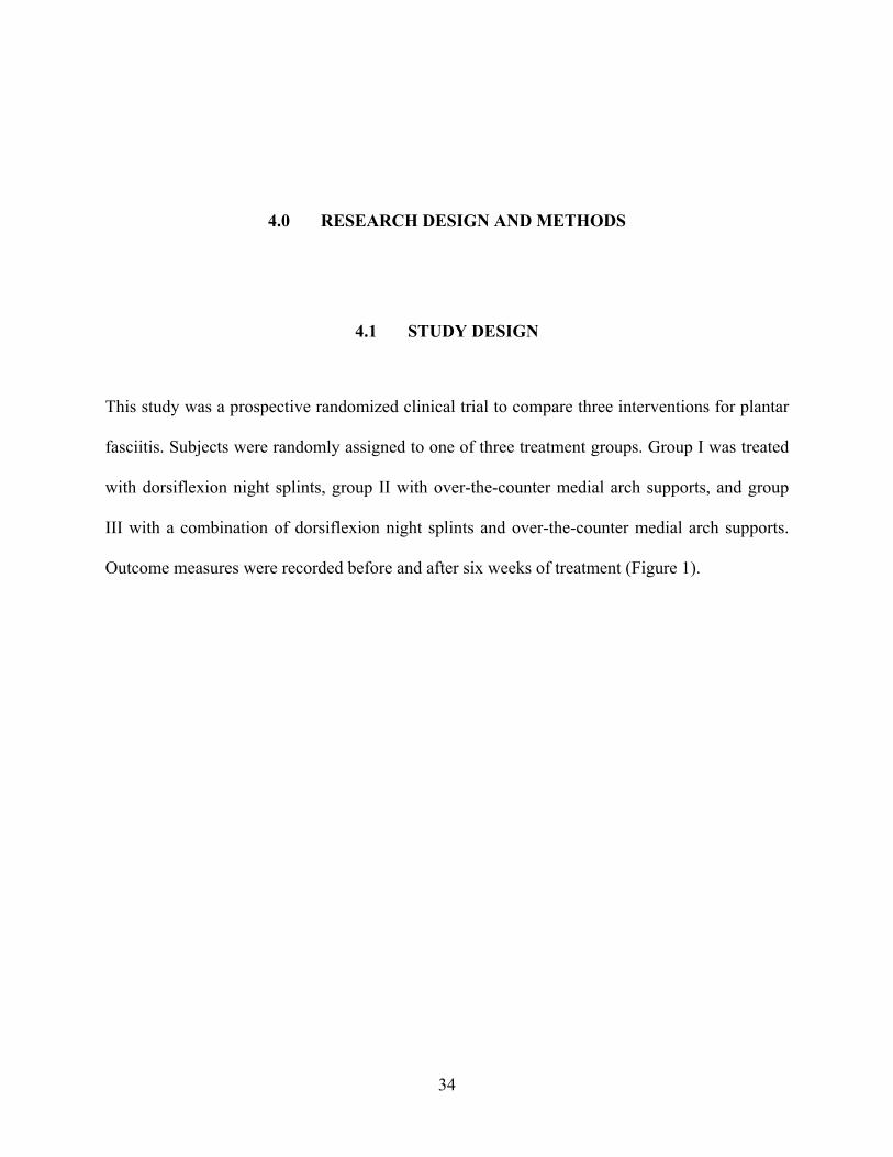
4.0 RESEARCH DESIGN AND METHODS
4.1 STUDY DESIGN
This study was a prospective randomized clinical trial to compare three interventions for plantar
fasciitis. Subjects were randomly assigned to one of three treatment groups. Group I was treated
with dorsiflexion night splints, group II with over-the-counter medial arch supports, and group
III with a combination of dorsiflexion night splints and over-the-counter medial arch supports.
Outcome measures were recorded before and after six weeks of treatment (Figure 1).
34
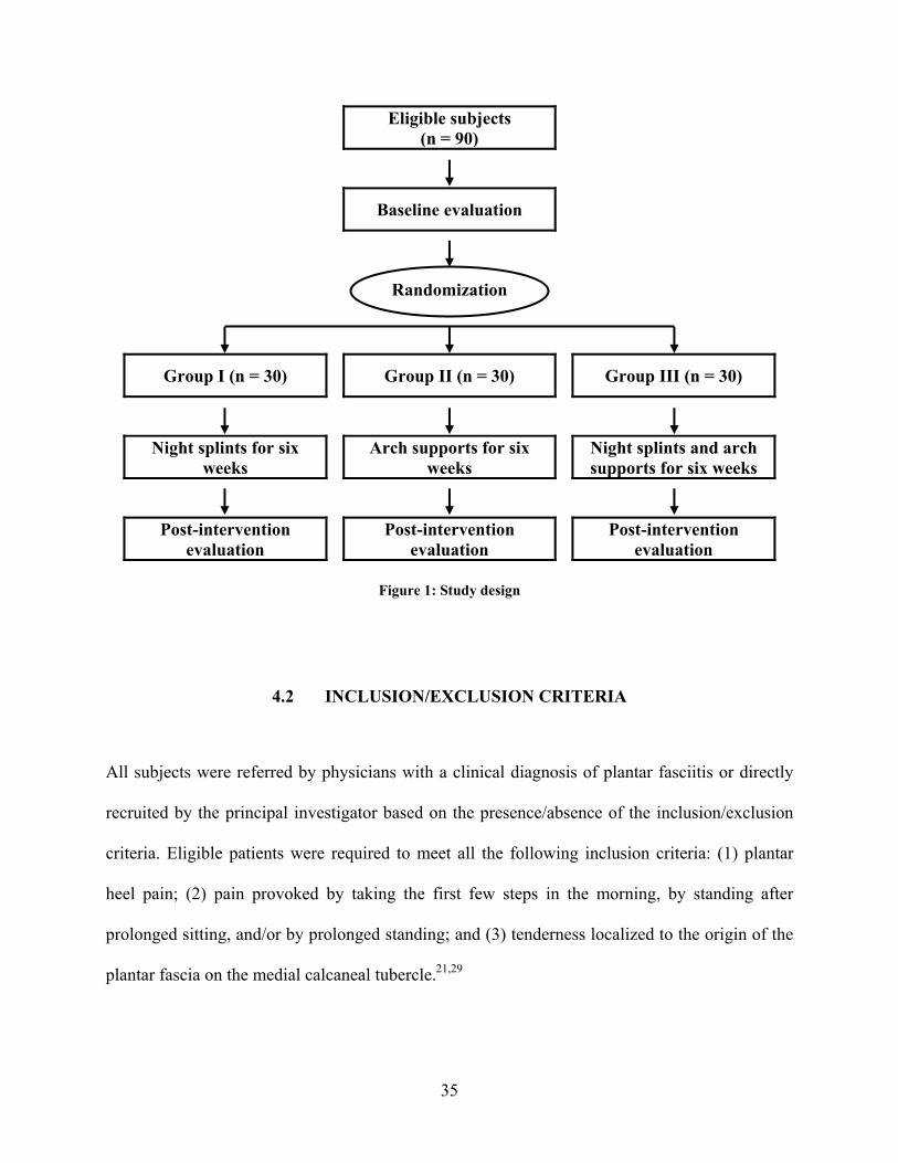
Eligible subjects (n = 90)
Baseline evaluation
Group I (n = 30) Group II (n = 30) Group III (n = 30)
Randomization
Night splints for six weeks Arch supports for six
weeksNight splints and arch supports for six weeks
Post-intervention evaluation Post-intervention
evaluationPost-intervention
evaluation
Figure 1: Study design
4.2 INCLUSION/EXCLUSION CRITERIA
All subjects were referred by physicians with a clinical diagnosis of plantar fasciitis or directly
recruited by the principal investigator based on the presence/absence of the inclusion/exclusion
criteria. Eligible patients were required to meet all the following inclusion criteria: (1) plantar
heel pain; (2) pain provoked by taking the first few steps in the morning, by standing after
prolonged sitting, and/or by prolonged standing; and (3) tenderness localized to the origin of the
plantar fascia on the medial calcaneal tubercle.21,29
35
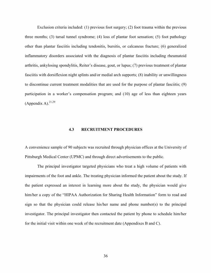
Exclusion criteria included: (1) previous foot surgery; (2) foot trauma within the previous
three months; (3) tarsal tunnel syndrome; (4) loss of plantar foot sensation; (5) foot pathology
other than plantar fasciitis including tendonitis, bursitis, or calcaneus fracture; (6) generalized
inflammatory disorders associated with the diagnosis of plantar fasciitis including rheumatoid
arthritis, ankylosing spondylitis, Reiter’s disease, gout, or lupus; (7) previous treatment of plantar
fasciitis with dorsiflexion night splints and/or medial arch supports; (8) inability or unwillingness
to discontinue current treatment modalities that are used for the purpose of plantar fasciitis; (9)
participation in a worker’s compensation program; and (10) age of less than eighteen years
(Appendix A).21,29
4.3 RECRUITMENT PROCEDURES
A convenience sample of 90 subjects was recruited through physician offices at the University of
Pittsburgh Medical Center (UPMC) and through direct advertisements to the public.
The principal investigator targeted physicians who treat a high volume of patients with
impairments of the foot and ankle. The treating physician informed the patient about the study. If
the patient expressed an interest in learning more about the study, the physician would give
him/her a copy of the “HIPAA Authorization for Sharing Health Information” form to read and
sign so that the physician could release his/her name and phone number(s) to the principal
investigator. The principal investigator then contacted the patient by phone to schedule him/her
for the initial visit within one week of the recruitment date (Appendixes B and C).
36

Additionally, the principal investigator directly recruited subjects to the study via public
advertisements in terms of notices or flyers posted in public places, on the internet, and/or in
local newspapers (Appendix D).
During the initial visit, the principal investigator first explained the study to the patient
including the overall purpose of the study, the experimental procedures that would be performed,
and the potential benefits and risks of the interventions. However, the patient was blinded to the
research questions and hypotheses. If the patient decided to proceed, the principal investigator
gave him/her a copy of the informed consent, as approved by the University of Pittsburgh
Institutional Review Board (IRB), to read at his/her own desired speed, and encouraged him/her
to ask any questions or raise any concerns. If the patient chose to enroll in the study, he/she
signed the consent form (Appendix E).
4.4 STUDY PROTOCOL
After informed consent had been obtained, the principal investigator assigned the subject a
unique three-digit code, obtained demographic information, and performed the baseline
evaluation. The baseline evaluation included: (1) height; (2) weight; (3) medial longitudinal arch
height; (4) range of pain-free passive ankle joint dorsiflexion; (5) plantar heel tenderness; (6)
plantar heel pain; and (7) disability imposed by the heel pain/plantar fasciitis. The principal
investigator then randomly assigned the subject to one of three six-week intervention groups
using a computer-generated randomization schedule. Subjects were randomized in blocks of 15
so that, after every 15 subjects, there was an equal number of subjects in each group. The random
group assignments were placed in sealed opaque envelopes. The randomization had been
37

concealed to the investigator until an opaque envelope containing the group assignment was
opened. The principal investigator opened the next envelope in the series and informed the
subject of the treatment he/she would receive only after the baseline information had been
collected. The principal investigator then provided the treatment modality that corresponds with
the group assignment to the subject and instructed him/her on its use. The initial visit required
about 30 minutes of the subject’s time. At the end of the initial visit, the principal investigator
scheduled the subject for the six-week follow-up visit (Appendix F).
At three weeks, the principal investigator contacted the subject by phone to answer any
questions, discuss any concerns, encourage continued participation, and record compliance. The
phone call required about 5 minutes of the subject’s time (Appendix G).
During the follow-up visit, the principal investigator performed the post-intervention
evaluation, recorded compliance, and discharged the subject. The post-intervention evaluation
included: (1) range of pain-free passive ankle joint dorsiflexion; (2) plantar heel tenderness; (3)
plantar heel pain; and (4) disability imposed by the heel pain/plantar fasciitis. The follow-up visit
required about 15 minutes of the subject’s time. The principal investigator was blinded to the
baseline evaluation data and the group assignment of the subject during the follow-up visit. If the
subject did not return for the follow-up visit, the principal investigator contacted him/her by
phone to encourage continued participation and reschedule the appointment. Both the initial and
follow-up visits took place at the UPMC Center for Sports Medicine (Appendix H).
Group I wore dorsiflexion night splints (Healwell Plantar Fasciitis Night Splints) for six
weeks. Each participant in this group received a dorsiflexion night splint made of polypropylene,
which holds the ankle in about 5° of dorsiflexion.29,32 A firm foam wedge was anchored to the
distal aspect of the foot plate of the splint with Velcro. The wedge was positioned such that its
38
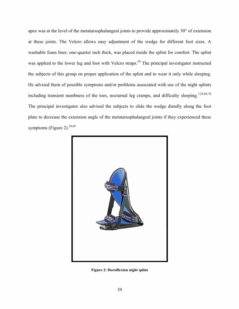
apex was at the level of the metatarsophalangeal joints to provide approximately 30° of extension
at these joints. The Velcro allows easy adjustment of the wedge for different foot sizes. A
washable foam liner, one-quarter inch thick, was placed inside the splint for comfort. The splint
was applied to the lower leg and foot with Velcro straps.29 The principal investigator instructed
the subjects of this group on proper application of the splint and to wear it only while sleeping.
He advised them of possible symptoms and/or problems associated with use of the night splints
including transient numbness of the toes, nocturnal leg cramps, and difficulty sleeping.7,29,69,78
The principal investigator also advised the subjects to slide the wedge distally along the foot
plate to decrease the extension angle of the metatarsophalangeal joints if they experienced these
symptoms (Figure 2).29,69
Figure 2: Dorsiflexion night splint
39
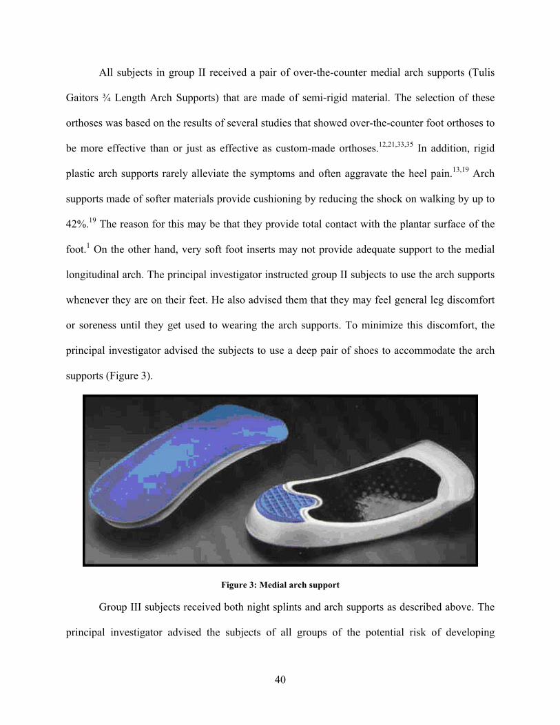
All subjects in group II received a pair of over-the-counter medial arch supports (Tulis
Gaitors ¾ Length Arch Supports) that are made of semi-rigid material. The selection of these
orthoses was based on the results of several studies that showed over-the-counter foot orthoses to
be more effective than or just as effective as custom-made orthoses.12,21,33,35 In addition, rigid
plastic arch supports rarely alleviate the symptoms and often aggravate the heel pain.13,19 Arch
supports made of softer materials provide cushioning by reducing the shock on walking by up to
42%.19 The reason for this may be that they provide total contact with the plantar surface of the
foot.1 On the other hand, very soft foot inserts may not provide adequate support to the medial
longitudinal arch. The principal investigator instructed group II subjects to use the arch supports
whenever they are on their feet. He also advised them that they may feel general leg discomfort
or soreness until they get used to wearing the arch supports. To minimize this discomfort, the
principal investigator advised the subjects to use a deep pair of shoes to accommodate the arch
supports (Figure 3).
Figure 3: Medial arch support
Group III subjects received both night splints and arch supports as described above. The
principal investigator advised the subjects of all groups of the potential risk of developing
40
pressure sores or ulcers from the use of night splints and/or arch supports. However, he provided
them with well-padded night splints and/or arch supports to minimize this risk. In addition, the
fact that those with decreased plantar foot sensation were excluded from the study, would also
minimize this risk. No other medications, stretching, or strengthening exercises were prescribed
to all groups. In addition, all subjects were instructed to discontinue use of any other intervention
modalities, except medications that are used for reasons other than plantar fasciitis, at least three
days before the initial visit, and were encouraged not to change their activity level during the six-
week enrollment in the study because these confounding factors would threaten the internal
validity of the study and could potentially mask the differences between the groups. Night splints
and arch supports were free of charge to all subjects.
4.5 MEASUREMENTS/INSTRUMENTATION
Each patient provided demographic information at the initial visit by completing questions
pertaining to age, gender, unilateral or bilateral involvement, involved side (if both feet were
involved, only the most symptomatic foot, as identified by the patient, was evaluated and treated,
with the exception that patients in group II and group III received a pair of the arch supports to
prevent any imbalances during weight bearing), duration of symptoms prior to treatment (in
months), average number of hours per day during which the patient is on his/her feet, number of
previous corticosteroid injections, and current medication use.
In addition, the principal investigator measured the height (in centimeters) and weight (in
kilograms) of each patient. He also measured medial longitudinal arch height as the arch index.
To do so, he positioned the patient in standing and measured both the navicular height and foot
41

length with a ruler.79,80 The navicular height was measured by marking the navicular tuberosity
on the medial side of the foot and measuring the perpendicular distance (in centimeters) from the
marked point to the floor.79-82 Foot length was measured by marking the medial side of the first
metatarsophalangeal joint and measuring the distance (in centimeters) from the marked point to
the most posterior point of the calcaneus.79,80 The arch index was then calculated, as described by
Cowan et al., by dividing the navicular height by the foot length.80
In 1997, McCrory and colleagues conducted a study to evaluate the validity of the arch
index as a predictor of the medial longitudinal arch height. They found a correlation coefficient
of r = 0.71 between normalized navicular height measured from weight bearing radiographs and
the arch index. It was concluded that the arch index provides a simple quantitative means of
assessing height of the medial longitudinal arch.83
Medial longitudinal arch height was also measured using the navicular drop test. The
principal investigator positioned the patient in standing and placed his/her foot in subtalar neutral
position by palpating the medial and lateral heads of the talus. The subtalar joint is in neutral
position when there is equal prominence of the talar head medially and laterally. The patient was
instructed to hold this position and navicular height was assessed by measuring the perpendicular
distance (in millimeters) from the previously marked navicular tuberosity to the floor, using a
ruler. The patient was then instructed to relax his/her foot and the navicular height was
reassessed. The difference between these two measurements is the navicular drop.84
Sell et al. found acceptable intra- and inter-tester reliability for measuring navicular
height in the subtalar neutral position (intra-class correlation coefficients of 0.92 and 0.87,
respectively) and in resting stance (intra-class correlation coefficients of 0.96 and 0.95,
respectively). Additionally, Sell et al. reported acceptable levels of intra- and inter-tester
42

reliability of the navicular drop (intra-class correlation coefficients of 0.83 and 0.73,
respectively).84
At the three-week phone call and the six-week follow-up visit, each patient answered
question(s), according to his/her group assignment, about the average percentage of sleeping
hours wearing the night splint and/or the average percentage of weight bearing hours using the
arch support during the last three weeks.78
Outcome measures included: (1) the range of pain-free passive ankle joint dorsiflexion;
(2) plantar heel tenderness; (3) plantar heel pain; and (4) disability imposed by the heel
pain/plantar fasciitis.
The principal investigator tested the range of pain-free passive ankle joint dorsiflexion in
two positions: supine, with the legs fully extended, and sitting. He placed the patient’s foot in
subtalar neutral position by palpating the medial and lateral heads of the talus while supinating
and pronating the subtalar joint. The position between pronation and supination when there is
equal prominence of the talar head medially and laterally was defined as the subtalar neutral
position. From the subtalar neutral position, the principal investigator passively dorsiflexed the
patient's ankle through the maximum pain-free range of ankle dorsiflexion. He aligned the
stationary arm of the goniometer with the fibular head and the moving arm parallel to the lateral
border of the calcaneus. Range of motion (ROM) value was recorded as positive (+) when the
ROM is beyond neutral (0°). The value was recorded as negative (-) when the ROM does not
reach neutral. Several studies have been conducted to examine the validity and intra- and inter-
tester reliability of the goniometer. The results of these studies indicated that goniometric
measurements are both valid and reliable, with the intra-tester reliability being strong (intra-class
43
correlation coefficients ranging from 0.825 to 0.997) and the inter-tester reliability being
moderate (intra-class correlation coefficients ranging from 0.43 to 0.99).85-91
Evaluation also included plantar heel tenderness, which was measured using a pressure
algometer. To assess tenderness, the principal investigator first positioned the patient in supine
with the legs fully extended and palpated and marked the origin of the plantar fascia at the
medial calcaneal tubercle. He then dorsiflexed the ankle and toes passively and applied the
algometer over the mark placed on the medial calcaneal tubercle.4,5,7,92 The algometer contact
head was aligned perpendicularly to the skin and the principal investigator gradually increased
the algometer pressure until the patient reported pain. The algometer reading, which represents
the pressure needed to elicit pain (in Newton per square centimeter), was recorded. Higher
algometer scores indicated greater pressure tolerance and, hence, less tenderness. Lower
algometer scores indicated less pressure tolerance and, thus, greater tenderness.92 The reliability
and validity of pressure algometer as a measure of tenderness have been documented in the
literature.93-97
Plantar heel pain and disability imposed by the heel pain/plantar fasciitis were measured
using the Pain and Disability sub-scales of the Foot Function Index (FFI), respectively. Each of
these sub-scales consists of nine items. All items are rated using a visual analogue scale that
consists of a horizontal 100 millimeter line, to which, no numbers or divisions are attached.
Verbal anchors, representing opposite extremes of the dimension being measured, were placed at
either end of the line. The principal investigator instructed the patient to place a mark on the line
in a position which best represented his/her experience in the past week, or to answer the
question as not applicable (NA) if he/she did not perform or was not involved in the activity in
question, which removes that question from scoring. The principal investigator then assigned a
44
score between 0 and 100 to the item by measuring the distance (in millimeters) from the anchor
on the left hand side of the line to the mark placed by the patient. He then obtained the sub-scale
score by adding the scores of the items and dividing by the number of the applicable items in that
sub-scale. Thus, the score for each sub-scale ranged from 0 to 100, with 0 representing the best
and 100 representing the worst possible scenario.98
The Pain sub-scale measures the level of foot pain in a variety of situations. The
dimension of measurement used for this sub-scale was severity of pain, and the anchors for the
visual analogue scale were “no pain” and “worst pain imaginable.” The Disability sub-scale
describes the difficulty in performing various activities due to foot problems. The measurement
dimension employed by this sub-scale was the degree of difficulty, and the visual analogue scale
anchors were “no difficulty” and “so difficult unable.”98
The FFI is a validated short and simple measure of foot pain and disability.98 Although it
was originally designed to assess the effect of foot orthoses on foot pathology in patients with
rheumatoid arthritis, it has been suggested by its developers that it is not limited to this group of
patients.99 Recently, several studies have used the FFI in research relating to different foot
pathologies, including plantar fasciitis, unrelated to rheumatoid arthritis.12,100,101 The FFI has
been examined for test-retest reliability, internal consistency, validity, and responsiveness on 87
patients with rheumatoid arthritis. It had good test-retest reliability (intra-class correlation
coefficients ranging from 0.69 to 0.87) with a one-week interval between the two tests. It also
had a high degree of internal consistency (Cronbach’s alpha ranging from 0.73 to 0.96) and
validity. In addition, the FFI was sensitive enough to detect changes in clinical status over a
period of six months.98
45
4.6 DATA MANAGEMENT
Only the patient’s ID code was used to identify patients on all data recording forms. The
principal investigator kept patient information, including his/her name, phone number(s), ID
linkage code, consent form, and “HIPAA Authorization for Sharing Health Information” form in
a file that was separate from the data recording forms. He kept this file and the IRB and data
recording forms in two locked drawers located in a locked office in the Department of Physical
Therapy in the School of Health and Rehabilitation Sciences at the University of Pittsburgh. The
principal investigator entered available data and verified the entered data once per week on his
own laptop computer, which has a password not known to anyone other than him. It was the
responsibility of the principal investigator to keep the data secured at all times. The data were
entered twice to insure accuracy and were coded into SPSS software. The principal investigator
then reviewed the entered data and backed-up the file. He evaluated the data periodically. For
missing data points, the “worst case scenario” was used (the least difference in each outcome
measure of each group between the post-intervention and baseline evaluation data was added to
the baseline evaluation data) in order to adhere with the principle of intention-to-treat analysis.
4.7 DATA ANALYSIS
4.7.1 Power analysis
Power estimates based on a standard deviation (SD) of 2.3, which was obtained from a recent
study with a similar design, revealed that a sample size of 30 subjects per group, taking into
46
account a 30% drop-out rate, would result in greater than 80% power to detect a 2 centimeter
mean difference on the Pain sub-scale of the FFI between group I and III and group II and III.102
4.7.2 Description of statistical procedures
Using SPSS software, the principal investigator first described the demographic, compliance,
baseline and post-intervention evaluation data of each group using means and SDs for
continuous variables and frequencies and percentages for categorical variables. He also tested the
normality and homogeneity of variances of the data. Then, he compared the demographic,
compliance, and baseline evaluation data of the three groups using analysis of variance
(ANOVA) test for each continuous variable and the chi-square test for each categorical variable
to determine if there were significant differences among the groups. Kruskal-Wallis non-
parametric test was used if the data showed significant skewness and/or heterogeneity of
variances. An overall significance level was maintained at p-value less than 0.05.
4.7.2.1 Analysis of specific aim 1: To examine whether there will be any difference between
the efficacy of three different treatment regimens: (1) dorsiflexion night splints; (2) medial
arch supports; and (3) dorsiflexion night splints and medial arch supports together, in the
management of plantar fasciitis in terms of: (1) the range of pain-free passive ankle joint
dorsiflexion; (2) plantar heel tenderness; (3) plantar heel pain; and (4) disability imposed
by the heel pain/plantar fasciitis.
Specific aim 1 was analyzed using analysis of covariance (ANCOVA) test to compare the
post-intervention scores of each outcome measure between the treatment groups in order to
examine if there was a significant difference. The baseline scores of the outcome measure being
47

tested as well as any other demographic and/or compliance variables that were significantly
different among the groups and were significantly correlated with the outcome measure being
tested were entered as covariates to “statistically equate” the groups.
If the post-intervention scores of any outcome measure showed a significant difference
between the groups, post-hoc analyses were explored to determine if there were significant
differences between group I and III and/or group II and III. Kruskal-Wallis non-parametric test
was used if the data had significant skewness and/or heterogeneity of variances.
4.7.2.2 Analysis of specific aim 2: To investigate whether patients with plantar fasciitis who
have less passive dorsiflexion of the ankle joint will benefit from a dorsiflexion night splint
more than those with greater passive dorsiflexion of the ankle joint in terms of: (1) the
range of pain-free passive ankle joint dorsiflexion; (2) plantar heel tenderness; (3) plantar
heel pain; and (4) disability imposed by the heel pain/plantar fasciitis.
Specific aim 2 was analyzed using multiple regression analysis with the post-intervention
scores of each outcome measure being the criterion and the baseline range of pain-free passive
ankle joint dorsiflexion being the predictor after controlling for the effect of baseline scores of
the outcome measure being tested, with the exception that simple regression analysis was used
with the difference between the post-intervention and baseline scores being the criterion to
analyze the range of pain-free passive ankle joint dorsiflexion because it is not possible to
control for the effect of baseline score of the range of pain-free passive ankle joint dorsiflexion
and then use it as a predictor or to use two predictors (the baseline range of pain-free passive
ankle joint dorsiflexion with the knee straight and bent) that are highly correlated (r = 0.78, p-
value < 0.001). This analysis was applied to group I only.
48
4.7.2.3 Analysis of specific aim 3: To investigate whether patients with plantar fasciitis who
have a lower medial longitudinal arch will benefit from a medial arch support more than
those with a higher medial longitudinal arch in terms of: (1) the range of pain-free passive
ankle joint dorsiflexion; (2) plantar heel tenderness; (3) plantar heel pain; and (4) disability
imposed by the heel pain/plantar fasciitis.
Specific aim 3 was analyzed using multiple regression analysis with the post-intervention
scores of each outcome measure being the criterion and the medial longitudinal arch height being
the predictor after controlling for the effect of baseline scores of the outcome measure being
tested. This analysis was applied to group II only.
49
5.0 RESULTS
5.1 PROFILE OF PROSPECTIVE RANDOMIZED STUDY
Between August 2005 and July 2006, 91 patients with plantar fasciitis who met the
inclusion/exclusion criteria of the study were identified and recruited. One patient declined to be
randomized, leaving 90 patients enrolled in the study, 30 in each group. The first 18 patients
were referred to the study by physicians at the UPMC over a period of eight months. Because
patient recruitment through physician offices had been too slow, the principal investigator started
to directly recruit patients via public advertisements, and he successfully recruited the rest of the
patients (72 patients) during the next three months. Demographic, compliance, baseline and post-
intervention evaluation data demonstrated no significant differences between patients who were
recruited through physician offices and those recruited via public advertisements, with the
exception that patients who were recruited through physician offices had significantly shorter
duration of symptoms prior to treatment, fewer number of previous corticosteroid injections, and
greater tenderness than those recruited via public advertisements (p-value < 0.05).
Nine subjects (10%) dropped-out of the study, 2 from group I and 7 from group II. Seven
drop-outs were lost to follow-up, and 2 chose to withdraw their consent for participation in the
study, one because she felt that the intervention was ineffective, while the other because he
moved. The study sample consisted of 23 men (25.6%) and 67 women (74.4%) (Figure 4).
50

Eligible patients (n = 91)
Not randomized (n = 1)
Randomized (n = 90)
Group I (n = 30) Group II (n = 30) Group III (n = 30)
Night splints for six weeks Arch supports for six
weeksNight splints and arch supports for six weeks
Follow-up (n = 28) Drop-out (n = 2) Follow-up (n = 23)
Drop-out (n =7)Follow-up (n = 30) Drop-out (n = 0)
Lost to follow-up (n = 2) Withdrawn (n = 0) Lost to follow-up (n = 5)
Withdrawn (n = 2)Lost to follow-up (n = 0)
Withdrawn (n = 0)
Figure 4: Profile of prospective randomized study
5.2 DESCRIPTION OF DEMOGRAPHIC, COMPLIANCE, BASELINE AND POST-
INTERVENTION EVALUATION DATA
The demographic, compliance, baseline and post-intervention evaluation data of each group were
described using mean, median, SD, minimum, maximum, skewness, and kurtosis for continuous
variables, and frequency and percentage for categorical variables.
51

5.2.1 Subjects’ characteristics (Table 1)
Table 1: Subjects' characteristics
Group Demographic variable
Group I
mean / median (SD) minimum / maximum
skewness (kurtosis) or
frequency (percentage)
Group II
mean / median (SD) minimum / maximum
skewness (kurtosis) or
frequency (percentage)
Group III
mean / median (SD) minimum / maximum
skewness (kurtosis) or
frequency (percentage)
Age 50.23 / 50.00 (11.37)
27.00 / 74.00 -0.124 (-0.245)
48.24 / 51.00 (12.73) 24.00 / 74.00
-0.313 (-0.293)
49.07 / 52.00 (10.13) 26.00 / 67.00 -1.028 (0.549)
Male 11 (36.70) 5 (16.70) 7 (23.30) Gender
Female 19 (63.30) 25 (83.30) 23 (76.70)
Unilateral 13 (43.30) 15 (50.00) 9 (30.00) Unilateral or bilateral
involvement Bilateral 17 (56.70) 15 (50.00) 21 (70.00)
Right 14 (46.70) 16 (53.30) 17 (56.70) Involved side
Left 16 (53.30) 14 (46.70) 13 (43.30)
Duration of symptoms prior to treatment
(in months)
16.88 / 9.50 (18.29) 1.00 / 60.00
1.307 (0.356)
18.26 / 6.00 (25.04) 0.50 / 120.00 2.731 (9.268)
8.78 / 6.00 (8.08) 1.00 / 36.00
1.823 (3.534)
Average number of hours per day during
which the patient is on his/her feet
7.25 / 5.50 (4.26) 1.00 / 17.00
0.658 (-0.636)
7.29 / 7.00 (3.74) 1.50 / 14.00
0.313 (-0.861)
7.47 / 7.00 (3.51) 2.00 / 15.00
0.423 (-0.679)
Number of previous corticosteroid
injections
0.30 / 0.00 (0.79) 0.00 / 3.00
2.927 (8.083)
0.38 / 0.00 (0.78) 0.00 / 3.00
2.163 (4.249)
0.43 / 0.00 (1.04) 0.00 / 4.00
2.555 (5.720)
Height (in centimeters)
171.63 / 170.60 (10.65) 156.20 / 199.40 0.606 (0.144)
168.33 / 167.80 (9.33) 144.10 / 188.50 -0.069 (1.279)
169.29 / 168.80 (8.74) 151.20 / 194.20 0.825 (1.778)
Weight (in kilograms)
93.38 / 92.10 (22.06) 59.40 / 137.30 0.259 (-0.927)
87.10 / 85.60 (20.19) 54.50 / 135.70 0.545 (-0.139)
88.20 / 86.55 (22.93) 51.50 / 179.10 2.063 (7.798)
52
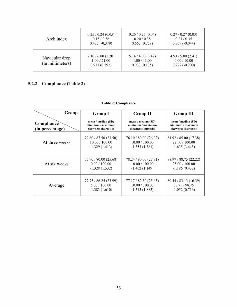
Arch index 0.25 / 0.24 (0.05)
0.15 / 0.36 0.435 (-0.379)
0.26 / 0.25 (0.04) 0.20 / 0.38
0.667 (0.759)
0.27 / 0.27 (0.03) 0.21 / 0.35
0.369 (-0.044)
Navicular drop (in millimeters)
7.10 / 6.00 (5.20) 1.00 / 21.00
0.933 (0.292)
5.14 / 4.00 (3.42) 1.00 / 13.00
0.933 (0.135)
4.93 / 5.00 (2.41) 0.00 / 10.00
0.227 (-0.200)
5.2.2 Compliance (Table 2)
Table 2: Compliance
Group
Compliance (in percentage)
Group I
mean / median (SD) minimum / maximum
skewness (kurtosis)
Group II
mean / median (SD) minimum / maximum
skewness (kurtosis)
Group III
mean / median (SD) minimum / maximum
skewness (kurtosis)
At three weeks 79.60 / 87.50 (23.50)
10.00 / 100.00 -1.329 (1.413)
76.10 / 80.00 (26.02) 10.00 / 100.00 -1.353 (1.381)
81.92 / 85.00 (17.38) 22.50 / 100.00 -1.635 (3.445)
At six weeks 75.90 / 80.00 (25.68)
0.00 / 100.00 -1.328 (1.532)
78.24 / 90.00 (27.71) 10.00 / 100.00 -1.462 (1.149)
78.97 / 88.75 (22.22) 25.00 / 100.00 -1.186 (0.432)
Average 77.75 / 86.25 (23.99)
5.00 / 100.00 -1.383 (1.610)
77.17 / 82.50 (25.63) 10.00 / 100.00 -1.515 (1.883)
80.44 / 83.13 (16.39) 38.75 / 98.75 -1.052 (0.716)
53
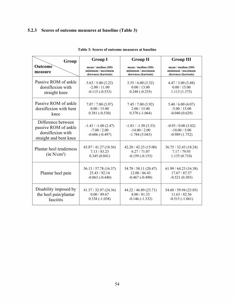
5.2.3 Scores of outcome measures at baseline (Table 3)
Table 3: Scores of outcome measures at baseline
Group Outcome measure
Group I
mean / median (SD) minimum / maximum
skewness (kurtosis)
Group II
mean / median (SD) minimum / maximum
skewness (kurtosis)
Group III
mean / median (SD) minimum / maximum
skewness (kurtosis)
Passive ROM of ankle dorsiflexion with
straight knee
5.63 / 5.00 (3.22) -2.00 / 11.00
-0.115 (-0.533)
5.55 / 6.00 (3.32) 0.00 / 13.00
0.248 (-0.255)
4.47 / 3.00 (3.48) 0.00 / 15.00
1.113 (1.375)
Passive ROM of ankle dorsiflexion with bent
knee
7.07 / 7.00 (3.97) 0.00 / 15.00
0.381 (-0.530)
7.45 / 7.00 (3.92) 2.00 / 15.00
0.370 (-1.064)
5.40 / 6.00 (4.07) -5.00 / 15.00
-0.040 (0.629)
Difference between passive ROM of ankle
dorsiflexion with straight and bent knee
-1.43 / -1.00 (2.47) -7.00 / 2.00
-0.606 (-0.497)
-1.83 / -1.50 (3.33) -14.00 / 2.00
-1.784 (5.043)
-0.93 / 0.00 (3.02) -10.00 / 5.00
-0.989 (1.752)
Plantar heel tenderness (in N/cm²)
43.87 / 41.27 (18.56) 7.13 / 83.23
0.345 (0.041)
42.20 / 42.23 (15.00) 6.27 / 71.07
-0.159 (-0.153)
36.75 / 32.43 (18.24) 7.17 / 79.93
1.135 (0.710)
Plantar heel pain 56.13 / 57.78 (16.37)
25.43 / 92.14 -0.063 (-0.440)
54.70 / 58.11 (20.47) 12.00 / 86.43
-0.467 (-0.490)
61.99 / 64.23 (16.38) 17.67 / 87.57 -0.521 (0.303)
Disability imposed by the heel pain/plantar
fasciitis
41.37 / 32.97 (24.36) 0.00 / 89.67
0.338 (-1.038)
44.22 / 46.89 (23.71) 8.00 / 81.33
-0.146 (-1.332)
54.60 / 59.94 (23.05) 11.63 / 82.56
-0.515 (-1.061)
54
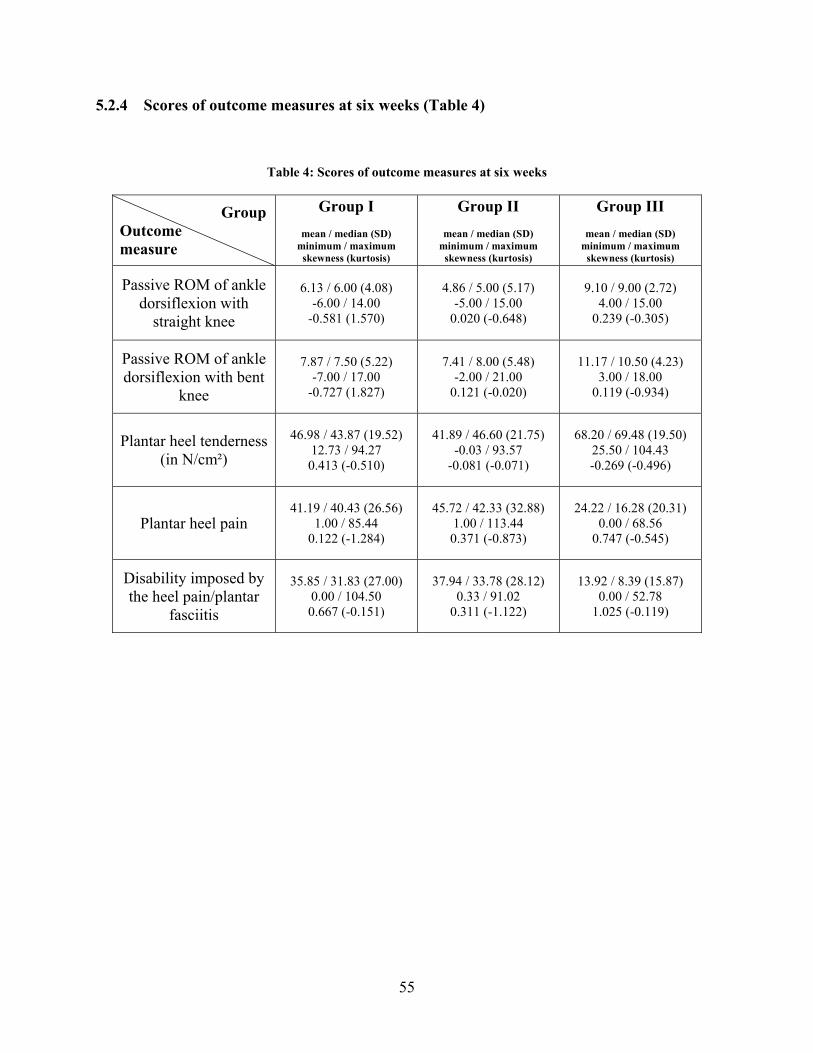
5.2.4 Scores of outcome measures at six weeks (Table 4)
Table 4: Scores of outcome measures at six weeks
Group Outcome measure
Group I
mean / median (SD) minimum / maximum
skewness (kurtosis)
Group II
mean / median (SD) minimum / maximum
skewness (kurtosis)
Group III
mean / median (SD) minimum / maximum
skewness (kurtosis)
Passive ROM of ankle dorsiflexion with
straight knee
6.13 / 6.00 (4.08) -6.00 / 14.00
-0.581 (1.570)
4.86 / 5.00 (5.17) -5.00 / 15.00
0.020 (-0.648)
9.10 / 9.00 (2.72) 4.00 / 15.00
0.239 (-0.305)
Passive ROM of ankle dorsiflexion with bent
knee
7.87 / 7.50 (5.22) -7.00 / 17.00
-0.727 (1.827)
7.41 / 8.00 (5.48) -2.00 / 21.00
0.121 (-0.020)
11.17 / 10.50 (4.23) 3.00 / 18.00
0.119 (-0.934)
Plantar heel tenderness (in N/cm²)
46.98 / 43.87 (19.52) 12.73 / 94.27 0.413 (-0.510)
41.89 / 46.60 (21.75) -0.03 / 93.57
-0.081 (-0.071)
68.20 / 69.48 (19.50) 25.50 / 104.43 -0.269 (-0.496)
Plantar heel pain 41.19 / 40.43 (26.56)
1.00 / 85.44 0.122 (-1.284)
45.72 / 42.33 (32.88) 1.00 / 113.44 0.371 (-0.873)
24.22 / 16.28 (20.31) 0.00 / 68.56
0.747 (-0.545)
Disability imposed by the heel pain/plantar
fasciitis
35.85 / 31.83 (27.00) 0.00 / 104.50 0.667 (-0.151)
37.94 / 33.78 (28.12) 0.33 / 91.02
0.311 (-1.122)
13.92 / 8.39 (15.87) 0.00 / 52.78
1.025 (-0.119)
55
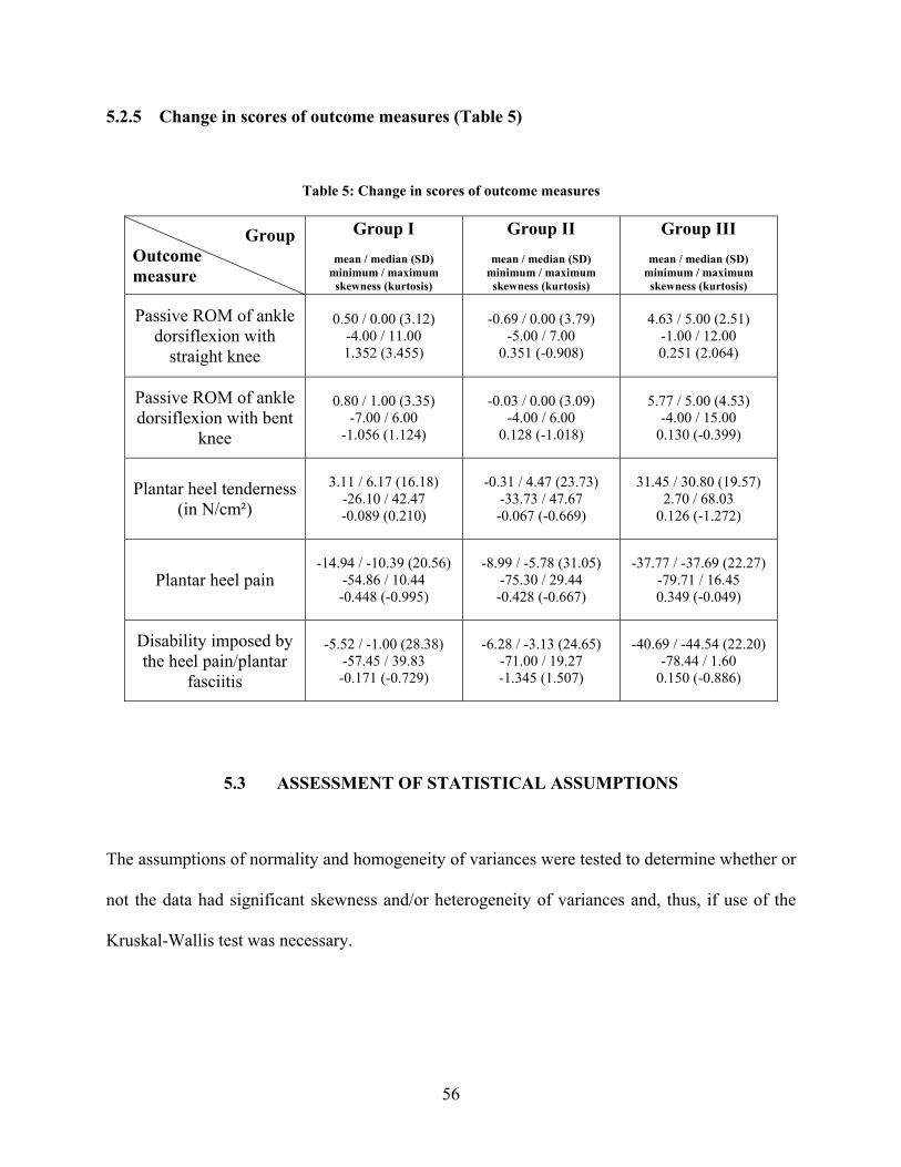
5.2.5 Change in scores of outcome measures (Table 5)
Table 5: Change in scores of outcome measures
Group Outcome measure
Group I
mean / median (SD) minimum / maximum
skewness (kurtosis)
Group II
mean / median (SD) minimum / maximum
skewness (kurtosis)
Group III
mean / median (SD) minimum / maximum
skewness (kurtosis)
Passive ROM of ankle dorsiflexion with
straight knee
0.50 / 0.00 (3.12) -4.00 / 11.00 1.352 (3.455)
-0.69 / 0.00 (3.79) -5.00 / 7.00
0.351 (-0.908)
4.63 / 5.00 (2.51) -1.00 / 12.00 0.251 (2.064)
Passive ROM of ankle dorsiflexion with bent
knee
0.80 / 1.00 (3.35) -7.00 / 6.00
-1.056 (1.124)
-0.03 / 0.00 (3.09) -4.00 / 6.00
0.128 (-1.018)
5.77 / 5.00 (4.53) -4.00 / 15.00
0.130 (-0.399)
Plantar heel tenderness (in N/cm²)
3.11 / 6.17 (16.18) -26.10 / 42.47 -0.089 (0.210)
-0.31 / 4.47 (23.73) -33.73 / 47.67 -0.067 (-0.669)
31.45 / 30.80 (19.57) 2.70 / 68.03
0.126 (-1.272)
Plantar heel pain -14.94 / -10.39 (20.56)
-54.86 / 10.44 -0.448 (-0.995)
-8.99 / -5.78 (31.05) -75.30 / 29.44 -0.428 (-0.667)
-37.77 / -37.69 (22.27) -79.71 / 16.45 0.349 (-0.049)
Disability imposed by the heel pain/plantar
fasciitis
-5.52 / -1.00 (28.38) -57.45 / 39.83 -0.171 (-0.729)
-6.28 / -3.13 (24.65) -71.00 / 19.27 -1.345 (1.507)
-40.69 / -44.54 (22.20) -78.44 / 1.60
0.150 (-0.886)
5.3 ASSESSMENT OF STATISTICAL ASSUMPTIONS
The assumptions of normality and homogeneity of variances were tested to determine whether or
not the data had significant skewness and/or heterogeneity of variances and, thus, if use of the
Kruskal-Wallis test was necessary.
56
5.3.1 Normality test of demographic, compliance, baseline and post-intervention
evaluation data
The assumption of normality was assessed by testing skewness and kurtosis of the demographic,
compliance, baseline and post-intervention evaluation data of the three groups.
57

5.3.1.1 Normality test of subjects’ characteristics
The assumption of normality was not met for all demographic continuous variables (-1.96
> t > 1.96) except average number of hours per day during which the patient is on his/her feet
and arch index (-1.96 < t < 1.96) (Table 6).
Table 6: Normality test of subjects’ characteristics
GroupDemographic variable
Group I t
Group II
t
Group III t
Skewness -0.290 -0.721 -2.407* Age*
Kurtosis -0.294 -0.347 0.659
Skewness 3.061* 6.293* 4.269* Duration of symptoms prior to treatment*
Kurtosis 0.427 10.968* 4.242*
Skewness 1.541 0.721 0.991 Average number of hours per day during
which the patient is on his/her feet
Kurtosis -0.764 -1.019 -0.815
Skewness 6.855* 4.984* 5.984* Number of previous corticosteroid
injections* Kurtosis 9.703* 5.028* 6.867*
Skewness 1.419 -0.159 1.932 Height*
Kurtosis 0.173 1.514 2.134*
Skewness 0.607 1.256 4.831* Weight*
Kurtosis -1.113 -0.164 9.361*
Skewness 1.019 1.537 0.864 Arch index
Kurtosis -0.455 0.898 -0.053
Skewness 2.185* 2.150* 0.532 Navicular drop*
Kurtosis 0.351 0.160 -0.240
* -1.96 > t > 1.96
58
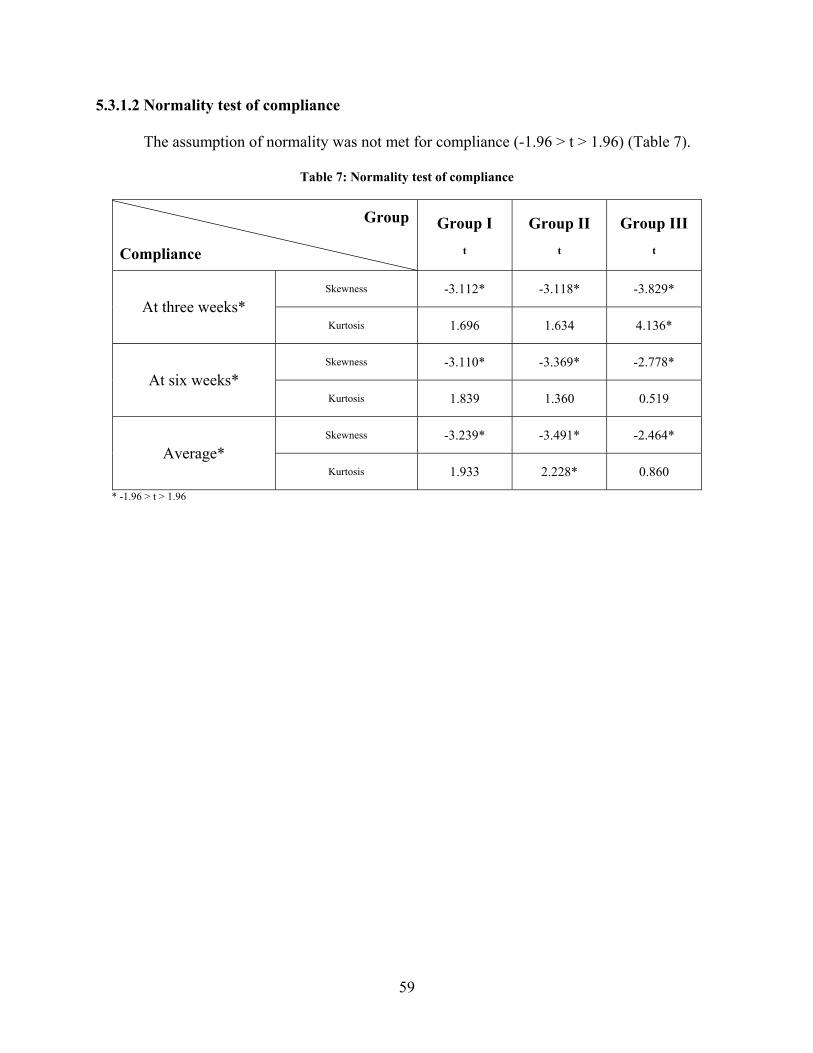
5.3.1.2 Normality test of compliance
The assumption of normality was not met for compliance (-1.96 > t > 1.96) (Table 7).
Table 7: Normality test of compliance
Group Compliance
Group I t
Group II
t
Group III t
Skewness -3.112* -3.118* -3.829* At three weeks*
Kurtosis 1.696 1.634 4.136*
Skewness -3.110* -3.369* -2.778* At six weeks*
Kurtosis 1.839 1.360 0.519
Skewness -3.239* -3.491* -2.464* Average*
Kurtosis 1.933 2.228* 0.860
* -1.96 > t > 1.96
59
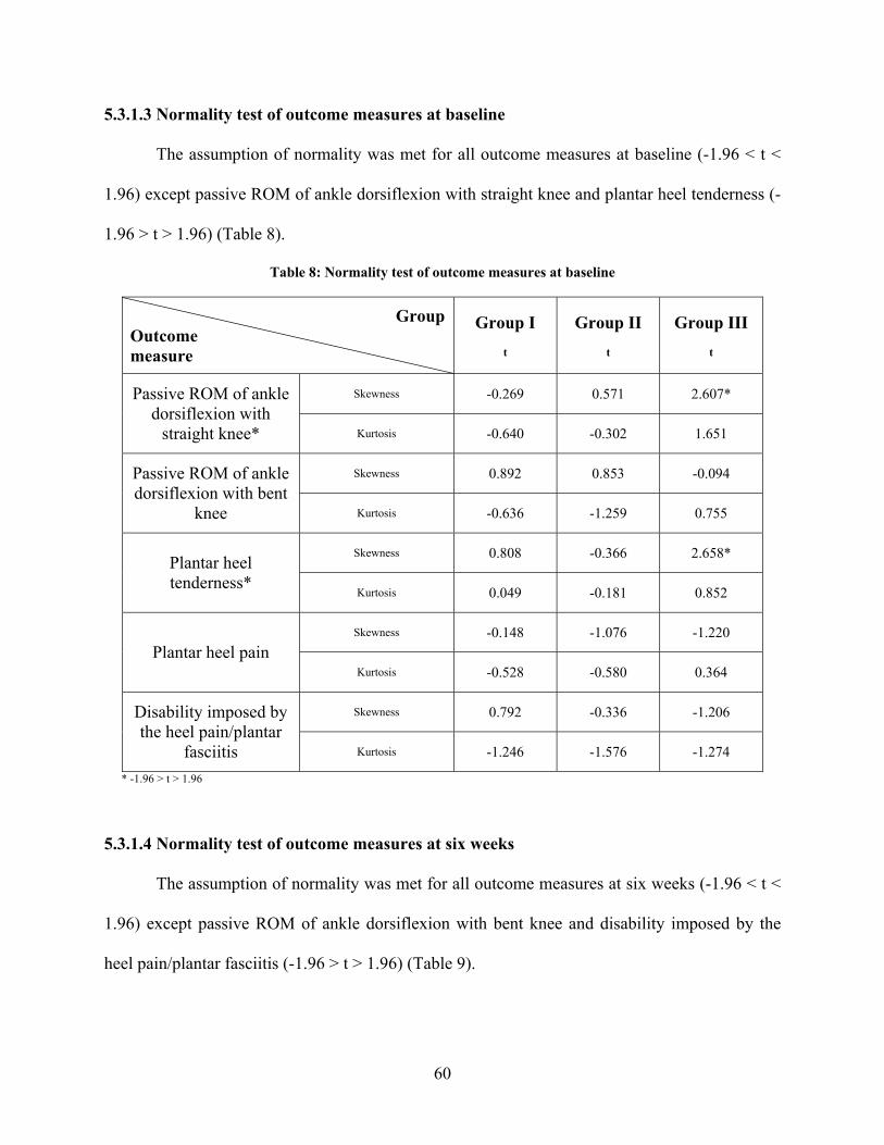
5.3.1.3 Normality test of outcome measures at baseline
The assumption of normality was met for all outcome measures at baseline (-1.96 < t <
1.96) except passive ROM of ankle dorsiflexion with straight knee and plantar heel tenderness (-
1.96 > t > 1.96) (Table 8).
Table 8: Normality test of outcome measures at baseline
GroupOutcome measure
Group I t
Group II
t
Group III t
Skewness -0.269 0.571 2.607* Passive ROM of ankle dorsiflexion with
straight knee* Kurtosis -0.640 -0.302 1.651
Skewness 0.892 0.853 -0.094 Passive ROM of ankle dorsiflexion with bent
knee Kurtosis -0.636 -1.259 0.755
Skewness 0.808 -0.366 2.658* Plantar heel tenderness*
Kurtosis 0.049 -0.181 0.852
Skewness -0.148 -1.076 -1.220 Plantar heel pain
Kurtosis -0.528 -0.580 0.364
Skewness 0.792 -0.336 -1.206 Disability imposed by the heel pain/plantar
fasciitis Kurtosis -1.246 -1.576 -1.274
* -1.96 > t > 1.96
5.3.1.4 Normality test of outcome measures at six weeks
The assumption of normality was met for all outcome measures at six weeks (-1.96 < t <
1.96) except passive ROM of ankle dorsiflexion with bent knee and disability imposed by the
heel pain/plantar fasciitis (-1.96 > t > 1.96) (Table 9).
60

Table 9: Normality test of outcome measures at six weeks
GroupOutcome measure
Group I t
Group II
t
Group III t
Skewness -1.361 0.046 0.560 Passive ROM of ankle dorsiflexion with
straight knee Kurtosis 1.885 -0.767 -0.366
Skewness -1.703 0.279 0.279 Passive ROM of ankle dorsiflexion with bent
knee* Kurtosis 2.193* -0.024 -1.121
Skewness 0.967 -0.187 -0.630 Plantar heel tenderness
Kurtosis -0.612 -0.084 -0.595
Skewness 0.286 0.855 1.749 Plantar heel pain
Kurtosis -1.541 -1.033 -0.654
Skewness 1.562 0.717 2.400* Disability imposed by the heel pain/plantar
fasciitis* Kurtosis -0.181 -1.328 -0.143
* -1.96 > t > 1.96
5.3.2 Test of homogeneity of variances of demographic, compliance, baseline and post-
intervention evaluation data
The assumption of homogeneity of variances of the demographic, compliance, baseline and post-
intervention evaluation data among the groups was assessed using Levene’s test.
61
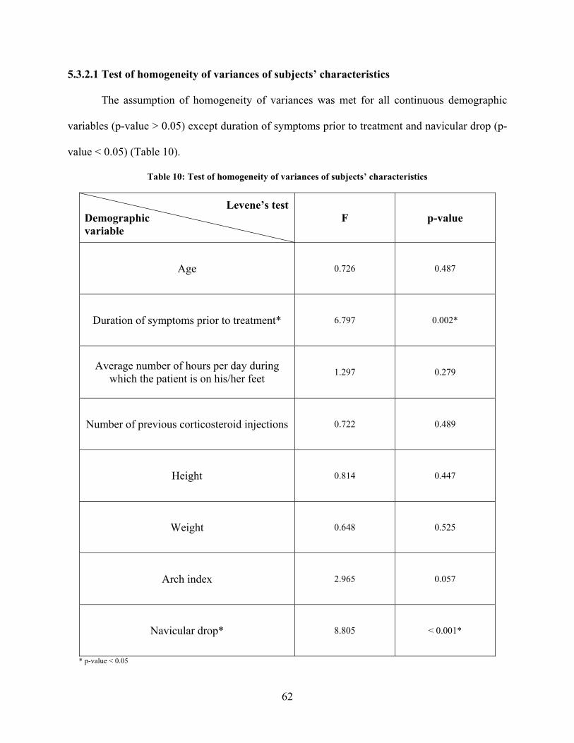
5.3.2.1 Test of homogeneity of variances of subjects’ characteristics
The assumption of homogeneity of variances was met for all continuous demographic
variables (p-value > 0.05) except duration of symptoms prior to treatment and navicular drop (p-
value < 0.05) (Table 10).
Table 10: Test of homogeneity of variances of subjects’ characteristics
Levene’s testDemographic variable
F p-value
Age 0.726 0.487
Duration of symptoms prior to treatment* 6.797 0.002*
Average number of hours per day during which the patient is on his/her feet
1.297 0.279
Number of previous corticosteroid injections 0.722 0.489
Height 0.814 0.447
Weight 0.648 0.525
Arch index 2.965 0.057
Navicular drop* 8.805 < 0.001*
* p-value < 0.05
62
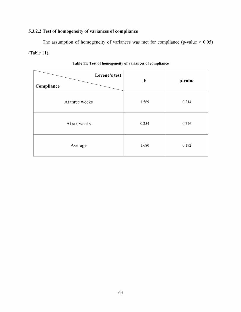
5.3.2.2 Test of homogeneity of variances of compliance
The assumption of homogeneity of variances was met for compliance (p-value > 0.05)
(Table 11).
Table 11: Test of homogeneity of variances of compliance
Levene’s test Compliance
F p-value
At three weeks 1.569 0.214
At six weeks 0.254 0.776
Average 1.680 0.192
63

5.3.2.3 Test of homogeneity of variances of outcome measures at baseline
The assumption of homogeneity of variances was met for all outcome measures at
baseline (p-value > 0.05) (Table 12).
Table 12: Test of homogeneity of variances of outcome measures at baseline
Levene’s testOutcome measure
F p-value
Passive ROM of ankle dorsiflexion with straight knee 0.041 0.960
Passive ROM of ankle dorsiflexion with bent knee 0.036 0.965
Plantar heel tenderness 0.464 0.630
Plantar heel pain 0.882 0.418
Disability imposed by the heel pain/plantar fasciitis 0.078 0.925
5.3.2.4 Test of homogeneity of variances of outcome measures at six weeks
The assumption of homogeneity of variances was not met for all outcome measures at six
weeks (p-value < 0.05) except passive ROM of ankle dorsiflexion with bent knee and plantar
heel tenderness (p-value > 0.05) (Table 13).
64
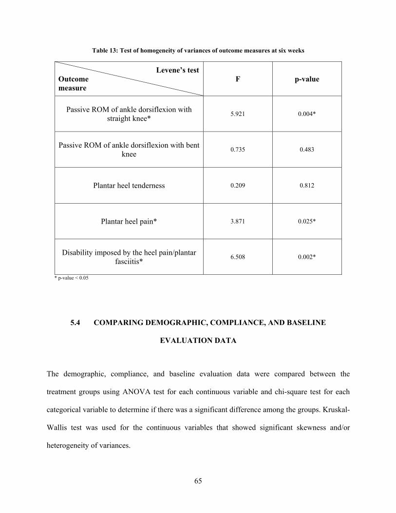
Table 13: Test of homogeneity of variances of outcome measures at six weeks
Levene’s testOutcome measure
F p-value
Passive ROM of ankle dorsiflexion with straight knee* 5.921 0.004*
Passive ROM of ankle dorsiflexion with bent knee 0.735 0.483
Plantar heel tenderness 0.209 0.812
Plantar heel pain* 3.871 0.025*
Disability imposed by the heel pain/plantar fasciitis* 6.508 0.002*
* p-value < 0.05
5.4 COMPARING DEMOGRAPHIC, COMPLIANCE, AND BASELINE
EVALUATION DATA
The demographic, compliance, and baseline evaluation data were compared between the
treatment groups using ANOVA test for each continuous variable and chi-square test for each
categorical variable to determine if there was a significant difference among the groups. Kruskal-
Wallis test was used for the continuous variables that showed significant skewness and/or
heterogeneity of variances.
65
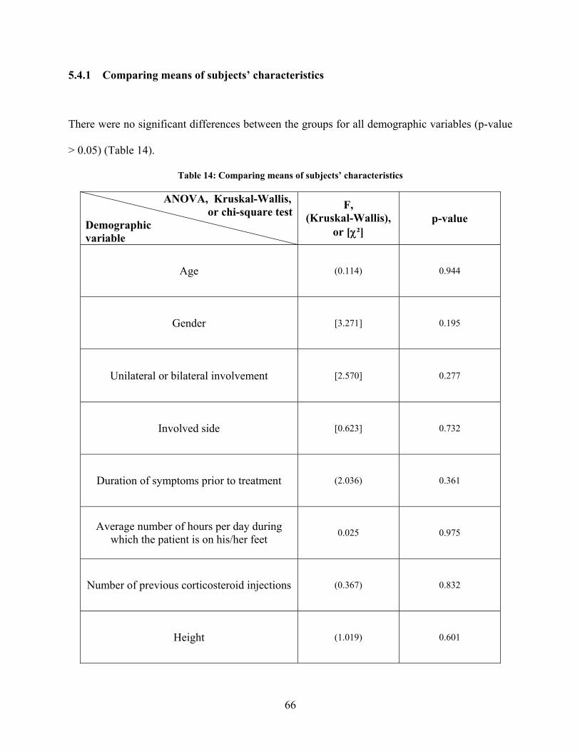
5.4.1 Comparing means of subjects’ characteristics
There were no significant differences between the groups for all demographic variables (p-value
> 0.05) (Table 14).
Table 14: Comparing means of subjects’ characteristics
ANOVA, Kruskal-Wallis, or chi-square testDemographic variable
F, (Kruskal-Wallis),
or [χ²] p-value
Age (0.114) 0.944
Gender [3.271] 0.195
Unilateral or bilateral involvement [2.570] 0.277
Involved side [0.623] 0.732
Duration of symptoms prior to treatment (2.036) 0.361
Average number of hours per day during which the patient is on his/her feet
0.025 0.975
Number of previous corticosteroid injections (0.367) 0.832
Height (1.019) 0.601
66
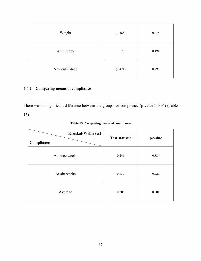
Weight (1.488) 0.475
Arch index 1.670 0.194
Navicular drop (2.421) 0.298
5.4.2 Comparing means of compliance
There was no significant difference between the groups for compliance (p-value > 0.05) (Table
15).
Table 15: Comparing means of compliance
Kruskal-Wallis test Compliance
Test statistic p-value
At three weeks 0.246 0.884
At six weeks 0.639 0.727
Average 0.208 0.901
67
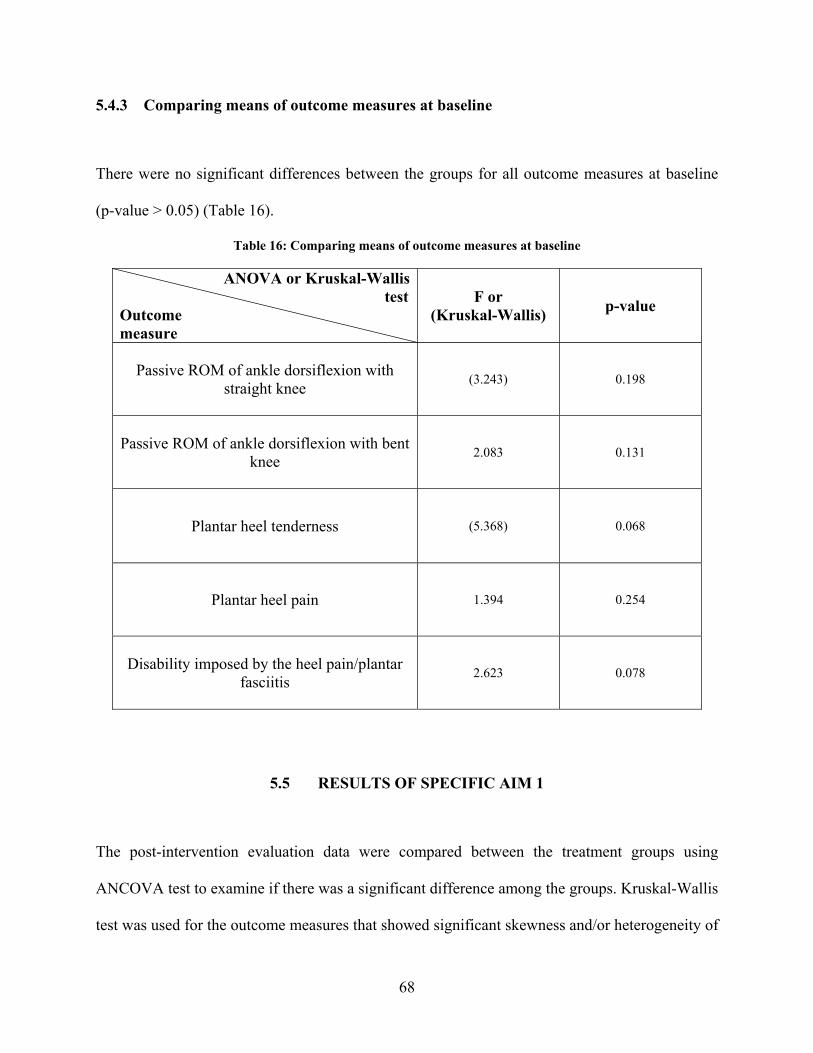
5.4.3 Comparing means of outcome measures at baseline
There were no significant differences between the groups for all outcome measures at baseline
(p-value > 0.05) (Table 16).
Table 16: Comparing means of outcome measures at baseline
ANOVA or Kruskal-Wallis testOutcome measure
F or (Kruskal-Wallis) p-value
Passive ROM of ankle dorsiflexion with straight knee (3.243) 0.198
Passive ROM of ankle dorsiflexion with bent knee 2.083 0.131
Plantar heel tenderness (5.368) 0.068
Plantar heel pain 1.394 0.254
Disability imposed by the heel pain/plantar fasciitis 2.623 0.078
5.5 RESULTS OF SPECIFIC AIM 1
The post-intervention evaluation data were compared between the treatment groups using
ANCOVA test to examine if there was a significant difference among the groups. Kruskal-Wallis
test was used for the outcome measures that showed significant skewness and/or heterogeneity of
68
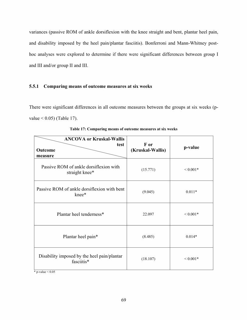
variances (passive ROM of ankle dorsiflexion with the knee straight and bent, plantar heel pain,
and disability imposed by the heel pain/plantar fasciitis). Bonferroni and Mann-Whitney post-
hoc analyses were explored to determine if there were significant differences between group I
and III and/or group II and III.
5.5.1 Comparing means of outcome measures at six weeks
There were significant differences in all outcome measures between the groups at six weeks (p-
value < 0.05) (Table 17).
Table 17: Comparing means of outcome measures at six weeks
ANCOVA or Kruskal-Wallis testOutcome measure
F or (Kruskal-Wallis) p-value
Passive ROM of ankle dorsiflexion with straight knee* (15.771) < 0.001*
Passive ROM of ankle dorsiflexion with bent knee* (9.045) 0.011*
Plantar heel tenderness* 22.097 < 0.001*
Plantar heel pain* (8.485) 0.014*
Disability imposed by the heel pain/plantar fasciitis* (18.107) < 0.001*
* p-value < 0.05
69

5.5.2 Post-hoc analyses of outcome measures at six weeks
Post-hoc analyses showed no significant difference between group I and II for all outcome
measures at six weeks (p-value > 0.05). However, all outcome measures showed significant
differences between group I and III and group II and III at six weeks (p-value < 0.017). In other
words, a dorsiflexion night splint and medial arch support together were more effective in the
treatment of plantar fasciitis than a dorsiflexion night splint or medial arch support each by itself
in terms of increasing the range of pain-free passive ankle dorsiflexion, relieving heel tenderness
and pain, and reducing disability imposed by the heel pain/plantar fasciitis (Table 18).
Table 18: Post-hoc analyses of outcome measures at six weeks
Group
Outcome measure
Group I & II p-value (2-tailed)
Bonferroni
or (Mann-Whitney)
Group I & III p-value (1-tailed)
Bonferroni
or (Mann-Whitney)
Group II & III p-value (1-tailed)
Bonferroni
or (Mann-Whitney)
Passive ROM of ankle dorsiflexion with
straight knee* (0.232) (0.001**) (< 0.001**)
Passive ROM of ankle dorsiflexion with bent
knee* (0.711) (0.007**) (0.004**)
Plantar heel tenderness* 0.836 < 0.001* < 0.001*
Plantar heel pain* (0.773) (0.006**) (0.007**)
Disability imposed by the heel pain/plantar
fasciitis* (0.574) (< 0.001**) (< 0.001**)
* p-value < 0.05 ** p-value < 0.017 (0.05/3)
70
5.6 RESULTS OF SPECIFIC AIM 2
The post-intervention evaluation data of group I were regressed on the baseline passive ROM of
ankle dorsiflexion with the knee straight and bent after controlling for the baseline evaluation
data.
5.6.1 Regression of group I outcome measures on baseline passive ROM of ankle
dorsiflexion with straight knee
Baseline passive ROM of ankle dorsiflexion with the knee straight was not a useful predictor of
the success of treatment with a dorsiflexion night splint for all outcome measures (p-value >
0.05). In other words, patients with plantar fasciitis who had less passive dorsiflexion of the
ankle joint with straight knee did not benefit from a dorsiflexion night splint more than those
with greater passive dorsiflexion of the ankle joint with straight knee in terms of increasing the
range of pain-free passive ankle dorsiflexion, relieving heel tenderness and pain, and reducing
disability imposed by the heel pain/plantar fasciitis (Table 19).
71
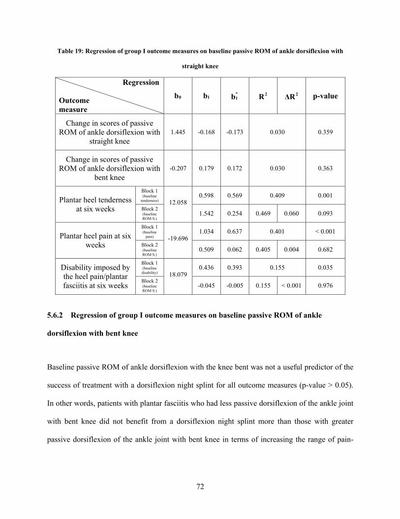
Table 19: Regression of group I outcome measures on baseline passive ROM of ankle dorsiflexion with
straight knee
Regression
Outcome measure
b0 b1 b*1 R2 ΔR2 p-value
Change in scores of passive ROM of ankle dorsiflexion with
straight knee 1.445 -0.168 -0.173 0.030 0.359
Change in scores of passive ROM of ankle dorsiflexion with
bent knee -0.207 0.179 0.172 0.030 0.363
Block 1 (baseline
tenderness) 0.598 0.569 0.409 0.001 Plantar heel tenderness
at six weeks Block 2 (baseline ROM S.)
12.058
1.542 0.254 0.469 0.060 0.093
Block 1 (baseline
pain) 1.034 0.637 0.401 < 0.001 Plantar heel pain at six
weeks Block 2 (baseline ROM S.)
-19.696
0.509 0.062 0.405 0.004 0.682
Block 1 (baseline disability)
0.436 0.393 0.155 0.035 Disability imposed by the heel pain/plantar fasciitis at six weeks
Block 2 (baseline ROM S.)
18.079
-0.045 -0.005 0.155 < 0.001 0.976
5.6.2 Regression of group I outcome measures on baseline passive ROM of ankle
dorsiflexion with bent knee
Baseline passive ROM of ankle dorsiflexion with the knee bent was not a useful predictor of the
success of treatment with a dorsiflexion night splint for all outcome measures (p-value > 0.05).
In other words, patients with plantar fasciitis who had less passive dorsiflexion of the ankle joint
with bent knee did not benefit from a dorsiflexion night splint more than those with greater
passive dorsiflexion of the ankle joint with bent knee in terms of increasing the range of pain-
72
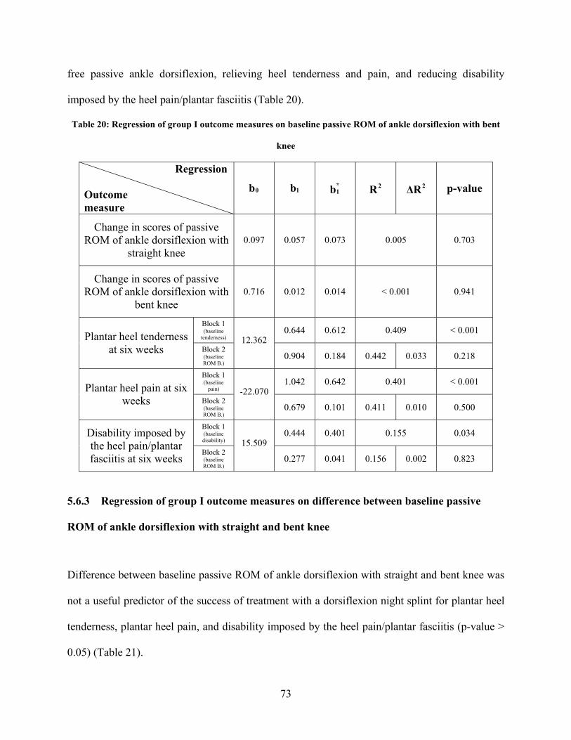
free passive ankle dorsiflexion, relieving heel tenderness and pain, and reducing disability
imposed by the heel pain/plantar fasciitis (Table 20).
Table 20: Regression of group I outcome measures on baseline passive ROM of ankle dorsiflexion with bent
knee
Regression
Outcome measure
b0 b1 b*1 R2 ΔR2 p-value
Change in scores of passive ROM of ankle dorsiflexion with
straight knee 0.097 0.057 0.073 0.005 0.703
Change in scores of passive ROM of ankle dorsiflexion with
bent knee 0.716 0.012 0.014 < 0.001 0.941
Block 1 (baseline
tenderness) 0.644 0.612 0.409 < 0.001 Plantar heel tenderness
at six weeks Block 2 (baseline ROM B.)
12.362
0.904 0.184 0.442 0.033 0.218
Block 1 (baseline
pain) 1.042 0.642 0.401 < 0.001 Plantar heel pain at six
weeks Block 2 (baseline ROM B.)
-22.070
0.679 0.101 0.411 0.010 0.500
Block 1 (baseline disability)
0.444 0.401 0.155 0.034 Disability imposed by the heel pain/plantar fasciitis at six weeks
Block 2 (baseline ROM B.)
15.509
0.277 0.041 0.156 0.002 0.823
5.6.3 Regression of group I outcome measures on difference between baseline passive
ROM of ankle dorsiflexion with straight and bent knee
Difference between baseline passive ROM of ankle dorsiflexion with straight and bent knee was
not a useful predictor of the success of treatment with a dorsiflexion night splint for plantar heel
tenderness, plantar heel pain, and disability imposed by the heel pain/plantar fasciitis (p-value >
0.05) (Table 21).
73
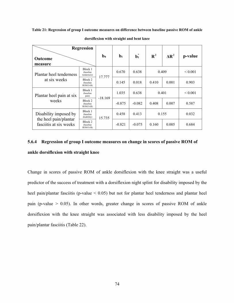
Table 21: Regression of group I outcome measures on difference between baseline passive ROM of ankle
dorsiflexion with straight and bent knee
Regression
Outcome measure
b0 b1 b*1 R2 ΔR2 p-value
Block 1 (baseline
tenderness) 0.670 0.638 0.409 < 0.001 Plantar heel tenderness
at six weeks Block 2 (baseline
ROM S-B)
17.777 0.145 0.018 0.410 0.001 0.903
Block 1 (baseline
pain) 1.035 0.638 0.401 < 0.001 Plantar heel pain at six
weeks Block 2 (baseline
ROM S-B)
-18.169 -0.875 -0.082 0.408 0.007 0.587
Block 1 (baseline disability)
0.458 0.413 0.155 0.032 Disability imposed by the heel pain/plantar fasciitis at six weeks
Block 2 (baseline
ROM S-B)
15.735
-0.821 -0.075 0.160 0.005 0.684
5.6.4 Regression of group I outcome measures on change in scores of passive ROM of
ankle dorsiflexion with straight knee
Change in scores of passive ROM of ankle dorsiflexion with the knee straight was a useful
predictor of the success of treatment with a dorsiflexion night splint for disability imposed by the
heel pain/plantar fasciitis (p-value < 0.05) but not for plantar heel tenderness and plantar heel
pain (p-value > 0.05). In other words, greater change in scores of passive ROM of ankle
dorsiflexion with the knee straight was associated with less disability imposed by the heel
pain/plantar fasciitis (Table 22).
74
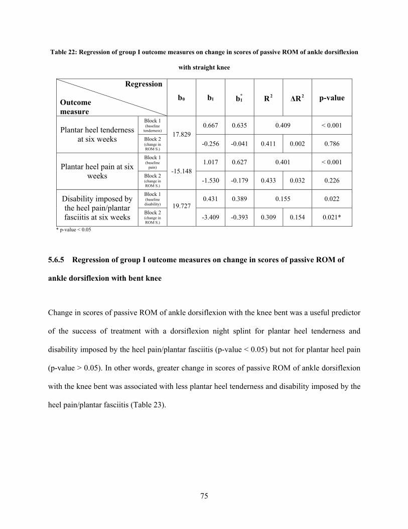
Table 22: Regression of group I outcome measures on change in scores of passive ROM of ankle dorsiflexion
with straight knee
Regression
Outcome measure
b0 b1 b*1 R2 ΔR2 p-value
Block 1 (baseline
tenderness) 0.667 0.635 0.409 < 0.001 Plantar heel tenderness
at six weeks Block 2 (change in ROM S.)
17.829 -0.256 -0.041 0.411 0.002 0.786
Block 1 (baseline
pain) 1.017 0.627 0.401 < 0.001 Plantar heel pain at six
weeks Block 2 (change in ROM S.)
-15.148 -1.530 -0.179 0.433 0.032 0.226
Block 1 (baseline disability)
0.431 0.389 0.155 0.022 Disability imposed by the heel pain/plantar fasciitis at six weeks
Block 2 (change in ROM S.)
19.727
-3.409 -0.393 0.309 0.154 0.021*
* p-value < 0.05
5.6.5 Regression of group I outcome measures on change in scores of passive ROM of
ankle dorsiflexion with bent knee
Change in scores of passive ROM of ankle dorsiflexion with the knee bent was a useful predictor
of the success of treatment with a dorsiflexion night splint for plantar heel tenderness and
disability imposed by the heel pain/plantar fasciitis (p-value < 0.05) but not for plantar heel pain
(p-value > 0.05). In other words, greater change in scores of passive ROM of ankle dorsiflexion
with the knee bent was associated with less plantar heel tenderness and disability imposed by the
heel pain/plantar fasciitis (Table 23).
75
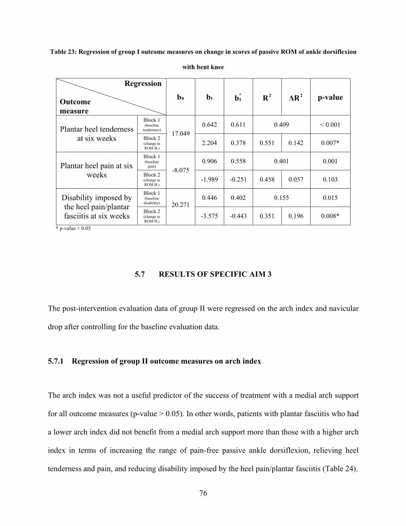
Table 23: Regression of group I outcome measures on change in scores of passive ROM of ankle dorsiflexion
with bent knee
Regression
Outcome measure
b0 b1 b*1 R2 ΔR2 p-value
Block 1 (baseline
tenderness) 0.642 0.611 0.409 < 0.001 Plantar heel tenderness
at six weeks Block 2 (change in ROM B.)
17.049 2.204 0.378 0.551 0.142 0.007*
Block 1 (baseline
pain) 0.906 0.558 0.401 0.001 Plantar heel pain at six
weeks Block 2 (change in ROM B.)
-8.075 -1.989 -0.251 0.458 0.057 0.103
Block 1 (baseline disability)
0.446 0.402 0.155 0.015 Disability imposed by the heel pain/plantar fasciitis at six weeks
Block 2 (change in ROM B.)
20.271
-3.575 -0.443 0.351 0.196 0.008*
* p-value < 0.05
5.7 RESULTS OF SPECIFIC AIM 3
The post-intervention evaluation data of group II were regressed on the arch index and navicular
drop after controlling for the baseline evaluation data.
5.7.1 Regression of group II outcome measures on arch index
The arch index was not a useful predictor of the success of treatment with a medial arch support
for all outcome measures (p-value > 0.05). In other words, patients with plantar fasciitis who had
a lower arch index did not benefit from a medial arch support more than those with a higher arch
index in terms of increasing the range of pain-free passive ankle dorsiflexion, relieving heel
tenderness and pain, and reducing disability imposed by the heel pain/plantar fasciitis (Table 24).
76

Table 24: Regression of group II outcome measures on arch index
Regression
Outcome measure
b0 b1 b*1 R2 ΔR2 p-value
Block 1 (baseline ROM S.)
1.076 0.680 0.464 < 0.001 Passive ROM of ankle dorsiflexion with
straight knee at six weeks
Block 2 (arch index)
-1.622
1.405 0.011 0.464 < 0.001 0.937
Block 1 (baseline ROM B.)
1.194 0.838 0.703 < 0.001 Passive ROM of ankle dorsiflexion with bent
knee at six weeks Block 2 (arch index)
-1.944
1.336 0.010 0.703 < 0.001 0.924
Block 1 (baseline
tenderness) 0.249 0.170 0.031 0.383 Plantar heel tenderness
at six weeks Block 2 (arch index)
36.238
-21.656 -0.042 0.033 0.002 0.830
Block 1 (baseline
pain) 0.627 0.391 0.159 0.040 Plantar heel pain at six
weeks Block 2 (arch index)
23.711
-46.816 -0.060 0.162 0.004 0.741
Block 1 (baseline disability)
0.661 0.550 0.303 0.002 Disability imposed by the heel pain/plantar fasciitis at six weeks Block 2
(arch index)
9.690
-0.603 -0.001 0.303 < 0.001 0.996
5.7.2 Regression of group II outcome measures on navicular drop
Navicular drop was not a useful predictor of the success of treatment with a medial arch support
for all outcome measures (p-value > 0.05). In other words, patients with plantar fasciitis who had
a greater navicular drop did not benefit from a medial arch support more than those with a
smaller navicular drop in terms of increasing the range of pain-free passive ankle dorsiflexion,
relieving heel tenderness and pain, and reducing disability imposed by the heel pain/plantar
fasciitis (Table 25).
77

Table 25: Regression of group II outcome measures on navicular drop
Regression
Outcome measure
b0 b1 b*1 R2 ΔR2 p-value
Block 1 (baseline ROM S.)
1.091 0.689 0.464 < 0.001 Passive ROM of ankle dorsiflexion with
straight knee at six weeks
Block 2 (navicular
drop)
-1.733
0.079 0.051 0.467 0.003 0.721
Block 1 (baseline ROM B.)
1.205 0.846 0.703 < 0.001 Passive ROM of ankle dorsiflexion with bent
knee at six weeks Block 2 (navicular
drop)
-2.178
0.101 0.061 0.707 0.004 0.563
Block 1 (baseline
tenderness) 0.253 0.173 0.031 0.371 Plantar heel tenderness
at six weeks Block 2 (navicular
drop)
28.666
0.338 0.052 0.034 0.003 0.784
Block 1 (baseline
pain) 0.635 0.396 0.159 0.040 Plantar heel pain at six
weeks Block 2 (navicular
drop)
11.602
-0.126 -0.013 0.159 < 0.001 0.944
Block 1 (baseline disability)
0.627 0.522 0.303 0.003 Disability imposed by the heel pain/plantar fasciitis at six weeks
Block 2 (navicular
drop)
17.203
-1.219 -0.147 0.324 0.021 0.369
78

6.0 DISCUSSION
6.1 DISCUSSION OF SPECIFIC AIM 1
Plantar fasciitis is an overuse injury causing inflammation at the origin of the plantar fascia and
is typically characterized by pain in the inferior heel region that is provoked by weight bearing
after a long period of non-weight bearing and by prolonged weight bearing.1,4,9,14,19,21,24,25 A
dorsiflexion night splint is used to relieve the pain associated with the first step in the morning
by preventing contracture of the plantar fascia and Achilles tendon overnight.4,5,7,15,19,23,24,26-32 A
medial arch support, on the other hand, relieves the end of the day pain by supporting the medial
longitudinal arch and, thus, preventing overstretch of plantar fascia during prolonged weight
bearing.1,7,21,24,33-37 Therefore, both night splints and arch supports may be necessary to treat
plantar fasciitis as they complement each other by both controlling nocturnal contracture of the
plantar fascia and Achilles tendon and reducing stresses imposed on the plantar fascia during the
day, respectively. In fact, many authors agree that the success of conservative treatment of
plantar fasciitis usually requires a combination of treatment modalities, rather than administering
only one treatment at a time.4,14,19,20 Although the isolated effectiveness of night splints and arch
supports in relieving symptoms of plantar fasciitis is well-established in the literature, no
previous study, to the best of our knowledge, has been conducted to evaluate the combined effect
of these treatment modalities.
79
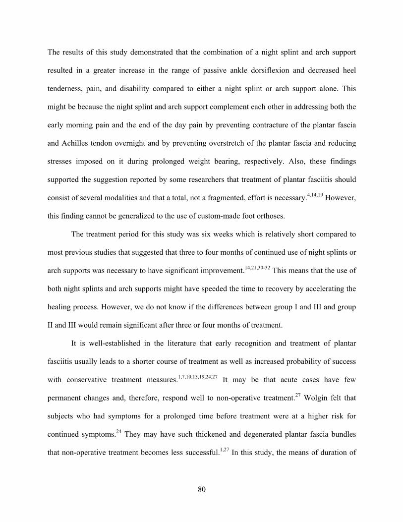
The results of this study demonstrated that the combination of a night splint and arch support
resulted in a greater increase in the range of passive ankle dorsiflexion and decreased heel
tenderness, pain, and disability compared to either a night splint or arch support alone. This
might be because the night splint and arch support complement each other in addressing both the
early morning pain and the end of the day pain by preventing contracture of the plantar fascia
and Achilles tendon overnight and by preventing overstretch of the plantar fascia and reducing
stresses imposed on it during prolonged weight bearing, respectively. Also, these findings
supported the suggestion reported by some researchers that treatment of plantar fasciitis should
consist of several modalities and that a total, not a fragmented, effort is necessary.4,14,19 However,
this finding cannot be generalized to the use of custom-made foot orthoses.
The treatment period for this study was six weeks which is relatively short compared to
most previous studies that suggested that three to four months of continued use of night splints or
arch supports was necessary to have significant improvement.14,21,30-32 This means that the use of
both night splints and arch supports might have speeded the time to recovery by accelerating the
healing process. However, we do not know if the differences between group I and III and group
II and III would remain significant after three or four months of treatment.
It is well-established in the literature that early recognition and treatment of plantar
fasciitis usually leads to a shorter course of treatment as well as increased probability of success
with conservative treatment measures.1,7,10,13,19,24,27 It may be that acute cases have few
permanent changes and, therefore, respond well to non-operative treatment.27 Wolgin felt that
subjects who had symptoms for a prolonged time before treatment were at a higher risk for
continued symptoms.24 They may have such thickened and degenerated plantar fascia bundles
that non-operative treatment becomes less successful.1,27 In this study, the means of duration of
80
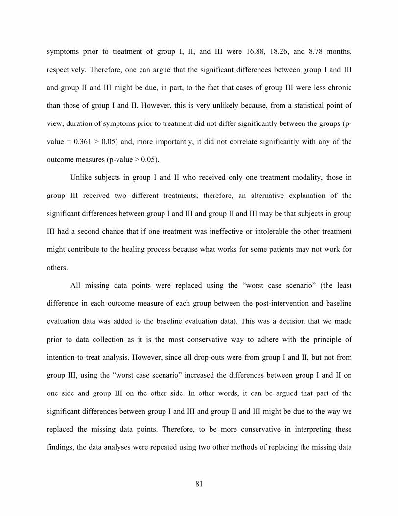
symptoms prior to treatment of group I, II, and III were 16.88, 18.26, and 8.78 months,
respectively. Therefore, one can argue that the significant differences between group I and III
and group II and III might be due, in part, to the fact that cases of group III were less chronic
than those of group I and II. However, this is very unlikely because, from a statistical point of
view, duration of symptoms prior to treatment did not differ significantly between the groups (p-
value = 0.361 > 0.05) and, more importantly, it did not correlate significantly with any of the
outcome measures (p-value > 0.05).
Unlike subjects in group I and II who received only one treatment modality, those in
group III received two different treatments; therefore, an alternative explanation of the
significant differences between group I and III and group II and III may be that subjects in group
III had a second chance that if one treatment was ineffective or intolerable the other treatment
might contribute to the healing process because what works for some patients may not work for
others.
All missing data points were replaced using the “worst case scenario” (the least
difference in each outcome measure of each group between the post-intervention and baseline
evaluation data was added to the baseline evaluation data). This was a decision that we made
prior to data collection as it is the most conservative way to adhere with the principle of
intention-to-treat analysis. However, since all drop-outs were from group I and II, but not from
group III, using the “worst case scenario” increased the differences between group I and II on
one side and group III on the other side. In other words, it can be argued that part of the
significant differences between group I and III and group II and III might be due to the way we
replaced the missing data points. Therefore, to be more conservative in interpreting these
findings, the data analyses were repeated using two other methods of replacing the missing data
81
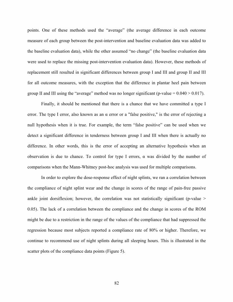
points. One of these methods used the “average” (the average difference in each outcome
measure of each group between the post-intervention and baseline evaluation data was added to
the baseline evaluation data), while the other assumed “no change” (the baseline evaluation data
were used to replace the missing post-intervention evaluation data). However, these methods of
replacement still resulted in significant differences between group I and III and group II and III
for all outcome measures, with the exception that the difference in plantar heel pain between
group II and III using the “average” method was no longer significant (p-value = 0.040 > 0.017).
Finally, it should be mentioned that there is a chance that we have committed a type I
error. The type I error, also known as an α error or a "false positive," is the error of rejecting a
null hypothesis when it is true. For example, the term “false positive” can be used when we
detect a significant difference in tenderness between group I and III when there is actually no
difference. In other words, this is the error of accepting an alternative hypothesis when an
observation is due to chance. To control for type I errors, α was divided by the number of
comparisons when the Mann-Whitney post-hoc analysis was used for multiple comparisons.
In order to explore the dose-response effect of night splints, we ran a correlation between
the compliance of night splint wear and the change in scores of the range of pain-free passive
ankle joint dorsiflexion; however, the correlation was not statistically significant (p-value >
0.05). The lack of a correlation between the compliance and the change in scores of the ROM
might be due to a restriction in the range of the values of the compliance that had suppressed the
regression because most subjects reported a compliance rate of 80% or higher. Therefore, we
continue to recommend use of night splints during all sleeping hours. This is illustrated in the
scatter plots of the compliance data points (Figure 5).
82
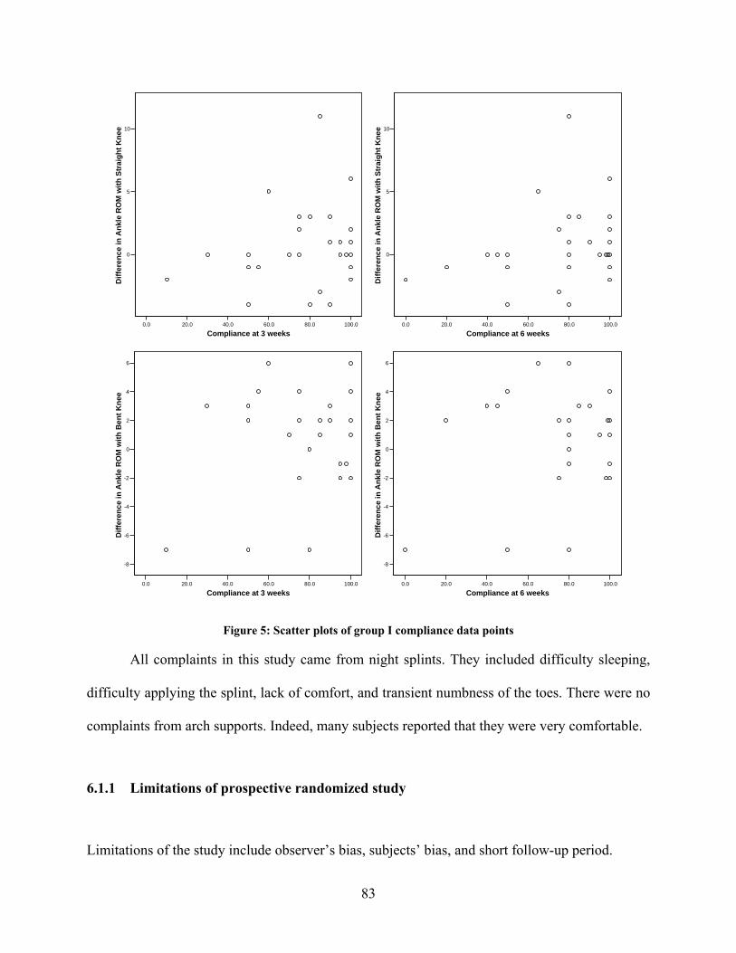
0.0 20.0 40.0 60.0 80.0 100.0
Compliance at 3 weeks
0
5
10
Diff
eren
ce in
Ank
le R
OM
with
Str
aigh
t Kne
e
0.0 20.0 40.0 60.0 80.0 100.0
Compliance at 6 weeks
0
5
10
Diff
eren
ce in
Ank
le R
OM
with
Str
aigh
t Kne
e
0.0 20.0 40.0 60.0 80.0 100.0
Compliance at 3 weeks
-8
-6
-4
-2
0
2
4
6
Diff
eren
ce in
Ank
le R
OM
with
Ben
t Kne
e
0.0 20.0 40.0 60.0 80.0 100.0
Compliance at 6 weeks
-8
-6
-4
-2
0
2
4
6
Diff
eren
ce in
Ank
le R
OM
with
Ben
t Kne
e
Figure 5: Scatter plots of group I compliance data points
All complaints in this study came from night splints. They included difficulty sleeping,
difficulty applying the splint, lack of comfort, and transient numbness of the toes. There were no
complaints from arch supports. Indeed, many subjects reported that they were very comfortable.
6.1.1 Limitations of prospective randomized study
Limitations of the study include observer’s bias, subjects’ bias, and short follow-up period.
83
6.1.1.1 Observer’s bias
In order to eliminate observer’s bias, we should, ideally, have blinded the principal
investigator from the group assignment of the subject by letting him perform the assessment
himself and hiring another physical therapist to do the randomization and provide the
corresponding treatment to the subject. However, we were unable to do that because of the
limited funds for the study. Therefore, we decided that the principal investigator should perform
the assessment before the randomization at the initial visit to be completely blinded to the group
assignment of the subject and, thus, to eliminate his bias toward any treatment group. We also
decided that he should have no access to the data recording form of the initial visit at the time of
the follow-up visit, and to record compliance, which would tell him to which treatment group the
subject belonged, only after finishing the follow-up assessment, with the hope that he would
forget the group assignment of the subject after six weeks of the subject’s participation in the
study. Although the principal investigator might not have been completely blinded to the group
assignment of the subject during the follow-up visit, he would be completely blinded to the
baseline measurements. In other words, even if he still could remember the subject’s assigned
group at the time of the follow-up visit, there would be no way that he could recall his/her
baseline measurements. In addition, the fact that most of the outcome measures of the study were
questionnaires that were completed by the subject (pain and disability) or tests that were
subjective in nature (tenderness), would minimize the effect of the principal investigator’s bias
on the follow-up assessment. In other words, ROM was the only objective outcome measure that
could be affected by the observer’s bias. We understand that the principal investigator might not
be completely blinded during the follow-up visit, and that his bias toward any treatment group
was not fully eliminated, but this was the best we could do to try to minimize the observer’s bias,
84

and we felt that it would result in a non-biased follow-up assessment and an acceptable level of
validity of the results.
6.1.1.2 Subjects’ bias
Subjects of the study were not blinded to their group assignment and, thus, their bias
toward any treatment group was not eliminated. In other words, those who were assigned to
group III might be psychologically affected by the fact they were in the combination group that
received two treatment modalities compared to group I and II that received only one treatment.
This is particularly important since all outcome measures, except ROM, were subjective in
nature. Therefore, one can argue that the significant differences between group I and III and
group II and III might be due, in part, to the subjects’ bias. However, the subjects were blinded to
the research questions and hypotheses of the study, which might minimize, but did not
completely eliminate, their bias. In addition, there was no possible way to blind the subjects from
their group assignment.
6.1.1.3 Short follow-up period
The follow-up period of this study was six weeks which is relatively short compared to
most previous studies on plantar fasciitis.14,21,30-32 Therefore, we do not have enough information
about the long-term benefits of the treatment measures used in this study. In other words, it is
unknown whether or not the improvement in the outcome measures would be retained for a long
period of time and whether or not a recurrence of symptoms would occur after discontinuing
treatment.
85
6.2 DISCUSSION OF SPECIFIC AIM 2 AND 3
The secondary hypotheses of this study were based on the suggested association between
tightness of the Achilles tendon and pes planus and their consequent excessive pronation with the
development of plantar fasciitis.1,22 A tight Achilles tendon is found in 78% of patients with
plantar fasciitis.4,13,16,17,24,39 It limits ankle joint dorsiflexion, which increases the load on the
intrinsic muscles of the foot and results in abnormal compensatory pronation of the subtalar joint
as ankle dorsiflexion progresses during the stance phase of gait.3,16,19,46,47,55
Between 81 and 86% of individuals with symptoms consistent with plantar fasciitis have
been classified on examination as having pes planus with excessive pronation.1 The theoretical
basis for this finding is the increased tension placed on the plantar fascia as a result of a lower
arch during standing and walking.7,13,19,42,54 In addition, increased pronation results in decreased
stability of the hindfoot, which produces additional stress on the origin of the central band of the
plantar fascia and may ultimately lead to plantar fasciitis.1,15,28
Excessive pronation results in an inability of the foot to supinate from mid to terminal
stance.3,51 Consequently, little load is conveyed through the lateral portion of the midfoot and
normal loading forces are inadequately supported by the bones and ligaments. The vertical
impulse is thus shifted away from the midfoot, and secondary structures, such as the plantar
fascia, must assume a greater load.6 Mann and Inman confirmed this by noting that heel
pronation increased the tension along the medial aspect of the heel.54
However, despite the wide acceptance of the association between these factors and
plantar fasciitis, research studies have not demonstrated that they are primary factors in the cause
of plantar fasciitis.1 Snook and Chrisman wrote, “It is reasonably certain that a condition which
86

has so many different theories of etiology and treatment does not have valid proof of any one
cause.”49 This thinking has been reiterated by other authors.11,24
The questionable validity of tightness of the Achilles tendon and pes planus as factors
that cause plantar fasciitis might be the reason behind the findings of this study that the range of
pain-free passive ankle joint dorsiflexion and medial longitudinal arch height were not useful
predictors of the success of treatment with a night splint and arch support in terms of all outcome
measures, respectively. However, greater change in the range of pain-free passive ankle joint
dorsiflexion following treatment with a night splint was associated with less plantar heel
tenderness and disability imposed by the heel pain/plantar fasciitis.
An alternative explanation may be that we have committed a type II error. The type II
error, often denoted as a β error or a "false negative," is the error of not rejecting a null
hypothesis when it is false. In other words, this is the error of failing to accept an alternative
hypothesis because of inadequate power.
In addition, it can be argued that the range of pain-free passive ankle joint dorsiflexion
and medial longitudinal arch height were not useful predictors of the success of treatment with a
night splint and arch support because there was a restriction in the range of the values of the
ROM and arch height that had suppressed the regression. However, this is very unlikely because
the ROM and arch height data points were randomly scattered through all the ranges of their
scatter plots (Figure 6 and 7). Figure 7 also shows that neither high nor low values of the medial
longitudinal arch height were useful predictors of the success of treatment with an arch support
since the arch height data points did not follow a curved pattern.
87
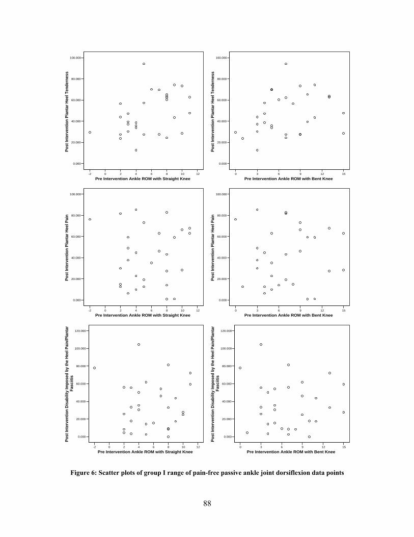
-2 0 2 4 6 8 10 12
Pre Intervention Ankle ROM with Straight Knee
0.000
20.000
40.000
60.000
80.000
100.000
Post
Inte
rven
tion
Plan
tar H
eel T
ende
rnes
s
0 3 6 9 12 15
Pre Intervention Ankle ROM with Bent Knee
0.000
20.000
40.000
60.000
80.000
100.000
Post
Inte
rven
tion
Plan
tar H
eel T
ende
rnes
s
-2 0 2 4 6 8 10 12
Pre Intervention Ankle ROM with Straight Knee
0.000
20.000
40.000
60.000
80.000
100.000
Post
Inte
rven
tion
Plan
tar H
eel P
ain
0 3 6 9 12 15
Pre Intervention Ankle ROM with Bent Knee
0.000
20.000
40.000
60.000
80.000
100.000
Post
Inte
rven
tion
Plan
tar H
eel P
ain
-2 0 2 4 6 8 10 12
Pre Intervention Ankle ROM with Straight Knee
0.000
20.000
40.000
60.000
80.000
100.000
120.000
Post
Inte
rven
tion
Disa
bilit
y Im
pose
d by
the
Heel
Pai
n/Pl
anta
r Fa
sciit
is
0 3 6 9 12 15
Pre Intervention Ankle ROM with Bent Knee
0.000
20.000
40.000
60.000
80.000
100.000
120.000
Post
Inte
rven
tion
Disa
bilit
y Im
pose
d by
the
Heel
Pai
n/Pl
anta
r Fa
sciit
is
Figure 6: Scatter plots of group I range of pain-free passive ankle joint dorsiflexion data points
88
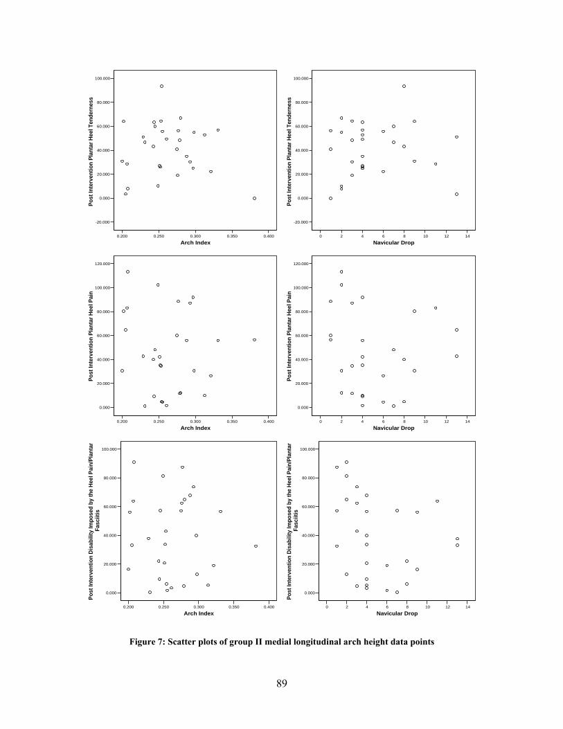
0.200 0.250 0.300 0.350 0.400
Arch Index
-20.000
0.000
20.000
40.000
60.000
80.000
100.000
Post
Inte
rven
tion
Plan
tar H
eel T
ende
rnes
s
0 2 4 6 8 10 12 14
Navicular Drop
-20.000
0.000
20.000
40.000
60.000
80.000
100.000
Post
Inte
rven
tion
Plan
tar H
eel T
ende
rnes
s
0.200 0.250 0.300 0.350 0.400
Arch Index
0.000
20.000
40.000
60.000
80.000
100.000
120.000
Post
Inte
rven
tion
Plan
tar H
eel P
ain
0 2 4 6 8 10 12 14
Navicular Drop
0.000
20.000
40.000
60.000
80.000
100.000
120.000
Post
Inte
rven
tion
Plan
tar H
eel P
ain
0.200 0.250 0.300 0.350 0.400
Arch Index
0.000
20.000
40.000
60.000
80.000
100.000
Post
Inte
rven
tion
Disa
bilit
y Im
pose
d by
the
Heel
Pai
n/Pl
anta
r Fa
sciit
is
0 2 4 6 8 10 12 14
Navicular Drop
0.000
20.000
40.000
60.000
80.000
100.000
Post
Inte
rven
tion
Disa
bilit
y Im
pose
d by
the
Heel
Pai
n/Pl
anta
r Fa
sciit
is
Figure 7: Scatter plots of group II medial longitudinal arch height data points
89
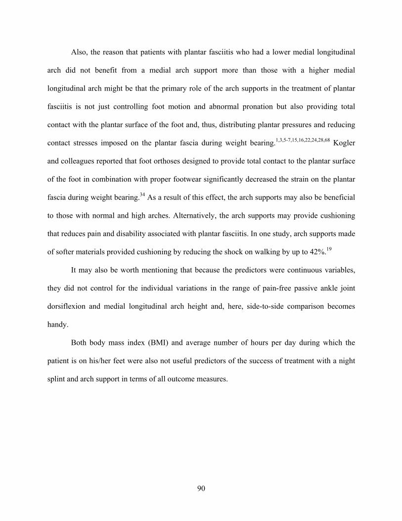
Also, the reason that patients with plantar fasciitis who had a lower medial longitudinal
arch did not benefit from a medial arch support more than those with a higher medial
longitudinal arch might be that the primary role of the arch supports in the treatment of plantar
fasciitis is not just controlling foot motion and abnormal pronation but also providing total
contact with the plantar surface of the foot and, thus, distributing plantar pressures and reducing
contact stresses imposed on the plantar fascia during weight bearing.1,3,5-7,15,16,22,24,28,68 Kogler
and colleagues reported that foot orthoses designed to provide total contact to the plantar surface
of the foot in combination with proper footwear significantly decreased the strain on the plantar
fascia during weight bearing.34 As a result of this effect, the arch supports may also be beneficial
to those with normal and high arches. Alternatively, the arch supports may provide cushioning
that reduces pain and disability associated with plantar fasciitis. In one study, arch supports made
of softer materials provided cushioning by reducing the shock on walking by up to 42%.19
It may also be worth mentioning that because the predictors were continuous variables,
they did not control for the individual variations in the range of pain-free passive ankle joint
dorsiflexion and medial longitudinal arch height and, here, side-to-side comparison becomes
handy.
Both body mass index (BMI) and average number of hours per day during which the
patient is on his/her feet were also not useful predictors of the success of treatment with a night
splint and arch support in terms of all outcome measures.
90
7.0 CONCLUSION
Based on the results of this study, it was concluded that a dorsiflexion night splint and medial
arch support together may be more effective in the treatment of plantar fasciitis than a
dorsiflexion night splint or medial arch support each by itself in terms of increasing the range of
pain-free passive ankle dorsiflexion, relieving heel tenderness and pain, and reducing disability
imposed by the heel pain/plantar fasciitis because, together, they address both the early morning
pain and the end of the day pain, respectively.
Patients with plantar fasciitis who have less passive dorsiflexion of the ankle joint do not
benefit from a dorsiflexion night splint more than those with greater passive dorsiflexion of the
ankle joint in terms of increasing the range of pain-free passive ankle dorsiflexion, relieving heel
tenderness and pain, and reducing disability imposed by the heel pain/plantar fasciitis. However,
greater change in the passive dorsiflexion of the ankle joint is associated with less plantar heel
tenderness and disability imposed by the heel pain/plantar fasciitis.
Patients with plantar fasciitis who have a lower medial longitudinal arch do not benefit
from a medial arch support more than those with a higher medial longitudinal arch in terms of
increasing the range of pain-free passive ankle dorsiflexion, relieving heel tenderness and pain,
and reducing disability imposed by the heel pain/plantar fasciitis.
Further studies may include, but are not limited to, evaluation of the effectiveness of
dorsiflexion night splints and medial arch supports in the treatment of plantar fasciitis for a
91
longer follow-up period. These studies may also compare the effects of different models and
brands of night splints and arch supports. For instance, comparing the effects of adjustable versus
non-adjustable night splints, and full length versus ¾ length arch supports. They may also
investigate whether foot taping is a useful predictor of the success of treatment with an arch
support.
In addition, the efficacy of different modalities of physical therapy such as cryotherapy
and hydrotherapy needs to be assessed. Although these modalities have been described as
effective in the management of plantar fasciitis, no studies have been conducted on patients with
plantar fasciitis to determine their actual effectiveness. Future studies may also examine the
effects of different combinations of treatment modalities. Despite the wide agreement that the
success of conservative care for the treatment of patients with plantar fasciitis requires a
combination of treatment modalities, there is no consensus about which treatments are the best or
the most cost-effective, and there is inconsistency in the treatments provided by various
practitioners.
92
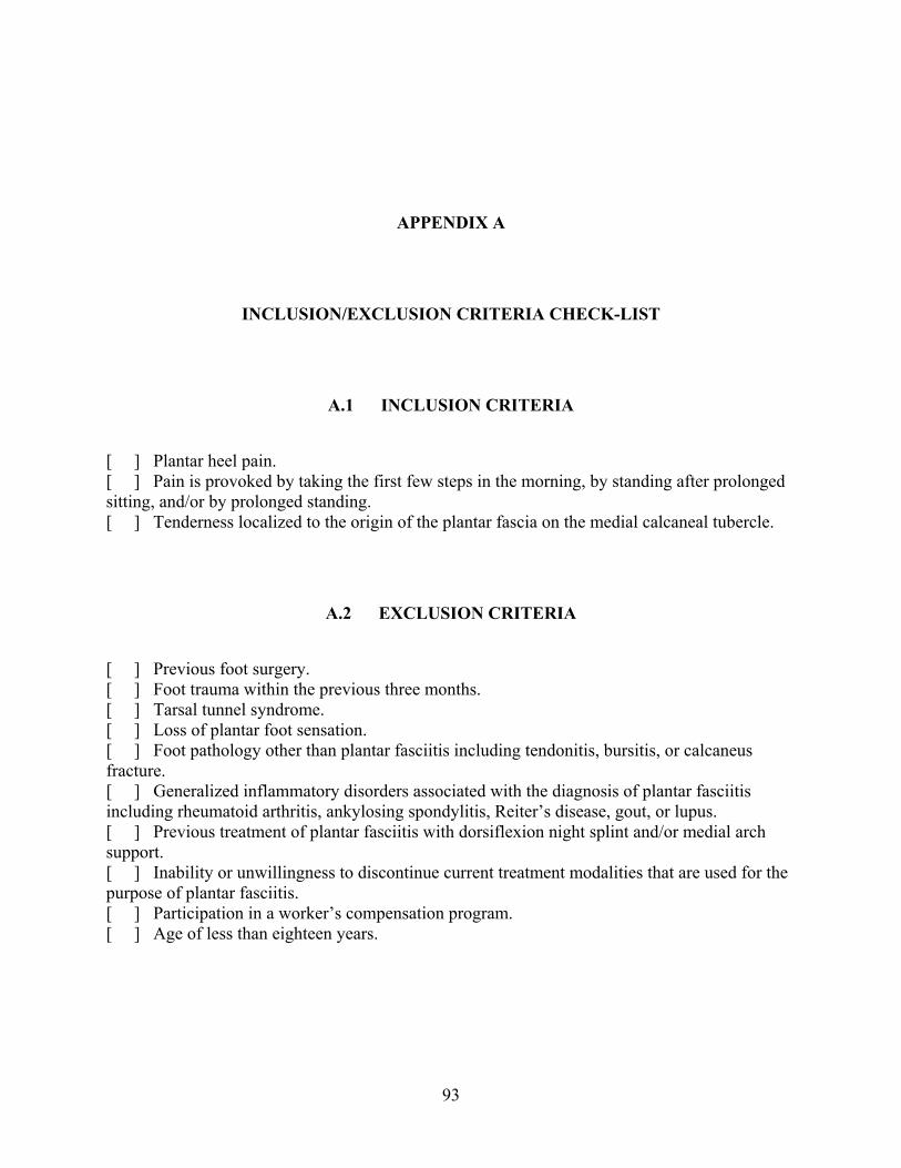
APPENDIX A
INCLUSION/EXCLUSION CRITERIA CHECK-LIST
A.1 INCLUSION CRITERIA
[ ] Plantar heel pain. [ ] Pain is provoked by taking the first few steps in the morning, by standing after prolonged sitting, and/or by prolonged standing. [ ] Tenderness localized to the origin of the plantar fascia on the medial calcaneal tubercle.
A.2 EXCLUSION CRITERIA
[ ] Previous foot surgery. [ ] Foot trauma within the previous three months. [ ] Tarsal tunnel syndrome. [ ] Loss of plantar foot sensation. [ ] Foot pathology other than plantar fasciitis including tendonitis, bursitis, or calcaneus fracture. [ ] Generalized inflammatory disorders associated with the diagnosis of plantar fasciitis including rheumatoid arthritis, ankylosing spondylitis, Reiter’s disease, gout, or lupus. [ ] Previous treatment of plantar fasciitis with dorsiflexion night splint and/or medial arch support. [ ] Inability or unwillingness to discontinue current treatment modalities that are used for the purpose of plantar fasciitis. [ ] Participation in a worker’s compensation program. [ ] Age of less than eighteen years.
93
APPENDIX B
HIPAA AUTHORIZATION FOR SHARING HEALTH INFORMATION
94
AUTHORIZATION FOR THE SHARING OF HEALTH INFORMATION RELATED TO POSSIBLE PARTICIPATION IN A RESEARCH STUDY
Title of Research Study: Conservative Treatment of Plantar Fasciitis with Dorsiflexion Night Splint and Medial Arch Supports: a Prospective Randomized Study Research Study Investigators: Principal Investigator: Ahmad Al-Ghadir, MS, PT, PhD Candidate, Department of Physical Therapy, Room 6010A Forbes Tower, Pittsburgh, PA 15260. Telephone: 412-726-1826 Co-Investigators: Anthony Delitto, PhD, PT, FAPTA, Associate Professor and Chair, Department of Physical Therapy, Room 6036 Forbes Tower, Pittsburgh, PA 15260. Telephone: 412- 383-6631 Dane Wukich, MD, Assistant Professor, Department of Orthopaedic Surgery, Suite 1010 Kaufmann Medical Building, Pittsburgh, PA 15213. Telephone: 412-687-3900 James Irrgang, PhD, PT, ATC, Associate Professor and Vice Chair of Clinical Services, Department of Physical Therapy, Room 6010A Forbes Tower, Pittsburgh, PA 15260. Telephone: 412-647-1237 Ray Burdett, PhD, PT, CPed, Associate Professor and Assistant Dean of Undergraduate Programs, Department of Physical Therapy, Room 6022 Forbes Tower, Pittsburgh, PA 15260. Telephone: 412-383-6704
Page 1 of 4 Patient’s Initials: _____
95
B.1 WHAT IS THE PURPOSE OF THIS AUTHORIZATION?
Your doctor or a member of your doctor’s health care staff has discussed with you that you may be eligible to take part in the above-named research study. You have indicated an interest in learning more about this research study from the researchers who are involved in conducting the study. Thus, your authorization (permission) is being requested to:
• share the fact that you are interested in participating in this study with the involved researchers;
• share only your medical diagnosis which suggests you may be eligible to take part in this study with the involved researchers; and
• allow the involved researchers to contact you so as to permit additional discussions of this study with you and/or to provide you with information on how you may take part in this study.
B.2 WHAT INFORMATION ABOUT ME WILL BE SHARED WITH THE RESEARCHERS?
If you give your permission, the following information about you will be shared (for example, by telephone or FAX) with the researchers involved in the conduct of the above-named research study:
• your name, address, and telephone number • only your medical diagnosis which suggests you may be eligible for this research study • your interest in being contacted for the research • a copy of this signed document
B.3 TO WHOM WILL THE ABOVE INFORMATION BE GIVEN?
We will share this information with one of the researchers listed above or a member of their research staff. This information will be used by the researchers to evaluate if you are eligible to participate in this research study and/or to contact you to further discuss this research study with you.
These researchers recognize the importance of maintaining the confidentiality (privacy) of your health information; however, it is not possible for us to guarantee its confidentiality after we have provided it to them.
Page 2 of 4 Patient’s Initials: _____
96
B.4 FOR HOW LONG IS AUTHORIZATION VALID?
Once this information has been shared with the researchers, this authorization form will expire. We will not continue to share your future health information with these researchers, nor will we share your health information with any other researchers unless you sign a separate authorization form that permits us to do so.
B.5 IS MY PERMISSION TO PROVIDE THIS INFORMATION TO THE RESEARCHERS VOLUNTARY?
Your permission to provide this information to the researchers is completely voluntary. Whether or not you provide your permission will have no affect on your current or future medical care or your relationship with your doctor or health care provider. Whether or not you provide your permission will have no affect on your current or future relationship with the University of Pittsburgh or University of Pittsburgh Medical Center.
B.6 MAY I WITHDRAW, AT A FUTURE DATE, MY PERMISSION TO PROVIDE THIS INFORMATION TO THE RESEARCHERS?
You may withdraw, at any time, your permission to provide this information to the researchers. However, once this information has been shared with the researchers, the information will be in their possession. Hence, should you decide to withdraw your permission after your information has been given to the researchers you should send a written and dated notice of this decision to the principal investigator of this research study at the address listed above. Upon receipt of this request, the researchers will destroy your information that was provided to them. If you wish to withdraw your permission to provide this information to the researchers before it is given to them, you should contact, by telephone, your doctor or a member of your doctor’s health care staff. With receipt of this request, your information will not be shared with the researchers.
Your decision to withdraw your permission to provide this information to the researchers will have no effect on your current or future medical care or your relationship with your doctor or health care provider. Your decision to withdraw your permission will have no affect on your current or future relationship with the University of Pittsburgh or University of Pittsburgh Medical Center. ****************************************************************************** VOLUNTARY AUTHORIZATION
Page 3 of 4 Patient’s Initials: _____
97

All of the above has been explained to me. By signing below I give my permission to share the information, specified above, with the researchers, identified above, for the purposes described. ____________________________________ Printed Name of Patient ____________________________________ ________________________ Signature of Patient Date
Page 4 of 4
98
APPENDIX C
PATIENT’S INFORMATION CHART
-Name: …………………………………………………… -Phone numbers: home: ………………………… work: ………………………… cellular: ………………………… -Date (MM/DD/YYYY): ………………………… -Gender: …………… -Foot size: ……………
99
APPENDIX D
6035 Forbes Tower
Pittsburgh, Pennsylvania 15260
412-383-6630
Fax: 412-383-6629
http://www.shrs.pitt.edu/physicaltherapy/
University of Pittsburgh PUBLIC ADVERTISEMENT
School of Health and Rehabilitation Sciences Department of Physical Therapy
RESEARCH PARTICIPANTS NEEDED The Department of Physical Therapy in the School of Health and Rehabilitation Sciences at the University of Pittsburgh is currently conducting a study to examine the combined effect of night splints and arch supports in the treatment of plantar fasciitis.
If you are at least eighteen years of age and have inferior HEEL PAIN that is provoked by taking the first few steps in the morning or by prolonged standing, you may qualify to take part in this study.
Participation in this study will require you to attend two visits at the University of Pittsburgh Medical Center (UPMC) Center for Sports Medicine. The initial visit will last for approximately 30 minutes. The follow-up visit will take place after six weeks of treatment with a night splint, an arch support, or a combination of both, and will last for approximately 15 minutes. During these visits, your heel tenderness will be measured and you will be asked to complete several questionnaires regarding the pain and difficulty you have performing everyday activities because of your heel pain.
Neither you nor your health insurance provider will be charged for the costs of any assessment performed or treatment provided for the purpose of this study.
If you are interested in participating or would like further information concerning this study, please contact AHMAD at 412-726-1826 or [email protected]
100
APPENDIX E
6035 Forbes Tower
Pittsburgh, Pennsylvania 15260
412-383-6630
Fax: 412-383-6629
http://www.shrs.pitt.edu/physicaltherapy/
University of Pittsburgh CONSENT FORM
School of Health and Rehabilitation Sciences Department of Physical Therapy Approval Date: June 28, 2006 Renewal Date: June 27, 2007 University of Pittsburgh Institutional Review Board IRB Number: 0506173
CONSENT TO ACT AS AN EXPERIMENTAL PARTICIPANT IN A RESEARCH STUDY
TITLE: Conservative Treatment of Plantar Fasciitis with Dorsiflexion Night Splints and Medial Arch Supports: a Prospective Randomized Study PRINCIPAL INVESTIGATOR: Ahmad Al-Ghadir, MS, PT, PhD Candidate, Department of Physical Therapy, Room 6010A Forbes Tower, Pittsburgh, PA 15260. Telephone: 412-726-1826 CO-INVESTIGATORS: Anthony Delitto, PhD, PT, FAPTA, Associate Professor and Chair, Department of Physical Therapy, Room 6036 Forbes Tower, Pittsburgh, PA 15260. Telephone: 412-383-6631 Dane Wukich, MD, Assistant Professor, Department of Orthopaedic Surgery, Suite 1010 Kaufmann Medical Building, Pittsburgh, PA 15213. Telephone: 412-687-3900 James Irrgang, PhD, PT, ATC, Associate Professor and Vice Chair of Clinical Services, Department of Physical Therapy, Room 6010A Forbes Tower, Pittsburgh, PA 15260. Telephone: 412-647-1237 Ray Burdett, PhD, PT, CPed, Associate Professor and Assistant Dean of Undergraduate Programs, Department of Physical Therapy, Room 6022 Forbes Tower, Pittsburgh, PA 15260. Telephone: 412-383-6704 SOURCE OF SUPPORT: Saudi Arabian Cultural Mission
Page 1 of 10 Patient’s Initials: _____
101
Approval Date: June 28, 2006 Renewal Date: June 27, 2007 University of Pittsburgh Institutional Review Board IRB Number: 0506173
E.1 WHY IS THIS RESEARCH BEING DONE?
Plantar fasciitis is an overuse injury causing inflammation at the origin of the plantar fascia (the muscles and tendons on the bottom of the foot) and is characterized by plantar heel pain that is provoked by taking the first few steps in the morning and by prolonged standing. The literature provides evidence to support the use of dorsiflexion night splints (splints that hold the foot and toes in extended position during sleep to stretch and, thus, prevent tightness of the plantar fascia and Achilles tendon) and medial arch supports in the treatment of plantar fasciitis. A night splint is used to address early morning pain by preventing tightness of the plantar fascia and Achilles tendon overnight. An arch support, on the other hand, addresses the end of the day pain by preventing overstretch of the plantar fascia during prolonged standing. Therefore, both night splints and arch supports may be necessary to treat plantar fasciitis as they complement each other by controlling tightness of the plantar fascia and Achilles tendon that develops overnight and reducing stresses imposed on the plantar fascia during the day, respectively. No previous study, to the best of our knowledge has been conducted to examine the combined effect of night splints and arch supports in the treatment of plantar fasciitis. The purpose of this study is to compare the effect of combining a night splint and arch support to the effect of either a night splint or arch support alone on heel tenderness, pain, and disability.
E.2 WHO IS BEING ASKED TO TAKE PART IN THIS RESEARCH STUDY?
You are being asked to participate in this study because you have been diagnosed with or have symptoms of plantar fasciitis. You are also being asked to participate in this study because you have no previous foot surgery, traumatic foot injury, other conditions affecting the foot such as arthritis, or prior treatment for your plantar fasciitis with a night splint or arch support. In addition, you are not being treated for a work-related injury and you are at least eighteen years of age.
Page 2 of 10 Patient’s Initials: _____
102
Approval Date: June 28, 2006 Renewal Date: June 27, 2007 University of Pittsburgh Institutional Review Board IRB Number: 0506173
E.3 WHAT PROCEDURES WILL BE PERFORMED FOR RESEARCH PURPOSES?
If you decide to take part in this research study, you will undergo the following procedures: • In order to participate in this study, you will be required to discontinue use of current
treatment modalities, except medications that are used for reasons other than plantar fasciitis, at least three days before the initial visit.
• During the initial visit, the principal investigator will first obtain background information about you such as your age, sex, involved side, duration of symptoms prior to treatment, average number of hours per day during which you are on your feet, number of previous corticosteroid injections, and current medication use (will require about 5 minutes of your time). You will then be asked to complete several questionnaires regarding the pain and difficulty you have performing everyday activities because of your heel pain (will require about 10 minutes of your time), and the investigator will measure your height, weight, arch height, range of motion, and heel tenderness (will require about 10 minutes of your time). Once the measurements are completed, the investigator will randomly assign you (for example, by flip of a coin) to one of three six-week intervention groups. The investigator will then provide you with either the night splint and/or arch support (you may receive the night splint alone, the arch support alone, or a combination of both) and will give you instructions for its use (will require about 5 minutes of your time). If you are given the night splint, you will be instructed to wear it only while sleeping; and if you are given the arch support, you will be instructed to use it whenever you are on your feet. At the end of the initial visit, the investigator will schedule you for the follow-up visit in six weeks.
• At three weeks, the investigator will contact you by phone to answer any questions, discuss any concerns, encourage continued participation, and record compliance (will require about 5 minutes of your time).
• During the follow-up visit, you will complete questionnaires regarding your pain and ability to perform daily activities (will require about 10 minutes of your time), and the investigator will measure your range of motion and heel tenderness and will record your compliance (will require about 5 minutes of your time). If you do not return for the follow-up visit, the investigator will contact you by phone to encourage continued participation and reschedule the appointment. You will be permitted to keep the night splint and/or arch support after the study is completed.
• Both the initial and follow-up visits will take place at the University of Pittsburgh Medical Center (UPMC) Center for Sports Medicine.
Page 3 of 10 Patient’s Initials: _____
103

Approval Date: June 28, 2006 Renewal Date: June 27, 2007 University of Pittsburgh Institutional Review Board IRB Number: 0506173
E.4 WHAT ARE THE POSSIBLE RISKS, SIDE EFFECTS, AND DISCOMFORTS OF THIS RESEARCH STUDY?
Possible symptoms and/or problems associated with the use of night splints may include transient numbness of the toes, leg cramps, and difficulty sleeping. These risks are common, that is, they occur in 10 to 25% (10 to 25 out of 100 individuals) of the people who use the night splints. To minimize these risks, the principal investigator will advise you to adjust the wedge placed under your toes. Some patients may feel general leg discomfort or soreness until they get used to wearing the arch supports. Development of general leg discomfort with the use of arch supports occurs infrequently, that is, it occurs in 1 to 10% (1 to 10 out of 100 individuals) of the people who use the arch supports. To minimize this, the principal investigator will advise you to use a deep pair of shoes to accommodate the arch supports. In addition, some patients may develop pressure sores or ulcers from the use of night splints and/or arch supports. However, the principal investigator will provide you with a well-padded night splint and/or arch support to minimize these risks. With such well-cushioned night splints and arch supports, these risks become rare, that is, they occur in less than 1% (less than 1 out of 100 individuals) of the people who use well-padded night splints and/or arch supports. Moreover, the fact that those with decreased plantar foot sensation will be excluded from the study will also minimize these risks. All these risks are mild in severity.
E.5 WHAT ARE THE POSSIBLE BENEFITS FROM TAKING PART IN THIS RESEARCH STUDY?
Participation in this study may lead to decreased heel tenderness and pain and improved ability to perform daily activities. However, these benefits cannot be guaranteed. Information gained from this study may also lead to a more effective treatment plan of plantar fasciitis, which could benefit future patients.
Page 4 of 10 Patient’s Initials: _____
104

Approval Date: June 28, 2006 Renewal Date: June 27, 2007 University of Pittsburgh Institutional Review Board IRB Number: 0506173
E.6 WHAT TREATMENTS OR PROCEDURES ARE AVAILABLE IF I DECIDE NOT TO TAKE PART IN THIS RESEARCH STUDY?
If you decide not to participate in this research study, you will still receive the medical care and/or physical therapy as prescribed by your physician, which may or may not include the use of a night splint and/or arch support. Alternative treatments may include non-steroidal anti-inflammatory drugs (NSAIDs), corticosteroid injections, ice massage, footwear, foot taping, heel pads, walking casts, stretching and strengthening exercises, and/or surgical intervention.
E.7 IF I AGREE TO TAKE PART IN THIS RESEARCH STUDY, WILL I BE TOLD OF ANY NEW RISKS THAT MAY BE FOUND DURING THE COURSE OF THE
STUDY?
You will be promptly notified if any new information develops during the conduct of this research study that may cause you to change your mind about continuing to participate.
E.8 WILL I OR MY INSURANCE PROVIDER BE CHARGED FOR THE COSTS OF ANY PROCEDURES PERFORMED AS PART OF THIS RESEARCH STUDY?
Neither you nor your insurance provider will be charged for the costs of any of the research procedures performed for the purpose of this research study. Funds from the Saudi Arabian Cultural Mission will cover the costs of all services associated with this study including night splints and arch supports. You or your insurer will be billed for any standard care services not done for the purpose of the research study and will be responsible for any co-pays, co-insurances, or deductibles.
Page 5 of 10 Patient’s Initials: _____
105
Approval Date: June 28, 2006 Renewal Date: June 27, 2007 University of Pittsburgh Institutional Review Board IRB Number: 0506173
E.9 WILL I BE PAID IF I TAKE PART IN THIS RESEARCH STUDY?
You will not be paid for participation in this study.
E.10 WHO WILL PAY IF I AM INJURED AS A RESULT OF TAKING PART IN THIS RESEARCH STUDY?
University of Pittsburgh researchers and their associates who provide services at UPMC recognize the importance of your voluntary participation in their research studies. These individuals and their staffs will make reasonable efforts to minimize, control, and treat any injuries that may arise as a result of your participation in this study. If you believe that you are injured as a result of the research procedures being performed, please contact the principal investigator or one of the co-investigators listed on the first page of this form immediately. Emergency medical treatment for injuries solely and directly related to your participation in this research study will be provided to you by the hospitals of UPMC. It is possible that UPMC may bill your insurance provider for the costs of this emergency treatment, but none of these costs will be charged directly to you. If your research-related injury requires medical care beyond this emergency treatment, you will be responsible for the costs of this follow-up care unless otherwise specifically stated below. You will not receive any monetary payment for, or associated with, any injury that you suffer in relation to this research.
E.11 WHO WILL KNOW ABOUT MY PARTICIPATION IN THIS RESEARCH STUDY?
Page 6 of 10 Patient’s Initials: _____
106
Approval Date: June 28, 2006 Renewal Date: June 27, 2007 University of Pittsburgh Institutional Review Board IRB Number: 0506173 To ensure that the confidentiality of any information obtained about you from this research study is maintained, records associated with your participation in this study will be stored in a locked file cabinet. Your identity on these records will be indicated by a unique three-digit code rather than by your name. Information linking your code to your identity will be accessible only to the investigators and their research team and will be stored in a locked file separate from the research records. You will not be identified by name in any publication of research results unless you sign a separate form giving your permission (release).
E.12 WILL THIS RESEARCH STUDY INVOLVE THE USE OR DISCLOSURE OF MY IDENTIFIABLE MEDICAL INFORMATION?
This research study will involve the recording of current and/or future identifiable medical information from your hospital and/or other (e.g., physician office) records. The information that will be recorded will include information concerning diagnosis and treatment of plantar fasciitis. This information will be used to help describe the general characteristics of people who participated in the study.
This research study will not result in any identifiable information that will be placed into your medical records.
E.13 WHO WILL HAVE ACCESS TO IDENTIFIABLE INFORMATION RELATED TO MY PARTICIPATION IN THIS RESEARCH STUDY?
In addition to the investigators listed on the first page of this authorization (consent) form and their research staff, the following individuals will or may have access to identifiable information (which may include your identifiable medical information) related to your participation in this research study:
• Authorized representatives of the University of Pittsburgh Research Conduct and Compliance Office may review your identifiable research information (which may include your identifiable medical information) for the purpose of monitoring the appropriate conduct of this research study.
• In unusual cases, the investigators may be required to release identifiable information (which may include your identifiable medical information) related to your participation in this research study in response to an order from a court of law.
Page 7 of 10 Patient’s Initials: _____
107

Approval Date: June 28, 2006 Renewal Date: June 27, 2007 University of Pittsburgh Institutional Review Board IRB Number: 0506173
• If the investigators learn that you or someone with whom you are involved is in serious danger or potential harm, they will need to inform the appropriate agencies, as required by Pennsylvania law.
E.14 FOR HOW LONG WILL THE INVESTIGATORS BE PERMITTED TO USE AND DISCLOSE IDENTIFIABLE INFORMATION RELATED TO MY
PARTICIPATION IN THIS RESEARCH STUDY?
The investigators may continue to use and disclose, for the purposes described above, identifiable information (which may include your identifiable medical information) related to your participation in this research study for an indefinite period of time. It is a University policy that all research records must be maintained for at least five years following study completion.
E.15 MAY I HAVE ACCESS TO MY MEDICAL INFORMATION THAT RESULTS FROM MY PARTICIPATION IN THIS RESEARCH STUDY?
In accordance with the UPMC Notices of Privacy Practices document that you have been provided, you are permitted access to information (including information resulting from your participation in this research study) contained within your medical records filed with your health care provider.
E.16 IS MY PARTICIPATION IN THIS RESEARCH STUDY VOLUNTARY?
Page 8 of 10 Patient’s Initials: _____
108
Approval Date: June 28, 2006 Renewal Date: June 27, 2007 University of Pittsburgh Institutional Review Board IRB Number: 0506173 Your participation in this research study, to include the use and disclosure of your identifiable medical information for the purposes described above, is completely voluntary. (Note, however, that if you do not provide your consent for the use and disclosure of your identifiable medical information for the purposes described above, you will not be allowed, in general, to participate in the research study). Whether or not you provide your consent for participation in this research study will have no effect on your current or future relationship with the University of Pittsburgh. Whether or not you provide your consent for participation in this research study will have no effect on your current or future medical care at a UPMC hospital or affiliated healthcare provider or your current or future relationship with a health care insurance provider.
Your physician may be involved as an investigator in this research study. As both your physician and a research investigator, he/she is interested both in your medical care and the conduct of this research study. Before agreeing to participate in this research study, or at any time during your study participation, you may discuss your care with another physician who is not associated with this research study. You are not under any obligation to participate in any research study offered by your physician.
E.17 MAY I WITHDRAW, AT A FUTURE DATE, MY CONSENT FOR PARTICIPATION IN THIS RESEARCH STUDY?
You may withdraw, at any time, your consent for participation in this research study, to include the use and disclosure of your identifiable medical information for the purposes described above. (Note, however, that if you withdraw your consent for the use and disclosure of your identifiable medical information for the purposes described above, you will also be withdrawn, in general, from further participation in this research study). Any identifiable research information recorded for, or resulting from, your participation in this research study prior to the date that you formally withdrew your consent may continue to be used and disclosed by the investigators for the purposes described above.
To formally withdraw your consent for participation in this research study you should provide a written and dated notice of this decision to the principal investigator of this research study at the address listed on the first page of this form.
Your decision to withdraw your consent for participation in this research study will have no effect on your current or future relationship with the University of Pittsburgh. Your decision to withdraw your consent for participation in this research study will have no effect on your current or future medical care at a UPMC hospital or affiliated health care provider or your current or future relationship with a health care insurance provider.
Page 9 of 10 Patient’s Initials: _____
109
Approval Date: June 28, 2006 Renewal Date: June 27, 2007 University of Pittsburgh Institutional Review Board IRB Number: 0506173
E.18 IF I AGREE TO PARTICIPATE IN THIS RESEARCH STUDY, CAN I BE REMOVED FROM THE STUDY WITHOUT MY CONSENT?
The investigators may withdraw your participation if for example you are unable to tolerate or comply with the use of the night splint or arch support. Any identifiable research or medical information recorded for, or resulting from your participation in this research study prior to the date that you are withdrawn from participation may continue to be used and disclosed by the investigators for the purposes described. ****************************************************************************** VOLUNTARY CONSENT The above information has been explained to me and all of my questions have been answered. Any future questions I have about this research study will be answered by a qualified individual or by the investigator(s) listed on the first page of this consent document at the telephone number(s) given. I understand that I may always request that my questions be answered by a listed investigator. Any questions I have about my rights as a research participant will be answered by the Human Subject Protection Advocate, Institutional Review Board Office, University of Pittsburgh (866-212-2668). A copy of this consent form will be given to me. By signing this form I agree to participate in this research study. ____________________________________ ________________________ Signature of Patient Date CERTIFICATION OF INFORMED CONSENT I certify that I have explained the nature and purpose of this research study to the above-named individual(s), and I have discussed the potential benefits and possible risks of study participation. Any questions the individual(s) have about this study have been answered, and we will always be available to address future questions as they arise. ____________________________________ ________________________ Printed Name of Person Obtaining Consent Role in Research Study ____________________________________ ________________________ Signature of Person Obtaining Consent Date
Page 10 of 10
110
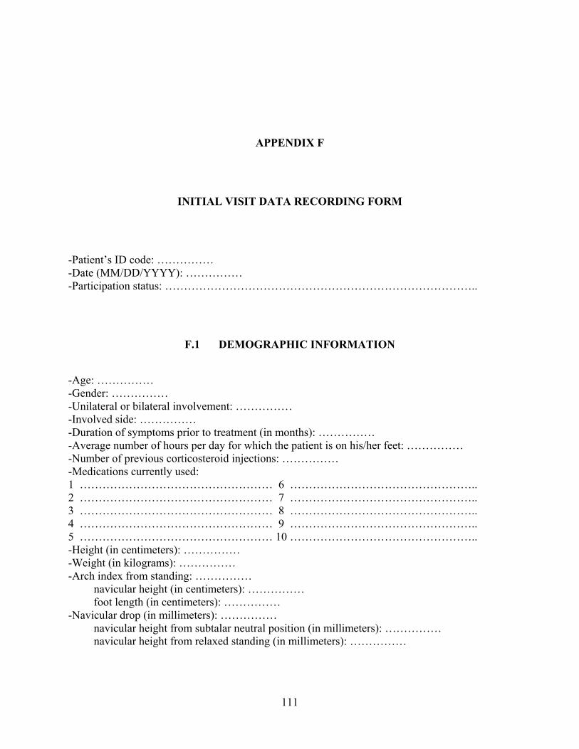
APPENDIX F
INITIAL VISIT DATA RECORDING FORM
-Patient’s ID code: …………… -Date (MM/DD/YYYY): …………… -Participation status: ………………………………………………………………………..
F.1 DEMOGRAPHIC INFORMATION
-Age: …………… -Gender: …………… -Unilateral or bilateral involvement: …………… -Involved side: …………… -Duration of symptoms prior to treatment (in months): …………… -Average number of hours per day for which the patient is on his/her feet: …………… -Number of previous corticosteroid injections: …………… -Medications currently used: 1 …………………………………………… 6 ………………………………………….. 2 …………………………………………… 7 ………………………………………….. 3 …………………………………………… 8 ………………………………………….. 4 …………………………………………… 9 ………………………………………….. 5 …………………………………………… 10 ………………………………………….. -Height (in centimeters): …………… -Weight (in kilograms): …………… -Arch index from standing: …………… navicular height (in centimeters): …………… foot length (in centimeters): …………… -Navicular drop (in millimeters): …………… navicular height from subtalar neutral position (in millimeters): …………… navicular height from relaxed standing (in millimeters): ……………
111
F.2 BASELINE EVALUATION
-Range of pain-free passive ankle joint dorsiflexion with straight knee: …………… -Range of pain-free passive ankle joint dorsiflexion with bent knee: …………… -Plantar heel tenderness: ……………
112

-Plantar heel pain (place a mark on the line in a position which best represents your experience in the past week, or answer the question not applicable (NA) if you did not perform or were not involved in the activity in question): ……………
How severe is your heel pain: NA 1. At its worst? No _______________________________________________ Worst pain ________ pain imaginable 2. After you get up in the morning with the first few steps? No _______________________________________________ Worst pain ________ pain imaginable 3. At the end of the day? No _______________________________________________ Worst pain ________ pain imaginable 4. When you walk barefoot? No _______________________________________________ Worst pain ________ pain imaginable 5. When you stand barefoot? No _______________________________________________ Worst pain ________ pain imaginable 6. When you walk wearing shoes? No _______________________________________________ Worst pain ________ pain imaginable 7. When you stand wearing shoes? No _______________________________________________ Worst pain ________ pain imaginable 8. When you walk wearing orthotics? No _______________________________________________ Worst pain ________ pain imaginable 9. When you stand wearing orthotics? No _______________________________________________ Worst pain ________ pain imaginable
113
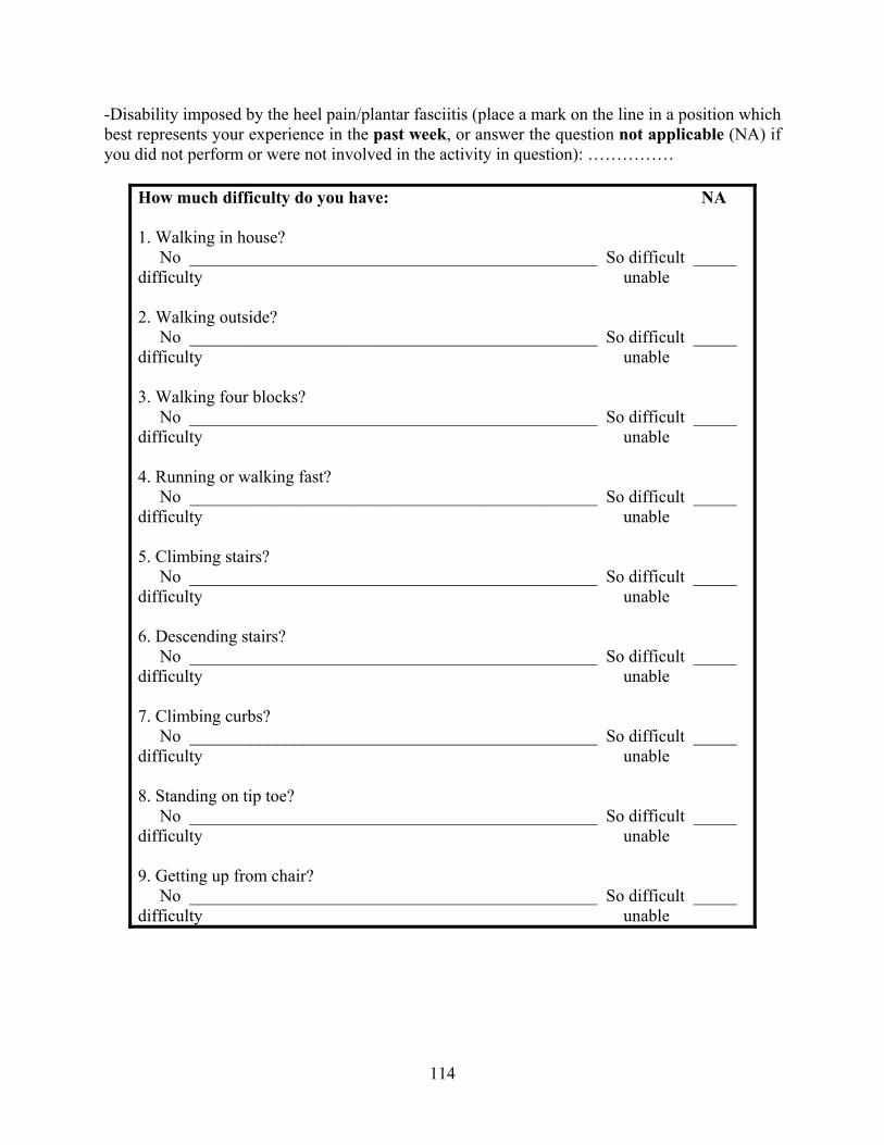
-Disability imposed by the heel pain/plantar fasciitis (place a mark on the line in a position which best represents your experience in the past week, or answer the question not applicable (NA) if you did not perform or were not involved in the activity in question): ……………
How much difficulty do you have: NA 1. Walking in house? No _______________________________________________ So difficult _____ difficulty unable 2. Walking outside? No _______________________________________________ So difficult _____ difficulty unable 3. Walking four blocks? No _______________________________________________ So difficult _____ difficulty unable 4. Running or walking fast? No _______________________________________________ So difficult _____ difficulty unable 5. Climbing stairs? No _______________________________________________ So difficult _____ difficulty unable 6. Descending stairs? No _______________________________________________ So difficult _____ difficulty unable 7. Climbing curbs? No _______________________________________________ So difficult _____ difficulty unable 8. Standing on tip toe? No _______________________________________________ So difficult _____ difficulty unable 9. Getting up from chair? No _______________________________________________ So difficult _____ difficulty unable
114

F.3 RANDOMIZATION
-Assigned group: ……………
F.4 TREATMENT
-Provide the treatment modality that corresponds with the group assignment to the patient and instruct him/her on its use: …………… -Instruct the patient to discontinue use of any other intervention modalities, except medications that are used for reasons other than plantar fasciitis, at least three days before the initial visit, and encourage him/her not to change his/her activity level during the six-week enrollment in the study: ……………
115

APPENDIX G
PHONE CALL DATA RECORDING FORM
-Patient’s ID code: …………… -Date (MM/DD/YYYY): …………… -Participation status: ………………………………………………………………………..
G.1 COMPLIANCE
-What is the average percentage of sleeping hours wearing the night splint during the last three weeks: …………… -What is the average percentage of weight bearing hours using the arch support during the last three weeks: ……………
G.2 QUESTIONS AND CONCERNS
-Questions: 1 …………………………………………………………………………………………… 2 …………………………………………………………………………………………… 3 …………………………………………………………………………………………… 4 …………………………………………………………………………………………… 5 …………………………………………………………………………………………… -Concerns: 1 …………………………………………………………………………………………… 2 …………………………………………………………………………………………… 3 …………………………………………………………………………………………… 4 …………………………………………………………………………………………… 5 ……………………………………………………………………………………………
116

APPENDIX H
FOLLOW-UP VISIT DATA RECORDING FORM
-Patient’s ID code: …………… -Date (MM/DD/YYYY): …………… -Participation status: ………………………………………………………………………..
H.1 POST-INTERVENTION EVALUATION
-Range of pain-free passive ankle joint dorsiflexion with straight knee: …………… -Range of pain-free passive ankle joint dorsiflexion with bent knee: …………… -Plantar heel tenderness: ……………
117
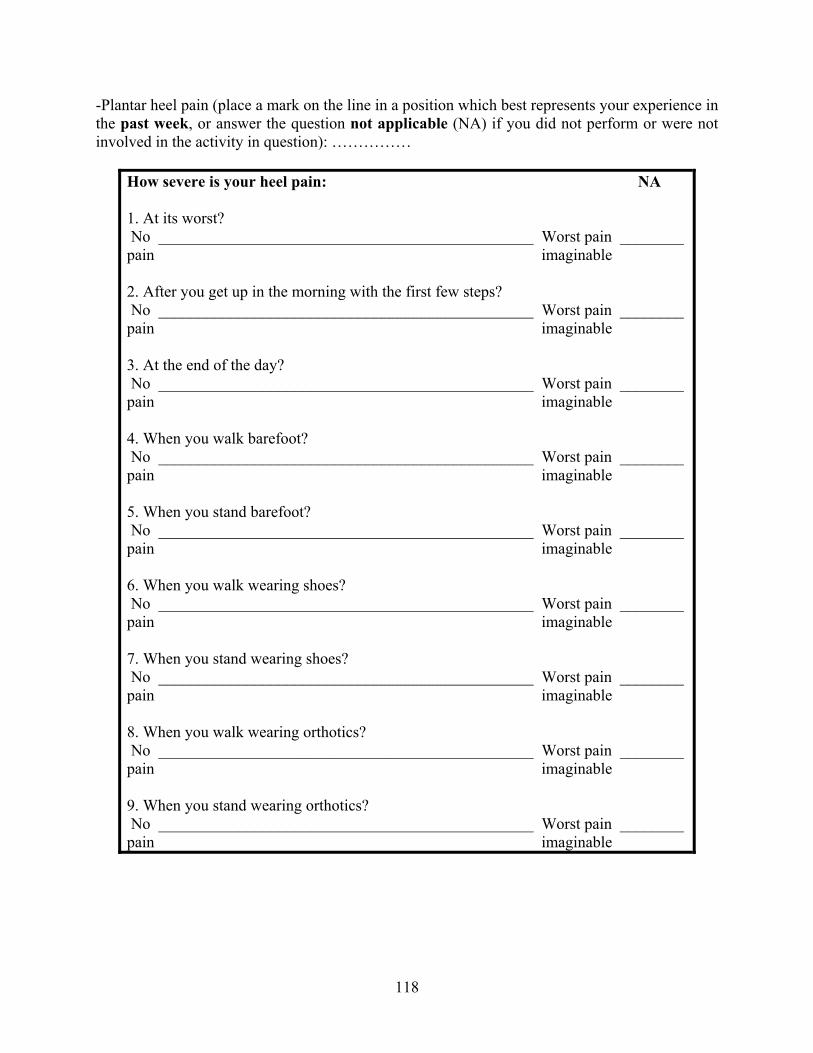
-Plantar heel pain (place a mark on the line in a position which best represents your experience in the past week, or answer the question not applicable (NA) if you did not perform or were not involved in the activity in question): ……………
How severe is your heel pain: NA 1. At its worst? No _______________________________________________ Worst pain ________ pain imaginable 2. After you get up in the morning with the first few steps? No _______________________________________________ Worst pain ________ pain imaginable 3. At the end of the day? No _______________________________________________ Worst pain ________ pain imaginable 4. When you walk barefoot? No _______________________________________________ Worst pain ________ pain imaginable 5. When you stand barefoot? No _______________________________________________ Worst pain ________ pain imaginable 6. When you walk wearing shoes? No _______________________________________________ Worst pain ________ pain imaginable 7. When you stand wearing shoes? No _______________________________________________ Worst pain ________ pain imaginable 8. When you walk wearing orthotics? No _______________________________________________ Worst pain ________ pain imaginable 9. When you stand wearing orthotics? No _______________________________________________ Worst pain ________ pain imaginable
118

-Disability imposed by the heel pain/plantar fasciitis (place a mark on the line in a position which best represents your experience in the past week, or answer the question not applicable (NA) if you did not perform or were not involved in the activity in question): ……………
How much difficulty do you have: NA 1. Walking in house? No _______________________________________________ So difficult _____ difficulty unable 2. Walking outside? No _______________________________________________ So difficult _____ difficulty unable 3. Walking four blocks? No _______________________________________________ So difficult _____ difficulty unable 4. Running or walking fast? No _______________________________________________ So difficult _____ difficulty unable 5. Climbing stairs? No _______________________________________________ So difficult _____ difficulty unable 6. Descending stairs? No _______________________________________________ So difficult _____ difficulty unable 7. Climbing curbs? No _______________________________________________ So difficult _____ difficulty unable 8. Standing on tip toe? No _______________________________________________ So difficult _____ difficulty unable 9. Getting up from chair? No _______________________________________________ So difficult _____ difficulty unable
119
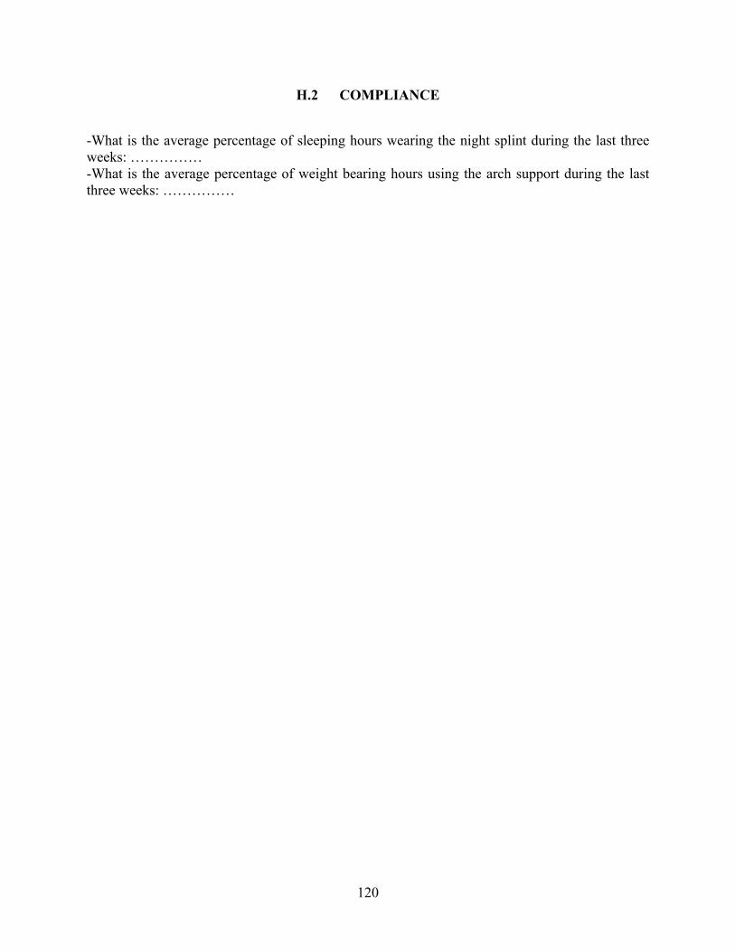
H.2 COMPLIANCE
-What is the average percentage of sleeping hours wearing the night splint during the last three weeks: …………… -What is the average percentage of weight bearing hours using the arch support during the last three weeks: ……………
120
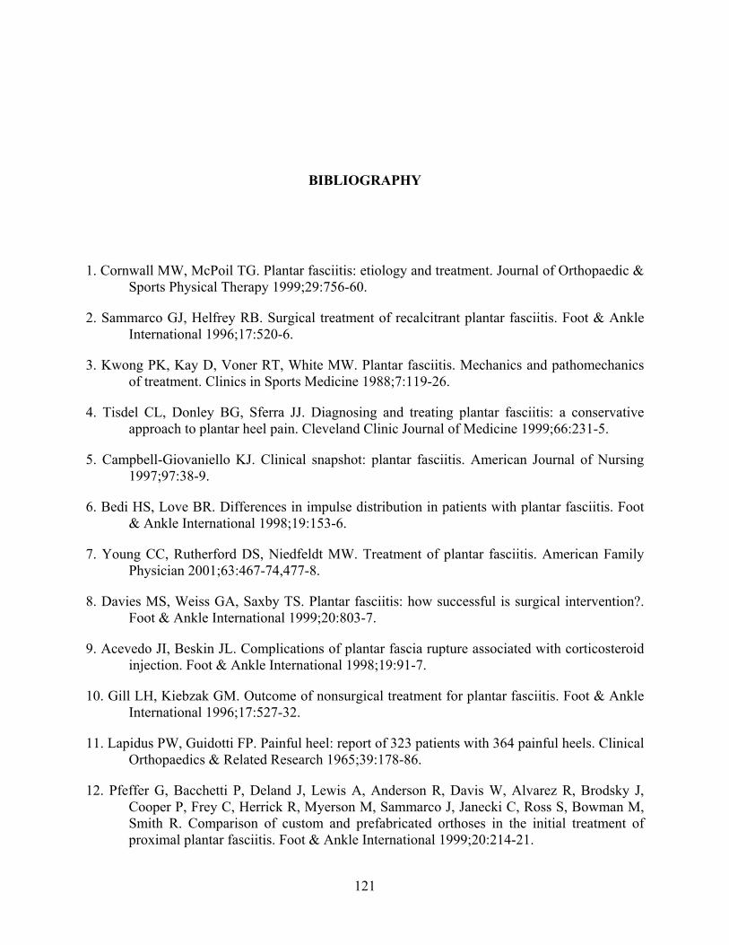
BIBLIOGRAPHY
1. Cornwall MW, McPoil TG. Plantar fasciitis: etiology and treatment. Journal of Orthopaedic & Sports Physical Therapy 1999;29:756-60.
2. Sammarco GJ, Helfrey RB. Surgical treatment of recalcitrant plantar fasciitis. Foot & Ankle International 1996;17:520-6.
3. Kwong PK, Kay D, Voner RT, White MW. Plantar fasciitis. Mechanics and pathomechanics of treatment. Clinics in Sports Medicine 1988;7:119-26.
4. Tisdel CL, Donley BG, Sferra JJ. Diagnosing and treating plantar fasciitis: a conservative approach to plantar heel pain. Cleveland Clinic Journal of Medicine 1999;66:231-5.
5. Campbell-Giovaniello KJ. Clinical snapshot: plantar fasciitis. American Journal of Nursing 1997;97:38-9.
6. Bedi HS, Love BR. Differences in impulse distribution in patients with plantar fasciitis. Foot & Ankle International 1998;19:153-6.
7. Young CC, Rutherford DS, Niedfeldt MW. Treatment of plantar fasciitis. American Family Physician 2001;63:467-74,477-8.
8. Davies MS, Weiss GA, Saxby TS. Plantar fasciitis: how successful is surgical intervention?. Foot & Ankle International 1999;20:803-7.
9. Acevedo JI, Beskin JL. Complications of plantar fascia rupture associated with corticosteroid injection. Foot & Ankle International 1998;19:91-7.
10. Gill LH, Kiebzak GM. Outcome of nonsurgical treatment for plantar fasciitis. Foot & Ankle International 1996;17:527-32.
11. Lapidus PW, Guidotti FP. Painful heel: report of 323 patients with 364 painful heels. Clinical Orthopaedics & Related Research 1965;39:178-86.
12. Pfeffer G, Bacchetti P, Deland J, Lewis A, Anderson R, Davis W, Alvarez R, Brodsky J, Cooper P, Frey C, Herrick R, Myerson M, Sammarco J, Janecki C, Ross S, Bowman M, Smith R. Comparison of custom and prefabricated orthoses in the initial treatment of proximal plantar fasciitis. Foot & Ankle International 1999;20:214-21.
121
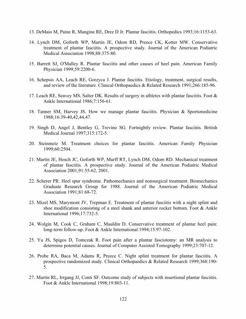
13. DeMaio M, Paine R, Mangine RE, Drez D Jr. Plantar fasciitis. Orthopedics 1993;16:1153-63.
14. Lynch DM, Goforth WP, Martin JE, Odom RD, Preece CK, Kotter MW. Conservative treatment of plantar fasciitis. A prospective study. Journal of the American Podiatric Medical Association 1998;88:375-80.
15. Barrett SJ, O'Malley R. Plantar fasciitis and other causes of heel pain. American Family Physician 1999;59:2200-6.
16. Schepsis AA, Leach RE, Gorzyca J. Plantar fasciitis. Etiology, treatment, surgical results, and review of the literature. Clinical Orthopaedics & Related Research 1991;266:185-96.
17. Leach RE, Seavey MS, Salter DK. Results of surgery in athletes with plantar fasciitis. Foot & Ankle International 1986;7:156-61.
18. Tanner SM, Harvey JS. How we manage plantar fasciitis. Physician & Sportsmedicine 1988;16:39-40,42,44,47.
19. Singh D, Angel J, Bentley G, Trevino SG. Fortnightly review. Plantar fasciitis. British Medical Journal 1997;315:172-5.
20. Steinmetz M. Treatment choices for plantar fasciitis. American Family Physician 1999;60:2504.
21. Martin JE, Hosch JC, Goforth WP, Murff RT, Lynch DM, Odom RD. Mechanical treatment of plantar fasciitis. A prospective study. Journal of the American Podiatric Medical Association 2001;91:55-62, 2001.
22. Scherer PR. Heel spur syndrome. Pathomechanics and nonsurgical treatment. Biomechanics Graduate Research Group for 1988. Journal of the American Podiatric Medical Association 1991;81:68-72.
23. Mizel MS, Marymont JV, Trepman E. Treatment of plantar fasciitis with a night splint and shoe modification consisting of a steel shank and anterior rocker bottom. Foot & Ankle International 1996;17:732-5.
24. Wolgin M, Cook C, Graham C, Mauldin D. Conservative treatment of plantar heel pain: long-term follow-up. Foot & Ankle International 1994;15:97-102.
25. Yu JS, Spigos D, Tomczak R. Foot pain after a plantar fasciotomy: an MR analysis to determine potential causes. Journal of Computer Assisted Tomography 1999;23:707-12.
26. Probe RA, Baca M, Adams R, Preece C. Night splint treatment for plantar fasciitis. A prospective randomized study. Clinical Orthopaedics & Related Research 1999;368:190-5.
27. Martin RL, Irrgang JJ, Conti SF. Outcome study of subjects with insertional plantar fasciitis. Foot & Ankle International 1998;19:803-11.
122
28. Plantar fasciitis. Repeated corticosteroid injections are safe. Canadian Family Physician 1998;44:45,51.
29. Powell M, Post WR, Keener J, Wearden S. Effective treatment of chronic plantar fasciitis with dorsiflexion night splints: a crossover prospective randomized outcome study. Foot & Ankle International 1998;19:10-8.
30. Batt ME, Tanji JL, Skattum N. Plantar fasciitis: a prospective randomized clinical trial of the tension night splint. Clinical Journal of Sport Medicine 1996;6:158-62.
31. Ryan J. Use of posterior night splints in the treatment of plantar fasciitis. American Family Physician 1995;52:891-8,901-2.
32. Wapner KL, Sharkey PF. The use of night splints for treatment of recalcitrant plantar fasciitis. Foot & Ankle International 1991;12:135-7.
33. McPoil TG, Cornwall MW. The effect of foot orthoses on transverse tibial rotation during walking. Journal of the American Podiatric Medical Association 2000;90:2-11.
34. Kogler GF, Solomonidis SE, Paul JP. In-vivo method for quantifying the effectiveness of the longitudinal arch support mechanism of a foot orthosis. Clinical Biomechanics 1995;10:245-52.
35. Brown GP, Donatelli R, Catlin PA, Wooden MJ. The effect of two types of foot orthoses on rearfoot mechanics. Journal of Orthopaedic & Sports Physical Therapy 1995;21:258-67.
36. O'Brien D, Martin WJ. A retrospective analysis of heel pain. Journal of the American Podiatric Medical Association 1985;75:416-8.
37. Campbell JW, Inman VT. Treatment of plantar fasciitis and calcaneal spurs with the UC-BL shoe insert. Clinical Orthopaedics & Related Research 1974;103:57-62.
38. Ogden JA, Alvarez R, Levitt R, Cross GL, Marlow M. Shock wave therapy for chronic proximal plantar fasciitis. Clinical Orthopaedics & Related Research 2001;387:47-59.
39. Sellman JR. Plantar fascia rupture associated with corticosteroid injection. Foot & Ankle International 1994;15:376-81.
40. Kane D, Greaney T, Shanahan M, Duffy G, Bresnihan B, Gibney R, FitzGerald O. The role of ultrasonography in the diagnosis and management of idiopathic plantar fasciitis. Rheumatology 2001;40:1002-8.
41. Reeve F, Laughlin RT, Wright DG. Endoscopic plantar fascia release: a cross-sectional anatomic study. Foot & Ankle International 1997;18:398-401.
42. Jerosch J. Endoscopic release of plantar fasciitis-a benign procedure?. Foot & Ankle International 2000;21:511-3.
123
43. Huang CK, Kitaoka HB, An KN, Chao EY. Biomechanical evaluation of longitudinal arch stability. Foot & Ankle International 1993;14:353-7.
44. Goulet MJ. Role of soft orthosis in treating plantar fasciitis. Suggestion from the field. Physical Therapy 1984;64:1544.
45. Hicks JH. The mechanics of the foot: II. The plantar aponeurosis and the arch. Journal of Anatomy 1954;88:25-31.
46. Chandler TJ, Kibler WB. A biomechanical approach to the prevention, treatment and rehabilitation of plantar fasciitis. Sports Medicine 1993;15:344-52.
47. Kibler WB, Goldberg C, Chandler TJ. Functional biomechanical deficits in running athletes with plantar fasciitis. American Journal of Sports Medicine 1991;19:66-71.
48. Daly PJ, Kitaoka HB, Chao EY. Plantar fasciotomy for intractable plantar fasciitis: clinical results and biomechanical evaluation. Foot & Ankle International 1992;13:188-95.
49. Snook GA, Chrisman OD. The management of subcalcaneal pain. Clinical Orthopaedics & Related Research 1972;82:163-8.
50. de Souza H, Reed L. An inexpensive "orthosis" for plantar fasciitis. Medical Journal of Australia 1997;167:509.
51. Brown C. A review of subcalcaneal heel pain and plantar fasciitis. Australian Family Physician 1996;25:875-81,884-5.
52. Hill JJ Jr, Cutting PJ. Heel pain and body weight. Foot & Ankle International 1989;9:254-6.
53. Riddle DL, Pulisic M, Pidcoe P, Johnson RE. Risk factors for plantar fasciitis: a matched case-control study. Journal of Bone & Joint Surgery 2003;85:872-7.
54. Mann R, Inman VT. Phasic activity of intrinsic muscles of the foot. Journal of Bone & Joint Surgery 1964;46:469-74.
55. Boyd HS. Plantar fasciitis. Orthopaedic Review 1992;21:116.
56. Schoenborn CA, Adams PF, Barnes PM. Body weight status of adults: United States, 1997-98. Advance Data 2002;330:1-15.
57. Clearihan L. Pain relief for plantar fasciitis. Australian Family Physician 1999;28:697.
58. Gudeman SD, Eisele SA, Heidt RS Jr, Colosimo AJ, Stroupe AL. Treatment of plantar fasciitis by iontophoresis of 0.4% dexamethasone. A randomized, double-blind, placebo-controlled study. American Journal of Sports Medicine 1997;25:312-6.
59. Davis PF, Severud E, Baxter DE. Painful heel syndrome: results of nonoperative treatment. Foot & Ankle International 1994;15:531-5.
124
60. MacLellan GE, Vyvyan B. Management of pain beneath the heel and Achilles tendonitis with visco-elastic heel inserts. British Journal of Sports Medicine 1981;15:117-21.
61. Wong SM, Griffith JF, Tang A, Hui AC. Re: the role of ultrasonography in the diagnosis and management of idiopathic plantar fasciitis. Rheumatology 2002;41:835-6.
62. Crawford F, Atkins D, Young P, Edwards J. Steroid injection for heel pain: evidence of short-term effectiveness. A randomized controlled trial. Rheumatology 1999;38:974-7.
63. Kane D, Greaney T, Bresnihan B, Gibney R, FitzGerald O. Ultrasound guided injection of recalcitrant plantar fasciitis. Annals of the Rheumatic Diseases 1998;57:383-4.
64. Leach R, Jones R, Silva T. Rupture of the plantar fascia in athletes. Journal of Bone & Joint Surgery 1978;60:537-9.
65. Kennedy JC, Willis RB. The effects of local steroid injections on tendons: a biomechanical and microscopic correlative study. American Journal of Sports Medicine 1976;4:11-21.
66. Rompe JD, Schoellner C, Nafe B. Evaluation of low-energy extracorporeal shock-wave application for treatment of chronic plantar fasciitis. Journal of Bone & Joint Surgery 2002;84:335-41.
67. Rompe JD, Hopf C, Nafe B, Burger R. Low-energy extracorporeal shock wave therapy for painful heel: a prospective controlled single-blind study. Archives of Orthopaedic & Trauma Surgery 1996;115:75-9.
68. Riddle DL, Freeman DB. Management of a patient with a diagnosis of bilateral plantar fasciitis and Achilles tendinitis. A case report. Physical Therapy 1988;68:1913-6.
69. Zamorski M. Plantar fasciitis, posterior night splints and activity during recovery. American Family Physician 1996;53:1993-4.
70. DiGiovanni BF, Nawoczenski DA, Lintal ME, Moore EA, Murray JC, Wilding GE, Baumhauer JF. Tissue-specific plantar fascia-stretching exercise enhances outcomes in patients with chronic heel pain. A prospective, randomized study. Journal of Bone & Joint Surgery 2003;85:1270-7.
71. Howell D. Plantar fasciitis. Journal of Orthopaedic & Sports Physical Therapy 2000;30:217.
72. Vohra PK, Giorgini RJ, Sobel E, Japour CJ, Villalba MA, Rostkowski T. Long-term follow-up of heel spur surgery. A 10-year retrospective study. Journal of the American Podiatric Medical Association 1999;89:81-8.
73. Tomczak RL, Haverstock BD. A retrospective comparison of endoscopic plantar fasciotomy to open plantar fasciotomy with heel spur resection for chronic plantar fasciitis/heel spur syndrome. Journal of Foot & Ankle Surgery 1995;34:305-11.
125
74. Murphy GA, Pneumaticos SG, Kamaric E, Noble PC, Trevino SG, Baxter DE. Biomechanical consequences of sequential plantar fascia release. Foot & Ankle International 1998;19:149-52.
75. Arangio GA, Chen C, Kim W. Effect of cutting the plantar fascia on mechanical properties of the foot. Clinical Orthopaedics & Related Research 1997;339:227-31.
76. Dailey JM. Differential diagnosis and treatment of heel pain. Clinics in Podiatric Medicine & Surgery 1991;8:153-66.
77. McCarthy DJ, Gorecki GE. The anatomical basis of inferior calcaneal lesions: a cryomicrotomy study. Journal of the American Podiatry Association 1979;69:527-36.
78. Berlet GC, Anderson RB, Davis WH, Kiebzak GM. A prospective trial of night splinting in the treatment of recalcitrant plantar fasciitis: the ankle dorsiflexion dynasplint. Orthopedics 2002;25:1273-5.
79. Wen DY, Puffer JC, Schmalzried TP. Lower extremity alignment and risk of overuse injuries in runners. Medicine & Science in Sports & Exercise 1997;29:1291-8.
80. Cowan DN, Jones BH, Robinson JR. Foot morphologic characteristics and risk of exercise-related injury. Archives of Family Medicine 1993;2:773-7.
81. Warren BL. Anatomical factors associated with predicting plantar fasciitis in long-distance runners. Medicine & Science in Sports & Exercise 1984;16:60-3.
82. Warren BL, Jones CJ. Predicting plantar fasciitis in runners. Medicine & Science in Sports & Exercise 1987;19:71-3.
83. McCrory JL, Young MJ, Boulton AJM, Cavanagh PR. Arch index as a predictor of arch height. The Foot: International Journal of Clinical Foot Science 1997;7:79-81.
84. Sell KE, Verity TM, Worrell TW, Pease BJ, Wigglesworth J. Two measurement techniques for assessing subtalar joint position: a reliability study. Journal of Orthopaedic & Sports Physical Therapy 1994;19:162-7.
85. Gogia PP, Braatz JH, Rose SJ, Norton BJ. Reliability and validity of goniometric measurements at the knee. Physical Therapy 1987;67:192-5.
86. Brosseau L, Tousignant M, Budd J, Chartier N, Duciaume L, Plamondon S, O'Sullivan JP, O'Donoghue S, Balmer S. Intratester and intertester reliability and criterion validity of the parallelogram and universal goniometers for active knee flexion in healthy subjects. Physiotherapy Research International 1997;2:150-66.
87. Brosseau L, Balmer S, Tousignant M, O'Sullivan JP, Goudreault C, Goudreault M, Gringras S. Intra- and intertester reliability and criterion validity of the parallelogram and universal goniometers for measuring maximum active knee flexion and extension of patients with knee restrictions. Archives of Physical Medicine & Rehabilitation 2001;82:396-402.
126

88. Elveru RA, Rothstein JM, Lamb RL. Goniometric reliability in a clinical setting. Subtalar and ankle joint measurements. Physical Therapy 1988;68:672-7.
89. Youdas JW, Bogard CL, Suman VJ. Reliability of goniometric measurements and visual estimates of ankle joint active range of motion obtained in a clinical setting. Archives of Physical Medicine & Rehabilitation 1993;74:1113-8.
90. Clapper MP, Wolf SL. Comparison of the reliability of the Orthoranger and the standard goniometer for assessing active lower extremity range of motion. Physical Therapy 1988;68:214-8.
91. Ekstrand J, Wiktorsson M, Oberg B, Gillquist J. Lower extremity goniometric measurements: a study to determine their reliability. Archives of Physical Medicine & Rehabilitation 1982;63:171-5.
92. Klaiman MD, Shrader JA, Danoff JV, Hicks JE, Pesce WJ, Ferland J. Phonophoresis versus ultrasound in the treatment of common musculoskeletal conditions. Medicine & Science in Sports & Exercise 1998;30:1349-55.
93. Fischer AA. Clinical use of tissue compliance meter for documentation of soft tissue pathology. Clinical Journal of Pain 1987;3:23-30.
94. Fischer AA. Pressure algometry over normal muscles. Standard values, validity and reproducibility of pressure threshold. Pain 1987;30:115-26.
95. Fischer AA. Pressure threshold meter: its use for quantification of tender spots. Archives of Physical Medicine & Rehabilitation 1986;67:836-8.
96. Nussbaum EL, Downes L. Reliability of clinical pressure-pain algometric measurements obtained on consecutive days. Physical Therapy 1998;78:160-9.
97. Reeves JL, Jaeger B, Graff-Radford SB. Reliability of the pressure algometer as a measure of myofascial trigger point sensitivity. Pain 1986;24:313-21.
98. Budiman-Mak E, Conrad KJ, Roach KE. The Foot Function Index: a measure of foot pain and disability. Journal of Clinical Epidemiology 1991;44:561-70.
99. Budiman-Mak E, Roach KE, Hedeker D, Caraballada R, Burks D, Moore H. Impacts of foot orthoses on pain and disability in rheumatoid arthritics. Journal of Clinical Epidemiology 1996;49:1-7.
100. Caselli MA, Clark N, Lazarus S, Velez Z, Venegas L. Evaluation of magnetic foil and PPT Insoles in the treatment of heel pain. Journal of the American Podiatric Medical Association 1997;87:11-6.
101. Caselli MA, Levitz SJ, Clark N, Lazarus S, Velez Z, Venegas L. Comparison of Viscoped and PORON for painful submetatarsal hyperkeratotic lesions. Journal of the American Podiatric Medical Association 1997;87:6-10.
127

102. Wiley JP, Aggarwala R, Donaldson C, Lun VMY, Yanagawa TL. Comparison of foot orthosis, physiotherapy and night splints in the treatment of plantar fasciitis. Medicine & Science in Sports & Exercise 2002;34:134.
128
Recommended