Connective Tissue, Ligaments & Raphe
Dr Ibrar Wazir
Lecturer Anatomy KGMC
1. What is Connective tissue, Its properties, Derived from which embryonic layer, Components, Types & Functions
2. What are Ligaments, Its types, Blood & Nerve supply & Functions
3. Understand Raphe
TISSUE• Tissue is an ensemble of similar cells from
same origin that together carry out specific function
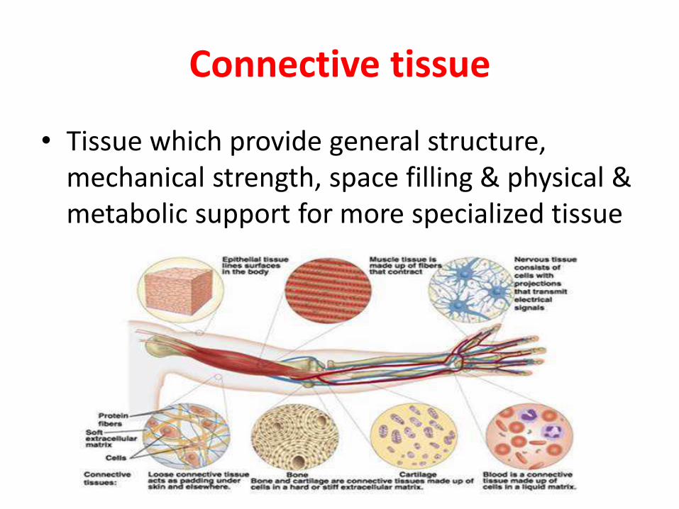
• There are four basic type of tissue i.e.
i. Connective tissue
ii. Epithelial tissue
iii. Muscular tissue
iv. Nervous tissue
Connective tissue
• Tissue which provide general structure, mechanical strength, space filling & physical & metabolic support for more specialized tissue
Properties of CT
CT have three structural properties
1. Tensile strength: to resist pulling, stretching & tearing i.e. provided by collagen family
2. Elasticity: facilitate return to original shape after normal physiological mechanical distortion i.e. provided by elastin fibrils
3. Volume (i.e. bulk/substance): provided by glycoproteins & complex carbohydrates with profound water binding ability forming a wet gel known as ground substance
Derived from which Embryonic Layer
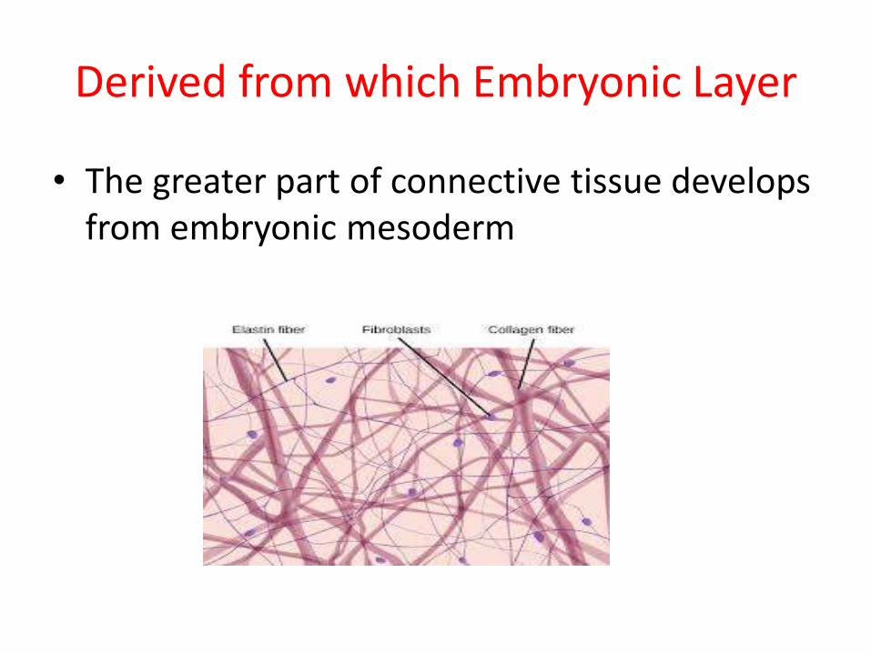
• The greater part of connective tissue develops from embryonic mesoderm
Components
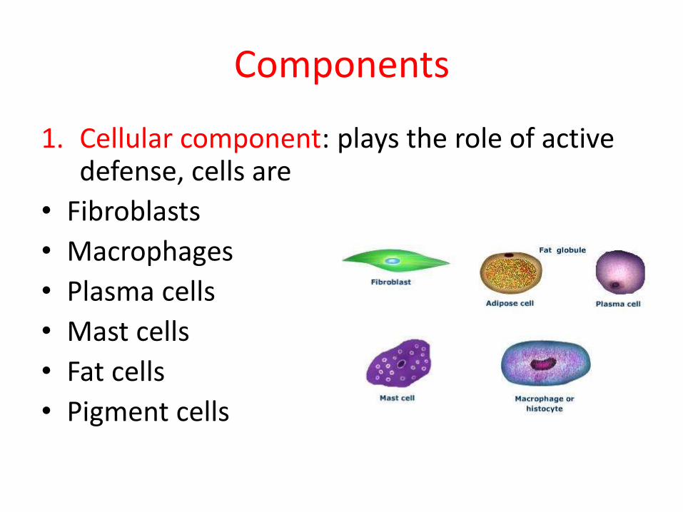
1. Cellular component: plays the role of active defense, cells are
• Fibroblasts
• Macrophages
• Plasma cells
• Mast cells
• Fat cells
• Pigment cells
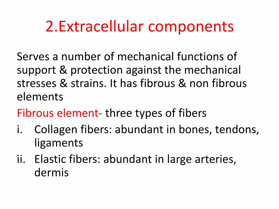
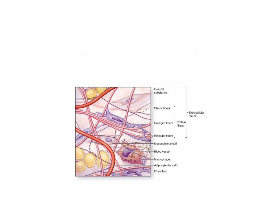
2.Extracellular components
Serves a number of mechanical functions of support & protection against the mechanical stresses & strains. It has fibrous & non fibrous elements
Fibrous element- three types of fibers
i. Collagen fibers: abundant in bones, tendons, ligaments
ii. Elastic fibers: abundant in large arteries, dermis
iii. Reticular fibers: abundant in lymph nodes, spleen, glands
Non fibrous elements- formed by ground substance which is viscous, gel like substance with high water contents. Major components are proteoglycans, multiadhesive glycoproteins, glycosaminoglycan's. Located in between fibers & cells
Types
Connective tissue can be broadly divided in to
A. Connective tissue proper
B. Special connective tissue
Connective tissue proper is further divided into
i. Loose connective tissue: located commonly under epithelia throughout dermis, lamina propria, layers surrounding glands & ducts fibers, ground substance
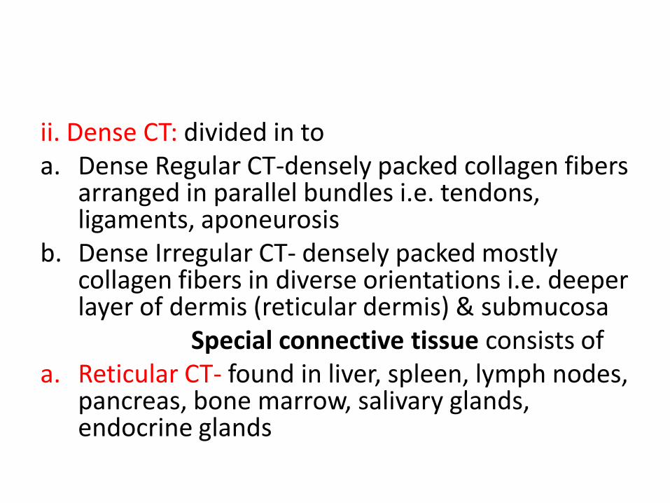
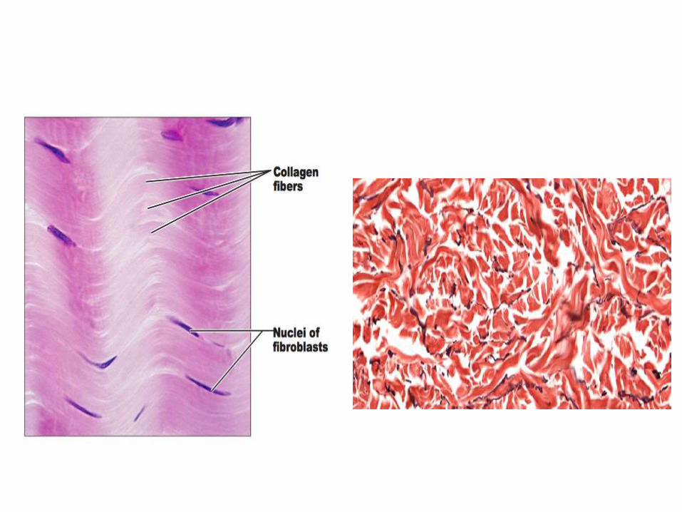
ii. Dense CT: divided in toa. Dense Regular CT-densely packed collagen fibers
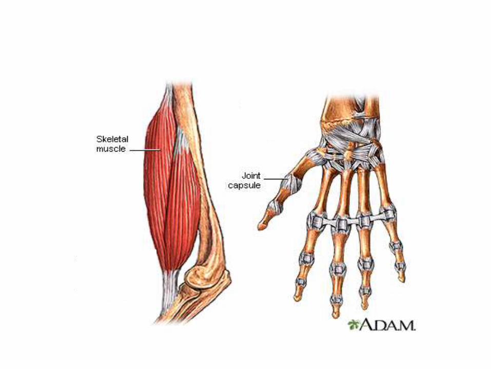
arranged in parallel bundles i.e. tendons, ligaments, aponeurosis
b. Dense Irregular CT- densely packed mostly collagen fibers in diverse orientations i.e. deeper layer of dermis (reticular dermis) & submucosa
Special connective tissue consists ofa. Reticular CT- found in liver, spleen, lymph nodes,
pancreas, bone marrow, salivary glands, endocrine glands
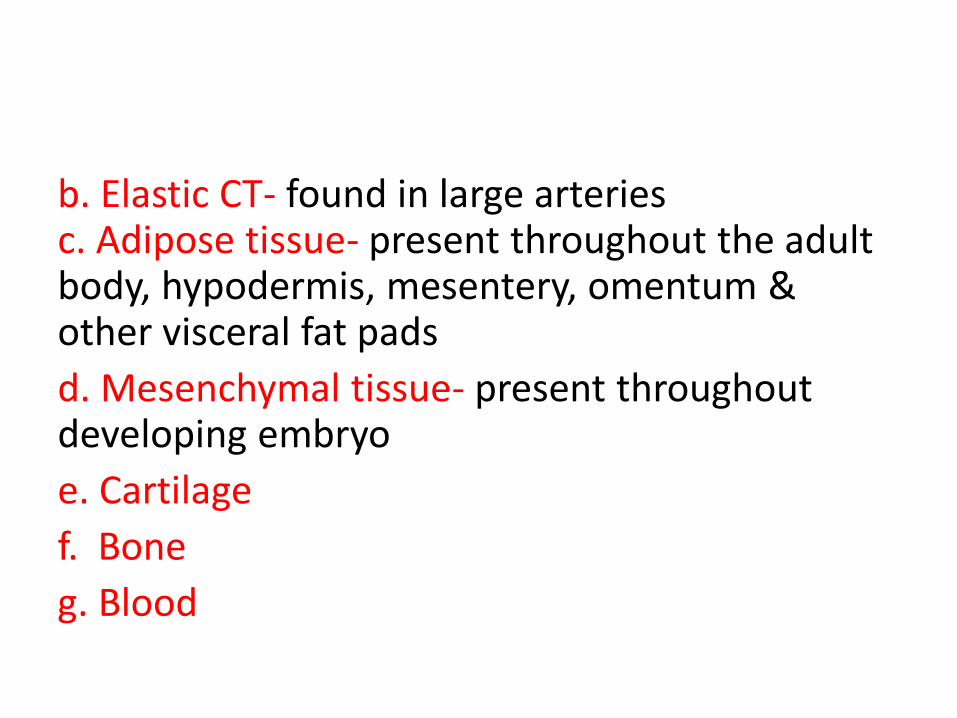
b. Elastic CT- found in large arteries c. Adipose tissue- present throughout the adult body, hypodermis, mesentery, omentum & other visceral fat pads
d. Mesenchymal tissue- present throughout developing embryo
e. Cartilage
f. Bone
g. Blood
Functions
I. As a packing material CT provide a supporting matrix for many highly organized structures
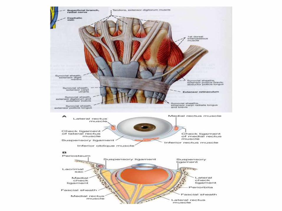
II. It forms restraining mechanism of the body in the form of retinacula, check ligaments
III. It provides surface coating of the body in the form of superficial fascia which stores fat & conserves body heat
IV. The ensheathing layers of deep fascia preserves the characteristic contour of the limbs & aid circulation in the veins & circulation
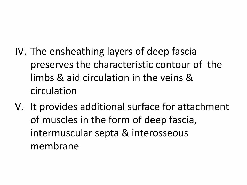
V. It provides additional surface for attachment of muscles in the form of deep fascia, intermuscular septa & interosseous membrane
VI. It forms facial planes which provide convenient pathway for vessels (blood vessels & lymphatic's) &nerves
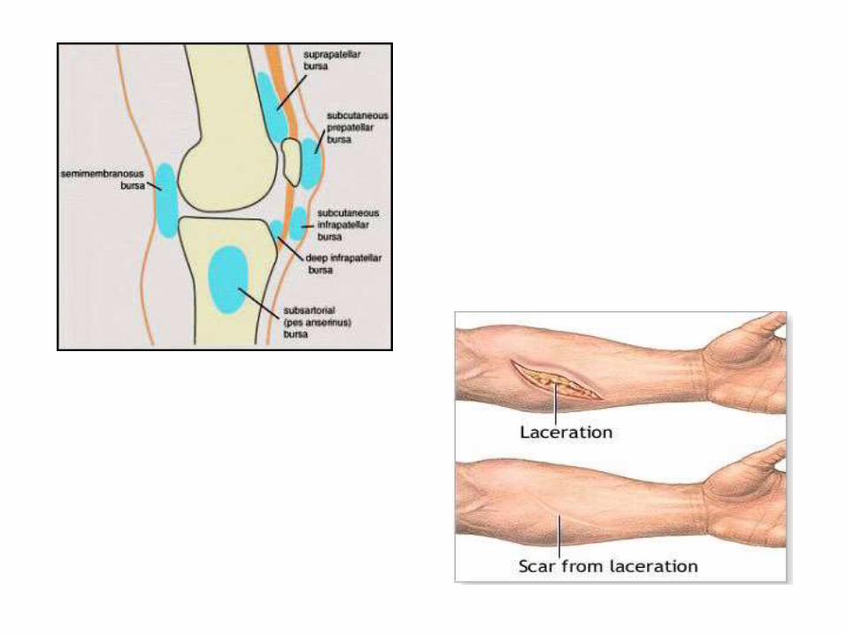
VII. In places where it is loose in texture ( loose CT) facilitate movements b/w the adjacent structures & by forming bursal sacs it minimizes friction and pressure effects
VIII. CT help in repair of injuries where by fibroblast lay down collagen fibers to form the scar tissue
IX. Macrophages of CT serves a defensive function. Plasma cells are capable of producing antibodies. Mast cells responsible for various inflammatory, allergic & hypersensitivity reactions. Pigment cells protect the skin against ultra violet radiations.
X. CT contains Mesenchymal cells capable of transforming into each type of CT
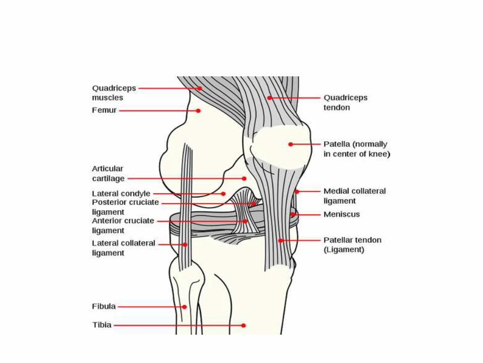
LIGAMENTS
Ligament is a cord or band of connective tissue uniting/connecting two structures i.e. bone to bone or cartilage. Commonly found in association with joints. Composed of 70% collagen, along with ground substance & a larger percentage of elastin then tendons
TYPES:
A. According to composition: two types
Collagenous- mostly composed of dense bundles of collagen fibers & are unstretchable under normal conditions e.g. collateral ligaments of the elbow joint, deltoid ligament
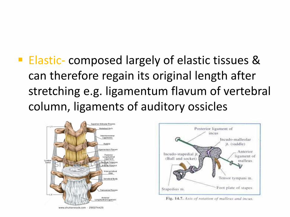
Elastic- composed largely of elastic tissues & can therefore regain its original length after stretching e.g. ligamentum flavum of vertebral column, ligaments of auditory ossicles
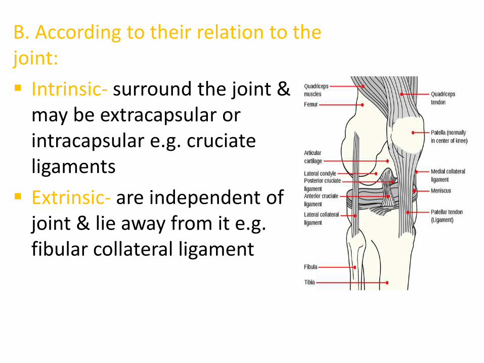
B. According to their relation to the joint:
Intrinsic- surround the joint & may be extracapsular or intracapsular e.g. cruciate ligaments
Extrinsic- are independent of joint & lie away from it e.g. fibular collateral ligament
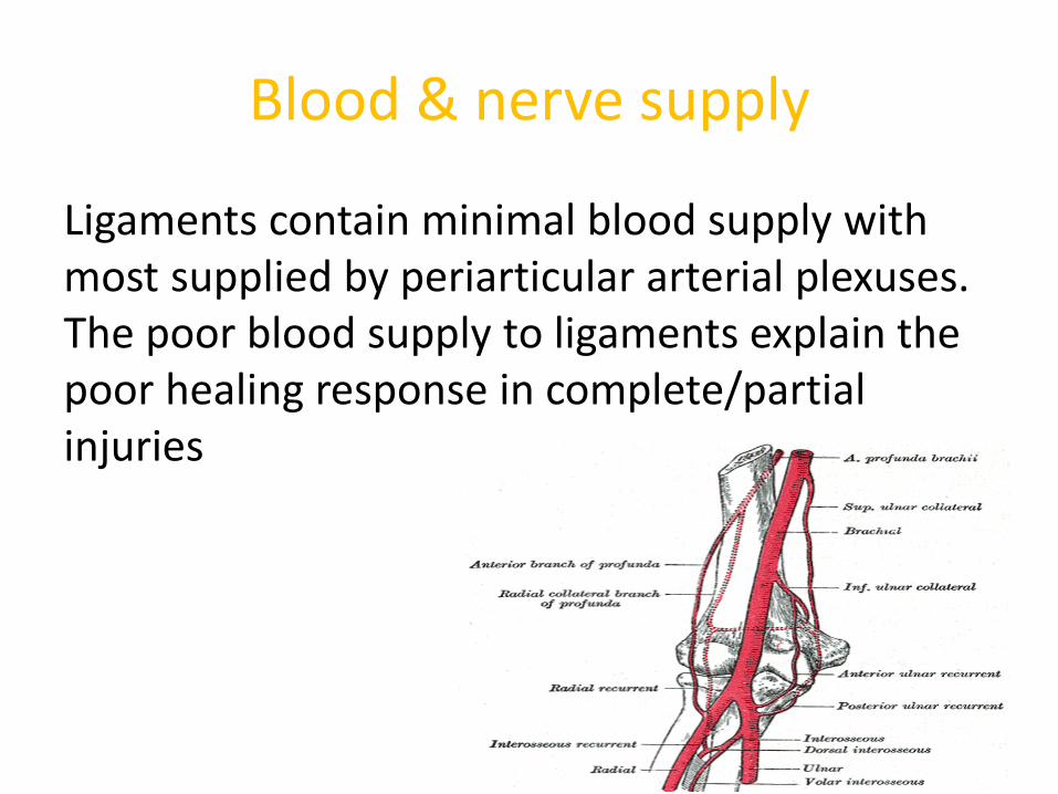
Blood & nerve supply
Ligaments contain minimal blood supply with most supplied by periarticular arterial plexuses. The poor blood supply to ligaments explain the poor healing response in complete/partial injuries
Nerves of the joint ramify on its ligaments.
Because of rich nerve supply, most ligaments serve as sense organs in acting as important reflex mechanism so are important in monitoring the position & movement of the joint
Functions
I. Hold the skeleton together
II. Transmit load from bone to bone
III. Stabilize joint in concert with normal osseous alignment & musculotendinous stability
IV. Their sensory function makes them important reflex organs
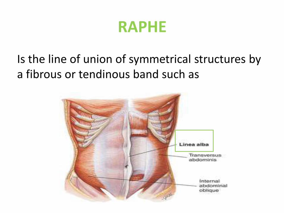
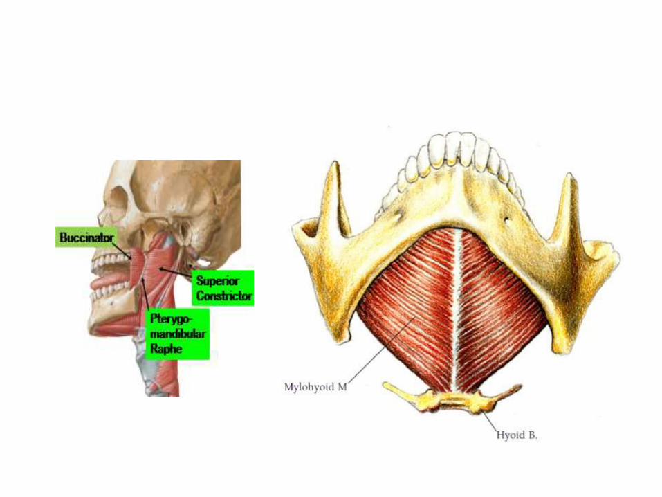
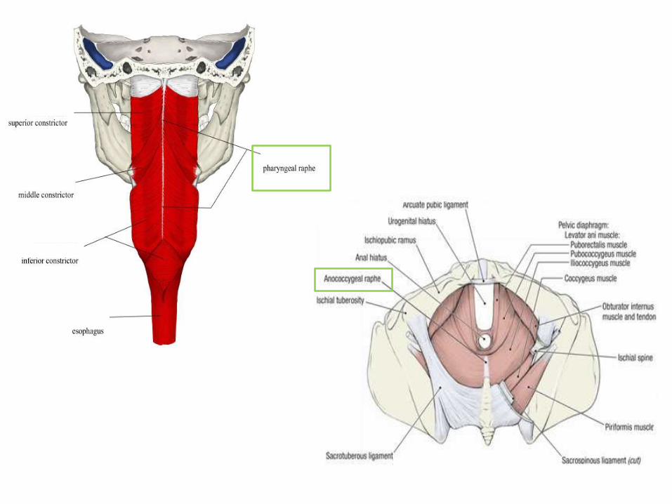
RAPHE
Is the line of union of symmetrical structures by a fibrous or tendinous band such as
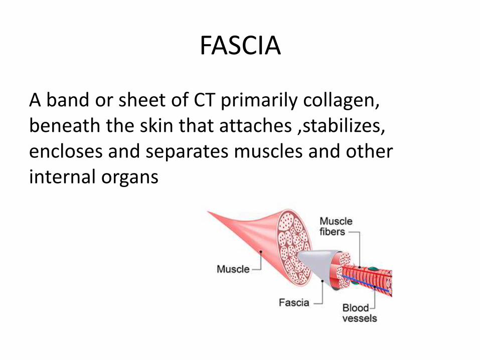
FASCIA
A band or sheet of CT primarily collagen, beneath the skin that attaches ,stabilizes, encloses and separates muscles and other internal organs
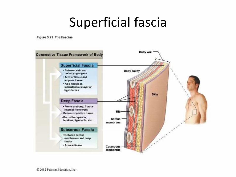
Superficial fascia
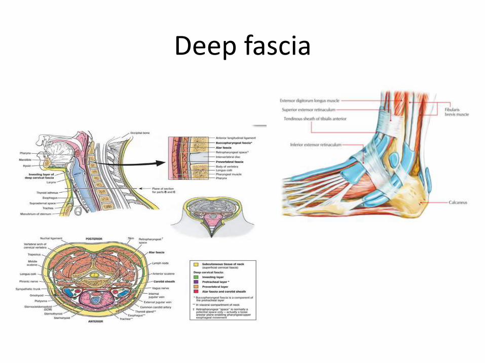
Deep fascia

THANKS
Recommended