Comparative Study on Production of Cellulase in Fresh and Sea Water by Fursarium subglutinans MTCC 11891
A THESIS SUBMITTED IN PARTIAL FULFILLMENT
OF THE REQUIREMENT FOR THE DEGREE OF
Master of Technology
in
Biotechnology
by
SHARMILA D
212BM2004
Under the Supervision of
Dr. A. Thirugnanam
Department of Biotechnology and Medical Engineering
National Institute of Technology Rourkela
Odisha, 769 008, India
May 2014

Department of Biotechnology and Medical Engineering
National Institute of Technology Rourkela, Odisha-769008
CERTIFICATE
This is to certify that the thesis entitled “Comparative Study on production of cellulase
in fresh and sea water by Fursarium subglutinans MTCC 11891 by SHARMILA D
(212BM2004), in partial fulfilment of the requirements for the award of the degree of
Master of Technology in Biotechnology during the session 2012-2014 in the Department
of Biotechnology and Medical Engineering, National Institute of Technology Rourkela, is
an authentic work carried out by her under my supervision and guidance. To the best of my
knowledge, the matter embodied in the thesis has not been submitted to any other
University/Institute for the award of any Degree or Diploma.
Place: NIT Rourkela Dr. A. Thirugnanam (Supervisor) Date: 28 May 2014. Assistant Professor, Biotechnology and Medical Engineering,
National Institute of Technology Rourkela Odisha-769008(India).
ii

ACKNOWLEDGEMENT
Successful completion of this project is the outcome of consistent guidance and
assistance from many people, faculty and friends and I am extremely fortunate to have got
these all along the completion of the project.
I owe my profound gratitude and respect to my project guide, Prof. A.
Thirugnanam, Department of Biotechnology and Medical Engineering, NIT Rourkela for
his invaluable academic support and professional guidance, regular encouragement and
motivation at various stages of this project. I also thank Prof. R. Jayabalan, Dept. of Life
Science, NIT Rourkela for his consistent guidance, lab support, technical advice during my
entire project duration, without which this work would not have been possible for me to
complete. I place on record my sincere gratitude to Prof. Krishna Pramanik, Head,
Department of Biotechnology and Medical Engineering, NIT Rourkela for her constant
encouragement.
I would like to express my sincere thanks to Ms. Indira Dash, Mr. Eldin M.J.,
Mr.Ajay Dethose and Ms. Moumita Sahoo, Research Scholars, Department of Life
Science, NIT Rourkela for their valuable help during conduction of my experiments. I
would also like to thank Ms. Tejinder Kaur, Ms. Reshmi Dey, Mr. Shreeshan Jena,
Ms. Chandrika Kumari, Mr. Krishna Kumar Ramajayam, Research Scholars and all
my M.Tech batch mates in the Department of Biotechnology and Medical Engineering,
NIT Rourkela, for their regular support, help and motivation.
I would also thank my Institution and my faculty members without whom this
project would have been a distant reality. I also extend my thanks to my family, friends,
and well-wishers.
Place: NIT Rourkela SHARMILA D Date: 25th May 2014 211BM2004
Dept. of Biotechnology and Medical Engineering National Institute of Technology Rourkela
Rourkela-769 008, Odisha (India)
iii

TABLE OF CONTENTS
SL.No. CONTENTS Page No.
Acknowledgement……………………………………………………………………….. iii
Abbreviations…………………………………………………………………………... vi
List of figures…………………………………………………………………...……….. vii
List of tables…………………………………………………………………................... viii
Abstract…………………………………………………………………………...….….. ix
1. Chapter 1 - Introduction…………………………………………………...... 1
2. Chapter 2 - Literature review……………………………………………….. 5
2.1 Fungi……………………………………………………………………........ 6
2.2 Cellulose and hemicellulose…………………...……………………............ 6
2.3 Cellulose and hemicellulose biodegradation………………...…….............. 7
2.4 Cellulase enzyme…………………..…………………………….................. 8
2.5 Pretreatment of ligno cellulosic material……………………...…….…….. 9
2.6 Submerged fermentation for cellulase production…………………............ 9
2.7 Instruments………………………………………………………………...... 10
2.8 Techniques…………………………………………………………………... 11
3. Chapter 3 - Materials and methods…………………………………...…...... 14
3.1 Collection of sample ...…………………...…………………...…................. 15
3.2 Preparation of media………………………………………………............... 15
3.3 Isolation of fungi………………………..…...……………………............... 15
3.4 Preparation of permanent slides……………………………...….................. 15
3.5 Slant culture…………………………...……………………………............. 16
3.6 Screening of cellulase producing fungus…………………………............... 17
3.7 Pretreatment of rice straw………………………………...……................... 17
3.8 Determination of concentration of metal ions in sea water…………............ 17
3.9 Production of cellulase using CMC as substrate in Mandels media............... 18
3.10 Production of cellulase using pretreated rice straw as substarte in Mandel’s
media and sea water……..……...……………………………….
19
3.11 Effect of time on cellulase production…………………………….............. 20
3.12 Effect of pH on cellulase activity………………………………….............. 20
3.13 Effect of temperature on cellulase activity………………………............... 20
iv

3.14 Effect of metal ions on cellulase activity…………………..……................ 20
3.15 Partial purification of enzyme………..…………………………………..... 20
3.16 Application of crude enzyme extract in ethanol production……..........…… 21
4. Chapter 4 - Results and discussion………….…………………………........ 22
4.1 Isolation, screening and characterization of fungi………………………….. 23
4.1.1 Isolation of fungal cultures………………………………………………… 23
4.1.2 Identification of cellulase producing fungi using congo red assay……….... 24
4.1.2.2 Estimation of reducing sugar using DNS assay…………………….............. 25
4.1.3 Screening of fungal cultures for its growth in sea water …………................ 26
4.1.4 Characterization of fungal cultures …...……………………….…................ 26
4.2 Production of cellulase enzyme and optimization of environmental
conditions……………………………………………………………………
27
4.2.1 Pretreatment of rice straw…………………………...………......................... 27
4.2.2 Determination of metal ions in sea water …………………...……….......... 28
4.2.3 Enzyme production using pretreated rice straw in Mandel’s media and sea
water…………..………..…..…......................................................................
29
4.2.4 Effect of time on cellulase production……………...………………..……… 29
4.2.5 Effect of pH on cellulase activity………………………….………….......… 30
4.2.6 Effect of temperature on cellulase activity…………...................................... 31
4.2.7 Effect of different metal catalyst on cellulase activity……………………… 32
4.3 Partial purification of cellulase using anion exchange chromatography……. 34
4.4 Application of crude enzyme extract in the ethanol production…………….. 34
5. Chapter 5 - Summary and conclusion…..……...…………………............... 35
6. References …....................…………………………………………….......... 37
v

ABBREVIATIONS
AAS - Atomic absorption spectroscopy
CMC - Carboxyl methyl cellulose
DNS - Di-nitro salicylic acid
mg - Milligram
μg - Microgram
ml - Millilitre
SEM - Scanning electron microscope
Units/ ml/ min- International unit of enzyme
nm – Nanometre
CCD - Charge-coupled device
CMOS – Complementary metal–oxide–semiconductor
STR - Stirred tank reactor
UV – Ultra violet
FPA – Filter paper assay
CBH - Cellobiohydrolases
EG – Endo glucanase
DPX – Di-butyl phthalate and Xylene
MTCC - Microbial Type Culture Collection
PDA – Potato dextrose agar
rpm – Revolutions per minute
vi

LIST OF FIGURES
SL. No. Figure Page No.
1 Conversion of cellulose to glucose by cellulase enzyme………………... 8
2 Oxidation and reduction reactions in reducing sugar analysis…………... 11
3 Basic instrumentation of flame atomic absorption………………………. 12
4 Atomization of flame in absorption spectroscopy……………………….. 13
5 Instrumentation of flame atomic absorption spectroscopy……………… 18
6 Anion exchange chromatography set-up for purification of cellulose…... 21
7 Three different colonies of fungus isolated from paddy field…………… 24
8 Pure culture of three fungal culture isolated from paddy field………….. 24
9 Identification of cellulase activity by zone of clearance………………… 25
10 Cellulase activity of various fungal isolates by DNS assay……………... 25
11 Scanning electron microscopic image of rice straw before pretreatment.. 27
12 Scanning microscopic image cross sectional area of rice straw before pretreatment…………... 27
13 Scaninnig microscopic image of rice straw after pretreatment…………. 28
14 Different ions concentration quantified in sea water using atomic absorption spectroscopy…………………………………………………. 29
15 Profile of cellulase production in shake flask using Mandel’s media…… 30
16 Profile of cellulase production in shake flask using sea water media…… 30
17 Effect of pH on cellulase activity for basal media produced cellulose….. 31
18 Effect of pH on cellulase activity for sea water media produced cellulose…………………………………………………………………. 31
19 Effect of temperature on cellulase activity for basal media produced cellulose………………………………………………………………..... 32
20 Effect of temperature on cellulase activity for sea water produced cellulose…………………………………………………………………... 32
21 Effect of metal ions on cellulase activity for basal media produced cellulose…………………………………………………………………... 33
22 Effect of metal ions on cellulase activity for sea water produced media 33
vii
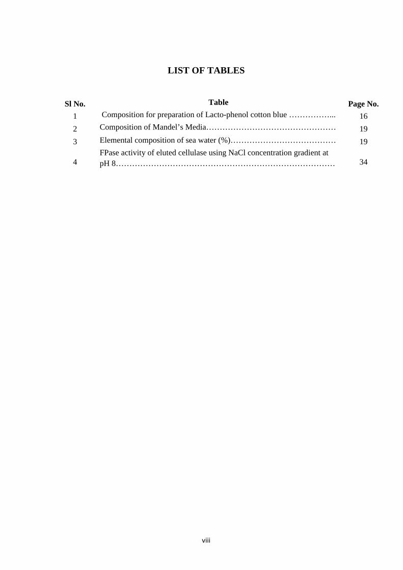
LIST OF TABLES
Sl No. Table Page No. 1 Composition for preparation of Lacto-phenol cotton blue ……………... 16 2 Composition of Mandel’s Media………………………………………… 19 3 Elemental composition of sea water (%)………………………………… 19
4 FPase activity of eluted cellulase using NaCl concentration gradient at pH 8……………………………………………………………………… 34
viii

ABSTRACT
Cellulase is one of the most required industrial enzymes and it is the costliest
process in bio fuel production. The present research work is based on the production of
cellulase enzyme from agricultural waste like rice straw. Sea water was used as a media
with the help of Fusarium subglutinans. The fungal strain was isolated from harvested rice
straw and cellulose was produced by DNS method. The pre-treatment of rice straw was
done using alkali treatment and minced well to make the cellulose structure to be simple.
The morphology of the pre-treated rice straw was observed using scanning electron
microscopy (SEM). The sea water was characterized using atomic adsorption spectroscopy
to find the presence of metal ions like iron, cobalt, zinc which is essential for the growth of
fungus. The enzyme production was carried out by submerged fermentation in a shaker
incubator at 37˚C for 21 days. The production was estimated at regular interval of time by
reducing sugar analysis of cellulose to glucose. The enzyme produced was characterized
using filter paper assay and analysed for its activity in different environments like pH,
temperature and metal ions (copper, zinc, cobalt, magnesium, manganese, iron). The
produced enzyme was then purified using anion exchange chromatography and checked
for its activity. The production and activity of the enzyme was found to be better when
compared to normal strains. When 5 mM metal ions like iron and manganese was added,
the activity was even more than normal strain. The purified enzyme can be used for the
hydrolysis of cellulose in juice production, as animal feed and the unprocessed enzyme can
be used for bio fuel production and paper pulp industry.
Keywords: Cellulose, DNS assay, Fusariun subglutinans, chromatography, bio-fuel
production.
ix

Chapter-1
Introduction

1.1 Cellulase
Cellulases are a group of enzymes produced primarily by fungi, bacteria, and protozoans that
hydrolyze cellulose [1]. Termites and microbial intestinal symbionts of some termites also
produce cellulase. Several kinds of cellulases are structurally and mechanistically different
[2].
Five different types of cellulases are known based on catalytic action they undergo. They are:
• Endocellulase – It randomly cleaves internal bonds at amorphous sites and creates new
chain ends.
• Exocellulase – It cleaves two to four units from the ends of the exposed chains produced
by endocellulase, resulting in formation of cellobiose.
• Cellobiase or beta-glucosidase – It breaks down the product of exocellulase enzyme into
individual monosaccharides.
• Oxidative Cellulases – It depolymerizes cellulose by radical reactions, as for instance
cellobiose dehydrogenase.
• Cellulose Phosphorylases – It depolymerize cellulose using phosphates instead of water.
Primary activity of cellulase is to breakdown cellulose to beta-glucose. This kind of cellulase
is produced in symbiotic intestinal bacteria of herbivores. Besides ruminants, most animals
don’t produce cellulase intrinsically and are able to partially break down cellulose through
fermentation. Thus, they cannot use energy from fibrous plant material [3]. Enzymes that
hydrolyze hemicellulose are said to be hemicellulase and are usually classified under
cellulase. Enzymes cleaving lignin are occasionally classified as cellulase, but this is
considered incorrect [4].
1.2 Cellulose
Cellulose is a main renewable form of carbohydrates, with about 1011 tons synthesized
annually. It is an unbranched β-1, 4-linked homopolymer of glucose, but cellulose have lots
of variation in their chain lengths according to origin and in the degrees of interaction
between chains [4, 5]. Cellulose is an important component of the primary cell wall of
angiosperms and algae. Some species of bacteria secrete it into biofilms. Cellulose is the most
abundant organic polymer on earth. The cellulose content of cotton fiber is 90%, wood is 40–
50% and dried hemp is about 45%. It has the greatest potential for bioethanol production [5].
However, cellulose must be hydrolyzed to obtain fermentable sugars and the cellulolytic
enzymes are central to the processing of biomass for bioethanol production [6]. 2

1.3 Cellulose degradation
The two major ways to degrade cellulose to glucose are: Chemical and Enzymatic. Each
molecule of cellulose is an unbranched polymer of 1 to 1000 million D-glucose units, linked
together with beta-1, 4 glycosidic bonds. However, they differ in the crystalline structures
and bindings by other biochemicals [7].
Two types of hydrogen bonds are present in cellulose molecules: one kind that forms between
the CH3OH group and the oxygen in the pyranose ring within the same molecule and the
other one that forms between the C6H5OH group of one molecule and the oxygen of the
glucosidic bond of another molecule. Ordinarily, the beta-1, 4 glycoside bonds easy to break.
However, because of these hydrogen bonds, cellulose forms very tightly packed crystallites.
These crystals are sometimes so tight that fluids (such as water, cellulase) cannot enter and
break or dissolve; only exogluconase, a subgroup of cellulase that attacks the terminal
glucosidic bond, is able to effectively degrade it [8]. Water’s inability to penetrate cellulose
explains why cellulose is insoluble. On the other hand, amorphous cellulose allows the
penetration of endogluconase, another subgroup of cellulase that catalyzes the hydrolysis of
internal bonds. This difference in the crystalline structure is that the hydrolysis rate is much
faster for amorphous cellulose than crystalline cellulose [9]. The process of breaking the
glucosidic bonds that hold the glucose basic units together to form a large cellulose molecule
is called hydrolysis because a water molecule must be supplied to render each broken bond
inactive. The chemical compounds surrounding the cellulose in plants, (for example lignin)
also limit the diffusion of the enzyme into the reaction sites and thus play an important role in
determination of hydrolysis rate. Sometimes, wood chips are pretreated with acid at
approximately 160°C to strip hemicellulose and lignin before they are treated with an enzyme
or a mixture of enzymes. Generally, 20 to 70% glucose yield can be expected after 24 hours
[9, 10]. The kinetics of cellulose hydrolysis has been widely studied, and Michealis-Menten
types of rate expressions with substrate or product inhibition terms have been proposed to
describe the observed reaction kinetics [11].
Many fungi and bacteria produce cellulase and do it extracellularly. Although it is common to
refer to a mixture of compounds that can degrade cellulose as cellulase, it is composed of
many distinctive enzymes [12]. Recent research has shown that one of the components is
relatively inert with the ability of recognizing and attaching itself to the surface of the
cellulose mass, in addition to the ability of recognizing and holding onto another protein
component that exhibits enzymatic activities. Thus, the chance of reaction is significantly
3

enhanced by a proximity effect, because the active enzyme is held onto the surface of a solid
substrate by an inert protein which acts as a glue [13].
1.4 Fungal source of cellulase enzyme
The conversion of cellulose into glucose is a two-step process in Trichoderma viride.
In the first step, beta-1, 4 glucanase breaks the glucosidic linkage to cellobiose, which is a
glucose dimer with a beta-1, 4 bond as opposed to maltose, a counterpart with an alpha-1, 4
bond. Subsequently, this beta-1, 4 glucosidic linkages is broken by beta-glucosidase.
The species most often used to study the production of cellulase are white-rot fungal cultures
of Trichoderma ressei and Trichoderma viride. Other fungal species often used are Fusarium
solani, Aspergillus niger, Penicillium funicolsum, and Cellulomonas sp [14]. The bacterial
species Clostridium thermocellum and Clostridium thermosaccharolyticum also represent
promising candidates for cellulase production since they are thermophilic (less contamination
problem and faster rate at a high temperature), anaerobic (no oxygen transfer limitation), and
ethanologenic (conversion of cellulose to ethanol via glucose with a single culture) [14, 15].
Aim
To produce, characterize and purify cellulase using fresh and sea water media by Fusarium
subglutinans (MTCC 11891) and also to use it in bioethanol production.
Objectives
• To isolate and characterize the cellulase producing fungus from soil.
• Comparative study on the production of cellulase using fresh and sea water media
• To optimize the environmental conditions for maximum cellulase activity.
• To use the produced cellulase in ethanol production.
4

Chapter -2
Literature review
5

2.1 Fungi
Fungi preferentially metabolize wood polysaccharides and produce an array of cellulolytic
and hemicellulolytic activities that may contribute to the degradation of plant cell wall
material. They are also capable of some direct transformation of lignin from the outer layers
of the cell walls, however they leave the middle lamella intact [16]. In addition to the
important role of fungi in carbon cycling, they are also involved in many biotechnological
processes. These processes include: brewing, winemaking, baking, cheese making and the
preparation of other fermented food (e.g. tempe, miso, angkak, soy sauce) together with
edible mushroom production are the most important fungal applications. Production of
enzymes (amylase, cellulase, invertase, lipase, pectinase, proteinase, rennin and xylanase),
organic acids (citric, itaconic and lactic acids), antibiotics and other pharmaceuticals
(penicillin, mevinolin, cephalosporin, griseofulvin and cyclosporine) by fungi are common
processes that have been reviewed [17]. Fungi are able to survive in intense environmental
conditions. They can tolerate a broad range of temperature, pH, humidity, salinity, oxygen
levels and ultraviolet radiation. Hence, their habitat varies from tropics to poles [18]. They
prefer to be in an acidic environment but their metabolism occurs conveniently in pH range
of 3.7 and 8.6 [19].
2.2 Cellulose and hemicellulose
Cellulose is commonly found as a crystal and is enclosed by amorphous cellulose
hemicellulose and lignin. Crystalline cellulose is resistant to the action of individual
cellulases due to structural rigidity. Hence, efficient conversion of cellulose to
monosaccharaides needs combination of many cellulases. Amorphous regions occur near the
crystal surface and are prone to enzymatic attack [20]. The rate of cellulose degradation
directly depends on crystallinity index and substances with which the cellulose is associated
e.g. Lignin [21].
Hemicelluloses, the third most abundant constituents of plant cell walls found in nature,
represent about 20 to 35% of the lignocellulose dry mass. They are heterogeneous polymers
of pentoses (D-xylose, L-arabinose), hexoses (D-mannose, D-glucose, and D-galactose) and
sugar acids. Links are formed by β-1,4- glycosidic bonds, but β-1,3-, β-1,6-, α -1,2-, α- 1,3-
and α -1,6- glycosidic bonds have also been reported [reference needed]. Hemicelluloses are
chemically bound to or cross-linked to other polysaccharides, proteins and lignin. They are
linked to pectin in primary cell walls and to lignin in secondary cell walls. This linked matrix
has protective role for the plant cell [22]. The major hemicelluloses of hardwood and annual
6

plants are xylans (15–30%), which probably interact with lignin and cellulose more than any
other hemicellulose. The main hemicelluloses of softwood are galactoglucomannans (15–
20%), arabinoglucoroxylan and arabinogalactan. Hemicelluloses are degraded readily due to
their amorphous nature and low degree of polymerization (approximately 70–200) as
compared to cellulose [23].
2.3 Cellulose and hemicellulose biodegradation
The interaction between both populations leads to a complete degradation of cellulose, which
releases CO2 and water under aerobic conditions, or carbon dioxide, methane and water under
anaerobic conditions. The hydrolysis of cellulose in fungi is carried out by cascade of
cellulolytic enzymes which are of many kinds like, extracellular endoglucanases,
cellobiohydrolases and β-glucosidases, which synergistically degrade cellulose [24].
Endoglucanases (EGs) represent less than 20% of the total protein in Hypocreajecorina
(anomorph Trichodermareesei) and preferentially hydrolyse cellulose microfibrils in the
amorphic parts of the fibril, releasing new terminal ends. Cellobiohydrolases (CBHs), which
may account for 20% to 60 % of the total cellulase proteins of fungi act on the existing or
endoglucanase-generated chain ends. In H. jecorina, CBH I attack reducing ends and CBH II
the non-reducing ends of the fiber. CBHs and EGs can degrade amorphous cellulose, but
CBHs are the only enzymes that efficiently degrade the crystalline form of cellulose. Both
enzymes release cellobiose molecules. The effective hydrolysis of cellulose also requires β-
glucosidases (BGL), which break down cellobiose and release two glucose molecules [25].
Products of cellulose degradation are made available as carbon and nitrogen sources for
either cellulolytic organisms or other microbes living in the environment containing
cellulosic materials [26]. The ability to produce cellulolytic enzymes is widespread among
microorganisms, but only a limited number of species are actually able to degrade native
cellulose in its crystalline form. Cellulolytic microorganisms (fungi and bacteria) can
establish synergistic relationships with non-cellulolytic species in cellulosic wastes [27]. The
hydrolysis of hemicellulose requires similar types of enzymes to those required for cellulose
hydrolysis. However, more enzymes are required for the complete degradation of
hemicellulose, because of the greater complexity of hemicellulose compared to cellulose
[28]. Hemicellulases hydrolyze glycosidic linkages in hemicelluloses and are classified
according to their substrate specificities [29]. The total enzymatic degradation of
hemicellulose polymers requires the action of “endo-type” enzymes that liberate short
oligosaccharides, which are subsequently degraded by sidegroup cleaving enzymes and “exo-
type” enzymes and finally monomeric sugars and acetic acid are formed. Similar to the
7

cellulases, hemicellulases act synergistically [30]. In fungi the production of cellulases may
also be regulated by factors other than induction and repression by sugars. Several lignin-
related aromatic compounds, which are found in association with cellulose in nature,
stimulate or inhibit cellulase production. This feature is common to some wood-rotting white
and brown rot basidiomycetes such as Phlebi aradiata, Trametes gibbosa and Trametes
coriolus [31].
2.4 Cellulase enzyme
Usually for production of cellulase enzyme filamentous fungi are widely used. Figure 1
shows the multi enzyme have the combination of three enzymes used for cleavage at
amorphous site in cellulose. These enzymes have various production levels when it comes to
fermentation process and it also depends upon the reactor shape and design [32].
Figure 1 - Conversion of cellulose to glucose by cellulase enzyme [36]
Cellulase activity is good at 50˚C in pH 5.8. Cellulose production from Pencillium
funiculosum displayed better stability over six days of incubation [33]. Cellulase production
depends upon biomass and cellulose concentration but it is not only an essential source but it
acts as inducer. mRNA quantities of cellulase increased because of gene silencing and down
regulation of cre 1 [34]. Maximum production of cellulase was produced by plant pathogenic
8

fungi, which produce degrading enzymes to break the cell wall polymers of plant and
structural complex of substrate (example : If paper or fruit peels were used, it is easy but with
kraft pulp was bit tough, because breaking the complex structure is highly complicated, so
pretreatment is necessary) [35].
2.5 Pretreatment of ligno cellulosic materials
Pretreatment process is most important to get the easily accessible with cellulose by
removing lignin, xylan and other molecules which are main ingredients in lingo cellulosic
materials. Physiochemical and structural factors decides the degradation of native
lignocellulosic biomass. Pretreatment step is necessary to improve the accessibility of
cellulose when we use enzyme as a catalyst. This step changes the degree of polymerization,
crystallinity index, lignin-carbohydrate linkages and increase the porosity of lignocelluloses
[37, 38]. Selection of pretreatment depends on how much the accessibility to cellulose is
obtained. This step is most eminent step in the total cost and time reduction of fermentation
process. In this process at high solid loading, when there is release of high amount of sugars
then the bottlenecks we have to deal with are mass transfer limitations, increased medium
viscosity and generation of high concentration of inhibitors. Cellulosic materials have low
density and are highly hygroscopic, thus slurries will become paste like material after
pretreatment [39, 40]. To avoid high viscosity issues, caused by pretreatment we have to
reduce particle size of slurry [40].
2.6 Submerged fermentation for cellulase production
Submerged Fermentation is a type of fermentation which is used for most of the industrial
enzyme production because of its ease of process and separation. It utilizes the liquid
substance such as molasses and broth. The biomolecules are secreted into fermentation broth
and after the process it’s easy to purify it. This process is highly suitable for the microbes that
require high moisture conditions e.g. bacteria and Fungus. The yield of product in this
fermentation differs for each substrate so it is important to select the correct substrate and
conditions for fermentation should be optimized [41]. Cellulase production is carried out in
aerobic aseptic culture. The growth medium contains salts, nutrients, surfactant and inducers
which are required for the fungus to survive and grow in medium. The important metal ions
which are required for cellulose production are iron, cobalt, copper, magnesium, calcium,
potassium, manganese and ammonium. The microbial contamination is avoided using
antibiotics and the sterilization of equipments before inoculation [42]. Steam sterilization is
done at 121oC for 20 minutes. The quantitative aspects of cellulose production were
9

improved between the years of 1972-1989. The maximum fold increase in enzyme
production happened in this period. The progress in cellulose production was averaged
doubling every two years due to the strain improvement and optimization of fermentation
process. Genencor International, Novo Nordisk, Rohm Finland are few of the companies
producing cellulose using submerged fermentation [43].
2.7 Instruments
2.7.1 Optical microscope
The optical microscope, commonly known as the "light microscope", is a type of microscope
which uses visible light and a system of lenses to magnify images of small samples. Optical
microscopes are the oldest types of microscope and were in compound form in the 17th
century. Basic optical microscopes can be very simple, although there are many complex
designs which aim to improve resolution and sample contrast [44].
The image from an optical microscope can be captured by normal light-sensitive cameras to
generate a micrograph. Originally images were captured by photographic film but modern
developments in CMOS and charge-coupled device (CCD) cameras allow the capture of
digital images. Purely digital microscopes are now available which use a CCD camera to
examine a sample, showing the resulting image directly on a computer screen without the
need for eyepieces [45, 46].
2.7.2 Scanning electron microscope (SEM)
A scanning electron microscope (SEM) is a type of electron microscope that produces images
of a sample by scanning it with a focused beam of electrons. The electrons interact with
atoms in the sample and are used to study sample's surface topography and composition [47].
The electron beam is generally scanned in a raster scan pattern, and the beam's position is
combined with the detected signal to produce an image. SEM can achieve resolution better
than 1 nanometer. Specimens can be observed in high vacuum, in low vacuum, and (in
environmental SEM) in wet conditions [48].
The most common mode of detection is by secondary electrons emitted by atoms excited by
the electron beam. The number of secondary electrons is a function of the angle between the
surface and the beam [49]. On a flat surface, the plume of secondary electrons is mostly
contained by the sample, but on a tilted surface, the plume is partially exposed and more
10

electrons are emitted. By scanning the sample and detecting the secondary electrons, an
image displaying the tilt of the surface is created [50].
2.7.3 UV spectroscopy
UV Spectroscopy is an absorption spectroscopy or reflectance spectroscopy in the ultraviolet-
visible spectral region. This means it uses light in the visible and adjacent (near-UV and near-
infrared (NIR)) ranges [51]. The absorption or reflectance in the visible range directly affects
the perceived color of the chemicals involved. In this region of the electromagnetic spectrum,
molecules undergo electronic transitions.
If the compound is more concentrated more light will be absorbed by the sample; within
small ranges, the Beer-Lambert law holds and the absorbance between samples vary with
concentration linearly. Beer-Lambert law is a law which states that the amount of light
absorbed by a body is proportional to the amount of absorbing particles in it. This means that
it will be proportional to the concentration of that additive multiplied by the thickness of that
body (the path length of the light through the body). Samples are usually prepared in
cuvettes; depending on the region of interest, they may be constructed of glass, plastic
(visible spectrum region of interest), or quartz (Far UV spectrum region of interest) [51, 52].
2.8 Techniques
2.8.1 Glucose estimation by DNS
This method is used to test the presence of free carbonyl group (C=O), the so-called reducing
sugars. Figure 2 reveals the oxidation of the aldehyde functional group present in glucose and
the ketone functional group in fructose. Simultaneously, 3, 5-dinitrosalicylic acid (DNS) is
reduced to 3-amino, 5-nitrosalicylic acid under alkaline conditions:
Figure 2 - Oxidation and reduction reactions in reducing sugar analysis
Since dissolved oxygen can interfere with glucose oxidation, sulfite, which itself is not
necessary for the color reaction, is added in the reagent to absorb the dissolved oxygen [53].
11

The above reaction scheme shows that one mole of sugar will react with one mole of 3, 5-
dinitrosalicylic acid. However, it is suspected that there are may side reactions, and the actual
reaction stoichiometry is more complicated than that previously described. The type of side
reaction depends on the exact nature of the reducing sugars [54]. Different reducing sugars
generally yield different color intensities; thus, it is necessary to calibrate for each sugar. In
addition to the oxidation of the carbonyl groups in the sugar, other side reaction such as the
decomposition of sugar also competes for the availability of 3, 5-dinitrosalicylic acid [55].
2.8.2 Filter paper assay
Filter Paper Activity/Assay (FPA) is used to determine the quantity of cellulolytic enzymes
required to hydrolyze lignocellulosic biomass. The FPA was initially proposed as a simple,
reproducible and quantitative method that predicts enzyme action under practical
saccharification conditions. This measures the ability of the cellulose to hydrolyze both
crystalline and amorphous regions of cellulose [56]. Therefore, it is generally believed that
FPase assay is the best measure of the activities of both endo- and exo- type in a cellulase
with a strip of filter paper (Whatman #1, 50mg, 1 X 6 cm) for 1hour reaction at the given
temperature [57].
2.8.3 Atomic absorption spectroscopy
This instrumentation is a setup which is used to detect metals and metalloids in sample
present in environment. The principle depends upon the fact that ground state metals to
absorb light of particular wavelength was sourced and amount of light absorbed can be
obtained against a standard curve.
Figure 3 - Basic instrumentation of flame atomic absorption [59]
12

Figure 4 – Schematic of atomization of flame in an absorption spectroscopy
Figure 3 and 4 the basic instrumentation setup has seven fundamental parts (1) Light source
(2) Atom cell (3) monochromator (4) Detector (5) Amplifier (6) Signal display (7) Data
station.
Hallow cathode lamp is a common light source which has glass tube with an inert gas (neon
or argon) at 1Nm-2 – 5Nm-2. To produce atoms from the sample two systems are commonly
used (1) Sucking a sample solution into the flame (2) Dropping of sample through electro
thermal atomization, which is placed inside a heated graphite tube [59, 60].
13

Chapter - 3
Materials and methods
14

3.1 Collection of sample
The soil samples were collected from the agricultural fields located on the outskirts of
Rourkela city (22.2492° N, 84.8828° E), Sundergarh district, Odisha, India. Rourkela city is
strategically located in the northwestern border of the Indian state of Odisha. Over a period of
year, the temperature varies from 7°C to 47°C. All soil samples were collected in the month
of June, 2013 from six randomly chosen agricultural fields (paddy fields). The collected
samples were kept in plastic bags. Samples were stored at 4°C until use for culturing fungi
(not more than 48 h).
3.2 Preparation of media
Potato Dextrose Agar (PDA, Himedia) medium was used for the isolation of fungi from soil.
3.9 ml of PDA medium was added to 100 ml of distilled water and autoclaved at 121°C, 15
psi for 15 min.
3.3 Isolation of fungi
Serial dilution method was followed to isolate the fungal strains from soil sample.
3.3.1 Stock Preparation
20 g of soil sample was diluted in 100 ml of sterile distilled water.
3.3.2 Serial dilution
Eight test tubes each containing 9 ml of sterile distilled water was taken. To the first test tube,
1 ml stock was added and mixed well. From the first test tube, 1 ml was transferred to second
test tube and mixed well. This was continued till the eight test tubes. From eighth test tube 1
ml of stock solution was discarded. Hence, the dilution factor from first to eighth test tube
was 10-1 to 10-8. From each test tube three different samples of volume 5 µl, 10 µl and 15 µl
were added to PDA plates and were incubated at 30°C for three days [4].
3.4 Preparation of permanent slides
For the preparation of permanent slides, lactophenol and lactophenol with cotton blue stains
were used. The slides were sealed with cover slip.
15

3.4.1 Stain Used
3.4.1.1 Preparation of lactophenol cotton blue
Table 1: Composition for preparation of Lacto-phenol cotton blue
Components Contents
Cotton Blue 0.5 g
Phenol 25 g
Lactic Acid 25 g
Glycerol (pure) 50 g
Distilled water 100 ml
Lactophenol was readily prepared by heating the phenol in a water bath until it dissolved and
then lactic acid and glycerol was added. The volume was then made up to 100 ml with
distilled water after which cotton blue was added.
3.4.2 Preparation of permanent slide
A drop of lactophenol blue solution was placed on a slide. Using an inoculating needle, the
fungal culture was spread carefully to make a thin layer. An edge of the coverslip was placed
on the drop and slowly spread over it. Air bubbles should be avoided under the coverslip. The
slide with the fungal culture was heat fixed by passing it carefully over a flame. The slide was
then observed under a microscope.
3.4.3 Phenotypic characterization of fungi
Sporulating isolates were identified with the help of standard manuals. Further confirmation
of the fungal species was done after observation under a light microscope and pictures were
taken using a Sony 8.1 MP digital camera. The fungi samples were sent to Microbial Type
Culture Collection and Gene Bank (MTCC), Chandigarh, Punjab, India for identification.
3.5 Slant culture
The culture made on the slanting surface of a solidified PDA medium in a test tube was tilted
to provide a greater area for growth. The microorganisms that showed effective cellulose
degrading capacity were maintained as slant culture.
16

3.6 Identification of cellulase producing fungi using congo red staining
method
Congo red staining was done using 0.1% congo red stain and 1% sodium hydroxide solution.
The fungal culture was grown in petri plate using PDA medium with 1% carboxyl methyl
cellulose (Himedia) for seven days at 37˚C in an incubator. Then 15 ml of congo red solution
was poured, left for 5 min and destained using sodium hydroxide [5].
3.7 Estimation of reducing sugars using DNS assay
DNS assay was done using DNS reagent (3, 5-dinitrosalicylic acid (DNS) - 10 g, sodium
sulfite- 0.5 g, and sodium hydroxide-10 g in 1litre of distilled water) and 40% potassium
sodium tartarate. The fungal culture was grown in Mandel’s media as a substrate of carboxyl
methyl cellulose (CMC) for 21 days. The culture was taken and centrifuged (REMI
centrifuge / C- 24BL) at 4˚C and 15,000 rpm for 10 min to separate the fungal biomass. Then
the crude extract was filtered with silica gel ‘G’ (Rankem) packed column for removal of
large agglomerates in processed broth. Again it was centrifuged under the same condition.
After that 3 ml of extract was taken and added to 3ml of DNS reagent and incubated at 90˚C
for 5 min in water bath [7]. When it was warm, 1 ml of 40 % Rochelle’s salt solution was
added and observed under UV-visible spectrometer (Double beam spectrop C2203,
Systronics) at 510 nm. Reducing sugar present in the sample was calculated using the
standard graph plotted by known glucose concentration.
3.7 Pretreatment of rice straw
Pretreatment was done to remove the lignin, hemicellulose and xylan from the rice straw. It
was done by alkali treatment using 1 M sodium hydroxide (4 g of sodium hydroxide in 100
ml of distilled water) at 121˚C, 15 psi for 20 min in autoclave. The processed rice straw was
then filtered using nylon cloth and the liquid was discarded. Then the pretreated rice straw
residue was used for cellulase production.
3.8 Collection of sea water Sea water was collected from Gopalpur, Orissa (19.27˚N, 84.92˚E) on the Bay of Bengal
coast during the month of July, 2013 using 50 ml ficol tube (Tarson) and stored at 4˚C. The
stored water was filtered using whatman filter paper No. 1 (F1000-C, Himedia) and used for
cellulase production process.
17
3.8.1 Determination of concentration of metal ions in sea water using
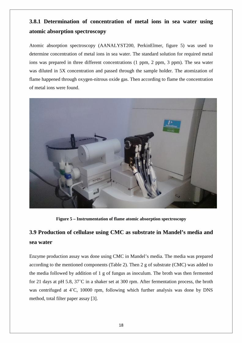
atomic absorption spectroscopy
Atomic absorption spectroscopy (AANALYST200, PerkinElmer, figure 5) was used to
determine concentration of metal ions in sea water. The standard solution for required metal
ions was prepared in three different concentrations (1 ppm, 2 ppm, 3 ppm). The sea water
was diluted in 5X concentration and passed through the sample holder. The atomization of
flame happened through oxygen-nitrous oxide gas. Then according to flame the concentration
of metal ions were found.
Figure 5 – Instrumentation of flame atomic absorption spectroscopy
3.9 Production of cellulase using CMC as substrate in Mandel’s media and
sea water
Enzyme production assay was done using CMC in Mandel’s media. The media was prepared
according to the mentioned components (Table 2). Then 2 g of substrate (CMC) was added to
the media followed by addition of 1 g of fungus as inoculum. The broth was then fermented
for 21 days at pH 5.8, 37˚C in a shaker set at 300 rpm. After fermentation process, the broth
was centrifuged at 4˚C, 10000 rpm, following which further analysis was done by DNS
method, total filter paper assay [3].
18

Table 2 – Composition of Mandel’s Media
Ingredients Contents
KH2PO4 2 g/l
CaCl2.H2O 0.4 g/l
MgSO4.7H2O 0.3 g/l
FeSO4.7H2O 5 g/l
MnSO4.4H2O 1.6 g/l
CoCl2.6H2O 1.4 g/l
Distilled water 1 litre
Table 3 - Elemental composition of sea water (%)
Element Percent Element Percent
Oxygen 85.84 Sulphur 0.091
Hydrogen 10.82 Calcium 0.04
Chloride 1.94 Potassium 0.04
Sodium 1.08 Bromine 0.0067
Magnesium 0.1292 Carbon 0.0028
3.10 Enzyme production using pretreated rice straw in Mandel’s media
and sea water
Enzyme production assay was done using pretreated rice straw in Mandel’s media. The media
was prepared according to the mentioned components in table 2, then 2 g of substrate (Rice
Straw) was added to the media. It was followed by addition of 1g of fungus as inoculum. The
19

broth was then fermented for 21 days at pH 5.8, 37˚C in a shaker set at 300 rpm. After
fermentation process, the broth was centrifuged at 4˚C, 10000 rpm. Further analysis was done
by DNS method and total filter paper assay. Similar process was carried out using sea water
as media. The composition of both media and sea water is same as shown in Table (2 and 3).
3.11 Effect of time on cellulase production
Cellulase production was assessed at regular intervals to check the production rate. It was
done by monitoring the glucose production by breakdown of cellulose. For this, 3 ml of
fermentation broth was taken and centrifuged at 4˚C and 10000 rpm. DNS reagent was
freshly prepared and 3 ml of it was added to the sample along with 1 ml of Rochelle’s salt.
Then sample’s absorbance was taken at 560 nm using a U-V spectrophotometer [61].
3.12 Effect of pH on cellulase activity
The pH of reaction is affected by the enzyme activity. The pH of crude enzyme samples were
adjusted to 4, 5, 6, 7, 8 and 9 in different flasks using 0.1 M NaCl and 0.1 M HCl. The
samples were later collected and assayed to find the maximum activity [61].
3.13 Effect of temperature on cellulase activity
The temperature of reaction affects the enzyme activity. Beyond a critical temperature, the
enzyme activity will decrease. Enzyme activity was assayed in flasks maintained at different
temperatures of 30˚C, 40˚C, 50˚C, 60˚C, 70˚C in a water bath. The cultured filtrates were
later collected and assayed to find the maximum activity [61].
3.14 Effect of metal ions on cellulase activity
The effect of different metal ions on enzyme activity was studied. Different metal salts,
Mn(II)SO4, Fe(II)SO4, Cu(II)SO4, Mg(II)SO4 and Zn(II)SO4 were added to the fermentation
broth in concentration of 5mM on to separate flasks and its cellulose activity was studied
[61].
3.15 Purification of cellulase
Purification of crude enzyme was done using ion exchange chromatography. Crude enzyme
was poured to DEAE sepharose (Sigma Aldrich) packed syringe column which was
equilibrated using a buffer (5 mM EDTA, 20 mM tris base (Himedia) at pH 8) shown in
figure 6. Elution was done by passing linear gradient of 0.2 M – 1.2 M NaCl through the
20

column. The eluted enzyme was collected as a fraction of 10 ml and tested for cellulase
activity [62].
Figure 6 – Anion exchange chromatography set-up for purification of cellulase.
3.16 Application of crude enzyme extract in ethanol production
Crude enzyme was applied through the silica gel packed column and further subjected to
centrifugation at 10000 rpm, 4˚C. Saw dust was pretreated using 1 M NaCl and 10 ml of
crude enzyme was added along with 2 g of saw dust. It was incubated in the water bath with
the following optimized parameters (80˚C, pH 5.8, MnSO4.7H2O (5 mM) as catalyst) for 1
hr. Then 2 ml of Saccharomyces cerevisiae culture was inoculated to the broth. After 5 days,
production of ethanol was estimated by gravimetric analysis using the following formula
[63].
21

Chapter - 4
Results and discussion
22

This chapter is organized in to three sections. Section 4.1 discusses about the results obtained
in the isolation, characterization and screening of various fungal cultures for the potential
production of cellulases. Section 4.2 deals with the results obtained in various batches for
optimizing the environmental conditions such as pH, temperature, time and metal ions to
maximize the cellulase production. In addition, evaluating the possibility of growing fungal
cultures on sea water rather than the basal medium were also carried out and reported in this
section. 4.3Cellulase activities checked with saw dust was reported. Section 4.4 deals with its
purification using anion exchange chromatography technique results.
4.1 Isolation, screening and characterization of fungi
4.1.1 Isolation of fungal cultures
The collected soil samples (100 g of each from the five random locations) on agricultural
fields at the depth of 8-10 cm were mixed well and homogenized. The homogenized soil
samples were subjected to two different techniques to isolate the fungal cultures present in the
soil. One is direct plating technique and the other is serial dilution method. 100 mg of
homogenized soil samples were directly sprinkled to the medium and incubated at room
temperature for a week in case of direct plating technique. Since the fungal diversity was quite
high in the inoculated soil sample and each species occupied a specific niche, merged colonies
were observed in this method. Either the inoculated soil volume or the medium level has to be
reduced to achieve the better isolation of fungal colonies since the area available for the fungal
cultures to spread and occupy their niches will be high in this situation.
The direct plating technique involves the sprinkling of a known amount of soil to the medium.
On contrary, the serial dilution technique involves the systematic dispersion of the soil fungi
out of which the very minimal amount used to inoculate and incubated further. Since the serial
dilution was done up to 10-8 times, the fungal cultures could be easily isolated in the respective
plates. Three predominant fungal cultures were observed in this petriplate as shown in Figure
7. These fungal colonies were well distinct it was isolated further through sub culturing
techniques. These colonies were named as F1, F2 and F3. Figure 8 shows the three distinct
predominant fungal cultures isolated from the soils collected on agricultural fields.
23

Figure 7 - Three different colonies of fungus isolated from paddy field
Figure 8 - Pure isolates of three fungal cultures (F1, F2, F3) isolated from paddy field
4.1.2. Identification of cellulase producing fungi using Congo red assay
As a preliminary qualitative study, the isolated fungal cultures were screened for its cellulose
activity by Congo red assay. Out of the three fungal isolates, F3 showed the significant zone
F1
F2 F3
24

of clearance. The zone of clearance in Congo red assay clearly depicts the cellulase activity
of F3 culture as shown in Figure 9.
Figure 9 - Identification of cellulase activity by zone of clearance
4.1.2.2 Estimation of reducing sugar using DNS assay
Cellulase activity was measured by the DNS method through the determination of the
liberated amount of reducing sugars. Glucose was used as a standard in this assay and all the
experiments were done in triplicates. One unit of enzyme activity was defined as the amount
of enzyme that releases 1 mol of glucose per minute per ml. All the three fungal isolates were
able to degrade the cellulose present in the medium confirmed that all the fungal isolates have
expressed its cellulase activity. Figure 10 revealed that F1 has lowest cellulase activity of
5.86 µg/ml. F2 and F3 isolates have 10 and 30% greater cellulase activity than the isolate F1.
Among the three fungal isolates, F3 have greatest cellulase activity of 7.67 µg/ml as shown in
Figure 9. Since F3 could be the promising candidate for high cellulase production, it was
concluded to recheck the cellulase activity.
Figure 10 - Production of cellulase by fungal isolates
Zone of clearance
25

4.1.3 Screening of fungal cultures for its growth in sea water
The novel attempt has been made to explore the possibilities of cultivating fungal cultures on
CMC broth that was already adjusted for pH with sea water. Three fungal strains previously
isolated from the paddy agricultural fields were inoculated on the CMC broth adjusted for pH
with sea water and incubated further for 14 days.
Visual observation of growth of fungal isolates on CMC broth adjusted for pH with sea water
revealed that F1 fungal isolate could not sustain in the newly saline environment. The growth
rate of F1 was almost hindered as it might be highly sensitive to the saline conditions, no
significant growth was observed. In case of F2 isolate growth on CMC broth adjusted for pH
with sea water, the growth rate was relatively higher as compared than that of F1. Yet within
the incubation time of seven days, notable cell death was observed. At the end of tenth day
the F2 fungal isolate was completely death.
On contrary, significant glucose concentrations were observed in CMC broth adjusted for pH
with sea water inoculated with F3 fungal isolate. It showed faster growth by depleting the
cellulose and converting it to glucose. Substantial spreading of the mycelium was also
observed with this fungal strain. This particular characteristic reveals that the strain could
have acclimatized to the saline conditions and it might have aided in sustaining the salinity
environment. This further reaffirmed that the F3 fungal isolate could be the promising
candidate for cellulase productions even with sea water.
4.1.4 Characterization of fungal cultures
The isolated fungal cultures were identified by performing traditional methods of
classification and identification of the organisms based on morphological, physiological,
biochemical, and nutritional characteristics performed at the Microbial Type Culture
Collection Center and Gene Bank (MTCC & GB), Institute of Microbial Technology
(IMTECH), Chandigarh, India.
Various biochemical tests such as assimilation of sugars, utilization of starch, urea, tyrosine,
asparagine and citrate components, and nitrate reduction, tolerance range of pH, NaCl and
temperature were performed. Cultural characteristics were studied on different media
regarding their growth intensity, growth pattern, colour of aerial mycelia and formation of
soluble pigments. F3 fungal isolate was identified as Fusarium subglutinans (MTCC 11891).
This fungal strain was deposited in the MTCC & GB of IMTECH, India. White aerial
26

mycelium growing rapidly, tinged with purple sometimes, microconidia abundant, oval,
usually single celled, may become 1-3 septate, produced in false heads, polyphialidic,
macroconidia abundant, slightly sickle shaped to almost straight with delicate walls and basal
cells foot shaped are the salient features of this fungus. This was abundant source in plants as
plant pathogen but have the ability to deplete the cellulose into glucose for its own growth.
4.2 Production of cellulase enzyme and optimization of environmental
conditions
4.2.1 Pretreatment of rice straw
Figure 11 - Scanning electron micrographs of rice straw before pretreatment
Figure 12 - Scanning electron micrographs cross sectional area of rice straw before
pretreatment
27

Figure 13 – Scanning elcetron micrographs of rice straw after pretreatment
Sodium hydroxide pretreatment for rice straw was reported as an effective method. Lignin
was removed by breaking the ester bonding between lignin and it enabled availability of
cellulose to fungus. Figure 11 and 12 show the fibre and the cross sectional area of rice straw
in which the arrangement of xylan, lignin and cellulose fiber. The SEM image in figure 13
showed the maximum removal of xylan, lignin and the cellulose fibers which act as substrate
for cellulase production. .The optimum concentration used for pretreatment was 1 M at 121oC
for 15 min. The rate of increase in depletion of ester bond was dependent on rate of increase
in various factors like temperature, concentration of base and time. The SEM image in figure.
11 shows the single rice straw before pretreatment fulfilled with lignin and xylan
crosslinking.
4.2.2 Determination of metal ions in sea water
Figure 14 – Elemental analysis of sea water by atomic absorption spectroscopy
28

Growth media was supplemented with metal ions which are essential for the cellular and
subcellular functions of fungus. Similarly, when sea water was used as basal medium, it is
important to determine the adequate metal ions. So, a screening test was performed and all
required ions were found to be in appropriate concentrations which were given in Figure 14.
4.2.3 Enzyme production using pretreated rice straw in Mandel’s media
and sea water
The production process was extended up to 21 days while the growth was initiated at 5th day.
The fungus was grown on the surface of the cellulose fiber. At a regular interval of time the
hydrolysis of cellulose to glucose was analyze by DNS assay and the cellulase activity was
analyzed using filter paper assay, at the end of the process, it was seen that the cotton used as
stopper was also depleted because of its cellulose content. Fungus was found to be grown on
the surface of the cellulose fiber. Cotton which was used as the stopper for the flask found to
be hydrolyzed due to fungal action which may be replaced by a non-cellulosic material. The
fungal growth was observed half the time in sea water then that in media. Even the activity
was less compared to basal media cellulase but it was not that much less compared to
Trichoderma viride in free cell formation in Stirred tank reactor [64]. The production of
enzyme could be increased by giving alternate stress, nutrient deprivation stress. The fast
depletion of cellulose was due to the pretreatment because it decreased the crystallinity and
increased the porosity in rice straw cellulose fiber. A steady increase in FPase activity was
significant due to the improved cellulase production due to extracellular protein secretion. At
the end of fermentation (21 days), enzyme production was observed to be increased.
Diminished production volume aids the availability of an increased surfaced area a factor
which is difficult to achieve using conical flasks. Hence tray type reactors could be
considered to enhance the production of enzyme. Paddle type agitators may be implemented
to solve aeration related issues.
4.2.4 Effect of time on cellulase production
The flasks were incubated in shaker for 21 days, the degradation of biomass started at 3rd day
approximately. The cellulase activity was analyzed from 4th day by taking out 3 ml of sample
and replaced by media. The decrease in biomass concentration was present on day 14 due to
the increase in cellulase activity of basal media and sea water media alike. After 21 days
cellulase activity was recorded as 277.5 Units/ ml/ min and 126.72 Units/ ml/ min in basal
media and sea water showed in figure 15 and 16 respectively. To avoid the contamination the
29

process was terminated on 21 days and moved on further studies and moved on to further
studies.
Figure 15 – Production of cellulase by Fusarium subglutinans MTCC 11891 in shake flask using
Mandel’s media
Figure 16 – Production of cellulase production by Fusarium subglutinans MTCC 11891 in shake
flask using sea water media
4.2.5 Effect of PH on cellulase activity
Effect of pH was analyzed from range of pH 4 to pH 9. There was no much variation in
FPase activity of crude enzyme from basal media and sea water produced cellulase in
different pH. Figure 17 and 18 shown that at pH 9 cellulase activity started decreasing and
reached 275.4 Units/ ml/ min for basal media produced cellulase and 177 Units/ ml/ min for
30

sea water produced cellulase. The maximum activity was observed at pH 5 as 292.53 Units/
ml/ min and 184 Units/ ml/ min for basal media and sea water produced cellulase. From the
above results it was obtained that the enzyme was pH independent. These crude enzymes
could be used in many industrial processes where the pH fluctuations are dominant.
Figure 17 – Effect of pH on cellulase activity in fresh water
Figure 18 – Effect of pH on celluse activity in sea water
4.2.6 Effect of temperature on cellulase activity
Effect of temperature was analyzed from a range of 37oC to 80oC showed in figure 19 and 20.
It was revealed that there was gradual increase in the activity of crude enzyme from 37oC
[260 Units/ ml/ min for basal media produced cellulase and 143.28 Units/ ml/ min for sea
water produced cellulase] to 80oC [347.43 Units/ ml/ min for basal media produced cellulase
and 232 Units/ ml/ min for sea water produced cellulase]. Therefore it is highly possible that
enzyme is thermo stable and showed its maximum activity up to 80˚C. When FPase assay
was done at 80oC for both enzymes, the hydrolysis was performed very rapidly. 31

Figure 19 – Effect of temperature on cellulase activity in fresh water
The optimum temperature for both crude enzymes were 75oC – 80oC, because the increase in
temperature would increase the kinetic energy of the enzyme system so the number of
collisions per unit volume increases and enzyme would have collide with substrate and bind
to active site.
Figure 20 – Effect of temperature on cellulase activity in sea water`
Above results indicated that the enzyme extract can be used directly from the process and for
the industries where high temperature plays vital role [e.g. Paper pulp industry].
32
4.2.7 Effect of different metal catalyst on cellulase activity
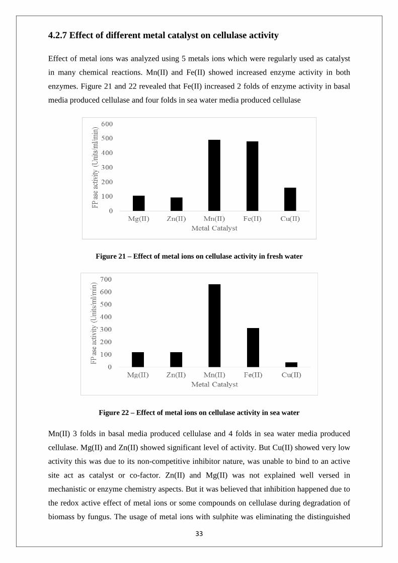
Effect of metal ions was analyzed using 5 metals ions which were regularly used as catalyst
in many chemical reactions. Mn(II) and Fe(II) showed increased enzyme activity in both
enzymes. Figure 21 and 22 revealed that Fe(II) increased 2 folds of enzyme activity in basal
media produced cellulase and four folds in sea water media produced cellulase
Figure 21 – Effect of metal ions on cellulase activity in fresh water
Figure 22 – Effect of metal ions on cellulase activity in sea water
Mn(II) 3 folds in basal media produced cellulase and 4 folds in sea water media produced
cellulase. Mg(II) and Zn(II) showed significant level of activity. But Cu(II) showed very low
activity this was due to its non-competitive inhibitor nature, was unable to bind to an active
site act as catalyst or co-factor. Zn(II) and Mg(II) was not explained well versed in
mechanistic or enzyme chemistry aspects. But it was believed that inhibition happened due to
the redox active effect of metal ions or some compounds on cellulase during degradation of
biomass by fungus. The usage of metal ions with sulphite was eliminating the distinguished
33

effects of other combinations. As stated earlier the metal ions have proved their effect of
metal ions on cellulase activity.
4.3 Partial purification of cellulase using anion exchange chromatography
Partial purification was done using anion exchange chromatography. When the pH of protein
was higher than pI value the charge of the protein would be negative. The pI of the cellulase
enzyme was nearly 5.2. So that the pH of the enzyme crude extract was increased upto pH 8
and crude sample was applied for elution. After elution using various linear gradient of NaCl
(0.2 to 1.2 M) followed by FPase activity, it was shown that at 0.8 and 1 M, cellulase was
eluted.
Table 4 – Effect of concentration of NaCl on elution of cellulase in anion exchange
chromatography
Concentration of NaCl (M) FPase activity (Units/ml/min)
0.2 35.32
0.4 49.08
0.6 52.73
0.8 298.68
1 273.12
1.2 45.62
1.4 40.73
1.6 38.95
4.4 Application of crude enzyme extract in the ethanol production
Saw dust was chosen as major source of saccharification because of its availability and low
cost. The production of ethanol at 7th day of fermentation was studied using gravimetric
method and estimated as 15.76%. The reduced production of ethanol was due to the
decreased hydrolysis of cellulose to glucose. This was due to its tightly packed arrangement
of cellulose in saw dust.
34

CHAPTER - 5
Summary and conclusion
35

Substantial research was carried out on the production and characterization of
cellulase in fresh water and sea water.
Rice straw is an important agricultural lignocellulose waste which can be used as
renewable energy source. Pretreatment using alkali treatment improves the utilization
of rice straw.
Fusarium subglutinans (MTCC 11891) was found to be eminent filamentous fungi for
the production of cellulase activity.
Usage of sea water as an alternative media provides the cost reduction and utilization
of saw dust increase the waste consumption.
The cellulase showed activity at various pH and it is also capable to sustain at high
temperature around 80˚C (thermo stable).
The estimation of ethanol revealed that this crude enzyme can be used for bio-ethanol
production.
36

REFERENCES
37

1. Rabelo S.C., Filho R.M., Costa A.C., “A comparison between lime and alkaline hydrogen peroxide pretreatments of sugarcane bagasse for ethanol production”, Applied Biochemistry and Biotechnology, 2008, 148, 45–58.
2. Zhou J., Wang Y.H., Chu J., Zhuang Y.P., Zhang S.L., Yin P., “Identification and
purification of the main components of cellulases from a mutant strain of Trichoderma viride”, Bioresource Technology, 2008, 99, 6826–33.
3. Murashima K., Kosugi A., Doi R.H., “Synergistic effects of cellulosomal xylanase
and cellulases from Clostridium cellulovorans on plant cell wall degradation”, Journal of bacteriology, 185, 1518–24.
4. Isabelle H.G., Antoine M., Alain D., Gwénaël J., Daniel M., Sabrina L., Hughes M.,
Jean-Claude S., Frédéric M., Marcel A, “Comparative secretome analyses of two Trichoderma reesei RUT-C30 and CL847 hypersecretory strains”, Biotechnology and Biofuels 1, 2005, 18, 15-24
5. Jorgensen H., Vibe-Pedersen J., Larsen J., Felby C. “Liquefaction of lignocellulose at
high-solids concentrations”, Biotechnology and Bioengineering, 2010, 96, 862–70.
6. Kuo, C.H., Lee, C.K. “Enhanced enzymatic hydrolysis of sugarcane bagasse by N-methylmorpholine-N-oxide pretreatment”, Bioresource Technology, 2003, 100, 866–71.
7. Xia, L.M., Shen, X.L., “High-yield cellulase production by Trichoderma reseei ZU-02 on corn cob residue”, Bioresource Technology, 2004, 91, 259–62.
8. Xu, Z.N., Yang, S.T., “Production of mycophenolic acid by Penicillium
brevicompactum immobilized in a rotating fibrous-bed bioreactor”, Enzyme Microbial Technology, 2004 40, 623–28.
9. Voragen A.G., Beldman J.G., Rombouts, F.N., “Cellulases of mutant strain of
Trichoderma viride QM9414”, Methods Enzymology, 1988, 160, 243–51.
10. Adsul M.G., Ghule J.E., Shaikh H., Singh R., Bastawde K.B., Gokhale D.V., Varma A.J., “Enzymatic hydrolysis of delignified bagasse polysaccharides”, Carbohydrate polymers, 2005, 62, 6–10.
11. Sassner, P., Galbe, M., Zacchi, G., “Bioethanol production based on simultaneous
saccharification and fermentation of steam-pretreated Salix at high dry-matter content”. Enzyme Microbial Technology, 2006, 39, 756–62.
12. Ververis, C., Georghiou, K., Danielidis, D, “Cellulose, hemicelluloses, lignin and ash
content of some organic materials and their suitability for use as paper pulp supplements”, Bioresource Technology, 2001, 98, 296–301.
13. Ruiz, E., Cara, C., Ballesteros, M., Manzanares, P., Ballesteros, I., Castro, E.,
“Ethanol production from pretreated olive tree wood and sunflower stalks by an SSF process”. Applied Biochemistry and Biotechnology, 2006, vol, 129–32.
14. Maeda, R.N., Silva, M. M.P., Santa Anna, L.M.M., Pereira, J.R.N., “Nitrogen source
optimization for cellulase production by Penicillium funiculosum, using a sequential
38

experimental design methodology and the desirability function”, Applied Biochemistry and Biotechnology, 2010, 161, 411–22.
15. Rao M., Gaikwad S., Mishra, C., Deshpande V., “Induction and catabolite repression
of cellulase in Penicillium funiculosum”. Applied Biochemistry and Biotechnology, 1998, 19, 129–37.
16. Mandels M., Weber J., “The production of cellulases. Advance Chemical Services,
1969, 95, 391–14.
17. Ghose T.K., “Measurement of cellulase activities”, Pure and Applied Chemistry, 1987, 59, 257–68.
18. Rao, M., Gaikwad, S., Deshpande V., “Catabolite repression of cellulase in
Penicillium funiculosum”. Applied Biochemistry and Biotechnology, 1988, 19, 129–37.
19. Lynd L.R., Wemer P.J., Zyl, W.H.V., Pretorius I.S., “Microbial cellulose utilization:
fundamentals and biotechnology”. Microbiology and Molecular Biology Reviews, 2002, 66, 506–57.
20. Rudolf A., Alkasrawi M., Zacchi G., Lidén G., “A comparison between batch and
fed-batch simultaneous saccharification and fermentation of steam pretreated spruce”. Enzyme Microbial Technology, 2005, 37, 195–205.
21. Linde M., Galbe M., Zacchi G., “Simultaneous saccharification and fermentation of
steam-pretreated barley straw at low enzyme loadings and low yeast concentration”. Enzyme Microbial Technology, 2006, 40, 1100–07.
22. Jacobsen S.E., Wyman C.E., “Cellulose and hemicellulose hydrolysis models for
application to current and novel pretreatment processes”, Applied Biochemistry and Biotechnology, 2000, 84–86, 81–96.
23. Zhu J.Y., Zhu W., Bryan O.P., Dien B.S., Tian, S., Gleisner, R., et al., “Ethanol
production from SPORL-pretreated lodge pole pine: preliminary evaluation of mass balance and process energy efficiency”, Applied Microbiology and Biotechnology, 2010, 86, 1355–65.
24. Vásquez M.P., Silva J.N.C., Souza Jr., M.B., Pereira Jr., N., “Enzymatic hydrolysis and optimization to ethanol production by simultaneous saccharification and fermentation”, Applied Biochemistry and Biotechnology, 2007, 137, 141–54.
25. Santos D.S., Camelo A.C., Rodrigues K.C., Carlos L.C., Pereira Jr., N., “Ethanol
production from sugarcane bagasse by Zymomonas mobilis using simultaneous saccharification and fermentation (SSF) process”, Applied Biochemistry and Biotechnology, 2010, 161, 93–105.
26. Aro N., Pakula, T., Penttilâ M., “Transcriptional regulation of plant cell wall
degradation by filamentous fungi”, FEMS Microbiology Reviews, 2005, 29, 719–39.
27. Baldrian P., Valaskova V., “Degradation of cellulose by basidiomycetous fungi”, FEMS Microbiology Reviews, 2008, 32, 501–21.
39

28. Demain A.L., Newcomb M., Wu J.H.D., “Cellulase, clostridia, and ethanol”, Microbiology and Molecular Biology Reviews, 2001, 69, 124–54.
29. Girard D., Converse A., “Recovery of cellulase from lignaceous hydrolysis residue”,
Applied Biochemistry and Biotechnology, 1993, 39–40, 521–33.
30. Waeonukul R., Kosugi A., Tachaapaikoon C., Pason P., Ratanakhanokchai K., Prawitwong P., Deng L., Sait, M., Mori Y., “Efficient saccharification of ammonia soaked rice straw by combination of Clostridium thermocellum cellulosome and Thermoanaerobacter brockii b-glucosidase”, Bioresource and Technology, 2012, 107, 352–357.
31. Kotiranta P., Karlsson J., Siika-aho M., Medve, J., Viikari L., Tjerneld F., Tenkanen
M., “Adsorption and activity of Trichoderma reesei cellobiohydrolase I, endoglucanase II, and the corresponding core proteins on steam pretreated willow”, Applied Biochemistry and. Biotechnology, 2010, 81, 81–90.
32. Tu M., Chandra R.P., Saddler J.N., “Evaluating the distribution of cellulases and the
recycling of free cellulases during the hydrolysis of lignocellulosic substrates”, Biotechnology Progress, 2007, 23, 398–406.
33. Tachaapaikoon C., Kosugi A., Pason P., Waeonuku, R., Ratanakhanokchai K., Kyu
K., Arai T., Murata Y., Mori Y., “Isolation and characterization of a new cellulosome-producing Clostridium thermocellum strain”, Biodegradation, 2012, 23, 57–68.
34. Ramos L.P., Breuil C., Saddler J.N., “The use of enzyme recycling and the influence
of sugar accumulation on cellulose hydrolysis by Trichoderma cellulases”, Enzyme Microbial Technology, 1993, 15, 19–25.
35. Qi B., Chen X., Su Y., Wa, Y., “Enzyme adsorption and recycling during hydrolysis
of wheat straw lignocellulose”, Bioresource Technology, 2012, 102, 2881–2889.
36. Galbe M, Zacchi G. Pretreatment of lignocellulosic materials for efficient bioethanol production”, Advances in Biochemical Engineering/Biotechnology, 2007, 108, 41–65.
37. Philippoussis A, Diamantopoulou P, Papadopoulou K, Lakhtar H, Roussos
S,Parissopoulos G., “Biomass, laccase and endoglucanase production by Lentinula edodes during solid state fermentation of reed grass, bean stalksand wheat straw residues”, World Journal of Microbiology and Biotechnology, 2011, 27, 285–97.
38. Chandra M., Kalra A., Sharma PK., Kumar H., Sangwan RS., “Optimizationof cellulases production by Trichoderma citrinoviride on marc of Artemisiaannua and its application for bioconversion process”, Biomass and Bioenergy, 2010, 34, 805–11.
39. Shuler ML, Kargi F., “Bioprocess engineering basic concepts”, 2nd ed. Prentice Hall;
2002.
40. Chen D, Guo Y, Huang R, Lu Q, Huang J., “Pretreatment by ultra-high pressure explosion with homogenizer facilitates cellulase digestion”, Bioresource Technology 2010, 101, 5592–600.
40

41. Limayem A, Ricke SC., “Lignocellulosic biomass for bioethanol production: current perspectives, potential issues and future prospects”, Progress in Energy and Combustion Science 2012, 38, 449–67.
42. Du R, Su R, Li X, Tantai X, Liu Z, Yang J., “Controlled adsorption of cellulase onto
pretreated corncob by pH adjustment”, Cellulose, 2012, 19, 371–80.
43. Jabasingh SA, Nachiyar CV., “Utilization of pretreated bagasse for the sustainable bioproduction of cellulase by Aspergillus nidulans MTCC344 using response surface methodology”, Industrial Crops and Products 2011, 34, 1564–71
44. Cunha FM, Esperanca MN, Zangirolami TC, Badino AC, Farinas CS., “Sequential solid state and submerged cultivation of Aspergillus niger on sugarcane bagasse for the production of cellulase”, Bioresource Technology 2012, 112, 270–4.
45. Dawson R. M. C., Elliott W. H. Elliott., K. M. Jones., “Data for biochemical research”, Oxford University Press, New York, 3rd edition, 1991.
46. Dhungana S., C. H. Taboy D. S. Anderson K. G. Vaughan P., Aisen T. A. Mietzner., A. L. Crumblis., “The influence of the synergistic anion on iron chelation by ferric binding protein, a bacterial transferrin”, Protocol NATL. Academy of science, U. S. A. 2006, 100, 3659–64.
47. Falkowski P. G., T. Fenchel., E. F. Delong., “The microbial engines that drive earth’s biogeochemical cycles”, Science, 2008, 320, 1034–39.
48. Ferchak J. D., E. K. Pye., “Effect of cellobiose, glucose, ethanol, and metal ions on the cellulase enzyme complex of Thermomonospora fusca”, Biotechnology and Bioengineering 2004, 25, 2865–72.
49. Florence T. M., “The production of hydroxyl radical from hydrogen peroxide”, Journal of Inorganic Biochemistry, 1984, 22, 221–30.
50. Gardner R. M., K. C. Doerner., B. A. White., “White purification and characterization of an exo-beta-1, 4-glucanase from Ruminococcus flavefaciens FD-1”, Journal of Bacteriology, 2010, 169, 4581–88.
51. Harada K.., Tanaka Y., Fukuda W., Hashimoto K., Murata., “Degradation of rice bran hemicellulose by Paenibacillus sp. strain HC1: gene cloning, characterization and function of beta-D-glucosidase as an enzyme involved in degradation”, Microbiology, 2005, 184, 215–24.
52. Heredia A., J. Fernandez-Bolafios, and R. Guillen., “Inhibitors of cellulolytic activity in olive fruits (Olea europaea, Hojiblanca var.)”, Biotechnology and its applications, 1989, 189, 216–18.
53. Hewit, L. F., “Oxidation-reduction potentials in bacteriology and biochemistry”, E&S Livingstone, Edinburgh, United Kingdom, 6th edition, 1950.
54. Himmel M. E., S. Y. Ding, D. K. Johnson, W. S. Adney, M. R. Nimlos, J. W.Brady., T. D. Foust., “Biomass recalcitrance: engineering plants and enzymes for biofuels production”. Science, 2007, 315, 804–07.
41

55. Kim D. W., Y. H. Jang C. S. Kim., N.S. Lee., “Effect of metal ions on the degradation
and adsorption of two cellobiohydrolases on microcrystalline cellulose. Bull. Korean Chemistry Society, 2001, 22, 716–20.
56. Kumar R., S. Singh., O. V. Singh., “Bioconversion of lignocellulosic biomass: biochemical and molecular perspectives”. Journal of Microbiology and Biotechnoogy, 2008, 35, 377–91.
57. Lee, T.-K., H. Kim., “Molecular cloning and expression of an endo-_-1,4-D-glucanase I (Avicelase I) gene from Bacillus cellulyticus K-12 and characterization of the recombinant enzyme”, Applied Biochemistry and Biotechnology, 1999, 80, 121–40.
58. Lever M., “A new reaction for colorimetric determination of carbohydrates”, Analytical Biochemistry, 1999, 47, 273–79.
59. Li D.C., M. Lu, Y.-L. Li, J. Lu., “Purification and characterization of an endocellulase from the thermophilic fungus Chaetomium thermophilum CT2”. Enzyme Microbial Technology 33, 932–37.
60. Li, Y. H., M. Ding, J. Wang, G.-J. Xu, and F. Zhao., “A novel thermoacidophilic endoglucanase, Ba-EGA, from a new cellulose-degrading bacterium, Bacillus sp. AC-1”, Applied Microbioogy and Biotechnology, 2006, 70, 430–36.
61. Lide D. R., “CRC handbook of chemistry and physics”, CRC Press, Boca Raton, FL. 73rd edition, 1991.
62. Liu L., J. Sun M. Li S. Wang, H. Pei, and J. Zhang., “Enhanced enzymatic hydrolysis and structural features of corn stover by FeCl3 pretreatment”, Bioresource Technology, 2009, 100, 5853–58.
63. Liu Z. D., R. C. Hider., “Design of clinically useful iron (III) - selective chelators”. Medical Research Reviews, 2001, 22, 26–64.
64. Lynd L. R., M. S. Laser D. Bransby B. E. Dale, B. Davison, R. Hamilton, M. Himmel, M. Keller J. D. McMillan J. Sheehan, and C. E. Wyman., “ How biotech can transform biofuels”, Nature Biotechnology, 2008, 26, 169-72.
42
Recommended