NAACCR Webinar Series 2017-2018 12/7/17
Uterus 1
COLLECTING CANCER DATA: UTERUS2017‐2018 NAACCR WEBINAR SERIES
Q&A
• Please submit all questions concerning webinar content through the Q&A panel.
• Reminder:
• If you have participants watching this webinar at your site, please collect their names and emails.
• We will be distributing a Q&A document in about one week. This document will fully answer questions asked during the webinar and will contain any corrections that we may discover after the webinar.
2

NAACCR Webinar Series 2017-2018 12/7/17
Uterus 2
3
Fabulous Prizes
AGENDA
• Primary Site/Multiple Primary and Histology Rules
• Staging
• Quiz 1
• Treatment
• Quiz 2
• Case Scenarios
4
NAACCR Webinar Series 2017-2018 12/7/17
Uterus 3
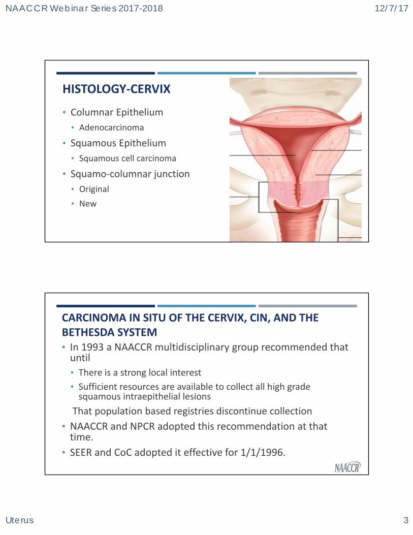
HISTOLOGY‐CERVIX
• Columnar Epithelium
• Adenocarcinoma
• Squamous Epithelium
• Squamous cell carcinoma
• Squamo‐columnar junction
• Original
• New
CARCINOMA IN SITU OF THE CERVIX, CIN, AND THE BETHESDA SYSTEM• In 1993 a NAACCR multidisciplinary group recommended that until
• There is a strong local interest
• Sufficient resources are available to collect all high grade squamous intraepithelial lesions
That population based registries discontinue collection
• NAACCR and NPCR adopted this recommendation at that time.
• SEER and CoC adopted it effective for 1/1/1996.
NAACCR Webinar Series 2017-2018 12/7/17
Uterus 4
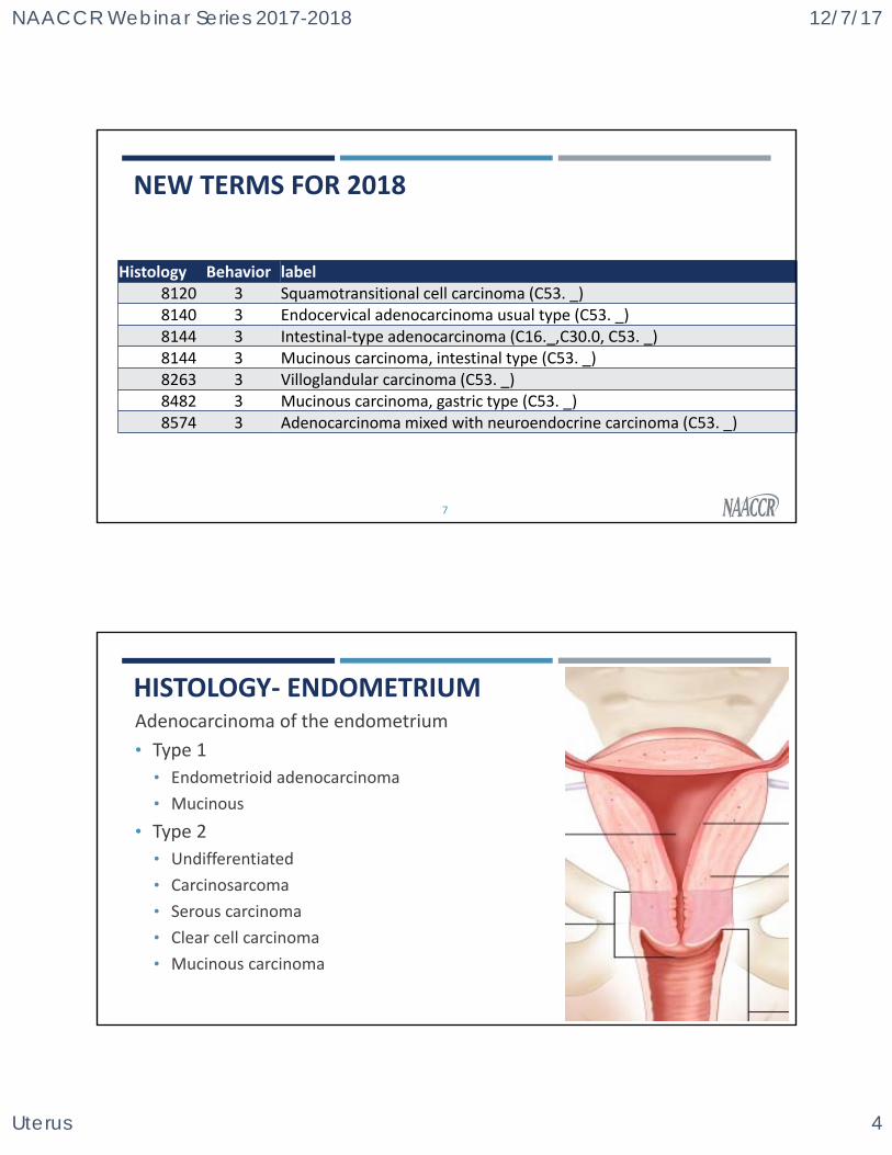
NEW TERMS FOR 2018
Histology Behavior label8120 3 Squamotransitional cell carcinoma (C53. _)8140 3 Endocervical adenocarcinoma usual type (C53. _)8144 3 Intestinal‐type adenocarcinoma (C16._,C30.0, C53. _) 8144 3 Mucinous carcinoma, intestinal type (C53. _)8263 3 Villoglandular carcinoma (C53. _) 8482 3 Mucinous carcinoma, gastric type (C53. _)8574 3 Adenocarcinoma mixed with neuroendocrine carcinoma (C53. _)
7
HISTOLOGY‐ ENDOMETRIUMAdenocarcinoma of the endometrium
• Type 1
• Endometrioid adenocarcinoma
• Mucinous
• Type 2
• Undifferentiated
• Carcinosarcoma
• Serous carcinoma
• Clear cell carcinoma
• Mucinous carcinoma
NAACCR Webinar Series 2017-2018 12/7/17
Uterus 5
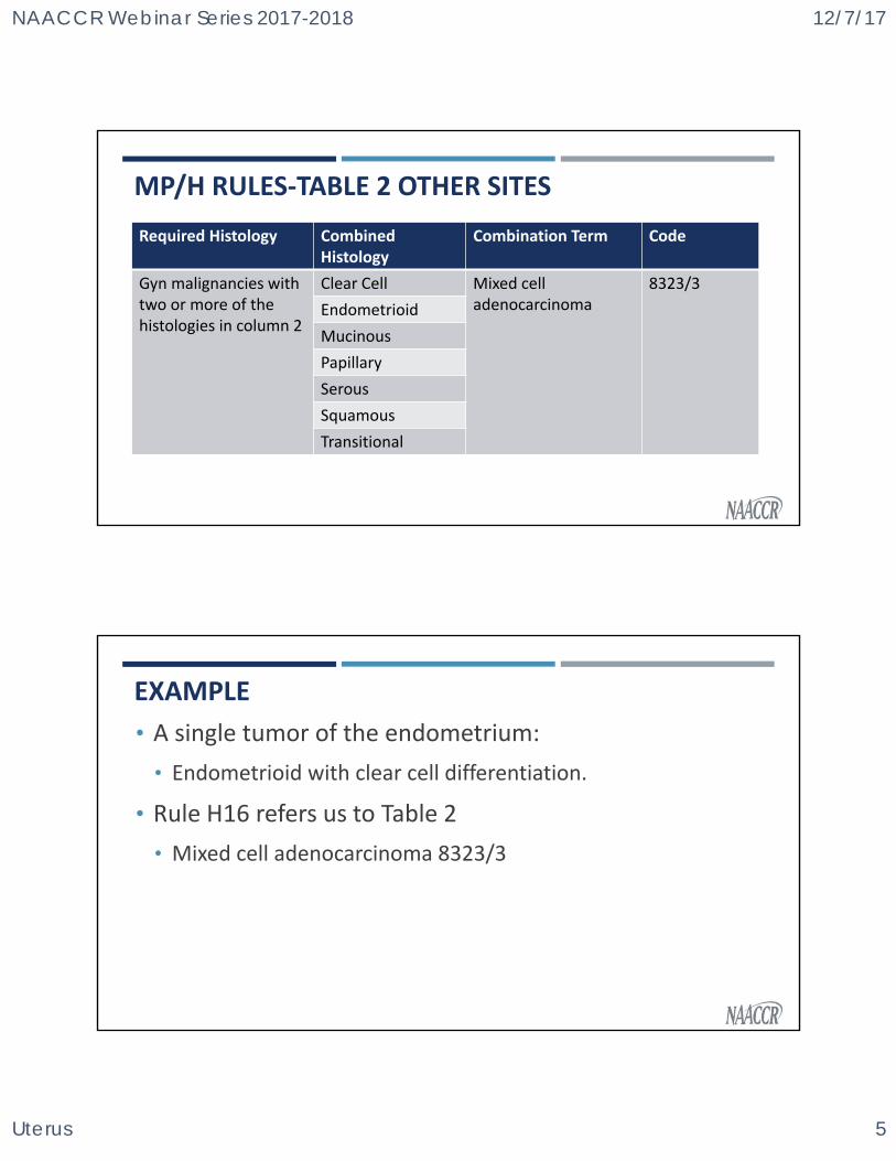
MP/H RULES‐TABLE 2 OTHER SITES
Required Histology Combined Histology
Combination Term Code
Gyn malignancies with two or more of the histologies in column 2
Clear Cell Mixed cell adenocarcinoma
8323/3
Endometrioid
Mucinous
Papillary
Serous
Squamous
Transitional
EXAMPLE
• A single tumor of the endometrium:
• Endometrioid with clear cell differentiation.
• Rule H16 refers us to Table 2
• Mixed cell adenocarcinoma 8323/3
NAACCR Webinar Series 2017-2018 12/7/17
Uterus 6
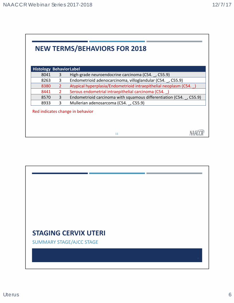
NEW TERMS/BEHAVIORS FOR 2018
11
Histology BehaviorLabel8041 3 High‐grade neuroendocrine carcinoma (C54. _, C55.9) 8263 3 Endometrioid adenocarcinoma, villoglandular (C54. _, C55.9) 8380 2 Atypical hyperplasia/Endometrioid intraepithelial neoplasm (C54. _) 8441 2 Serous endometrial intraepithelial carcinoma (C54. _)8570 3 Endometrioid carcinoma with squamous differentiation (C54. _, C55.9)8933 3 Mullerian adenosarcoma (C54. _, C55.9)
Red indicates change in behavior
STAGING CERVIX UTERISUMMARY STAGE/AJCC STAGE
12
NAACCR Webinar Series 2017-2018 12/7/17
Uterus 7
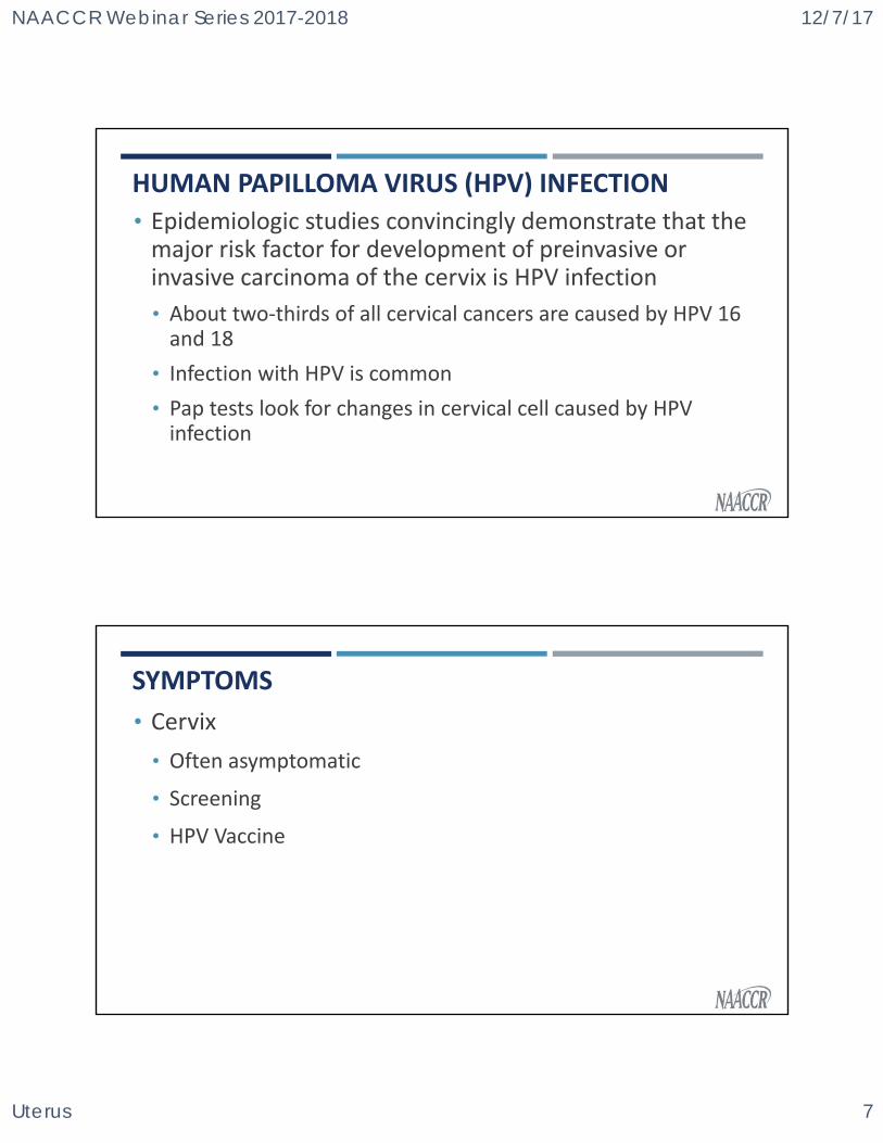
HUMAN PAPILLOMA VIRUS (HPV) INFECTION
• Epidemiologic studies convincingly demonstrate that the major risk factor for development of preinvasive or invasive carcinoma of the cervix is HPV infection
• About two‐thirds of all cervical cancers are caused by HPV 16 and 18
• Infection with HPV is common
• Pap tests look for changes in cervical cell caused by HPV infection
SYMPTOMS
• Cervix
• Often asymptomatic
• Screening
• HPV Vaccine
NAACCR Webinar Series 2017-2018 12/7/17
Uterus 8
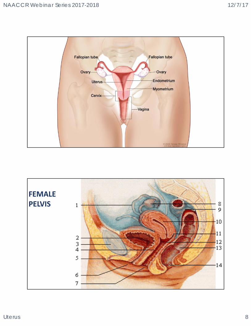
FEMALE PELVIS
NAACCR Webinar Series 2017-2018 12/7/17
Uterus 9
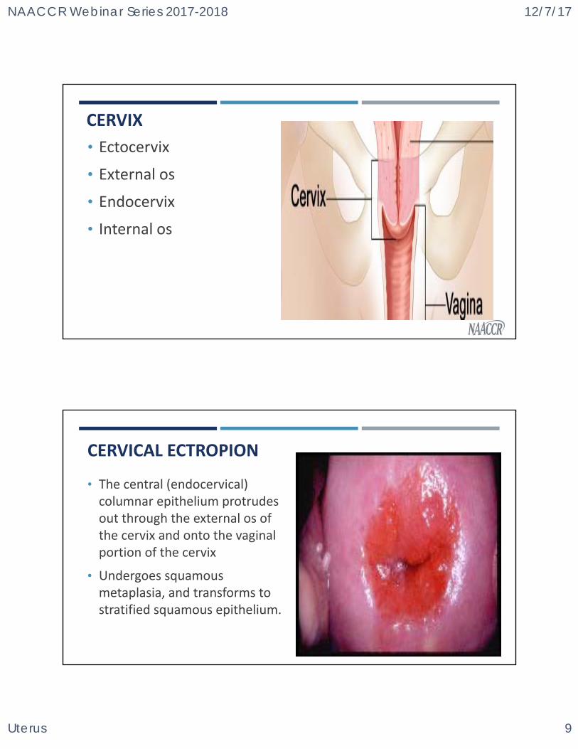
CERVIX
• Ectocervix
• External os
• Endocervix
• Internal os
CERVICAL ECTROPION
• The central (endocervical) columnar epithelium protrudes out through the external os of the cervix and onto the vaginal portion of the cervix
• Undergoes squamous metaplasia, and transforms to stratified squamous epithelium.
NAACCR Webinar Series 2017-2018 12/7/17
Uterus 10
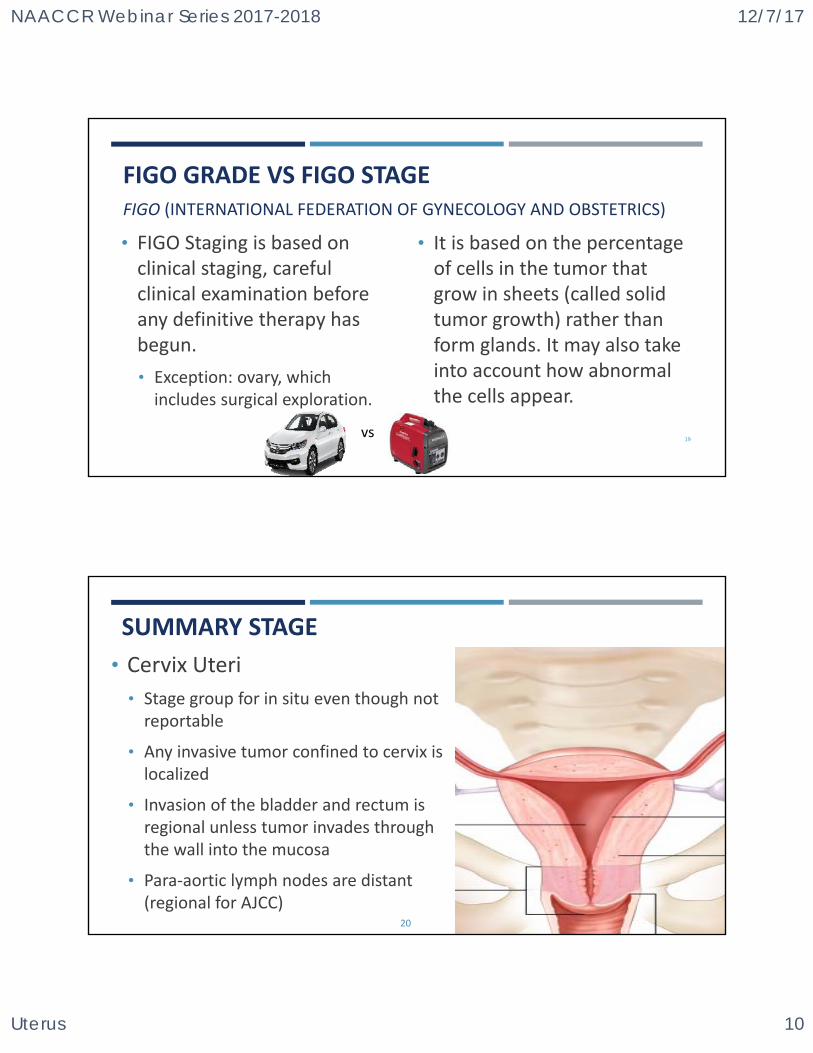
FIGO GRADE VS FIGO STAGEFIGO (INTERNATIONAL FEDERATION OF GYNECOLOGY AND OBSTETRICS)
• FIGO Staging is based on clinical staging, careful clinical examination before any definitive therapy has begun.
• Exception: ovary, which includes surgical exploration.
• It is based on the percentage of cells in the tumor that grow in sheets (called solid tumor growth) rather than form glands. It may also take into account how abnormal the cells appear.
19vs
SUMMARY STAGE
• Cervix Uteri
• Stage group for in situ even though not reportable
• Any invasive tumor confined to cervix is localized
• Invasion of the bladder and rectum is regional unless tumor invades through the wall into the mucosa
• Para‐aortic lymph nodes are distant (regional for AJCC)
20
NAACCR Webinar Series 2017-2018 12/7/17
Uterus 11
AJCC STAGE CERVIX UTERI
• 7th edition Chapter 35 page 397
• 8th edition Chapter 52 page 649
• AJCC ID‐52
• Errata‐Changes to Author List
21
FIGO STAGING OF CERVICAL CARCINOMAS
• Driven by the primary tumor (see T values)
• Stage I is confined to the cervix
• Stage II is carcinoma that extends beyond the cervix, but does not extend into the pelvic wall.
• Stage III is carcinoma that has extended into the pelvic sidewall
• Stage IV is carcinoma that has extended beyond the true pelvis or has clinically involved the mucosa of the bladder and/or rectum.
22http://screening.iarc.fr/viaviliappendix1.php
NAACCR Webinar Series 2017-2018 12/7/17
Uterus 12
RULES FOR CLASSIFICATION
• Clinical Staging
• FIGO uses clinical staging
• Determined prior to start of definitive therapy
• Clinical examination
• Palpation, inspection, colposcopy, endocervical curettage, hysteroscopy, cystoscopy, proctoscopy, intravenous urography, and x‐ray of lungs and skeleton
• Cone biopsy (usually)
• Lymph node status
• Radiologic‐guided fine needle aspiration, laparoscopic or peritoneal biopsy, or lymphadenectomy
23
RULES FOR CLASSIFICATION
• Clinical Staging
• CT, MRI, PET
• Ignore for staging
• May be used to make treatment plan
24
NAACCR Webinar Series 2017-2018 12/7/17
Uterus 13
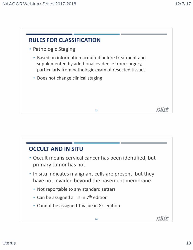
RULES FOR CLASSIFICATION
• Pathologic Staging
• Based on information acquired before treatment and supplemented by additional evidence from surgery, particularly from pathologic exam of resected tissues
• Does not change clinical staging
25
OCCULT AND IN SITU
• Occult means cervical cancer has been identified, but primary tumor has not.
• In situ indicates malignant cells are present, but they have not invaded beyond the basement membrane.
• Not reportable to any standard setters
• Can be assigned a Tis in 7th edition
• Cannot be assigned T value in 8th edition
26
NAACCR Webinar Series 2017-2018 12/7/17
Uterus 14
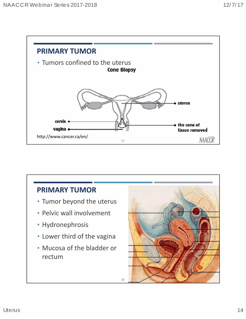
PRIMARY TUMOR
• Tumors confined to the uterus
27http://www.cancer.ca/en/
PRIMARY TUMOR
• Tumor beyond the uterus
• Pelvic wall involvement
• Hydronephrosis
• Lower third of the vagina
• Mucosa of the bladder or rectum
28
NAACCR Webinar Series 2017-2018 12/7/17
Uterus 15
29
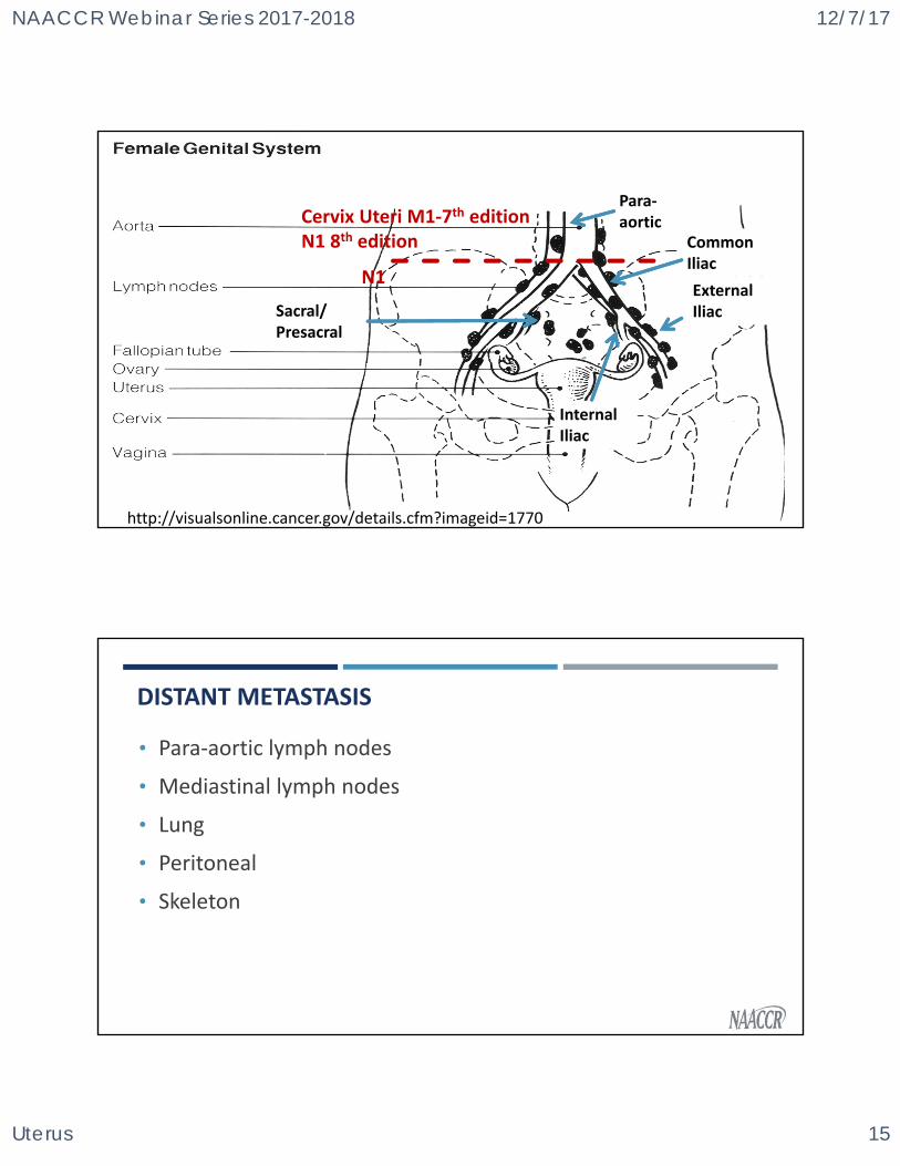
http://visualsonline.cancer.gov/details.cfm?imageid=1770
Common Iliac
ExternalIliac
InternalIliac
Para‐aortic
Sacral/ Presacral
N1
Cervix Uteri M1‐7th editionN1 8th edition
DISTANT METASTASIS
• Para‐aortic lymph nodes
• Mediastinal lymph nodes
• Lung
• Peritoneal
• Skeleton
NAACCR Webinar Series 2017-2018 12/7/17
Uterus 16
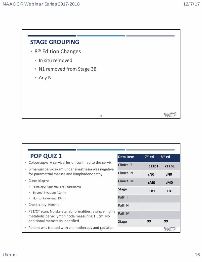
STAGE GROUPING
• 8th Edition Changes
• In situ removed
• N1 removed from Stage 3B
• Any N
31
POP QUIZ 1• Colposcopy: A cervical lesion confined to the cervix.
• Bimanual pelvic exam under anesthesia was negative for parametrial masses and lymphadenopathy.
• Cone biopsy:
• Histology: Squamous cell carcinoma
• Stromal invasion: 4.2mm
• Horizontal extent: 23mm
• Chest x‐ray: Normal
• PET/CT scan: No skeletal abnormalities; a single highly metabolic pelvic lymph node measuring 1.5cm. No additional metastasis identified.
• Patient was treated with chemotherapy and radiation. 32
Data Item 7th ed 8th ed
Clinical T
Clinical N
Clinical M
Stage
Path T
Path N
Path M
Stage
cT1b1
99
cT1b1
99
cN0
cM0
1B1
cN0
cM0
1B1
NAACCR Webinar Series 2017-2018 12/7/17
Uterus 17
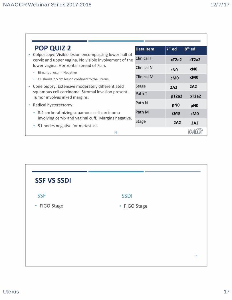
POP QUIZ 2• Colposcopy: Visible lesion encompassing lower half of
cervix and upper vagina. No visible involvement of the lower vagina. Horizontal spread of 7cm.
• Bimanual exam: Negative
• CT shows 7.5 cm lesion confined to the uterus.
• Cone biopsy: Extensive moderately differentiated squamous cell carcinoma. Stromal invasion present. Tumor involves inked margins.
• Radical hysterectomy:
• 8.4 cm keratinizing squamous cell carcinoma involving cervix and vaginal cuff. Margins negative.
• 51 nodes negative for metastasis
33
Data Item 7th ed 8th ed
Clinical T
Clinical N
Clinical M
Stage
Path T
Path N
Path M
Stage
cT2a2
pT2a2
cT2a2
pT2a2
cN0
cM0
2A2
cN0
cM0
2A2
pN0
cM0
2A2
pN0
cM0
2A2
SSF VS SSDI
SSF
• FIGO Stage
SSDI
• FIGO Stage
34
NAACCR Webinar Series 2017-2018 12/7/17
Uterus 18
QUESTIONS?
35
STAGING CORPUS UTERISUMMARY STAGE/AJCC STAGE
36
NAACCR Webinar Series 2017-2018 12/7/17
Uterus 19
ENDOMETRIAL CARCINOMA
• Risk factors
• Post menopausal estrogen therapy (unopposed)
• Obesity
• High‐fat diet
• Early menarche and late menopause
• Symptoms
• Abnormal vaginal bleeding (most often in postmenopausal period)
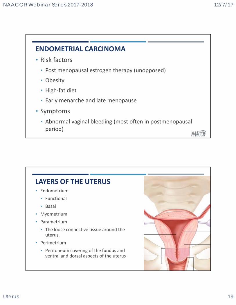
LAYERS OF THE UTERUS• Endometrium
• Functional
• Basal
• Myometrium
• Parametrium
• The loose connective tissue around the uterus.
• Perimetrium
• Peritoneum covering of the fundus and ventral and dorsal aspects of the uterus
NAACCR Webinar Series 2017-2018 12/7/17
Uterus 20
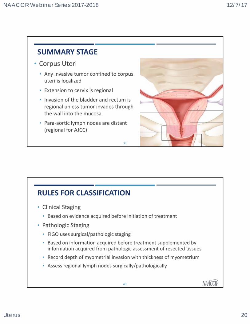
SUMMARY STAGE
• Corpus Uteri
• Any invasive tumor confined to corpus uteri is localized
• Extension to cervix is regional
• Invasion of the bladder and rectum is regional unless tumor invades through the wall into the mucosa
• Para‐aortic lymph nodes are distant (regional for AJCC)
39
RULES FOR CLASSIFICATION
• Clinical Staging
• Based on evidence acquired before initiation of treatment
• Pathologic Staging
• FIGO uses surgical/pathologic staging
• Based on information acquired before treatment supplemented by information acquired from pathologic assessment of resected tissues
• Record depth of myometrial invasion with thickness of myometrium
• Assess regional lymph nodes surgically/pathologically
40
NAACCR Webinar Series 2017-2018 12/7/17
Uterus 21
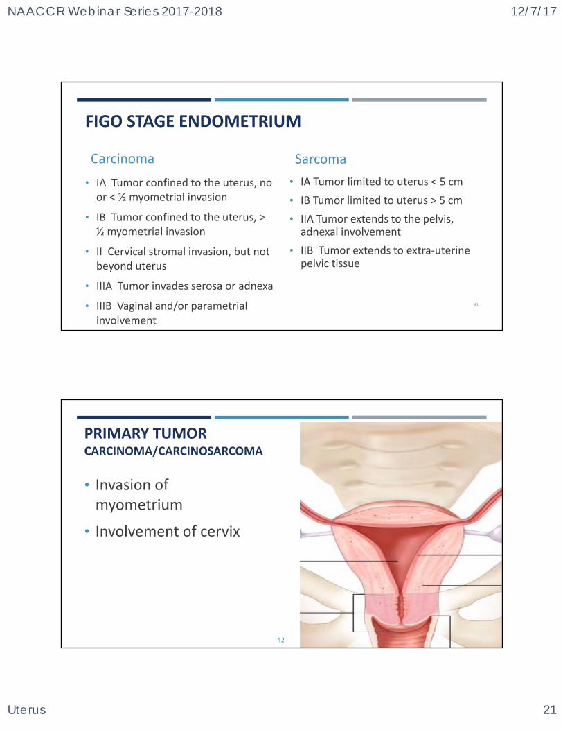
FIGO STAGE ENDOMETRIUM
Carcinoma
• IA Tumor confined to the uterus, no or < ½ myometrial invasion
• IB Tumor confined to the uterus, > ½ myometrial invasion
• II Cervical stromal invasion, but not beyond uterus
• IIIA Tumor invades serosa or adnexa
• IIIB Vaginal and/or parametrial involvement
Sarcoma
• IA Tumor limited to uterus < 5 cm
• IB Tumor limited to uterus > 5 cm
• IIA Tumor extends to the pelvis, adnexal involvement
• IIB Tumor extends to extra‐uterine pelvic tissue
41
PRIMARY TUMOR CARCINOMA/CARCINOSARCOMA
• Invasion of myometrium
• Involvement of cervix
42
NAACCR Webinar Series 2017-2018 12/7/17
Uterus 22
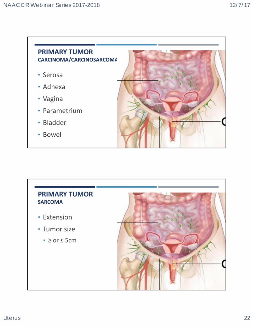
PRIMARY TUMOR CARCINOMA/CARCINOSARCOMA
• Serosa
• Adnexa
• Vagina
• Parametrium
• Bladder
• Bowel43
PRIMARY TUMOR SARCOMA
• Extension
• Tumor size
• ≥ or ≤ 5cm
44
NAACCR Webinar Series 2017-2018 12/7/17
Uterus 23
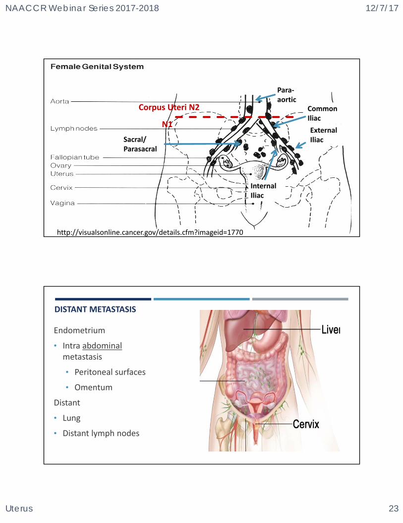
45
http://visualsonline.cancer.gov/details.cfm?imageid=1770
Common Iliac
ExternalIliac
InternalIliac
Para‐aortic
Sacral/ Parasacral
N1
Corpus Uteri N2
DISTANT METASTASIS
Endometrium
• Intra abdominal metastasis
• Peritoneal surfaces
• Omentum
Distant
• Lung
• Distant lymph nodes
NAACCR Webinar Series 2017-2018 12/7/17
Uterus 24
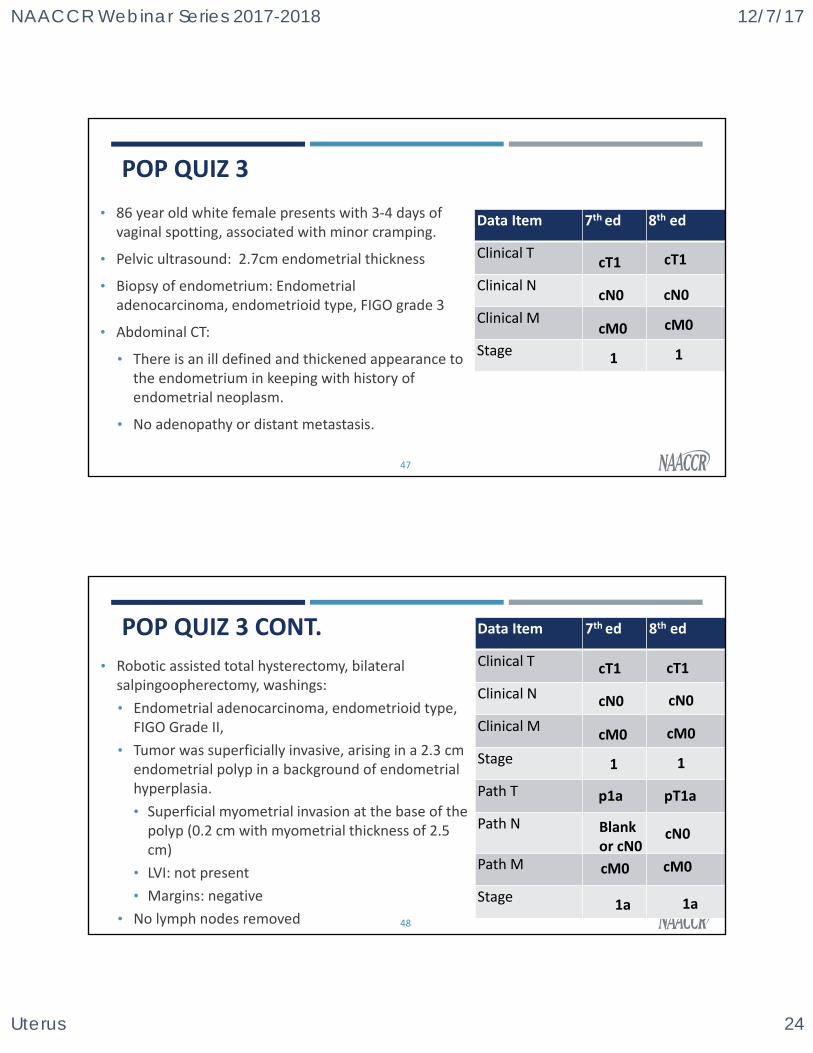
POP QUIZ 3
• 86 year old white female presents with 3‐4 days of vaginal spotting, associated with minor cramping.
• Pelvic ultrasound: 2.7cm endometrial thickness
• Biopsy of endometrium: Endometrial adenocarcinoma, endometrioid type, FIGO grade 3
• Abdominal CT:
• There is an ill defined and thickened appearance to the endometrium in keeping with history of endometrial neoplasm.
• No adenopathy or distant metastasis.
47
Data Item 7th ed 8th ed
Clinical T
Clinical N
Clinical M
Stage
cT1 cT1
cN0
cM0
1
cN0
cM0
1
POP QUIZ 3 CONT.
• Robotic assisted total hysterectomy, bilateral salpingoopherectomy, washings:
• Endometrial adenocarcinoma, endometrioid type, FIGO Grade II,
• Tumor was superficially invasive, arising in a 2.3 cm endometrial polyp in a background of endometrial hyperplasia.
• Superficial myometrial invasion at the base of the polyp (0.2 cm with myometrial thickness of 2.5 cm)
• LVI: not present
• Margins: negative
• No lymph nodes removed 48
Data Item 7th ed 8th ed
Clinical T
Clinical N
Clinical M
Stage
Path T
Path N
Path M
Stage
p1a pT1a
cT1 cT1
cN0
cM0
1
cN0
cM0
1
Blank or cN0
cN0
cM0cM0
1a 1a
NAACCR Webinar Series 2017-2018 12/7/17
Uterus 25
SSF VS SSDI
SSF
• FIGO Stage
• Peritoneal Cytology
• Number of Positive Pelvic Nodes
• Number of Examined Pelvic Nodes
• Number of Positive Para‐Aortic Nodes
• Number of Examined Para‐Aortic Nodes
SSDI
49
• FIGO Stage
• Peritoneal Cytology
• Number of Positive Pelvic Nodes
• Number of Examined Pelvic Nodes
• Number of Positive Para‐Aortic Nodes
• Number of Examined Para‐Aortic Nodes
QUESTIONS?
50
NAACCR Webinar Series 2017-2018 12/7/17
Uterus 26
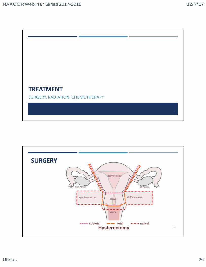
TREATMENTSURGERY, RADIATION, CHEMOTHERAPY
51
52
SURGERY
NAACCR Webinar Series 2017-2018 12/7/17
Uterus 27
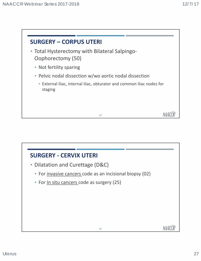
SURGERY – CORPUS UTERI
• Total Hysterectomy with Bilateral Salpingo‐Oophorectomy (50)
• Not fertility sparing
• Pelvic nodal dissection w/wo aortic nodal dissection
• External iliac, internal iliac, obturator and common iliac nodes for staging
53
SURGERY ‐ CERVIX UTERI
• Dilatation and Curettage (D&C)
• For invasive cancers code as an incisional biopsy (02)
• For In situ cancers code as surgery (25)
54

NAACCR Webinar Series 2017-2018 12/7/17
Uterus 28
SURGERY – CERVIX UTERI
• LEEP (Loop Electrocautery Excision Procedure)
• Local tumor destruction (15)
• No specimen sent to pathology
• Local tumor excision (28)
• Specimen sent to pathology
• Cone biopsy (27)
• With gross excision of lesion (24)
55
SURGERY – CERVIX UTERI
• Radical vaginal trachelectomy with laparoscopic lymphadenectomy procedure with or without SLN mapping
• Fertility sparing option
• Stage IA‐2
• Stage IB‐1
• Lesions of 2cm diameter or less
56
NAACCR Webinar Series 2017-2018 12/7/17
Uterus 29
SURGERY – CERVIX UTERI
• Radical hysterectomy with bilateral pelvic lymph node dissection with or without SLN mapping
• FIGO Stage IA‐2, IB and IIA lesion
• Fertility preservation is not desired
57
CHEMORADIATION ‐ CERVIX UTERI
• Advanced‐stage disease
• FIGO stage IIB and above
• Preferred regimens
• Cisplatin
• Cisplatin/fluorouracil
58
NAACCR Webinar Series 2017-2018 12/7/17
Uterus 30
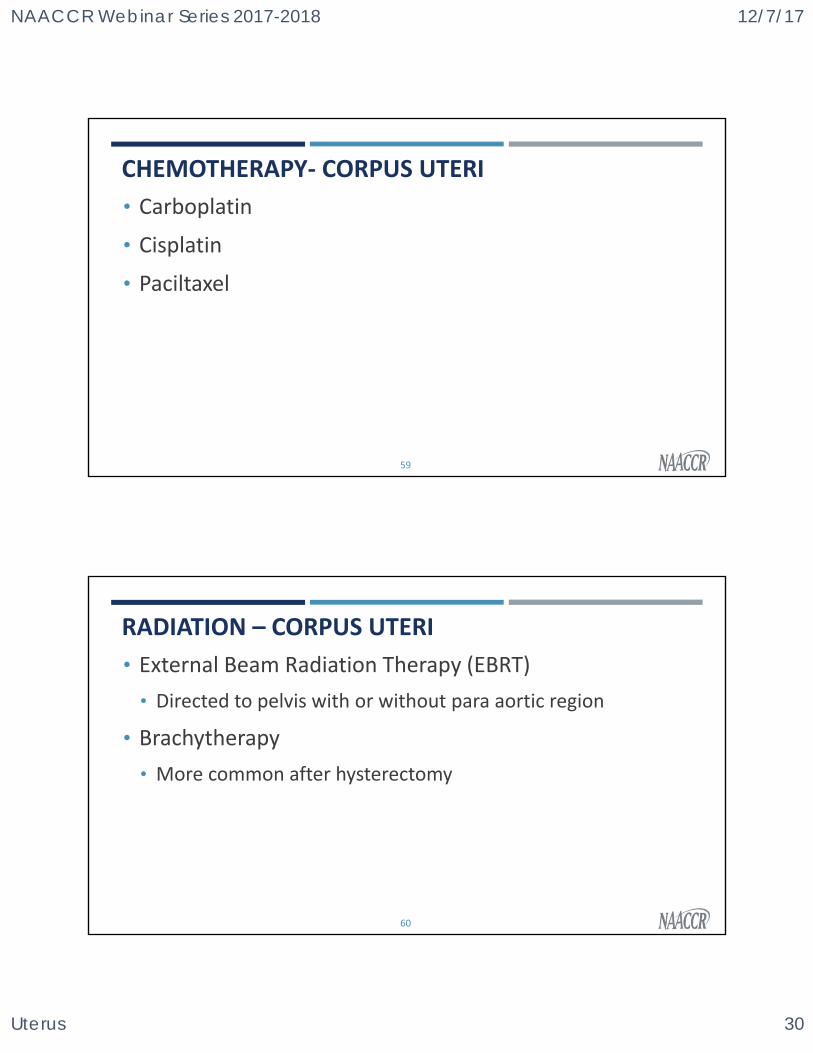
CHEMOTHERAPY‐ CORPUS UTERI
• Carboplatin
• Cisplatin
• Paciltaxel
59
RADIATION – CORPUS UTERI
• External Beam Radiation Therapy (EBRT)
• Directed to pelvis with or without para aortic region
• Brachytherapy
• More common after hysterectomy
60
NAACCR Webinar Series 2017-2018 12/7/17
Uterus 31
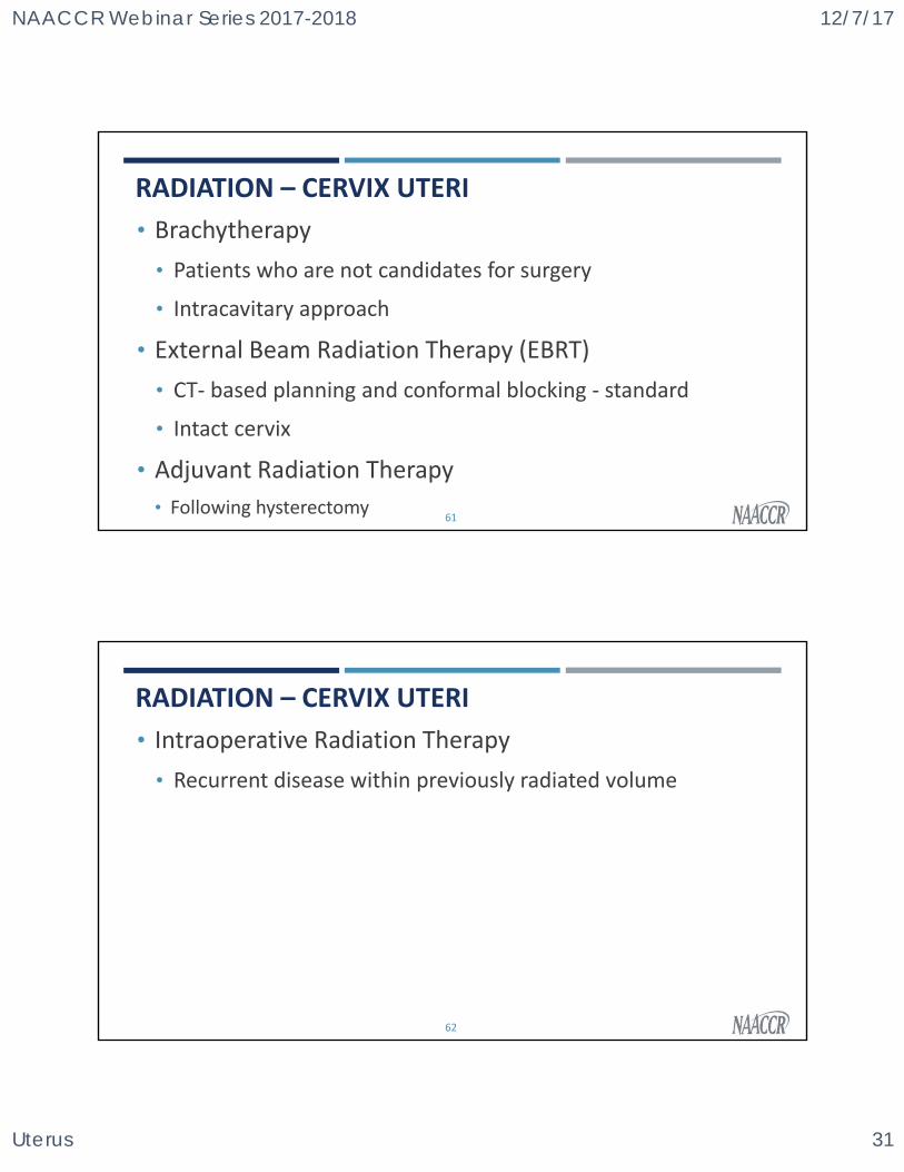
RADIATION – CERVIX UTERI
• Brachytherapy
• Patients who are not candidates for surgery
• Intracavitary approach
• External Beam Radiation Therapy (EBRT)
• CT‐ based planning and conformal blocking ‐ standard
• Intact cervix
• Adjuvant Radiation Therapy
• Following hysterectomy61
RADIATION – CERVIX UTERI
• Intraoperative Radiation Therapy
• Recurrent disease within previously radiated volume
62
NAACCR Webinar Series 2017-2018 12/7/17
Uterus 32
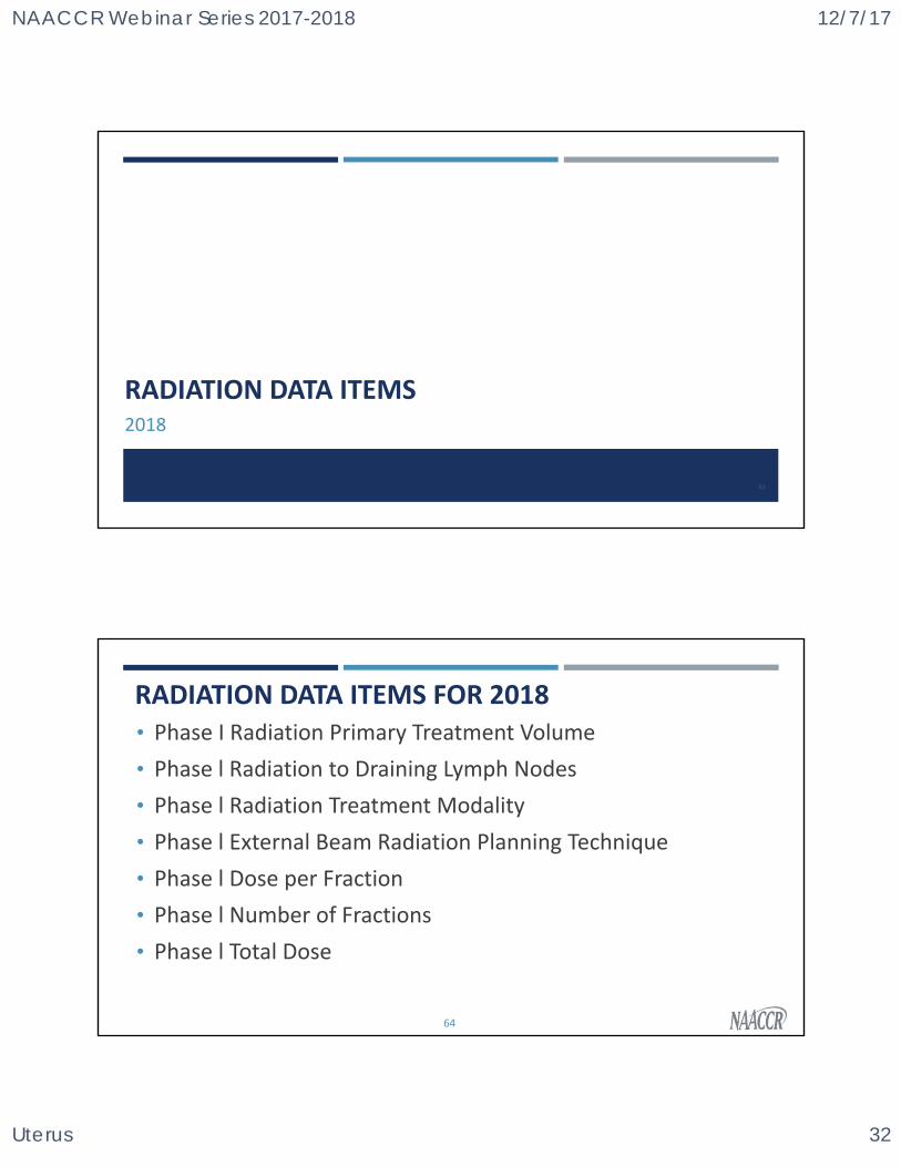
RADIATION DATA ITEMS2018
63
RADIATION DATA ITEMS FOR 2018• Phase I Radiation Primary Treatment Volume
• Phase l Radiation to Draining Lymph Nodes
• Phase l Radiation Treatment Modality
• Phase l External Beam Radiation Planning Technique
• Phase l Dose per Fraction
• Phase l Number of Fractions
• Phase l Total Dose
64
NAACCR Webinar Series 2017-2018 12/7/17
Uterus 33
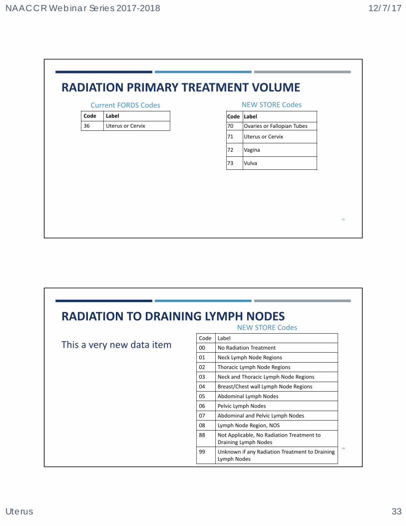
RADIATION PRIMARY TREATMENT VOLUMENEW STORE Codes
Code Label
36 Uterus or Cervix
Current FORDS Codes
65
Code Label
70 Ovaries or Fallopian Tubes
71 Uterus or Cervix
72 Vagina
73 Vulva
RADIATION TO DRAINING LYMPH NODESNEW STORE Codes
66
Code Label
00 No Radiation Treatment
01 Neck Lymph Node Regions
02 Thoracic Lymph Node Regions
03 Neck and Thoracic Lymph Node Regions
04 Breast/Chest wall Lymph Node Regions
05 Abdominal Lymph Nodes
06 Pelvic Lymph Nodes
07 Abdominal and Pelvic Lymph Nodes
08 Lymph Node Region, NOS
88 Not Applicable, No Radiation Treatment to Draining Lymph Nodes
99 Unknown if any Radiation Treatment to Draining Lymph Nodes
This a very new data item
NAACCR Webinar Series 2017-2018 12/7/17
Uterus 34
67
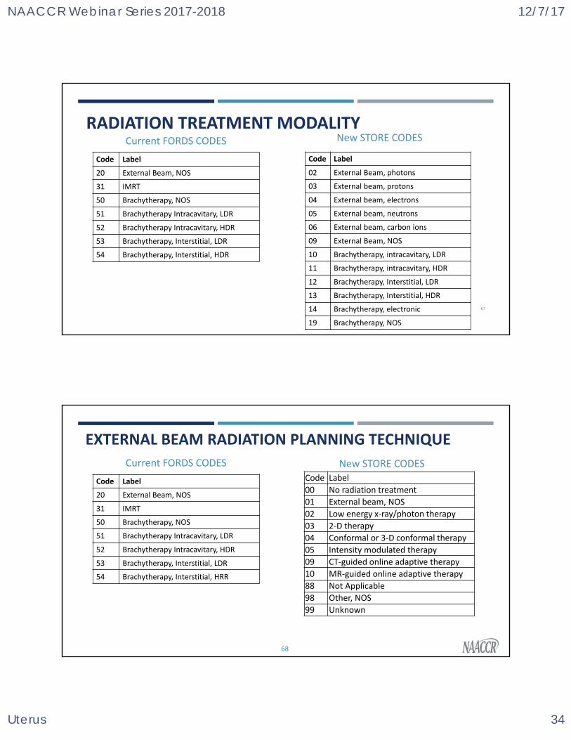
RADIATION TREATMENT MODALITY
Code Label
02 External Beam, photons
03 External beam, protons
04 External beam, electrons
05 External beam, neutrons
06 External beam, carbon ions
09 External Beam, NOS
10 Brachytherapy, intracavitary, LDR
11 Brachytherapy, intracavitary, HDR
12 Brachytherapy, Interstitial, LDR
13 Brachytherapy, Interstitial, HDR
14 Brachytherapy, electronic
19 Brachytherapy, NOS
New STORE CODES
Code Label
20 External Beam, NOS
31 IMRT
50 Brachytherapy, NOS
51 Brachytherapy Intracavitary, LDR
52 Brachytherapy Intracavitary, HDR
53 Brachytherapy, Interstitial, LDR
54 Brachytherapy, Interstitial, HDR
Current FORDS CODES
EXTERNAL BEAM RADIATION PLANNING TECHNIQUE
68
New STORE CODES
Code Label
20 External Beam, NOS
31 IMRT
50 Brachytherapy, NOS
51 Brachytherapy Intracavitary, LDR
52 Brachytherapy Intracavitary, HDR
53 Brachytherapy, Interstitial, LDR
54 Brachytherapy, Interstitial, HRR
Current FORDS CODES
Code Label
00 No radiation treatment
01 External beam, NOS
02 Low energy x‐ray/photon therapy
03 2‐D therapy
04 Conformal or 3‐D conformal therapy
05 Intensity modulated therapy
09 CT‐guided online adaptive therapy
10 MR‐guided online adaptive therapy
88 Not Applicable
98 Other, NOS
99 Unknown
NAACCR Webinar Series 2017-2018 12/7/17
Uterus 35
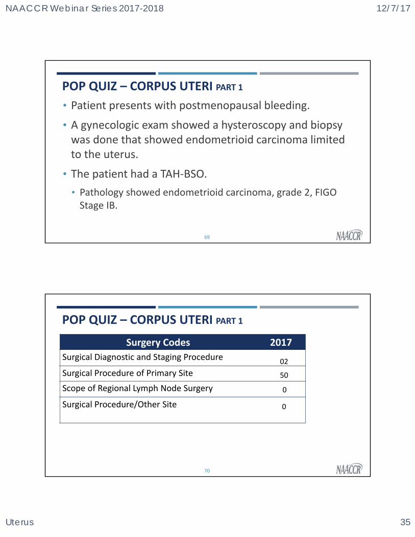
POP QUIZ – CORPUS UTERI PART 1
• Patient presents with postmenopausal bleeding.
• A gynecologic exam showed a hysteroscopy and biopsy was done that showed endometrioid carcinoma limited to the uterus.
• The patient had a TAH‐BSO.
• Pathology showed endometrioid carcinoma, grade 2, FIGO Stage IB.
69
Surgery Codes 2017
Surgical Diagnostic and Staging Procedure
Surgical Procedure of Primary Site
Scope of Regional Lymph Node Surgery
Surgical Procedure/Other Site
POP QUIZ – CORPUS UTERI PART 1
70
02
50
0
0
NAACCR Webinar Series 2017-2018 12/7/17
Uterus 36
POP QUIZ –CORPUS UTERI PART 2
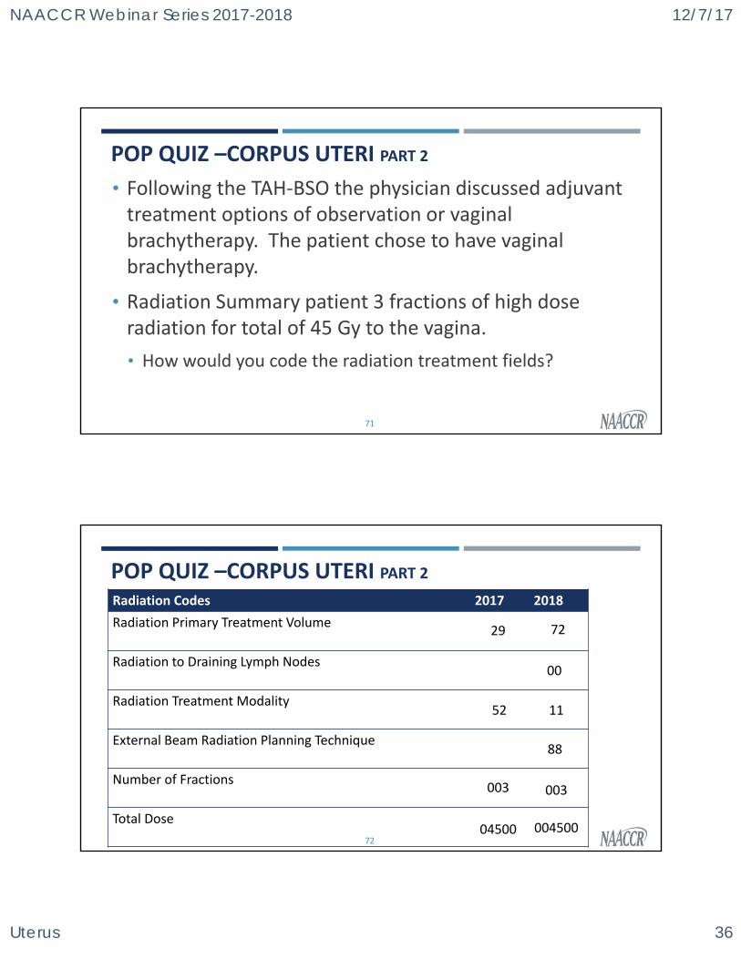
• Following the TAH‐BSO the physician discussed adjuvant treatment options of observation or vaginal brachytherapy. The patient chose to have vaginal brachytherapy.
• Radiation Summary patient 3 fractions of high dose radiation for total of 45 Gy to the vagina.
• How would you code the radiation treatment fields?
71
POP QUIZ –CORPUS UTERI PART 2
72
Radiation Codes 2017 2018
Radiation Primary Treatment Volume
Radiation to Draining Lymph Nodes
Radiation Treatment Modality
External Beam Radiation Planning Technique
Number of Fractions
Total Dose
29
52
003
04500
72
00
11
88
003
004500
NAACCR Webinar Series 2017-2018 12/7/17
Uterus 37
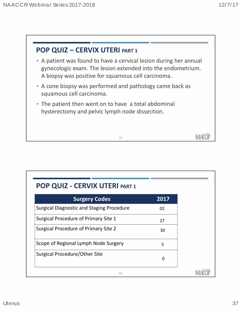
POP QUIZ – CERVIX UTERI PART 1
• A patient was found to have a cervical lesion during her annual gynecologic exam. The lesion extended into the endometrium. A biopsy was positive for squamous cell carcinoma.
• A cone biopsy was performed and pathology came back as squamous cell carcinoma.
• The patient then went on to have a total abdominal hysterectomy and pelvic lymph node dissection.
73
POP QUIZ ‐ CERVIX UTERI PART 1
Surgery Codes 2017
Surgical Diagnostic and Staging Procedure
Surgical Procedure of Primary Site 1
Surgical Procedure of Primary Site 2
Scope of Regional Lymph Node Surgery
Surgical Procedure/Other Site
74
02
30
5
27
0
NAACCR Webinar Series 2017-2018 12/7/17
Uterus 38
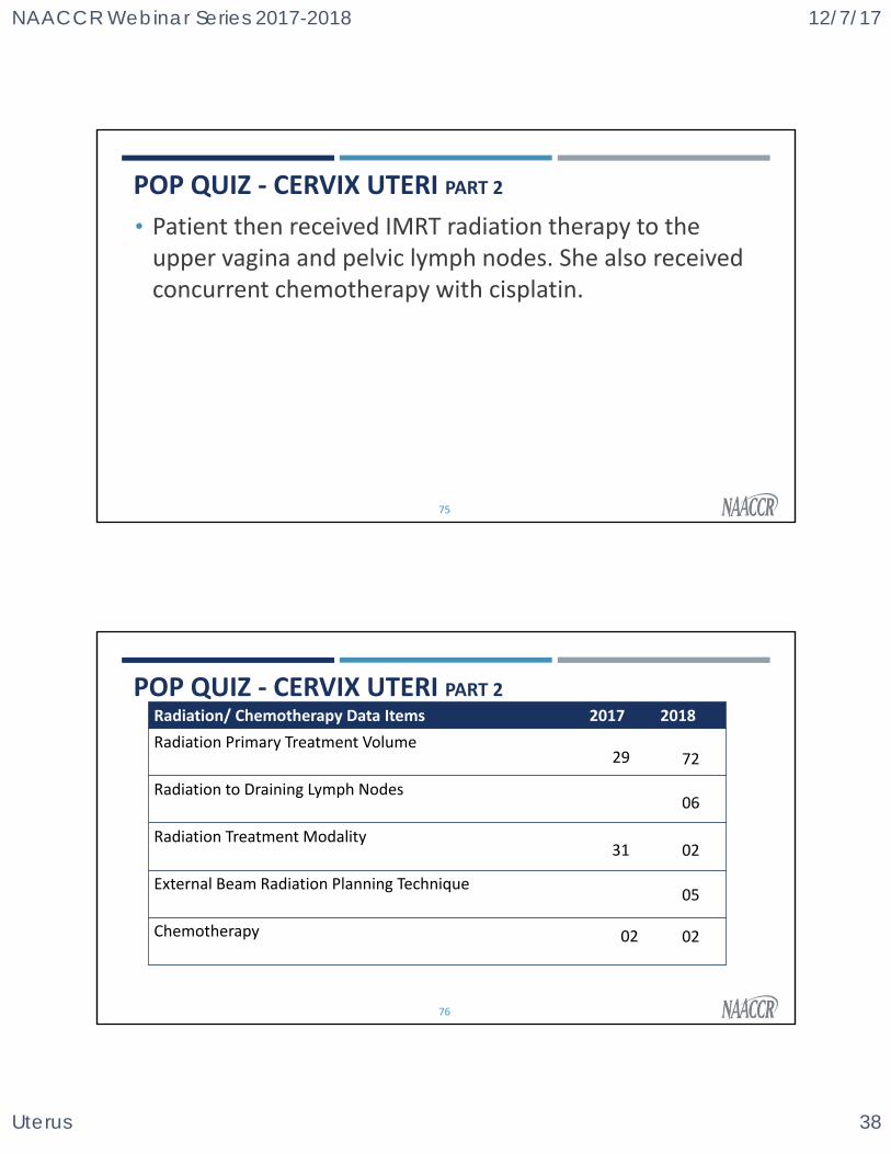
POP QUIZ ‐ CERVIX UTERI PART 2
• Patient then received IMRT radiation therapy to the upper vagina and pelvic lymph nodes. She also received concurrent chemotherapy with cisplatin.
75
POP QUIZ ‐ CERVIX UTERI PART 2
76
Radiation/ Chemotherapy Data Items 2017 2018
Radiation Primary Treatment Volume
Radiation to Draining Lymph Nodes
Radiation Treatment Modality
External Beam Radiation Planning Technique
Chemotherapy 02 02
72
06
02
05
29
31

NAACCR Webinar Series 2017-2018 12/7/17
Uterus 39
QUESTIONS?QUIZ 2
CASE SCENARIOS
77
COMING UP….
• Collecting Cancer Data: GIST and Soft Tissue Sarcomas
• 01/11/2018
• Collecting Cancer Data: Stomach and Esophagus
• 02/01/2018
78

NAACCR Webinar Series 2017-2018 12/7/17
Uterus 40
Fabulous Prizes Winners
79
Fabulous Prizes
CE CERTIFICATE QUIZ/SURVEY
• Phrase
• FIGO
• Link
http://www.surveygizmo.com/s3/4041165/Uterus‐2017
80

NAACCR Webinar Series 2017-2018 12/7/17
Uterus 41
JIM HOFFERKAMP [email protected]
ANGELA MARTIN [email protected]
81
Recommended