Clinical Anatomy of the knee
Mr CM Gupte Mr Alvin Chen
Overview • Knee joint function • Surface anatomy • Bones • Ligaments • Tendons • Examination • Disease processes
The Knee Joint
• Poorly constructed in terms of stability - femur round, tibia flat.
• Comprised of four bones. • Femur • Tibia • Fibula • Patella
The knee joint
• Load bearing / Force transmission
• Locomotion
• Proprioception
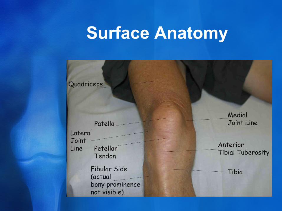
Surface Anatomy
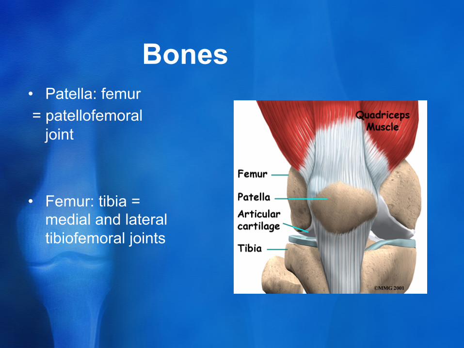
Bones • Patella: femur = patellofemoral
joint
• Femur: tibia = medial and lateral tibiofemoral joints
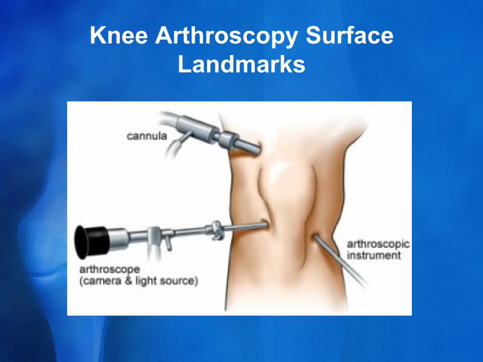
Knee Arthroscopy Surface Landmarks
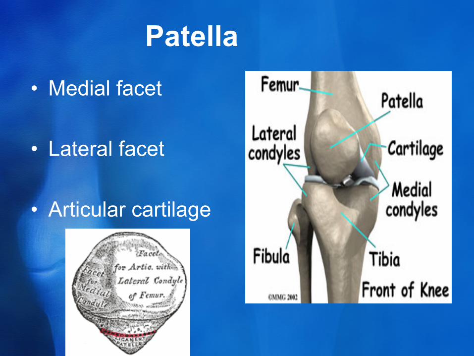
Patella • Medial facet
• Lateral facet
• Articular cartilage
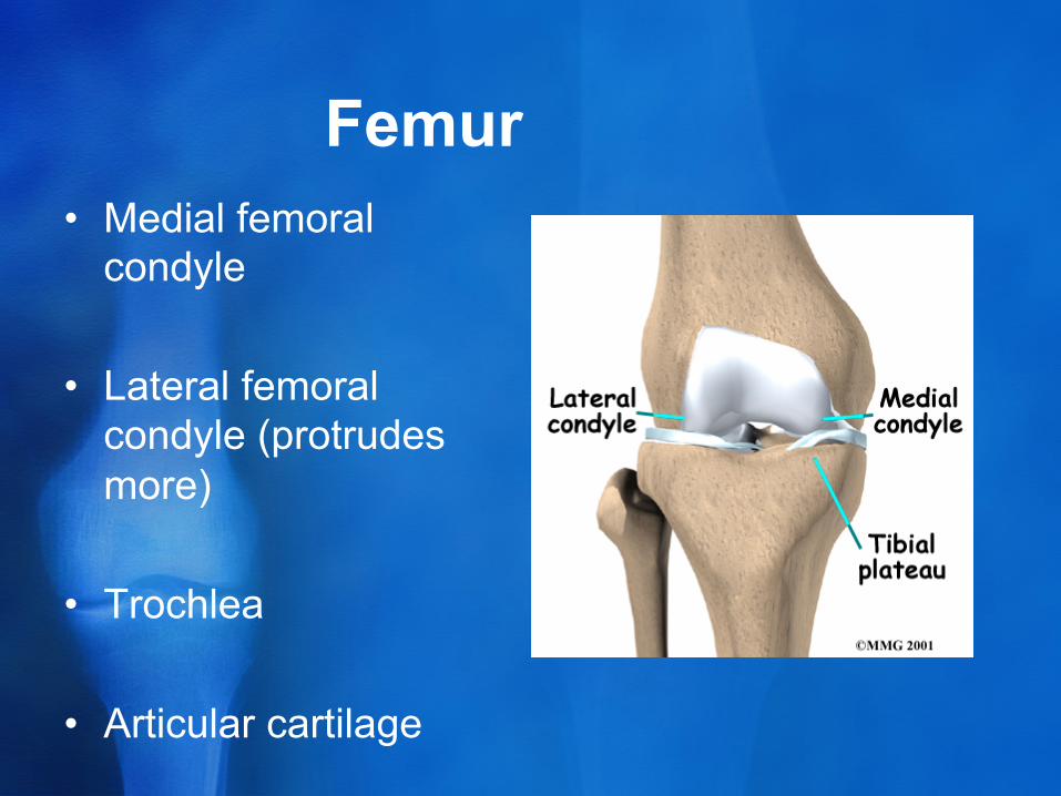
Femur • Medial femoral
condyle
• Lateral femoral condyle (protrudes more)
• Trochlea
• Articular cartilage
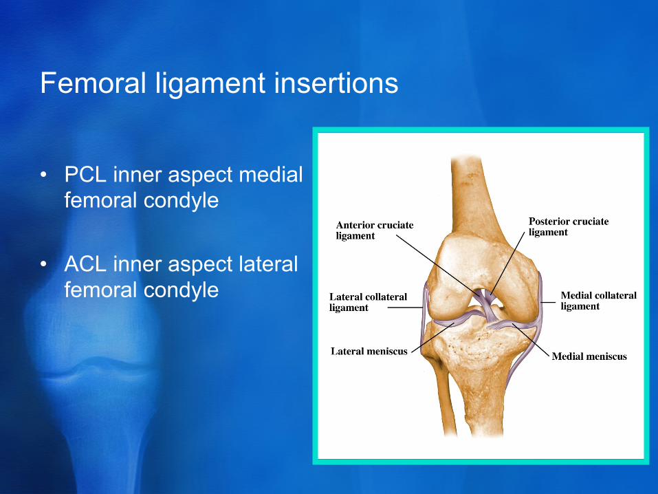
Femoral ligament insertions
• PCL inner aspect medial femoral condyle
• ACL inner aspect lateral femoral condyle
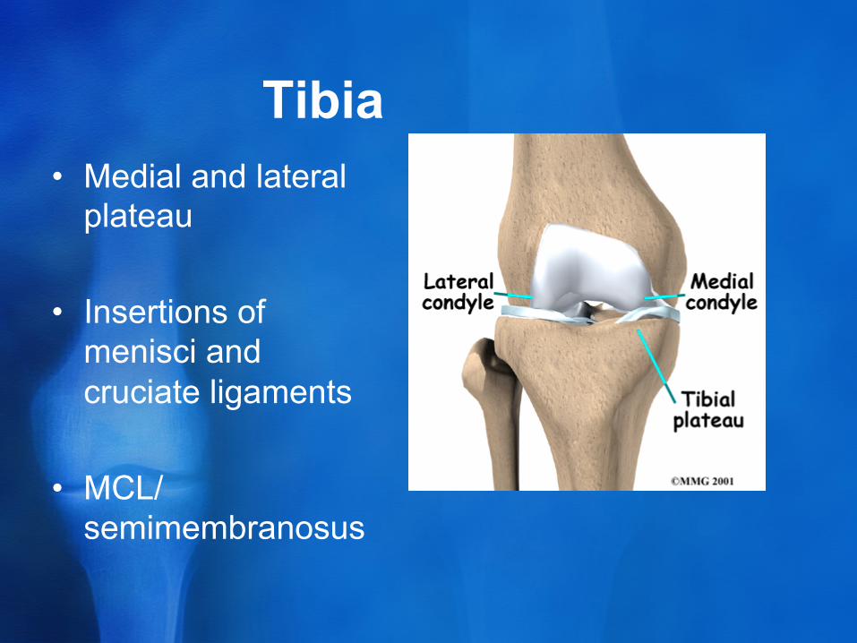
Tibia • Medial and lateral
plateau
• Insertions of menisci and cruciate ligaments
• MCL/ semimembranosus
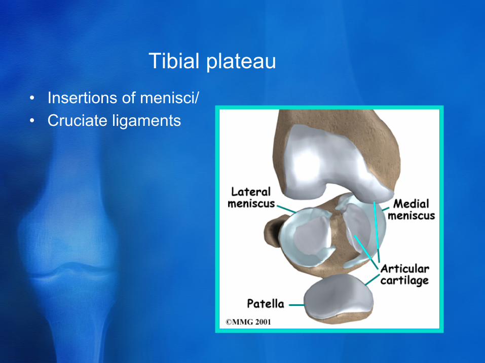
Tibial plateau • Insertions of menisci/ • Cruciate ligaments
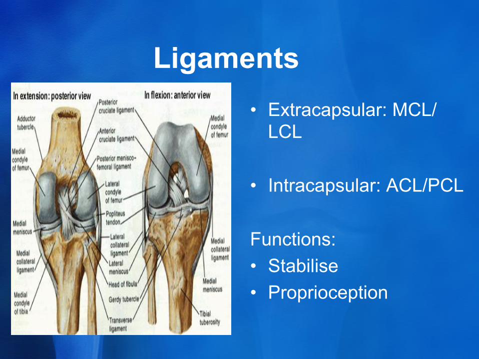
Ligaments • Extracapsular: MCL/
LCL
• Intracapsular: ACL/PCL
Functions: • Stabilise • Proprioception
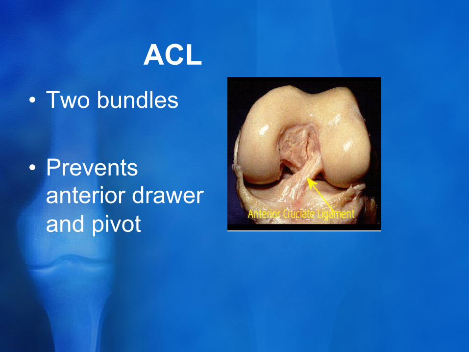
ACL • Two bundles
• Prevents anterior drawer and pivot
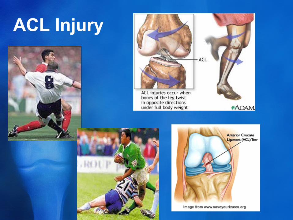
ACL Injury
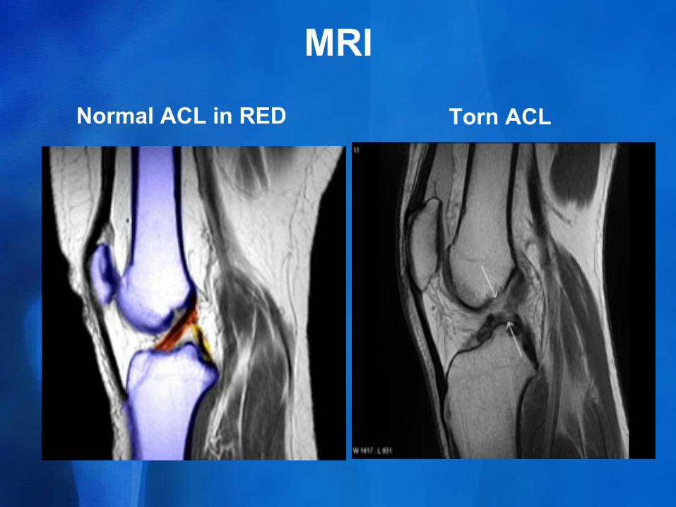
MRI
Normal ACL in RED Torn ACL
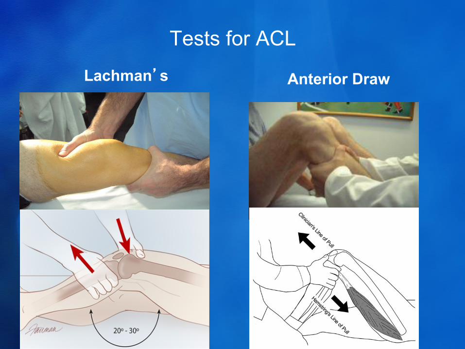
Tests for ACL
Lachman’s Anterior Draw
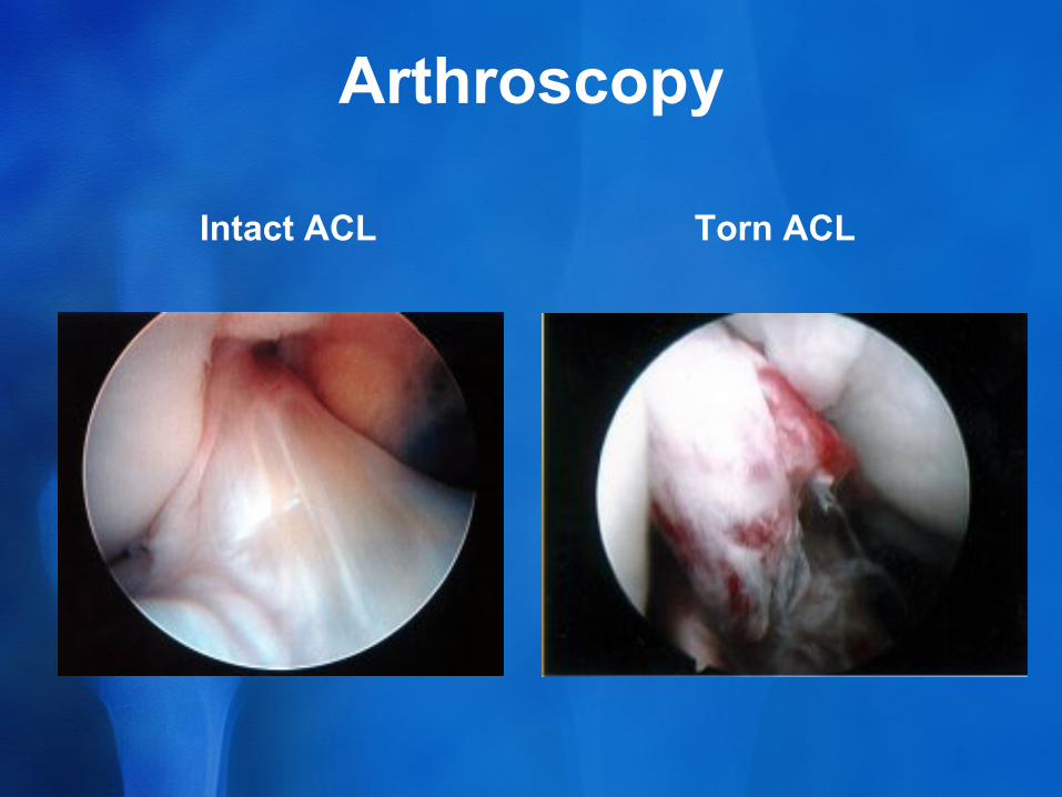
Arthroscopy
Intact ACL Torn ACL
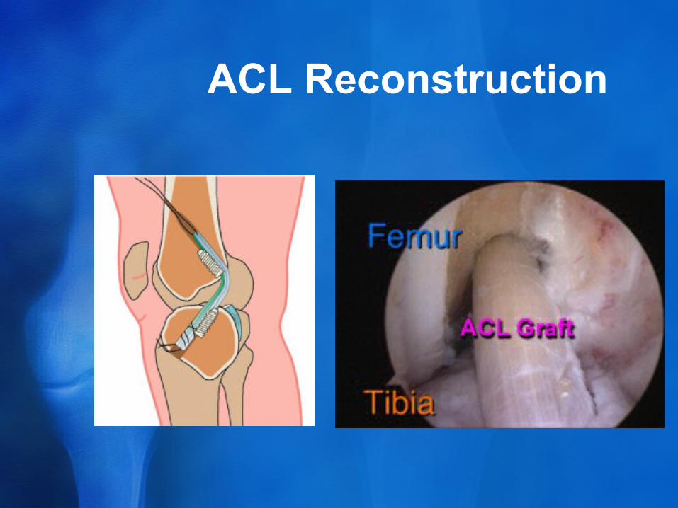
ACL Reconstruction
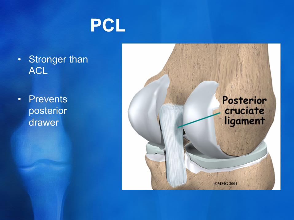
PCL • Stronger than
ACL
• Prevents posterior drawer
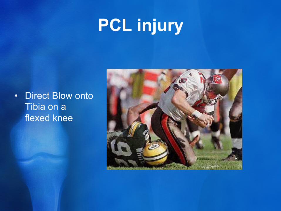
PCL injury
• Direct Blow onto Tibia on a flexed knee
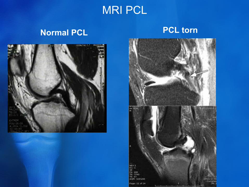
MRI PCL
Normal PCL PCL torn
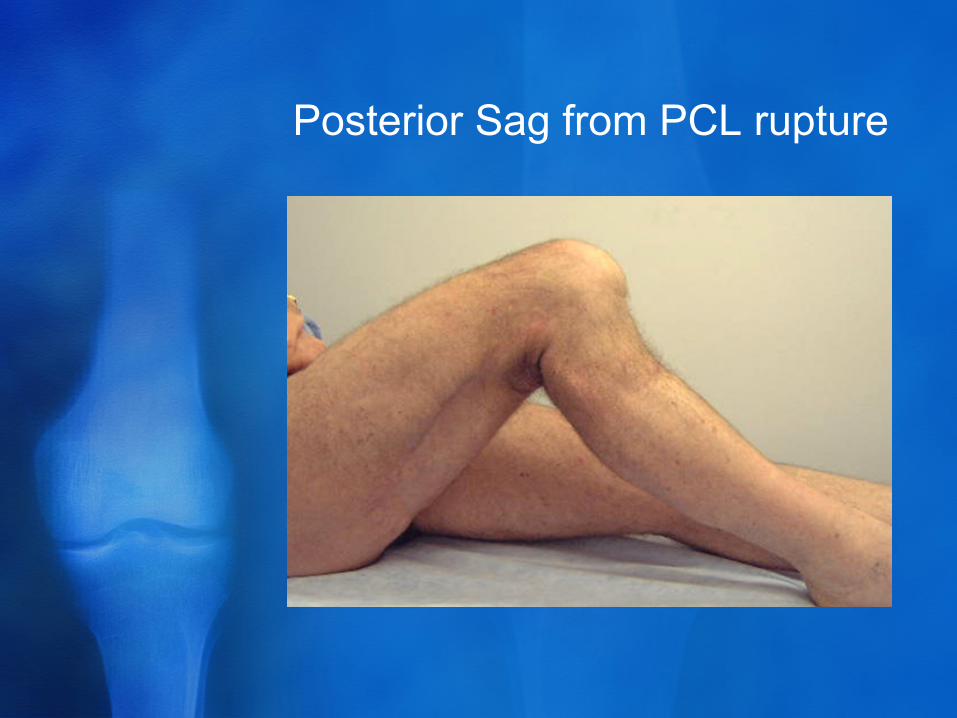
Posterior Sag from PCL rupture
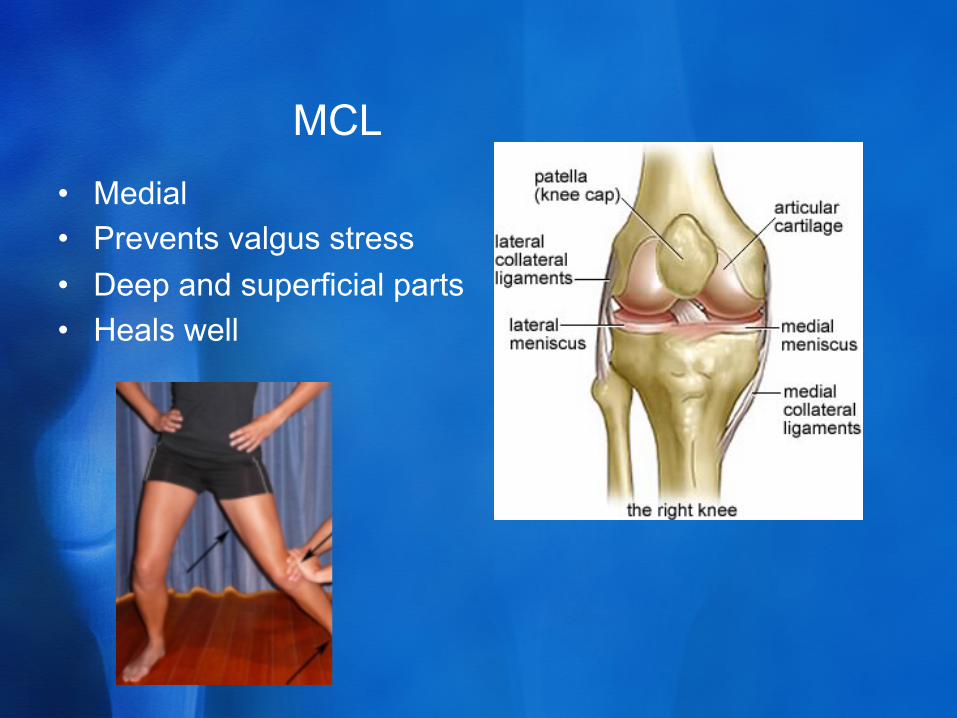
MCL • Medial • Prevents valgus stress • Deep and superficial parts • Heals well
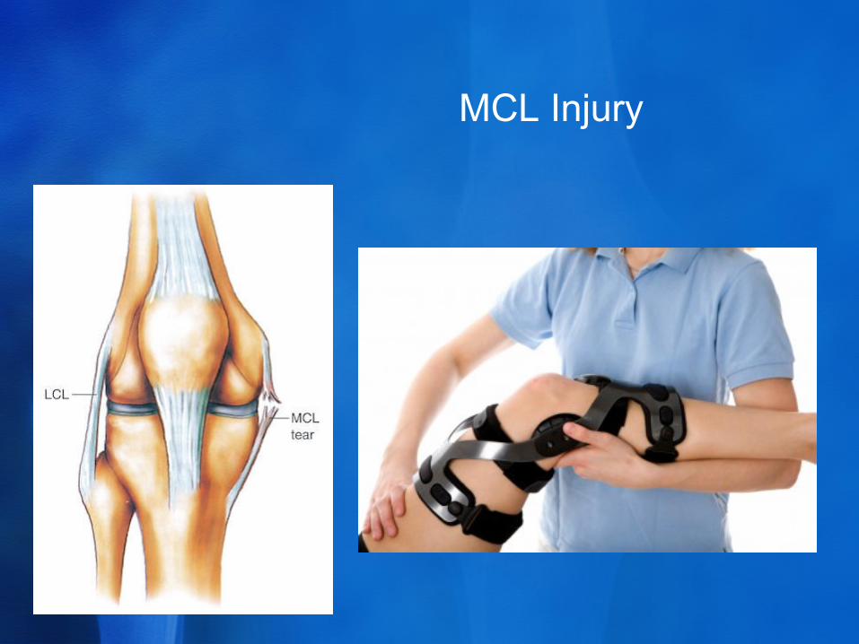
MCL Injury
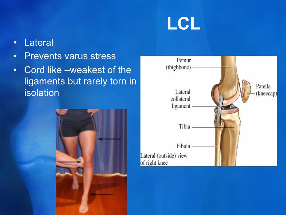
LCL • Lateral • Prevents varus stress • Cord like –weakest of the
ligaments but rarely torn in isolation
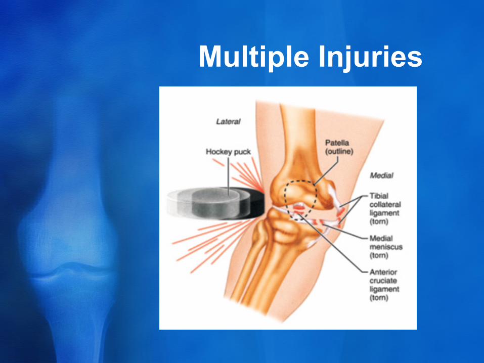
Multiple Injuries
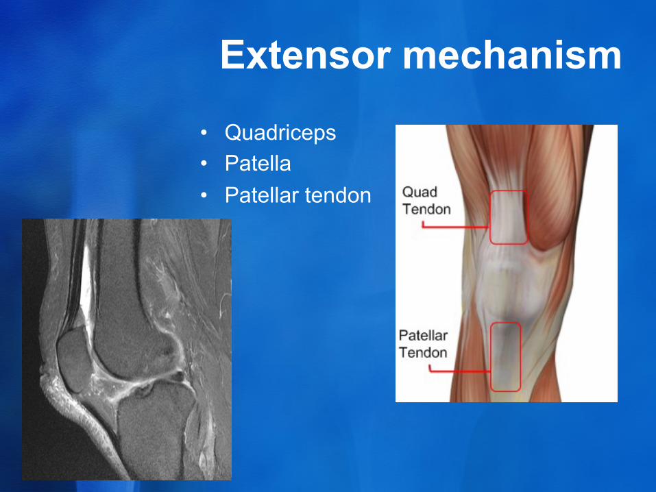
Extensor mechanism
• Quadriceps • Patella • Patellar tendon

Quadriceps
• VMO • Rectus femoris • Vastus intermedius • Vastus lateralis • Extend knee
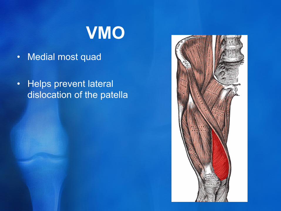
VMO • Medial most quad
• Helps prevent lateral dislocation of the patella
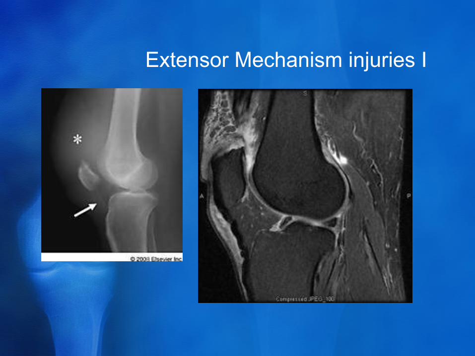
Extensor Mechanism injuries I
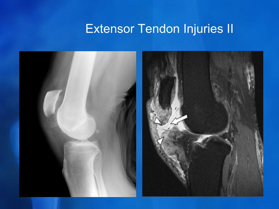
Extensor Tendon Injuries II
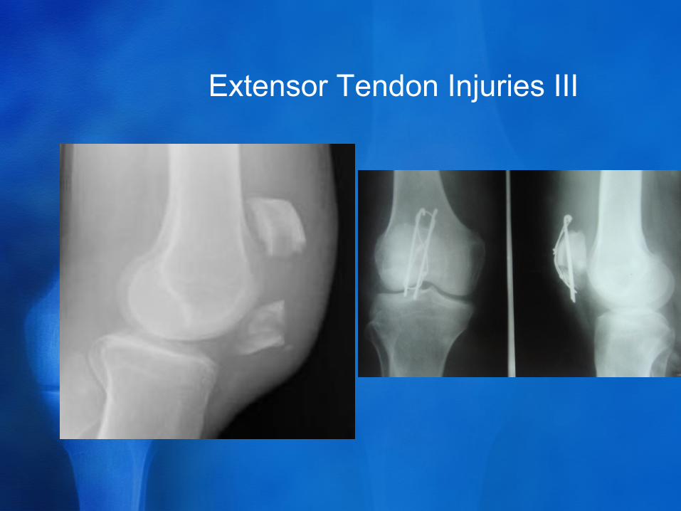
Extensor Tendon Injuries III
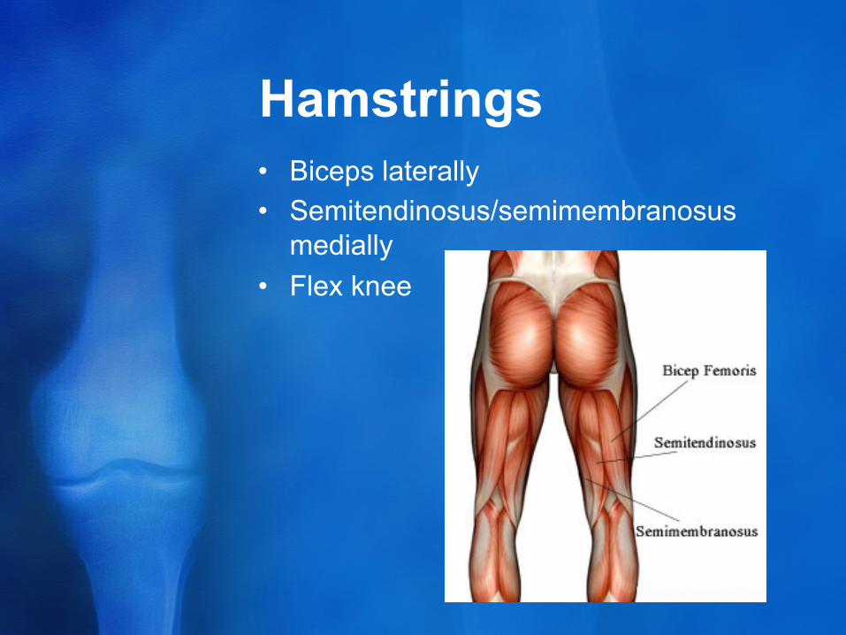
Hamstrings • Biceps laterally • Semitendinosus/semimembranosus
medially • Flex knee
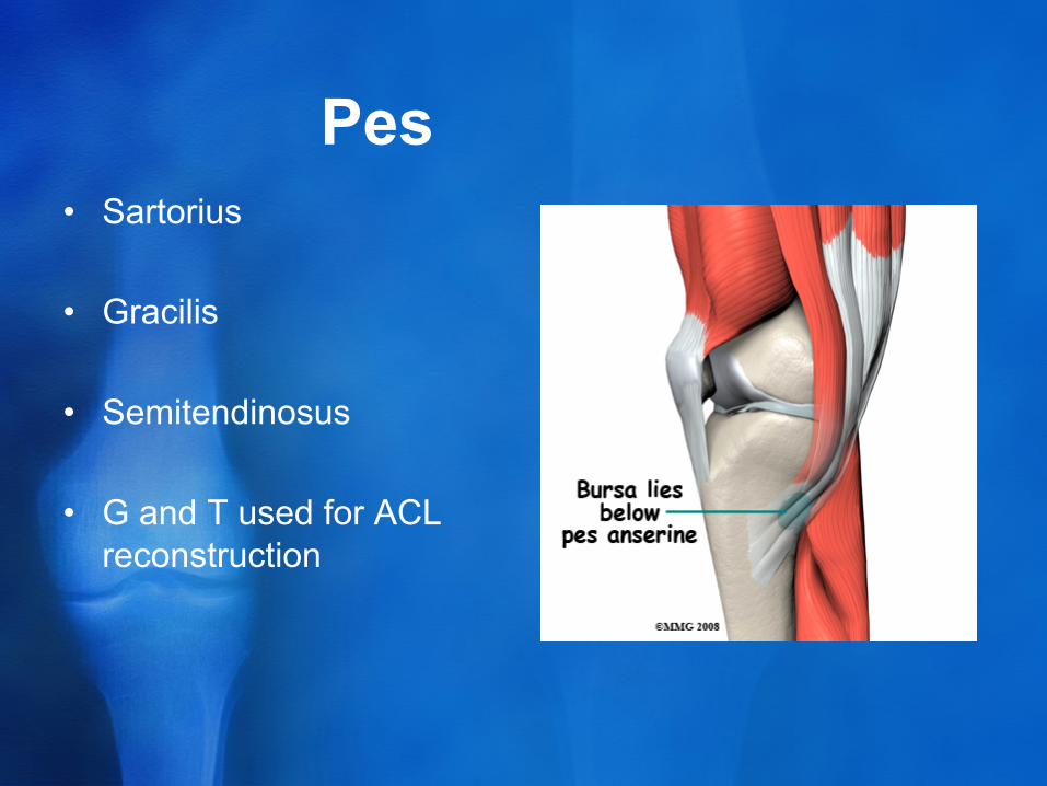
Pes • Sartorius • Gracilis
• Semitendinosus
• G and T used for ACL reconstruction
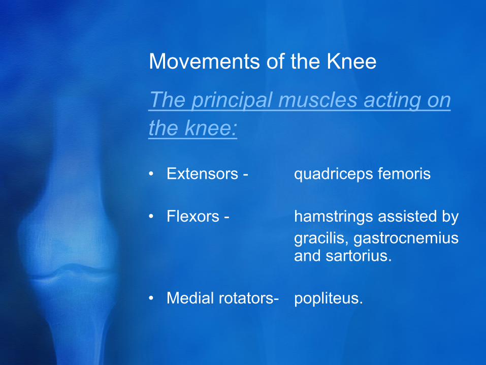
Movements of the Knee
The principal muscles acting on the knee: • Extensors - quadriceps femoris • Flexors - hamstrings assisted by gracilis, gastrocnemius
and sartorius.
• Medial rotators- popliteus.
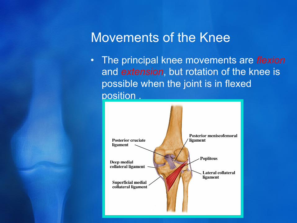
Movements of the Knee • The principal knee movements are flexion
and extension, but rotation of the knee is possible when the joint is in flexed position .
Examination • Look
• Feel • Move
• Think about structures and what you’re dong to them

Diseases • OA • Meniscal tears • Ligament injuries • Patellofemoral tracking • Tendon injuries • Inflammatory arthritis • Infection/tumours
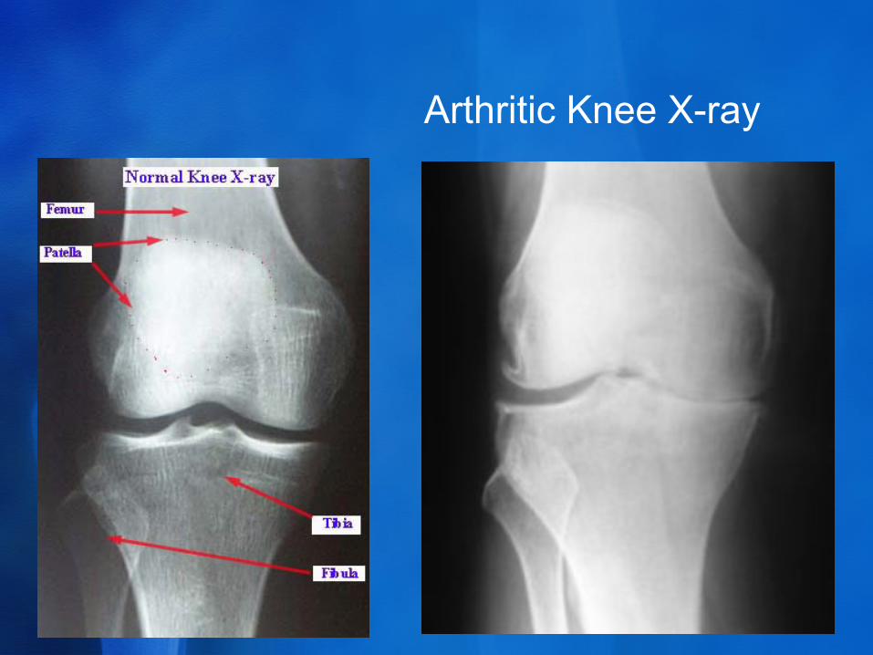
Arthritic Knee X-ray
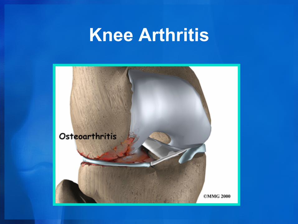
Knee Arthritis
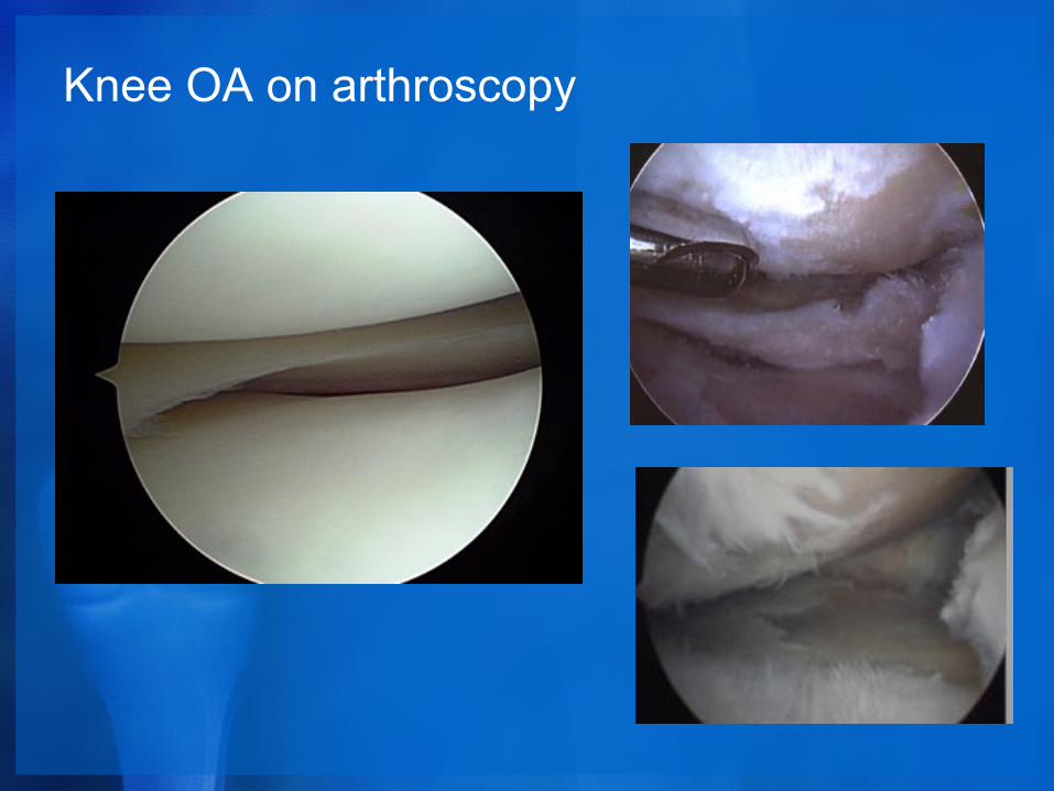
Knee OA on arthroscopy
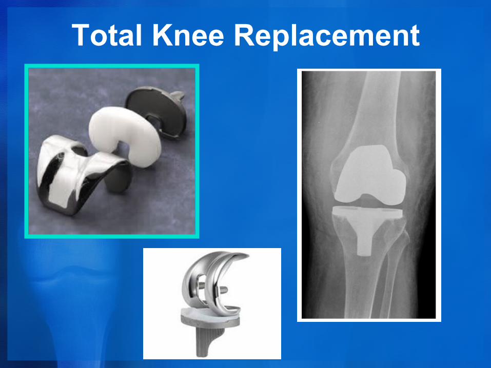
Total Knee Replacement
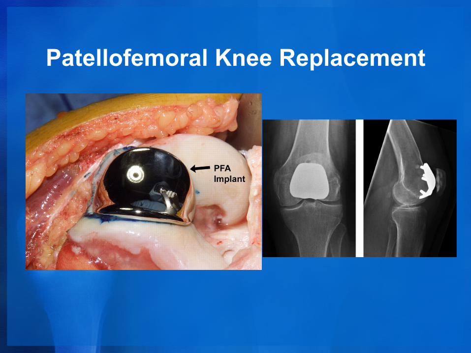
Patellofemoral Knee Replacement
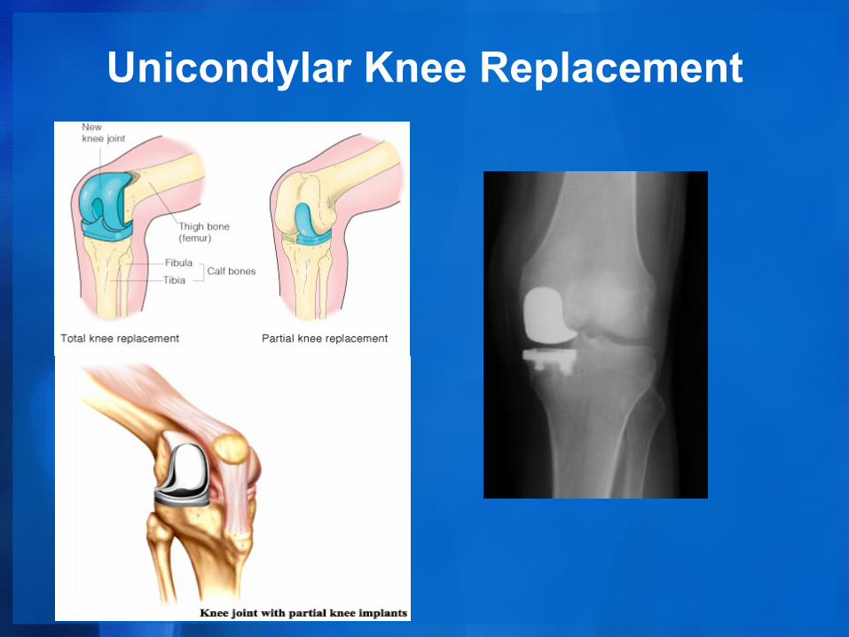
Unicondylar Knee Replacement
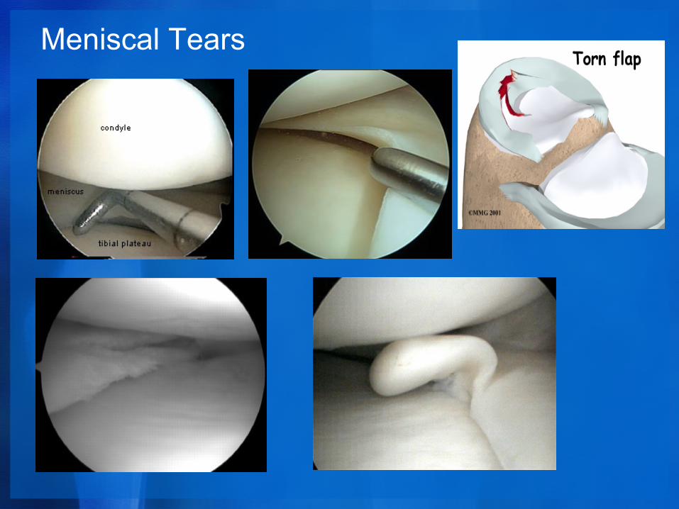
Meniscal Tears
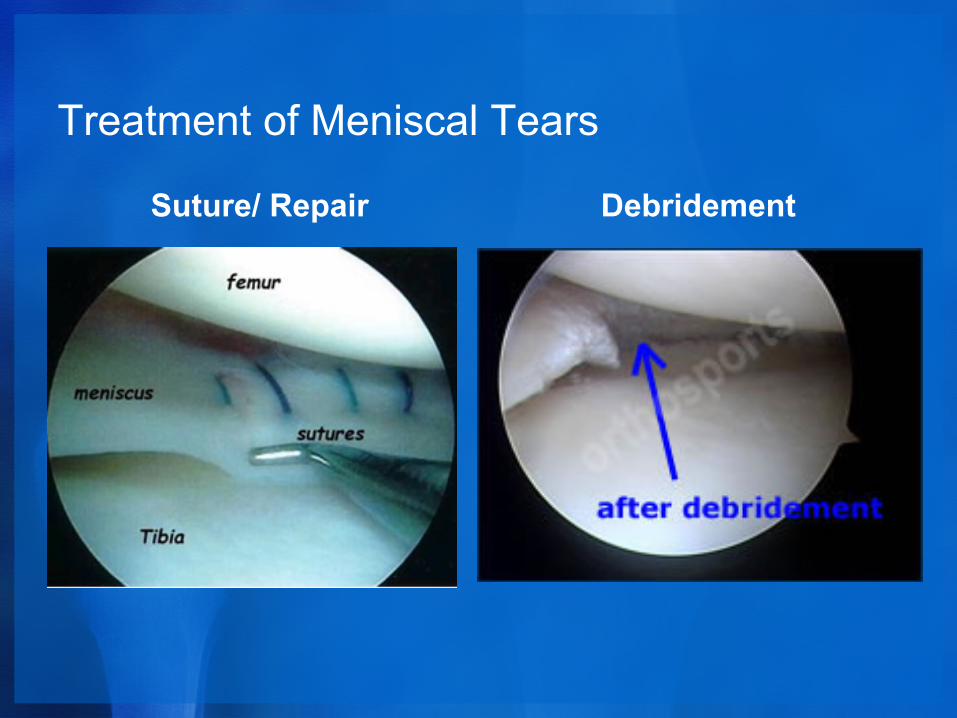
Treatment of Meniscal Tears
Suture/ Repair Debridement
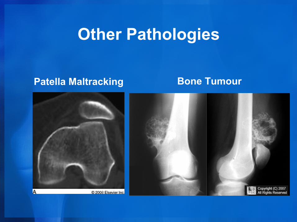
Other Pathologies
Patella Maltracking Bone Tumour

Any Questions ?
Recommended