1 Reflux Disease
Gastro
-oeso
ph
ag
eal R
efl
ux D
isease
in A
du
lts
Refl
ux D
isease
fifth EDition 2011
RE-PRintED 2011
©Digestive Health Foundation
Digestive Health Foundation
Cl
iniC
al
uP
Da
tE

2 Reflux Disease
Table of contents
1. Gastro-oesophageal reflux disease 5
Terminology and the significance of reflux 5
Epidemiology 5
Natural History 5
Pathogenesis 5
Clinical Features 6
2. Diagnosis 6
Symptoms 6
Therapeutic trial 7
Investigations: The role of endoscopy 7
3. Management 8
Empirically treated patients 9
Patients who have undergone endocopic assessment 9
Risks of acid suppression and acid suppressive medication 9
Fundoplication 9
Endoscopic therapies for gastro-oesphageal reflux disease 9
Barretts Oesophagus 10
Page

4 Reflux Disease
Gastro-oesophageal Reflux Disease in Adults
Reflux Diseasefifth EDition 2011
acknowledgementsThis booklet is a publication of the
Digestive Health Foundation, the
educational and health promotional
body of the Gastroenterological
Society of Australia, the professional
body representing the specialty of
gastrointestinal and liver disease in
Australia. Members of the Society
are drawn from physicians, surgeons,
scientists and other medical specialties
with an interest in GI Disorders.
This edition was updated by Associate
Professor Geoffrey Hebbard, Director of
Gastroenterology, The Royal Melbourne
Hospital on behalf of the Digestive
Health Foundation.
The Digestive Health Foundation of the
Gastroenterological Society of Australia
has prepared this document and every
care has been taken in its compilation.
The document is intended to be used as
a guide only and not as an authoritative
statement on every conceivable step or
circumstance that may or could relate to
the management of gastrooesophageal
reflux disease in adults.
Clinicians should use this document as
an aid in relation to gastrooesophageal
reflux disease and not as a complete
authoritative statement. Medical
understanding of gastrooesophageal
reflux is evolving. This document reflects
this understanding as at February 2011
and may be subject to updating and
review at a later date.
The Gastroenterological Society of
Australia and the compilers of the
document shall not be liable to users of
the document nor to any other person,
firm, company or other body for any
loss, direct, indirect or consequential, on
whatsoever account for any omission
or negligent misstatement contained
therein, or by reason of, arising from
or in relation to any such user, by any
other person, company of body relying
or acting upon any matter contained
therein or arising thereout.
This document was sponsored wholly and solely by the Gastroenterological Society of Australia.
Printed by:
Gastroenterological Society of australiaPo Box 508, Mulgrave
Victoria 3170, Australia
© Copyright. Gastroenterological Society of australia and Gastroenterological nurses College of australia.
This booklet is copyright and all rights reserved. It may not be reproduced in whole or in part without permission from the Gastroenterological Society of Australia.
Digestive Health Foundation
Reflux Disease 5
1 GaStRo-oESoPhaGEal REflux DiSEaSE
terminology and the significance
of Reflux
• Reflux of gastric contents into the
oesophagus is a normal physiological
event, occurring usually during the
postprandial period.
• Gastro-oesophageal reflux disease
occurs when reflux exposes the patient
to the risk of physical complications,
or symptoms lead to a significant
impairment of wellbeing or quality
of life.
• Clinically significant impairment of
wellbeing (quality of life) usually occurs
when symptoms are present on two or
more days a week.
• Reflux disease carries the risk of
reflux (peptic) oesophagitis and
complications such as columnar-lined
(Barrett’s) oesophagus and oesophageal
adenocarcinoma. However, the absolute
risk of oesophageal cancer in an
individual with reflux is small.
• Most patients with symptoms of
Gastro-oesophageal reflux do not have
endoscopically visible lesions. Only about
one-third of patients with reflux disease
have reflux oesophagitis as evidenced
by endoscopically visible mucosal breaks
(erosions or ulceration) or columnar-lined
(Barrett’s) oesophagus.
Epidemiology
• Reflux disease is common. Between
15-20% of adults experience heartburn
at least once a week.
• Obesity (BMI >30kg/m2), alcohol
consumption (> 7 standard drinks a
week) and a first-degree relative with
heartburn increase the risk of having
reflux symptoms.
• Patients with connective tissue diseases
such as scleroderma, chronic respiratory
diseases such as asthma and cystic
fibrosis, institutionalised and intellectually
handicapped people, and patients
nursed in a supine position for prolonged
periods are at increased risk of reflux
disease and its complications.
natural history
• In most patients, reflux disease is a
chronic disorder. Mild symptoms may
vary in intensity and occur only on some
days. With increasing severity, symptoms
tend to occur daily.
• Most patients will therefore require
long term management, although this
should be individualised to control the
patient’s symptoms and reduce the risk
of complications.
Pathogenesis
• Gastro-oesophageal reflux disease
occurs as a result of excessive exposure
of the oesophagus to gastric contents.
The most damaging components are
acid and pepsin, although bile and
pancreatic enzymes may contribute in
some patients.
• The excessive exposure to gastric
contents is largely the result of an
increased frequency of acid reflux
episodes, but impaired clearance of
stomach contents from the oesophagus
or impaired mucosal defences may also
be an important factor in some patients.
• Under normal conditions, reflux is
prevented by the tonic activity of the
lower oesophageal sphincter (LOS),
supported by the crural diaphragm.
Reflux episodes occur primarily because
of defective LOS function. The major
event that leads to reflux is transient
LOS relaxation. An important minority
of reflux episodes occurs because of
defective basal LOS pressure, often
associated with hiatal hernia.
• A number of factors may aggravate
reflux symptoms. These include dietary
components such as fat, chocolate,
caffeine and alcohol, as well as smoking
and some drugs.
• Hiatal hernia is associated with reflux
disease, however reflux disease may also
be present without hiatal hernia, and
the presence of a hiatal hernia does not
necessarily mean that reflux disease is
present or should be treated.
Reflux disease is the most common cause of heartburn and indigestion in western society
6 Reflux Disease
Clinical featuresSymptoms directly related to reflux episodes and Symptoms caused by complications of reflux disease.
Symptoms may be broadly grouped
into those which are directly related to
reflux episodes, and those caused by
complacations of reflux disease.
Symptoms directly related to reflux episodes
• Heartburn
This is the hallmark symptom of reflux
disease. It is characterised by a feeling
of burning rising up from the stomach or
lower chest towards the neck. It is typically
provoked by meals, especially those
containing fatty or highly spiced food or by
bending, straining or lying down. It is usually
eased or relieved by antacids.
• Regurgitation
This is the other characteristic symptom
of reflux disease. Regurgitated material is
usually re-swallowed but is sometimes so
voluminous that it may be mistaken for
‘vomiting’. Some patients may experience
regurgitation as their predominant
symptom, particulary if gastric acid
secretion is suppressed.
• Waterbrash
Oesophageal acidification may cause such
sudden and brisk stimulation of salivation
that the patient’s mouth fills with saliva. This
may be associated with nausea.
• Atypical symptoms
Reflux may cause a number of other
symptoms not easily identified as being due
to reflux. These include angina-like chest
pain, throat/voice changes, cough, asthma,
excessive belching, dyspepsia and nausea.
These symptoms are nonspecific and do not
in themselves imply the presence of reflux,
although it should be considered in the
differential diagnosis.
Symptoms caused by complications of reflux disease
• Dysphagia
Dysphagia may be due either to
inflammation, defective oesophageal
peristalsis or heightened oesophageal
sensitivity. Dysphagia that is associated with
symptoms of bolus impaction is suggestive
of a stricture or ring.
• Odynophagia (painful swallowing)This is usually associated with severe
oesophagitis but may also be due to
excessive sensitivity of the oesophageal
mucosa.
• Bleeding from oesophagitis
Haematemesis may be a presenting
symptom but is rarely severe. Occasionally,
iron deficiency may result from severe
oesophagitis (however other causes must
be excluded).
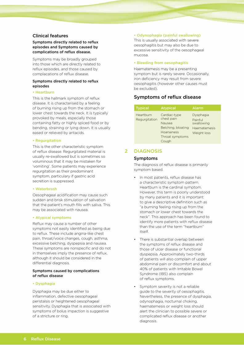
Symptoms of reflux disease
2 DiaGnoSiS
SymptomsThe diagnosis of reflux disease is primarily
symptom based.
• In most patients, reflux disease has
a characteristic symptom pattern.
Heartburn is the cardinal symptom.
However, this term is poorly understood
by many patients and it is important
to give a descriptive definition such as
“a burning feeling rising up from the
stomach or lower chest towards the
neck”. This approach has been found to
identify more patients with reflux disease
than the use of the term “heartburn”
itself.
• There is substantial overlap between
the symptoms of reflux disease and
those of ulcer disease or functional
dyspepsia. Approximately two-thirds
of patients will also complain of upper
abdominal pain or discomfort and about
40% of patients with Irritable Bowel
Syndrome (IBS) also complain
of reflux symptoms.
• Symptom severity is not a reliable
guide to the severity of oesophagitis.
Nevertheless, the presence of dysphagia,
odynophagia, nocturnal choking,
haematemesis or weight loss should
alert the clinician to possible severe or
complicated reflux disease or another
diagnosis.
typical atypical alarm
Heartburn
Regurgitation
Cardiac-type chest pain
Nausea
Belching, bloating
Hoarseness
Throat symptoms
Cough
Dysphagia
Painful swallowing
Haematemesis
Weight loss
Reflux Disease 7
therapeutic trial• A symptom-based diagnosis can be
supplemented by the use of a therapeutic
trial of high dose proton pump inhibitor
for two to four weeks. This test has
a sensitivity and specificity for reflux
disease that is comparable to that
of oesophageal pH monitoring and
substantially superior to endoscopy.
investigations: the role of endoscopyWhen to investigate?
• It is inappropriate to investigate every
patient with suspected reflux symptoms.
Young patients who have longstanding
mild, typical reflux symptoms and no
alarm symptoms may be given a trial of
therapy without investigation.
investigations should be done when:
• the diagnosis is unclear because
symptoms are either non-specific or
atypical for reflux disease, or are ‘mixed’
with gastroduodenal symptoms such as
the presence of epigastric pain.
• symptoms that persist or progress on
therapy symptoms suggest severe or
complicated oesophagitis is present; for
example haematemesis, odynophagia,
persistent dysphagia or weight loss
• other diagnoses seem possible,
for example:
– infective or drug-induced
oesophagitis
– oesophageal malignancy
– gastroduodenal disorders
– myocardial infarction or ischaemia.
Which investigation?
- Endoscopy
Upper GI endoscopy is the investigation
of first choice as it:
• is the most specific test for reflux
oesophagitis (although not sensitive
– see below)
• is the most accurate means of
diagnosing other mucosal lesions such
as infective oesophagitis, peptic ulcer
disease, malignancy or other upper gut
abnormalities that may be difficult to
distinguish on history from reflux disease
• is the only effective way to grade
oesophagitis, which has relevance to the
choice of therapy and management of
reflux disease (see below)
• is the most sensitive method for
diagnosing columnar-lined or Barrett’s
oesophagus
• is useful for recognition and management
of peptic stricture.
• However, endoscopy has a limited
role in the diagnosis of reflux disease,
as only approximately one third of
patients with reflux symptoms have
diagnostic endoscopic abnormalities.
In these ‘endoscopy negative’ patients,
distal oesophageal biopsies (which
may demonstrate microscopic changes
of oesophagitis in approximately 25%
of cases/individuals/patients) are not
recommended as routine practice, as this
is a relatively expensive test that does not
influence choice of therapy, which should
be directed at symptom control. Potential
roles for endoscopy are outlined in Table 1.
• In patients with alarm symptoms,
endoscopy should be performed
promptly, prior to empirical therapy.
Early endoscopy is also indicated in
patients with atypical symptoms or
when symptoms fail to respond to initial
therapy. Endoscopic reassessment should
be performed within 6 months of anti-
reflux surgery to exclude any new or
unexpected pathology.
approximately two thirds of patients with Gastro-oesophageal reflux disease are ‘endoscopy negative’ i.e. have no diagnostic oesophageal mucosal abnormalities
investigations are warranted if the diagnosis is unclear; symptoms persist or are refractory to treatment; complications are suspected or; alarm symptoms are present.
a diganosis based on symptoms can be aided by trial of treatment
the history is the single most useful method for the diagnosis of reflux disease
typical atypical alarm
Heartburn
Regurgitation
Cardiac-type chest pain
Nausea
Belching, bloating
Hoarseness
Throat symptoms
Cough
Dysphagia
Painful swallowing
Haematemesis
Weight loss

Reflux Disease 8
• The role of endoscopy in patients with
longstanding ‘medically controlled’ Gastro-
oesophageal reflux disease is less clear.
Evidence suggests that symptomatic
control equates to endoscopic healing
(even in severe Los Angeles classification
Grade C or D oesophagitis) so endoscopic
assessment is only necessary if there
is a relapse whilst on maximal or near
maximal medical therapy. On the other
hand, a ‘once in a life-time’ endoscopy in
this subset of patients may be reasonable
to assist discussions about long-term
management and to exclude the potential
complications of severe oesophagitis.
There is, however, no convincing evidence
that screening all patients with reflux for
Barrett’s oesophagus would be of benefit.
Endoscopic surveillance in known Barrett’s
oesophagus is discussed in Section 4.
Barium swallow and meal
• This is an inappropriate primary
diagnostic test as it is insensitive and not
specific for reflux disease.
• It may be useful however, to assess
and plan management in patients
with persistent dysphagia when a
complicating stricture is suspected or for
the assessment of large hiatal hernia.
24-hour ambulatory oesophageal ph monitoring
• This investigation tests the amount of
exposure of the oesophagus to acid and
whether symptoms are related to the
occurrence of reflux.
• pH monitoring is useful in patients in
whom the diagnosis and relationship
between symptoms and reflux remains
unclear despite a therapeutic trial of acid
suppression and endoscopy.
3 ManaGEMEnt
• As the severity of Gastro-oesophageal
reflux disease varies significantly
between patients, management needs
to be individualised. The aims of
management are to control symptoms
(in the majority of patients) and to
prevent complications (in a small but
important minority).
• Although gastric acid secretion is normal
and the cause of the disease is lower
oesophageal sphincter dysfunction,
the mainstay of treatment is acid
suppression.
- The proton pump inhibitors (PPI)
are the most potent agents and
are most effective if taken 30-60
minutes before food. Some PPIs are
now available without prescription
in Australia.
- H2 receptor antagonists are less
potent, but may have a more rapid
onset of action. These are available
over the counter, so are very
accessible to patients.
- Antacids have a rapid onset of
action, but are ineffective in long-
term management other than for
mild intermittent or occasional
breakthrough symptoms.
- Prokinetic agents have little role in
the management of reflux disease..
TABLE 1indications for early endoscopy:
• Alarm symptoms (including dysphagia,
odynophagia, weight loss, haematemesis,
anaemia)
• Diagnostic uncertainty such as mixed,
non-specific or atypical symptoms
• Symptoms refractory to initial treatment
• Pre-operative assessment
• Provision of reassurance when verbal
reassurance is inadequate.
Endoscopy may also be appropriate:
• For patients with long-standing frequent
troublesome symptoms
• To tailor drug treatment
• To detect and manage Barrett’s
oesophagus.

Reflux Disease 9
• Lifestyle measures have not been
demonstrated to have significant effects
in healing oesophagitis, however dietary
changes (avoidance of precipitating
foods), weight loss (reduction in weight
in overweight or obese individuals), and
nocturnal posture (elevation of the torso)
may be useful in controlling symptoms
in some patients. Patients should not be
placed on unnecessarily restrictive diets.
Empirically treated patients
• In the group of patients where empirical
treatment is commenced and who
have an adequate therapeutic response
indicating that reflux is the likely cause
of their symptoms, it is appropriate to
cease or reduce therapy and allow the
patient to use acid suppression on an
intermittent basis as required. It is likely
that symptoms will return in 70% of
cases, and patients will adjust the dose of
medication to the severity and frequency
of symptoms.
• Atypical symptoms (ENT, respiratory)
often require a more prolonged/higher
dose trial of therapy.
Patients who have undergone
endoscopic assessment
• Patients with no oesophagitis or milder
grades of oesophagitis at endoscopy
(Los Angeles Grades A and B) can be
treated in a similar manner to empirically
treated patients, and ‘stepped down’
to the lowest level of acid suppression
where symptoms are adequately
controlled.
• Patients with severe oesophagitis (Los
Angeles Grades C and D) should be
treated with regular (standard dose) PPI
on an ongoing basis to maintain mucosal
healing. Initial healing and symptom relief
are more rapid in these patients when
treated with formulations of PPI that
provide a greater degree of acid control.
• Patients with complications such as
strictures will require endoscopic therapy
(e.g. dilatation).
Risks of acid suppression and
acid suppressive medication
• Long term acid suppression with PPIs
has been associated with a slight
increase in the rates of bacterial
gastroenteritis, community acquired
pneumonia, osteoporosis and hip
fracture, as well as bacterial overgrowth
of the small intestine and outbreaks
of Clostridium difficile colitis in elderly
patients in hospitals. The absolute risk of
these complications is very low.
• The evidence for a clinically significant
interaction with Clopidogrel is weak.
• There is a low incidence of interstitial
nephritis with PPIs. However this
should be considered in patients with
a deterioration in renal function on PPI
therapy.
• Patients on warfarin should have their
INR checked following the introduction
of acid suppressive medication, as
with the commencement of any new
medication.
fundoplication
• Fundoplication is an effective treatment
for Gastro-oesophageal reflux disease,
and can be performed laparoscopically,
but carries the small risks associated with
laparoscopic abdominal surgery.
• Fundoplication is of particular value
in patients whose symptoms cannot
be controlled adequately by medical
therapy, and may be considered in
patients who wish to avoid long-term
medication and are willing to accept the
small risks involved.
Endoscopic therapies for
gastro-oesophageal reflux disease
• A number of therapies that can be
delivered endoscopically have been
developed for the treatment of reflux
disease. These include suturing devices,
injection of inert substances, plication
devices and devices that deliver
radiofrequency energy to the
Gastro-oesophageal junction.
• Experience with these techniques
is relatively limited. They do not
significantly reduce exposure of the
distal oesophagus to acid, and many

have already been removed from the
market because of lack of efficacy or
complications (including death).
• These treatments should not be used
by inexperienced operators or outside
a program (e.g. a clinical trial) where
complications can be easily reported.
Barretts Oesophagus
• Metaplasia of the lining of the lower
oesophagus to an intestinal phenotype
(Barrett’s oesophagus) is related to
Gastro-oesophageal reflux, and confers
an increased risk of oesophageal
adenocarcinoma.
• Although the relative risk is significantly
increased, the absolute risk of an
individual with Barrett’s oesophagus
developing adenocarcinome of the
oesophagus remains low (<2% lifetime
risk), and very few patients with Barrett’s
oesophagus die of oesophageal
adenocarcinoma.
• If Barrett’s oesophagus is found at
endoscopy, patients should be offered
the opportunity to enroll into a screening
program where regular biopsies
are taken to assess for dysplasia or
carcinoma, with appropriate follow-up
management where required.
Reflux Disease 10

Reflux Disease 11

Reflux Disease 12
Digestive Health Foundationc/- GESA
PO Box 508, Mulgrave 3170Victoria, Australia
Telephone: 1300 766 176Facsimile: (03)9802 8533
E-mail: [email protected]
3247
2
Digestive Health Foundation
Recommended