112 BIOLOGY STUDENT’S COMPANION RESOURCES [SES DB014]
CHAPTER 8 : REPRODUCTION and DEVELOPMENT
SUBTOPIC 8.1: Types of Reproduction
LEARNING OUTCOMES:
a) Define sexual and asexual reproduction.
SUBTOPIC 8.2: Sexual Reproduction in Flowering Plants
LEARNING OUTCOMES:
a) State the general structures and functions of the reproductive organs in flowering plants
b) Describe the development of a pollen grain and formation of male gamete
c) Describe the development of ovule, embryo sac and formation of female gamete
d) Explain double fertilization in the formation of seed
MAIN IDEAS /
KEY POINT EXPLANATION NOTES
Definition of
sexual and
asexual
reproduction
Definition of asexual reproduction
• A process by which an individual inherits all of its genes from a
single parent thus, being genetically identical to the parent
Definition of sexual reproduction
• A process in which new organisms are created by combining the
genetic information from two individuals of different sexes via
fertilization
General
structures and
functions of the
reproductive
organs in
flowering plants
• The flower is a unique structure that is specialized for sexual
reproduction.
• The four types of floral organs are sepal, petal, stamen and carpel
113 BIOLOGY STUDENT’S COMPANION RESOURCES [SES DB014]
MAIN IDEAS /
KEY POINT EXPLANATION NOTES
Floral organ Structure and function
Sepal • Sepal forms the outer and lower ring of
flowers
• Usually green and more leaf-like in
appearance
• Enclose the flower before it opens
• The group of sepals is called calyx
• Function is to cover and protect the young
flower bud
Petal • Petal are broad, flat and thin but vary in
shape and brightly colored
• The group of petals is called corolla
• Function is to attract animals pollinators to
the flower
Stamen • The male reproductive organs of flower that
consists of :
• Anther
• Filament
Functions :
• Anther contain pollen sacs that produce
pollen grains (male gametophyte)
• Filament is the stalk that holds the anther
Carpel • The female reproductive organs of flower
that consists of :
• Stigma (sticky structure on top)
• Style (neck like structure)
• Ovary
Functions :
• Stigma act as a landing platform for pollen
grains
• Style is the structure in which pollen tube
grows
• Ovary is the structure that contains one or
more ovules. Female gamete develops in its
ovule
Terminologies used :
• Microsporangium refers to the diploid (2n) pollen sac of anther
• Megasporangium refers to the diploid (2n) ovule in ovaries
• Male sporocyte : Diploid (2n) microsporocyte or microspore
mother cell
• Female sporocyte : Diploid (2n) megasporocyte or megaspore
mother cell
• Male spore : Haploid (n) microspore
• Female spore : Haploid (n) megaspore

114 BIOLOGY STUDENT’S COMPANION RESOURCES [SES DB014]
MAIN IDEAS /
KEY POINT EXPLANATION NOTES
• Male gametophyte refers to haploid (n) pollen grain
• Female gametophyte refers to haploid (n) embryo sac
• Male gamete refers to haploid (n) sperm cell
• Female gamete refers to haploid (n) egg cell
Development of
a pollen grain
and formation of
male gamete
❖ Pollen grain (male gametophyte) forms in the pollen sac of
anther (microsporangium)
❖ Each anther contains four pollen sacs
❖ Each pollen sac contains numerous diploid (2n) microspore
mother cells (microsporocytes)
❖ Each diploid (2n) microsporocyte undergoes meiosis
❖ Produce 4 haploid (n) microspores (tetrad)
❖ Each microspore (n) undergoes mitosis
❖ Producing a generative cell (n) and a tube cell (n)
❖ Both cells are encased in haploid (n) immature pollen grain
❖ Pollen grain is surrounded by exine (the outer wall) and intine
(the inner layer)
❖ When pollination occur (pollen grains land on stigma), the
generative cell (n) divides by mitosis into two haploid (n)
sperm cells and now, pollen grain (n) is matured

115 BIOLOGY STUDENT’S COMPANION RESOURCES [SES DB014]
MAIN IDEAS /
KEY POINT EXPLANATION NOTES

116 BIOLOGY STUDENT’S COMPANION RESOURCES [SES DB014]
MAIN IDEAS /
KEY POINT EXPLANATION NOTES
Development of
the ovule,
embryo sac and
the formation of
the female
gamete
❖ Diploid, 2n ovule (megasporangium) develop from carpel tissue
which is held by funicle.
❖ Young ovule contain nucellus. Nucellus is encased within two
layers of integuments
❖ Integuments form tiny hole at one end known as micropyle and
the other end is called chalaza
❖ In nucellus, diploid (2n) ovule
(megasporangium) contain one
megasporocyte / megaspore mother cell (2n)
❖ Each megasporocyte (2n) undergoes meiosis
❖ Producing four haploid (n) megaspores
❖ Three of the megaspores (n) degenerate

117 BIOLOGY STUDENT’S COMPANION RESOURCES [SES DB014]
MAIN IDEAS /
KEY POINT EXPLANATION NOTES
❖ The one surviving megaspore (n) undergoes three times mitotic
division (without cytokinesis)
❖ Producing eight haploid nuclei within the haploid embryo sac
(female gametophyte)
❖ Eight haploid nuclei are arranged in groups of four nuclei at two
opposite poles
❖ One nucleus from each pole moves to the center, forming polar
nuclei (2n)
❖ Three haploid nuclei at the pole near to micropyle form 1
haploid egg cell (female gamete) and 2 haploid synergid cells
❖ 3 haploid nuclei at the pole away from micropyle, form the
antipodal cells
❖ Embryo sac thus consist of 8 haploid nuclei contained within 7
cells
Double
fertilization in
the formation of
seed
Definition of double fertilization :
The union of the 2 sperm cells (n) with different nuclei of the
embryo sac.
One haploid (n) sperm cell fertilize the egg cell (n) forming diploid
(2n) zygote. Other one sperm cell (n) fuses with polar nuclei (2n)
forming triploid (3n) primary endosperm
After double fertilization
each ovule develops into
seed. Ovary develops
into fruit. Endosperm
become food-storing
tissue of the seed.

118 BIOLOGY STUDENT’S COMPANION RESOURCES [SES DB014]
MAIN IDEAS /
KEY POINT EXPLANATION NOTES
✓ Pollen grains land on stigma
✓ At the time of pollination, the pollen grain typically consists of
only 1 tube cell (n) and 1 generative cell (n)
✓ Pollen grain absorbs water and germinates by producing a
pollen tube (a long tube that delivers sperm cell to the embryo
sac).
✓ As the pollen tube elongates through the style, generative cell
(n) divides by mitosis and forms 2 sperm cells (n)
✓ The tube nucleus leads ahead of the 2 sperm cells as the tip of
pollen tube grows toward micropyle in response to chemical
attractants produced by the synergid cells.
✓ The arrival of pollen tube at micropyle initiates the death of one
of the two synergid cells, providing a passageway into embryo
sac.
✓ Tube nucleus and the 2 sperm cells (n) are then discharged from
pollen tube.
✓ Double fertilization occur after the 2 sperm cells (n) reach the
embryo sac.
✓ Near the time of double fertilization, tube nucleus, the other one
synergid cell and the antipodal cells degenerate
✓ In double fertilization, one haploid (n) sperm cell fertilize the
egg cell (n) forming diploid (2n) zygote.
✓ Other one sperm cell (n) fuses with polar nuclei (2n) forming
triploid (3n) primary endosperm.
polar nuclei

119 BIOLOGY STUDENT’S COMPANION RESOURCES [SES DB014]
CHAPTER 8 : REPRODUCTION and DEVELOPMENT
SUBTOPIC 8.3 : Human Reproductive System
LEARNING OUTCOMES:
a) Describe the structure of spermatozoa.
b) Describe the stages of spermatogenesis.
c) Describe the structure of the secondary oocyte.
d) Describe the stages of oogenesis.
e) Outline female reproductive cycle and its hormonal control:
i. Ovarian cycle
ii. Uterine/menstrual cycle
MAIN IDEAS /
KEY POINT EXPLANATION NOTES
Male Reproductive Organ : Testes (singular: testis) consist of many highly
coiled tubes called seminiferous tubule.
Seminiferous tubule is the site of spermatogenesis or sperms production.
Once produced, sperm cells swim inside the lumen of seminiferous tubule.
Structure of
spermatozoa
Four major parts of
spermatozoa :
✓ Head
✓ Neck
✓ Midpiece / middle
piece
✓ Tail

120 BIOLOGY STUDENT’S COMPANION RESOURCES [SES DB014]
MAIN IDEAS /
KEY POINT EXPLANATION NOTES
Structure
of sperm
Characteristics
Head • Composed of a haploid nucleus
Acrosome • Located at the tip of head
• Consist of special vesicle that contain hydrolytic
enzyme (that help the sperm penetrate an ovum)
Neck • Short and have one pair of centriole
Midpiece • Contain mitochondria (provide ATP for the
movement of the sperm’s tail)
Tail • Is the sperm flagellum that move the sperm to the
ovum
Stages in
spermatogenesis
❖ Spermatogenesis begins during puberty
❖ Occurs in the seminiferous tubule of testes
1) In embryonic testis, diploid (2n) primordial germ cell differentiate into
spermatogonia (2n)
2) At puberty, each spermatogonia (2n) divide by mitosis forming type A
and type B spermatogonia (2n)
3) Type A spermatogonia (2n) remains at basement membrane to maintain
the layer of germinal cells – continue divide by mitosis producing large
number of spermatogonia (2n)
4) Type B spermatogonia (2n) differentiate into primary spermatocytes (2n)
5) Each primary spermatocyte (2n) undergo meiosis I forming 2 haploid (n)
secondary spermatocyte
6) Each secondary spermatocyte (n) undergo meiosis II forming 2 haploid
(n) spermatids – there are 4 spermatids (n) from 1 primary
spermatocytes (n). Spermatids are rounded, non-motile cells.
7) Spermatids (n) undergo spermiogenesis – Spermatid elongates, discard
excess cytoplasm to become lighter and form tail. Spermatids become
mature and motile spermatozoa / sperm cells

121 BIOLOGY STUDENT’S COMPANION RESOURCES [SES DB014]
MAIN IDEAS /
KEY POINT EXPLANATION NOTES
122 BIOLOGY STUDENT’S COMPANION RESOURCES [SES DB014]
MAIN IDEAS /
KEY POINT EXPLANATION NOTES
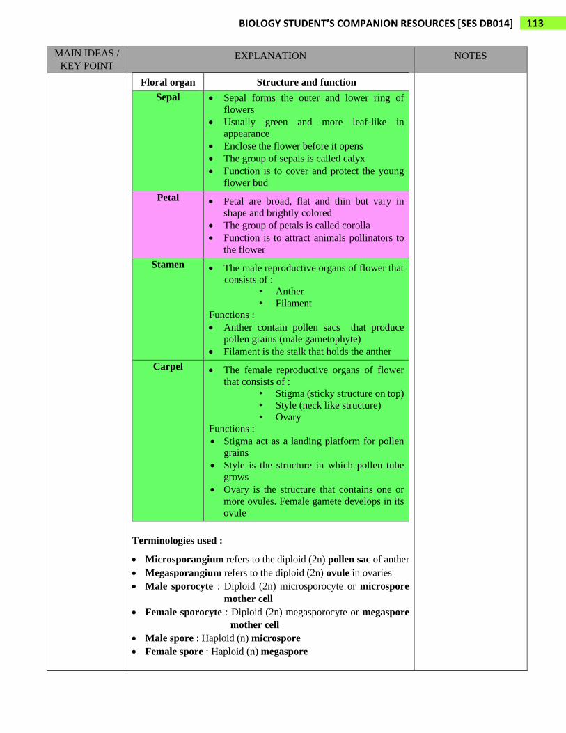
Structure of
secondary
oocyte
❖ Female has a pair of ovaries.
❖ The outer layer of each ovary is packed with follicles
❖ Each follicle consist of an oocyte surrounded by a group of support cells.
❖ Every month, one primary follicle containing a primary oocyte develops
and matures into Graafian follicle.
❖ Secondary oocyte is found
inside matured Graafian follicle.
❖ During ovulation, secondary
oocyte is released from ovary.

123 BIOLOGY STUDENT’S COMPANION RESOURCES [SES DB014]
MAIN IDEAS /
KEY POINT EXPLANATION NOTES
❖ Structures of secondary oocyte :
✓ Corona radiata (a group of granulosa cells) :
- Secretory cells in Graafian follicle that surrounds the
secondary oocyte.
- Supply nutrients to the developing oocyte.
✓ Zona pellucida
- A layer of glycoprotein that surrounds the plasma membrane
of secondary oocyte
✓ First polar body
- One small cell that is produced in meiosis I during the
development of an oocyte and finally degenerates
Stages of
oogenesis
The development of mature oocytes in ovaries starts during the
embryonic development
1) In embryonic ovaries, diploid (2n) primordial germ cells differentiate
into oogonia (2n)
2) Oogonia (2n) undergo mitosis forming large numbers of oogonia (2n)
3) Oogonia (2n) differentiate by increasing in size forming primary
oocyte (2n)
4) Each primary oocyte (2n) starts meiosis I but stop at Prophase I.
Primary oocyte (2n) enter resting stage until puberty
5) At puberty, primary oocytes (2n) complete Meiosis I forming small
haploid (n) first polar body and large secondary oocyte (n)
6) Secondary oocyte undergo Meiosis II but stop at metaphase II –
During ovulation, secondary oocyte is released from ovary into
Fallopian tube.
7) If secondary oocyte (n) is fertilized by sperm cell (n) then, meiosis II
is completed forming one large ovum (n) and small second polar
body (n). The second polar body will finally degenerate.

124 BIOLOGY STUDENT’S COMPANION RESOURCES [SES DB014]
MAIN IDEAS /
KEY POINT EXPLANATION NOTES

125 BIOLOGY STUDENT’S COMPANION RESOURCES [SES DB014]
MAIN IDEAS /
KEY POINT EXPLANATION NOTES
Female
reproductive
cycles and its
hormonal
control

126 BIOLOGY STUDENT’S COMPANION RESOURCES [SES DB014]
MAIN IDEAS /
KEY POINT EXPLANATION NOTES
❖ Ovarian Cycle (relate with ovaries) is divided into :
- Follicular phase (Day 1 – 13)
development of follicles
- Luteal phase (Day 15-28)
development and degeneration of corpus luteum
• Menstrual/Uterine Cycle (relate with endometrial wall of uterus)
is divided into :
- Menstrual flow phase (Day 1 – 5)
rupture of endometrial wall
- Proliferative phase (Day 6 – 14)
repairing of endometrial wall
- Secretory phase (Day 15 – 28)
maintain thickening of endometrial wall
• Follicular phase correspond with menstrual flow phase and
proliferative phase
• Luteal phase correspond with secretory phase

127 BIOLOGY STUDENT’S COMPANION RESOURCES [SES DB014]
MAIN IDEAS /
KEY POINT EXPLANATION NOTES
Ovarian Cycle
1. Ovarian cycle begins when hypothalamus secrete gonadotropin-releasing
hormone (GnRH), which stimulate anterior pituitary
2. to secrete small amounts of follicle stimulating hormone (FSH) and
luteinizing hormone (LH)
3. FSH stimulate follicle growth, aided by LH
4. Follicles start to secrete estrogen. Estrogen concentration slowly rise during
most of the follicular phase. Low levels of estrogen inhibit secretion of
pituitary hormones, keeping levels of FSH and and LH relatively low.
5. Estrogen secretion by the follicles begin to rise steeply
6. The FSH and LH levels increase markedly. High level of estrogen
stimulates GnRH secretion by causing hypothalamus to increase the
secretion of GnRH. High level of estrogen also increases the secretion of
LH
7. LH surge (peak in LH level) stimulate maturation of follicle into Graafian
follicle. In response to FSH and LH surge, ovulation occur releasing the
secondary oocyte.
8. The luteal phase follows ovulation. LH stimulates formation of corpus
luteum from the ruptured follicle. Stimulated by LH, corpus luteum secrete
progesterone and estrogen. High level of estrogen and progesterone, exert
negative feedback on hypothalamus and anterior pituitary. Results in, low
level of FSH and LH in order to prevent maturation of another oocyte if
pregnancy occur.
If pregnancy does not occur, low level of GnRH at the end of luteal phase
cause corpus luteum to disintegrate, triggering sharp decline in estrogen and
progesterone. Low level of estrogen and progesterone cause the
hypothalamus and anterior pituitary is no longer inhibited. Anterior
pituitary secrete enough FSH to stimulate the growth of new follicles,
initiating next ovarian cycle.

128 BIOLOGY STUDENT’S COMPANION RESOURCES [SES DB014]
MAIN IDEAS /
KEY POINT EXPLANATION NOTES
Uterine / Menstrual Cycle
9. Prior to ovulation, estrogen secreted in increasing amounts by growing
follicles stimulates the endometrium to thicken. This is the proliferative
phase of uterine cycle.
After ovulation, estrogen and progesterone secreted by corpus luteum
stimulate maintenance and further development of the uterine lining,
enlargement of arteries and growth of endometrial glands. This is the
secretory phase of uterine cycle. If an embryo has not implanted in
endometrium by the end of secretory phase, corpus luteum disintegrates.
10. Low level of estrogen and progesterone causes arteries in endometrium to
constrict and uterine lining disintegrates, releasing blood that is shed along
with endometrial tissue and fluid. The result is menstruation (menstrual
flow phase of uterine cycle). During this phase, a new set of ovarian
follicles begin to grow. The first day of menstrual flow is designated day 1
of the new female reproductive cycle.

129 BIOLOGY STUDENT’S COMPANION RESOURCES [SES DB014]
CHAPTER 8 : REPRODUCTION and DEVELOPMENT
SUBTOPIC 8.4 : Fertilization
LEARNING OUTCOMES :
a) Describe briefly the stages that lead to fertilization.
i. Capacitation
ii. Acrosomal reaction
iii. Fusion of sperm head membrane and oocyte
iv. Cortical reaction
MAIN IDEAS /
KEY POINT EXPLANATION NOTES
Stages that lead
to fertilization
➢ Haploid (n) secondary oocyte / ovum is fertilized by sperm cell (n) forming a
diploid (2n) zygote
➢ Occurs in the Fallopian tube
➢ Fertilization occurs in 4 stages :
i. Capacitation
ii. Acrosomal reaction
iii. Fusion of sperm head membrane and oocyte
iv. Cortical reaction
1) Capacitation
❖ Sperm activating process.
• Several changes in the outer surface of the sperm occur :
✓ Removal of glycoprotein layer, cholesterol and plasma protein
• Effects of capacitation :
✓ Motility of sperm cells increase
✓ Removal of glycoprotein layer allows the binding of sperm cell to
secondary oocyte / ovum
✓ Removal of cholesterol increase membrane fluidity
✓ Easier to release hydrolytic enzymes
130 BIOLOGY STUDENT’S COMPANION RESOURCES [SES DB014]
MAIN IDEAS /
KEY POINT EXPLANATION NOTES
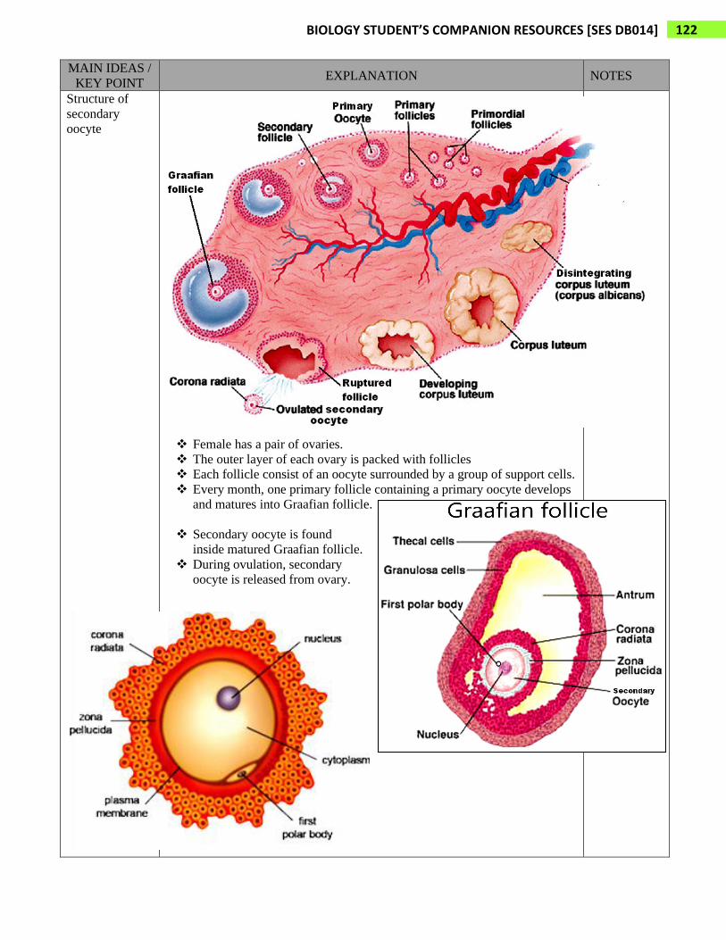
2) Acrosomal Reaction
• Before sperm cell reach the secondary oocyte,
– It moves through the layers of granulosa cells (corona radiata) and
reach zona pellucida
• Head of sperm cells bind to receptors on zona pellucida
– triggers the acrosome to burst, releasing hydrolytic enzyme
• The enzyme digest a path through zona pellucida to the surface of
secondary oocyte.
acrosomal vesicle hydrolytic enzyme
3) Fusion of Sperm Head Membrane and Oocyte
• Membrane proteins of sperm head bind to receptors on plasma membrane
of the secondary oocyte
• The two membrane fuse
– Releasing the nucleus and centrosome of sperm cells into the
cytoplasm of secondary oocyte

131 BIOLOGY STUDENT’S COMPANION RESOURCES [SES DB014]
MAIN IDEAS /
KEY POINT EXPLANATION NOTES
4) Cortical Reaction
• Immediately after the penetration of one sperm cell into secondary oocyte,
cortical granules release enzymes into zona pellucida via exocytosis to
destroy the sperm cell receptor
• These enzymes also thicken and harden the zona pellucida forming
fertilization envelope
• The entry of sperm cell triggers the completion of meiosis II forming
haploid (n) ovum and second polar body (n)
• At this stage, the nuclei of the sperm cell and ovum is known as pronuclei
• Fusion of male pronuclei and female pronuclei produce a diploid (2n)
zygote.
Recommended