General introduction
Chapter 1

10
Chapter 1
Hearing is very important in everyday life. Humans depend on their hearing in
a number of respects, such as for communication, socializing, learning, listen-
ing and to be warned for approaching danger. So when hearing is lost, it can
have a disabling effect on a person’s life. It has been estimated that in the
Netherlands > 1 million people have an average hearing loss of at least 35 dB
at the frequencies involved in speech perception (1, 2, and 4 kHz). In general,
two types of hearing loss can be distinguished: conductive and sensorineural
hearing loss. Conductive hearing loss is due to a blockage of the anatomical
cascade that conducts the sound waves from the outer to the inner ear.
Examples are middle ear infections, perforation of the eardrum, and otosclero-
sis, a disorder in which the stapes may become immobile because of exces-
sive growth of the bone. The other type of hearing loss is called
“sensorineural” and refers to damage of the cochlea and/or auditory nerve.
Sensorineural hearing loss can be induced by aging (presbycusis), loud music
or noise, viral or bacterial infections and drugs (such as aminoglycoside antibi-
otics or the anti-cancer drug cisplatin).
In this thesis will be investigated whether the side effects of cisplatin upon the
auditory system can be reduced or even prevented.
The peripheral auditory systemThe peripheral auditory system can be subdivided into three parts: the outer,
middle, and inner ear. The outer ear consists of the auricle and the external
auditory canal and plays a role in sound localization, partly by frequency-selec-
tive modification of the sound wave, while it is transferred to the tympanic
membrane (eardrum). Subsequently, these acoustic vibrations progress along
the tympanic cavity (= middle ear) via the cascade of three tiny ossicles: the
malleus (hammer), the incus (anvil) and the stapes (stirrup). Since there will
be an energy loss when sound is transferred directly from air to the fluid in the
cochlea, the ossicles amplify the sound and transfer the sound-induced vibra-
tions via the stapes to the oval window, efficiently converting the sound waves
into vibrations of the cochlear fluids.
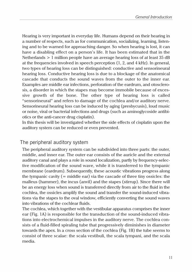
The cochlea, which together with the vestibular apparatus comprises the inner
ear (Fig. 1A) is responsible for the transduction of the sound-induced vibra-
tions into electrochemical impulses in the auditory nerve. The cochlea con-
sists of a fluid-filled spiraling tube that progressively diminishes in diameter
towards the apex. In a cross section of the cochlea (Fig. 1B) the tube seems to
consist of three scalae: the scala vestibuli, the scala tympani, and the scala
media.
11
General Introduction
At its base, the scala vestibuli is sealed off by the oval window membrane, to
which the stapes is connected. The scala tympani is closed at its base by
another thin elastic membrane: the round window membrane. The scala tym-
pani and the scala vestibuli are in open connection at the apex of the cochlea
by an opening known as the helicotrema. In between these two compart-
ments lies the scala media, which is separated from the scala vestibuli by
Reissner’s membrane and from the scala tympani by the basilar membrane.
On top of the latter is situated the organ of Corti, which contains ±16000
receptor cells (hair cells).
The scala tympani and scala vestibuli contain perilymph, which is like normal
extracellular fluid in composition and is at or near ground potential. The scala
media contains endolymph, which is more like intracellular fluid with high lev-
els of K+ and low levels of Na+. K+ provides the major charge carrier for sen-
sory transduction in the hair cells (Wangemann, 2002). The electrolyte
composition and potential of the endolymph (+ 80 mV) is regulated by an
energy-consuming mechanism involving multiple ion transport processes in
12
Chapter 1
OHC
SV
StV
SGNB
A
C
Figure 1: A. The structure of the human inner ear, containing the vestibular apparatus and the cochlea. B.
Cross section of the cochlea that shows the arrangement of the three scalae: the scala tympani (ST), scala vestibuli
(SV) and scala media (SM). The stria vascularis (StV) is situated on he lateral wall of the scala media. SGN: Spiral
ganglion cells; BM: basilar membrane; RM Reissner’s membrane. C. Detailed structure of the organ of Corti, which
contains the receptor cells: outer hair cells (OHCs) and inner hair cells (IHCs). TM: tectorial membrane.
the stria vascularis located in the lateral wall of the cochlea. The stria vascu-
laris is a complex, multilayered structure, containing three layers of different
cell types. Facing the endolymphatic space is a luminal layer of marginal cells,
which are characterized by the presence of numerous basolateral membranes
that are rich in mitochondria. The middle layer of the stria vascularis is com-
posed of the capillaries and intermediate cells. Facing the spiral ligament there
is a layer of multiple flat, basal cells.
The sensory epithelium of the inner ear, the organ of Corti, (Fig. 1C) is posi-
tioned on top of the basilar membrane. It contains two types of hair cells: the
outer hair cells (OHCs) and the inner hair cells (IHCs). The OHCs are arranged
in three rows and the IHCs in a single row. Both types contain a bundle of hair-
like structures, the so-called stereocilia, on the surface facing the scala media.
The tops of the stereocilia of the OHCs are inserted into holes in the tectorial
membrane. When a sound-induced vibration reaches the cochlea, the basilar
membrane moves up and down because of differences in the fluid pressure
between the scala vestibuli and the scala tympani. This movement of the basil-
ar membrane is accompanied by a shearing motion between the organ of
Corti and the tectorial membrane, causing the stereocilia to bend. In response
to these movements the hair cells generate a stream of electrical signals that
code the frequency, intensity and duration of the sound. The electrical signals
are generated in both IHCs and OHCs, but the neural information predomi-
nantly originates from the IHCs, which receive 90-95% of the afferent nerve
fibers (Spoendlin, 1972). The signals are transported through the eighth cranial
nerve (vestibulocochlear nerve) to the brain. The OHCs are responsible for the
sensitivity and frequency selectivity of the cochlea. It is thought that OHCs can
generate forces, by actively contracting and relaxing, enhancing the basilar
membrane motion (Brownell et al., 1985).
Several factors, such as noise, bacteria, viruses, aging, drugs and other chemi-
cal agents, may cause hearing loss (ototoxicity). Some of the clinically applied
ototoxic agents, such as aminoglycosides and cisplatin, have such a critical
role in the treatment of serious, life-threatening diseases that the ototoxic risk
can considered to be of less importance.
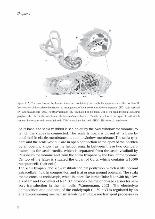
CisplatinThe biological activity of cis-diamminedichloroplatinum (II) or cisplatin (Fig. 2)
was discovered in 1965 by Rosenberg and co-workers during their studies to
the effects of an electric current on bacterial growth. They noticed that an
electrical field caused inhibition of Escherichia coli cell division (Rosenberg et
13
General Introduction
al., 1965). Further investigation indicated that the active agents responsible for
this effect were platinum salts, which were produced at the electrode during
electrolysis (Rosenberg et al., 1967). Several platinum complexes were tested
for their biological activity and some of them, including cisplatin, suppressed
cell division and induced filamentous growth of bacteria (Howle and Gale,
1970), which was known to be an indicator of DNA damage.
Therefore, it was plausible to assume that cisplatin would also interfere with
cell division in eukaryotes and subsequent studies revealed that cisplatin treat-
ment indeed results in arrested growth of tumors. Surprisingly, the trans-iso-
mer of cisplatin had no effect on tumor growth (Rosenberg et al., 1969).
The first clinical trials with cisplatin
started in the 1970s. Nowadays, cis-
platin is a widely used antineoplastic
agent. Cisplatin-based combination
chemotherapy displays significant
efficacy in the treatment of testis
tumors, ovarian carcinoma, squa-
mous cell carcinoma of the head and
neck, and non-small-cell carcinoma
of the lung. This anti-tumor effect is
due to a covalent binding between
the platinum atom and genomic or
mitochondrial DNA. Once cisplatin
enters the cell the chlorine atoms are
replaced by water, resulting in the formation of a positively charged aquated
species that can react easily with nitrogen or sulphur atoms in intracellular
macromolecules to form protein-, RNA-, or DNA-adducts. If there is another
potentially reactive site nearby, cisplatin can react further to form intra- and
inter-strand crosslinks (Kartalou and Essigmann, 2001), eventually leading to
apoptotic (programmed) cell death of tumor cells. The clinical use of cisplatin,
however, is limited by dose-dependent side effects, such as renal dysfunction,
peripheral neuropathies, hearing loss, nausea, vomiting, and myelosuppres-
sion. Severe nephrotoxicity was the most important dose limiting finding in
early clinical trials. With forced diuresis, this side effect has become more
manageable, leaving peripheral neuropathies and ototoxicity as the major side
effects of concern.
14
Chapter 1
Pt
Cl NH3
NH3
Cl
Figure 2: chemical structure of cisplatin
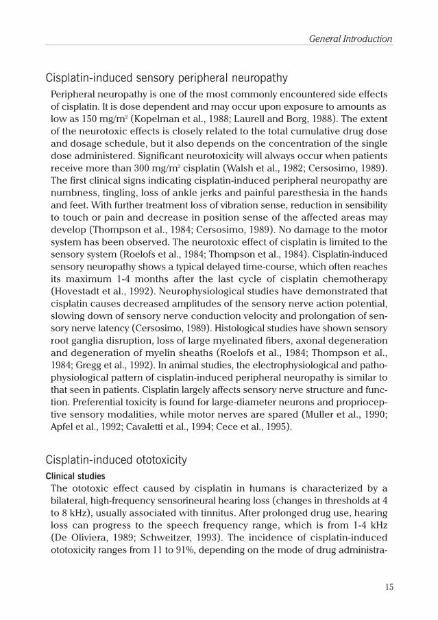
Cisplatin-induced sensory peripheral neuropathyPeripheral neuropathy is one of the most commonly encountered side effects
of cisplatin. It is dose dependent and may occur upon exposure to amounts as
low as 150 mg/m2 (Kopelman et al., 1988; Laurell and Borg, 1988). The extent
of the neurotoxic effects is closely related to the total cumulative drug dose
and dosage schedule, but it also depends on the concentration of the single
dose administered. Significant neurotoxicity will always occur when patients
receive more than 300 mg/m2 cisplatin (Walsh et al., 1982; Cersosimo, 1989).
The first clinical signs indicating cisplatin-induced peripheral neuropathy are
numbness, tingling, loss of ankle jerks and painful paresthesia in the hands
and feet. With further treatment loss of vibration sense, reduction in sensibility
to touch or pain and decrease in position sense of the affected areas may
develop (Thompson et al., 1984; Cersosimo, 1989). No damage to the motor
system has been observed. The neurotoxic effect of cisplatin is limited to the
sensory system (Roelofs et al., 1984; Thompson et al., 1984). Cisplatin-induced
sensory neuropathy shows a typical delayed time-course, which often reaches
its maximum 1-4 months after the last cycle of cisplatin chemotherapy
(Hovestadt et al., 1992). Neurophysiological studies have demonstrated that
cisplatin causes decreased amplitudes of the sensory nerve action potential,
slowing down of sensory nerve conduction velocity and prolongation of sen-
sory nerve latency (Cersosimo, 1989). Histological studies have shown sensory
root ganglia disruption, loss of large myelinated fibers, axonal degeneration
and degeneration of myelin sheaths (Roelofs et al., 1984; Thompson et al.,
1984; Gregg et al., 1992). In animal studies, the electrophysiological and patho-
physiological pattern of cisplatin-induced peripheral neuropathy is similar to
that seen in patients. Cisplatin largely affects sensory nerve structure and func-
tion. Preferential toxicity is found for large-diameter neurons and propriocep-
tive sensory modalities, while motor nerves are spared (Muller et al., 1990;
Apfel et al., 1992; Cavaletti et al., 1994; Cece et al., 1995).
Cisplatin-induced ototoxicityClinical studies
The ototoxic effect caused by cisplatin in humans is characterized by a
bilateral, high-frequency sensorineural hearing loss (changes in thresholds at 4
to 8 kHz), usually associated with tinnitus. After prolonged drug use, hearing
loss can progress to the speech frequency range, which is from 1-4 kHz
(De Oliviera, 1989; Schweitzer, 1993). The incidence of cisplatin-induced
ototoxicity ranges from 11 to 91%, depending on the mode of drug administra-
15
General Introduction
tion, dosage per treatment and cumulative dose (De Oliviera, 1989; Waters et
al., 1991). Also, age and pre-existing hearing loss can influence the severity of
cisplatin ototoxicity (Fausti et al., 1984). Bolus injections of 60 mg/m2, adminis-
tered once a week, have been shown to cause significant threshold differ-
ences after 6-12 months of treatment (Aguilar-Markulis et al., 1981). At
cumulative doses of 270 mg/m2 the first significant changes in auditory thresh-
old appear, especially at the high frequencies (Schaefer et al., 1985). At doses
of more than 450 mg/m2, 88% of the patients show a high-frequency hearing
loss (> 4 kHz) (McHaney et al., 1983). Only sporadically (incomplete) recovery
of cisplatin-induced hearing loss has been reported (Aguilar-Markulis et al.,
1981; Vermorken et al., 1983; Melamed et al., 1985; Laurell and Jungnelius,
1990). Histopathological studies in humans have shown loss of OHCs and IHCs
in the basal turn of the cochlea, degeneration of the stria vascularis, significant
decrease in the number of spiral ganglion cells, and damage to the cuticular
plate (Wright and Schaefer, 1982; Strauss et al., 1983; Hinojosa et al., 1995;
Hoistad et al., 1998).
Experimental studies In vitro cisplatin-models generally concern the toxicity of cisplatin with respect
to isolated cochlear OHCs (Saito et al., 1991, 1996; Sha et al., 2001; Devarajan
et al., 2002) and cochlear explants (Clerici et al., 1996; Zheng and Gao, 1996;
Kopke et al., 1997; Liu et al., 1998). However, most of the studies about the oto-
toxic effects of cisplatin have been performed in vivo in rodents: e.g., hamsters
(Melamed et al., 2000; Kaltenbach et al., 2002), chinchillas (Ford et al., 1997;
Tsukasaki et al., 2000), gerbils (Sie et al., 1997, 1999; Alam et al., 2000), rats
(Laurell et al., 1995, 1997; Meech et al., 1998; Hatzopoulos et al., 1999, 2001,
2002), guinea pigs (Tange, 1984; Schweitzer et al., 1986; Kohn et al., 1988;
Laurell and Engström, 1989, Laurell and Bagger-Sjöbäck 1991b; Schweitzer,
1993; Saito et al., 1994a, b; 1997a, b; Kohn et al., 1997; De Groot et al., 1997;
Cardinaal et al., 2000a-c; Klis et al., 2000, 2002), and sporadically in other mam-
mals such as dogs (Sockalingam et al., 2002) and rhesus monkeys (Stadnicki
et al., 1975). In these studies cisplatin was administered by intraperitoneal
injection at doses ranging from 0.75 to 4 mg/kg given repeatedly one to five
times per week for a total of 1-8 weeks or as a single dose of 5-18 mg/kg by
intraperitoneal injection or intravenous infusion.
The estimation of the onset of ototoxicity has been performed by measuring
the auditory brain stem response (ABR), electrocochleography (ECochG) or
by measuring the otoacoustic emissions (OAE). In animals the electrophysio-
logical and pathophysiological pattern of cisplatin-induced ototoxicity is simi-
16
Chapter 1
lar to that seen in patients. Cisplatin induces a dose-related permanent sen-
sorineural hearing loss starting at the high frequencies. Pathophysiological
studies in guinea pigs have shown that chronic cisplatin administration leads
to loss of OHCs, and at high doses also to loss of IHCs, with those in the basal
turn more severely affected than the ones in the middle and apical turns
(Nakai et al., 1982; Tange, 1984, Hoeve et al., 1988; Saito and Aran, 1994b;
Cardinaal et al., 2000a). Laurell and Bagger-Sjöbäck (1991a) have shown that
the morphological changes in the cochlea of guinea pigs after cisplatin expo-
sure occur in three stages. The first stage includes disturbance of the support-
ing cells surrounding the OHCs. The second stage was characterized by
degeneration of the OHCs; one of the first signs is loss of stereocilia and intra-
cellular vacuolation. The IHCs usually remain intact until all the OHCs have
degenerated. In the final stage collapse of the entire organ of Corti occurs. The
effects of cisplatin are not limited to the hair cells. Boheim and Bichler (1985)
have shown that cisplatin destroys the efferent auditory nerve fibers near the
OHCs. Others have found histological changes in the spiral ganglion cells of
guinea pigs, consisting of vacuolation of their cytoplasm (Cardinaal et al.,
2000b) and cell shrinkage (Van Ruijven et al., personal communication).
Furthermore, damage to the stria vascularis was observed in several studies in
rats and guinea pigs (Kohn et al., 1988, 1997; Meech et al., 1998; Campbell et
al., 1999; Cardinaal et al., 2000a, b). This damage consisted of blebbing and
vacuolation of the marginal cells and atrophy of the intermediate cells.
Besides these morphological changes, a smaller than normal endocochlear
potential (EP) was observed after administration of cisplatin to chinchillas
(Ford et al., 1997) and guinea pigs (Komune et al., 1981; Konishi et al., 1983;
Laurell and Engström 1989; Klis et al., 2000, 2002).
A number of animal studies (Stadnicki et al., 1975; Nakai et al., 1982; Stengs et
al., 1997; Cardinaal et al., 2000b; Klis et al., 2000, 2002) demonstrated that sev-
eral of the cisplatin-induced ototoxic effects (OHC damage, increase of hear-
ing threshold, decease of EP) recover after cessation of the cisplatin treatment.
Summarizing, cisplatin seems to have at least three targets in the cochlea, the
organ of Corti, the stria vascularis and the spiral ganglion cells. Presently, the
relation between the respective effects on these targets, e.g., whether one is
causally related to the other and how these targets are involved in recovery, is
unknown.
17
General Introduction
Protection against cisplatin-induced side effectsSeveral attempts have been made to prevent cisplatin-induced side effects,
e.g. by changing the dose, the method of administration or even by replacing
cisplatin with a non-toxic analogue (e.g. carboplatin). However, these efforts
proved to be unsatisfactory. Thus, another approach was investigated to over-
come the toxic effects of cisplatin: pharmacological intervention. Several
classes of compounds, such as neurotrophins and sulphur-containing com-
pounds, have been found to protect against cisplatin-induced neuro- and oto-
toxicity. In this section the most significant results from experiments with
different classes of compounds that protect against cisplatin-induced ototoxic-
ity will be reviewed.
FosfomycinThe first agent tested for its possible protection against cisplatin ototoxicity
was fosfomycin. Both Ohtani et al. (1985) and Schweitzer et al. (1986, 1993)
showed that significantly less OHC loss occurs when animals are treated with
1 mg/kg/day cisplatin in combination with 300 mg/kg/day fosfomycin.
However, these results could not be reproduced in later studies performed by
Church et al. (1995) and Kaltenbach et al. (1997), in which higher doses of cis-
platin (3 mg/kg/day, once every other day) were used in combination with fos-
fomycin co-treatment. These seemingly contradictory results could be
explained by the observation of Ohtani et al. (1985) that at high concentrations
of cisplatin (5 mg/kg/day) fosfomycin has no protective effect.
Sulphur-containing compoundsThe application of sulphur-containing compounds was based on the hypothe-
sis that damage caused by cisplatin is due to formation of free radicals, which
interfere with the antioxidant defense system, resulting in oxidative stress
(Rybak et al., 1995, Ravi et al., 1995). Severe oxidative stress produces major
disruption of cell metabolism, resulting in cell death (Evans and Halliwell,
1999). Several compounds that may reduce free radical formation, have been
described, e.g. sodium thiosulfate (STS), diethyldithiocarbamate (DDTC), 4-
methylthiobenzoic acid (MTBA), L- and D-methionine, lipoic acid, and salicyl-
ate. In 1985, it was shown that sodium thiosulfate, already known to prevent
cisplatin nephrotoxicity, prevents from neurotoxicity when administered
simultaneously with cisplatin in patients (Markman et al., 1985). This com-
pound also seemed to protect guinea pigs from cisplatin-induced hearing loss
(Otto et al., 1988; Church et al., 1995; Kaltenbach et al., 1997; Saito et al.,
1997a). However, it has been shown that STS reacts with cisplatin to form
18
Chapter 1
covalently bound complexes, thus hampering cisplatin’s anti-tumor activity
(Howell et al., 1982). Therefore, recent experiments have focused on the
direct application of STS into the inner ear in order to selectively bind cisplatin
in the cochlea (Wang et al., 2002). Another protective agent is the sulphur-con-
taining amino acid methionine. Both the naturally occurring L-methionine and
its synthetic analog D-methionine have been tested for their otoprotective
properties. Campbell et al. (1996, 1999) have showed excellent protection by
D-methionine from cisplatin ototoxicity in rats. Unfortunately, also L- and D-
methionine lowered the systemic exposure to cisplatin (Reser et al., 1999;
Ekborn et al., 2002; Vrana and Brabec, 2002). Therefore, topical administration
of L- or D-methionine directly onto the round window membrane has been
studied (Li et al., 2001; Korver et al., 2002). With this approach both com-
pounds completely protect the inner ear from cisplatin-induced ototoxicity.
Other sulphur-containing compounds that provide protection against cisplatin
ototoxicity after systemic application are diethyldithiocarbamate (Church et
al., 1995; Kaltenbach et al., 1997; Rybak et al., 1995; Walker et al., 1994), 4-
methylthiobenzoic acid (Rybak et al., 1997; Kamimura et al., 1999), salicylate
(Li et al., 2002) and lipoic acid (Rybak et al., 1999a-c). However, excessive
amounts of these compounds are necessary to realize a protective effect
against cisplatin ototoxicity; the concentration of the sulphur-containing com-
pounds exceeds the cisplatin dose by 5 to 100 times (e.g., 300 mg/kg D-methio-
nine versus 8 mg/kg cisplatin). At these concentrations the sulphur-containing
compounds are known to react directly with cisplatin resulting in a lowered
cytotoxic effect of the drug (Ekborn et al., 2002).
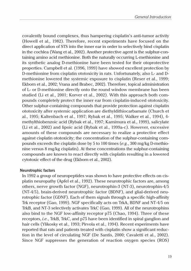
Neurotrophic factorsIn 1992 a group of neuropeptides was shown to have protective effects on cis-
platin neuropathy (Apfel et al., 1992). These neurotrophic factors are, among
others, nerve growth factor (NGF), neurotrophin-3 (NT-3), neurotrophin-4/5
(NT-4/5), brain-derived neurotrophic factor (BDNF), and glial-derived neu-
rotrophic factor (GDNF). Each of them signals through a specific high-affinity
Trk receptor (Gao, 1999). NGF specifically acts on TrkA, BDNF and NT-4/5 on
TrkB, and NT-3 selectively activates TrkC (Gao, 1999). All of the neurotrophins
also bind to the NGF low-affinity receptor p75 (Chao, 1994). Three of these
receptors, i.e., TrkB, TrkC, and p75 have been identified in spiral ganglion and
hair cells (Ylikosky et al., 1993; Pirvola et al., 1994). Recent experiments have
reported that rats and patients treated with cisplatin show a significant reduc-
tion in the level of circulating NGF (De Santis, 2000; Cavaletti et al., 2002).
Since NGF suppresses the generation of reaction oxygen species (ROS)
19
General Introduction
(Dugan et al., 1997), reduction of NGF could result in an increased generation
of ROS, eventually leading to peripheral neuropathy or ototoxicity. Most of the
experiments with growth factors have been performed in vitro with organo-
typic culture of cochlear explants or vestibular neuro-epithelia. BDNF and NT-
4/5 were found to delay further degeneration of spiral ganglion cells. This
protective effect, although somewhat smaller, was also observed when NT-3
was administered to spiral ganglion cells (Zheng et al., 1995, 1996). However,
no attenuation of cisplatin-induced hair cell loss was observed with these
compounds (Zheng and Gao, 1996). The results of studies performed with
NGF are rather conflicting. Some authors have reported that NGF protects spi-
ral ganglion cells from cisplatin-induced toxicity (Malgrange et al., 1994), while
others consider NGF to be ineffective (Zheng et al., 1995, 1996). Kuang et al.
(1999) recently showed that locally delivered GDNF protects guinea pig
cochleas when it was administered in combination with systemically applied
cisplatin. This is the only study known to show protection in vivo by one of the
above co-treatments at a dose in the µg-range, suggesting a specific mecha-
nism against ototoxicity at the hair cell level. Recently, another group of pep-
tides known as melanocortins has been demonstrated to protect against
cisplatin-induced ototoxicity in vivo at this dose range.
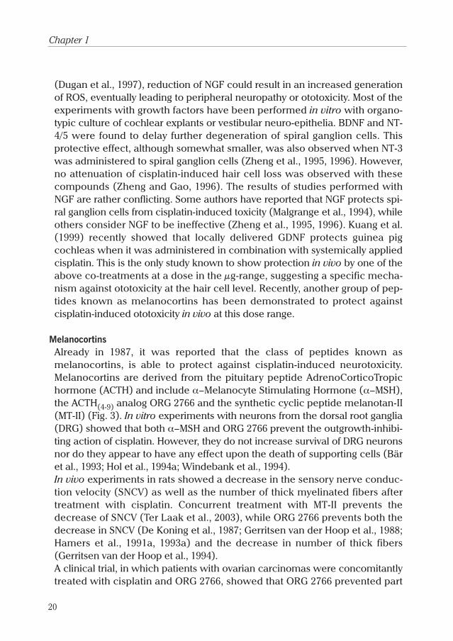
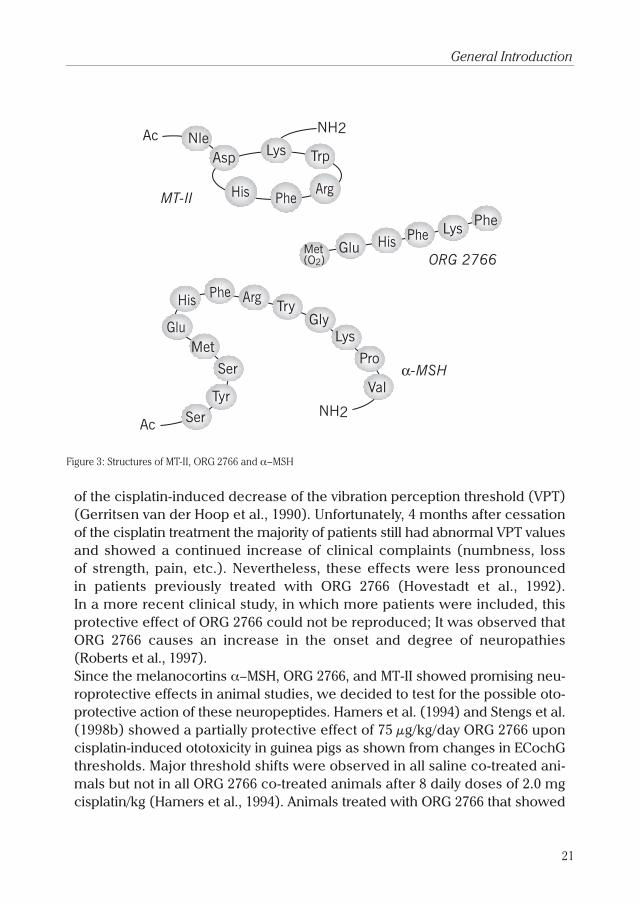
MelanocortinsAlready in 1987, it was reported that the class of peptides known as
melanocortins, is able to protect against cisplatin-induced neurotoxicity.
Melanocortins are derived from the pituitary peptide AdrenoCorticoTropic
hormone (ACTH) and include α−Melanocyte Stimulating Hormone (α−MSH),
the ACTH(4-9) analog ORG 2766 and the synthetic cyclic peptide melanotan-II
(MT-II) (Fig. 3). In vitro experiments with neurons from the dorsal root ganglia
(DRG) showed that both α−MSH and ORG 2766 prevent the outgrowth-inhibi-
ting action of cisplatin. However, they do not increase survival of DRG neurons
nor do they appear to have any effect upon the death of supporting cells (Bär
et al., 1993; Hol et al., 1994a; Windebank et al., 1994).
In vivo experiments in rats showed a decrease in the sensory nerve conduc-
tion velocity (SNCV) as well as the number of thick myelinated fibers after
treatment with cisplatin. Concurrent treatment with MT-II prevents the
decrease of SNCV (Ter Laak et al., 2003), while ORG 2766 prevents both the
decrease in SNCV (De Koning et al., 1987; Gerritsen van der Hoop et al., 1988;
Hamers et al., 1991a, 1993a) and the decrease in number of thick fibers
(Gerritsen van der Hoop et al., 1994).
A clinical trial, in which patients with ovarian carcinomas were concomitantly
treated with cisplatin and ORG 2766, showed that ORG 2766 prevented part
20
Chapter 1
of the cisplatin-induced decrease of the vibration perception threshold (VPT)
(Gerritsen van der Hoop et al., 1990). Unfortunately, 4 months after cessation
of the cisplatin treatment the majority of patients still had abnormal VPT values
and showed a continued increase of clinical complaints (numbness, loss
of strength, pain, etc.). Nevertheless, these effects were less pronounced
in patients previously treated with ORG 2766 (Hovestadt et al., 1992).
In a more recent clinical study, in which more patients were included, this
protective effect of ORG 2766 could not be reproduced; It was observed that
ORG 2766 causes an increase in the onset and degree of neuropathies
(Roberts et al., 1997).
Since the melanocortins α−MSH, ORG 2766, and MT-II showed promising neu-
roprotective effects in animal studies, we decided to test for the possible oto-
protective action of these neuropeptides. Hamers et al. (1994) and Stengs et al.
(1998b) showed a partially protective effect of 75 µg/kg/day ORG 2766 upon
cisplatin-induced ototoxicity in guinea pigs as shown from changes in ECochG
thresholds. Major threshold shifts were observed in all saline co-treated ani-
mals but not in all ORG 2766 co-treated animals after 8 daily doses of 2.0 mg
cisplatin/kg (Hamers et al., 1994). Animals treated with ORG 2766 that showed
21
General Introduction
Ac Nle
MT-II
ORG 2766
Asp
His
His
PheArg
TrpLys
NH2
Met(O2)
Lys PhePhe
Glu
α-MSH
Arg
Lys
NH2
Pro
Val
Ac SerTyr
Try
Ser
MetGlu
His Phe
Gly
Figure 3: Structures of MT-II, ORG 2766 and α−MSH
no protection, tended to have slightly worse CAP input-output curves than the
saline co-treated ones, but OHC survival was significantly better in these ‘non-
responders’ than in saline-treated controls (De Groot et al., 1997). Heijmen et
al. (1999) demonstrated that treatment of albino guinea pigs with daily injec-
tions of cisplatin (2 mg/kg/day i.p. for 8 days) and concomitant injections of
α−MSH (75 µg/kg/day s.c. for 9 days) results in a considerable number of ani-
mals with preserved hearing after cessation of cisplatin treatment. This was
not found in the cisplatin/saline treated group.
Thus, these experiments have demonstrated that ACTH-derived neuropeptides
(melanocortins) are able to protect against cisplatin-induced ototoxicity.
However, the mechanism by which these neuropeptides exert their otoprotec-
tive effect is not yet known. Their protective effect cannot be due to a direct
interaction between the melanocortins and cisplatin, since both α−MSH and
ORG 2766 were administered in much smaller doses than cisplatin. The actual
target might be the intermediate cells in the stria vascularis. Meyer zum
Gottesberge (2000) has suggested that these intermediate cells, which are
actually melanocytes (Hilding and Ginzberg, 1977), are under α−MSH control.
α−MSH may act as an emergency system in the regulation of inner ear home-
ostasis and function. ACTH-derived neuropeptides, such as α−MSH, are known
to strongly bind to G-protein-coupled melanocortin (MC) receptors (Mountjoy
et al., 1992). Therefore, α−MSH and ORG 2766 may exert their protective action
by activating a MC-like receptor in the intermediate cells, thus preventing cis-
platin from damaging the stria vascularis. Although the epidermal melanocytes
have been shown to contain a MC-receptor that specifically binds to α−MSH
(Tsatmali et al., 2002), the presence of such a MC-receptor has yet to be
demonstrated in the inner ear. Moreover, none of the known MC-receptors
binds to ORG 2766. So the actual mechanism of action in the prevention of cis-
platin-induced ototoxicity of the ACTH-derived neuropeptides is still unclear.
Methods for studying ototoxicityIn this thesis we are mostly interested in the prevention of cisplatin-induced
ototoxicity. Cisplatin causes structural damage and functional loss in several
tissues of the cochlea and the auditory nerve. In order to study these effects
experimentally we have used two approaches: electrocochleography and his-
tology.
22
Chapter 1
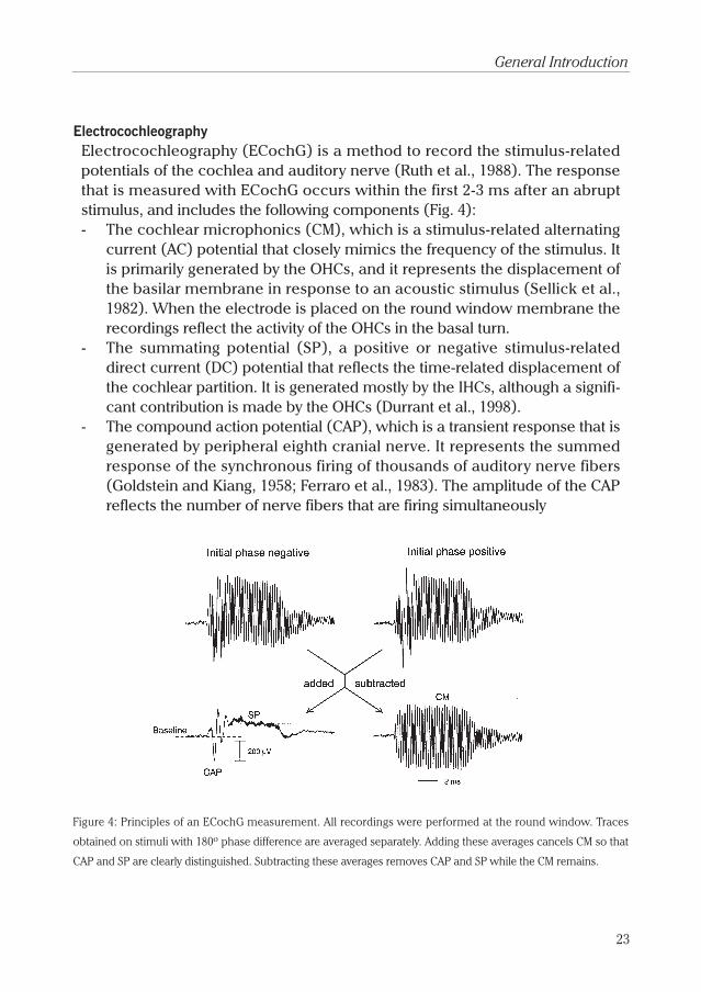
ElectrocochleographyElectrocochleography (ECochG) is a method to record the stimulus-related
potentials of the cochlea and auditory nerve (Ruth et al., 1988). The response
that is measured with ECochG occurs within the first 2-3 ms after an abrupt
stimulus, and includes the following components (Fig. 4):
- The cochlear microphonics (CM), which is a stimulus-related alternating
current (AC) potential that closely mimics the frequency of the stimulus. It
is primarily generated by the OHCs, and it represents the displacement of
the basilar membrane in response to an acoustic stimulus (Sellick et al.,
1982). When the electrode is placed on the round window membrane the
recordings reflect the activity of the OHCs in the basal turn.
- The summating potential (SP), a positive or negative stimulus-related
direct current (DC) potential that reflects the time-related displacement of
the cochlear partition. It is generated mostly by the IHCs, although a signifi-
cant contribution is made by the OHCs (Durrant et al., 1998).
- The compound action potential (CAP), which is a transient response that is
generated by peripheral eighth cranial nerve. It represents the summed
response of the synchronous firing of thousands of auditory nerve fibers
(Goldstein and Kiang, 1958; Ferraro et al., 1983). The amplitude of the CAP
reflects the number of nerve fibers that are firing simultaneously
Figure 4: Principles of an ECochG measurement. All recordings were performed at the round window. Traces
obtained on stimuli with 180ο phase difference are averaged separately. Adding these averages cancels CM so that
CAP and SP are clearly distinguished. Subtracting these averages removes CAP and SP while the CM remains.
23
General Introduction
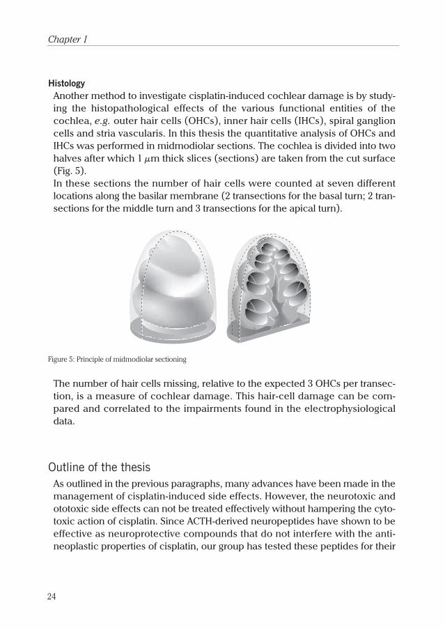
HistologyAnother method to investigate cisplatin-induced cochlear damage is by study-
ing the histopathological effects of the various functional entities of the
cochlea, e.g. outer hair cells (OHCs), inner hair cells (IHCs), spiral ganglion
cells and stria vascularis. In this thesis the quantitative analysis of OHCs and
IHCs was performed in midmodiolar sections. The cochlea is divided into two
halves after which 1 µm thick slices (sections) are taken from the cut surface
(Fig. 5).
In these sections the number of hair cells were counted at seven different
locations along the basilar membrane (2 transections for the basal turn; 2 tran-
sections for the middle turn and 3 transections for the apical turn).
Figure 5: Principle of midmodiolar sectioning
The number of hair cells missing, relative to the expected 3 OHCs per transec-
tion, is a measure of cochlear damage. This hair-cell damage can be com-
pared and correlated to the impairments found in the electrophysiological
data.
Outline of the thesisAs outlined in the previous paragraphs, many advances have been made in the
management of cisplatin-induced side effects. However, the neurotoxic and
ototoxic side effects can not be treated effectively without hampering the cyto-
toxic action of cisplatin. Since ACTH-derived neuropeptides have shown to be
effective as neuroprotective compounds that do not interfere with the anti-
neoplastic properties of cisplatin, our group has tested these peptides for their
24
Chapter 1
possible otoprotective action. In previous studies, in which the drugs were
administered during a fixed number of days, it was shown that both α−MSH
and ORG 2766 have a beneficial effect on cisplatin-induced ototoxicity.
However, since only part of the animals was protected, further optimization of
the melanocortin treatment was considered to be necessary. Furthermore, the
mechanism underlying the action of melanocortins had to be investigated.
This thesis is based on a longitudinal animal model in which an implanted
electrode allows repeated measurements of cochlear sensitivity. In the first
part the compounds are administrated systemically, in the second part we
focus on a cochlear model in which the compounds can be delivered directly
into the cochlea via a mini-osmotic pump system.
In the first study (Chapter 2) the longitudinal animal model is used to investi-
gate the otoprotective effects of the new and more potent melanocortin-recep-
tor agonist melanotan-II (MT-II). This synthetic melanocortin has been
effective in the protection of cisplatin-induced peripheral neuropathy (Ter
Laak et al., 2003). The objective of the study was to investigate whether MT-II
is able to delay the occurrence of cisplatin-induced ototoxicity and to effect
subsequent recovery. Animals were implanted with a permanent electrode
and treated daily with cisplatin and MT-II until a 40 dB CAP threshold shift at 8
kHz occurred. Subsequently, cisplatin treatment was stopped and CAP recov-
ery was studied for another 2 weeks.
A subsequent study was performed to test the protective effects of the
melanocortin peptides α−MSH and ORG 2766 in the same longitudinal animal
model (Chapter 3). We investigated whether these peptides delay the occur-
rence of the cisplatin-induced shift in auditory threshold, and whether they
effect the subsequent recovery of the cochlear action potential (CAP) and
endocochlear potential (EP). Both peptides showed significant ameliorating
effects on the recovery of the CAP.
Since it was suggested that this recovery might be due to reversible strial fail-
ure and EP recovery, in the third study (Chapter 4) we have investigated the
time course of EP recovery and whether the recovery of the EP is influenced
by α−MSH co-treatment. In an experimental set-up similar to that of the sec-
ond study, the EP and CAP were measured 1, 2, or 3 days after the criterion
threshold shift at 8 kHz was reached.
25
General Introduction
To gain more insight into the mechanism underlying α−MSH action, the sec-
ond objective of this thesis, we switched to the cochlear model in which
effects of cisplatin and melanocortins could be studied without the possibly
confounding influence of systemic administration. In the fourth study
(Chapter 5) this animal model was used; cisplatin was administered directly
into the cochlea via a mini-osmotic pump system while α−MSH or saline were
administered daily by systemic injection. This approach decreased interanimal
variability, which made it easier to quantify the efficacy of systemic α−MSH co-
treatment. Furthermore, since cisplatin was delivered directly to the cochlea,
any ameliorating effects of α−MSH would indicate that treatment with α−MSH
probably involves a cochlear target.
To complement the previous study a mirror experiment was performed in the
fifth study (Chapter 6). Guinea pigs that were implanted with a permanent
electrode and a mini-osmotic pump, pumping either saline or α−MSH, were
co-treated systemically with cisplatin until the 40 dB threshold shift at 8 kHz
was reached. Then, cisplatin treatment was stopped, but intracochlear perfu-
sion and electrocochleography were continued for 10 days to evaluate possi-
ble effects of local α−MSH treatment on recovery.
26
Chapter 1
Recommended