Open Access
1Bond AR, et al. Open Heart 2018;5:e000716. doi:10.1136/openhrt-2017-000716
AbstrActBackground The right ventricle (RV) is not designed to sustain high pressure leading to failure. There are no current medications to help RV contraction, so further information is required on adaption of the RV to such hypertension.Methods The Right Ventricle in Children (RVENCH) study assessed infants with congenital heart disease undergoing cardiac surgery with hypertensive RV. Clinical and echocardiographic data were recorded, and samples of RV were taken from matched infants, analysed for proteomics and compared between pathologies and with clinical and echocardiographic outcome data.Results Those with tetralogy of Fallot (TOF) were significantly more cyanosed than those with ventricular septal defect (median oxygen saturation 83% vs 98%, P=0.0038), had significantly stiffer RV (tricuspid E wave/A wave ratio 1.95 vs 0.84, P=0.009) and had most had restrictive physiology. Gene ontology in TOF, with enrichment analysis, demonstrated significant increase in proteins of contractile mechanisms and those of calmodulin, actin binding and others associated with contractility than inventricular septal defect. Structural proteins were also found to be higher in association with sarcomeric function: Z-disc, M-Band and thin-filament proteins. Remaining proteins associated with actin binding, calcium signalling and myocyte cytoskeletal development. Phosphopeptide enrichment led to higher levels of calcium signalling proteins in TOF.Conclusion This is the first demonstration that those with an RV, which is stiff and hypertensive in TOF, have a range of altered proteins, often in calcium signalling pathways. Information about these alterations might guide treatment options both in terms of individualised therapy or inotropic support for the Right ventricle when hypertensive due to pulmoanry hypertension or congenital heart disease.
BaCkgRoundIt is estimated that about 1% of all live-born babies are affected by cardiac congenital malformations. Intracardiac surgical repair in these patients is associated with a low mortality in infants and children (<5%). More impor-tantly, a growing number of patients survive into adulthood with hypertensive right ventri-cles (RV). Heart failure due to RV dysfunction is a major cause of morbidity in this popula-tion, and a significant proportion of them will die as a result. There is recent evidence that the
► Additional material is published online only. To view please visit the journal online (http:// dx. doi. org/ 10. 1136/ openhrt- 2017- 000716).
To cite: Bond AR, Iacobazzi D, Abdul-Ghani S, et al. Changes in contractile protein expression are linked to ventricular stiffness in infants with pulmonary hypertension or right ventricular hypertrophy due to congenital heart disease. Open Heart 2018;5:e000716. doi:10.1136/openhrt-2017-000716
Received 3 September 2017Revised 15 November 2017Accepted 18 November 2017
1Clinical Sciences, Bristol Heart Institute, Bristol Royal Infirmary, Bristol, UK2Proteomics Facility, University of Bristol, Bristol, UK3Department of Congenital Heart Disease, King David Building, Bristol, UK
Correspondence toProfessor Robert M R Tulloh; Robert. Tulloh@ bristol. ac. uk
Changes in contractile protein expression are linked to ventricular stiffness in infants with pulmonary hypertension or right ventricular hypertrophy due to congenital heart disease
Andrew R Bond,1 Dominga Iacobazzi,1 Safa Abdul-Ghani,1 Mohammed Ghorbel,1 Kate Heesom,2 Mariangela Wilson,2 Christopher Gillett,3 Sarah J George,1 Massimo Caputo,1,3 Saadeh Suleiman,1 Robert M R Tulloh1,3
Basic and translational research
Key questions
What is already known about this subject? ► Proteomic changes in myocardium have been studied in adult patients with or without ischaemic arrest, and our laboratory has identified genetic changes in the myocardium of children undergoing cardiac surgery.
What does this study add? ► To date, this is a unique study that identifies the proteomic differences in right ventricular myocardium from infants undergoing cardiac surgery for cyanotic and acyanotic heart diseases with a hypertensive right ventricle.
How might this impact on clinical practice? ► It is of clinical concern how to manage such children, as to the timing of surgery, the type of inotrope that might benefit the child with each cardiac condition and individual mechanisms that might be accessed in precision medicine to determine the best therapy for each child. This study is the first step in that process to identify the mechanisms involved and the best treatment for the individual child.
on 8 April 2019 by guest. P
rotected by copyright.http://openheart.bm
j.com/
Open H
eart: first published as 10.1136/openhrt-2017-000716 on 3 January 2018. Dow
nloaded from
Open Heart
2 Bond AR, et al. Open Heart 2018;5:e000716. doi:10.1136/openhrt-2017-000716
mortality is increasing in the teenage population who are dying of the complications of cardiac surgery performed over a decade ago, and it is our responsibility to improve this for the next generation.1 The gravity of the problem is well explained by the 2009 WHO annual report that puts the disability-adjusted life-year for congenital heart disease higher than diseases such as diabetes or hyperten-sion.2 At present, pharmacotherapy of heart failure may improve clinical symptoms, but there is no targeted phar-macotherapy to improve RV systolic and diastolic function or to prevent or even reverse the remodelling of the RV. Significant morbidity secondary to RV dysfunction leads to greater length of time on the paediatric intensive care unit (PICU) postoperatively and more expensive repeat inter-ventions and reoperations. Since each day on the PICU can cost up to £2000, and each reoperation costs £25000, it will be beneficial to the healthcare economy to improve the quality of individualised care we deliver to these chil-dren.
One drawback in this area is the fact that patients with RV dysfunction are grouped together despite the fact that there are different congenital cardiac disorders that will trigger different functional, molecular and cellular remodelling in the RV. It is therefore vital to dissect potential differences in cell signalling and remodelling in patients with different causes of RV dysfunction. Consequently, a greater under-standing of the underlying mechanisms of RV function in different congenital cardiac disorder groups will help to formulate strategies targeting individual cardiac disorders. The National Heart, Lung, and Blood Institute (Bethesda, Maryland, USA) has been awarded top priority to research on pathophysiology of the RV. RV dysfunction is caused by changes in the cell fate and tissue composition. These changes occur due to cell signalling that may be initiated by various factors including oxidative stress and lead to alter-ations in cell survival, structural (cytoskeletal) and extra-cellular proteins, protease activity and calcium handling properties.3 From our recent studies, we have shown that chronic hypoxaemia in cyanotic children with tetralogy of Fallot (TOF) induced the expression of genes associated with apoptosis and reduced the expression of genes associ-ated with normal myocyte contractility and function.3
It is well established that convergence of the signalling pathways activated by tumour growth factor-β, Wnts and cadherin are critical for development and its importance re-emerge in disease. It plays a key role in cell fate (growth and survival) and in matrix synthesis and remodelling, which are all pertinent processes in RV dysfunction. We already have extensive evidence for their important role in blood vessel remodelling and propose that modulation of these signalling pathways contribute to RV dysfunction. Although right ventricular failure (RVF) is the hallmark of pulmonary arterial hypertension (PAH), the mechanism of RVF is unclear. Development of PAH-induced RVF is associated with an increased reactive oxygen species (ROS) production. Increases in oxidative stress lead to generation of nitro-tyrosine residues in tissue inhibitor of metallopro-teinase and liberate active matrix metalloproteinase.4
The purpose of this study is to investigate the cellular signalling changes associated with RV hypertension in the setting of congenital heart disease. This would lead to a better understanding of the determinants of RV function in patients with hypertrophy associated with different congen-ital cardiac malformations. It was proposed that:1. Changes in key myocardial cellular signalling pathways
triggered by hypertension-induced RV dysfunction in congenital cardiac disorders depend on the type of malformation and systemic oxygen saturation.
2. The pathology-induced cellular and molecular chang-es will result in differences in postoperative stay within the hospital.
3. Identifying differences in the cellular signalling pathways between those with large septal defects and those with TOF might enable us to tailor therapies for each pathology to improve outcome.
The clinical outcome in our patients often depends on the ability of the RV to cope with the anatomical substrate and the effect of cardiopulmonary bypass. We hoped to be able to understand better the effect of these and hence to modify our treatment accordingly. This might lead to better and perhaps individualised care for our patients undergoing cardiopulmonary bypass for congenital heart diseases.
Patients and methodsInclusion criteriaWe compared patients with RV hypertension who under-went cardiopulmonary bypass and consented for inclu-sion between 1 July 2015 and 31 March 2017. We report on two groups of children with right ventricular pressures at systemic level: group 1: pulmonary hypertension due to large septal defects, such as ventricular septal defect (VSD) and group 2: obstructed RV (as in TOF). In group 1, left to right shunt was the cause of RV hypertension. In group 2, the children were cyanosed, and the RV was functioning under a significant obstructive afterload.
Exclusion criteriaChildren with additional syndromes were excluded (apart from Trisomy 21) due to the unknown effect on ventricular function and protein analysis. Parents who were not able to consider the information at least 1 week before opera-tion were excluded, hence excluding any children under-going emergency surgery. In addition, we excluded those with sepsis or coexisting morbidity, which might alter the protein analysis.
Fully informed consent was obtained from parents prior to admission for operation. Clinical data were obtained from patient’s clinical records and Heartsuite Database at the time of cardiac surgery. Demographic data were recorded, including details of cardiac anatomy and systemic oxygen saturations. At operation, tissue samples were obtained from right ventricular myocardium as soon as possible after instituting cardiopulmonary bypass to minimise the effect of this on protein analysis.
Protein analysis technique is online supplement.
on 8 April 2019 by guest. P
rotected by copyright.http://openheart.bm
j.com/
Open H
eart: first published as 10.1136/openhrt-2017-000716 on 3 January 2018. Dow
nloaded from
3Bond AR, et al. Open Heart 2018;5:e000716. doi:10.1136/openhrt-2017-000716
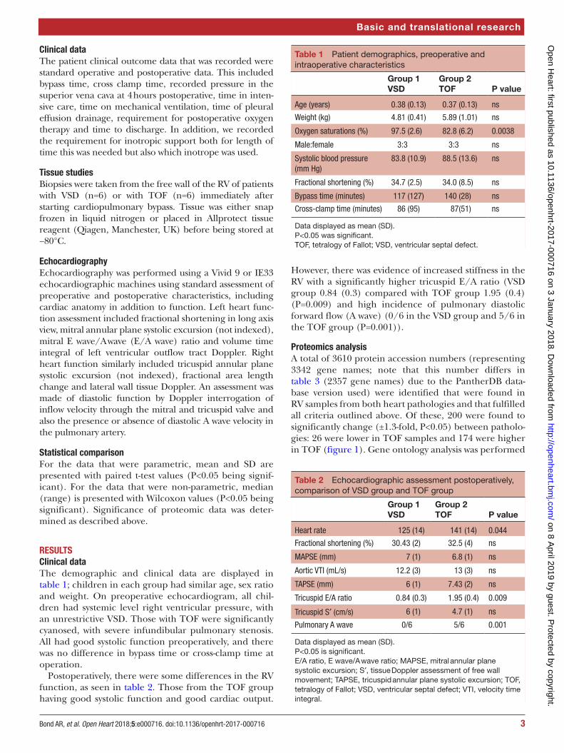
Table 1 Patient demographics, preoperative and intraoperative characteristics
Group 1 VSD
Group 2 TOF P value
Age (years) 0.38 (0.13) 0.37 (0.13) ns
Weight (kg) 4.81 (0.41) 5.89 (1.01) ns
Oxygen saturations (%) 97.5 (2.6) 82.8 (6.2) 0.0038
Male:female 3:3 3:3 ns
Systolic blood pressure (mm Hg)
83.8 (10.9) 88.5 (13.6) ns
Fractional shortening (%) 34.7 (2.5) 34.0 (8.5) ns
Bypass time (minutes) 117 (127) 140 (28) ns
Cross-clamp time (minutes) 86 (95) 87(51) ns
Data displayed as mean (SD).P<0.05 was significant.TOF, tetralogy of Fallot; VSD, ventricular septal defect.
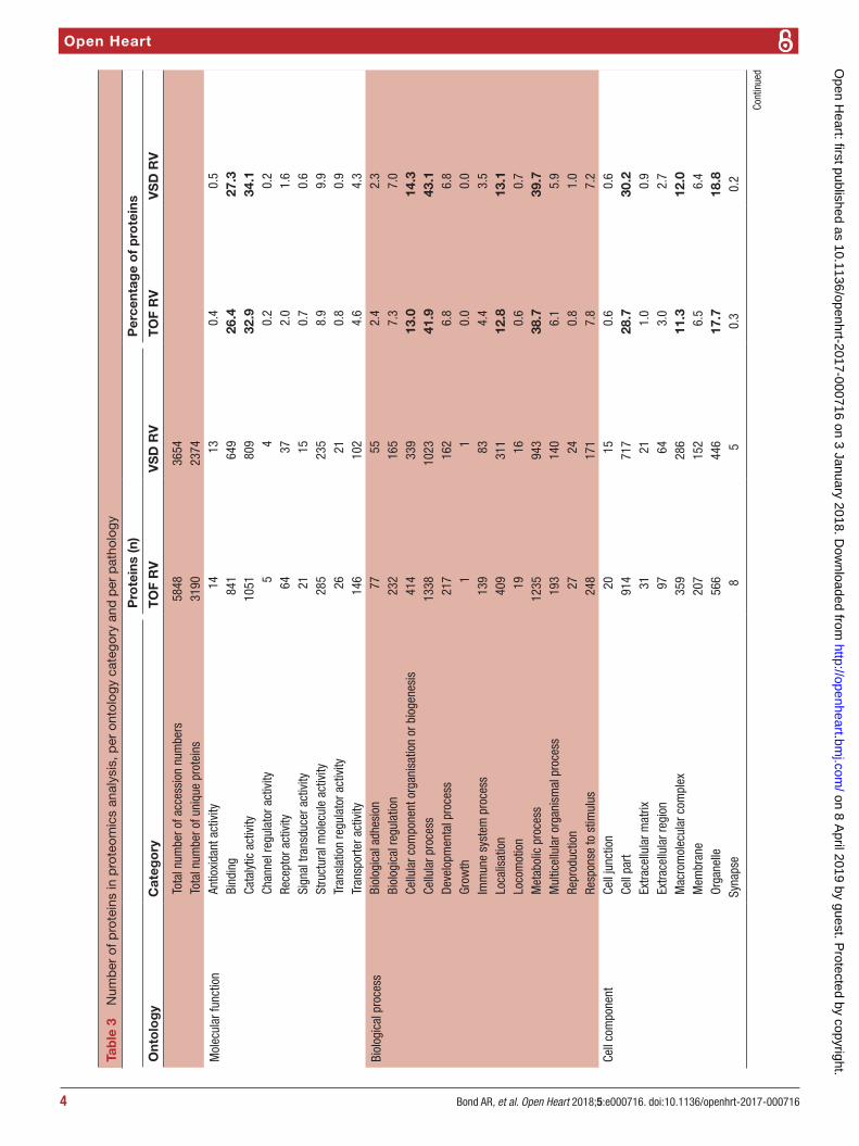
Table 2 Echocardiographic assessment postoperatively, comparison of VSD group and TOF group
Group 1 VSD
Group 2 TOF P value
Heart rate 125 (14) 141 (14) 0.044
Fractional shortening (%) 30.43 (2) 32.5 (4) ns
MAPSE (mm) 7 (1) 6.8 (1) ns
Aortic VTI (mL/s) 12.2 (3) 13 (3) ns
TAPSE (mm) 6 (1) 7.43 (2) ns
Tricuspid E/A ratio 0.84 (0.3) 1.95 (0.4) 0.009
Tricuspid S′ (cm/s) 6 (1) 4.7 (1) ns
Pulmonary A wave 0/6 5/6 0.001
Data displayed as mean (SD).P<0.05 is significant.E/A ratio, E wave/A wave ratio; MAPSE, mitral annular plane systolic excursion; S′, tissue Doppler assessment of free wall movement; TAPSE, tricuspid annular plane systolic excursion; TOF, tetralogy of Fallot; VSD, ventricular septal defect; VTI, velocity time integral.
Basic and translational research
Clinical dataThe patient clinical outcome data that was recorded were standard operative and postoperative data. This included bypass time, cross clamp time, recorded pressure in the superior vena cava at 4 hours postoperative, time in inten-sive care, time on mechanical ventilation, time of pleural effusion drainage, requirement for postoperative oxygen therapy and time to discharge. In addition, we recorded the requirement for inotropic support both for length of time this was needed but also which inotrope was used.
Tissue studiesBiopsies were taken from the free wall of the RV of patients with VSD (n=6) or with TOF (n=6) immediately after starting cardiopulmonary bypass. Tissue was either snap frozen in liquid nitrogen or placed in Allprotect tissue reagent (Qiagen, Manchester, UK) before being stored at −80°C.
EchocardiographyEchocardiography was performed using a Vivid 9 or IE33 echocardiographic machines using standard assessment of preoperative and postoperative characteristics, including cardiac anatomy in addition to function. Left heart func-tion assessment included fractional shortening in long axis view, mitral annular plane systolic excursion (not indexed), mitral E wave/A wave (E/A wave) ratio and volume time integral of left ventricular outflow tract Doppler. Right heart function similarly included tricuspid annular plane systolic excursion (not indexed), fractional area length change and lateral wall tissue Doppler. An assessment was made of diastolic function by Doppler interrogation of inflow velocity through the mitral and tricuspid valve and also the presence or absence of diastolic A wave velocity in the pulmonary artery.
Statistical comparisonFor the data that were parametric, mean and SD are presented with paired t-test values (P<0.05 being signif-icant). For the data that were non-parametric, median (range) is presented with Wilcoxon values (P<0.05 being significant). Significance of proteomic data was deter-mined as described above.
RESulTSClinical dataThe demographic and clinical data are displayed in table 1; children in each group had similar age, sex ratio and weight. On preoperative echocardiogram, all chil-dren had systemic level right ventricular pressure, with an unrestrictive VSD. Those with TOF were significantly cyanosed, with severe infundibular pulmonary stenosis. All had good systolic function preoperatively, and there was no difference in bypass time or cross-clamp time at operation.
Postoperatively, there were some differences in the RV function, as seen in table 2. Those from the TOF group having good systolic function and good cardiac output.
However, there was evidence of increased stiffness in the RV with a significantly higher tricuspid E/A ratio (VSD group 0.84 (0.3) compared with TOF group 1.95 (0.4) (P=0.009) and high incidence of pulmonary diastolic forward flow (A wave) (0/6 in the VSD group and 5/6 in the TOF group (P=0.001)).
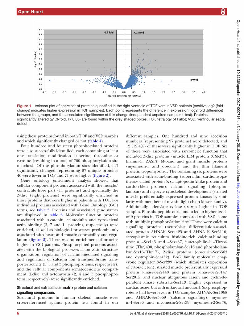
Proteomics analysisA total of 3610 protein accession numbers (representing 3342 gene names; note that this number differs in table 3 (2357 gene names) due to the PantherDB data-base version used) were identified that were found in RV samples from both heart pathologies and that fulfilled all criteria outlined above. Of these, 200 were found to significantly change (±1.3-fold, P<0.05) between patholo-gies: 26 were lower in TOF samples and 174 were higher in TOF (figure 1). Gene ontology analysis was performed
on 8 April 2019 by guest. P
rotected by copyright.http://openheart.bm
j.com/
Open H
eart: first published as 10.1136/openhrt-2017-000716 on 3 January 2018. Dow
nloaded from
Open Heart
4 Bond AR, et al. Open Heart 2018;5:e000716. doi:10.1136/openhrt-2017-000716
Tab
le 3
N
umb
er o
f pro
tein
s in
pro
teom
ics
anal
ysis
, per
ont
olog
y ca
tego
ry a
nd p
er p
atho
logy
Pro
tein
s (n
)P
erce
ntag
e o
f p
rote
ins
Ont
olo
gy
Cat
ego
ryT
OF
RV
VS
D R
VT
OF
RV
VS
D R
V
Tota
l num
ber o
f acc
essi
on n
umbe
rs58
4836
54To
tal n
umbe
r of u
niqu
e pr
otei
ns31
9023
74
Mol
ecul
ar fu
nctio
nAn
tioxi
dant
act
ivity
1413
0.4
0.5
Bind
ing
841
649
26.4
27.3
Cata
lytic
act
ivity
1051
809
32.9
34.1
Chan
nel r
egul
ator
act
ivity
54
0.2
0.2
Rece
ptor
act
ivity
6437
2.0
1.6
Sign
al tr
ansd
ucer
act
ivity
2115
0.7
0.6
Stru
ctur
al m
olec
ule
activ
ity28
523
58.
99.
9Tr
ansl
atio
n re
gula
tor a
ctiv
ity26
210.
80.
9Tr
ansp
orte
r act
ivity
146
102
4.6
4.3
Biol
ogic
al p
roce
ssBi
olog
ical
adh
esio
n77
552.
42.
3Bi
olog
ical
regu
latio
n23
216
57.
37.
0Ce
llula
r com
pone
nt o
rgan
isat
ion
or b
ioge
nesi
s41
433
913
.014
.3Ce
llula
r pro
cess
1338
1023
41.9
43.1
Deve
lopm
enta
l pro
cess
217
162
6.8
6.8
Grow
th1
10.
00.
0Im
mun
e sy
stem
pro
cess
139
834.
43.
5Lo
calis
atio
n40
931
112
.813
.1Lo
com
otio
n19
160.
60.
7M
etab
olic
pro
cess
1235
943
38.7
39.7
Mul
ticel
lula
r org
anis
mal
pro
cess
193
140
6.1
5.9
Repr
oduc
tion
2724
0.8
1.0
Resp
onse
to s
timul
us24
817
17.
87.
2Ce
ll co
mpo
nent
Cell
junc
tion
2015
0.6
0.6
Cell
part
914
717
28.7
30.2
Extra
cellu
lar m
atrix
3121
1.0
0.9
Extra
cellu
lar r
egio
n97
643.
02.
7M
acro
mol
ecul
ar c
ompl
ex35
928
611
.312
.0M
embr
ane
207
152
6.5
6.4
Orga
nelle
566
446
17.7
18.8
Syna
pse
85
0.3
0.2
Cont
inue
d
on 8 April 2019 by guest. P
rotected by copyright.http://openheart.bm
j.com/
Open H
eart: first published as 10.1136/openhrt-2017-000716 on 3 January 2018. Dow
nloaded from
5Bond AR, et al. Open Heart 2018;5:e000716. doi:10.1136/openhrt-2017-000716
Pro
tein
s (n
)P
erce
ntag
e o
f p
rote
ins
Ont
olo
gy
Cat
ego
ryT
OF
RV
VS
D R
VT
OF
RV
VS
D R
V
Prot
ein
clas
sCa
lciu
m-b
indi
ng p
rote
in85
642.
72.
7Ce
ll ad
hesi
on m
olec
ule
5238
1.6
1.6
Cell
junc
tion
prot
ein
2118
0.7
0.8
Chap
eron
e63
492.
02.
1Cy
tosk
elet
al p
rote
in21
016
76.
67.
0De
fens
e/im
mun
ity p
rote
in40
311.
31.
3En
zym
e m
odul
ator
268
200
8.4
8.4
Extra
cellu
lar m
atrix
pro
tein
5540
1.7
1.7
Hydr
olas
e25
518
98.
08.
0Is
omer
ase
5442
1.7
1.8
Liga
se79
562.
52.
4Ly
ase
4338
1.3
1.6
Mem
bran
e tra
ffic
prot
ein
9369
2.9
2.9
Nucl
eic
acid
bin
ding
412
323
12.9
13.6
Oxid
ored
ucta
se19
816
56.
27.
0Re
cept
or86
542.
72.
3Si
gnal
ling
mol
ecul
e10
780
3.4
3.4
Stor
age
prot
ein
11
0.0
0.0
Stru
ctur
al p
rote
in27
200.
80.
8Su
rfact
ant
11
0.0
0.0
Tran
scrip
tion
fact
or11
073
3.4
3.1
Tran
sfer
/car
rier p
rote
in83
642.
62.
7Tr
ansf
eras
e22
717
17.
17.
2Tr
ansm
embr
ane
rece
ptor
regu
lato
ry/a
dapt
or p
rote
in13
100.
40.
4Tr
ansp
orte
r12
381
3.9
3.4
Vira
l pro
tein
10
0.0
0.0
Pro
tein
s ar
e al
so s
how
n as
a p
erce
ntag
e of
the
num
ber
of u
niq
ue p
rote
ins
per
pat
holo
gy. T
he o
ntol
ogy
cate
gorie
s w
ith t
he 1
0 hi
ghes
t p
erce
ntag
e of
pro
tein
s p
er
pat
holo
gy a
re g
iven
in b
old
.R
V, r
ight
ven
tric
le; T
OF,
tet
ralo
gy o
f Fal
lot;
VS
D, v
entr
icul
ar s
epta
l def
ect.
Tab
le 3
C
ontin
ued
Basic and translational research
on 8 April 2019 by guest. P
rotected by copyright.http://openheart.bm
j.com/
Open H
eart: first published as 10.1136/openhrt-2017-000716 on 3 January 2018. Dow
nloaded from
Open Heart
6 Bond AR, et al. Open Heart 2018;5:e000716. doi:10.1136/openhrt-2017-000716
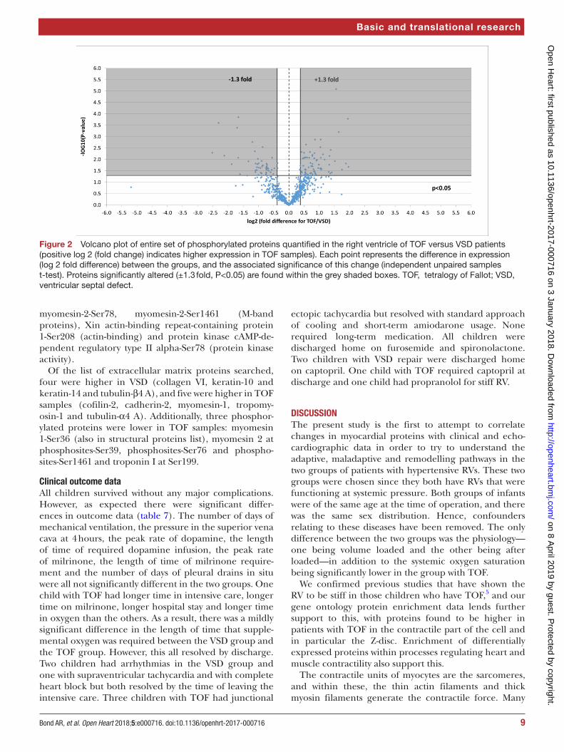
Figure 1 Volcano plot of entire set of proteins quantified in the right ventricle of TOF versus VSD patients (positive log2 (fold change) indicates higher expression in TOF samples). Each point represents the difference in expression (log2 fold difference) between the groups, and the associated significance of this change (independent unpaired samples t-test). Proteins significantly altered (±1.3-fold, P<0.05) are found within the grey shaded boxes. TOF, tetralogy of Fallot; VSD, ventricular septal defect.
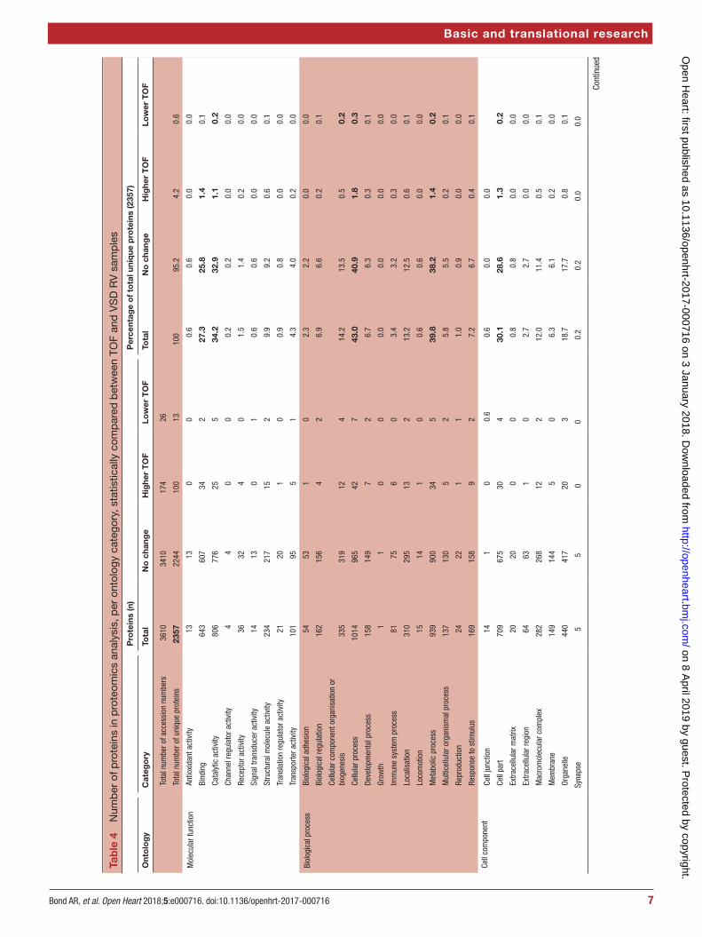
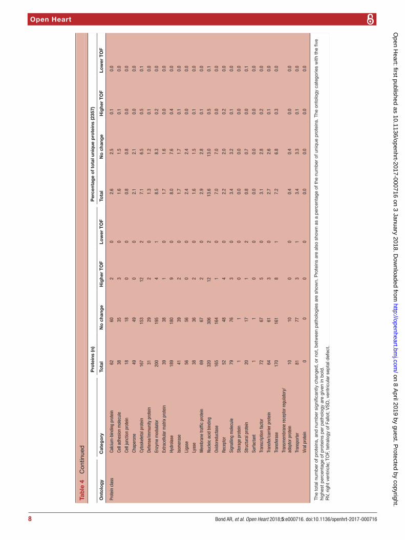
using these proteins found in both TOF and VSD samples and which significantly changed or not (table 4).
Four hundred and fourteen phosphorylated proteins were also successfully identified, each containing at least one translation modification at serine, threonine or tyrosine (resulting in a total of 700 phosphorylation site matches). Of the phosphorylation sites identified, 117 significantly changed representing 97 unique proteins: 46 were lower in TOF and 71 were higher (figure 2).
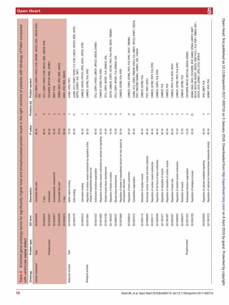
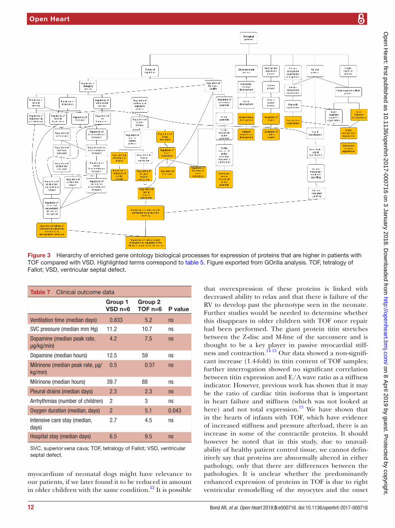
Gene ontology enrichment analysis showed that cellular component proteins associated with the muscle/contractile fibre part (11 proteins) and specifically the Z-disc (eight proteins) were significantly enriched in those proteins that were higher in patients with TOF. For individual proteins associated with Gene Ontology (GO) terms, see table 5. Proteins and associated gene names are displayed in table 6. Molecular function proteins associated with α-catenin, calmodulin and cytoskeletal actin binding (3, 7 and 14 proteins, respectively) were enriched, as well as biological processes predominantly associated with heart and muscle contractility and regu-lation (figure 3). There was no enrichment of proteins higher in VSD patients. Phosphorylated proteins associ-ated with the biological processes actomyosin structure organisation, regulation of calcium-mediated signalling and regulation of calcium ion transmembrane trans-porter activity (5, 3 and 3 phosphoproteins, respectively), and the cellular components somatodendritic compart-ment, Z-disc and actomyosin (2, 4 and 5 phosphopro-teins, respectively) were significantly enriched.
Structural and extracellular matrix protein and calcium signalling comparisonsStructural proteins in human skeletal muscle were cross-referenced against protein lists found in our
different samples. One hundred and nine accession numbers (representing 97 proteins) were detected, and 12 (12.4%) of these were significantly higher in TOF. Six of these were associated with sarcomeric function that included Z-disc proteins (muscle LIM protein (CSRP3), filamin-C, ZASP), M-band and giant muscle proteins (myomesin-1 and obscurin) and the thin filament protein, tropomyosin-1. The remaining six proteins were associated with actin-binding (supervillin, cardiomyopa-thy-associated protein 5, synaptopodin 2-like protein and cordon-bleu protein), calcium signalling (phospho-lamban) and myocyte cytoskeletal development (striated muscle preferentially expressed protein kinase; by simi-larity with members of myosin light chain kinase family). Additionally, adenylate cyclase six was higher in TOF samples. Phosphopeptide enrichment led to higher levels of 9 proteins in TOF samples compared with VSD, some with multiple phosphorylation sites. These were calcium signalling proteins (neuroblast differentiation-associ-ated protein AHNAK–Ser4425 and AHNA K–Ser5110, sarcoplasmic reticulum histidine-rich calcium-binding protein –Ser145 and –Ser457, junctophilin-2 –Threo-nine (Thr)490, phospholamban-Ser16 and phospholam-ban-Ser16/Thr17), Z-disk proteins (obscurin-Ser5563 and dystrophin-Ser432), BAG family molecular chap-erone regulator 3-Ser289 (which stimulates expression of cytoskeleton), striated muscle preferentially expressed protein kinase-Ser2448 and protein kinase-Ser2014/Ser2015, and nuclear ubiquitous casein and cyclin-de-pendent kinase substrate-Ser113 (highly expressed in cardiac tissue, but with unknown function). Six phosphop-roteins had lower levels in TOF samples: AHNAK-Ser1068 and AHNAK-Ser5369 (calcium signalling), myomes-in-1-Ser36 and myomesin-2-Ser39, myomesin-2-Ser76,
on 8 April 2019 by guest. P
rotected by copyright.http://openheart.bm
j.com/
Open H
eart: first published as 10.1136/openhrt-2017-000716 on 3 January 2018. Dow
nloaded from
7Bond AR, et al. Open Heart 2018;5:e000716. doi:10.1136/openhrt-2017-000716
Tab
le 4
N
umb
er o
f pro
tein
s in
pro
teom
ics
anal
ysis
, per
ont
olog
y ca
tego
ry, s
tatis
tical
ly c
omp
ared
bet
wee
n TO
F an
d V
SD
RV
sam
ple
s
Pro
tein
s (n
)P
erce
ntag
e o
f to
tal u
niq
ue p
rote
ins
(235
7)
Ont
olo
gy
Cat
ego
ryTo
tal
No
cha
nge
Hig
her
TO
FLo
wer
TO
FTo
tal
No
cha
nge
Hig
her
TO
FLo
wer
TO
F
Tota
l num
ber o
f acc
essi
on n
umbe
rs36
1034
1017
426
Tota
l num
ber o
f uni
que
prot
eins
2357
2244
100
1310
095
.24.
20.
6
Mol
ecul
ar fu
nctio
nAn
tioxi
dant
act
ivity
1313
00
0.6
0.6
0.0
0.0
Bind
ing
643
607
342
27.3
25.8
1.4
0.1
Cata
lytic
act
ivity
806
776
255
34.2
32.9
1.1
0.2
Chan
nel r
egul
ator
act
ivity
44
00
0.2
0.2
0.0
0.0
Rece
ptor
act
ivity
3632
40
1.5
1.4
0.2
0.0
Sign
al tr
ansd
ucer
act
ivity
1413
01
0.6
0.6
0.0
0.0
Stru
ctur
al m
olec
ule
activ
ity23
421
715
29.
99.
20.
60.
1
Tran
slat
ion
regu
lato
r act
ivity
2120
10
0.9
0.8
0.0
0.0
Tran
spor
ter a
ctiv
ity10
195
51
4.3
4.0
0.2
0.0
Biol
ogic
al p
roce
ssBi
olog
ical
adh
esio
n54
531
02.
32.
20.
00.
0
Biol
ogic
al re
gula
tion
162
156
42
6.9
6.6
0.2
0.1
Cellu
lar c
ompo
nent
org
anis
atio
n or
bi
ogen
esis
335
319
124
14.2
13.5
0.5
0.2
Cellu
lar p
roce
ss10
1496
542
743
.040
.91.
80.
3
Deve
lopm
enta
l pro
cess
158
149
72
6.7
6.3
0.3
0.1
Grow
th1
10
00.
00.
00.
00.
0
Imm
une
syst
em p
roce
ss81
756
03.
43.
20.
30.
0
Loca
lisat
ion
310
295
132
13.2
12.5
0.6
0.1
Loco
mot
ion
1514
10
0.6
0.6
0.0
0.0
Met
abol
ic p
roce
ss93
990
034
539
.838
.21.
40.
2
Mul
ticel
lula
r org
anis
mal
pro
cess
137
130
52
5.8
5.5
0.2
0.1
Repr
oduc
tion
2422
11
1.0
0.9
0.0
0.0
Resp
onse
to s
timul
us16
915
89
27.
26.
70.
40.
1
Cell
com
pone
ntCe
ll ju
nctio
n 14
10
0.6
0.6
0.0
0.0
Cell
part
709
675
304
30.1
28.6
1.3
0.2
Extra
cellu
lar m
atrix
2020
00
0.8
0.8
0.0
0.0
Extra
cellu
lar r
egio
n64
631
02.
72.
70.
00.
0
Mac
rom
olec
ular
com
plex
282
268
122
12.0
11.4
0.5
0.1
Mem
bran
e14
914
45
06.
36.
10.
20.
0
Orga
nelle
440
417
203
18.7
17.7
0.8
0.1
Syna
pse
55
00
0.2
0.2
0.0
0.0
Cont
inue
d
Basic and translational research
on 8 April 2019 by guest. P
rotected by copyright.http://openheart.bm
j.com/
Open H
eart: first published as 10.1136/openhrt-2017-000716 on 3 January 2018. Dow
nloaded from
Open Heart
8 Bond AR, et al. Open Heart 2018;5:e000716. doi:10.1136/openhrt-2017-000716
Pro
tein
s (n
)P
erce
ntag
e o
f to
tal u
niq
ue p
rote
ins
(235
7)
Ont
olo
gy
Cat
ego
ryTo
tal
No
cha
nge
Hig
her
TO
FLo
wer
TO
FTo
tal
No
cha
nge
Hig
her
TO
FLo
wer
TO
F
Prot
ein
clas
sCa
lciu
m-b
indi
ng p
rote
in62
602
02.
62.
50.
10.
0
Cell
adhe
sion
mol
ecul
e38
353
01.
61.
50.
10.
0
Cell
junc
tion
prot
ein
1818
00
0.8
0.8
0.0
0.0
Chap
eron
e49
490
02.
12.
10.
00.
0
Cyto
skel
etal
pro
tein
167
153
122
7.1
6.5
0.5
0.1
Defe
nse/
imm
unity
pro
tein
3129
20
1.3
1.2
0.1
0.0
Enzy
me
mod
ulat
or20
019
54
18.
58.
30.
20.
0
Extra
cellu
lar m
atrix
pro
tein
3938
10
1.7
1.6
0.0
0.0
Hydr
olas
e18
918
09
08.
07.
60.
40.
0
Isom
eras
e41
392
01.
71.
70.
10.
0
Liga
se56
560
02.
42.
40.
00.
0
Lyas
e38
362
01.
61.
50.
10.
0
Mem
bran
e tra
ffic
prot
ein
6967
20
2.9
2.8
0.1
0.0
Nucl
eic
acid
bin
ding
320
306
122
13.6
13.0
0.5
0.1
Oxid
ored
ucta
se16
516
41
07.
07.
00.
00.
0
Rece
ptor
5248
40
2.2
2.0
0.2
0.0
Sign
allin
g m
olec
ule
7976
30
3.4
3.2
0.1
0.0
Stor
age
prot
ein
11
00
0.0
0.0
0.0
0.0
Stru
ctur
al p
rote
in20
171
20.
80.
70.
00.
1
Surfa
ctan
t1
10
00.
00.
00.
00.
0
Tran
scrip
tion
fact
or72
675
03.
12.
80.
20.
0
Tran
sfer
/car
rier p
rote
in64
613
02.
72.
60.
10.
0
Tran
sfer
ase
170
161
81
7.2
6.8
0.3
0.0
Tran
smem
bran
e re
cept
or re
gula
tory
/ad
apto
r pro
tein
1010
00
0.4
0.4
0.0
0.0
Tran
spor
ter
8177
31
3.4
3.3
0.1
0.0
Vira
l pro
tein
00
00
0.0
0.0
0.0
0.0
The
tota
l num
ber
of p
rote
ins,
and
num
ber
sig
nific
antly
cha
nged
, or
not,
bet
wee
n p
atho
logi
es a
re s
how
n. P
rote
ins
are
also
sho
wn
as a
per
cent
age
of t
he n
umb
er o
f uni
que
pro
tein
s. T
he o
ntol
ogy
cate
gorie
s w
ith t
he fi
ve
high
est
per
cent
age
of p
rote
ins
per
pat
holo
gy a
re g
iven
in b
old
.R
V, r
ight
ven
tric
le; T
OF,
tet
ralo
gy o
f Fal
lot;
VS
D, v
entr
icul
ar s
epta
l def
ect.
Tab
le 4
C
ontin
ued
on 8 April 2019 by guest. P
rotected by copyright.http://openheart.bm
j.com/
Open H
eart: first published as 10.1136/openhrt-2017-000716 on 3 January 2018. Dow
nloaded from
9Bond AR, et al. Open Heart 2018;5:e000716. doi:10.1136/openhrt-2017-000716
Figure 2 Volcano plot of entire set of phosphorylated proteins quantified in the right ventricle of TOF versus VSD patients (positive log 2 (fold change) indicates higher expression in TOF samples). Each point represents the difference in expression (log 2 fold difference) between the groups, and the associated significance of this change (independent unpaired samples t-test). Proteins significantly altered (±1.3 fold, P<0.05) are found within the grey shaded boxes. TOF, tetralogy of Fallot; VSD, ventricular septal defect.
Basic and translational research
myomesin-2-Ser78, myomesin-2-Ser1461 (M-band proteins), Xin actin-binding repeat-containing protein 1-Ser208 (actin-binding) and protein kinase cAMP-de-pendent regulatory type II alpha-Ser78 (protein kinase activity).
Of the list of extracellular matrix proteins searched, four were higher in VSD (collagen VI, keratin-10 and keratin-14 and tubulin-β4 A), and five were higher in TOF samples (cofilin-2, cadherin-2, myomesin-1, tropomy-osin-1 and tubulin-α4 A). Additionally, three phosphor-ylated proteins were lower in TOF samples: myomesin 1-Ser36 (also in structural proteins list), myomesin 2 at phosphosites-Ser39, phosphosites-Ser76 and phospho-sites-Ser1461 and troponin I at Ser199.
Clinical outcome dataAll children survived without any major complications. However, as expected there were significant differ-ences in outcome data (table 7). The number of days of mechanical ventilation, the pressure in the superior vena cava at 4 hours, the peak rate of dopamine, the length of time of required dopamine infusion, the peak rate of milrinone, the length of time of milrinone require-ment and the number of days of pleural drains in situ were all not significantly different in the two groups. One child with TOF had longer time in intensive care, longer time on milrinone, longer hospital stay and longer time in oxygen than the others. As a result, there was a mildly significant difference in the length of time that supple-mental oxygen was required between the VSD group and the TOF group. However, this all resolved by discharge. Two children had arrhythmias in the VSD group and one with supraventricular tachycardia and with complete heart block but both resolved by the time of leaving the intensive care. Three children with TOF had junctional
ectopic tachycardia but resolved with standard approach of cooling and short-term amiodarone usage. None required long-term medication. All children were discharged home on furosemide and spironolactone. Two children with VSD repair were discharged home on captopril. One child with TOF required captopril at discharge and one child had propranolol for stiff RV.
diSCuSSionThe present study is the first to attempt to correlate changes in myocardial proteins with clinical and echo-cardiographic data in order to try to understand the adaptive, maladaptive and remodelling pathways in the two groups of patients with hypertensive RVs. These two groups were chosen since they both have RVs that were functioning at systemic pressure. Both groups of infants were of the same age at the time of operation, and there was the same sex distribution. Hence, confounders relating to these diseases have been removed. The only difference between the two groups was the physiology—one being volume loaded and the other being after loaded—in addition to the systemic oxygen saturation being significantly lower in the group with TOF.
We confirmed previous studies that have shown the RV to be stiff in those children who have TOF,5 and our gene ontology protein enrichment data lends further support to this, with proteins found to be higher in patients with TOF in the contractile part of the cell and in particular the Z-disc. Enrichment of differentially expressed proteins within processes regulating heart and muscle contractility also support this.
The contractile units of myocytes are the sarcomeres, and within these, the thin actin filaments and thick myosin filaments generate the contractile force. Many
on 8 April 2019 by guest. P
rotected by copyright.http://openheart.bm
j.com/
Open H
eart: first published as 10.1136/openhrt-2017-000716 on 3 January 2018. Dow
nloaded from
Open Heart
10 Bond AR, et al. Open Heart 2018;5:e000716. doi:10.1136/openhrt-2017-000716
Tab
le 5
E
nric
hed
gen
e on
tolo
gy t
erm
s fo
r si
gnifi
cant
ly h
ighe
r to
tal a
nd p
hosp
hory
late
d p
rote
in le
vels
in t
he r
ight
ven
tric
le o
f pat
ient
s w
ith t
etra
logy
of F
allo
t co
mp
ared
w
ith v
entr
icul
ar s
epta
l def
ect
Ont
olo
gy
Pro
tein
typ
eG
O t
erm
Des
crip
tio
nP
val
ue P
rote
ins
(n)
Pro
tein
sym
bo
l
Cellu
lar c
ompo
nent
Tota
lGO
:004
4449
Cont
ract
ile fi
bre
part
4E-0
511
CFL2
, CM
YA5,
CSR
P3, F
HOD3
, FLN
C, M
YOM
1, M
YOZ2
, NEB
L, O
BSCN
, RYR
2,
SVIL
GO:0
0300
18Z-
disc
9E-0
58
CFL2
, CSR
P3, F
HOD3
, FLN
C, M
YOZ2
, NEB
L, O
BSCN
, RYR
2
Phos
phor
ylat
edGO
:004
2641
Acto
myo
sin
6E-0
54
CDC4
2BPB
, MYO
18A,
NEB
L, S
ORBS
1
GO:0
0364
77So
mat
oden
driti
c co
mpa
rtmen
t1E
-04
2M
APT,
STX
4
GO:0
0444
49Co
ntra
ctile
fibr
e pa
rt7E
-04
5AH
NAK,
BAG
3, J
PH2,
NEB
L, O
BSCN
GO:0
0300
18Z-
disc
8E-0
44
BAG3
, JPH
2, N
EBL,
OBS
CN
Mol
ecul
ar fu
nctio
nTo
tal
GO:0
0452
94Al
pha-
cate
nin
bind
ing
1E-0
53
AJUB
A, C
DH2,
PKP
2
GO:0
0037
79Ac
tin b
indi
ng4E
-05
14AJ
UBA,
CFL
2, C
SRP3
, FHO
D3, F
LII,
FLNC
, LIM
CH1,
MYO
Z2, N
EBL,
SNT
A1,
SNTB
2, S
ORBS
1, S
VIL,
TW
F2
GO:0
0055
16Ca
lmod
ulin
bin
ding
9E-0
47
CAM
K2D,
OBS
CN, P
PP3C
C, R
YR2,
SNT
A1, S
NTB2
, STR
N
Biol
ogic
al p
roce
ssGO
:001
0881
Regu
latio
n of
car
diac
mus
cle
cont
ract
ion
by re
gula
tion
of th
e re
leas
e of
seq
uest
ered
cal
cium
ion
3E-0
54
CAM
K2D,
GST
M2,
PLN
, RYR
2
GO:0
0310
32Ac
tom
yosi
n st
ruct
ure
orga
nisa
tion
9E-0
57
CFL2
, CSR
P3, F
HOD3
, LIM
CH1,
MYO
Z2, O
BSCN
, SOR
BS1
GO:0
0108
82Re
gula
tion
of c
ardi
ac m
uscl
e co
ntra
ctio
n by
cal
cium
ion
sign
allin
g9E
-05
4CA
MK2
D, G
STM
2, P
LN, R
YR2
GO:0
0147
06St
riate
d m
uscl
e tis
sue
deve
lopm
ent
1E-0
46
CFL2
, CSR
P3, M
YOM
1, P
LN, S
EMA3
C, S
VIL,
GO:1
9035
22Re
gula
tion
of b
lood
circ
ulat
ion
1E-0
49
ADCY
6, C
AMK2
D, C
SRP3
, GST
M2,
PKP
2, P
LN, R
YR2,
SNT
A1, T
MEM
65
GO:0
0605
37M
uscl
e tis
sue
deve
lopm
ent
1E-0
46
CFL2
, CSR
P3, M
YOM
1, P
LN, S
EMA3
C, S
VIL
GO:0
0108
80Re
gula
tion
of re
leas
e of
seq
uest
ered
cal
cium
ion
into
cyt
osol
by
sarc
opla
smic
retic
ulum
2E-0
44
CAM
K2D,
GST
M2,
PLN
, RYR
2
GO:0
0080
16Re
gula
tion
of h
eart
cont
ract
ion
2E-0
48
CAM
K2D,
CSR
P3, G
STM
2, P
KP2,
PLN
, RYR
2, S
NTA1
, TM
EM65
GO:0
0070
10Cy
tosk
elet
on o
rgan
isat
ion
3E-0
416
AJUB
A, C
FL2,
CSR
P3, E
RBB2
IP, F
HOD3
, FLI
I, LI
MCH
1, M
YOZ2
, NTM
T1, O
BSCN
, PK
P2, P
RKCD
BP, S
HANK
1, S
ORBS
1, S
VIL,
TUB
A4A
GO:0
0551
19Re
laxa
tion
of c
ardi
ac m
uscl
e4E
-04
3CA
MK2
D, G
STM
2, P
LN
GO:0
0860
05Ve
ntric
ular
car
diac
mus
cle
cell
actio
n po
tent
ial
4E-0
43
PKP2
, RYR
2, S
NTA1
GO:0
0551
17Re
gula
tion
of c
ardi
ac m
uscl
e co
ntra
ctio
n4E
-04
5CA
MK2
D, G
STM
2, P
KP2,
PLN
, RYR
2
GO:0
0020
26Re
gula
tion
of th
e fo
rce
of h
eart
cont
ract
ion
4E-0
44
CAM
K2D,
CSR
P3, P
LN, R
YR2
GO:1
9010
77Re
gula
tion
of re
laxa
tion
of m
uscl
e6E
-04
2CA
MK2
D, P
LN
GO:1
9018
97Re
gula
tion
of re
laxa
tion
of c
ardi
ac m
uscl
e6E
-04
2CA
MK2
D, P
LN
GO:0
0020
27Re
gula
tion
of h
eart
rate
8E-0
45
CAM
K2D,
PKP
2, P
LN, R
YR2,
SNT
A1
GO:0
0069
42Re
gula
tion
of s
triat
ed m
uscl
e co
ntra
ctio
n1E
-03
5CA
MK2
D, G
STM
2, P
KP2,
PLN
, RYR
2
GO:0
0900
75Re
laxa
tion
of m
uscl
e1E
-03
3CA
MK2
D, G
STM
2, P
LN
Phos
phor
ylat
edGO
:003
1032
Acto
myo
sin
stru
ctur
e or
gani
satio
n1E
-05
5CD
C42B
PB, M
YLK3
, MYO
18A,
OBS
CN, S
ORBS
1
GO:0
0507
89Re
gula
tion
of b
iolo
gica
l pro
cess
3E-0
425
AHNA
K, B
AG3,
BVE
S, C
DC42
BPB,
JPH
2, K
ANK2
, KPN
A4, L
RRFI
P2, M
APT,
M
YLK3
, MYO
18A,
NUC
KS1,
OBS
CN, O
SBP,
PLCL
1, P
LN, P
SIP1
, RBM
20, R
SF1,
SC
N7A,
SGT
A, S
ORBS
1, S
PEG,
STX
4, Z
BTB7
A
GO:0
0508
48Re
gula
tion
of c
alci
um-m
edia
ted
sign
allin
g4E
-04
3JP
H2, M
APT,
PLN
GO:1
9010
19Re
gula
tion
of c
alci
um io
n tra
nsm
embr
ane
trans
porte
r act
ivity
4E-0
43
AHNA
K, J
PH2,
PLN
on 8 April 2019 by guest. P
rotected by copyright.http://openheart.bm
j.com/
Open H
eart: first published as 10.1136/openhrt-2017-000716 on 3 January 2018. Dow
nloaded from
11Bond AR, et al. Open Heart 2018;5:e000716. doi:10.1136/openhrt-2017-000716
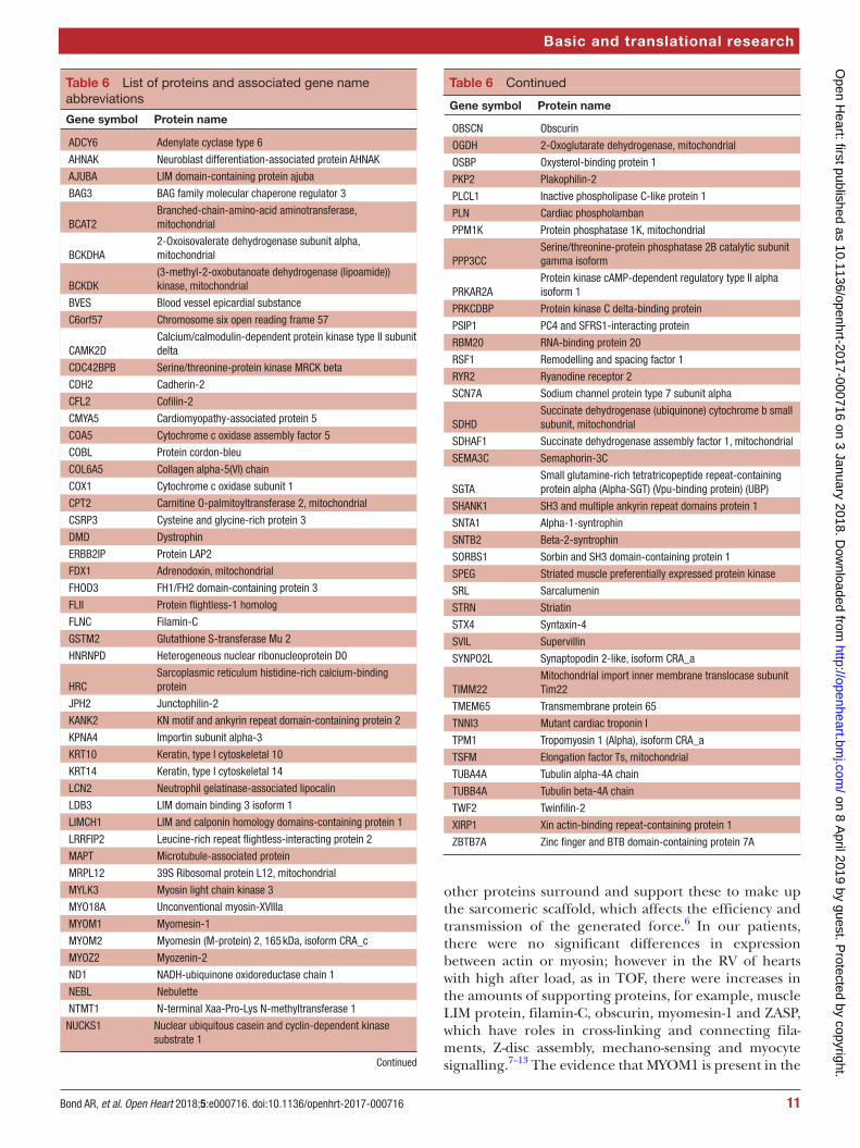
Table 6 List of proteins and associated gene name abbreviations
Gene symbol Protein name
ADCY6 Adenylate cyclase type 6
AHNAK Neuroblast differentiation-associated protein AHNAK
AJUBA LIM domain-containing protein ajuba
BAG3 BAG family molecular chaperone regulator 3
BCAT2Branched-chain-amino-acid aminotransferase, mitochondrial
BCKDHA2-Oxoisovalerate dehydrogenase subunit alpha, mitochondrial
BCKDK(3-methyl-2-oxobutanoate dehydrogenase (lipoamide)) kinase, mitochondrial
BVES Blood vessel epicardial substance
C6orf57 Chromosome six open reading frame 57
CAMK2DCalcium/calmodulin-dependent protein kinase type II subunit delta
CDC42BPB Serine/threonine-protein kinase MRCK beta
CDH2 Cadherin-2
CFL2 Cofilin-2
CMYA5 Cardiomyopathy-associated protein 5
COA5 Cytochrome c oxidase assembly factor 5
COBL Protein cordon-bleu
COL6A5 Collagen alpha-5(VI) chain
COX1 Cytochrome c oxidase subunit 1
CPT2 Carnitine O-palmitoyltransferase 2, mitochondrial
CSRP3 Cysteine and glycine-rich protein 3
DMD Dystrophin
ERBB2IP Protein LAP2
FDX1 Adrenodoxin, mitochondrial
FHOD3 FH1/FH2 domain-containing protein 3
FLII Protein flightless-1 homolog
FLNC Filamin-C
GSTM2 Glutathione S-transferase Mu 2
HNRNPD Heterogeneous nuclear ribonucleoprotein D0
HRCSarcoplasmic reticulum histidine-rich calcium-binding protein
JPH2 Junctophilin-2
KANK2 KN motif and ankyrin repeat domain-containing protein 2
KPNA4 Importin subunit alpha-3
KRT10 Keratin, type I cytoskeletal 10
KRT14 Keratin, type I cytoskeletal 14
LCN2 Neutrophil gelatinase-associated lipocalin
LDB3 LIM domain binding 3 isoform 1
LIMCH1 LIM and calponin homology domains-containing protein 1
LRRFIP2 Leucine-rich repeat flightless-interacting protein 2
MAPT Microtubule-associated protein
MRPL12 39S Ribosomal protein L12, mitochondrial
MYLK3 Myosin light chain kinase 3
MYO18A Unconventional myosin-XVIIIa
MYOM1 Myomesin-1
MYOM2 Myomesin (M-protein) 2, 165 kDa, isoform CRA_c
MYOZ2 Myozenin-2
ND1 NADH-ubiquinone oxidoreductase chain 1
NEBL Nebulette
NTMT1 N-terminal Xaa-Pro-Lys N-methyltransferase 1
NUCKS1 Nuclear ubiquitous casein and cyclin-dependent kinase substrate 1
Continued
Gene symbol Protein name
OBSCN Obscurin
OGDH 2-Oxoglutarate dehydrogenase, mitochondrial
OSBP Oxysterol-binding protein 1
PKP2 Plakophilin-2
PLCL1 Inactive phospholipase C-like protein 1
PLN Cardiac phospholamban
PPM1K Protein phosphatase 1K, mitochondrial
PPP3CCSerine/threonine-protein phosphatase 2B catalytic subunit gamma isoform
PRKAR2AProtein kinase cAMP-dependent regulatory type II alpha isoform 1
PRKCDBP Protein kinase C delta-binding protein
PSIP1 PC4 and SFRS1-interacting protein
RBM20 RNA-binding protein 20
RSF1 Remodelling and spacing factor 1
RYR2 Ryanodine receptor 2
SCN7A Sodium channel protein type 7 subunit alpha
SDHDSuccinate dehydrogenase (ubiquinone) cytochrome b small subunit, mitochondrial
SDHAF1 Succinate dehydrogenase assembly factor 1, mitochondrial
SEMA3C Semaphorin-3C
SGTASmall glutamine-rich tetratricopeptide repeat-containing protein alpha (Alpha-SGT) (Vpu-binding protein) (UBP)
SHANK1 SH3 and multiple ankyrin repeat domains protein 1
SNTA1 Alpha-1-syntrophin
SNTB2 Beta-2-syntrophin
SORBS1 Sorbin and SH3 domain-containing protein 1
SPEG Striated muscle preferentially expressed protein kinase
SRL Sarcalumenin
STRN Striatin
STX4 Syntaxin-4
SVIL Supervillin
SYNPO2L Synaptopodin 2-like, isoform CRA_a
TIMM22Mitochondrial import inner membrane translocase subunit Tim22
TMEM65 Transmembrane protein 65
TNNI3 Mutant cardiac troponin I
TPM1 Tropomyosin 1 (Alpha), isoform CRA_a
TSFM Elongation factor Ts, mitochondrial
TUBA4A Tubulin alpha-4A chain
TUBB4A Tubulin beta-4A chain
TWF2 Twinfilin-2
XIRP1 Xin actin-binding repeat-containing protein 1
ZBTB7A Zinc finger and BTB domain-containing protein 7A
Table 6 Continued
Basic and translational research
other proteins surround and support these to make up the sarcomeric scaffold, which affects the efficiency and transmission of the generated force.6 In our patients, there were no significant differences in expression between actin or myosin; however in the RV of hearts with high after load, as in TOF, there were increases in the amounts of supporting proteins, for example, muscle LIM protein, filamin-C, obscurin, myomesin-1 and ZASP, which have roles in cross-linking and connecting fila-ments, Z-disc assembly, mechano-sensing and myocyte signalling.7–13 The evidence that MYOM1 is present in the
on 8 April 2019 by guest. P
rotected by copyright.http://openheart.bm
j.com/
Open H
eart: first published as 10.1136/openhrt-2017-000716 on 3 January 2018. Dow
nloaded from
Open Heart
12 Bond AR, et al. Open Heart 2018;5:e000716. doi:10.1136/openhrt-2017-000716
Figure 3 Hierarchy of enriched gene ontology biological processes for expression of proteins that are higher in patients with TOF compared with VSD. Highlighted terms correspond to table 5. Figure exported from GOrilla analysis. TOF, tetralogy of Fallot; VSD, ventricular septal defect.
Table 7 Clinical outcome data
Group 1 VSD n=6
Group 2 TOF n=6 P value
Ventilation time (median days) 0.833 5.2 ns
SVC pressure (median mm Hg) 11.2 10.7 ns
Dopamine (median peak rate, µg/kg/min)
4.2 7.5 ns
Dopamine (median hours) 12.5 59 ns
Milrinone (median peak rate, µg/kg/min)
0.5 0.57 ns
Milrinone (median hours) 39.7 88 ns
Pleural drains (median days) 2.3 2.3 ns
Arrhythmias (number of children) 2 3 ns
Oxygen duration (median, days) 2 5.1 0.043
Intensive care stay (median, days)
2.7 4.5 ns
Hospital stay (median days) 6.5 9.5 ns
SVC, superior vena cava; TOF, tetralogy of Fallot; VSD, ventricular septal defect.
myocardium of neonatal dogs might have relevance to our patients, if we later found it to be reduced in amount in older children with the same condition.12 It is possible
that overexpression of these proteins is linked with decreased ability to relax and that there is failure of the RV to develop past the phenotype seen in the neonate. Further studies would be needed to determine whether this disappears in older children with TOF once repair had been performed. The giant protein titin stretches between the Z-disc and M-line of the sarcomere and is thought to be a key player in passive myocardial stiff-ness and contraction.14 15 Our data showed a non-signifi-cant increase (1.4-fold) in titin content of TOF samples; further interrogation showed no significant correlation between titin expression and E/A wave ratio as a stiffness indicator. However, previous work has shown that it may be the ratio of cardiac titin isoforms that is important in heart failure and stiffness (which was not looked at here) and not total expression.15 We have shown that in the hearts of infants with TOF, which have evidence of increased stiffness and pressure afterload, there is an increase in some of the contractile proteins. It should however be noted that in this study, due to unavail-ability of healthy patient control tissue, we cannot defin-itively say that proteins are abnormally altered in either pathology, only that there are differences between the pathologies. It is unclear whether the predominantly enhanced expression of proteins in TOF is due to right ventricular remodelling of the myocytes and the onset
on 8 April 2019 by guest. P
rotected by copyright.http://openheart.bm
j.com/
Open H
eart: first published as 10.1136/openhrt-2017-000716 on 3 January 2018. Dow
nloaded from
13Bond AR, et al. Open Heart 2018;5:e000716. doi:10.1136/openhrt-2017-000716
Basic and translational research
of hypertrophy or a compensatory mechanism. Studies on aortas from rats with type 2 diabetes have shown that increased ROS caused by oxidative stress upregulates contractile protein expression resulting in vascular wall remodelling.16
Perhaps of greater importance is the differences in phosphorylation of proteins involved in the ventricle contractile processes. Phosphorylation of proteins indi-cates a change in protein function (either activation or deactivation) and enrichment was seen in proteins at various phosphorylation sites with higher levels in TOF, associated with calcium signalling and contractility pathways. Of the phosphorylation sites we detected in our proteomics analysis, many of them have not been detected previously in heart tissue (PhosphositePlus, accessed March 201717), the exceptions being the following (R denotes regulatory protein, and K denotes associated kinases where known): phospholamban-ser16 (R: protein phosphatase 1 regulatory inhibitor subunit 1A,18 K: cAMP-dependent protein kinase catalytic subunit alpha19) and -thr17 (R: tumour necrosis factor-α receptor subtypes 1 and 2,20 K: cAMP-dependent protein kinase catalytic subunit alpha19), myomesin 2-ser76 (K: cAMP-de-pendent protein kinase catalytic subunit alpha,21 protein kinase cAMP-dependent regulatory type II-α-ser78 and cardiac troponin I-ser199) (K: cAMP-dependent protein kinase catalytic subunit alpha).22 The interaction of cardiac troponin I-Ser199 with its putative kinase has been shown to be inhibited by tropomyosin I,23 which was significantly higher in TOF samples, resulting in altered force of contraction.
Previous studies have shown some changes in calcium signalling in congenital heart disease, but it has not been possible to determine whether this is a generic response to RV hypertension or to specific cardiac physiologies.24 Proteins associated with calcium signalling were detected with adenylate cyclase 6, calcium/calmodulin-dependent protein kinase, phospholamban, ryanodine receptor 2 and sarcalumenin all being significantly higher in TOF. Phosphosites for phospholamban at Ser16, sarco-plasmic reticulum histidine-rich calcium-binding protein at Ser145 and junctophilin-2 at Thr490 were higher in TOF. However, cardiac troponin I at Ser199 was lower. The finding for phospholamban-Ser16 is similar to our previous studies and was linked to hypoxaemia.25 26
Although children with TOF clearly are more cyanosed, it is not clear how this would affect the RV. The biopsies are taken from the cavity of the RV that would normally see the venous (desaturated) blood. Systemic desatura-tion would not be expected to affect the endocardial surface of the RV.27 28 It should be noted that hypoxia-in-ducible factor-1-alpha (HIF-1α) was not detected in our proteomics analysis, despite oxygen saturation levels indi-cating hypoxia, especially in the TOF patients. It would be expected that HIF-1α be present in all our samples as has previously been described for CHD,29 suggesting the abundance was too low to detect with our protocol. However, we know that the RV is stiff after definitive
cardiac surgery for TOF, which might relate to abnormal-ities of contractile proteins, secondary to an obstructed RV in comparison with the volume loaded RV of children with large post-tricuspid left to right shunt (VSD).30 For the first time, we have made a direct comparison between the hypertensive RV of children with VSD and the RV of children with TOF.
The novel findings for the rest of the proteins/phosphoproteins provide further evidence for maladapta-tion of the RV in cyanotic TOF patients with implications for increased stiffness after open-heart surgery. The lack of functional measurements, for instance, single permea-bilised cardiomyocyte measurements to assess systolic and diastolic properties, calcium sensitivity and cross-bridge kinetics, make it difficult to understand the functional relevance of the observed differences in cellular signal-ling between the two patient groups. It would be helpful to understand this in future studies, but the amount of tissue available from such small children limits the possi-bility under the current approvals for this study. A large piece of RV tissue would be required, and there is some doubt that the digestion process required to achieve single cells might render the sample unrepresentative.
Comparison with known cardiac diseaseGenes known to be affected in myocardial disorders with ventricular dysfunction (hypertrophic cardiomyopathy (HCM), dilated cardiomyopathy (DCM) and arrhythmo-genic right ventricular cardiomyopathy (ARVC) (KEGG (Kyoto Encyclopaedia of Genes and Genomes) pathways map0510, map0514 and map05412 respectively31) were compared with our proteomics data. The majority of the affected proteins in these diseases were also present in our samples (11/15 HCM, 9/13 DCM, 5/6 ARVC); however, only tropomyosin-1 (normally affected in HCM and DCM, but not ARVC) was significantly higher in TOF samples. This suggests there was no downregula-tion of contractility as seen in maladaptation and right ventricular cardiomyopathy but more likely the upregula-tion due to increased myocardial wall stress.32
ConCluSionFor the first time, it is now possible to suggest a cellular mechanism for the stiff RV in TOF, in that we have shown abnormalities in the calcium signalling pathways of the right ventricular myocardium. We have shown increased expression of myocardial contractile and extracellular proteins in TOF as compared with VSD, showing an adap-tive physiology that is unrelated to age. We have shown that there is also parallel abnormality of clinical, echo-cardiographic and outcome data that might allow us, in future, to customise the treatment of the individual child in relation to their post-translational proteomics. It is too early to relate one finding directly to the other, but further work needs to be performed to determine the relationship between these important changes in RV hypertension in congenital heart disease.
on 8 April 2019 by guest. P
rotected by copyright.http://openheart.bm
j.com/
Open H
eart: first published as 10.1136/openhrt-2017-000716 on 3 January 2018. Dow
nloaded from
Open Heart
14 Bond AR, et al. Open Heart 2018;5:e000716. doi:10.1136/openhrt-2017-000716
Contributors RMT, M-SG, SJG and MG designed the study and protocol. MC collected RV samples. CG performed and analysed the clinical and echocardiographic data. ARB, DI, SA-G and KH performed the laboratory analysis. RMT and AJB wrote the first draft, and all authors provided input to and approved the final version of the manuscript.
Funding Sparks, the Childrens charity * 13BTL01. This study was supported by the NIHR Biomedical Centre at the University Hospitals Bristol NHS Foundation Trust and the University of Bristol. University Hospitals Bristol NHS Foundation Trust department of research and innovation number CH/2014/4571 provided sponsorship.
disclaimer The views expressed in this publication are those of the author(s) and not necessarily those of the NHS, the National Institute for Health Research or the Department of Health.
Competing interests None declared.
Patient consent Detail has been removed from this case description/these case descriptions to ensure anonymity. The editors and reviewers have seen the detailed information available and are satisfied that the information backs up the case the authors are making.
Ethics approval Ethical approval was granted by the National Research Ethics Service number 14/NW/1256, IRAS 143683.
Provenance and peer review Not commissioned; internally peer reviewed.
open access This is an Open Access article distributed in accordance with the Creative Commons Attribution Non Commercial (CC BY-NC 4.0) license, which permits others to distribute, remix, adapt, build upon this work non-commercially, and license their derivative works on different terms, provided the original work is properly cited and the use is non-commercial. See: http:// creativecommons. org/ licenses/ by- nc/ 4. 0/
© Article author(s) (or their employer(s) unless otherwise stated in the text of the article) 2018. All rights reserved. No commercial use is permitted unless otherwise expressly granted.
RefeRences 1. Knowles RL, Bull C, Wren C, et al. Mortality with congenital heart
defects in England and Wales, 1959-2009: exploring technological change through period and birth cohort analysis. Arch Dis Child 2012;97:861–5.
2. Murray CJ, Richards MA, Newton JN, et al. UK health performance: findings of the global burden of disease study 2010. Lancet 2013;381:997–1020.
3. Ghorbel MT, Cherif M, Jenkins E, et al. Transcriptomic analysis of patients with tetralogy of Fallot reveals the effect of chronic hypoxia on myocardial gene expression. J Thorac Cardiovasc Surg 2010;140:337–45.
4. Qipshidze N, Tyagi N, Metreveli N, et al. Autophagy mechanism of right ventricular remodeling in murine model of pulmonary artery constriction. Am J Physiol Heart Circ Physiol 2012;302:H688–96.
5. Chaturvedi RR, Shore DF, Lincoln C, et al. Acute right ventricular restrictive physiology after repair of tetralogy of Fallot: association with myocardial injury and oxidative stress. Circulation 1999;100:1540–7.
6. Agarkova I, Perriard JC. The M-band: an elastic web that crosslinks thick filaments in the center of the sarcomere. Trends Cell Biol 2005;15:477–85.
7. van der Ven PF, Obermann WM, Lemke B, et al. Characterization of muscle filamin isoforms suggests a possible role of gamma-filamin/ABP-L in sarcomeric Z-disc formation. Cell Motil Cytoskeleton 2000;45:149–62.
8. Lin C, Guo X, Lange S, et al. Cypher/ZASP is a novel A-kinase anchoring protein. J Biol Chem 2013;288:29403–13.
9. Fürst DO, Goldfarb LG, Kley RA, et al. Filamin C-related myopathies: pathology and mechanisms. Acta Neuropathol 2013;125:33–46.
10. Hoffmann C, Moreau F, Moes M, et al. Human muscle LIM protein dimerizes along the actin cytoskeleton and cross-links actin filaments. Mol Cell Biol 2014;34:3053–65.
11. Buyandelger B, Ng KE, Miocic S, et al. MLP (muscle LIM protein) as a stress sensor in the heart. Pflugers Arch 2011;462:135–42.
12. Reddy KB, Fox JE, Price MG, et al. Nuclear localization of Myomesin-1: possible functions. J Muscle Res Cell Motil 2008;29:1–8.
13. Lange S, Agarkova I, Perriard JC, et al. The sarcomeric M-band during development and in disease. J Muscle Res Cell Motil 2005;26:375–9.
14. Granzier H, Labeit S. Cardiac titin: an adjustable multi-functional spring. J Physiol 2002;541(Pt 2):335–42.
15. Nagueh SF, Shah G, Wu Y, et al. Altered titin expression, myocardial stiffness, and left ventricular function in patients with dilated cardiomyopathy. Circulation 2004;110:155–62.
16. Chettimada S, Ata H, Rawat DK, et al. Contractile protein expression is upregulated by reactive oxygen species in aorta of Goto-Kakizaki rat. Am J Physiol Heart Circ Physiol 2014;306:H214–24.
17. Hornbeck PV, Zhang B, Murray B, et al. PhosphoSitePlus, 2014: mutations, PTMs and recalibrations. Nucleic Acids Res 2015;43:D512–20.
18. Rodriguez P, Mitton B, Nicolaou P, et al. Phosphorylation of human inhibitor-1 at Ser67 and/or Thr75 attenuates stimulatory effects of protein kinase a signaling in cardiac myocytes. Am J Physiol Heart Circ Physiol 2007;293:H762–9.
19. Valverde CA, Mundiña-Weilenmann C, Said M, et al. Frequency-dependent acceleration of relaxation in mammalian heart: a property not relying on phospholamban and SERCA2a phosphorylation. J Physiol 2005;562(Pt 3):801–13.
20. Defer N, Azroyan A, Pecker F, et al. TNFR1 and TNFR2 signaling interplay in cardiac myocytes. J Biol Chem 2007;282:35564–73.
21. Obermann WM, van der Ven PF, Steiner F, et al. Mapping of a myosin-binding domain and a regulatory phosphorylation site in M-protein, a structural protein of the sarcomeric M band. Mol Biol Cell 1998;9:829–40.
22. Kooij V, Zhang P, Piersma SR, et al. PKCα-specific phosphorylation of the troponin complex in human myocardium: a functional and proteomics analysis. PLoS One 2013;8:e74847.
23. Wijnker PJ, Li Y, Zhang P, et al. A novel phosphorylation site, Serine 199, in the C-terminus of cardiac troponin I regulates calcium sensitivity and susceptibility to calpain-induced proteolysis. J Mol Cell Cardiol 2015;82:93–103.
24. Wu Y, Feng W, Zhang H, et al. Ca²+-regulatory proteins in cardiomyocytes from the right ventricle in children with congenital heart disease. J Transl Med 2012;10:67.
25. Imura H, Caputo M, Parry A, et al. Age-dependent and hypoxia-related differences in myocardial protection during pediatric open heart surgery. Circulation 2001;103:1551–6.
26. Modi P, Imura H, Caputo M, et al. Cardiopulmonary bypass-induced myocardial reoxygenation injury in pediatric patients with cyanosis. J Thorac Cardiovasc Surg 2002;124:1035–6.
27. Jeewa A, Manickaraj AK, Mertens L, et al. Genetic determinants of right-ventricular remodeling after tetralogy of Fallot repair. Pediatr Res 2012;72:407–13.
28. Lee W, Yoo SJ, Roche SL, et al. Determinants and functional impact of restrictive physiology after repair of tetralogy of fallot: new insights from magnetic resonance imaging. Int J Cardiol 2013;167:1347–53.
29. Yin HL, Luo CW, Dai ZK, et al. Hypoxia-inducible factor-1α, vascular endothelial growth factor, inducible nitric oxide synthase, and endothelin-1 expression correlates with angiogenesis in congenital heart disease. Kaohsiung J Med Sci 2016;32:348–55.
30. Norgård G, Gatzoulis MA, Moraes F, et al. Relationship between type of outflow tract repair and postoperative right ventricular diastolic physiology in tetralogy of Fallot. Implications for long-term outcome. Circulation 1996;94:3276–80.
31. Kanehisa M, Goto S. KEGG: kyoto encyclopedia of genes and genomes. Nucleic Acids Res 2000;28:27–30.
32. Zagorski J, Sanapareddy N, Gellar MA, et al. Transcriptional profile of right ventricular tissue during acute pulmonary embolism in rats. Physiol Genomics 2008;34:101–11.
on 8 April 2019 by guest. P
rotected by copyright.http://openheart.bm
j.com/
Open H
eart: first published as 10.1136/openhrt-2017-000716 on 3 January 2018. Dow
nloaded from
Recommended