Ch. 2 continued….
Biological Molecules
4 Classes of
ORGANIC
MACROMOLECULES

Carbon: The Backbone of Life
• Living organisms consist mostly of carbon-based compounds (ORGANIC)
• Organic chemistry- study of compounds containing carbon, bonded to H and O (CHO)
© 2011 Pearson Education, Inc.

WHY Carbon???
• With 4 valence electrons, C forms 4 covalent bonds with a variety of atoms (wants 8!) ability makes large, complex molecules
© 2011 Pearson Education, Inc.

= protons
-
= neutrons
-
= electrons-
-
_______________=
center of an atom.
Home to protons
and neutrons.
Carbon Atom
Positively charged
No charge
electrons travel in
regions outside the
nucleus called
orbitals
Nucleus

Figure 4.5
(a) Length
Ethane 1-Butene
(c) Double bond position
2-ButenePropane
(b) Branching (d) Presence of rings
Butane 2-Methylpropane
(isobutane)Cyclohexane Benzene

Hydrocarbons
• Hydrocarbons are organic molecules consisting of only carbon and hydrogen
• Lipid (fats)- have long hydrocarbon chains
• Hydrocarbon chains = many bonds = release a large amount of energy when broken apart
© 2011 Pearson Education, Inc.

Figure 4.6
Nucleus
Fat droplets
(b) A fat molecule(a) Part of a human adipose cell
10 m
Chemical Functional Groups
• Functional groups – parts of organic molecules that are most commonly involved in chemical reactions
• The number and arrangement of functional groups = unique properties of molecule
© 2011 Pearson Education, Inc.

Figure 4.UN02
Estradiol
Testosterone
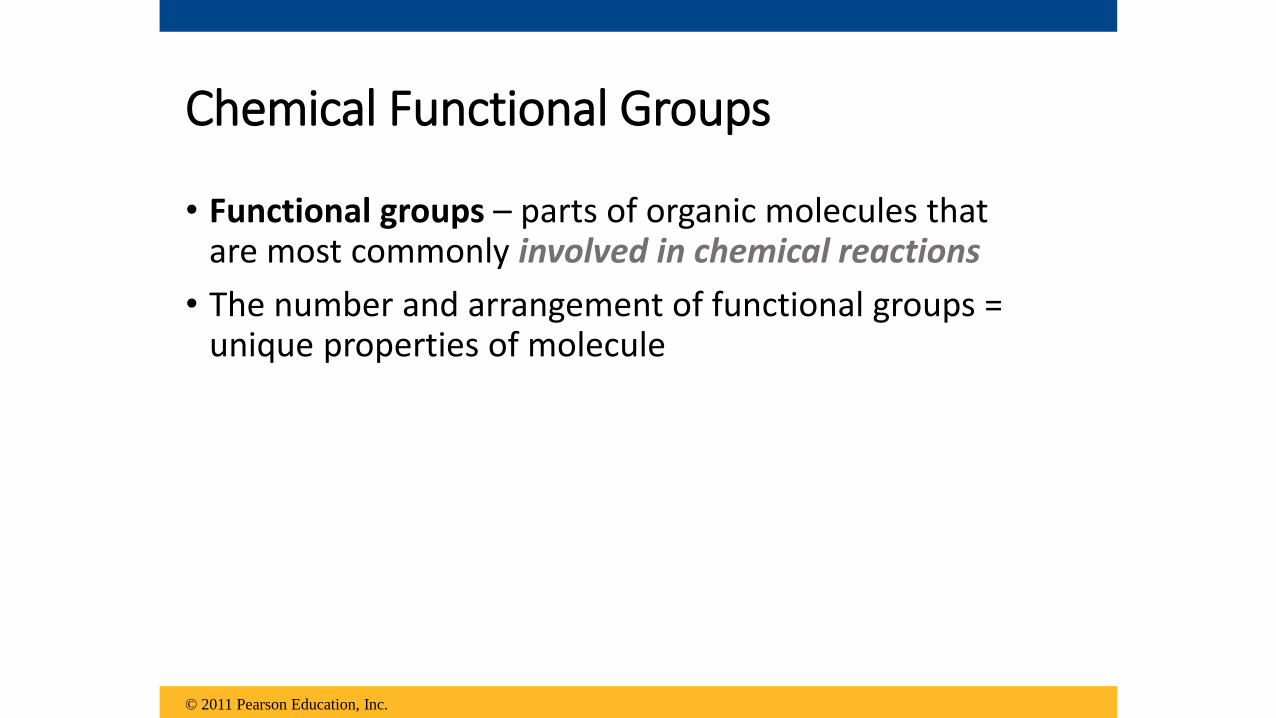
• The 7 functional groups most important in the chemistry of life:
1. Hydroxyl group2. Carbonyl group3. Carboxyl group4. Amino group5. Sulfhydryl group6. Phosphate group7. Methyl group
*NEED TO MEMORIZE WHAT IT LOOKS LIKE, WHERE TO FIND IT (macromolecule), and HOW IT WILL INTERACT (particularly with H2O)
© 2011 Pearson Education, Inc.

Figure 4.9-a
STRUCTURE
CHEMICALGROUP Hydroxyl
NAME OF
COMPOUND
EXAMPLE
Ethanol
Alcohols (Their specific names
usually end in -ol.)
(may be written HO—)
Carbonyl
Ketones if the carbonyl group is
within a carbon skeleton
Aldehydes if the carbonyl group
is at the end of the carbon skeleton
Carboxyl
Acetic acidAcetone
Propanal
Carboxylic acids, or organic acids
FUNCTIONAL
PROPERTIES• Is polar as a result of the
electrons spending more time
near the electronegative oxygen
atom.
• Can form hydrogen bonds with
water molecules, helping dissolve
organic compounds such as
sugars.
• A ketone and an aldehyde may be
structural isomers with different
properties, as is the case for
acetone and propanal.
• Ketone and aldehyde groups are
also found in sugars, giving rise
to two major groups of sugars:
ketoses (containing ketone
groups) and aldoses (containing
aldehyde groups).
• Found in cells in the ionized form
with a charge of 1 and called a
carboxylate ion.
Nonionized Ionized
• Acts as an acid; can donate an
H+ because the covalent bond
between oxygen and hydrogen
is so polar:

Figure 4.9-b
Amino Sulfhydryl Phosphate Methyl
Methylated compoundsOrganic phosphates
(may be
written HS—)
ThiolsAmines
Glycine Cysteine
• Acts as a base; can
pick up an H+ from the
surrounding solution
(water, in living
organisms):
Nonionized Ionized
• Found in cells in the
ionized form with a
charge of 1+.
• Two sulfhydryl groups can
react, forming a covalent
bond. This “cross-linking”
helps stabilize protein
structure.
• Cross-linking of cysteines
in hair proteins maintains
the curliness or straightness
of hair. Straight hair can be
“permanently” curled by
shaping it around curlers
and then breaking and
re-forming the cross-linking
bonds.
• Contributes negative charge to
the molecule of which it is a part
(2– when at the end of a molecule,
as above; 1– when located
internally in a chain of
phosphates).
• Molecules containing phosphate
groups have the potential to react
with water, releasing energy.
• Arrangement of methyl
groups in male and female
sex hormones affects their
shape and function.
• Addition of a methyl group
to DNA, or to molecules
bound to DNA, affects the
expression of genes.
Glycerol phosphate 5-Methyl cytidine

Figure 4. UN04
Adenosine
What is this structure??
What does it do for cells?

Figure 4. UN05
AdenosineAdenosine
Reactswith H2O
Inorganicphosphate
ATP ADP
Energy

Is this molecule soluble in water?
A.yesB.no

Is this molecule soluble in water?
A.yesB.no

The general structure of amino acids are shown in this figure. What functional groups are highlighted in salmon and yellow, respectively?
a) Amino and carboxyl
b) Amino and carbonyl
c) Hydroxyl and carbonyl
d) Methyl and carboxyl
e) Methyl and hydroxyl

The general structure of amino acids are shown in this figure. What functional groups are highlighted in salmon and yellow, respectively?
a) Amino and carboxyl
b) Amino and carbonyl
c) Hydroxyl and carbonyl
d) Methyl and carboxyl
e) Methyl and hydroxyl

What functional group is commonly used in cells to transfer energy from one organic molecule to another?
a) carboxyl
b) sulfhydryl
c) hydroxyl
d) phosphate
e) amino

What functional group is commonly used in cells to transfer energy from one organic molecule to another?
a) carboxyl
b) sulfhydryl
c) hydroxyl
d) phosphate
e) amino

Review
• Identify the functional groups in this structure. What macromolecule is it?

ALL living things are made up of 4
classes of large biological molecules:
1. Carbohydrates
2. Lipids
3. Proteins
4. Nucleic acids
© 2011 Pearson Education, Inc.

Macromolecules, polymers, monomers
• Polymer - long molecule consisting of many smaller building blocks
• Monomers- smaller building-block molecules of polymers
• Polymers:• Carbohydrates
• Proteins
• Nucleic acids
*Lipids – consider an exception; but usually built from smaller pieces
© 2011 Pearson Education, Inc.

Analogy of macromolecules

The Synthesis & Breakdown of Polymers
• Condensation reaction (AKA dehydration synthesis)-
when two monomers bond together through the LOSS of a water molecule (DEHYDRATES!)
• Hydrolysis- breaks chemical bonds of polymers into monomers, USING water; essentially the reverse of the dehydration reaction (“Lyse”- break, loosen)
© 2011 Pearson Education, Inc.
© 2011 Pearson Education, Inc.
Animation: Polymers
Right-click slide / select “Play”

Figure 5.2(a) Dehydration reaction: synthesizing a polymer
Short polymer Unlinked monomer
Dehydration removesa water molecule,forming a new bond.
Longer polymer
(b) Hydrolysis: breaking down a polymer
Hydrolysis addsa water molecule,breaking a bond.
1
1
1
2 3
2 3 4
2 3 4
1 2 3

The Diversity of Polymers
• Macromolecules vary among cells of an organism, vary more within a species, and vary even more between species
• An immense variety of polymers can be built from a small set of monomers
• Macromolecules Tutorial/ Animations
HO
© 2011 Pearson Education, Inc.

that consist of
Section 2-3
Carbohydrates Lipids Nucleic acids Proteins
Monosaccharides Fatty acids
& GlycerolNucleotides Amino Acids
Carbon
Compounds
include
which contain
that consist of that consist of that consist of
which contain which contain which contain
Carbon,hydrogen,
oxygen
Carbon,hydrogen,
Oxygen *
Carbon,hydrogen,oxygen, nitrogen,
phosphorus
Carbon,hydrogen,oxygen,
Nitrogen *
Go to
Section:
Concept Summary
* Phosphorus = phospholipid (2 tails) * Sulfur Types of Bonds: - Covalent (Alpha or beta
glycosidic)- Hydrogen (b/w
Types of Bonds: - Covalent and hydrogen- Ester linkages b/w hydrocarbon FA chain tails and glycerol head
Types of Bonds: - Covalent (b/w sugar/ phosphates and sugar/bases)- Hydrogen (b/w base pairs)
Types of Bonds: - PEPTIDE (b/w a.a.)
PRIMARY- Hydrogen (b/w R
groups of a.a) –SECONDARY
- disulfide bridges/ionic/ H bonds, hydrophobic interactions (b/w R groups of a.a) _TERT

Class: Elements made from:
Example: Functions: (SUBUNITS) Monomer and Basic Structural Diagrams
Carbohy-drate
C,H,O1:2:1
GLUCOSEC6H12O6
Mono-FructoseGlucoseGalactose
Di-LactoseSucrose
Poly-Starch GlycogenCellulose
1.Stores Short Term Energy
- Animals ONLY=
- Plants ONLY=
2. Structural
support within cells:-
-
MONOSACCHARIDE =
Mono + mono =
Mono + mono ++mono (X 100) =
Lipids Mostly
C, H
Verylittle
O
1. Stores LONG TERM Energy
2. Form cellmembranes
3. Waterproof coverings
4. Chemical messengers
5. Insulation6. Protection
1 glycerol3 fatty acids
Saturated-
Unsaturated-
GLYCOGEN
STARCH
PLANT CELL WALLS (CELLULOSE)
CHITIN (“KITE-IN”)
(INSECT EXOSKELETON; Fungi cell walls)
SINGLE SUGAR
POLYSACCHARIDE
FATSOILSWAXESSTEROIDSTestosteroneEstrogenCholesterol
NO DOUBLE BONDS IN FATTY ACID
(SINGLE bonds=Straight lines = solids)
@ LEAST 1 DOUBLE BOND (double = kink in the leg; can’t fit closely= liquids)
DISACCHARIDES
Saturated fat!

Nucleic acids
C HO P N
Stores and transmits hereditary information
Help in reproduction of cells
NUCLEOTIDES =
1.
2.
3.
ProteiNs CH O N*S
Shape of protein’s determine their functions:1. HELPS CONTROL
RATE OF REACTIONS (ENZYMES)
2. pump small molecules in and out of the cell
3. Aids in cell movement
4. Structural support-muscles (ACTIN/MYOSIN)5. Antibodies of immune system
AMINO ACIDS =
CLASS: Elements made of:
Examples: Functions: Monomer and Structural diagrams:
DNA/RNA
5-C SUGAR
PHOSPHATE
GROUP
NITROGENOUS
BASE
20 DIFFERENT
KINDS
HELD
TOGETHER BY
PEPTIDE BONDS!
*R GROUP- variable
that identifies each
of the 20 a.a.
Hemoglobin
Insulin
Collagen
Lactase
Trypsin
Pepsin

Carbohydrates (“-ose”)
• Serve as fuel and building material in cell walls
• Carbohydrates- include sugars and polymers of sugars
• Monosaccharides- single sugars; simplest carbohydrates
• Disaccharides – 2 monosaccharides held together by glycosidic bond
• Polysaccharides- macromolecules composed of hundreds of monosaccharide building blocks
© 2011 Pearson Education, Inc.

Monosaccharides
• Usually have molecular formulas that are multiples of CH2O (1 C: 2 H: 1 O)
• Glucose (C6H12O6)- most common
© 2011 Pearson Education, Inc.

Figure 5.3c
Aldose (Aldehyde Sugar) Ketose (Ketone Sugar)
Hexoses: 6-carbon sugars (C6H12O6)
Glucose Galactose Fructose

• FUNCTION: Monosaccharides serve as a major fuel for cells and as raw material for building molecules
© 2011 Pearson Education, Inc.

Figure 5.4
(a) Linear and ring forms
(b) Abbreviated ring structure
1
2
3
4
5
6
6
5
4
32
1 1
23
4
5
6
1
23
4
5
6

Disaccharides
• Formed when a CONDENSATION reaction
• Joins 2 monosaccharides = covalent bond called a glycosidic linkage
© 2011 Pearson Education, Inc.

Figure 5.5
(a) Dehydration reaction in the synthesis of maltose
(b) Dehydration reaction in the synthesis of sucrose
Alpha Glucose Alpha Glucose
Alpha Glucose
Maltose
Beta Fructose Sucrose
1–4glycosidic
linkage
1–2glycosidic
linkage
1 4
1 2

© 2011 Pearson Education, Inc.
Animation: DisaccharideRight-click slide / select “Play”
Polysaccharides
• Polymers of sugars
• storage and structural roles
• Structure and function determined by sugar monomers AND the positions of glycosidiclinkages
© 2011 Pearson Education, Inc.

Polysaccharides: Storage in PLANTS
• Starch- storage polysaccharide of PLANTS, consists entirely of glucose monomers
• store starch as granules within chloroplasts and other plastids or in roots (i.e. carrots, potatoes…)
• NEVER FOUND IN ANIMALS!
• Combination of AMYLOSE and AMYLOPECTIN• Amylose = Alpha glucose = Unbranching chain
(“STRAIGHT”) = Carbons #1, 4 bonding• Amylopectin = Alpha glucose = branching chain =
• 1, 4 AND Carbons #1, 6 bonding
© 2011 Pearson Education, Inc.

Figure 5.6
(a) Starch:a plant polysaccharide
(b) Glycogen:an animal polysaccharide
Chloroplast Starch granules
Mitochondria Glycogen granules
Amylopectin
Amylose
Glycogen
1 m
0.5 m

Polysaccharides: Storage in ANIMALS
•Glycogen - storage polysaccharide in animals
• Stores mainly in liver and muscle cells
• Like Amylopectin = Alpha glucose = branchingchain= Carbons #1, 4 and 1, 6 bonding
• Difference = more branching chains
© 2011 Pearson Education, Inc.
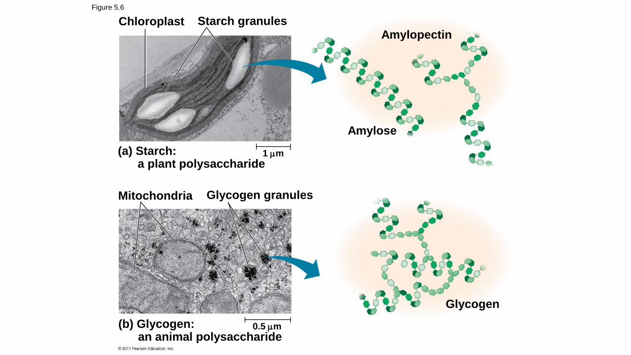
Figure 5.6
(a) Starch:a plant polysaccharide
(b) Glycogen:an animal polysaccharide
Chloroplast Starch granules
Mitochondria Glycogen granules
Amylopectin
Amylose
Glycogen
1 m
0.5 m
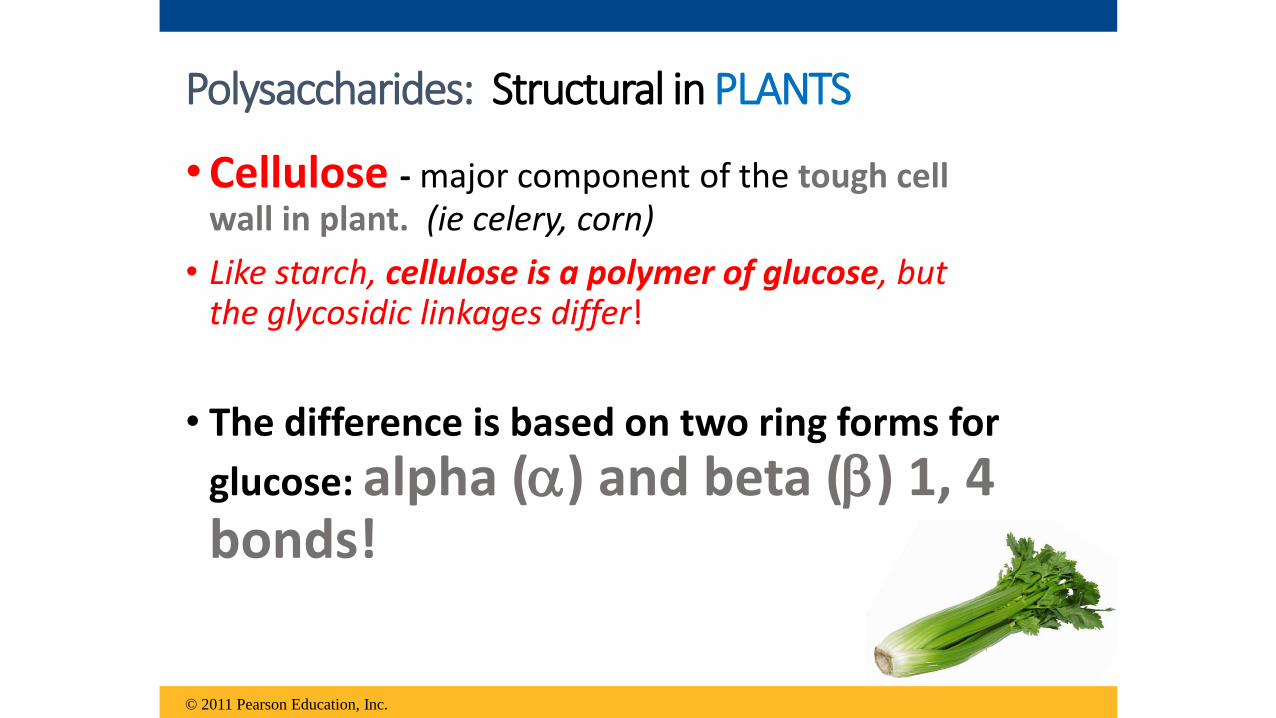
• Cellulose - major component of the tough cell wall in plant. (ie celery, corn)
• Like starch, cellulose is a polymer of glucose, but the glycosidic linkages differ!
• The difference is based on two ring forms for
glucose: alpha () and beta () 1, 4 bonds!
© 2011 Pearson Education, Inc.
Polysaccharides: Structural in PLANTS

Figure 5.7
(a) and glucosering structures
(b) Starch: 1–4 linkage of glucose monomers (c) Cellulose: 1–4 linkage of glucose monomers
Glucose Glucose
4 1 4 1
4141
= OH BELOW the ring on C1; same side = OH ABOVE the O; alternation of sides
C1 = the carbon to the right of the O


• Chitin- major component of the tough exoskeletons of invertebrates (ie cockroaches, crabs) AND cell walls of many fungi
• Like cellulose (1, 4 BETA bonds); ALSO HAS NITROGEN!
Polysaccharides: Structural in ANIMALS * not in Cambridge book!


© 2011 Pearson Education, Inc.

Figure 5.9
Chitin is used to make a strong and flexiblesurgical thread that decomposes after thewound or incision heals.

© 2011 Pearson Education, Inc.
Polymers with glucose are helical• Starch (amylose/amylopectin)• Glycogen
Polymers with glucose are straight• Cellulose- parallel strands of long cellulose
molecules group into microfibrils (strongbuilding structure for plants)
• Chitin
Glycosidic Linkages and Shape

Cell wall
Microfibril
Cellulosemicrofibrils in aplant cell wall
Cellulosemolecules
Glucosemonomer
10 m
0.5 m
Figure 5.8
Starch and
Cellulose
Animation
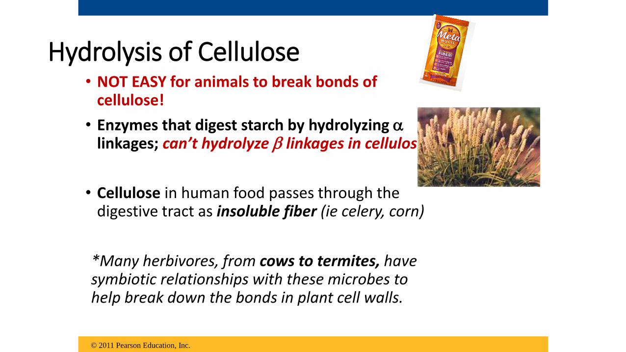
Hydrolysis of Cellulose• NOT EASY for animals to break bonds of
cellulose!
• Enzymes that digest starch by hydrolyzing linkages; can’t hydrolyze linkages in cellulose!
• Cellulose in human food passes through the digestive tract as insoluble fiber (ie celery, corn)
*Many herbivores, from cows to termites, have symbiotic relationships with these microbes to help break down the bonds in plant cell walls.
© 2011 Pearson Education, Inc.


Which polysaccharide has the greatest number of branches?
a) cellulose
b) starch
c) amylose
d) glycogen
Which polysaccharide has the greatest number of branches?
a) cellulose
b) starch
c) amylose
d) glycogen

a) proteinsb) starchc) nucleic acidsd) fatty acids
If actively growing cells are fed 14C-labeled
glucose, what macromolecules will become
radioactive first?
a) proteinsb) starchc) nucleic acidsd) fatty acids
If actively growing cells are fed 14C-labeled
glucose, what macromolecules will become
radioactive first?

Why are human enzymes that digest starch unable to digest cellulose?
a) Cellulose is made of amino-containing sugars that cannot be metabolized.
b) Cellulose is only in plants, therefore humans do not have enzymes to break plant polysaccharides.
c) Cellulose has beta-glycosidic linkages; starch-digesting enzymes break only alpha-glycosidic linkages.
d) Cellulose has alpha-glycosidic linkages that only bacterial enzymes can break.
Carbohydrate Lab Tests –POTENTIAL Practical Q!
1. Reducing/Non- Reducing Sugar test
2. Starch test
• Read p. 32-36 in sugar and starch lab tests Cambridge chapter
• Predict the results of the various test you will be doing of unknowns
(write in left margin of data table)
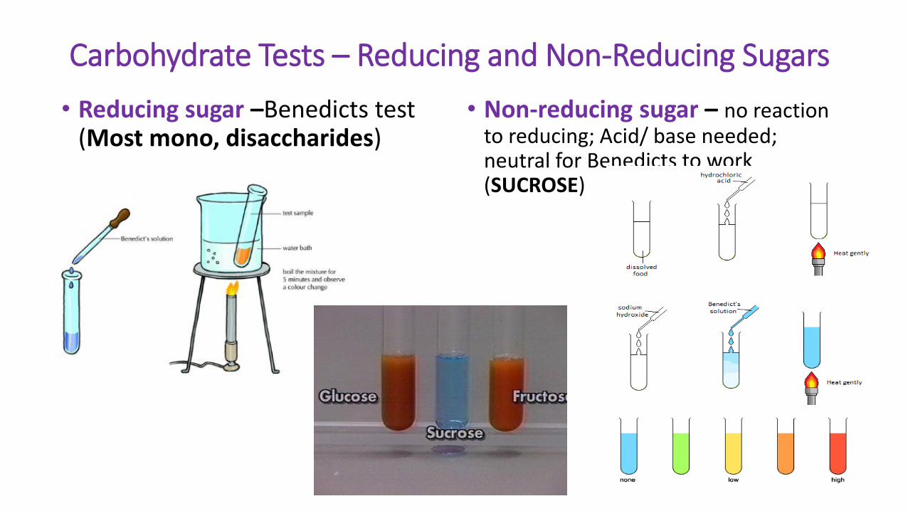
Carbohydrate Tests – Reducing and Non-Reducing Sugars
• Reducing sugar –Benedicts test (Most mono, disaccharides)
• Non-reducing sugar – no reaction to reducing; Acid/ base needed; neutral for Benedicts to work (SUCROSE)

Carbohydrate Tests – Presence of Starch test
• Potassium-Iodide (K2I) Solution= Strong + only for plant tissues (Storage carbohydrate in starchy plants- carrots, potatoes, etc)

LIPIDS

Lipids*Sometimes considered the class of macromolecules NOT formed of polymers
• Diverse group of hydrophobic molecules• having little or no affinity for water
• consist mostly of hydrocarbons
(formed of nonpolar covalent bonds)
• The most biologically important lipids:• Fats (Triglycerides)
• Phospholipids – cell membranes
• Steroids – hormones; cholesterol in cell membranes
© 2011 Pearson Education, Inc.
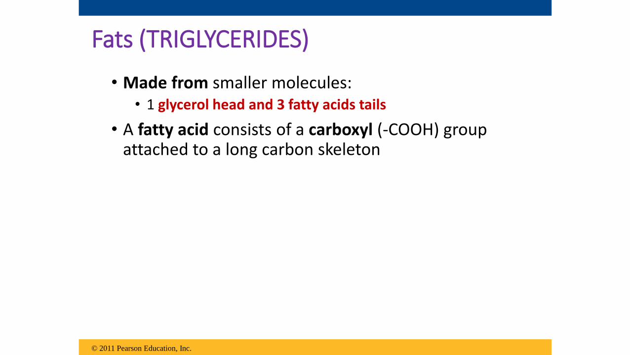
Fats (TRIGLYCERIDES)
• Made from smaller molecules: • 1 glycerol head and 3 fatty acids tails
• A fatty acid consists of a carboxyl (-COOH) group attached to a long carbon skeleton
© 2011 Pearson Education, Inc.

Figure 5.10
(a) One of three dehydration reactions in the synthesis of a fat
(b) Fat molecule (triacylglycerol)
Fatty acid(in this case, palmitic acid)
Glycerol
Ester linkage – cov. bond in fats holding glycerol head to tails
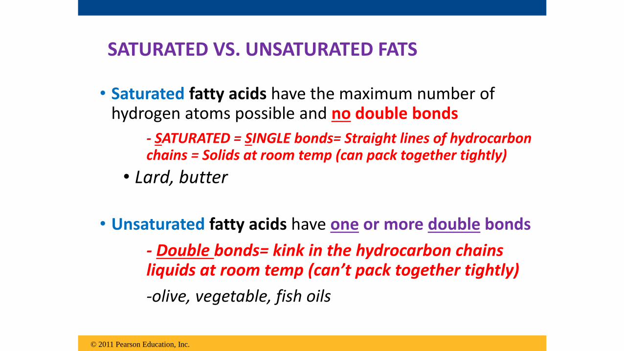
• Saturated fatty acids have the maximum number of hydrogen atoms possible and no double bonds
- SATURATED = SINGLE bonds= Straight lines of hydrocarbon chains = Solids at room temp (can pack together tightly)
• Lard, butter
• Unsaturated fatty acids have one or more double bonds
- Double bonds= kink in the hydrocarbon chains liquids at room temp (can’t pack together tightly)
-olive, vegetable, fish oils
© 2011 Pearson Education, Inc.
SATURATED VS. UNSATURATED FATS

© 2011 Pearson Education, Inc.
Animation: FatsRight-click slide / select “Play”

Figure 5.11
(a) Saturated fat(b) Unsaturated fat
Structuralformula of asaturated fatmolecule
Space-fillingmodel of stearicacid, a saturatedfatty acid
Structuralformula of anunsaturated fatmolecule
Space-filling modelof oleic acid, anunsaturated fattyacid
Cis double bondcauses bending.
A diet rich in saturated fats may contribute to cardiovascular disease through plaque deposits
Hydrogenation is the process of converting unsaturated fats to saturated fats by adding hydrogen

Functions of Lipids
• Long –term energy storage in adipose cells
• Structure of cell membranes
• Cushions vital organs
• Insulates the body
• Chemical messengers- Hormones
© 2011 Pearson Education, Inc.

Phospholipids
• In a phospholipid, two fatty acids and a phosphate group are attached to glycerol
• The two fatty acid tails are hydrophobic, but the phosphate group and its attachments form a hydrophilic head
© 2011 Pearson Education, Inc.

Figure 5.12
Choline
Phosphate
Glycerol
Fatty acids
Hydrophilichead
Hydrophobictails
(c) Phospholipid symbol(b) Space-filling model(a) Structural formula
Hyd
rop
hil
ic h
ea
dH
yd
rop
ho
bic
ta
ils

Figure 5.13
Hydrophilichead
Hydrophobictail
WATER
WATER
Fluid mosaic model
Phospholipids are the component of all cell membranes

Steroids
• Steroids are lipids characterized by a carbon skeleton consisting of 4 fused carbon rings
• Sex hormones – estrogen and testosterone
• Cholesterol- important component in animal cell membranes; helps maintain structure and shape of cell
© 2011 Pearson Education, Inc.
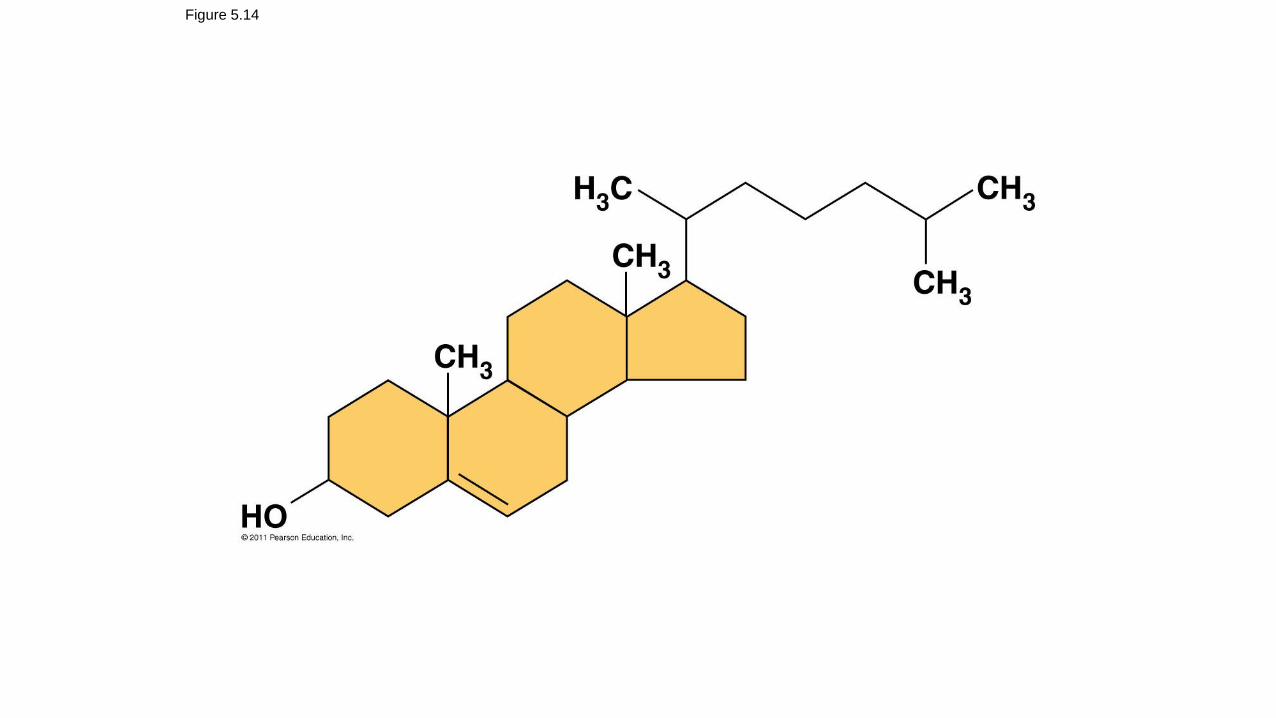
Figure 5.14

PROTEINS

Proteins• Proteins account for more than 50% of the dry mass
of most cells
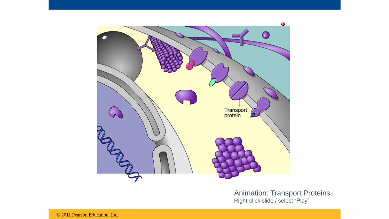
• Many functions include : 1. Structural support (hair/fingernails, skeletal muscle)2. Storage (albumin in eggs, seeds,milk)3. Transport (channels in cell membranes, hemoglobin)4. Cellular communications (hormones- insulin,
enzymes)5. Movement (skeletal muscle, flagella, centrioles) 6. Defense against foreign substances (ie. Enzymes and
antibodies)
© 2011 Pearson Education, Inc.

Figure 5.15-a
Enzymatic proteins Defensive proteins
Storage proteins Transport proteins
Enzyme Virus
Antibodies
Bacterium
Ovalbumin Amino acidsfor embryo
Transportprotein
Cell membrane
Function: Selective acceleration of chemical reactions
Example: Digestive enzymes catalyze the hydrolysis
of bonds in food molecules.
Function: Protection against disease
Example: Antibodies inactivate and help destroy
viruses and bacteria.
Function: Storage of amino acids Function: Transport of substances
Examples: Casein, the protein of milk, is the major
source of amino acids for baby mammals. Plants have
storage proteins in their seeds. Ovalbumin is the
protein of egg white, used as an amino acid source
for the developing embryo.
Examples: Hemoglobin, the iron-containing protein of
vertebrate blood, transports oxygen from the lungs to
other parts of the body. Other proteins transport
molecules across cell membranes.

Figure 5.15-b
Hormonal proteins
Function: Coordination of an organism’s activities
Example: Insulin, a hormone secreted by the
pancreas, causes other tissues to take up glucose,
thus regulating blood sugar concentration
Highblood sugar
Normalblood sugar
Insulinsecreted
Signalingmolecules
Receptorprotein
Muscle tissue
Actin Myosin
100 m 60 m
Collagen
Connectivetissue
Receptor proteins
Function: Response of cell to chemical stimuli
Example: Receptors built into the membrane of a
nerve cell detect signaling molecules released by
other nerve cells.
Contractile and motor proteins
Function: Movement
Examples: Motor proteins are responsible for the
undulations of cilia and flagella. Actin and myosin
proteins are responsible for the contraction of
muscles.
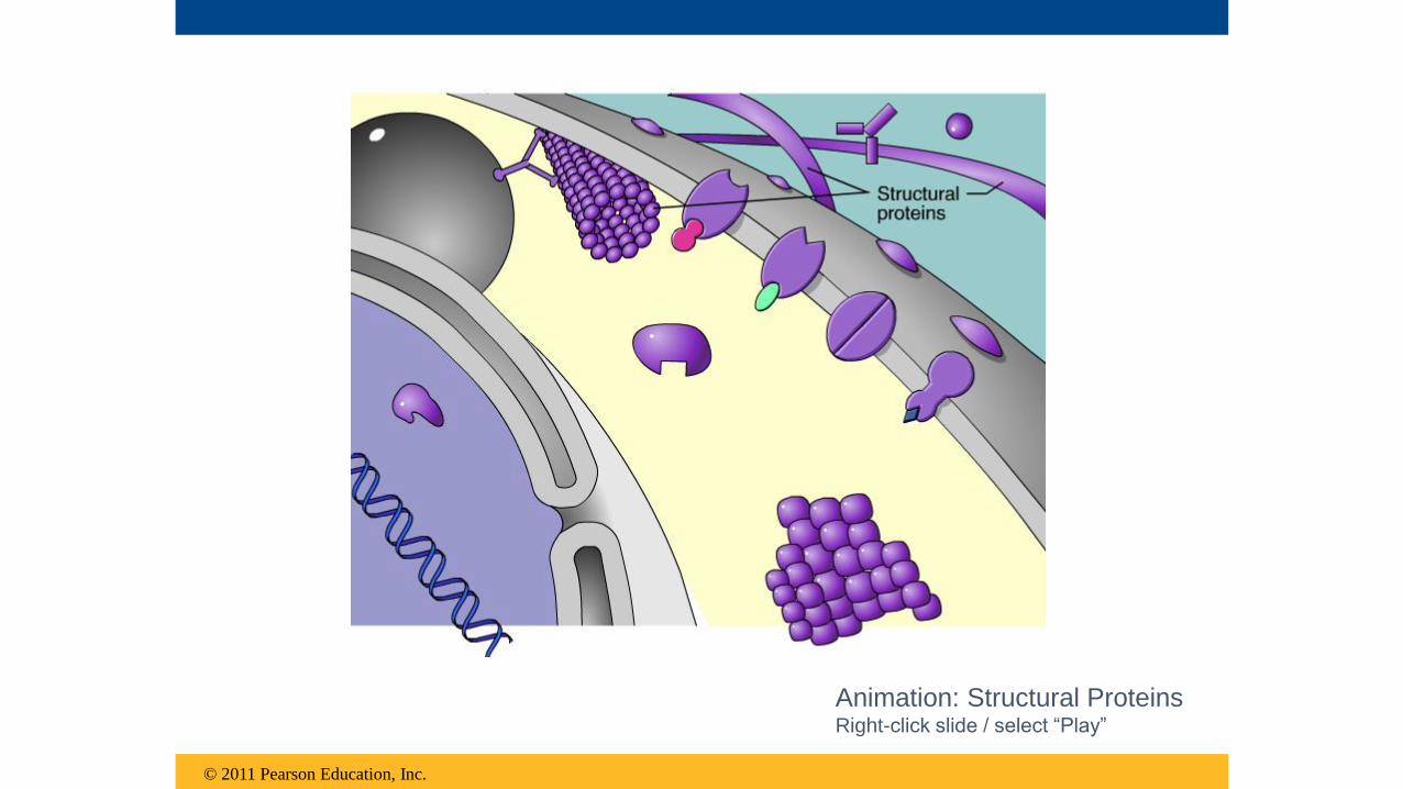
Structural proteins
Function: Support
Examples: Keratin is the protein of hair, horns,
feathers, and other skin appendages. Insects and
spiders use silk fibers to make their cocoons and webs,
respectively. Collagen and elastin proteins provide a
fibrous framework in animal connective tissues.
© 2011 Pearson Education, Inc.
Animation: Structural ProteinsRight-click slide / select “Play”

© 2011 Pearson Education, Inc.
Animation: Storage ProteinsRight-click slide / select “Play”
© 2011 Pearson Education, Inc.
Animation: Transport ProteinsRight-click slide / select “Play”

© 2011 Pearson Education, Inc.
Animation: Receptor ProteinsRight-click slide / select “Play”

© 2011 Pearson Education, Inc.
Animation: Contractile ProteinsRight-click slide / select “Play”

© 2011 Pearson Education, Inc.
Animation: Defensive ProteinsRight-click slide / select “Play”

© 2011 Pearson Education, Inc.
Animation: Hormonal ProteinsRight-click slide / select “Play”

© 2011 Pearson Education, Inc.
Animation: Sensory ProteinsRight-click slide / select “Play”

© 2011 Pearson Education, Inc.
Animation: Gene Regulatory ProteinsRight-click slide / select “Play”

• Enzymes are a type of protein that acts as a catalystto speed up chemical reactions
• Enzymes function as workhorses that carry out the processes of life
© 2011 Pearson Education, Inc.

© 2011 Pearson Education, Inc.
Animation: EnzymesRight-click slide / select “Play”

Structure of Proteins
Monomers = Amino acids • organic molecules with carboxyl and amino
groups
• differ in their properties due to differing side chains, called R groups
• Amino acids are linked by peptide bonds
• Polypeptides are unbranched polymers built from the same set of 20 amino acids
© 2011 Pearson Education, Inc.
Side chain (R group)
Aminogroup
Carboxylgroup
carbon

Figure 5.16
Nonpolar side chains; hydrophobic
Side chain(R group)
Glycine(Gly or G)
Alanine(Ala or A)
Valine(Val or V)
Leucine(Leu or L)
Isoleucine(Ile or I)
Methionine(Met or M)
Phenylalanine(Phe or F)
Tryptophan(Trp or W)
Proline(Pro or P)
Polar side chains; hydrophilic
Serine(Ser or S)
Threonine(Thr or T)
Cysteine(Cys or C)
Tyrosine(Tyr or Y)
Asparagine(Asn or N)
Glutamine(Gln or Q)
Electrically charged side chains; hydrophilic
Acidic (negatively charged)
Basic (positively charged)
Aspartic acid(Asp or D)
Glutamic acid(Glu or E)
Lysine(Lys or K)
Arginine(Arg or R)
Histidine(His or H)
20 Different Amino Acids separated by their R
groups; properties of each are indicative of R group
BASIC RULES TO KNOW: Charges or OH attached
= hydrophilic(+ bases, - acids)
• Rings, CH3 or H = nonpolar, hydrophobic
NONPOLAR and POLAR (water!) DON’T MIX well!!

Figure 5.17
Peptide bond
New peptidebond forming
Sidechains
Back-bone
Amino end(N-terminus)
Peptidebond
Carboxyl end(C-terminus)

Protein Shape and Function
• A functional protein consists of one or more polypeptides precisely twisted, folded, and coiled into a unique shape
• The sequence of amino acids determines a protein’s 3-D structure; structure determines its function
© 2011 Pearson Education, Inc.

Figure 5.18
(a) A ribbon model (b) A space-filling model
Groove
Groove

Figure 5.19
Antibody protein Protein from flu virus
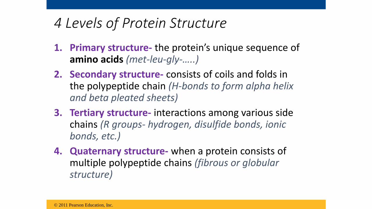
4 Levels of Protein Structure
1. Primary structure- the protein’s unique sequence of amino acids (met-leu-gly-…..)
2. Secondary structure- consists of coils and folds in the polypeptide chain (H-bonds to form alpha helix and beta pleated sheets)
3. Tertiary structure- interactions among various side chains (R groups- hydrogen, disulfide bonds, ionic bonds, etc.)
4. Quaternary structure- when a protein consists of multiple polypeptide chains (fibrous or globular structure)
© 2011 Pearson Education, Inc.

© 2011 Pearson Education, Inc.
Animation: Protein Structure IntroductionRight-click slide / select “Play”

Figure 5.20aPrimary structure
Aminoacids
Amino end
Carboxyl end
Primary structure of transthyretin

© 2011 Pearson Education, Inc.
Animation: Primary Protein StructureRight-click slide / select “Play”

Figure 5.20b
Secondarystructure
Tertiarystructure
Quaternarystructure
Hydrogen bond
helix
pleated sheet
strand
Hydrogenbond
Transthyretinpolypeptide
Transthyretinprotein

© 2011 Pearson Education, Inc.
Animation: Secondary Protein StructureRight-click slide / select “Play”

Secondary structure
Hydrogen bond
helix
pleated sheet
strand, shown as a flatarrow pointing towardthe carboxyl end
Hydrogen bond
Figure 5.20c

© 2011 Pearson Education, Inc.
Animation: Tertiary Protein StructureRight-click slide / select “Play”

Figure 5.20f
Hydrogenbond
Disulfidebridge
Polypeptidebackbone
Ionic bond
Hydrophobic
interactions and
van der Waals
interactions

Figure 5.20g
Quaternary structure
Transthyretinprotein
(four identicalpolypeptides)

Figure 5.20h
Collagen
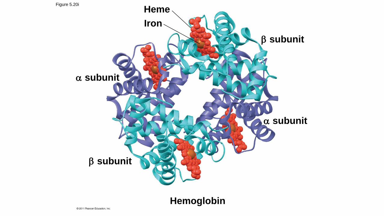
Hemoglobin
Heme
Iron
subunit
subunit
subunit
subunit
Figure 5.20i
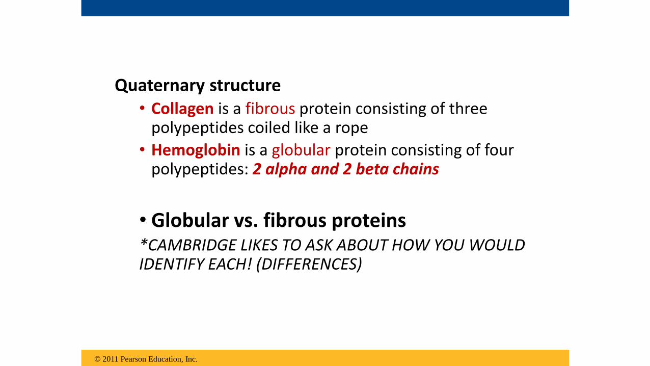
Quaternary structure • Collagen is a fibrous protein consisting of three
polypeptides coiled like a rope
• Hemoglobin is a globular protein consisting of four polypeptides: 2 alpha and 2 beta chains
• Globular vs. fibrous proteins*CAMBRIDGE LIKES TO ASK ABOUT HOW YOU WOULD IDENTIFY EACH! (DIFFERENCES)
© 2011 Pearson Education, Inc.

© 2011 Pearson Education, Inc.
Animation: Quaternary Protein StructureRight-click slide / select “Play”

Sickle-Cell Disease: A Change in Primary Structure
• A slight change in primary structure can affect a protein’s structure and ability to function
• Sickle-cell disease, an inherited blood disorder, results from a single amino acid substitution in the protein hemoglobin
© 2011 Pearson Education, Inc.

Figure 5.21
PrimaryStructure
Secondaryand TertiaryStructures
QuaternaryStructure
FunctionRed BloodCell Shape
subunit
subunit
Exposedhydrophobicregion
Molecules do notassociate with oneanother; each carriesoxygen.
Molecules crystallizeinto a fiber; capacityto carry oxygen isreduced.
Sickle-cellhemoglobin
Normalhemoglobin
10 m
10 m
Sic
kle
-cell h
em
og
lob
inN
orm
al
hem
og
lob
in
1
2
3
4
5
6
7
1
2
3
4
5
6
7

Denaturation of Proteins
• This loss of a protein’s native structure is called denaturation; becomes biologically inactive
• Alterations in • pH
• salt concentration
• temperature
• other environmental factors can cause a protein to unravel
© 2011 Pearson Education, Inc.

Figure 5.22
Normal protein Denatured protein
tu

Nucleic Acids

Functions of Nucleic acids:
• store, transmit, and help express hereditaryinformation
• DNA and RNA• programmed unit of inheritance called a gene =
a.a. sequence for protein synthesis
Monomers = nucleotides
© 2011 Pearson Education, Inc.

Figure 5.25-1
Synthesis ofmRNA
mRNA
DNA
NUCLEUS
CYTOPLASM
1

Figure 5.25-2
Synthesis ofmRNA
mRNA
DNA
NUCLEUS
CYTOPLASM
mRNA
Movement ofmRNA intocytoplasm
1
2

Figure 5.25-3
Synthesis ofmRNA
mRNA
DNA
NUCLEUS
CYTOPLASM
mRNA
Ribosome
AminoacidsPolypeptide
Movement ofmRNA intocytoplasm
Synthesisof protein
1
2
3

Figure 5.26
Sugar-phosphate backbone5 end
5C
3C
5C
3C
3 end
(a) Polynucleotide, or nucleic acid
(b) Nucleotide
Phosphategroup Sugar
(pentose)
Nucleoside
Nitrogenousbase
5C
3C
1C
Nitrogenous bases
Cytosine (C) Thymine (T, in DNA) Uracil (U, in RNA)
Adenine (A) Guanine (G)
Sugars
Deoxyribose (in DNA) Ribose (in RNA)
(c) Nucleoside components
Pyrimidines
Purines
EACH nucleotide= 1) a nitrogenous
base2) a pentose sugar, 3) one or more
phosphate groups

2 Group of nitrogenous bases:
1. Pyrimidines (cytosine, thymine, and uracil) have a single ring (SMALLER)
2. Purines (adenine and guanine) ring fused to a another ring (BIGGER!)
• In DNA, the sugar is deoxyribose; in RNA, the sugar is ribose
© 2011 Pearson Education, Inc.

The Structures of DNA and RNA Molecules
• RNA molecules - single polypeptide chains
• DNA molecules - double helix
• One DNA molecule includes many genes
© 2011 Pearson Education, Inc.

• The nitrogenous bases in DNA pair up and form hydrogen bonds: adenine (A) always with thymine (T), and guanine (G) always with cytosine (C)
• Called complementary base pairing
• Complementary pairing can also occur between two RNA molecules or between parts of the same molecule
• In RNA, thymine is replaced by uracil (U) so A and U pair
© 2011 Pearson Education, Inc.
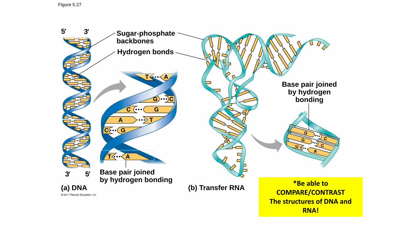
Figure 5.27
Sugar-phosphatebackbones
Hydrogen bonds
Base pair joinedby hydrogen bonding
Base pair joinedby hydrogen
bonding
(b) Transfer RNA(a) DNA
5 3
53
*Be able to COMPARE/CONTRAST
The structures of DNA and RNA!

REVIEW QUESTIONS!

Lipids
a) are made from glycerol and fatty acids.
b) contain nitrogen.
c) have low energy content.
d) are acidic when mixed with water.
e) do not dissolve well in water.
All lipids

Compared to tropical fish, arctic fish oils have
a) more unsaturated fatty acids.
b) more cholesterol.
c) fewer unsaturated fatty acids.
d) more trans-unsaturated fatty acids.
e) more hydrogenated fatty acids.
Lipids

Subunits and Metabolic Labeling
a) 35S-labeled sulfate
b) 32P-labeled phosphate
c) 14C-labeled leucine
d) 3H-labeled thymidine
e) 14C-labeled guanine
If you want to selectively label nucleic acids being
synthesized by cells, what radioactive compound
would you add to the medium?

Protein Structure and Amino Acids
a) on the exterior surface of the protein
b) in the interior of the protein, away from water
c) at the active site, binding oxygen
d) at the heme-binding site
Sickle-cell disease is caused by a mutation in the beta-
hemoglobin gene that changes a charged amino acid,
glutamic acid, to valine, a hydrophobic amino acid.
Where in the protein would you expect to find glutamic
acid?

Protein Structure
a) primaryb) tertiaryc) quarternaryd) all of the abovee) primary and tertiary
structures only
The sickle-cell hemoglobin
mutation alters what level(s)
of protein structure?

Macromolecular Structures and Bonds
a) Acidic pH denatures (unfolds and inactivates) proteins by disrupting their hydrogen bonds.
b) Citrus juice denatures proteins by disrupting their ionic bonds.
c) Citrus juice contains enzymes that hydrolyze peptide bonds to break apart proteins.
d) Citrus juice dissolves cell membranes by disrupting hydrophobic interactions.
Ceviche is prepared by marinating fresh raw fish in citrus juice for several hours, until the flesh becomes opaque and firm, as if cooked. How does citrus juice render the seafood safe to eat?

RNA and DNA
a) DNA encodes hereditary information; RNA does not.
b) DNA forms duplexes; RNA does not.
c) DNA contains thymine; RNA contains uracil.
d) all of the above
How does RNA differ from DNA?
Recommended