CEP11004, a novel inhibitor of the mixed lineage kinases,
suppresses apoptotic death in dopamine neurons of the substantia
nigra induced by 6-hydroxydopamine
Anindita Ganguly,* Tinmarla Frances Oo,* Margarita Rzhetskaya,* Robert Pratt,*
Olga Yarygina,* Takashi Momoi,� Nikolai Kholodilov* and Robert E. Burke*,�
Departments of *Neurology and �Pathology, The College of Physicians and Surgeons, Columbia University, New York,
New York, USA
�Division of Development and Differentiation, National Institute of Neuroscience, NCNP, Kodaira, Tokyo, Japan
Abstract
There is much evidence that the kinase cascade which leads
to the phosphorylation of c-jun plays an important signaling
role in the mediation of programmed cell death. We have
previously shown that c-jun is phosphorylated in a model of
induced apoptotic death in dopamine neurons of the sub-
stantia nigra in vivo. To determine the generality and func-
tional significance of this response, we have examined c-jun
phosphorylation and the effect on cell death of a novel mixed
lineage kinase inhibitor, CEP11004, in the 6-hydroxydopam-
ine model of induced apoptotic death in dopamine neurons.
We found that expression of total c-jun and Ser73-phosphor-
ylated c-jun is increased in this model and both colocalize with
apoptotic morphology. CEP11004 suppresses apoptotic death
to levels of 44 and 58% of control values at doses of 1.0 and
3.0 mg/kg, respectively. It also suppresses, to approximately
equal levels, the number of profiles positive for the activated
form of capase 9. CEP11004 markedly suppresses striatal
dopaminergic fiber loss in these models, to only 22% of con-
trol levels. We conclude that c-jun phosphorylation is a gen-
eral feature of apoptosis in living dopamine neurons and that
the mixed lineage kinases play a functional role as up-stream
mediators of cell death in these neurons.
Keywords: apoptosis, c-jun, c-jun kinase, Parkinson’s dis-
ease, programmed cell death.
J. Neurochem. (2004) 88, 469–480.
Programmed cell death may play a role in the pathogenesis of
the chronic neurodegenerative diseases and so may provide a
target for therapeutic interventions. Such interventions may
be more effective if they act up-stream in death pathways. In
the intrinsic, or mitochondrial, pathway of programmed cell
death, for example, it would seem desirable to intervene
before the release of mitochondrial death mediators (Yuan
and Yankner 2000).
One up-stream signaling cascade that mediates cell death
is that of c-jun N-terminal kinase (JNK) phosphorylation of
c-jun. Programmed cell death in neurons can be inhibited by
neutralizing antibodies to c-jun (Estus et al. 1994) or by
dominant negative mutants (Ham et al. 1995). Neurotrophic
factor withdrawal-induced death increases activity of JNK
(Xia et al. 1995; Park et al. 1996) and its activity is, in turn,
regulated by a complex up-stream kinase cascade (Fig. 1),
many components of which have also been implicated in the
regulation of neuronal cell death in vitro (Xu et al. 2001;
Bazenet et al. 1998; Gallo and Johnson 2002). This signaling
cascade is likely to be up-stream of mitochondrial release of
death mediators because inhibition of death with a dominant
negative c-jun blocks cytochrome c release in vitro (Whit-
field et al. 2001).
Studies with an indolocarbazole derivative, CEP-1347,
which inhibits the mixed lineage kinases (MLKs) (Maroney
Received June 2, 2003; revised manuscript received October 1, 2003;
accepted October 2, 2003.
Address correspondence and reprint requests to Robert E. Burke,
Department of Neurology, Room 308, Black Building, Columbia
University, 650 West 168th Street, New York, NY 10032, USA.
E-mail: [email protected]
Abbreviations used: ABC, avidin–biotin horseradish peroxidase
complexes; BSA, bovine serum albumin; DLK, dual leucine zipper
kinase; JNK, c-jun N-terminal kinase; MLK, mixed lineage kinase;
6OHDA, 6-hydroxydopamine; PB, phosphate buffer; PBS, phosphate-
buffered saline; PND, post-natal day; SN, substantia nigra; SNpc, SN
pars compacta; TH, tyrosine hydroxylase.
Journal of Neurochemistry, 2004, 88, 469–480 doi:10.1046/j.1471-4159.2003.02176.x
� 2003 International Society for Neurochemistry, J. Neurochem. (2004) 88, 469–480 469
et al. 2001), have supported the importance of the JNK/c-jun
signaling pathway in programmed cell death. In vitro, it
suppresses death induced by neurotrophic factor withdrawal,
oxidative stress and beta-amyloid treatment (Maroney et al.
1998, 1999; Bozyczko-Coyne et al. 2001). CEP-1347 also
suppresses neuron death in living animals in studies of
excitotoxicity (Saporito et al. 1998), natural cell death
(Glicksman et al. 1998), axotomy (Glicksman et al. 1998)
and noise-induced auditory hair cell loss (Pirvola et al. 2000).
We have had an interest in the role of the JNK/c-jun
signaling pathway in programmed cell death of dopamine
neurons of the substantia nigra (SN), which degenerate in
Parkinson’s disease. When these neurons are injured by the
neurotoxin 6-hydroxydopamine (6OHDA) or axotomy, they
show increased expression of c-jun and phosphorylated c-jun
(Jenkins et al. 1993; Herdegen et al. 1998). We have shown
that, when they are induced to die by apoptosis, following
early target deprivation, they up-regulate c-jun, phosphoryl-
ated c-jun and JNK with the onset of death and these proteins
colocalize with apoptotic morphology (Oo et al. 1999). The
probable functional role of JNK/c-jun signaling in regulating
the viability of these neurons is suggested by studies in the
MPTP neurotoxin model, which have demonstrated an
increased number of dopamine neurons following treatment
with CEP-1347 (Saporito et al. 1999). However, these studies
did not directly examine effects on cell death as opposed to
restorative effects on phenotype. Furthermore, studies in the
MPTP model are somewhat difficult to interpret in relation to
the canonical models of programmed cell death pathways
because the morphology of cell death following MPTP is
mixed. Acute treatment induces a necrotic form of cell death
(Jackson-Lewis et al. 1995) and, while chronic low doses of
MPTP induce apoptosis (Tatton and Kish 1997), they do not
do so exclusively; a necrotic morphological pattern is also
observed (Jackson-Lewis et al. 2000b). To explore the ability
of MLK inhibition to suppress dopamine neuron death and to
examine its role specifically in the intrinsic pathway we have
here examined the effect of a novel MLK inhibitor, CEP11004
(Murakata et al. 2002), on apoptotic death in a developmental
model induced by 6OHDA. We have previously shown that
the morphology of death in this model is exclusively apoptotic
(Marti et al. 1997) and we demonstrate herein that death is
associated with activation of caspase 9, an up-stream caspase
in the intrinsic pathway.
Materials and methods
6-hydroxydopamine lesion and CEP11004 treatment regimen
All animal procedures were performed in a facility accredited by the
Association for Assessment and Accreditation of Laboratory Animal
Care in accordance with protocols approved by the Institutional
Animal Care and Use Committee of Columbia University and the
principles outlined in the National Institute of Health Guide for the
Care and Use of Laboratory Animals. The 6OHDA lesions were
performed as previously described (Marti et al. 1997). Rat pups at
post-natal day (PND) 7 were pre-treated with 25 mg/kg des-
methylimipramine, anesthetized by hypothermia and placed prone
on an ice pack. The skull was exposed by a midline incision and a
burr hole was placed 3.0 mm lateral to the left of bregma on the
coronal suture. A 28-gauge cannula was then inserted vertically into
the striatum to a depth of 4.0 mm from the top of the skull.
6-Hydroxydopamine hydrobromide (Regis, Morton Grove, IL,
USA) was prepared at 15 lg (total weight)/1.0 lL in 0.9% NaCl/
0.02% ascorbic acid and infused by pump (Harvard Apparatus,
Holliston, MA, USA) at a rate of 0.25 lL/min. The cannula wasslowly withdrawn 2 min after the end of the infusion. After recovery
from anesthesia, pups were returned to the dams. A dose–response
analysis was performed with doses ranging from 1.0 to 15 lg and itwas determined that 2.5 lg was the minimally effective dose.
Therefore, this was the dose given in the CEP11004 experiments.
CEP11004 was initially dissolved in warm Solutol and then diluted
in phosphate-buffered saline (PBS) to a final Solutol concentration
of 10%. CEP11004 was administered subcutaneously the day before
the 6OHDA lesion and given once per day thereafter up to and
including post-lesion day 6.
Tissue processing and immunohistochemistry for c-jun,
phospho-c-jun, tyrosine hydroxylase and activated caspase 9
C-jun immunostaining
c-jun immunostaining was performed as previously described (Oo
et al. 1999). Pups were anesthetized byHalothane inhalation and then
perfused intracardially with 0.9% saline for 5 min by gravity,
followed by 4% paraformaldehyde in 0.1 M phosphate buffer (PB)
Fig. 1 The MAPK signaling pathways. A variety of cell stresses ini-
tially activate members of the Rho small GTPase family, Cdc42 and
Rac1 (Bazenet et al. 1998). These kinases act up-stream of members
of the mixed lineage kinase (MLK) family (Gallo and Johnson 2002)
including MLKs 1, 2, 3, dual leucine zipper kinase (DLK) and leucine
zipper kinase (LZK). These kinases are classified as MAPK kinase
kinases (MAP KKKs). The MLKs phosphorylate and activate MAPK
kinases (MAP KKs or MKKs). MKKs 4 and 7, in particular, have been
shown to activate c-jun N-terminal kinase (JNK) isoforms (Gallo and
Johnson 2002). JNK, in turn, phosphorylates and activates transcrip-
tion factors including c-jun.
470 A. Ganguly et al.
� 2003 International Society for Neurochemistry, J. Neurochem. (2004) 88, 469–480

for 10 min at room temperature. The brain was removed and the SN
was blocked. The SN was post-fixed in 4% paraformaldehyde in
0.1 M PB for 3 h at 4�C and then placed in 20% sucrose in 0.1 M PBfor 24 h prior to sectioning. Each SN was rapidly frozen by
immersion in isopentane on dry ice and then sectioned in a cryostat
at 30 lm. Sections were processed free-floating. They were initiallywashed with PBS, followed by PBS/0.5% bovine serum albumin
(BSA) and then by PBS/0.5% BSA with Triton 0.1% for 15 min at
4�C. After an additional wash in PBS, sections were incubated withantiserum to c-jun (Ab-1; Oncogene Science, Cambridge, MA, USA)
at 1 : 20 for 48 h at 4�C. Sections were then washed in PBS/0.5%BSA and then incubated with biotinylated Protein A, prepared in our
laboratory, at 1 : 100 for 60 min at room temperature. After a wash in
PBS/0.5%BSA, sections were incubated in avidin–biotin horseradish
peroxidase complexes (ABC; Vector Laboratories, Burlingame, CA,
USA) at 1 : 600 for 60 min at room temperature, followed by
incubation in diaminobenzidine in the presence of hydrogen peroxide
to generate a brown chromogen product. Sections were mounted to
slides subbed in gelatin and then thionin counterstained to identify
cellular morphology and intranuclear apoptotic chromatin clumps
(Janec and Burke 1993). The Ab-1 (Oncogene Science) to c-jun is an
affinity-purified rabbit polyclonal antibody raised against amino acids
209–225 in the DNA-binding domain in the C-terminal region of
v-jun. Previous investigators have shown that Ab-1 identifies a single
band of the appropriate molecular weight on western analysis of rat
brain homogenates and its regional distribution of immunostaining
correlates with expression of c-jun mRNA (Harlan and Garcia 1995).
Ser-73-phospho-c-jun immunostaining
Rat pups were anesthetized by hypothermia on wet ice in an
oxygenated chamber. They were perfused intracardially with 4%
paraformaldehyde/0.1 M PB/0.2 mM sodium orthovanadate at a rate
of 5.0 mL/min by peristaltic pump for 10 min. The brain was then
removed, the SN blocked and post-fixed in the same fixative
overnight. The SN block was then cryoprotected for 24 h in 20%
sucrose/0.1 M PB. The tissue was frozen as described and sectioned
at 30 lm on a cryostat. Sections were processed free-floating, as
described for c-jun. Sections were incubated with a rabbit anti-Ser73
phospho-c-jun polyclonal antibody (9164S; Cell Signaling Tech-
nology, Beverly, MA, USA) at 1 : 50 for 48 h. Thereafter, sections
were treated with biotinylated Protein A and ABC, as described for
c-jun. As for c-jun, sections were thionin counterstained to identify
intranuclear apoptotic chromatin clumps. Use of this antibody to
detect N-terminal phosphorylated c-jun has previously been reported
(Vaudano et al. 2001).
Tyrosine hydroxylase immunostaining
Animals were anesthetized and perfused as described for c-jun.
Brains were post-fixed for 1 week at 4�C and then cryoprotected.Representative coronal sections (30 lm) were taken from each of themajor planes encompassing the SN (Paxinos–Watson planes 2.7, 3.2,
3.7 and 4.2) (Paxinos and Watson 1982). After washes with PBS (2·for 15 min each), sections were incubated in primary antibody
(MAB5280; Chemicon, Temecula, CA, USA) mouse monoclonal
anti-tyrosine hydroxylase (TH) at 1 : 40 in PBS/10% normal horse
serum for 24 h at 4�C. Sections were then washed with PBS
(2· 15 min each) and incubated with biotinylated horse anti-mouseIgG (Vector Laboratories) at 1 : 50 in PBS/10% normal horse serum
at 4�C. Sections were washed in PBS/BSA (2· for 15 min each) atroom temperature and then incubated with ABC (Vector Laborator-
ies) at 1 : 600 for 1 h at room temperature. Following washes in PBS
(2· for 5 min each), sections were incubated in a solution of
diaminobenzidine (50 mg in 100 mLTris buffer, pH 7.6) containing
glucose oxidase, ammonium chloride and D(+) glucose to generate
H2O2. Following three 15-min washes in Tris buffer, pH 7.6,
sections were then mounted on gelatin-coated slides, left to dry
overnight at room temperature and thionin counterstained.
Activated caspase 9 immunostaining
Brains were processed initially as described for c-jun and then
incubated with a rabbit polyclonal antibody to the activated form of
caspase 9 (anti-m9D368) (Fujita et al. 2000) at 1 : 200 for 48 h at
4�C. The sections were then washed in PBS/BSA and treated withbiotinylated Protein A and ABC, as described for c-jun, except that
the ABC step was performed twice for increased sensitivity. The
sections were then incubated with diaminobenzidine and H2O2, as
described, and thionin counterstained. The antiactivated caspase 9
antibody has previously been characterized (Fujita et al. 2000).
Briefly, it was raised to the LDQLD368 C-terminal processing site of
caspase 9. This antiserum reacts specifically with the processed form
of expressed caspase 9 and not with the zymogen form. It detects
only a band corresponding to the processed form of caspase 9 in
staurosporine-treated wild type mouse embryonic fibroblasts and
detects no bands in caspase 9 null mouse embryonic fibroblasts.
Immunoreactivity detected by anti-m9D368 is augmented in the
brains of homozygous null bcl-2 mice and observed in conjunction
with positive TUNEL labeling.
Quantitative morphological analysis of c-jun, Ser73-phospho-c-jun-
positive and activated-caspase 9-positive profiles
For each brain, immunostained sections (with thionin counterstain)
were classified according to location within the SN pars compacta
(SNpc), based on planes comparable to planes 4.2, 3.7 or 3.2 in the
Paxinos–Watson atlas of adult rat brain (Paxinos and Watson 1982).
At least two sections within each of these planes were selected and
the SN in its entirety was scanned visually on both the non-injected
(Control) and the 6OHDA-injected (Experimental) sides at 600·.Thus, ‡ 6 sections per animal were scanned. A profile was counted aspositive if it contained brown chromogen nuclear staining well above
background levels (as shown in Figs 2 and 3). The values for number
of profiles were averaged among all of the individual sections
scanned within a specific plane to obtain an overall value for that
plane; these values were then added to obtain a measure of the
number of profiles for each region on each side of each brain. No
attempt was made to correct for double-counting error or to determine
the absolute number of positive neurons per brain. For this reason, the
counts are referred to as positive ‘profiles’ (Coggeshall and Lekan
1996). It should be noted, however, that double-counting error would
not be expected to be large, given the small size of the nucleus in
comparison to the section thickness (Saper 1996).
Qualitative morphological analysis of apoptosis in dopamine
neurons
In the TH immunoperoxidase-stained sections, apoptosis was
identified at the light microscope level by performing a thionin
counterstain and visualizing intranuclear chromatin clumps as one or
CEP11004 and apoptotic death 471
� 2003 International Society for Neurochemistry, J. Neurochem. (2004) 88, 469–480
more intensely basophilic, homogeneously stained, round and
distinctly bounded structures. We have previously shown for natural
cell death in dopamine neurons, and for induction of this death event
by either striatal lesion or the injection of 6OHDA, that apoptotic
profiles so identified are confirmed to be apoptotic by electron
microscopy (Macaya et al. 1994; Jackson-Lewis et al. 2000a).
Profiles so identified can also be confirmed as apoptotic by TUNEL
labeling, suppressed silver staining and immunostaining for activa-
ted caspase 3 and caspase cleavage products (Macaya et al. 1994;
Marti et al. 1997; Jeon et al. 1999; Jackson-Lewis et al. 2000a; Oo
et al. 2002).
Quantitative morphological analysis of apoptosis in neurons
of the substantia nigra pars compacta
Apoptotic profiles in each SN were quantified by selecting two to
three sections from each of Paxinos–Watson (Paxinos and Watson
1982) SN planes 4.2, 3.7 and 3.2 and scanning the entire SN at 600·.Apoptotic profiles were identified as cellular profiles containing one
or more distinct, rounded basophilic chromatin clumps. Free
extracellular chromatin clumps, lying outside an identifiable cellular
profile, were not counted. The number of profiles in the two sections
in a given plane were averaged to provide a measure for that plane
and the averages for the three planes were then added to provide an
index of the number of profiles for each SN. We have previously
shown, using a physical disector technique (Gundersen 1986), that
apoptotic profiles, as defined, are rarely split by the microtome blade
(Oo and Burke 1997). Clarke and Oppenheim (1995) have
demonstrated a similar result. Thus, apoptotic profiles, identified
by focusing through the section, represent unique and unbiased
counts. To assess the number of apoptotic profiles derived from
dopaminergic neurons of the SNpc, we have previously used two sets
of criteria: cellular and regional. An apoptotic profile was considered
to be dopaminergic by the cellular criterion if its cytoplasm was TH
positive. It was considered to be derived from a dopamine neuron by
the regional criterion if it was within 15 lm of two TH-positive
neurons. We used this regional criterion because dopamine neurons
lose their cytoplasm as they undergo apoptosis, making strict
phenotypic identification on the basis of cytoplasmic markers
impossible (Macaya et al. 1994). We have shown for natural cell
death in SNpc that there is a precise temporal and numerical
correlation between numbers of profiles counted using these two
criteria (r ¼ 0.939) (Oo and Burke 1997), suggesting that they are
likely to be derived from the same population.
Immunofluorescence double labeling for tyrosine hydroxylase and
Ser73-phospho-c-jun
Animals were anesthetized, perfused and their brains were removed
and frozen as described for phospho-c-jun. Sections were then
rinsed in Tris-buffered saline (pH 7.4)/0.2% Triton/2% normal goat
serum/2% normal base serum. They were then incubated with anti-
TH at 1 : 40 and anti-Ser73-phospho-c-jun at 1 : 50 for 48 h at 4�C.Following rinsing in Tris-buffered saline, they were then incubated
with horse anti-mouse-Texas Red (Vector Laboratories) at 1 : 75
and biotinylated goat anti-rabbit (Vector Laboratories) at 1 : 75 for
1 h at room temperature. Following a Tris-buffered saline rinse, they
were incubated with fluorescein-conjugated avidin (Vector Labor-
atories) at 1 : 100 for 1 h. The sections were then mounted on
gelatin-coated slides, dried, treated with Hoechst 33342 (0.0004%)
(Molecular Probes, Eugene, OR, USA) (to demonstrate apoptotic
chromatin clumps), rinsed and coverslipped with antifade medium
(Dako, Carpinteria, CA, USA). The sections were then viewed
under epifluorescence with an Eclipse 800 microscope (Nikon,
Melville, NY, USA) or with an LSM 510 Multiphoton Confocal
Microscope (Zeiss, Thornwood, NY, USA).
Striatal tyrosine hydroxylase-positive fiber staining and
quantification
Animals were anesthetized and perfused as described for TH neuron
staining. One week post-fixation, brains were frozen (without
cryoprotection) on fine powdered dry ice. Striatal sections were
cut at 30 lm from Paxinos–Watson plane 8.7–10.2. Sections were
rinsed in PBS and then treated with PBS/BSA and PBS/BSA/0.1%
Triton. Sections were then incubated with a rabbit anti-TH
polyclonal antibody (Calbiochem, San Diego, CA, USA) for 48 h
at 4�C, treated with biotinylated Protein A and ABC as described
and incubated with diaminobenzidine and H2O2. The optical density
of the TH immunoperoxidase staining was determined as previously
described (Burke et al. 1990) using an Analytical Imaging Station
(Imaging Research, Inc., St Catharines, Canada).
Northern analysis
The SN was microdissected from PND14 animals as previously
described (El-Khodor et al. 2001). RNAwas isolated from each SN
individually using an RNAeasy mini kit (Qiagen, Valencia, CA,
USA). Each piece of tissue was homogenized in 350 lL of buffer
(a)
(b)
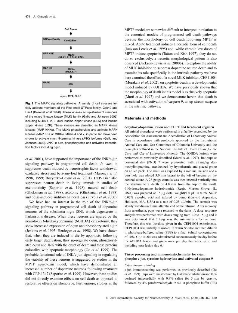
Fig. 2 The expression of c-jun is induced in substantia nigra pars
compacta (SNpc) following intrastriatal injection of 6-hydroxydopam-
ine (6OHDA). (a) Brown peroxidase staining for c-jun was observed
within cell nuclei. In a number of instances, as in this profile, c-jun
immunostaining colocalized with basophilic intranuclear chromatin
clumps (arrow), characteristic of apoptosis. Bar, 10 lm. (b) Quantita-
tive analysis of the number of c-jun-positive profiles in SNpc demon-
strates an about eightfold induction following intrastriatal 6OHDA
(**p < 0.02, ANOVA; n ¼ 7 vehicle injected; n ¼ 10 6OHDA injected).
CON, Control, non-injected side; EXP, 6OHDA- or vehicle-injected
side. SN, substantia nigra.
472 A. Ganguly et al.
� 2003 International Society for Neurochemistry, J. Neurochem. (2004) 88, 469–480
provided by passing it through a 27-gauge needle with a 1-mL
syringe 10 times. Further steps of the RNA isolation were carried
out according to the manufacturer’s instructions. RNA concentration
was then determined by measuring absorption at 260 nm on a
GenQuant spectrophotometer (Pharmacia, Cambridge, UK); 10 lgof each RNA sample per lane were electrophoresed in 1.4%
agarose–formaldehyde gels and equal loading of lanes was
confirmed by ethidium bromide staining. RNA was transferred onto
an Immobilon-Ny+ membrane (Millipore Corporation, Bedford,
MA, USA) using a capillary system (Sambrook et al. 1989)
overnight in 2· saline sodium citrate. After transfer, RNA was
cross-linked to the membrane by UV exposure in a FUNA-UV-
Linker (Spectroline, Westbury, NY, USA). Hybridization was
performed overnight at 67�C in ULTRAhyb (Ambion, Austin, TX,USA). Washing was carried out according to the manufacturer’s
recommendations. The membrane was then dried in air, wrapped
and exposed to a phosphorimager cassette. Radioactivity of bands
was determined using a Phosphor Imager 445 SI and ImageQuant
software.
Molecular probes for northern analysis
To create molecular probes for rat members of the MLK family we
utilized known human and mouse sequences to identify candidates
for rat homologues in rat expressed sequence tag databases. Based
on these sequences, primers were designed and RT–PCR was
performed on mRNA derived from rat brain. Each candidate
sequence was cloned and sequenced and, in each case, an open
reading frame analysis revealed a highly homologous amino acid
sequence to the human or mouse homologue of each corresponding
MLK family member. Our sequences for these rat homologues have
been entered in GenBank as the following Accession numbers:
MLK1, AY240866; MLK2, AY240867; MLK3, AY240868; dual
leucine zipper kinase (DLK), AY240864 and leucine zipper kinase,
AY240865.
Non-radioactive in situ hybridization
Brains from PND14 animals were rapidly removed, frozen on dry
ice and sectioned through the SN at 14 lm. Sections were thawmounted onto slides and stored at ) 80�C until use. On the day ofhybridization, the sections were warmed, fixed by immersion in 4%
paraformaldehyde/PBS, washed and then acetylated in triethanol-
amine/acetic anhydride solution. Following a wash in PBS, sections
were pre-hybridized as previously described (Burke et al. 1994).
Digoxigenin-11-UTP riboprobes were prepared as per the manufac-
turer’s instructions (Roche Diagnostics GmbH, Penzberg, Germany).
The size and integrity of labeled probe were confirmed by gel
(a) (b)
(c) (d)
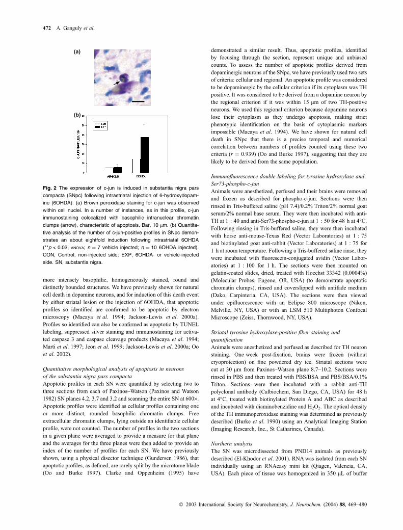
Fig. 3 Expression of Ser73-phospho-c-jun is induced in substan-
tia nigra pars compacta (SNpc) following intrastriatal injection of
6-hydroxydopamine (6OHDA). (a and b) Low power photomicrographs
of Ser73-phospho-c-jun immunostaining following intrastriatal 6OHDA.
These micrographs represent the non-injected contralateral side (a)
and the ipsilateral, injected side (b) of the same section. No staining is
observed on the non-injected side. Robust staining is observed
throughout the SNpc on the injected side. (c) At a cellular level, Ser73-
phospho-c-jun staining was observed in the nuclei of neurons with
normal morphology. Occasionally apoptotic chromatin clumps were
observed, as shown here (arrow), but, even in these instances,
neuronal morphology appeared normal. Bar, 10 lm. (d) Quantitative
analysis of the number of phospho-c-jun-positive profiles demon-
strates a marked induction following intrastriatal 6OHDA (**p < 0.001,
ANOVA; n ¼ 4 vehicle-injected; n ¼ 4 6OHDA-injected). CON, Control,
non-injected side; EXP, 6OHDA- or vehicle-injected side; SN,
substantia nigra; SNpr: SN pars reticulata; P04-c-jun: phosphorylated
c-jun.
CEP11004 and apoptotic death 473
� 2003 International Society for Neurochemistry, J. Neurochem. (2004) 88, 469–480

electrophoresis. Hybridization was performed overnight at 68�C in ahumidified chamber. Sections were washed in 0.2· saline sodiumcitrate at 68�C and then incubated overnight with an anti-
digoxigenin antibody (Roche Diagnostics, Mannheim, Germany).
Following a wash in 0.1 M Tris/0.15 M NaCl, sections were
incubated with a solution containing nitro blue tetrazolium and 5-
bromo-4-chloro-3-indolyl-phosphate in a darkened humidified
chamber overnight. Sections were then washed in Tris/EDTA
(pH 8.0) and coverslipped with aqueous mounting medium (Dako).
Results
Expression of c-jun and Ser73-phospho-c-jun are induced
in substantia nigra pars compacta following
6-hydroxydopamine
Following intrastriatal injection of 6OHDA at PND7, the
expression of c-jun in SNpc was examined at post-lesion day
6, the time of peak cell death (Marti et al. 1997) in this
model. A clear induction was observed, with the mean
number of profiles increased about eightfold in comparison
to vehicle-injected animals (Fig. 2). The immunostaining for
c-jun at a cellular level was localized to the nucleus in all
profiles (Fig. 2a). The large majority (97.2 ± 1.0% for
n ¼ 10 animals) of cellular profiles with c-jun-positive
nuclei had normal-appearing neuronal morphology on thio-
nin counterstain. The remaining profiles (2.8 ± 1.1%) dem-
onstrated intensely basophilic, round chromatin clumps,
characteristic of apoptosis (Fig. 2a). There was a trend for
the number of c-jun-positive profiles to be increased in the
contralateral SNpc of 6OHDA-injected animals in compar-
ison to vehicle-injected animals (Fig. 2b) suggesting a
modest toxin effect on the non-injected side, but this did
not achieve significance.
Following intrastriatal injection of 6OHDA, there was also
a clear induction of phosphorylation of c-jun (Fig. 3).
Interestingly, this induction was more robust, and more
specific for injury, than the induction of total c-jun protein.
c-jun protein was observed in a small but consistent number
of cells even on the contralateral side of vehicle-injected
animals. However, no Ser73-phospho-c-jun profiles were
observed in that condition (Fig. 3d). While a small number
of profiles were observed in SNpc following vehicle injection
(1.9 ± 1.0; n ¼ 4), there was a 28-fold increase in their
number following 6OHDA injection (Fig. 3d). As for c-jun,
the immunostaining for Ser73-phospho-c-jun was always
nuclear and the large majority of stained cells showed normal
neuronal morphology. Those few which showed colocaliza-
tion of Ser73-phospho-c-jun staining with intranuclear
apoptotic chromatin clumps showed normal neuronal mor-
phology, suggesting that this staining is an early event in the
course of apoptosis (Fig. 3c).
In order to confirm the expression of Ser73-phospho-c-jun
in dopaminergic neurons of the SNpc, fluorescent double
labeling was performed with immunostaining for TH. This
staining demonstrated phospho-c-jun staining exclusively
within TH-positive neurons of the SNpc ipsilateral to the
6OHDA injection (Fig. 4). As observed on thionin stain, the
neurons with phospho-c-jun-positive nuclei had normal
neuronal morphology (Fig. 4) even when apoptotic chroma-
tin was observed in the nucleus (Figs 4c–f).
Mixed lineage kinases are expressed in substantia nigra
during development
Having shown that c-jun protein and its phosphorylation are
induced in dopamine neurons of the SNpc following 6OHDA
injection, we next sought to determine whether critical
up-stream signaling components, particularly members of the
MLK family, are expressed. We, therefore, assayed for the
presence and relative abundance of mRNAs for the MLK
family members by northern analysis in microdissected
PND14 ventral mesencephalic tissues. As shown in Fig. 5,
mRNAs for MLK 1, 2, 3, DLK and leucine zipper kinase
were detectable in these tissues. The most abundant mRNA
was that of DLK, which was almost four times more
abundant than that of the next most abundant, MLK1. In
order to confirm this higher level of expression of DLK
anatomically, we performed in situ hybridization for DLK in
comparison to another representative member of the MLKs,
MLK3. This analysis demonstrated a much higher level of
expression of DLK in the SN (Figs 5c and d). At a cellular
level, this analysis revealed that high levels of expression of
DLK are observed in neurons of the SNpc.
Fig. 4 Ser73-phospho-c-jun immunostaining is observed exclusively
within tyrosine hydroxylase (TH)-positive neurons of the substantia
nigra pars compacta (SNpc) following intrastriatal 6-hydroxydopamine
injection. (a) An optical plane through the SNpc visualized at low
power by confocal microscopy demonstrates that intranuclear Ser73-
phospho-c-jun staining (green) is observed only in neurons with
TH-positive cytoplasm (red). Profile 1, positive for both labels, is
shown at higher power in (b). It has a normal polygonal shape with
normal-appearing, extended neural processes. Profile 2 in (a) is
TH positive but negative for phospho-c-jun. Occasional TH-positive
neurons (c) which were positive for intranuclear phospho-c-jun stain-
ing (d) also contained apoptotic nuclear chromatin clumps, revealed by
Hoechst 33342 dye (arrows in e and f).
474 A. Ganguly et al.
� 2003 International Society for Neurochemistry, J. Neurochem. (2004) 88, 469–480

CEP11004 suppresses 6-hydroxydopamine-induced
apoptosis in substantia nigra dopamine neurons
In order to test the ability of CEP11004 to suppress apoptosis
induced in dopamine neurons by 6OHDA, it was adminis-
tered on the day before lesion and then each day until killing
on the sixth post-lesion day. At a dose of 1.0 mg/kg/day,
CEP11004 suppressed apoptosis to levels only 44% of those
observed in vehicle-treated animals (Fig. 6a). To determine
whether further suppression could be achieved with a higher
dose, a second experiment was performed using 3.0 mg/kg/
day. While this regimen also achieved suppression of
apoptosis, it was somewhat less than that observed with
1.0 mg/kg/day; apoptosis was suppressed to levels 58% of
those observed in vehicle-treated animals (Fig. 6b). This
difference between the two doses, however, did not achieve
significance (p ¼ 0.3).
To confirm that apoptosis was suppressed in phenotypi-
cally defined dopamine neurons, we analyzed TH-positive
apoptotic profiles (Fig. 6c). There was a suppression to 48%
of vehicle controls, similar to that observed when both
cellular and regional counting criteria were used.
Caspase 9 is activated following 6-hydroxydopamine
injection and its activation is suppressed by CEP11004
To confirm that caspase 9 is activated when apoptosis is
induced by 6OHDA, immunohistochemistry was performed
with an antibody which exclusively recognizes the activated
form (Fujita et al. 2000). Immunostaining was positive
strictly in cellular profiles which also demonstrated classic
basophilic intranuclear chromatin clumps by thionin count-
erstain (Fig. 7a). Staining was observed in some profiles
exclusively within the cytoplasm (as shown in Fig. 7a) but,
in others, it was either strictly perinuclear or in both cellular
compartments. Given that 40–50% of apoptosis appears to be
Fig. 6 CEP11004 suppresses apoptosis in dopamine neurons of the
substantia nigra pars compacta (SNpc) induced by 6-hydroxydopam-
ine (6OHDA). (a) CEP11004 administered at a dose of 1.0 mg/kg/day
for 8 days, starting the day before lesion and continuing until the sixth
post-lesion day, suppressed apoptosis in substantia nigra (SN) to 44%
of vehicle treatment levels (**p ¼ 0.004, ANOVA; Tukey all pairwise
comparisons post-hoc; n ¼ 8, vehicle; n ¼ 7, 6OHDA). For this ana-
lysis all apoptotic profiles within the SNpc of tyrosine hydroxylase
(TH)-stained sections, meeting cellular or regional criteria as defined in
Materials and methods, were counted. CON, Control, non-injected
side; EXP, 6OHDA-injected side. (b) CEP11004 3.0 mg/kg/day also
suppressed apoptosis to 58% that of vehicle treatment (**p ¼ 0.005,
ANOVA; Tukey post-hoc; n ¼ 6, vehicle; n ¼ 7, 6OHDA). (c) The sec-
tions analyzed in (b) were also analyzed for only those profiles which
met cellular criteria for being dopaminergic, i.e. they had a TH-positive
cytoplasm. These TH-positive profiles also showed a significant sup-
pression by CEP11004 (**p < 0.02, ANOVA).
(a)
(b)
(c) (d) (e)
DLK
5 kb
3.6 kb
2.6 kb
MLK3 MLK2 MLK1LZK
Fig. 5 Northern and in situ hybridization analysis of mRNA expression
for members of the mixed lineage kinase (MLK) family in ventral
mesencephalon. (a) A representative northern analysis demonstrates
that mRNAs for dual leucine zipper kinase (DLK), MLK3, MLK2, leu-
cine zipper kinase (LZK) and MLK1 are detectable in ventral mesen-
cephalic tissue at post-natal day 14. For each mRNA species, samples
from two animals are electrophoresed in adjacent lanes. (b) Quantifi-
cation of radioactivity within bands by phosphorimager demonstrates
that the most abundant mRNA species is that of DLK (each column
represents the mean ± SEM for n ¼ 6 determinations). (**p < 0.001,
ANOVA) (c) Non-radioactive in situ hybridization demonstrates robust
expression of DLK in substantia nigra (SN) particularly the SN pars
compacta (SNpc). An adjacent section, processed in parallel, probed
for MLK3 (d) shows a much lower level of signal intensity. (e) A high
power photomicrograph of the section shown in (c) demonstrates
neuronal staining in the SNpc (arrows). Bar, 10 lm.
CEP11004 and apoptotic death 475
� 2003 International Society for Neurochemistry, J. Neurochem. (2004) 88, 469–480

resistant to CEP11004 in this model, we sought to determine
whether this resistance may be due to other death pathways
acting independently of caspase 9. Such a possibility would
predict that CEP11004 would suppress a greater percentage
of activated caspase 9 profiles than total profiles in SNpc. We
observed, however, that while CEP11004 suppressed caspase
9 profiles, to a level of 58% of vehicle-treated controls
(Fig. 7c), this level was not significantly different from that
observed for all thionin-stained profiles (67%) (Fig. 7b) or
for profiles in SNpc in TH-immunostained material meeting
either cellular or regional criteria (44%; Fig. 6a). We,
therefore, conclude that the cell death in this model which
is not suppressible by CEP11004 utilizes caspase 9 to a
similar degree as that which is suppressible.
CEP11004 suppresses degeneration of tyrosine
hydroxylase-positive striatal dopaminergic fibers
following 6-hydroxydopamine injection
As cell death mechanisms involved in the destruction of
axons may be different from those which are operative at the
level of the cell body (Raff et al. 2002), we investigated
whether CEP11004 also protects striatal dopaminergic fibers
in the 6OHDA model. Administration of CEP11004 led to a
clear preservation of striatal TH-positive fibers, quantified by
measurement of optical density of peroxidase reaction
product over the striatum (Fig. 8). Optical density values
were reduced by a mean of 0.023 units in vehicle-treated
animals, whereas the reduction was only 0.005 units in
CEP11004-treated animals. Thus, CEP-treated animals
showed a reduction only 22% of that observed in vehicle-
treated animals. In the ANOVA of the data presented in
Fig. 8(a), there was no significant difference between optical
(a)
(b)
(c)
Fig. 7 CEP11004 (CEP) suppresses activation of caspase 9 upon
induction of apoptosis by intrastriatal 6-hydroxydopamine (6OHDA).
(a) Immunoperoxidase staining of activated caspase 9 within the
cytoplasm of an apoptotic profile within the substantia nigra pars
compacta (SNpc). A single, basophilic apoptotic chromatin clump is
observed within the nucleus of this profile. Bar, 10 lm. (b) CEP11004
decreases the total number of apoptotic profiles identified by thionin
stain within SNpc [n ¼ 9 for both vehicle (VEH) and CEP11004
treatment groups; ANOVA, p < 0.001 for all groups; post-hoc Tukey,
**p ¼ 0.01 between vehicle and CEP11004 6OHDA (EXP) condi-
tions]. CON, Control, non-injected side; EXP, 6OHDA-injected side.
(c) In the same sections analyzed in (b) CEP11004 decreases the
number of activated caspase 9 profiles in the SNpc following intra-
striatal 6OHDA (ANOVA, p < 0.001 for all groups; post-hoc Tukey,
*p ¼ 0.05 between vehicle and CEP11004). SN, substantia nigra.
(a)
(b)
Fig. 8 CEP11004 protects striatal dopaminergic fibers in the intra-
striatal 6-hydroxydopamine (6OHDA) model. (a) Optical density
measurements of striatal tyrosine hydroxylase (TH) immunoperoxi-
dase reaction product reveal a 0.023 unit (62%) reduction by 6OHDA
in vehicle-treated animals. In the CEP11004-treated animals, there is
only a 0.005 unit (17%) reduction. CON, Control, non-injected side;
EXP, 6OHDA-injected side (*p < 0.05, post-hoc Tukey). (b) Repre-
sentative TH-stained coronal sections of the striatum in comparable
planes (Paxinos–Watson 9.7) from vehicle- and CEP11004-treated
animals. There is preservation of staining on the 6OHDA-treated side
in the CEP11004-treated animals. NS, not significant.
476 A. Ganguly et al.
� 2003 International Society for Neurochemistry, J. Neurochem. (2004) 88, 469–480

densities in the 6OHDA-treated striatum and in the contra-
lateral control striatum in CEP11004-treated animals.
Discussion
The present study confirms previous observations, made at a
regional level (Vaudano et al. 2001), that increased expres-
sion of c-jun and phospho-c-jun accompanies the death of
SNpc dopamine neurons induced by intrastriatal injection of
6OHDA and demonstrate, at a cellular level, colocalization
with apoptotic morphology. Using a suppressed silver stain
technique, which is capable of detecting necrosis (Nadler and
Evenson 1983; Jackson-Lewis et al. 1995), and variant
morphologies of programmed cell death (Oo et al. 1996) we
have previously shown that the morphology of cell death in
this intrastriatal 6OHDA model is exclusively apoptotic
throughout its time course (Marti et al. 1997). We have
previously shown that increased expression of c-jun and
phospho-c-jun also accompanies the exclusive induction of
apoptosis in a developmental target injury model (Macaya
et al. 1994; Oo et al. 1999) and an axotomy model
(El-Khodor and Burke 2002) (and unpublished observa-
tions). We, therefore, conclude that increased expression of
c-jun and phospho-c-jun is a universal correlate of apoptotic
death in dopamine neurons during development. This is the
case in spite of the fact that, while these models share the
classic light microscope features of neuronal apoptosis, they
differ in terms of the cellular distribution of activated caspase
3 (Jeon et al. 1999) and caspase cleavage products (Oo et al.
2002), indicating that there are likely to be different
underlying cellular mechanisms.
Increased expression of c-jun or phospho-c-jun also
accompanies induction of apoptosis in dopamine neurons
in the adult setting. Intrastriatal injection of 6OHDA in adults
induces apoptosis (Marti et al. 2002) and this lesion has been
associated with increased expression of c-jun (Jenkins et al.
1993; Vaudano et al. 2001) and phospho-c-jun (Vaudano
et al. 2001). Likewise, low, chronic doses of MPTP induce
apoptosis (Tatton and Kish 1997) and this regimen induces
phosphorylation of MKK4, JNK and c-jun (Saporito et al.
2000; Xia et al. 2001). However, the precise relationship
between c-jun signaling and any specific pathway of cell
death in these adult models is somewhat difficult to identify
because mixed morphologies of cell death occur in both the
adult 6OHDA (Marti et al. 1997) and the low dose MPTP
model (Jackson-Lewis et al. 2000b). In these studies,
expression of c-jun-related signaling molecules was not
identified in conjunction with either apoptotic morphology or
caspase activation at a cellular level. In fact, one possible, but
equally interesting, interpretation of these studies may be that
c-jun signaling is associated with necrosis or variant
morphologies of programmed cell death either instead of,
or in addition to, apoptosis. This may also be the case in an
adult medial forebrain bundle axotomy model in which
apoptotic morphology and in situ end labeling are not
observed (Crocker et al. 2001) and yet increased expression
of c-jun and phospho-c-jun have been reported (Herdegen
et al. 1998). In any case, we can conclude, on the basis of the
present work, that c-jun signaling is a common feature of
apoptosis in dopamine neurons during development and,
based on studies by others, that it is also a common feature of
cell death in adult models, where the relationship to
apoptosis, specifically, remains to be defined.
The present study also demonstrates that the association
between c-jun and phospho-c-jun both in time and spatially
at a cellular level with apoptosis is likely to indicate a
functional role because inhibition of the MLKs, which act as
up-stream mediators of c-jun phosphorylation, by adminis-
tration of CEP11004 suppresses apoptotic cell death. This
conclusion is supported by observations in the aforemen-
tioned adult models of dopamine neuron degeneration.
Saporito et al. (1999) have demonstrated that treatment with
CEP1347 results in an increased number of dopamine
neurons following MPTP. This investigation, however, did
not directly assess effects on the magnitude of cell death so it
is possible that the increase in dopamine neuron number
could have been due to restorative effects on phenotype,
conversion of phenotype or recruitment of new neurons.
However, our finding that CEP11004 inhibits cell death
directly is supported by observations of Xia et al. (2001)
who demonstrated that viral transfection of dopamine
neurons with the JNK-binding domain of JNK-interacting
protein-1 suppresses activated caspase 3 induction by low
dose MPTP. It is further supported by the observations of
Crocker et al. (2001) who demonstrated, in the adult
axotomy model, that viral transfection of dopamine neurons
with a dominant negative form of c-jun prevents the loss of
Fluorogold retrogradely labeled neurons. Our observations
extend these results by demonstrating in vivo that it is
possible to suppress apoptotic cell death by intervening at the
level of the MLKs, up-stream of JNK and c-jun. It is
important to note that CEP11004 was not likely to be
protective in the 6OHDA model on the basis of inhibition
of 6OHDA uptake into terminals by the dopamine trans-
porter. Saporito et al. (1999) have shown that the closely
related analog, CEP1347, does not inhibit the dopamine
transporter.
The present demonstration of an increased abundance of
apoptotic profiles immunopositive for the activated form of
caspase 9 in the SNpc following intrastriatal 6OHDA
suggests that there is utilization of the canonical intrinsic,
or mitochondrial, pathway of programmed cell death (Bud-
ihardjo et al. 1999). The ability of CEP11004 to suppress the
number of these profiles suggests that the MLKs are
up-stream of caspase 9. This conclusion is supported by the
observations of Harris et al. (2002) who demonstrated, for
sympathetic ganglia in culture, that another MLK inhibitor,
CEP1347, blocks mitochondrial cytochrome c release.
CEP11004 and apoptotic death 477
� 2003 International Society for Neurochemistry, J. Neurochem. (2004) 88, 469–480

We have found that 3.0 mg/kg/day of CEP11004 does not
suppress death to any greater degree than 1.0 mg/kg/day,
suggesting that suppression to levels of 44–58% is maximal in
this model. It is unlikely that this apparent resistance is due to
the presence of DLK as the dominant MLK in SN. While
CEP11004 is more potent at MLKs 1, 2 and 3, it also inhibits
DLK (D. Bozycko-Coyne, personal communication). It is also
unlikely that the resistant population represents a different
phenotype of dying cells. When the analysis of the effect of
CEP11004 is restricted to TH-positive apoptotic profiles in the
SNpc, the level of death suppression at 3.0 mg/kg remains
48%, similar to the level of suppression for all apoptotic
profiles in SNpc at 1.0 mg/kg.We interpret the existence of this
resistant cell death to mean that there are alternative pathways
of death in this model which do not utilize the MLKs. These
alternative pathways, however, utilize the activation of caspase
9 in the intrinsic pathway to the same degree as the MLK
pathway because suppression of caspase 9 profiles was similar
to the overall suppression of cell death.
The ability of CEP11004 to inhibit apoptotic death in
dopamine neurons at the level of theMLKs, up-stream of other
components of the JNK/c-jun kinase cascade and the activa-
tion of caspase 9, would suggest that it has the potential to
forestall apoptosis early in its course. Our observations support
this concept. The large majority of phospho-c-jun-positive
neurons showed normal neuronal morphology and
TH-positive phenotype, suggesting an early stage in the
biochemical and morphological process of cell death. An
alternative interpretation, that phospho-c-jun positivity in
some instances serves as a marker for a process unrelated to
cell death, such as a regenerative response, is unlikely for
several reasons. First, the large majority of TH-positive
neurons in this model die, so the markers expressed are more
likely to be playing a role in cell death. Second, if we only
consider those cellular instances in which apoptotic nuclear
chromatin clumps were observed, indicating a commitment to
die, even these profiles showed preserved neuronal morphol-
ogy and TH phenotype, indicating an early stage in the death
process. Third, while we did not conduct a time course study in
this model, in another model of induced apoptosis, that of early
target injury, the induction of c-jun occurred in phase with the
induction of death (Oo et al. 1999). Finally, the ability of
CEP11004 to directly suppress cell death, demonstrated
herein, indicates that a major role for this signaling pathway
is to mediate cell death rather than a regenerative response.
Our observation that CEP11004 treatment preserved
striatal dopaminergic fiber staining suggests that the MLKs,
in the c-jun signaling pathway, play a role not only in the
destruction of the cell soma but also in the destruction of
axons and terminal arborizations. It is important to recognize
this as a distinct role for the MLKs because it is now clear
that the molecular pathways which mediate axonal destruc-
tion are separate and distinct from those which mediate
soma destruction (Raff et al. 2002). The preservation of
TH-positive fibers in our study is unlikely to be a secondary
consequence of preservation of dopamine neurons because
the 6OHDA injection is directly into the striatum so
destruction of fibers is likely to occur initially. A role for
the c-jun signaling pathway in dopamine axonal degeneration
is supported by observations of other investigators in other
models. Saporito et al. (1999) demonstrated preservation of
striatal dopaminergic terminal markers by CEP1347 treat-
ment in the MPTP model as did Xia et al. (2001) following
viral transfection with the JNK-binding domain of JNK-
interacting protein-1. Similarly, Crocker et al. (2001) dem-
onstrated preservation of striatal dopaminergic fibers by viral
transfection with dominant negative c-jun following adult
axotomy. Thus, the c-jun signaling pathway may be an
important therapeutic target not only for the preservation of
dopamine neurons but also for their terminal axonal struc-
tures. This is a potentially important therapeutic goal because
striatal dopaminergic terminal markers are reduced out of
proportion to cell body loss in Parkinson’s disease, suggest-
ing that terminal structures may suffer the brunt of pathology
early in the course of the disease (Kish et al. 1988).
In conclusion, we have shown that phosphorylation of
c-jun is likely to be a universal feature of apoptosis of
dopamine neurons in a developmental setting. It is also
widely observed in adult models of induced death of these
neurons in which a variety of death morphologies, including
apoptosis, occur. We have shown that the kinase pathway
leading to c-jun phosphorylation is likely to be playing a
functional role in the apoptotic death process because
inhibition of the MLKs with CEP11004 directly suppresses
apoptosis. The c-jun phosphorylation kinase pathway is
likely to be acting, at least in part, through the intrinsic
pathway of programmed cell death by activating caspase 9.
Our cellular analysis of phospho-c-jun-immunopositive pro-
files suggests that MLK inhibition may intervene early in the
cell death process prior to irreversible morphological alter-
ation. Mixed lineage kinase inhibition may, therefore,
provide a therapeutic strategy for the treatment of neurode-
generative diseases affecting dopamine neurons, such as
Parkinson’s disease, in which programmed cell death has
been postulated to occur.
Acknowledgements
This work was supported by NS26836, NS38370, The Parkinson’s
Disease Foundation and the Lowenstein Foundation. CEP 11 004
was provided by Cephalm, Inc.
References
Bazenet C. E., Mota M. A. and Rubin L. L. (1998) The small GTP-
binding protein Cdc42 is required for nerve growth factor with-
drawal-induced neuronal death. Proc. Natl Acad. Sci. USA 95,
3984–3989.
478 A. Ganguly et al.
� 2003 International Society for Neurochemistry, J. Neurochem. (2004) 88, 469–480

Bozyczko-Coyne D., O’Kane T. M., Wu Z. L., Dobrzanski P., Murthy S.,
Vaught J. L. and Scott R. W. (2001) CEP-1347/KT-7515, an
inhibitor of SAPK/JNK pathway activation, promotes survival and
blocks multiple events associated with Abeta-induced cortical
neuron apoptosis. J. Neurochem. 77, 849–863.
Budihardjo I., Oliver H., Lutter M., Luo X. and Wang X. (1999) Bio-
chemical pathways of caspase activation during apoptosis. Annu.
Rev. Cell Dev. Biol. 15, 269–290.
Burke R. E., Cadet J. L., Kent J. D., Karanas A. L. and Jackson-Lewis V.
(1990) An assessment of the validity of densitometric measures of
striatal tyrosine hydroxylase-positive fibers: relationship to apo-
morphine-induced rotations in 6-hydroxydopamine lesioned rats.
J. Neurosci Methods, 35, 63–73.
Burke R. E., Franklin S. O. and Inturrisi C. E. (1994) Acute and
persistent suppression of preproenkephalin mRNA expression
in the striatum following developmental hypoxic-ischemic injury.
J. Neurochem. 62, 1878–1886.
Clarke P. G. H. and Oppenheim R. W. (1995) Neuron death in vertebrate
development: In vivo methods, in Methods in Cell Biology: Cell
Death (Schwartz L. M. and Osborne B. A., eds), pp. 277–321.
Academic Press, New York.
Coggeshall R. E. and Lekan H. A. (1996) Methods for determining
numbers of cells and synapses: a case for more uniform standards
of review. J. Comp. Neurol. 364, 6–15.
Crocker S. J., Lamba W. R., Smith P. D., Callaghan S. M., Slack R. S.,
Anisman H. and Park D. S. (2001) c-Jun mediates axotomy-
induced dopamine neuron death in vivo. Proc. Natl Acad. Sci. USA
98, 13 385–13 390.
El-Khodor B. F. and Burke R. E. (2002) Medial forebrain bundle
axotomy during development induces apoptosis in dopamine
neurons of the substantia nigra and activation of caspases in their
degenerating axons. J. Comp. Neurol. 452, 65–79.
El-Khodor B. F., Kholodilov N. G., Yarygina O. and Burke R. E. (2001)
The expression of mRNAs for the proteasome complex is devel-
opmentally regulated in the rat mesencephalon. Brain Res. Dev.
Brain Res. 129, 47–56.
Estus S., Zaks W. J., Freeman R. S., Gruda M., Bravo R. and Johnson E.
M. (1994) Altered gene expression in neurons during programmed
cell death identification of c-jun as necessary for neuronal apop-
tosis. J. Cell Biol. 127, 1717–1727.
Fujita E., Urase K., Egashira J. et al. (2000) Detection of caspase-9
activation in the cell death of the Bcl-x-deficient mouse embryo
nervous system by cleavage sites-directed antisera. Brain Res. Dev.
Brain Res. 122, 135–147.
Gallo K. A. and Johnson G. L. (2002) Mixed-lineage kinase control of
JNK and p38 MAPK pathways. Nat. Rev. Mol. Cell Biol. 3, 663–
672.
Glicksman M. A., Chiu A. Y., Dionne C. A. et al. (1998) CEP-1347/
KT7515 prevents motor neuronal programmed cell death and
injury-induced dedifferentiation in vivo. J. Neurobiol. 35, 361–370.
Gundersen H. J. G. (1986) Stereology of arbitrary particles. J. Micros-
copy 143, 3–45.
Ham J., Babij C., Whitfield J., Pfarr C. M., Lallemand D., Yaniv M. and
Rubin L. L. (1995) A c-jun dominant negative mutant protects
sympathetic neurons against programmed cell death. Neuron 14,
927–939.
Harlan R. E. and Garcia M. M. (1995) Charting of Jun family member
proteins in the rat forebrain and midbrain: immunocytochemical
evidence for a new Jun-related antigen. Brain Res. 692, 1–22.
Harris C. A., Deshmukh M., Tsui-Pierchala B., Maroney A. C. and
Johnson E. M. J. (2002) Inhibition of the c-Jun N-terminal kinase
signaling pathway by the mixed lineage kinase inhibitor CEP-1347
(KT7515) preserves metabolism and growth of trophic factor-
deprived neurons. J. Neurosci. 22, 103–113.
Herdegen T., Claret F. X., Kallunki T., Martin-Villalba A., Winter C.,
Hunter T. and Karin M. (1998) Lasting N-terminal phosphorylation
of c-Jun and activation of c-Jun N-terminal kinases after neuronal
injury. J. Neurosci. 18, 5124–5135.
Jackson-Lewis V., Jakowec M., Burke R. E. and Przedborski S. (1995)
Time course and morphology of dopaminergic neuronal death
caused by the neurotoxin 1-methyl-4-phenyl-1,2,3,6,-tetrahydro-
pyridine. Neurodegeneration 4, 257–269.
Jackson-Lewis V., Vila M., Djaldetti R., Guegan C., Liberatore G., Liu
J., O’Malley K. L., Burke R. E. and Przedborski S. (2000a)
Developmental cell death in dopaminergic neurons of the sub-
stantia nigra of mice. J. Comp. Neurol. 424, 476–488.
Jackson-Lewis V., Vila M., Liberatore G. and Przedborski S. (2000b)
Chronic MPTP: a truer model of Parkinson’s disease. Abstract Soc.
Neurosci. 26, 754.
Janec E. and Burke R. E. (1993) Naturally occurring cell death during
postnatal development of the substantia nigra of the rat. Mol. Cell
Neurosci. 4, 30–35.
Jenkins R., O’Shea R., Thomas K. L. and Hunt S. P. (1993) c-Jun
expression in substantia nigra neurons following striatal
6-hydroxydopamine lesions in the rat. Neuroscience 53, 447–
455.
Jeon B. S., Kholodilov N. G., Oo T. F., Kim S., Tomaselli K. J., Srin-
ivasan A., Stefanis L. and Burke R. E. (1999) Activation of
caspase-3 in developmental models of programmed cell death in
neurons of the substantia nigra. J. Neurochem. 73, 322–333.
Kish S. J., Shannak K. and Hornykiewicz O. (1988) Uneven pattern of
dopamine loss in the striatum of patients with idiopathic Parkin-
son’s disease. Pathophysiologic and clinical implications. N. Engl.
J. Med. 318, 876–880.
Macaya A., Munell F., Gubits R. M. and Burke R. E. (1994) Apoptosis
in substantia nigra following developmental striatal excitotoxic
injury. Proc. Natl Acad. Sci. USA 91, 8117–8121.
Maroney A. C., Glicksman M. A., Basma A. N. et al. (1998) Moto-
neuron apoptosis is blocked by CEP-1347 (KT 7515), a novel
inhibitor of the JNK signaling pathway. J. Neurosci. 18, 104–
111.
Maroney A. C., Finn J. P., Bozyczko-Coyne D., O’Kane T. M., Neff N.
T., Tolkovsky A. M., Park D. S., Yan C. Y., Troy C. M. and Greene
L. A. (1999) CEP-1347 (KT7515), an inhibitor of JNK activation,
rescues sympathetic neurons and neuronally differentiated PC12
cells from death evoked by three distinct insults. J. Neurochem. 73,
1901–1912.
Maroney A. C., Finn J. P., Connors T. J. et al. (2001) Cep-1347
(KT7515), a semisynthetic inhibitor of the mixed lineage kinase
family. J. Biol. Chem. 276, 25 302–25 308.
Marti M. J., James C. J., Oo T. F., Kelly W. J. and Burke R. E. (1997)
Early developmental destruction of terminals in the striatal target
induces apoptosis in dopamine neurons of the substantia nigra.
J. Neurosci. 17, 2030–2039.
Marti M. J, Saura J., Burke R. E., Jackson-Lewis V., Jimenez A.,
Bonastre M. and Tolosa E. (2002) Striatal 6-¢hydroxydopamineinduces apoptosis of nigral neurons in the adult rat. Brain Res. 958,
185–191.
Murakata C., Kaneko M., Gessner G. et al. (2002) Mixed lineage kinase
activity of indolocarbazole analogues. Bioorg. Med. Chem. Lett.
12, 147–150.
Nadler J. V. and Evenson D. A. (1983) Use of excitatory amino acids to
make axon-sparing lesions of hypothalamus. Meth. Enzymol. 103,
393–400.
Oo T. F., Blazeski R., Harrison S. M. W., Henchcliffe C., Mason C. A.,
Roffler-Tarlov S. and Burke R. E. (1996) Neuron death in the
substantia nigra of weaver mouse occurs late in development and is
not apoptotic. J. Neurosci. 16, 6134–6145.
CEP11004 and apoptotic death 479
� 2003 International Society for Neurochemistry, J. Neurochem. (2004) 88, 469–480

Oo T. F. and Burke R. E. (1997) The time course of developmental cell
death in phenotypically defined dopaminergic neurons of the
substantia nigra. Dev. Brain Res. 98, 191–196.
Oo T. F., Henchcliffe C., James D. and Burke R. E. (1999) Expression of
c-fos, c-jun, and c-jun N-terminal kinase (JNK) in a developmental
model of induced apoptotic death in neurons of the substantia
nigra. J. Neurochem. 72, 557–564.
Oo T. F., Siman R. and Burke R. E. (2002) Distinct nuclear and cyto-
plasmic localization of caspase cleavage products in two models of
induced apoptotic death in dopamine neurons of the substantia
nigra. Exp. Neurol. 175, 1–9.
Park D. S., Stefanis L., Yan C. Y. I., Farinelli S. E. and Greene L. A.
(1996) Ordering the cell-death pathway – differential-effects of
Bc12, an interleukin-1-converting enzyme family protease inhib-
itor, and other survival agents on jnk activation in serum nerve
growth factor-deprived PC12 cells. J. Biol. Chem. 271, 21 898–21
905.
Paxinos G. and Watson C. (1982) The Rat Brain in Stereotaxic
Coordinates. Academic Press, San Diego, CA.
Pirvola U., Xing-Qun L., Virkkala J., Saarma M., Murakata C., Camo-
ratto A. M., Walton K. M. and Ylikoski J. (2000) Rescue of
hearing, auditory hair cells, and neurons by CEP-1347/KT7515, an
inhibitor of c-Jun N-terminal kinase activation. J. Neurosci. 20,
43–50.
Raff M. C., Whitmore A. V. and Finn J. T. (2002) Axonal self-
destruction and neurodegeneration. Science 296, 868–871.
Sambrook J., Fritsch E. F. and Maniantis T. (1989) Molecular Cloning.
Cold Spring Harbor Laboratory Press, Cold Spring Harbor, NY.
Saper C. B. (1996) Any way you cut it: a new journal policy for the use
of unbiased counting methods. J. Comp. Neurol. 364, 5.
Saporito M. S., Brown E. R., Carswell S., DiCamillo A. M., Miller M.
S., Murakata C., Neff N. T., Vaught J. L. and Haun F. A. (1998)
Preservation of cholinergic activity and prevention of neuron death
by CEP-1347/KT-7515 following excitotoxic injury of the nucleus
basalis magnocellularis. Neuroscience 86, 461–472.
Saporito M. S., Brown E. M., Miller M. S. and Carswell S. (1999) CEP-
1347/KT-7515, an inhibitor of c-jun N-terminal kinase activation,
attenuates the 1-methyl-4-phenyl tetrahydropyridine-mediated loss
of nigrostriatal dopaminergic neurons In vivo. J. Pharmacol. Exp.
Ther. 288, 421–427.
Saporito M. S., Thomas B. A. and Scott R. W. (2000) MPTP activates
c-Jun NH(2)-terminal kinase (JNK) and its upstream regulatory
kinase MKK4 in nigrostriatal neurons in vivo. J. Neurochem. 75,
1200–1208.
Tatton N. A. and Kish S. J. (1997) In situ detection of apoptotic nuclei in
the substantia nigra compacta of 1-methyl-4-phenyl-1,2,3,6-tetra-
hydropyridine-treated mice using terminal deoxynucleotidyl
transferase labelling and acridine orange. Neuroscience 77, 1037–
1048.
Vaudano E., Rosenblad C. and Bjorklund A. (2001) Injury induced c-Jun
expression and phosphorylation in the dopaminergic nigral neurons
of the rat: correlation with neuronal death and modulation by glial-
cell-line-derived neurotrophic factor. Eur. J. Neurosci. 13, 1–14.
Whitfield J., Neame S. J., Paquet L., Bernard O. and Ham J. (2001)
Dominant-negative c-Jun promotes neuronal survival by reducing
BIM expression and inhibiting mitochondrial cytochrome c release.
Neuron 29, 629–643.
Xia X. G., Harding T., Weller M., Bieneman A., Uney J. B. and Schulz J.
B. (2001) Gene transfer of the JNK interacting protein-1 protects
dopaminergic neurons in the MPTP model of Parkinson’s disease.
Proc. Natl Acad. Sci. USA 98, 10 433–10 438.
Xia Z., Dickens M., Raingeaud J., Davis R. J. and Greenberg M. E.
(1995) Opposing effects of ERK and JNK-p38 MAP kinases on
apoptosis. Science 270, 1326–1331.
Xu Z., Maroney A. C., Dobrzanski P., Kukekov N. V. and Greene L. A.
(2001) The MLK family mediates c-Jun N-terminal kinase acti-
vation in neuronal apoptosis. Mol. Cell Biol. 21, 4713–4724.
Yuan J. and Yankner B. A. (2000) Apoptosis in the nervous system.
Nature 407, 802–809.
480 A. Ganguly et al.
� 2003 International Society for Neurochemistry, J. Neurochem. (2004) 88, 469–480
Recommended