Cellecta CRISPR
Pooled sgRNA Libraries
User Manual
Application: High-Throughput Knockout Screening
with CRISPR Pooled sgRNA Lentiviral Libraries
v2, 7/8/2016
Cellecta CRISPR Pooled sgRNA Libraries www.cellecta.com
[email protected] 2 of 38 v2, 7/8/2016
Contents
I. Introduction ......................................................................................................................................................... 4
I.A. Background............................................................................................................................................................ 4
I.B. CRISPR Pooled sgRNA Library General Information ......................................................................................... 5
I.B.1. Other Materials Available Separately from Cellecta ................................................................................. 5
I.B.2. Required Materials Needed from Other Vendors ..................................................................................... 5
I.B.3. Related Products and Services from Cellecta ............................................................................................ 7
I.C. Recommended Pilot Experiments .......................................................................................................................... 7
I.C.1. Doubling Time ................................................................................................................................................ 7
I.C.2. Calculating a Kill Curve .................................................................................................................................. 8
I.C.3. Check Toxicity of Polybrene ......................................................................................................................... 8
I.C.4. Promoter Validation ...................................................................................................................................... 8
II. Preparing Cas9 Expressing Cells ........................................................................................................................ 9
II.A. Packaging Protocol for Cas9 Expression Vectors .............................................................................................. 9
II.A.1. Day 0 – Plate Cells......................................................................................................................................... 9
II.A.2. Day 1 – Transfection ..................................................................................................................................... 9
II.A.3. Day 2 – DNase I Treatment........................................................................................................................ 10
II.A.4. Day 3 – Collect Lentiviral Supernatant ..................................................................................................... 10
II.B. Concentrating Virus ............................................................................................................................................... 11
II.C. Lentiviral Titer Estimation and Generation of Cas9 Cell Lines ......................................................................... 11
II.C.1. Transduction of Adherent Cells ................................................................................................................ 11
II.C.2. Alternative Transduction Protocol (Spinoculation) for Suspension Cells ............................................ 12
II.C.3. Blasticidin Resistance Assay & Titer Estimation ...................................................................................... 12
II.C.4. Generation of Cas9 Cell Line for Screens ................................................................................................ 13
III. sgRNA Library Packaging, Titering & Transduction ......................................................................................14
III.A. Packaging Protocol for Pooled Lentiviral sgRNA Libraries ............................................................................. 14
III.A.1. Day 0 – Plate Cells...................................................................................................................................... 14
III.A.2. Day 1 – Transfection .................................................................................................................................. 14
III.A.3. Day 2 – DNase I Treatment ...................................................................................................................... 15
III.A.4. Day 3 – Collect Lentiviral Supernatant .................................................................................................... 15
III.A.5. Concentrating Virus (Optional) ................................................................................................................ 16
III.B. Lentiviral Titer Estimation .................................................................................................................................. 16
III.B.1. Transduction for Titering .......................................................................................................................... 16
III.B.1.1. Transduction of Adherent Cells for Titering .................................................................................... 17
III.B.1.2. Alternative Transduction Protocol (Spinoculation) for Titering in Suspension Cells .................. 18
III.B.2. Lentiviral Titer Estimation: RFP assay ..................................................................................................... 18
III.B.3. Calculating the PuromycinR-Titer ............................................................................................................. 20
III.C. Transduction of Target Cells ................................................................................................................................ 21
III.C.1. Number of Starting Cells and Representation ....................................................................................... 21
III.C.2. Multiplicity of Infection ............................................................................................................................. 22
Cellecta CRISPR Pooled sgRNA Libraries www.cellecta.com
[email protected] 3 of 38 v2, 7/8/2016
III.C.3. Representation and Cell Propagation Techniques ................................................................................ 23
III.C.4. Transduction of Target Cells for Screening ............................................................................................ 23
IV. Loss-of-Function Screens ..................................................................................................................................24
IV.A. Positive Selection Screens (a.k.a. Rescue Screens) ........................................................................................... 24
IV.A.1. Length of the Screen ................................................................................................................................. 24
IV.A.2. MOI of Transduction ................................................................................................................................. 24
IV.A.3. Maintenance of the Cells .......................................................................................................................... 24
IV.A.4. Baseline Controls for Positive Selection Screens .................................................................................. 25
IV.B. Negative Selection Screens (a.k.a. Viability Screens) ........................................................................................ 25
IV.B.1. Length of the Screen ................................................................................................................................. 25
IV.B.2. Maintaining Library Representation throughout Screen ..................................................................... 25
IV.B.3. Baseline Controls for Negative Screens ................................................................................................. 26
V. Identifying the sgRNA Hits in Your Screen ....................................................................................................27
V.A. Genomic DNA Extraction for sgRNA (or barcode).............................................................................................. 27
V.B. Amplification of sgRNA (or barcodes) from Genomic DNA ............................................................................ 28
V.B.1. First Round of PCR ..................................................................................................................................... 28
V.B.2. Second Round of PCR ................................................................................................................................ 29
V.C. NGS of Pooled sgRNA (or barcodes) on Illumina NextSeq 500, HiSeq, or GAIIx ......................................... 31
VI. Troubleshooting ..................................................................................................................................................32
VI.A. Low lentiviral titer in supernatant (<106 TU/ml for sgRNA-only, or <105 TU/ml for Cas9 vectors) ............ 32
VI.A.1. Poor transfection efficiency ..................................................................................................................... 32
VI.A.2. Inefficient production of the virus........................................................................................................... 32
VI.B. Poor transduction efficiency .............................................................................................................................. 32
VI.C. Transduction affects target cell viability........................................................................................................... 33
VI.D. No expression of RFP, PuroR, BlastR, or sgRNA in target cells ....................................................................... 34
VI.E. Difficulties with sample preparation and NGS ................................................................................................ 34
VI.E.1. No PCR product ...................................................................................................................................... 34
VI.E.2. No sgRNA (or barcodes) present in NGS results ................................................................................ 34
VII. Other Information .............................................................................................................................................35
VII.A. Technical Support ................................................................................................................................................ 35
VII.B. Safety Guidelines ................................................................................................................................................. 37
VII.C. References ............................................................................................................................................................ 38
VII.D. Terms and Conditions ........................................................................................................................................ 38

Cellecta CRISPR Pooled sgRNA Libraries www.cellecta.com
[email protected] 4 of 38 v2, 7/8/2016
I. Introduction
I.A. Background
The CRISPR (Clustered Regularly Interspaced Short Palindromic Repeats) and CRISPR-associated
(Cas) genes denoted as CRISPR/Cas9 system is a targeted gene-editing tool adapted from
Streptococcus pyogenes that enables the permanent knockout of target genes. Single Guide RNAs
(sgRNA or gRNA) direct the Cas9 nuclease to a specific genomic region, upon which the Cas9 cleaves
the target gene and permanently knocks it out (Figure 1).
Figure 1. Guide RNA (sgRNA or gRNA) directs the Cas9 nuclease to a specific genomic
region, upon which the Cas9 cleaves the target gene and permanently knocks it out.
Cellecta has employed a dual-vector CRISPR/Cas9 lentiviral system for most CRISPR pooled sgRNA
libraries. With the dual-vector CRISPR system, Cas9 is transduced into target cells, which are then
selected for a high level of Cas9 expression. Generally, the higher the expression of Cas9, the more
efficient the knockout of the target gene will be. After a population of high-expressing Cas9 cells are
obtained, they are then transduced with the sgRNA library, such that most cells receive only one
copy of sgRNA, thus knocking out only one target gene per cell. Upon selecting the transduced cells
with a specific phenotype (e.g., cell death, cell proliferation, etc.), Next-Generation high throughput
deep sequencing (NGS) of the sgRNA sequences present in the genomic DNA of the remaining cells
quantifies the fractional representation of each sgRNA sequence remaining in the population.
The protocols below provide the instructions on how to package the plasmid form of a pooled
sgRNA library into viral particles, perform a loss-of-function screen, and prepare experimental
samples for Next Generation sequencing (NGS) and analysis of raw sequencing data sets.
Please read the entire user manual before proceeding with your experiment.

Cellecta CRISPR Pooled sgRNA Libraries www.cellecta.com
[email protected] 5 of 38 v2, 7/8/2016
I.B. CRISPR Pooled sgRNA Library General Information
For specific information on the pooled sgRNA library that you have purchased, please review the
Product Analysis Certificate (PAC) carefully. The PAC contains information such as:
Vector sequence and map used
Primer sequences required for sgRNA amplification and sequencing
Cloned sgRNA insert design for your library
Viral concentration (for viral libraries)
PACs for Premade CRISPR sgRNA Libraries can also be downloaded from the Cellecta website here:
https://www.cellecta.com/resources/product-manuals-and-certificates/.
I.B.1. Other Materials Available Separately from Cellecta
Cas9-Only Expression Vectors, in plasmid and packaged formats:
o pRCCB-CMV-Cas9-2A-Blast, Cat.#s SVC9B-PS (plasmid), SVC9B-VS (packaged)
o pRCCH-CMV-Cas9-2A-Hygro, Cat.#s SVC9-PS (plasmid), SVC9-VS (packaged)
CRISPRtest™ Functional Cas9 Activity Kit (Cat.# CRTEST)
LentiFuge™ Lentiviral Concentration Reagent (Cat.# LFVC1)
Ready-to-use Lentiviral Packaging Plasmid Mix (Cat.# CPCP-K2A). Libraries can be packaged
into lentiviral particles with nearly any 2nd or 3rd generation HIV-based lentiviral packaging
mix. Cellecta’s lentiviral packaging mix contains two plasmids: psPAX2 and pMD2.G, pre-
mixed in an appropriate ratio.
Other custom services are listed in Related Products and Services from Cellecta.
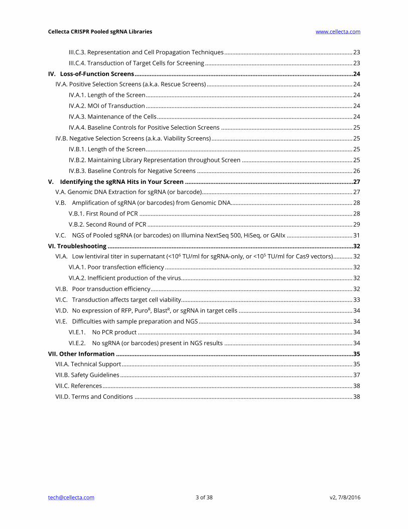
I.B.2. Required Materials Needed from Other Vendors
293T/17 Cell Line (ATCC, Cat.# CRL-11268™)
Dulbecco's Modified Eagle Medium (D-MEM) (1X) (Mediatech CellGro, Cat.# 15-013-CV)
NOTE: ADD FRESH GLUTAMINE (1X) at the time a sealed bottle of D-MEM is opened, even if
the label indicates glutamine has already been added. Glutamine in solution at 4°C has a
half-life of 1–2 months, so glutamine D-MEM purchased “off-the-shelf” from a supplier is to
be regarded as glutamine(-). In our experience, the addition of glutamine increases titer
approximately 2-fold. If D-MEM comes supplemented with stable L-Alanyl-L-Glutamine
dipeptide, addition of fresh glutamine is not necessary.
1M HEPES pH 7.2-7.6 (Mediatech, Cat.# 25-060-CI)
1M MgCl2 solution
Glutamine (L-Alanyl-L-Glutamine, Dipeptide L-glutamine) (Mediatech, Cat.# 25-015-CI)
Fetal Bovine Serum
Puromycin or other antibiotic (depending on vector)
Cellecta CRISPR Pooled sgRNA Libraries www.cellecta.com
[email protected] 6 of 38 v2, 7/8/2016
Blasticidin or Hygromycin (depending on vector)
D-PBS (Mediatech, Cat.# 21-031-CV)
Trypsin-EDTA (Mediatech, Cat.# 25-052-CI)
Polybrene (hexadimethrine bromide) (Sigma-Aldrich, Cat.# 107689)
Proteinase K from Tritirachium album (Sigma-Aldrich, Cat.# P4850)
500 ml, 0.2 μm PES filter units (Fisher Cat.# 09-741-02 or Thermo Fisher Cat.# 5660020)
Tissue Culture Plates and Related Tissue Culture Supplies
Lipofectamine® Transfection Reagent (Thermo Fisher, Cat.# 18324020)
Plus® Reagent (Thermo Fisher, Cat.# 11514015)
15-ml Falcon screw-cap tubes (12,000 RCF rated, P:CHCl3-resistant, Corning, Cat.# 352196)
Buffer P1 (50mM Tris-HCl pH 8.0, 10mM EDTA) (QIAGEN, Cat.# 19051)
RNase A (QIAGEN, Cat.# 19101)
Sonicator for Genomic DNA Shearing
Phenol:Chloroform:Isoamyl Alcohol 25:24:1 (Sigma-Aldrich, Cat.# P3803 or P2069)
DNase I, RNase-free (Epicentre, Cat.# D9905K)
Titanium Taq DNA polymerase with PCR buffer (Takara, Cat.# 639242)
dNTP Mix (10 mM each) (GE Healthcare, Cat.# 28-4065-52)
QIAquick PCR purification kit (QIAGEN, Cat.# 28104)
QIAquick Gel Extraction Kit (QIAGEN, Cat.# 28704)
DNeasy Blood & Tissue Kit (QIAGEN, Cat.# 69504)
QIAamp DNA Micro Kit (QIAGEN, Cat.# 56304)
PCR primers for sgRNA amplification from genomic DNA (IDT): See Library PAC
Next-Gen Sequencing (NGS) primers (IDT): See Library PAC
Primer for sequencing sgRNA inserts in sgRNA constructs (IDT): See Library PAC
PhiX Library (Illumina, Cat.# FC-110-3001)
NGS Kits (Illumina):
Platform Kit Type Illumina Cat.# Description
GAIIx™ Sequencing FC-104-5001 TruSeq SBS Kit v5 – GA (36-cycle)
Cluster Generation GD-203-5001 TruSeq SR Cluster Kit v5 – CS – GA
HiSeq®* Sequencing FC-401-3002 TruSeq SBS Kit v3 – HS (50 cycle)
Cluster Generation GD-401-3001 TruSeq SR Cluster Kit v3-cbot-HS
NextSeq® 500 Sequencing FC-404-2005 NextSeq 500 v2 Kit
* See Illumina website for information on HiSeq 2500 rapid run kits. We do not support NGS of samples on MiSeq.

Cellecta CRISPR Pooled sgRNA Libraries www.cellecta.com
[email protected] 7 of 38 v2, 7/8/2016
I.B.3. Related Products and Services from Cellecta
CRISPR Human Genome Knockout Libraries
Loss-of-Function Screens with Pooled sgRNA or shRNA Libraries
Next-Gen Sequencing of cell pellets, genomic DNA, or xenografts from knockout screens
Isogenic Knockout Cell Lines
Custom CRISPR sgRNA constructs
Linearized CRISPR sgRNA expression vectors
For more information, visit www.cellecta.com, email us at [email protected], or call 650-938-3910.
I.C. Recommended Pilot Experiments
In order to obtain reliable data from your genetic screen, we suggest appropriate planning
beforehand. We recommend the pilot studies below in your cell system of choice (the cell system
that will be used in your pooled library screen). Cell-type specific data from these pilot experiments
will provide you more confidence in your screen results.
I.C.1. Doubling Time
The doubling time is the time it takes your cells to double in number. It is useful to know the
doubling time of your cells so that you can plate the appropriate number for transduction with the
lentiviral library. Start with cells that have already been growing for a few weeks, rather than using
cells that have just been thawed from a frozen state. To calculate the doubling time, trypsinize your
cells as if you were going to split them. Count them using a hemacytometer or cell counter and keep
track of the number that you replate onto the cell culture plates. The starting number of cells is Xb.
Propagate the cells as you normally do, replacing media as necessary. The next time they are ready
to be split, trypsinize them as usual and count them again using a hemacytometer or cell counter.
The number of cells at the end is referred to as Xe. The cells should be in the log phase of growth to
calculate doubling time properly, so it is important to not let the cells become confluent. To calculate
the doubling time, use the following formula:
𝐷𝑜𝑢𝑏𝑙𝑖𝑛𝑔 𝑇𝑖𝑚𝑒 =𝑇(𝑙𝑛2)
ln (𝑋𝑒𝑋𝑏
)
where T = Time in any units
For example, let’s say that on Day 0, you count 2 × 10⁶ cells. Three (3) days later, you count the cells
at 16 × 106 cells.
Xb = 2 × 10⁶
T = 3 days
Xe = 16 × 106
𝐷𝑜𝑢𝑏𝑙𝑖𝑛𝑔 𝑇𝑖𝑚𝑒 =3(𝑙𝑛2)
ln (16,000,000
2,000,000)
=3(0.69)
ln (8)=
2.08
2.08 = 1 𝑑𝑎𝑦 (time unit same as used for T)

Cellecta CRISPR Pooled sgRNA Libraries www.cellecta.com
[email protected] 8 of 38 v2, 7/8/2016
I.C.2. Calculating a Kill Curve
Cellecta’s standard Cas9-only expression vector expresses a blasticidin resistance gene, while the
sgRNA library vector typically expresses a puromycin resistance gene. Cellecta also offers a Cas9
vector expressing a blasticidin resistance gene. For some customized libraries, other selection
markers such as Neo, Bleo, or Hygro may be substituted. Regardless of the selection marker the
plasmids contain, to successfully select cells transduced with Cas9 or the sgRNA library, you need to
know the concentration of antibiotic that kills untransduced cells within a given amount of time. We
recommend the following method for obtaining the “kill curve” for puromycin and blasticidin.
To create a puromycin kill curve, aliquot cells in a 12-well plate at such a density so they are at 72
hours from confluence. Add puromycin at the concentration of 0 μg/ml, 0.5 μg/ml, 1 μg/ml, 2 μg/ml,
5 μg/ml, and 10 μg/ml in six different wells. Mix and place the cells at 37°C in a CO2 incubator. Grow
cells under standard conditions for 72 hours, then count viable cells and determine the lowest
concentration of drug that kills at least 90% of cells in 72 hours. Use this concentration at the
puromycin selection step during the screen.
To calculate a blasticidin kill curve, follow the same steps and use the same concentrations as for the
puromycin kill curve. Determine the lowest concentration of drug that kills 99% of cells in 3-5 days.
Use this concentration of blasticidin at the Blasticidin Resistance Assay & Titer Estimation step.
If using hygromycin, follow the same steps above but use 0 μg/ml, 50 μg/ml, 100 μg/ml, 200 μg/ml,
400 μg/ml, and 800 μg/ml hygromycin in six different wells.
I.C.3. Check Toxicity of Polybrene
Polybrene is a transduction enhancement reagent used during transduction of the pooled lentiviral
sgRNA library into the target cells. Polybrene is a polycation that neutralizes charge interactions to
increase binding between the lentiviral envelope and the plasma membrane. The optimal
concentration of Polybrene depends on cell type and may need to be empirically determined.
Excessive exposure to Polybrene can be toxic to some cells.
We recommend performing a Polybrene toxicity titration before transducing your target cells. In a
12-well plate, grow cells in complete culture medium with a range of Polybrene concentrations (0
μg/ml, 1 μg/ml, 2 μg/ml, 3 μg/ml, 4 μg/ml, 5 μg/ml) for 24 hours. Then, replace old medium with
Polybrene-free complete culture medium and grow cells for an additional 72 hours. Check for
toxicity by counting viable cells. For your experiments, use the highest concentration of Polybrene
that results in less than 10% cell toxicity compared to no Polybrene (typically, 5 μg/ml is
recommended). For some cell types, you cannot use Polybrene.
I.C.4. Promoter Validation
If you have not used lentiviral vectors in your target cells before, we recommend conducting a pilot
experiment to determine which promoters will work best. Pol II promoters drive the expression of
markers such as TagRFP and the puromycin resistance gene for libraries and Cas9 and blasticidin
resistance genes for Cas9 vectors. Cellecta sells pre-packaged control viruses expressing different
marker genes from different promoters. You can use these ready-to-use products to determine
which promoter combination will work the best in your cells.
Cellecta CRISPR Pooled sgRNA Libraries www.cellecta.com
[email protected] 9 of 38 v2, 7/8/2016
II. Preparing Cas9 Expressing Cells
II.A. Packaging Protocol for Cas9 Expression Vectors
The following protocol describes the generation of pseudoviral packaged lentiviral Cas9-Only
Expression Vectors (see Other Materials Available from Cellecta) using Thermo Fisher’s
Lipofectamine® Transfection Reagent and Plus® Reagent. Other transfection reagents may be used,
but the protocol should be adjusted to fit the manufacturer’s protocol. The yield of recombinant
lentiviral particles typically produced under these optimized conditions is between 1 × 10⁶ TU/ml and
5 × 10⁶ TU/ml for sgRNA constructs, but CRISPR/Cas9 lentivectors have a much lower yield
(approximately 1 × 10⁵ TU/ml) because of the large size of the construct. The protocol below is for
one 15-cm plate, but you may need to use multiple plates.
1. Start growing 293T cells in D-MEM medium plus glutamine, supplemented with 10% FBS, 2 to 3
days prior to transfection.
II.A.1. Day 0 – Plate Cells
2. Twenty four (24) hours prior to transfection, plate 12.5 × 10⁶ 293T cells in a 15-cm plate* (or 150
cm2 flask). Use 30 ml of media per plate. Disperse the cells and ensure even distribution. At the
moment of transfection, the cells should have reached ~80% confluency. Increase or decrease
the number of 293T cells seeded if optimal confluency is not achieved in 24 hours. Incubate at
37°C in a CO2 incubator for 24 hours.
* The goal is to have the 293T cells reach 80% confluency by Day 1. You may want to calculate
the number of cells seeded empirically since cell counts and growth rate can vary.
II.A.2. Day 1 – Transfection
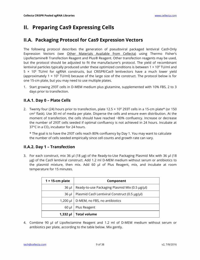
3. For each construct, mix 36 μl (18 μg) of the Ready-to-Use Packaging Plasmid Mix with 36 μl (18
μg) of the Cas9 lentiviral construct. Add 1.2 ml D-MEM medium without serum or antibiotics to
the plasmid mixture, then mix. Add 60 μl of Plus Reagent, mix, and incubate at room
temperature for 15 minutes.
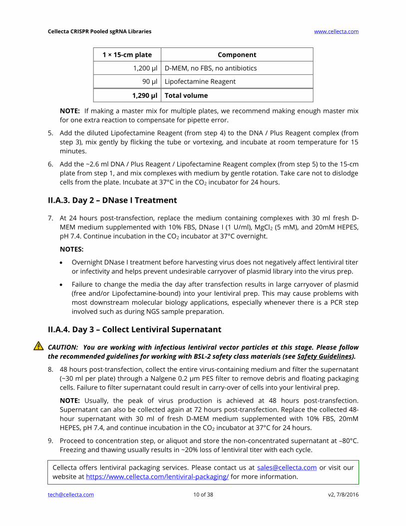
4. Combine 90 μl of Lipofectamine Reagent and 1.2 ml of D-MEM medium without serum or
antibiotics per plate, according to the table below. Mix gently.
1 × 15-cm plate Component
36 μl Ready-to-use Packaging Plasmid Mix (0.5 μg/μl)
36 μl Plasmid Cas9 Lentiviral Construct (0.5 μg/μl)
1,200 μl D-MEM, no FBS, no antibiotics
60 μl Plus Reagent
1,332 μl Total volume
Cellecta CRISPR Pooled sgRNA Libraries www.cellecta.com
[email protected] 10 of 38 v2, 7/8/2016
1 × 15-cm plate Component
1,200 μl D-MEM, no FBS, no antibiotics
90 μl Lipofectamine Reagent
1,290 μl Total volume
NOTE: If making a master mix for multiple plates, we recommend making enough master mix
for one extra reaction to compensate for pipette error.
5. Add the diluted Lipofectamine Reagent (from step 4) to the DNA / Plus Reagent complex (from
step 3), mix gently by flicking the tube or vortexing, and incubate at room temperature for 15
minutes.
6. Add the ~2.6 ml DNA / Plus Reagent / Lipofectamine Reagent complex (from step 5) to the 15-cm
plate from step 1, and mix complexes with medium by gentle rotation. Take care not to dislodge
cells from the plate. Incubate at 37°C in the CO2 incubator for 24 hours.
II.A.3. Day 2 – DNase I Treatment
7. At 24 hours post-transfection, replace the medium containing complexes with 30 ml fresh D-
MEM medium supplemented with 10% FBS, DNase I (1 U/ml), MgCl2 (5 mM), and 20mM HEPES,
pH 7.4. Continue incubation in the CO2 incubator at 37°C overnight.
NOTES:
Overnight DNase I treatment before harvesting virus does not negatively affect lentiviral titer
or infectivity and helps prevent undesirable carryover of plasmid library into the virus prep.
Failure to change the media the day after transfection results in large carryover of plasmid
(free and/or Lipofectamine-bound) into your lentiviral prep. This may cause problems with
most downstream molecular biology applications, especially whenever there is a PCR step
involved such as during NGS sample preparation.
II.A.4. Day 3 – Collect Lentiviral Supernatant
CAUTION: You are working with infectious lentiviral vector particles at this stage. Please follow
the recommended guidelines for working with BSL-2 safety class materials (see Safety Guidelines).
8. 48 hours post-transfection, collect the entire virus-containing medium and filter the supernatant
(~30 ml per plate) through a Nalgene 0.2 μm PES filter to remove debris and floating packaging
cells. Failure to filter supernatant could result in carry-over of cells into your lentiviral prep.
NOTE: Usually, the peak of virus production is achieved at 48 hours post-transfection.
Supernatant can also be collected again at 72 hours post-transfection. Replace the collected 48-
hour supernatant with 30 ml of fresh D-MEM medium supplemented with 10% FBS, 20mM
HEPES, pH 7.4, and continue incubation in the CO2 incubator at 37°C for 24 hours.
9. Proceed to concentration step, or aliquot and store the non-concentrated supernatant at –80°C.
Freezing and thawing usually results in ~20% loss of lentiviral titer with each cycle.
Cellecta offers lentiviral packaging services. Please contact us at [email protected] or visit our
website at https://www.cellecta.com/lentiviral-packaging/ for more information.

Cellecta CRISPR Pooled sgRNA Libraries www.cellecta.com
[email protected] 11 of 38 v2, 7/8/2016
II.B. Concentrating Virus
Although concentrating virus is optional, 100-fold concentration is recommended for the Cas9
construct since yield is typically low.
1. Transfer lentiviral supernatant to clear, sterile centrifuge tubes.
2. (Recommended) Add Cellecta’s LentiFuge™ Viral Concentration Reagent according to the
LentiFuge User Manual instructions (see Other Materials).
3. Centrifuge at 15,000 × g for at least 1 hour at 4°C. Mark the tubes to identify the location where
the pellet will be. At the end of centrifugation, you may or may not be able to see a pellet;
assume it is at the location of the mark.
4. Immediately discard the supernatant by aspirating.
5. Place the tubes on ice, resuspend the pellet (which may not be visible) in PBS, PBS/10%FBS, or
PBS/1%BSA, make aliquots, and freeze at –80°C. 100-fold concentration is recommended (e.g.,
resuspend in 1 ml PBS if starting from 100 ml supernatant).
II.C. Lentiviral Titer Estimation and Generation of Cas9 Cell Lines
Most of the commonly used mammalian cell lines can be effectively transduced by lentiviral
constructs. Relative titers can vary up to 50-fold depending on the chosen cell line. For this reason, it
is critical to determine the viral titer in the cells that you plan to use for the rest of the experiment.
The following section uses packaged lentiviral particles for transduction into example target cells
(HEK293).
NOTE: Lentiviral particles should only be opened within the laminar flow hood, and should be used
under Biosafety Level 2 (BSL-2) conditions (see Safety Guidelines).
II.C.1. Transduction of Adherent Cells
The following protocol has been optimized for HEK293 cells. For other adherent cell types,
parameters such as media, growth surface, time of detection, etc., will have to be adjusted.
Day 1
1. Quickly thaw the lentiviral particles in a water bath at 37°C. Transfer the thawed particles to a
laminar flow hood, gently mix by rotation, inversion, or gentle vortexing, and keep on ice.
CAUTION: Only open the tube containing the lentiviral particles in the laminar flow hood.
NOTE: Unused lentiviral stock can be refrozen at –80°C, but freezing and thawing usually results
in ~20% loss of lentiviral titer with each cycle.
Trypsinize and resuspend HEK293 cells at a density of 1 × 10⁵ cells/ml in D-MEM supplemented
with 10% FBS and 5 μg/ml Polybrene. Aliquot 1 ml/well in a 12-well plate and add 0 μl, 1 μl, 3.3 μl
and 10 μl of 100-fold concentrated Cas9 lentiviral stock to four different wells. Mix and return
cells to CO2 incubator. Grow cells under standard conditions for 24 hours.

Cellecta CRISPR Pooled sgRNA Libraries www.cellecta.com
[email protected] 12 of 38 v2, 7/8/2016
2. 24 hours after transduction, replace media with fresh D-MEM supplemented with 10% FBS.
NOTE: It is important to accurately record the original number of cells at time of transduction, as
this is critical in titer calculation. As a rule of thumb, cells should be transduced at such a density
that they will become confluent in 48 hours.
II.C.2. Alternative Transduction Protocol (Spinoculation) for Suspension Cells
The following protocol has been optimized for K-562 cells. For other cell types, parameters such as
media, growth surface, time of detection, etc. will have to be adjusted.
1. K-562 cells are transduced using spinoculation. This is performed using multi-well tissue culture
plates and a tabletop centrifuge capable of 1,200 × g and centrifugation of multi-well plates.
2. Grow K-562 cells and maintain them between 2 × 10⁵ and 1 × 10⁶ cells/ml. Do not let them
become too dense or let the medium become yellow at any point.
3. For lentiviral library titration, K-562 cells are resuspended at 2 × 10⁶ cells per ml in RPMI/10%FBS
supplemented with 20mM HEPES, pH7.4 and Polybrene, 5 μg/ml. 0.25-ml aliquots are placed
into each well in a 48-well plate (5 × 10⁵ cells/well total). This cell density has proven effective for
many suspension cell lines in-house at Cellecta. To each cell-containing well, add increasing
amounts of lentiviral stock to be titered. For Cas9 100-fold concentrated lentiviral stock, add 0 μl,
1 μl, 3 μl, 10 μl and 33ul of 100-fold concentrated virus. Close the plate, mix by gentle agitation,
wrap the perimeter with parafilm, and place the plate into centrifuge with an appropriate
balance and centrifuge at 1,200 × g at 25°C for 2 hours.
4. Following centrifugation, remove plate(s) from centrifuge, carefully remove parafilm, and place
in incubator. After 3 hours, feed cells with 0.25 ml additional complete medium per well (no
Polybrene).
5. At 24 hours after transduction, transfer cells to a 12-well plate and add 1ml fresh RPMI/10%FBS.
NOTE: Use larger vessels for large-scale genetic screen transductions. Scale up all volumes
accordingly.
II.C.3. Blasticidin Resistance Assay & Titer Estimation
1. 48 hours after transduction, split each transduction into two halves.
2. 72 hours after transduction, add blasticidin to one of each transduced pair. Use the
concentration of blasticidin calculated from the blasticidin kill curve. For HEK293 cells, we use 1
µg/ml blasticidin, but other cell lines may respond to other concentrations ranging from ~0.5 to
10 µg/ml. For hygromycin, we use 100 µg/ml, but you may need to test ranges from ~50 – 800
µg/ml depending on the cell line.
3. When you see 99% cell death in the samples containing blasticidin (usually after ~ 3-5 days) in
the no-virus control pair, count the cells from all the pairs and calculate the percent transduction
for each dilution:
𝑃𝑒𝑟𝑐𝑒𝑛𝑡 𝑇𝑟𝑎𝑛𝑠𝑑𝑢𝑐𝑡𝑖𝑜𝑛 = 100 ∗ 𝑐𝑒𝑙𝑙 𝑐𝑜𝑢𝑛𝑡 𝑖𝑛 𝑠𝑎𝑚𝑝𝑙𝑒 𝑤𝑖𝑡ℎ 𝑏𝑙𝑎𝑠𝑡𝑖𝑐𝑖𝑑𝑖𝑛
𝑐𝑒𝑙𝑙 𝑐𝑜𝑢𝑛𝑡 𝑖𝑛 𝑠𝑎𝑚𝑝𝑙𝑒 𝑤𝑖𝑡ℎ𝑜𝑢𝑡 𝑏𝑙𝑎𝑠𝑡𝑖𝑐𝑖𝑑𝑖𝑛
Use the virus dilution that gives 50% blasticidin-resistant cells (HR-50) as a starting point to generate
the Cas9 cell line for screens.

Cellecta CRISPR Pooled sgRNA Libraries www.cellecta.com
[email protected] 13 of 38 v2, 7/8/2016
II.C.4. Generation of Cas9 Cell Line for Screens
1. To generate the best Cas9 cell line for screens, transduce the parental cell line with increasing
amounts of virus corresponding to:
1x HR-50
2x HR-50
4x HR-50
8x HR-50
16x HR-50
2. 72 hours after transduction, start blasticidin selection (same concentration of blasticidin used to
calculate HR-50) and grow cells under selection for 2 weeks.
3. Select the Cas9 cell sample transduced with the highest amount of virus, which yielded the
highest number of cells after 2 weeks of blasticidin selection. This should ensure that you have
selected the cell sample expressing the highest non-toxic levels of Cas9.
Example:
Amount of Cas9 virus Cells after 2 weeks of
Blasticidin Selection
1x HR-50 7 × 10⁷ cells
2x HR-50 1 × 10⁸ cells
4x HR-50 1.5 × 10⁸ cells
8x HR-50 1.5 × 10⁸ cells
16x HR-50 1 × 10⁸ cells
Here, you would choose the cells treated with 8x HR-50 to expand the Cas9 cell line.
(4x HR-50 and 8x HR-50 have both the highest amount of cells; 8x HR-50 has higher copy
number of Cas9 than 4x HR-50).
4. Split the selected Cas9 cell line (“8x HR-50” in this case) into 3 samples, and continue selection
with 1X, 2X, and 4X increases in blasticidin concentration
Sample 1: Same blasticidin concentration used to calculate HR-50
Sample 2: 2x blasticidin concentration used to calculate HR-50
Sample 3: 4x blasticidin concentration used to calculate HR-50
5. After 1 week, select the Cas9 cells that survived the highest blasticidin concentration. Expand the
selected Cas9 cell sample. This is the Cas9 cell line you will use in your screen.
6. To assess the level of Cas9 activity in your cell lines, we recommend using Cellecta’s CRISPRtest™
Functional Cas9 Activity Kit (Cat.# CRTEST).
Cellecta CRISPR Pooled sgRNA Libraries www.cellecta.com
[email protected] 14 of 38 v2, 7/8/2016
III. sgRNA Library Packaging, Titering & Transduction
III.A. Packaging Protocol for Pooled Lentiviral sgRNA Libraries
The following protocol describes the generation of a packaged CRISPR lentiviral sgRNA library (in an
sgRNA-only vector) using Thermo Fisher’s Lipofectamine® and Plus® Reagents. Other transfection
reagents may be used, but the protocol should be adjusted to fit the manufacturer’s protocol. The
yield of recombinant lentiviral particles typically produced under these optimized conditions is 1-5 ×
106 TU/ml. The chart below indicates the number of 15-cm plates to use depending on the library
complexity. We do not recommend scaling down the lentiviral packaging protocol due to risk of
compromising the representation of the sgRNA library.
NOTE: For libraries in CRISPR/Cas9 sgRNA vectors (single-vector system), you should expect a much
lower yield (approximately 1 × 10⁵ TU/ml) because of the vectors’ large size.
Library Complexity Number of Plates to use Amount of Virus Produced
6K 2 × 15-cm plates 6 × 10⁷ TU
12K 4 × 15-cm plates 1.2 × 10⁸ TU
27K 8 × 15-cm plates 2.5 × 10⁸ TU
55K 16 × 15-cm plates 5 × 10⁸ TU
90K 32 × 15-cm plates 1 × 10⁹ TU
1. Start growing 293T cells in D-MEM medium plus glutamine, supplemented with 10% FBS without
antibiotics, 2 to 3 days prior to transfection.
III.A.1. Day 0 – Plate Cells
2. Twenty four (24) hours prior to transfection, plate 12.5 × 10⁶ 293T cells in each of the 15-cm
plates (or 150 cm2 flasks)*. Use 30 ml of media per plate. Disperse the cells and ensure even
distribution. At the moment of transfection, the cells should have reached 80% confluency.
Increase or decrease the number of 293T cells seeded if optimal confluency is not achieved in 24
hours. Incubate at 37°C in a CO2 incubator for 24 hours.
* The goal is to have the 293T cells reach 80% confluency by day 1. You may want to calculate
the number of seed cells empirically since cell counts can vary.
III.A.2. Day 1 – Transfection
3. In a sterile 50-ml or larger polypropylene tube, mix the Ready-to-use Packaging Plasmid Mix and
the Plasmid sgRNA Library, add the plasmid mixture to D-MEM medium without serum or
antibiotics, then mix. Add the Plus Reagent, mix, and incubate at RT for 15 minutes. Use the
table below for the volumes to use depending on the number of 15-cm plates you are using.
Cellecta CRISPR Pooled sgRNA Libraries www.cellecta.com
[email protected] 15 of 38 v2, 7/8/2016
4 × 15-cm plates 8 × 15-cm plates 16 × 15-cm plates Component
240 µl 480 μl 960 µl Packaging Plasmid Mix (0.5 μg/μl)
24 µl 48 μl 96 µl Plasmid sgRNA Library (1 μg/μl)
4,800 µl 9,600 μl 19,200 µl D-MEM, no FBS, no antibiotics
240 µl 480 μl 960 µl Plus Reagent
5,304 µl 10,608 μl 21,216 µl Total volume
4. Add Lipofectamine to the D-MEM medium without serum or antibiotics in order to make a
convenient master mix according to the table below. Mix gently.
4 × 15-cm plates 8 × 15-cm plates 16 × 15-cm plates Component
4,800 µl 9,600 μl 19,200 µl D-MEM, no FBS, no antibiotics
360 µl 720 μl 1,440 µl Lipofectamine Reagent
5,160 µl 10,320 μl 20,640 µl Total volume
5. Add the diluted Lipofectamine (from step 4) to the DNA / Plus Reagent complex (from step 3),
mix gently by flicking the tube or vortexing and incubate at room temperature for 15 minutes.
6. Add 2.5 ml of the DNA / Plus Reagent / Lipofectamine complex (from step 5) to each 15-cm plate
from step 2, and mix complexes with medium by gentle rotation. Take care not to dislodge cells
from the plate. Incubate at 37°C in the CO2 incubator for 24 hours.
III.A.3. Day 2 – DNase I Treatment
7. At 24 hours post-transfection, replace the medium containing complexes with 30 ml fresh D-
MEM medium supplemented with 10% FBS, DNase I (1 U/ml), MgCl2 (5 mM), and 20mM HEPES,
pH 7.4. Continue incubation in the CO2 incubator at 37°C overnight.
NOTES:
Overnight DNase I treatment before harvesting virus does not negatively affect lentiviral titer
or infectivity and helps prevent undesirable carryover of plasmid library into the virus prep.
Failure to change the media the day after transfection results in large carryover of plasmid
(free and/or Lipofectamine-bound) in your lentiviral prep. This may cause problems with
most downstream molecular biology applications, especially whenever there is a PCR step
involved such as during NGS sample preparation.
III.A.4. Day 3 – Collect Lentiviral Supernatant
CAUTION: You are working with infectious lentiviral vector particles at this stage. Please follow
the recommended guidelines for working with BSL-2 safety class materials (see Safety Guidelines).
8. At 48 hours post-transfection, collect the entire virus-containing medium from each plate and
filter the supernatant (~30 ml per plate) through a Nalgene 0.2 μm PES filter to remove debris
and floating packaging cells. Failure to filter supernatant could result in carry-over of cells into
your lentiviral prep.

Cellecta CRISPR Pooled sgRNA Libraries www.cellecta.com
[email protected] 16 of 38 v2, 7/8/2016
NOTE: Usually, the peak of virus production is achieved at 48 hours post-transfection.
Supernatant can also be collected at 72 hours post-transfection—replace the collected 48-hour
supernatant with 30 ml of fresh D-MEM medium supplemented with 10% FBS, 20mM HEPES
pH7.4 and continue incubation in the CO2 incubator at 37°C for 24 hours.
9. Proceed to concentration step, or aliquot and store the non-concentrated supernatant at –80°C.
Freezing and thawing usually results in ~20% loss of lentiviral titer with each cycle.
III.A.5. Concentrating Virus (Optional)
Although concentrating virus is optional, it is recommended if (1) very high titer virus stock is needed
to achieve desired MOI in hard-to-transduce target cells, (2) virus should be suspended in another
media (besides D-MEM/10%FBS) which is optimal for sensitive target cells, or (3) 18 hours post-
transduction baseline control is used in your screen (to minimize problems with possible plasmid
library carryover). However, because of the additional manipulation of samples, there is the added
risk of contamination and loss of virus.
The following protocol was optimized to concentrate virus with high recovery. The protocol assumes
that lentiviral supernatant was harvested 48 hours after transfection and filtered as in step 17
above.
1. Aliquot lentiviral supernatant in clear sterile centrifuge tubes.
2. (Recommended) Add Cellecta’s LentiFuge™ Concentration Reagent according to the LentiFuge
User Manual instructions.
3. Centrifuge at 15,000 × g for at least 1 hour at 4°C. Mark the tubes to identify the location where
the pellet will be. At the end of centrifugation, you may or may not be able to see a pellet—
assume it is at the location of the mark.
4. Immediately discard the supernatant by aspirating.
5. Place the tubes on ice, resuspend the pellet (which may not be visible) in PBS, PBS/10%FBS, or
PBS+1%BSA, make aliquots, and freeze at –80°C. 100-fold concentration is recommended (e.g.,
resuspend in 1 ml PBS if starting from 100 ml supernatant).
III.B. Lentiviral Titer Estimation
The following section uses packaged lentiviral particles for transduction into example target cells
(HEK293). Please note that lentiviral particles should only be opened within the laminar flow hood,
and should be used under biosafety Level 2 conditions.
III.B.1. Transduction for Titering
Lentiviral transductions are performed by mixing cells and virus in culture media supplemented with
Polybrene. For both adherent and suspension cells, transductions are initiated in suspension and
Cellecta offers lentiviral packaging services. Please contact us at [email protected] or visit
https://www.cellecta.com/lentiviral-packaging/ for more information.

Cellecta CRISPR Pooled sgRNA Libraries www.cellecta.com
[email protected] 17 of 38 v2, 7/8/2016
carried out overnight. Adherent cells are allowed to adhere to substrate during transduction and are
transduced at a cell density that allows for 2-3 population doublings before reaching confluence.
Suspension cells are typically transduced at higher density than standard growth density, and then
they are diluted to standard growth density 18-24 hours after transduction.
III.B.1.1. Transduction of Adherent Cells for Titering
The following protocol has been optimized for HEK293 cells. For other adherent cell types,
parameters such as media, growth surface, time of detection, etc. will have to be adjusted.
Day 1
1. Quickly thaw the lentiviral particles in a water bath at 37°C. Transfer the thawed particles to a
laminar flow hood, gently mix by rotation, inversion, or gentle vortexing, and keep on ice.
CAUTION: Only open the tube containing the lentiviral particles in the laminar flow hood.
NOTE: Unused lentiviral stock may be refrozen at –80°C, but freezing and thawing usually
results in ~20% loss of lentiviral titer with each cycle.
2. Trypsinize and resuspend HEK293 cells at a density of 1 × 10⁵ cells/ml in D-MEM supplemented
with 10% FBS and 5 μg/ml Polybrene. Aliquot 1 ml/well in a 12-well plate and add 0 μl, 3 μl, 10 μl,
33 μl, and 100 μl of non-concentrated lentiviral stock (supernatant filtered to remove cells and
cell debris, not concentrated) to six different wells. If concentrated virus is used, scale down virus
volumes accordingly. Mix and return cells to CO2 incubator. Grow cells under standard
conditions for 24 hours.
IMPORTANT:
At time of transduction, it is very important to accurately record the number of cells. This is
critical in titer calculation.
For adherent cells other than HEK293, cell density at time of transduction would have to be
adjusted for optimal transduction. Cell density typically depends on cell size. As a rule of
thumb, cells should be transduced at such a density that they would become confluent in ~48
hours.
Day 2
3. Between 16 to 24 hours post-transduction, replace media with fresh D-MEM supplemented with
10% FBS without Polybrene. Return cells to CO2 incubator, and grow under standard conditions
for an additional 48 hours.
NOTE: Avoid confluence during and after transduction. If necessary, trypsinize and replate cells.
Day 4 (72 hours after transduction)
4. Detach cells from the plate by trypsin treatment, block trypsin with FBS/media, centrifuge,
resuspend in 1X D-PBS, and determine the percentage of transduced (RFP-positive) cells by flow
cytometry.
NOTE: Attempting to determine the percentage of transduced cells by fluorescence microscopy
is not recommended. It is likely to lead to inaccurate titer estimation.
Cellecta CRISPR Pooled sgRNA Libraries www.cellecta.com
[email protected] 18 of 38 v2, 7/8/2016
IMPORTANT: Flow cytometry settings to detect RFP-positive cells are the following: Excitation:
561nm (530nm laser is still acceptable), Emission: 600/20 band-pass filter, or similar (for TagRFP).
5. Proceed to Lentiviral Titer estimation (RFP assay).
III.B.1.2. Alternative Transduction Protocol (Spinoculation) for Titering in
Suspension Cells
The following protocol has been optimized for K-562 cells. For other cell types, parameters such as
media, growth surface, time of detection, etc. will have to be adjusted.
1. K-562 cells are transduced (“infected”) using spinoculation. This is performed using multi-well
tissue culture plates and a tabletop centrifuge capable of 1,200 × g and centrifugation of multi-
well plates.
2. Grow K-562 cells and maintain them between 2 × 10⁵ and 1 × 10⁶ cells/ml. Do not let them
become too dense or let the medium become yellow at any point.
3. For lentiviral library titration, K-562 cells are resuspended at 2 × 10⁶ cells per ml in RPMI/10%FBS
supplemented with 20mM HEPES, pH 7.4 and Polybrene, 5 μg/ml. 0.5-ml aliquots are placed into
each well in a 24-well plate (1 × 10⁶ cells/well total). This cell density has proven effective for
many suspension cell lines in-house at Cellecta. To each cell-containing well, add increasing
amounts of lentiviral stock to be titered. For standard 100-fold concentrated lentiviral stock, add
0 μl, 0.3 μl 1 μl, 3 μl, and 10 μl virus. Close the plate, mix by gentle agitation, wrap the perimeter
with parafilm, and place the plate into centrifuge with an appropriate balance and centrifuge at
1,200 × g at 25°C for 2 hours.
4. Following centrifugation, remove plate(s) from centrifuge, carefully remove parafilm, and place
in incubator. After 3 hours, “feed” cells with 0.5 ml additional complete medium per well (no
Polybrene).
5. 24 hours after spinoculation, resuspend cells at 2 × 10⁵ cells/ml in RPMI/10%FBS in the
appropriate culture vessel and grow for additional 48 hours.
6. 72 hours after spinoculation, determine the percentage of transduced (RFP-positive) cells by flow
cytometry.
IMPORTANT: Flow cytometry settings to detect RFP-positive cells are the following: Excitation:
561nm (530nm laser is still acceptable), Emission: 600/20 band-pass filter, or similar (for TagRFP).
NOTE: Determining the percentage of transduced cells by fluorescence microscopy results in
inaccurate titer estimation and is not recommended.
7. Proceed to Lentiviral Titer estimation (RFP assay) below.
NOTE: Use larger vessels for large-scale genetic screen transductions. Scale up all volumes
accordingly.
III.B.2. Lentiviral Titer Estimation: RFP assay
Lentiviral sgRNA vectors that express the fluorescent protein TagRFP (excitation 560nm, emission
590nm) allow lentiviral titer estimation by flow cytometry (RFP assay) or by a combined flow
cytometry/puromycin resistance assay (RFP/PuroR assay). To check lentiviral titer, we recommend
always using the same cells you will use in the screen. Most of the commonly used mammalian cell
Cellecta CRISPR Pooled sgRNA Libraries www.cellecta.com
[email protected] 19 of 38 v2, 7/8/2016
lines can be effectively transduced by lentiviral constructs. Relative titers can vary up to 50-fold
depending on the chosen cell line.
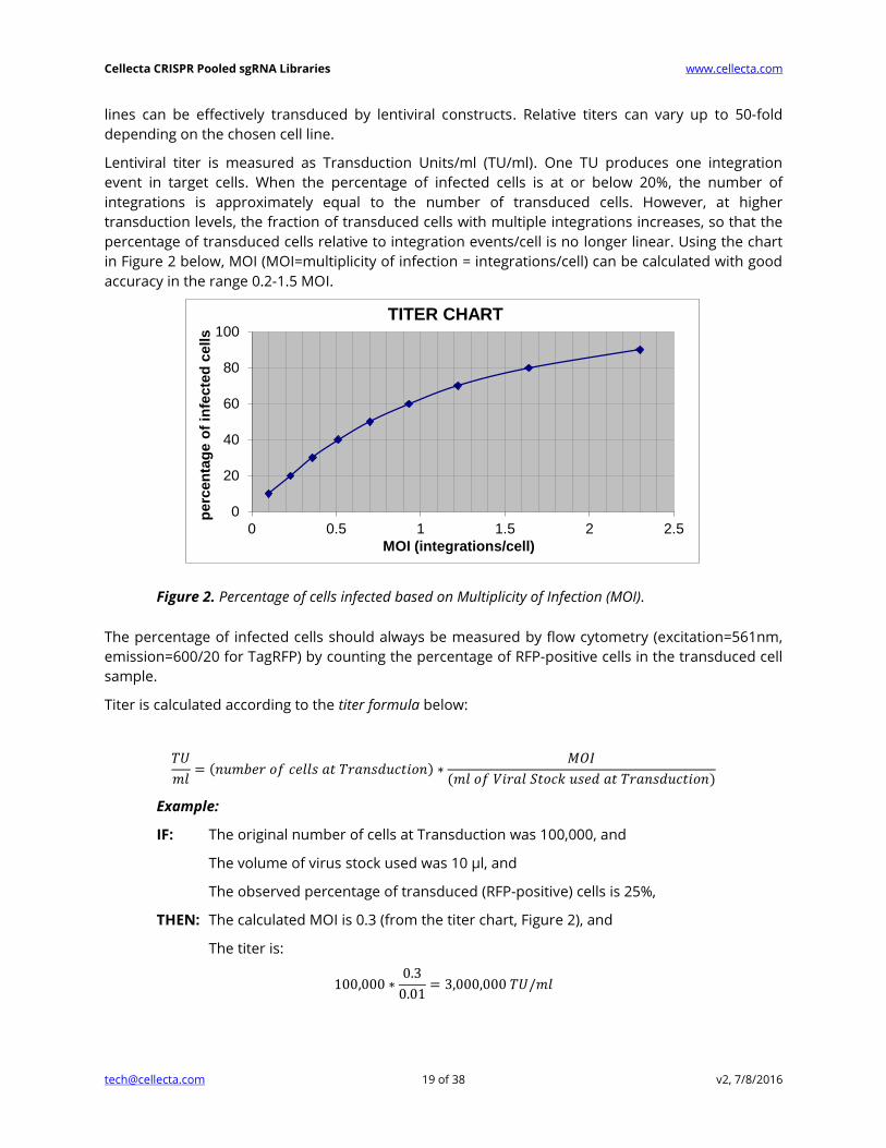
Lentiviral titer is measured as Transduction Units/ml (TU/ml). One TU produces one integration
event in target cells. When the percentage of infected cells is at or below 20%, the number of
integrations is approximately equal to the number of transduced cells. However, at higher
transduction levels, the fraction of transduced cells with multiple integrations increases, so that the
percentage of transduced cells relative to integration events/cell is no longer linear. Using the chart
in Figure 2 below, MOI (MOI=multiplicity of infection = integrations/cell) can be calculated with good
accuracy in the range 0.2-1.5 MOI.
Figure 2. Percentage of cells infected based on Multiplicity of Infection (MOI).
The percentage of infected cells should always be measured by flow cytometry (excitation=561nm,
emission=600/20 for TagRFP) by counting the percentage of RFP-positive cells in the transduced cell
sample.
Titer is calculated according to the titer formula below:
𝑇𝑈
𝑚𝑙= (𝑛𝑢𝑚𝑏𝑒𝑟 𝑜𝑓 𝑐𝑒𝑙𝑙𝑠 𝑎𝑡 𝑇𝑟𝑎𝑛𝑠𝑑𝑢𝑐𝑡𝑖𝑜𝑛) ∗
𝑀𝑂𝐼
(𝑚𝑙 𝑜𝑓 𝑉𝑖𝑟𝑎𝑙 𝑆𝑡𝑜𝑐𝑘 𝑢𝑠𝑒𝑑 𝑎𝑡 𝑇𝑟𝑎𝑛𝑠𝑑𝑢𝑐𝑡𝑖𝑜𝑛)
Example:
IF: The original number of cells at Transduction was 100,000, and
The volume of virus stock used was 10 μl, and
The observed percentage of transduced (RFP-positive) cells is 25%,
THEN: The calculated MOI is 0.3 (from the titer chart, Figure 2), and
The titer is:
100,000 ∗0.3
0.01= 3,000,000 𝑇𝑈/𝑚𝑙
0
20
40
60
80
100
0 0.5 1 1.5 2 2.5
perc
en
tag
e o
f in
fecte
d c
ells
MOI (integrations/cell)
TITER CHART
Cellecta CRISPR Pooled sgRNA Libraries www.cellecta.com
[email protected] 20 of 38 v2, 7/8/2016
Once titer is estimated, the amount of Lentiviral Stock necessary to transduce any given number of
target cells at any transduction efficiency (range of 10% - 80% infected cells) can be back-calculated
from the titer formula and titer chart above.
Example:
To transduce 20,000,000 cells at 50% transduction efficiency, with a lentiviral stock titer of
3,000,000 TU/ml, we calculated the required amount of lentiviral stock as follows:
We calculate the required MOI to achieve 50% transduction efficiency, using the titer chart:
50% transduction efficiency = 0.7 MOI
We calculate the volume of lentiviral stock required using the titer formula:
𝑇𝑈
𝑚𝑙= (𝑛𝑢𝑚𝑏𝑒𝑟 𝑜𝑓 𝑐𝑒𝑙𝑙𝑠 𝑎𝑡 𝑇𝑟𝑎𝑛𝑠𝑑𝑢𝑐𝑡𝑖𝑜𝑛) ∗
𝑀𝑂𝐼
(𝑚𝑙 𝑜𝑓 𝑉𝑖𝑟𝑎𝑙 𝑆𝑡𝑜𝑐𝑘 𝑢𝑠𝑒𝑑 𝑎𝑡 𝑇𝑟𝑎𝑛𝑠𝑑𝑢𝑐𝑡𝑖𝑜𝑛)
3,000,000 = (20,000,000) ∗0.7
(𝑚𝑙 𝑜𝑓 𝑉𝑖𝑟𝑎𝑙 𝑆𝑡𝑜𝑐𝑘 𝑢𝑠𝑒𝑑 𝑎𝑡 𝑇𝑟𝑎𝑛𝑠𝑑𝑢𝑐𝑡𝑖𝑜𝑛)
𝑉𝑖𝑟𝑎𝑙 𝑆𝑡𝑜𝑐𝑘 = 20,000,000 ∗ (0.7
3,000,000) = 4.67 𝑚𝑙
III.B.3. Calculating the PuromycinR-Titer
We recommend determining the fraction of RFP-positive cells
(at a given MOI) that will survive puromycin selection
beforehand, if puromycin selection of transduced cells is going
to be performed in the screen. Although RFP and puro-
resistance markers are expressed from the same promoter, not
all cells expressing detectable RFP are guaranteed to be puro-
resistant. A threshold of PuroR expression is required to confer
puromycin resistance. Depending on cell type, such a threshold
is associated with different levels of RFP co-expression.
Depending on the MOI used, a different percentage of RFP-
positive cells will express enough PuroR to survive puromycin
selection (i.e., the higher the MOI, the higher the percentage of
multiple integrants, so the higher the percentage of RFP-
positive cells expressing higher levels of PuroR).
In order to calculate which fraction of RFP-positive cells are
going to survive puromycin selection we recommend
performing the RFP lentiviral titer estimation protocol.
Based on assessed titer, perform a small-scale transduction
aiming at 50% infected cells: 3 days after transduction, split cells into 2 samples, grow cells with or
without puromycin for an additional 3 days, then analyze both samples by flow cytometry.
By looking at the RFP intensity of puromycin-treated cells, calculate the percentage of cells that
survived puromycin selection.
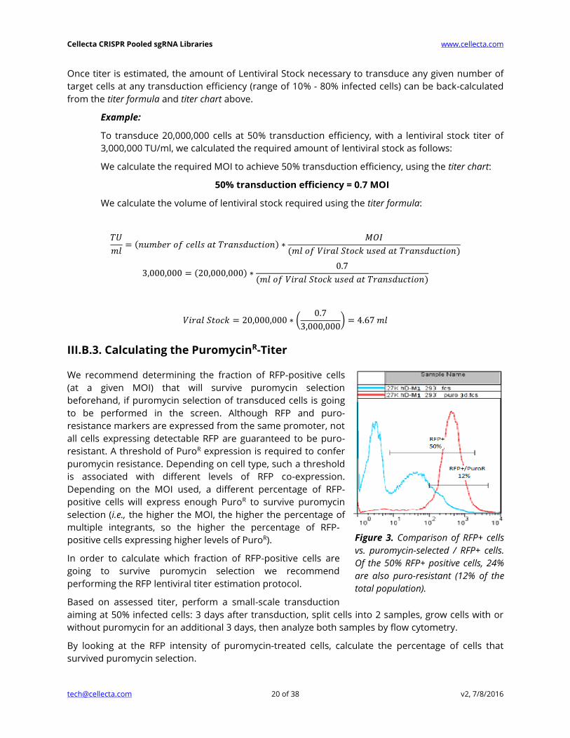
Figure 3. Comparison of RFP+ cells
vs. puromycin-selected / RFP+ cells.
Of the 50% RFP+ positive cells, 24%
are also puro-resistant (12% of the
total population).
Cellecta CRISPR Pooled sgRNA Libraries www.cellecta.com
[email protected] 21 of 38 v2, 7/8/2016
Figure 3 shows FACS analysis of transduced cells where no puromycin selection is indicated in blue,
and puromycin selection is indicated in red. 50% of cells were RFP-positive, and 24% of the RFP-
positive cells were also puromycin-resistant (12% of total).
IMPORTANT: The percentage of RFP-positive cells that are also puromycin-resistant is dependent
on MOI, as it increases with the increase of percentage RFP-positive cells bearing multiple
integrations. In the example above, 24% of RFP-positive cells (12% of total) were puromycin-resistant
when cells were infected at MOI 0.7 (50% RFP-positive cells). If the same cells were infected at the
recommended MOI of 0.5 (40% RFP-positive cells), less than 24% of RFP-positive cells would also be
puromycin-resistant cells. Conversely, if cells were infected at MOI 2 (85% RFP-positive cells), a much
higher percentage than 24% of RFP-positive cells would also be puromycin-resistant, due to a high
percentage of RFP-positive cells bearing multiple integrants and therefore expressing high levels of
the puromycin-resistance gene.
In the case described above, a 27K library genetic screen was started with at least 46 × 106 cells per
replicate and transduction. Cells were infected at MOI 0.7 (50% transduction efficiency) to obtain 23
× 106 infected (RFP-positive) cells, of which about 5.5 × 10⁶ will be puro-resistant (200 puro resistant
cells/sgRNA).
NOTES:
In your screening experiment, we do not recommend using an MOI greater than 0.5.
Using higher MOIs to achieve greater than 40% RFP-positive cells in order to obtain 20% or more
puro-resistant cells is not recommended. It is advised to limit the RFP-based MOI to 0.5 (40%
RFP-positive cells) and use enough cells at transduction to obtain the desired amount of
puromycin-resistant transduced cells (at least 200 cells/sgRNA).
IMPORTANT: When performing lentiviral transductions for a genetic screen, make sure to use
exactly the same conditions as in library titering. Accurately scale up volumes, surface areas, cell
number, and reagents to be used.
III.C. Transduction of Target Cells
Before transducing your target cells, the following steps must be completed:
1. Define the number of starting cells
2. Determine the MOI
3. Plan the experimental conditions to maintain representation
4. Calculate the titer of packaged sgRNA libraries
III.C.1. Number of Starting Cells and Representation
Pooled sgRNA library screens require quantification of changes in the fraction of cells bearing each
sgRNA sequence in selected vs. control cells (or starting library). A “hit” is identified when selected
cells have significantly more or fewer cells bearing a particular sgRNA sequence. Whether one is
looking at enrichment of sgRNA sequences in the selected cell population vs. control (positive
selection) or depletion of sgRNA sequenced in selected cell population vs. control (negative
selection), it is critical that the screens begin with a sufficient number of cells expressing each sgRNA
Cellecta CRISPR Pooled sgRNA Libraries www.cellecta.com
[email protected] 22 of 38 v2, 7/8/2016
to ensure that measured changes in the fraction of cells bearing any given sgRNA sequence are
statistically significant. This means that if there are very low numbers of cells bearing specific sgRNA
sequences at the start of the screen, random changes in a drifting population may be difficult to
differentiate from significant trends.
Simply put, a loss of 2 cells is a 20% change if there are only 10 initially vs. 2% if there are 100. For
this reason, at least a few hundred cells on average need to be infected with each sgRNA to initiate a
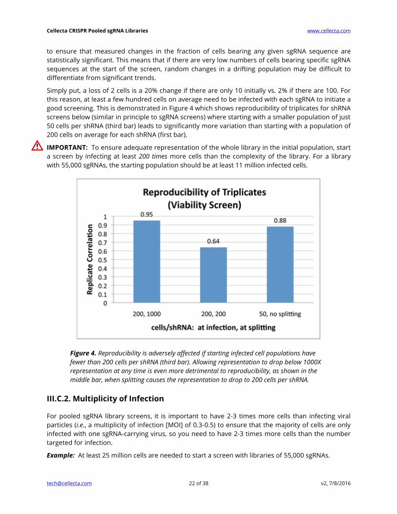
good screening. This is demonstrated in Figure 4 which shows reproducibility of triplicates for shRNA
screens below (similar in principle to sgRNA screens) where starting with a smaller population of just
50 cells per shRNA (third bar) leads to significantly more variation than starting with a population of
200 cells on average for each shRNA (first bar).
IMPORTANT: To ensure adequate representation of the whole library in the initial population, start
a screen by infecting at least 200 times more cells than the complexity of the library. For a library
with 55,000 sgRNAs, the starting population should be at least 11 million infected cells.
Figure 4. Reproducibility is adversely affected if starting infected cell populations have
fewer than 200 cells per shRNA (third bar). Allowing representation to drop below 1000X
representation at any time is even more detrimental to reproducibility, as shown in the
middle bar, when splitting causes the representation to drop to 200 cells per shRNA.
III.C.2. Multiplicity of Infection
For pooled sgRNA library screens, it is important to have 2-3 times more cells than infecting viral
particles (i.e., a multiplicity of infection [MOI] of 0.3-0.5) to ensure that the majority of cells are only
infected with one sgRNA-carrying virus, so you need to have 2-3 times more cells than the number
targeted for infection.
Example: At least 25 million cells are needed to start a screen with libraries of 55,000 sgRNAs.
Cellecta CRISPR Pooled sgRNA Libraries www.cellecta.com
[email protected] 23 of 38 v2, 7/8/2016
The lower the MOI, the more cells you need to start the screen so it is tempting to use a high MOI.
However, you should consider that a higher MOI produces a higher percentage of infected cells
bearing two or more different sgRNA constructs.
For most loss-of-function screens, we recommend optimizing conditions and performing genetic
screen transductions at no more than 0.5 MOI (a 40% transduction efficiency), which balances
these two considerations. Please note that to accurately calculate the MOI, it is critical to determine
the library titer directly in your target cells prior to beginning your experiment. Once conditions are
established to achieve ~40% transduction efficiency in the titering assay, scale up all conditions
proportionately to accommodate the larger amount of transduced cells needed for the genetic
screen.
III.C.3. Representation and Cell Propagation Techniques
To ensure a comprehensive screen, it is not simply sufficient to start with the right amount of cells.
During the screening process, incorrect propagation of the cells can completely undercut the
representation set up at the initiation of the screen. This is especially true for a negative selection
screen, such as a viability screen where one is interested in identifying sgRNAs that kill or inhibit
proliferation of cells, and, therefore, drop out of the population. It is critical to maintain the full
library representation that was initially used at the start of the screen.
If a portion of propagated cells is removed during propagation (e.g., cells are split), the
representation of the library can be skewed in the sample, which introduces significant random
noise. This effect is readily seen in the middle bar in Figure 4 where the effect of starting with
sufficient cells (i.e., 200-fold library complexity) is completely undercut by splitting cells during
propagation so that the final count of cells after 10 days is the same as the initial number of
transduced cells (i.e., 200-fold library complexity). The correlation between triplicates falls
dramatically when the cells are split to this degree.
IMPORTANT: If cells are ever to be discarded or samples split at any time during the screen, the
number of remaining cells in each sample should always exceed the complexity of the library by at
least 1,000-fold, as shown in the first bar in Figure 4. For example, keep at least 27 million cells after
every splitting step, for a 27K library. Also, before splitting or discarding, make sure you first pool all
cells from the same replicate together.
III.C.4. Transduction of Target Cells for Screening
Once you have calculated the titer of the packaged CRISPR pooled sgRNA library and you know the
MOI and number of starting cells you need to maintain representation, transduce your target cells
using the appropriate number and size of plates and the correct amount of virus. You will use the
Cas9-expressing cell line that you created in Section II. 72 hours after transduction, you can select
the cells with puromycin according to the concentration calculated from the puromycin kill curve
you calculated earlier as well.
Cellecta CRISPR Pooled sgRNA Libraries www.cellecta.com
[email protected] 24 of 38 v2, 7/8/2016
IV. Loss-of-Function Screens
IV.A. Positive Selection Screens (a.k.a. Rescue Screens)
Rescue screens allow for identification of genes that are functionally required for cell sensitivity to a
treatment or to understand underlying drug mechanisms. Positive screens are also known as
enrichment screens. Many positive screens use FACS to look for modulators of signaling molecules
such as NF-κB, p53, c-myc, HSF-1, and HIF-1α using fluorescent reporter cell lines, or cells expressing
specific antibody-detectable markers, such as specific receptors.
IV.A.1. Length of the Screen
A positive screen involves a selection that eliminates most of the cells. With this sort of screen, the
goal is to isolate a small population of cells with sgRNAs that enable the cells to pass through the
selection step. The critical factor here is the nature of the selection, which ultimately determines the
screen procedure.
NOTE: In most cases, it is advisable to wait at least two weeks after sgRNA library transduction
before carrying out the selection step. The two-week wait period is needed to allow knockout of the
alleles of the target gene in most transduced cell, and the development of the resistant phenotype
before applying selection. Cells should then be harvested as soon as positive selection is completed.
Growing and expanding clones after positive selection is not advised.
IV.A.2. MOI of Transduction
A positive screen involves isolation of a small population of cells with sgRNA sequences that will be
over-represented or enriched when compared to the starting library sgRNA counts. As with any
screen, to ensure reproducible and reliable results, it is critical that you transduce enough cells to
maintain sufficient representation of each sgRNA construct present in the library. The number of
cells stably transduced with the sgRNA library at the time of transduction should exceed the
complexity of the sgRNA library by at least 200-fold.
Example: The starting population should be at least 11 million infected cells to start a screen with a
library with 55,000 sgRNAs (see Figure 4).
IV.A.3. Maintenance of the Cells
A positive selection screen often involves the comparison of two types of samples: selected and
unselected (control) samples. After transduction and before selection, it is best practice not to
discard any cells. However, this is often not practical. If cells have to be discarded or split before
beginning the selection, the number of remaining cells in each sample should always exceed the
complexity of the library by at least 1,000-fold (e.g., keep at least 5.5 × 10⁷ cells after every splitting
step, for a 55K library).
IMPORTANT: After the selection step, all the cells in the selected samples should be collected for
genomic DNA purification and sgRNA PCR amplification. For the control samples, follow the above-
mentioned 1,000-fold rule: you should collect enough cells to equal 1,000-fold the complexity of the
library.
Cellecta CRISPR Pooled sgRNA Libraries www.cellecta.com
[email protected] 25 of 38 v2, 7/8/2016
Similarly, when amplifying sgRNAs from isolated DNA, you should always use all of the genomic
DNA recovered from cell samples, up to the amount corresponding to 1000X cells the library
size. For diploid cells, 55 million cells contain ~450 µg of genomic DNA.
IV.A.4. Baseline Controls for Positive Selection Screens
In order to calculate the enrichment-fold of the sgRNA sequences present in the selected
population, a baseline control is needed. Depending on the screen, the plasmid library itself can be
used as baseline, or pre-selection cells, or mock-selected cells.
IV.B. Negative Selection Screens (a.k.a. Viability Screens)
A standard dropout viability screen (negative selection screen) relies on the fact that some of the
sgRNAs in the screen are either cytotoxic or cytostatic (presumably by having knocked out an
essential target gene). Cells with sgRNAs that do not inhibit growth grow normally, populating the
culture; cells with lethal sgRNA do not propagate. The endpoint analysis involves looking for sgRNA
sequences that are underrepresented in the sample population relative to the original library.
IV.B.1. Length of the Screen
For a CRISPR dropout viability screen to work, the cells need to be cultured long enough for
knockouts to develop in all the alleles of each sgRNA’s target gene, and then for cells with unaffected
growth to significantly increase their proportion relative to the affected cells.
NOTE: The length of any particular screen may need to be altered depending on the specifics (e.g.,
cell growth rates, types of targets of interest, use of additional compounds, etc.).
1. Typically, we find allowing for 3 weeks after transduction to be a good starting point.
If the screen is not run long enough, all the sgRNA counts will be in a narrow range and it will be
difficult to identify significantly depleted sgRNA sequences from background variability. If the screen
is run too long, the range of representation of sgRNA sequences may become broader due to the
natural growth variance in different cells in the population. This phenomenon, often referred to as
genetic drift, can increase the background variance of the screen and make it difficult to identify
significantly depleted sgRNAs from background variability.
IV.B.2. Maintaining Library Representation throughout Screen
As mentioned above, the number of cells stably transduced with the sgRNA library at the time of
transduction should exceed the complexity of the sgRNA library by at least 200-fold. For a library
with 55,000 sgRNAs, the starting population should be at least 11 million infected cells. The MOI of
transduction should be kept at or below 0.5, to ensure that the majority of transduced cells carry
only one integrated provirus.
After transduction, the ideal is to never discard any cells at any time during the experiment (e.g., at
treatment, harvesting, DNA purification, etc.). However, this is often not practical—especially for a
negative screen where most of the cells propagate normally. If the number of cells becomes too
large and you are forced to discard a fraction, the number of remaining cells should always exceed
the complexity of the library by at least 1,000-fold (e.g., keep at least 55 million cells after every

Cellecta CRISPR Pooled sgRNA Libraries www.cellecta.com
[email protected] 26 of 38 v2, 7/8/2016
splitting step, for a 55K library). Similarly, when amplifying sgRNAs from isolated DNA, you should
always use all of the genomic DNA recovered from cell samples, up to a number of cells
corresponding to 1000X the library size.
IV.B.3. Baseline Controls for Negative Screens
In a simple screen aimed at identifying sgRNAs which are cytotoxic in a given cell line, we typically
use the library itself as the baseline control, since the sgRNA frequency distribution in plasmid and
packaged lentiviral library is virtually identical. The plasmid library has already been sequenced as
part of the QC when we made the library, so it is not necessary to re-sequence the library at this
point. If you would also like to use transduced cells as a baseline control, typically we recommend
harvesting and sequencing genomic DNA from them by about 18 hours post-transduction.
IMPORTANT: If using 18-hour post-transduction as baseline control, it is highly recommended to
use LentiFuge-purified virus (See Other Materials) for transduction in your experiment, to minimize
problems with plasmid library contamination in baseline.
In more complex experiments, aiming at identifying differential toxicity between isogenic cell lines,
or between compound-treated and non-treated cells, other baselines will be needed.
Cellecta CRISPR Pooled sgRNA Libraries www.cellecta.com
[email protected] 27 of 38 v2, 7/8/2016
V. Identifying the sgRNA Hits in Your Screen
V.A. Genomic DNA Extraction for sgRNA (or barcode)
Identification of sgRNA in the experimental samples requires amplification of the sgRNA portion of
the integrated lentiviral constructs from sample genomic DNA. Subsequent high-throughput
sequencing of sgRNAs by the Illumina NextSeq 500, HiSeq, or GAIIx is done to quantify each sgRNA
and generate digital expression data using Deconvolution software. We currently do not support
NGS of samples on the Illumina MiSeq.
If you are starting with fewer than 1 million cells, we recommend using the QIAGEN QIAamp DNA
Micro Kit, according to the manufacturer’s instructions, instead of using the protocol here.
If you are starting with between 1 million and 10 million cells, you can use the QIAGEN DNeasy
Blood and Tissue Kit for genomic DNA isolation as an alternative to the protocol below.
For samples with more than 10 million cells (most negative selection screens), you should follow the
protocol below, isolating genomic DNA. Use of a column-based kit may result in loss of genomic
DNA and loss of representation of sgRNAs that survived the screening.
NOTE: Use of disposable tubes is highly recommended in order to avoid contamination.
1. Suspend cell pellet in 5 ml QIAGEN buffer P1 (with RNase A) in 15 ml polypropylene
(phenol/chloroform resistant), Falcon screw-cap centrifuge tube (12,000 RCF rated).
2. Add 0.25 ml 10% SDS, mix and incubate 5 minutes at RT.
3. Using an ultrasonic homogenizer, sonicate to shear DNA into 10-100 kb sized fragments. To
prevent cross-contamination, thoroughly wash the ultrasound head with running water and dry
with clean paper towel between samples.
4. Add 10 μl of Proteinase K, mix and incubate 15 minutes at RT.
5. Add 5 ml Phenol:Chloroform:Isoamyl Alcohol solution, vortex hard and spin down 60 min, 20°C
at 8,000 rpm in JA-14 or equivalent rotor (Beckman).
6. At this point, there should be approximately 5 ml of clear upper phase. Transfer 4 ml of upper
phase to a new 15 ml disposable screw cap tube (same as in Step 1).
7. Add 0.5 ml 3M Sodium Acetate and 4 ml isopropanol, mix well, then spin down 30 min, 20°C at
8,000 rpm in a JA-14 or equivalent rotor.
8. In order to have a more visible pellet, compacted at the bottom of the tube, it is recommended
to incubate overnight at room temperature before centrifugation.
IMPORTANT: If starting material is less than 5 million cells, add carrier before centrifugation
(linear polyacrylamide, 25 μg/ml final) and spin down for a longer time (60 min).
9. Discard supernatant, add 10 ml 70% ethanol, spin down 5 min, 20°C at 8,000 rpm in JA-14 or
equivalent rotor.
10. Discard supernatant and air-dry pellet.
Cellecta offers sample prep, NGS, and analysis services. Please contact us at [email protected]
or visit Cellecta's NGS Services web page for more information.

Cellecta CRISPR Pooled sgRNA Libraries www.cellecta.com
[email protected] 28 of 38 v2, 7/8/2016
11. Dissolve DNA pellet in appropriate volume of dH2O to a concentration of ~2 mg/ml.
NOTE: Expected yield is about 10 μg per 1 million cells.
12. Incubate 30 minutes at 80°C before spectrophotometer reading.
V.B. Amplification of sgRNA (or barcodes) from Genomic DNA
The amplification of pooled sgRNAs from genomic DNA isolated from cell samples in the previous
step should be carried out by two rounds of PCR using Titanium Taq DNA polymerase mix (Takara).
For negative selection screens and non-selected (baseline) samples of positive selection screens, use
a total of 400 μg for 55K libraries, and scale up or down for larger or smaller libraries. For selected
samples from positive-selection screens where the number of cells is typically very low, use the
entire amount of genomic DNA recovered. The protocol was optimized using a Life Technologies
Veriti® Thermal Cycler. Use of other PCR enzymes and/or thermal cyclers may require additional
optimization.
The lentiviral sgRNA library and PCR primer designs include sequences complementary to the
sequences of the immobilized primers necessary for generating amplification clusters in Illumina’s
NextSeq 500, HiSeq, or GAIIx Flow Cells. Our library design is compatible with Single-Read and
Paired End Flow Cells. The primers have been designed to work with the NextSeq 500, but they are
also compatible with HiSeq and GAIIx systems as well. See Required Materials for the appropriate
Illumina catalog numbers. NGS of samples on the Illumina MiSeq is not supported.
Use 10 ng of plasmid sgRNA library as an amplification control in both the first and second rounds of
PCR. The product of the second round of PCR can be used as the NGS control.
V.B.1. First Round of PCR
The first round of PCR serves to amplify the sgRNAs (or barcodes) remaining in the genomic DNA
pool after the phenotypic screen is complete. We recommend not exceeding 100 µg in 100 µL total
volume per reaction.
1. Prepare a master mix according to the table below, where x is the volume of genomic DNA. For
each sample, prepare 4 × 100 µL reactions containing a total of 200 µg of genomic DNA.
X µl Genomic DNA (200 µg)
12 µl Forward 1st round PCR primer* (10 µM)
12 µl Reverse 1st round PCR primer* (10 µM)
8 µl 50X dNTP Mix (10 mM each)
40 µl 10X Titanium Taq Buffer
320 – x µl Deionized water
8 µl 50X Titanium Taq Polymerase
400 µl Total volume (Split into 4 × 100 µl reactions)
2. Perform PCR under the following cycling conditions.
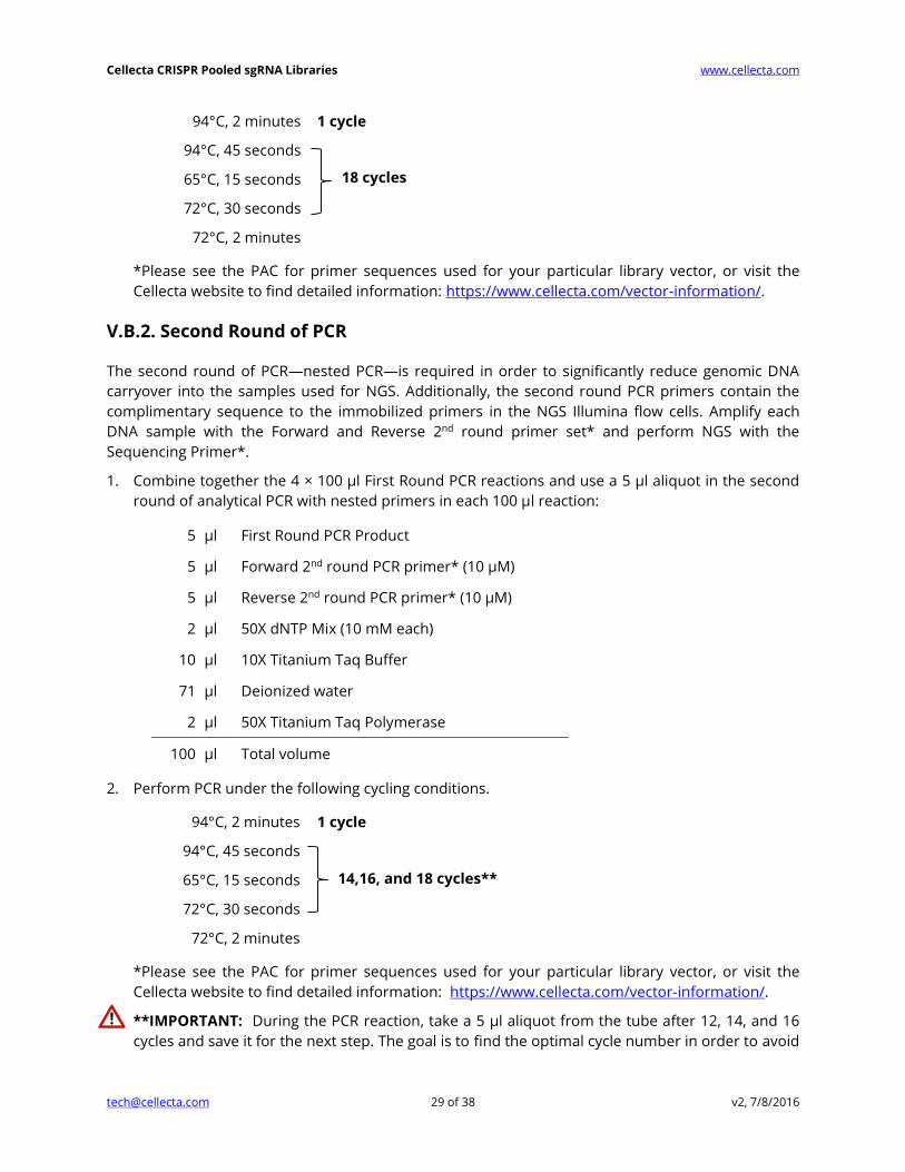
Cellecta CRISPR Pooled sgRNA Libraries www.cellecta.com
[email protected] 29 of 38 v2, 7/8/2016
94°C, 2 minutes 1 cycle
94°C, 45 seconds
65°C, 15 seconds
72°C, 30 seconds
72°C, 2 minutes
*Please see the PAC for primer sequences used for your particular library vector, or visit the
Cellecta website to find detailed information: https://www.cellecta.com/vector-information/.
V.B.2. Second Round of PCR
The second round of PCR—nested PCR—is required in order to significantly reduce genomic DNA
carryover into the samples used for NGS. Additionally, the second round PCR primers contain the
complimentary sequence to the immobilized primers in the NGS Illumina flow cells. Amplify each
DNA sample with the Forward and Reverse 2nd round primer set* and perform NGS with the
Sequencing Primer*.
1. Combine together the 4 × 100 µl First Round PCR reactions and use a 5 µl aliquot in the second
round of analytical PCR with nested primers in each 100 µl reaction:
5 µl First Round PCR Product
5 µl Forward 2nd round PCR primer* (10 µM)
5 µl Reverse 2nd round PCR primer* (10 µM)
2 µl 50X dNTP Mix (10 mM each)
10 µl 10X Titanium Taq Buffer
71 µl Deionized water
2 µl 50X Titanium Taq Polymerase
100 µl Total volume
2. Perform PCR under the following cycling conditions.
94°C, 2 minutes 1 cycle
94°C, 45 seconds
65°C, 15 seconds
72°C, 30 seconds
72°C, 2 minutes
*Please see the PAC for primer sequences used for your particular library vector, or visit the
Cellecta website to find detailed information: https://www.cellecta.com/vector-information/.
**IMPORTANT: During the PCR reaction, take a 5 µl aliquot from the tube after 12, 14, and 16
cycles and save it for the next step. The goal is to find the optimal cycle number in order to avoid
14,16, and 18 cycles**
18 cycles
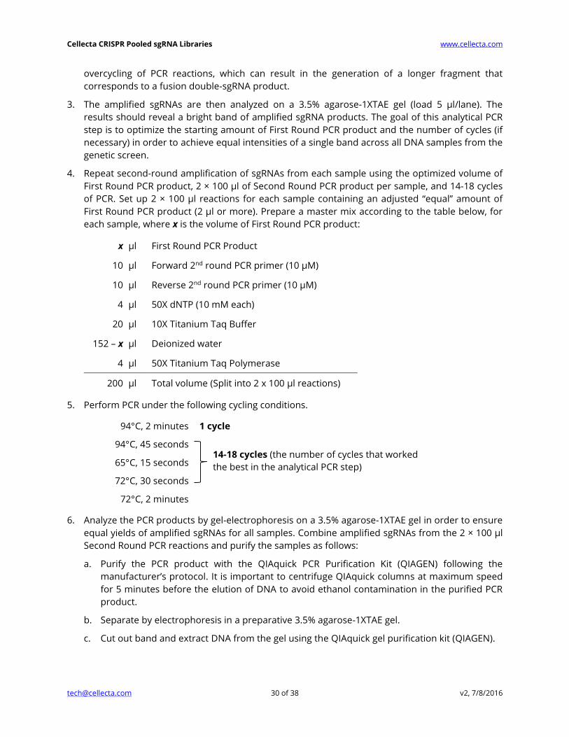
Cellecta CRISPR Pooled sgRNA Libraries www.cellecta.com
[email protected] 30 of 38 v2, 7/8/2016
overcycling of PCR reactions, which can result in the generation of a longer fragment that
corresponds to a fusion double-sgRNA product.
3. The amplified sgRNAs are then analyzed on a 3.5% agarose-1XTAE gel (load 5 µl/lane). The
results should reveal a bright band of amplified sgRNA products. The goal of this analytical PCR
step is to optimize the starting amount of First Round PCR product and the number of cycles (if
necessary) in order to achieve equal intensities of a single band across all DNA samples from the
genetic screen.
4. Repeat second-round amplification of sgRNAs from each sample using the optimized volume of
First Round PCR product, 2 × 100 µl of Second Round PCR product per sample, and 14-18 cycles
of PCR. Set up 2 × 100 µl reactions for each sample containing an adjusted “equal” amount of
First Round PCR product (2 µl or more). Prepare a master mix according to the table below, for
each sample, where x is the volume of First Round PCR product:
x µl First Round PCR Product
10 µl Forward 2nd round PCR primer (10 µM)
10 µl Reverse 2nd round PCR primer (10 µM)
4 µl 50X dNTP (10 mM each)
20 µl 10X Titanium Taq Buffer
152 – x µl Deionized water
4 µl 50X Titanium Taq Polymerase
200 µl Total volume (Split into 2 x 100 µl reactions)
5. Perform PCR under the following cycling conditions.
94°C, 2 minutes 1 cycle
94°C, 45 seconds
65°C, 15 seconds
72°C, 30 seconds
72°C, 2 minutes
6. Analyze the PCR products by gel-electrophoresis on a 3.5% agarose-1XTAE gel in order to ensure
equal yields of amplified sgRNAs for all samples. Combine amplified sgRNAs from the 2 × 100 µl
Second Round PCR reactions and purify the samples as follows:
a. Purify the PCR product with the QIAquick PCR Purification Kit (QIAGEN) following the
manufacturer’s protocol. It is important to centrifuge QIAquick columns at maximum speed
for 5 minutes before the elution of DNA to avoid ethanol contamination in the purified PCR
product.
b. Separate by electrophoresis in a preparative 3.5% agarose-1XTAE gel.
c. Cut out band and extract DNA from the gel using the QIAquick gel purification kit (QIAGEN).
14-18 cycles (the number of cycles that worked
the best in the analytical PCR step)

Cellecta CRISPR Pooled sgRNA Libraries www.cellecta.com
[email protected] 31 of 38 v2, 7/8/2016
d. Quantitate using A260 nm measurement using NanoDrop® spectrophotometer (or
equivalent) and adjust concentration to 10 nM.
V.C. NGS of Pooled sgRNA (or barcodes) on Illumina NextSeq 500,
HiSeq, or GAIIx
NGS of pooled amplified sgRNAs can be performed on the Illumina NextSeq 500 (high output flow
cells that give up to 400 million single reads or up to 800 million paired-end reads), HiSeq (80-100
million reads per sample), or GAIIx (20-30 million reads per sample) using the appropriate Cellecta
Sequencing Primer* and Illumina’s protocol for the respective sequencing machine. The final
concentration of primers in the reaction should be 300 nM for the NextSeq 500, or 500 nM for the
HiSeq or GAIIx. The number of cycles (read length) required depends on the length of the sgRNA or
barcode.
The sgRNA library and PCR primer designs include P7 and P5 sequences complementary to the
sequences of the immobilized primers necessary for generating amplification clusters in Illumina’s
NextSeq 500, HiSeq, or GAIIx flow cells.
Prepare your sequencing samples as follows:
1. Adjust purified PCR samples to 10 nM.
2. Prepare the library for sequencing according to the “Denaturing and Diluting Libraries for the
NextSeq 500” Illumina protocol (or the machine that you are using).
3. We include PhiX library as a spike-in from 5 to 15% (depending on the complexity of the library)
to increase the diversity of the library which helps generate better sequencing results.
4. Add sequencing primers as a spike-in to appropriate cartridge positions for the respective
sequencing machine.
*Please see the PAC for primer sequences used for your particular library vector, or visit the Cellecta
website to find detailed information: https://www.cellecta.com/vector-information/.
Cellecta offers sample prep, NGS, and analysis services. Please contact us at [email protected]
or visit Cellecta's NGS Services web page for more information.
Cellecta CRISPR Pooled sgRNA Libraries www.cellecta.com
[email protected] 32 of 38 v2, 7/8/2016
VI. Troubleshooting
VI.A. Low lentiviral titer in supernatant (<106 TU/ml for sgRNA-
only, or <105 TU/ml for Cas9 vectors)
VI.A.1. Poor transfection efficiency
(At 48 hours post-transfection, less than 80% of 293T cells are very brightly fluorescent)
Possible reason: 293T Cells have too high or too low density.
Solution: Plate fewer or more cells in order to have about 80% confluency at time of
transfection.
Possible reason: Plasmid DNA/Lipofectamine/Plus Reagent ratios are incorrect.
Solution: Optimize the ratios using the guidelines provided in the Lipofectamine protocol.
VI.A.2. Inefficient production of the virus
Possible reason: 293T Cells are of poor quality.
Solutions:
Optimize growth conditions, check growth medium, and don’t grow 293T cells for
more than 20 passages.
Check for mycoplasma contamination.
Do not overgrow the cells (do not allow the cells to reach more than 90% confluency
in order to keep the culture continuously in logarithmic growth phase).
Possible reason: Lentiviral supernatant harvested too early or too late.
Solution: Harvest supernatant 48 hours and 72 hours after transfection.
Possible reason: 293T cell media is too acidic at time of virus harvesting.
Solution: Make sure to replace media 24 hours before harvesting, and make sure to
supplement media with HEPES pH 7.4, 20mM final.
VI.B. Poor transduction efficiency
Possible reason: Target cells have too high or too low density.
Solution: Plate fewer or more cells in order to have 20-50% confluency at transduction
stage.
Cellecta CRISPR Pooled sgRNA Libraries www.cellecta.com
[email protected] 33 of 38 v2, 7/8/2016
Possible reason: Target cell line may be difficult to transduce.
Solutions:
Use a higher concentration of lentiviral particles.
Perform “Spinoculation” to improve transduction efficiency.
Check to see if Polybrene was added at 5 µg/ml.
Possible reason: Wrong amount of Polybrene added during transduction stage.
Solution: If Polybrene is toxic to the target cells, optimize Polybrene concentration in the
range of 0 – 5 µg/ml by performing a toxicity titration as described in I.C.3. Check Toxicity of
Polybrene Section.
Possible reason: Loss of lentiviral titer during storage.
Solution: Ensure storage of aliquoted packaged construct at –80°C. Each freeze-thaw cycle
typically causes reduction of the titer by ~20%. Use a fresh stock for transduction.
Possible reason: The RFP assay is performed too early.
Solution: Normally, the maximal expression of RFP from the integrated provirus is expected
to develop by 72 hours after transduction. However, some cells exhibit delayed expression.
Try the assay at a later time, such as 96 hours.
Possible reason: The RFP assay is performed with the wrong flow cytometry settings.
Solution: RFP+ cells are to be detected using a 561nm laser for excitation (530nm still
acceptable) and 600/20 band-pass filters (or similar) for detection (for TagRFP). Using blue
laser (488nm) for excitation leads to gross underestimation of viral titer.
Possible reason: In the RFP assay, the % of transduced cells is determined by fluorescence
microscopy instead of flow cytometry.
Solution: Use flow cytometry.
VI.C. Transduction affects target cell viability
Possible reason: Polybrene is toxic for target cells
Solution: Optimize the concentration and exposure time to Polybrene during the
transduction step. For some sensitive cells, Polybrene should not be used.
Possible reason: Virus-containing conditioned media is toxic to target cells.

Cellecta CRISPR Pooled sgRNA Libraries www.cellecta.com
[email protected] 34 of 38 v2, 7/8/2016
Solution: Concentrate and resuspend the virus in target cell growth media, PBS/10%FBS, or
PBS/1%BSA.
VI.D. No expression of RFP, PuroR, BlastR, or sgRNA in target cells
Possible reason: The promoter is not functional in target cells.
Solutions:
Change the target cells.
Contact Cellecta at [email protected] to have the constructs cloned in another
vector with different promoter.
VI.E. Difficulties with sample preparation and NGS
VI.E.1. No PCR product
Possible reasons: (1) Incorrect primers or bad reagents used, (2) missing reagents, (3) low
transduction of target cells, or (4) poor gDNA purification prep contains PCR inhibitors.
Solutions:
Include 10 ng of plasmid library DNA as a positive control. If it produces the correct
amplification product, the problem lies either with absent or low numbers of sgRNA (e.g., low
MOI or problems with the transduction efficiency) or impurities in genomic DNA that block
sgRNA amplification. Dilute the genomic DNA 2-5 fold and repeat the amplification step in
several test tubes. If the positive control does not produce the correct product, confirm use
of the correct primers and reagents.
Verify that primer sequences are correct.
VI.E.2. No sgRNA (or barcodes) present in NGS results
Possible reason #1: Incorrect First or Second round PCR primers or Sequencing Primer used.
Solution: Ensure that you use the correct First and Second Round PCR Primers and Cellecta
Sequencing Primer. See the Product Analysis Certificate (PAC) for the NGS primer sequences for
your particular library vector, or visit the Cellecta website to find detailed information for standard
vectors: https://www.cellecta.com/vector-information/.
Possible reason #2: Incorrect NGS reagent kit used.
Solution: Ensure that you or the NGS core facility uses the proper Illumina NGS reagent kit. For
details, please check the kit descriptions in the Required Materials section.
Cellecta CRISPR Pooled sgRNA Libraries www.cellecta.com
[email protected] 35 of 38 v2, 7/8/2016
VII. Other Information
VII.A. Technical Support
For help with using CRISPR Pooled Lentiviral sgRNA Libraries, please email technical support at
[email protected] with the answers to the questions below (if applicable).
Library Used:
1. Which library did you use, and which Module(s) (if applicable)?
2. What are the lot numbers?
Packaging the Library:
1. What was the lentiviral titer, and what was the total number of TU packaged?
2. How was the virus concentrated? (if applicable)
Transducing Target Cells:
1. What MOI did you use to transduce your target cells?
2. What target cells did you use?
3. How many replicates did you use? (i.e., duplicate, triplicate, etc.)
4. Did you use puromycin after transduction, and at what concentration?
5. For how long did you use puromycin on the cells?
CRISPR Screen:
1. Could you briefly explain your experiment?
2. How many infected cells were used?
Sample Preparation & NGS:
1. Describe the protocol you used to amplify the sgRNAs.
2. What NGS system and which Illumina NGS Kits did you use?
3. How much PCR product was used for NGS?
4. How many sequences were read per sample?
5. Would you be able to send us the raw data so that it may help us diagnose the issue?
Please refer to the questions above and contact us by phone or email:
Phone: +1 (650) 938-3910
Toll-Free: +1 (877) 938-3910
Fax: +1 (650) 938-3911

Cellecta CRISPR Pooled sgRNA Libraries www.cellecta.com
[email protected] 36 of 38 v2, 7/8/2016
E-mail:
Technical Support: [email protected]
General Information: [email protected]
Sales: [email protected]
Orders: [email protected]
Postal Mail: Cellecta, Inc.
320 Logue Ave.
Mountain View, CA 94043
For the latest technical news and updates, visit Cellecta’s blog at:
https://www.cellecta.com/blog-news/
Cellecta CRISPR Pooled sgRNA Libraries www.cellecta.com
[email protected] 37 of 38 v2, 7/8/2016
VII.B. Safety Guidelines
The HIV-based lentivector system is designed to maximize its biosafety features, which include:
A deletion in the enhancer of the U3 region of 3’ΔLTR ensures self-inactivation of the
lentiviral construct after transduction and integration into genomic DNA of the target cells.
The RSV promoter upstream of 5’LTR in the lentivector allows efficient Tat-independent
production of lentiviral RNA, reducing the number of genes from HIV-1 that are used in this
system.
Number of lentiviral genes necessary for packaging, replication and transduction is reduced
to three (gag, pol, rev). The corresponding proteins are expressed from different plasmids
lacking packaging signals and share no significant homology to any of the expression
lentivectors, pVSV-G expression vector, or any other vector to prevent generation of
recombinant replication-competent virus.
None of the HIV-1 genes (gag, pol, rev) are present in the packaged lentiviral genome, as they
are expressed from packaging plasmids lacking packaging signal—therefore, the lentiviral
particles generated are replication-incompetent.
Lentiviral particles will carry only a copy of your expression construct.
Despite the above safety features, use of HIV-based vectors falls within NIH Biosafety Level 2 criteria
due to the potential biohazard risk of possible recombination with endogenous lentiviral sequences
to form self-replicating virus or the possibility of insertional mutagenesis. For a description of
laboratory biosafety level criteria, consult the Centers for Disease Control Office of Health and Safety
Web site at:
http://www.cdc.gov/biosafety/publications/bmbl5/bmbl5_sect_iv.pdf
It is also important to check with the health and safety guidelines at your institution regarding the
use of lentiviruses and follow standard microbiological practices, which include:
Wear gloves and lab coat at all times when conducting the procedure.
Always work with lentiviral particles in a Class II laminar flow hood.
All procedures are performed carefully to minimize the creation of splashes or aerosols.
Work surfaces are decontaminated at least once a day and after any spill of viable material.
All cultures, stocks, and other regulated wastes are decontaminated before disposal by an
approved decontamination method such as autoclaving. Materials to be decontaminated
outside of the immediate laboratory area are to be placed in a durable, leak-proof, properly
marked (biohazard, infectious waste) container and sealed for transportation from the
laboratory.
Cellecta CRISPR Pooled sgRNA Libraries www.cellecta.com
[email protected] 38 of 38 v2, 7/8/2016
VII.C. References
For a complete list of References and Product Citations, please see:
https://www.cellecta.com/resources/publications/
VII.D. Terms and Conditions
Cellecta, Inc. Limited License
Cellecta grants the end user (the “Recipient”) of the CRISPR Pooled Lentiviral sgRNA Libraries and Vector (the “Product”) a non-transferable, non-
exclusive license to use the reagents for internal research use only as described in the enclosed protocols; in particular, research use only
excludes and without limitation, resale, repackaging, or use for the making or selling of any commercial product or service without the written
approval of Cellecta, Inc. - separate licenses are available for non-research use or applications. The Product is not to be used for human
diagnostics or included/used in any drug intended for human use. Care and attention should be exercised in handling the Product by following
appropriate research laboratory practices.
Cellecta’s liability is expressly limited to replacement of Product or a refund limited to the actual purchase price. Cellecta’s liability does not
extend to any damages arising from use or improper use of the Product, or losses associated with the use of additional materials or reagents.
This limited warranty is the sole and exclusive warranty. Cellecta does not provide any other warranties of any kind, expressed or implied,
including the merchantability or fitness of the Product for a particular purpose. Use of the Product for any use other than described expressly
herein may be covered by patents or subject to rights other than those mentioned. Cellecta disclaims any and all responsibility for injury or
damage that may be caused by the failure of the Recipient or any other person to use the Product in accordance with the terms and conditions
outlined herein.
The Recipient may refuse these licenses by returning the enclosed Product unused. By keeping or using the enclosed Product, you agree to be
bound by the terms of these licenses. The laws of the State of California shall govern the interpretation and enforcement of the terms of these
Licenses.
Limited Use Label Licenses
The Recipient acknowledges that the Product has been developed by Cellecta based on licenses from Third Parties and agrees with the Terms
of Limited Use for the Recipient provided by the Third Parties:
Life Technologies Corporation End-User Label License for the use of Lentiviral Expression System:
“This product or service (based upon the Lentiviral Expression System) is sublicensed from Life Technologies Corporation under U.S. Patent
Nos. 5,686,279; 5,834,256; 5,858,740; 5,994,136; 6,013,516; 6,051,427; 6,165,782; 6,218,187; 6,428,953; 6,924,144; 7,083,981 and 7,250,299 and
corresponding patents and applications in other countries for internal research purposes only. Use of this technology for gene therapy
applications or bioprocessing other than for nonhuman research use requires a license from GBP IP, LLC. Please contact GBP IP, LLC 537
Steamboat Road, Suite 200, Greenwich, CT 06830. Use of this technology to make or sell products or offer services for consideration in the
research market requires a license from Life Technologies Corporation, 5791 Van Allen Way, Carlsbad, CA 92008.”
Evrogen IP JSC End-User Label License for the use of lentiviral sgRNA constructs comprising TagRFP-encoded gene:
“This product is for internal non-commercial research use only. No rights are conveyed to modify or clone the gene encoding fluorescent
protein contained in this product. The right to use this product specifically excludes the right to validate or screen compounds. For information
on commercial licensing, contact Evrogen Licensing Department, email: [email protected]”.
Terms and Conditions are also available online at https://www.cellecta.com/company/legal-information/terms-and-conditions/.
© 2016 Cellecta, Inc. All Rights Reserved.
Trademarks
CELLECTA is a registered trademark of Cellecta, Inc. CRISPRtest and LentiFuge are trademarks of Cellecta, Inc. CRL-11268 is a trademark of
ATCC. Lipofectamine, Plus Reagent, NanoDrop, and Veriti are registered trademarks of Thermo Fisher Scientific. NextSeq and HiSeq are
registered trademarks of Illumina, Inc. GAIIx is a trademark of Illumina, Inc.
Recommended