CCHCS Care Guide: Chronic Wound Management
SUMMARY DECISION SUPPORT PATIENT EDUCATION/SELF MANAGEMENT
October 2018
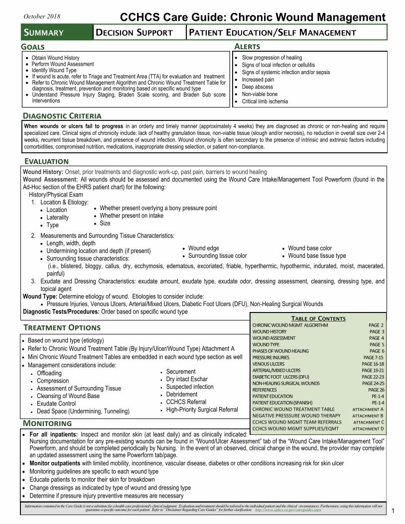
Obtain Wound History Perform Wound Assessment Identify Wound Type If wound is acute, refer to Triage and Treatment Area (TTA) for evaluation and treatment Refer to Chronic Wound Management Algorithm and Chronic Wound Treatment Table for
diagnosis, treatment, prevention and monitoring based on specific wound type Understand Pressure Injury Staging, Braden Scale scoring, and Braden Sub score
Interventions
For all inpatients: Inspect and monitor skin (at least daily) and as clinically indicated: Nursing documentation for any pre-existing wounds can be found in “Wound/Ulcer Assessment” tab of the “Wound Care Intake/Management Tool” Powerform, and should be completed periodically by Nursing. In the event of an observed, clinical change in the wound, the provider may complete an updated assessment using the same Powerform tab/page.
Monitor outpatients with limited mobility, incontinence, vascular disease, diabetes or other conditions increasing risk for skin ulcer
Monitoring guidelines are specific to each wound type
Educate patients to monitor their skin for breakdown
Change dressings as indicated by type of wound and dressing type
Determine if pressure injury preventive measures are necessary
Monitoring
Slow progression of healing
Signs of local infection or cellulitis
Signs of systemic infection and/or sepsis
Increased pain
Deep abscess
Non-viable bone
Critical limb ischemia
1
When wounds or ulcers fail to progress in an orderly and timely manner (approximately 4 weeks) they are diagnosed as chronic or non-healing and require specialized care. Clinical signs of chronicity include: lack of healthy granulation tissue, non-viable tissue (slough and/or necrosis), no reduction in overall size over 2-4 weeks, recurrent tissue breakdown, and presence of wound infection. Wound chronicity is often secondary to the presence of intrinsic and extrinsic factors including comorbidities, compromised nutrition, medications, inappropriate dressing selection, or patient non-compliance.
Information contained in the Care Guide is not a substitute for a health care professional's clinical judgment. Evaluation and treatment should be tailored to the individual patient and the clinical circumstances. Furthermore, using this information will not guarantee a specific outcome for each patient. Refer to “Disclaimer Regarding Care Guides” for further clarification. http://www.cphcs.ca.gov/careguides.aspx
Evaluation
Securement Dry intact Eschar Suspected infection Debridement CCHCS Referral High-Priority Surgical Referral
Diagnostic Criteria
Goals Alerts
Treatment Options
Wound History: Onset, prior treatments and diagnostic work-up, past pain, barriers to wound healing Wound Assessment: All wounds should be assessed and documented using the Wound Care Intake/Management Tool Powerform (found in the Ad-Hoc section of the EHRS patient chart) for the following: History/Physical Exam
1. Location & Etiology: Location Laterality Type
2. Measurements and Surrounding Tissue Characteristics: Length, width, depth Undermining location and depth (if present) Surrounding tissue characteristics: (i.e., blistered, bloggy, callus, dry, ecchymosis, edematous, excoriated, friable, hyperthermic, hypothermic, indurated, moist, macerated,
painful) 3. Exudate and Dressing Characteristics: exudate amount, exudate type, exudate odor, dressing assessment, cleansing, dressing type, and
topical agent Wound Type: Determine etiology of wound. Etiologies to consider include:
Pressure Injuries, Venous Ulcers, Arterial/Mixed Ulcers, Diabetic Foot Ulcers (DFU), Non-Healing Surgical Wounds Diagnostic Tests/Procedures: Order based on specific wound type
TABLE OF CONTENTS CHRONIC WOUND MGMT ALGORITHM PAGE 2 WOUND HISTORY PAGE 3 WOUND ASSESSMENT PAGE 4 WOUND TYPE PAGE 5 PHASES OF WOUND HEALING PAGE 6 PRESSURE INJURIES PAGE 7-15 VENOUS ULCERS PAGE 16-18 ARTERIAL/MIXED ULCERS PAGE 19-21 DIABETIC FOOT ULCERS (DFU) PAGE 22-23 NON-HEALING SURGICAL WOUNDS PAGE 24-25 REFERENCES PAGE 26 PATIENT EDUCATION PE-1-4 PATIENT EDUCATION (SPANISH) PE-1-4 CHRONIC WOUND TREATMENT TABLE attachment A NEGATIVE PRESSSURE WOUND THERAPY attachment B CCHCS WOUND MGMT TEAM REFERRALS attachment C CCHCS WOUND MGMT SUPPLIES/EQMT attachment D
Based on wound type (etiology)
Refer to Chronic Wound Treatment Table (By Injury/Ulcer/Wound Type) Attachment A
Mini Chronic Wound Treatment Tables are embedded in each wound type section as well
Management considerations include:
Offloading Compression Assessment of Surrounding Tissue Cleansing of Wound Base Exudate Control Dead Space (Undermining, Tunneling)
Whether present overlying a bony pressure point Whether present on intake
Size
Wound edge Surrounding tissue color
Wound base color Wound base tissue type
2
SUMMARY DECISION SUPPORT PATIENT EDUCATION/SELF MANAGEMENT
CCHCS Care Guide: Chronic Wound Management October 2018
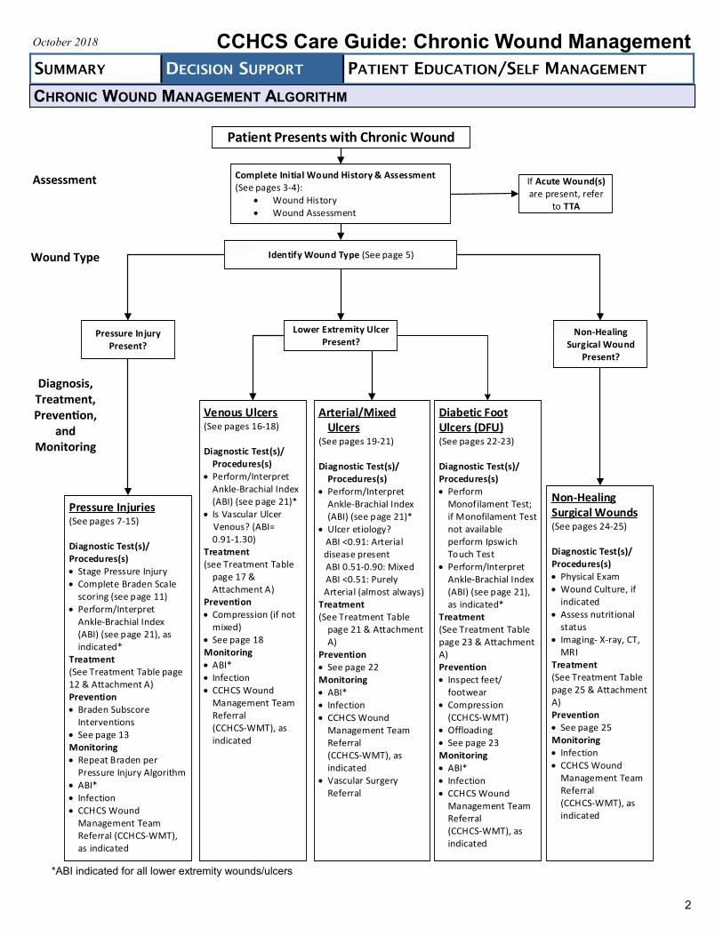
Patient Presents with Chronic Wound
Complete Initial Wound History & Assessment (See pages 3-4):
Wound History Wound Assessment
Pressure Injury Present?
If Acute Wound(s) are present, refer
to TTA
Identify Wound Type (See page 5)
Pressure Injuries(See pages 7-15)
Diagnostic Test(s)/Procedures(s) Stage Pressure Injury Complete Braden Scale
scoring (see page 11) Perform/Interpret
Ankle-Brachial Index (ABI) (see page 21), as indicated*
Treatment(See Treatment Table page 12 & Attachment A)Prevention Braden Subscore
Interventions See page 13Monitoring Repeat Braden per
Pressure Injury Algorithm ABI* Infection CCHCS Wound
Management Team Referral (CCHCS-WMT), as indicated
Venous Ulcers(See pages 16-18)
Diagnostic Test(s)/Procedures(s)
Perform/Interpret Ankle-Brachial Index (ABI) (see page 21)*
Is Vascular Ulcer Venous? (ABI=
0.91-1.30) Treatment(see Treatment Table
page 17 & Attachment A)
Prevention Compression (if not
mixed) See page 18Monitoring ABI* Infection CCHCS Wound
Management Team Referral (CCHCS-WMT), as indicated
Diabetic Foot Ulcers (DFU)(See pages 22-23)
Diagnostic Test(s)/Procedures(s) Perform
Monofilament Test; if Monofilament Test not available perform Ipswich Touch Test
Perform/Interpret Ankle-Brachial Index (ABI) (see page 21), as indicated*
Treatment(See Treatment Table page 23 & Attachment A)Prevention Inspect feet/
footwear Compression
(CCHCS-WMT) Offloading See page 23Monitoring ABI* Infection CCHCS Wound
Management Team Referral (CCHCS-WMT), as indicated
Lower Extremity Ulcer Present?
Arterial/Mixed Ulcers
(See pages 19-21)
Diagnostic Test(s)/Procedures(s)
Perform/Interpret Ankle-Brachial Index (ABI) (see page 21)*
Ulcer etiology? ABI <0.91: Arterial _disease present ABI 0.51-0.90: Mixed ABI <0.51: Purely _Arterial (almost always)Treatment (See Treatment Table
page 21 & Attachment A)
Prevention See page 22Monitoring ABI* Infection CCHCS Wound
Management Team Referral (CCHCS-WMT), as indicated
Vascular Surgery Referral
Non-Healing Surgical Wound
Present?
Non-Healing Surgical Wounds(See pages 24-25)
Diagnostic Test(s)/Procedures(s) Physical Exam Wound Culture, if
indicated Assess nutritional
status Imaging- X-ray, CT,
MRITreatment(See Treatment Table page 25 & Attachment A)Prevention See page 25Monitoring Infection CCHCS Wound
Management Team Referral (CCHCS-WMT), as indicated
CHRONIC WOUND MANAGEMENT ALGORITHM
*ABI indicated for all lower extremity wounds/ulcers
Assessment
Wound Type
Diagnosis, Treatment, Prevention,
and Monitoring
Parameters Descriptors
Onset When did it first occur?
Is it recurrent?
What is the patient’s description of the cause of the wound?
Any change in size or amount of drainage?
Prior Treatments and Diagnostic
Work-up
Dressings?
Antibiotic use?
Offloading or prevention strategies to alleviate mechanism of injury?
Diagnostic tests?
Previous consultations/referrals?
Pain History Past pain and past pain level related to the wound(s)?
Interventions tried for relief?
Effectiveness of interventions?
Barriers to
Wound Healing Barriers can be real or perceived, as well as intrinsic (relating to the patient), or extrinsic (relating to the patient’s environment)
Intrinsic Factors:
Ability to comprehend and understand instructions
Any physical limitations or mobility issues that may affect healing: Necrosis - dead tissues which create ideal conditions for bacterial growth Bacterial Infection - can delay wound healing by releasing toxins that damage tissue. Repeated
contamination with feces and urine can pose a serious challenge to wound healing Hemorrhage - the inflammatory phase of wound healing cannot take place until a fibrin clot forms or
hemostasis occurs High wound tension - may lead to wound dehiscence Nutritional status - protein deficiency reduces collagen formation Medical conditions - chronic illnesses such as diabetes, obesity, circulatory disorders etc. contribute
to decreased tissue perfusion Tissue perfusion - successful wound healing requires adequate blood supply to the site of damage Age - structural & moisture changes in the skin occurring with age can contribute to delayed healing Medications - steroids can inhibit wound healing Smoking - nicotine is a vasoconstrictor. Carbon monoxide prevents oxygen release; both decrease
tissue perfusion Varicose veins - lead to swelling; decrease perfusion to the damaged tissue Dehydration - fluids are needed for oxygen profusion. Granulation and epithelialization require a
moist environment for optimal healing
Willingness to be an active participant in care and treatment Extrinsic Factors:
Equipment not available
Institution specific limitations: building, housing, terrain challenges
Correctional/security challenges
3
SUMMARY DECISION SUPPORT PATIENT EDUCATION/SELF MANAGEMENT
CCHCS Care Guide: Chronic Wound Management October 2018
WOUND HISTORY
When performing an initial evaluation of a wound, conduct a thorough wound history and wound assessment. If the wound is located on the lower extremity, also perform a basic vascular exam. Assessment and documentation of wounds should be done in a systematic and consistent manner.
4
SUMMARY DECISION SUPPORT PATIENT EDUCATION/SELF MANAGEMENT
CCHCS Care Guide: Chronic Wound Management October 2018
WOUND ASSESSMENT
Parameters Descriptors
Location Describe the anatomical site of wound(s)
Morphology/
Dimensions Describe and calculate the following:
Color
Shape
Elevation (flat, elevated, depressed)
Size/Volume: Determine the length x width x depth (L x W x D) in centimeters (cm); (L x W for wounds without depth) Always measure in cm the longest measure for each axis: Length: 12 - 6 o’clock measure in cm Width: 9 - 3 o’clock measure in cm Depth: deepest point in cm If no depth, document “no appreciable depth.” If wound covered with slough/dry necrotic tissue, document
as “indeterminate.”
Note any tunneling or undermining
Temperature
Texture
Wound Base Assess tissue types: eschar, slough, granulation, epithelial
Drainage/
Exudate
Describe the:
Amount of drainage (none, scant, moderate, or large)
Color of the drainage
Odor (none, mild, moderate, or strong)
Moisture balance (surrounding skin is not wet; dressing is not adhered to wound base).
Is the drainage well contained by the dressing?
Surrounding
Skin
Physical skin assessment should include an assessment of: skin color, moisture, temperature, texture, mobility, turgor, skin lesions, the presence of edema, and the nails. Consider the following:
Intact or not intact? Color? Is there a palpable temperature change?
How does it feel to palpation: supple (normal), soft (fluctuant) or hard (indurated)? Does it blanch?
Is there edema present? Common causes – direct trauma, impaired venous return, systemic conditions
Texture: deviations from normal – rough, flaky, friable and easily broken skin. Patients on corticosteroids over a longer period of time may also develop thin skin
Vascularity Assess and document the following:
Pallor/Vascularity: Abnormal loss of color from normal skin or mucous membranes due to anemia, inadequate circulation, or edema
Capillary Refill: Tested to assess vascular flow to the hands and feet. Color should return in less than 3 seconds. Conditions that could cause blanch times >3 seconds include: dehydration, shock, peripheral vascular disease, or hypothermia
Current Pain Location, scale, quality(ies), onset, duration, exacerbating/relieving factors, comments Note: Uncontrolled pain can also lead to poor wound healing and increased infection rates
Edema Grading Scale
1+ Slight pitting, no viable distortion; disappears rapidly
2+ Somewhat deeper pit than in grade 1, but no readily detectable distortion; disappears in 10-15 seconds
3+ Pit is noticeably deep and may last more than 1 minute
4+ Pit is very deep and lasts as long as 2 to 5 minutes; dependent extremity is grossly distorted
5
SUMMARY DECISION SUPPORT PATIENT EDUCATION/SELF MANAGEMENT
CCHCS Care Guide: Chronic Wound Management October 2018
WOUND TYPE
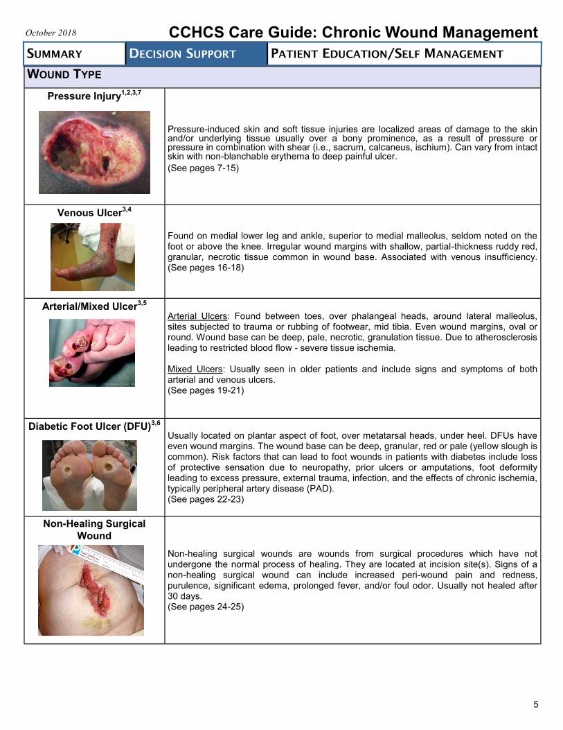
Pressure Injury1,2,3,7
Pressure-induced skin and soft tissue injuries are localized areas of damage to the skin and/or underlying tissue usually over a bony prominence, as a result of pressure or pressure in combination with shear (i.e., sacrum, calcaneus, ischium). Can vary from intact skin with non-blanchable erythema to deep painful ulcer.
(See pages 7-15)
Venous Ulcer3,4
Found on medial lower leg and ankle, superior to medial malleolus, seldom noted on the foot or above the knee. Irregular wound margins with shallow, partial-thickness ruddy red, granular, necrotic tissue common in wound base. Associated with venous insufficiency. (See pages 16-18)
Arterial/Mixed Ulcer3,5
Arterial Ulcers: Found between toes, over phalangeal heads, around lateral malleolus, sites subjected to trauma or rubbing of footwear, mid tibia. Even wound margins, oval or round. Wound base can be deep, pale, necrotic, granulation tissue. Due to atherosclerosis leading to restricted blood flow - severe tissue ischemia. Mixed Ulcers: Usually seen in older patients and include signs and symptoms of both arterial and venous ulcers. (See pages 19-21)
Diabetic Foot Ulcer (DFU)3,6 Usually located on plantar aspect of foot, over metatarsal heads, under heel. DFUs have even wound margins. The wound base can be deep, granular, red or pale (yellow slough is common). Risk factors that can lead to foot wounds in patients with diabetes include loss of protective sensation due to neuropathy, prior ulcers or amputations, foot deformity leading to excess pressure, external trauma, infection, and the effects of chronic ischemia, typically peripheral artery disease (PAD). (See pages 22-23)
Non-Healing Surgical
Wound
Non-healing surgical wounds are wounds from surgical procedures which have not undergone the normal process of healing. They are located at incision site(s). Signs of a non-healing surgical wound can include increased peri-wound pain and redness, purulence, significant edema, prolonged fever, and/or foul odor. Usually not healed after 30 days. (See pages 24-25)
6
SUMMARY DECISION SUPPORT PATIENT EDUCATION/SELF MANAGEMENT
CCHCS Care Guide: Chronic Wound Management October 2018
PHASES OF WOUND HEALING
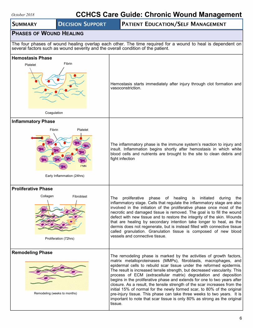
The four phases of wound healing overlap each other. The time required for a wound to heal is dependent on several factors such as wound severity and the overall condition of the patient.
Hemostasis Phase
Hemostasis starts immediately after injury through clot formation and vasoconstriction.
Inflammatory Phase
The inflammatory phase is the immune system’s reaction to injury and insult. Inflammation begins shortly after hemostasis in which white blood cells and nutrients are brought to the site to clean debris and fight infection
Proliferative Phase
The proliferative phase of healing is initiated during the inflammatory stage. Cells that regulate the inflammatory stage are also involved in the initiation of the proliferative phase once most of the necrotic and damaged tissue is removed. The goal is to fill the wound defect with new tissue and to restore the integrity of the skin. Wounds that are healing by secondary intention take longer to heal, as the dermis does not regenerate, but is instead filled with connective tissue called granulation. Granulation tissue is composed of new blood vessels and connective tissue.
Remodeling Phase
The remodeling phase is marked by the activities of growth factors, matrix metalloproteinases (MMPs), fibroblasts, macrophages, and epidermal cells to rebuild scar tissue under the reformed epidermis. The result is increased tensile strength, but decreased vascularity. This process of ECM (extracellular matrix) degradation and deposition begins in the proliferative phase and extends for one to two years after closure. As a result, the tensile strength of the scar increases from the initial 15% of normal for the newly formed scar, to 80% of the original pre-injury tissue. This phase can take three weeks to two years. It is important to note that scar tissue is only 80% as strong as the original tissue.
Remodeling (weeks to months)
Fibrin Platelet
Coagulation
Platelet Fibrin
Early Inflammation (24hrs)
Proliferation (72hrs)
Collagen Fibroblast
PRESSURE INJURIES1,2,3,7,8
SUMMARY DECISION SUPPORT PATIENT EDUCATION/SELF MANAGEMENT
CCHCS Care Guide: Chronic Wound Management October 2018
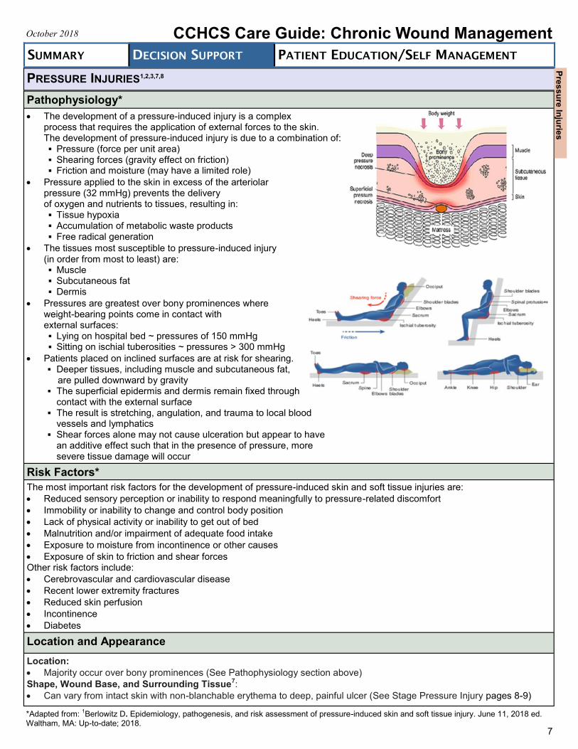
Pathophysiology*
The development of a pressure-induced injury is a complex process that requires the application of external forces to the skin. The development of pressure-induced injury is due to a combination of: Pressure (force per unit area) Shearing forces (gravity effect on friction) Friction and moisture (may have a limited role)
Pressure applied to the skin in excess of the arteriolar pressure (32 mmHg) prevents the delivery of oxygen and nutrients to tissues, resulting in: Tissue hypoxia Accumulation of metabolic waste products Free radical generation
The tissues most susceptible to pressure-induced injury (in order from most to least) are: Muscle Subcutaneous fat Dermis
Pressures are greatest over bony prominences where weight-bearing points come in contact with external surfaces: Lying on hospital bed ~ pressures of 150 mmHg Sitting on ischial tuberosities ~ pressures > 300 mmHg
Patients placed on inclined surfaces are at risk for shearing. Deeper tissues, including muscle and subcutaneous fat, are pulled downward by gravity The superficial epidermis and dermis remain fixed through
contact with the external surface The result is stretching, angulation, and trauma to local blood
vessels and lymphatics
Shear forces alone may not cause ulceration but appear to have an additive effect such that in the presence of pressure, more severe tissue damage will occur
Risk Factors*
The most important risk factors for the development of pressure-induced skin and soft tissue injuries are:
Reduced sensory perception or inability to respond meaningfully to pressure-related discomfort
Immobility or inability to change and control body position
Lack of physical activity or inability to get out of bed
Malnutrition and/or impairment of adequate food intake
Exposure to moisture from incontinence or other causes
Exposure of skin to friction and shear forces Other risk factors include:
Cerebrovascular and cardiovascular disease
Recent lower extremity fractures
Reduced skin perfusion
Incontinence
Diabetes
Location and Appearance
Location:
Majority occur over bony prominences (See Pathophysiology section above)
Shape, Wound Base, and Surrounding Tissue7:
Can vary from intact skin with non-blanchable erythema to deep, painful ulcer (See Stage Pressure Injury pages 8-9)
*Adapted from: 1Berlowitz D. Epidemiology, pathogenesis, and risk assessment of pressure-induced skin and soft tissue injury. June 11, 2018 ed. Waltham, MA: Up-to-date; 2018.
7
Pre
ss
ure
Inju
ries
PRESSURE INJURIES
Diagnostic Test(s) and Procedure(s): NPUAP Pressure Injury Stages7
STAGE PRESSURE INJURY*
SUMMARY DECISION SUPPORT PATIENT EDUCATION/SELF MANAGEMENT
CCHCS Care Guide: Chronic Wound Management October 2018
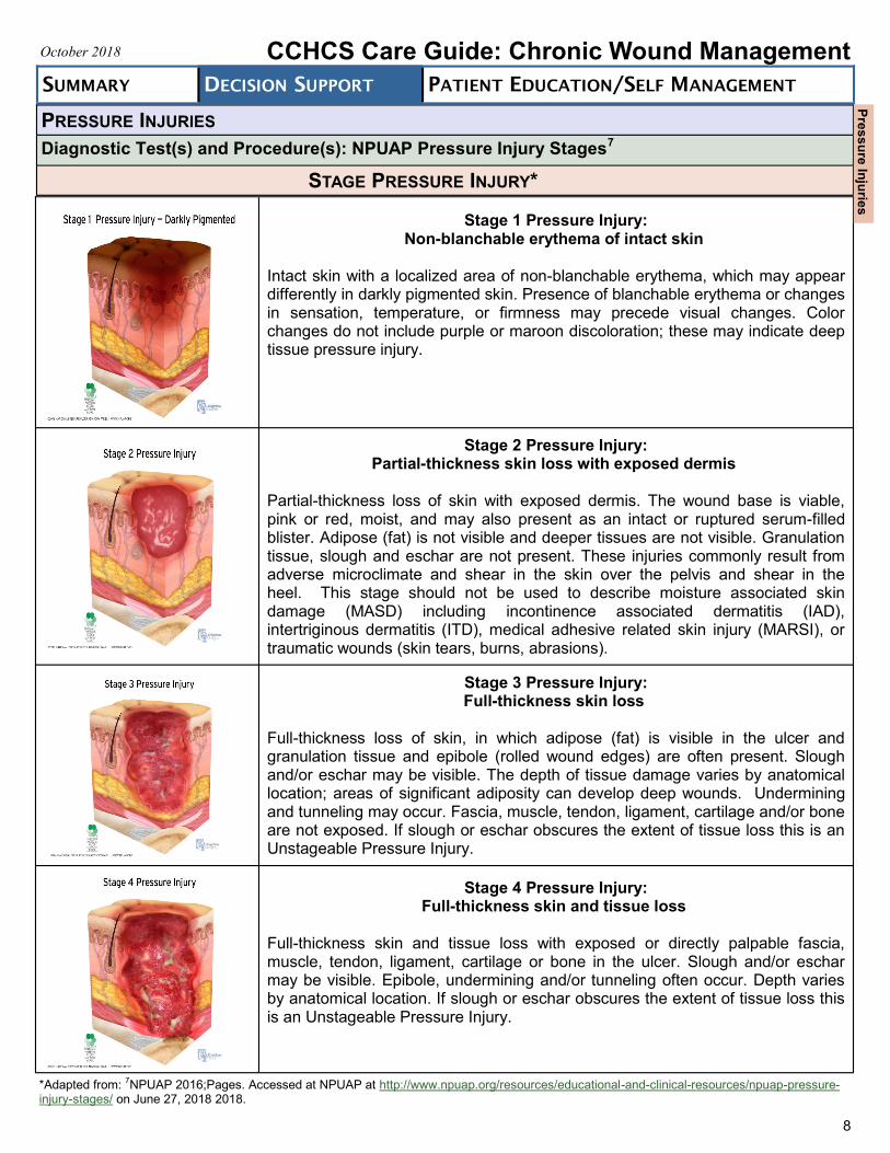
Stage 1 Pressure Injury:
Non-blanchable erythema of intact skin
Intact skin with a localized area of non-blanchable erythema, which may appear differently in darkly pigmented skin. Presence of blanchable erythema or changes in sensation, temperature, or firmness may precede visual changes. Color changes do not include purple or maroon discoloration; these may indicate deep tissue pressure injury.
Stage 2 Pressure Injury:
Partial-thickness skin loss with exposed dermis
Partial-thickness loss of skin with exposed dermis. The wound base is viable, pink or red, moist, and may also present as an intact or ruptured serum-filled blister. Adipose (fat) is not visible and deeper tissues are not visible. Granulation tissue, slough and eschar are not present. These injuries commonly result from adverse microclimate and shear in the skin over the pelvis and shear in the heel. This stage should not be used to describe moisture associated skin damage (MASD) including incontinence associated dermatitis (IAD), intertriginous dermatitis (ITD), medical adhesive related skin injury (MARSI), or traumatic wounds (skin tears, burns, abrasions).
Stage 3 Pressure Injury: Full-thickness skin loss
Full-thickness loss of skin, in which adipose (fat) is visible in the ulcer and granulation tissue and epibole (rolled wound edges) are often present. Slough and/or eschar may be visible. The depth of tissue damage varies by anatomical location; areas of significant adiposity can develop deep wounds. Undermining and tunneling may occur. Fascia, muscle, tendon, ligament, cartilage and/or bone are not exposed. If slough or eschar obscures the extent of tissue loss this is an Unstageable Pressure Injury.
Stage 4 Pressure Injury:
Full-thickness skin and tissue loss
Full-thickness skin and tissue loss with exposed or directly palpable fascia, muscle, tendon, ligament, cartilage or bone in the ulcer. Slough and/or eschar may be visible. Epibole, undermining and/or tunneling often occur. Depth varies by anatomical location. If slough or eschar obscures the extent of tissue loss this is an Unstageable Pressure Injury.
8
*Adapted from: 7NPUAP 2016;Pages. Accessed at NPUAP at http://www.npuap.org/resources/educational-and-clinical-resources/npuap-pressure-injury-stages/ on June 27, 2018 2018.
Pre
ss
ure
Inju
ries
PRESSURE INJURIES
Diagnostic Test(s) and Procedure(s): NPUAP Pressure Injury Stages7
(Continued)
STAGE PRESSURE INJURY (CONTINUED)
SUMMARY DECISION SUPPORT PATIENT EDUCATION/SELF MANAGEMENT
CCHCS Care Guide: Chronic Wound Management October 2018
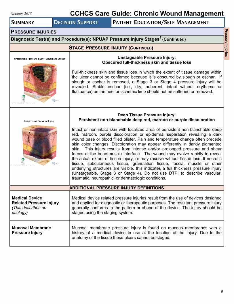
Unstageable Pressure Injury:
Obscured full-thickness skin and tissue loss Full-thickness skin and tissue loss in which the extent of tissue damage within the ulcer cannot be confirmed because it is obscured by slough or eschar. If slough or eschar is removed, a Stage 3 or Stage 4 pressure injury will be revealed. Stable eschar (i.e., dry, adherent, intact without erythema or fluctuance) on the heel or ischemic limb should not be softened or removed.
Deep Tissue Pressure Injury: Persistent non-blanchable deep red, maroon or purple discoloration
Intact or non-intact skin with localized area of persistent non-blanchable deep red, maroon, purple discoloration or epidermal separation revealing a dark wound base or blood filled blister. Pain and temperature change often precede skin color changes. Discoloration may appear differently in darkly pigmented skin. This injury results from intense and/or prolonged pressure and shear forces at the bone-muscle interface. The wound may evolve rapidly to reveal the actual extent of tissue injury, or may resolve without tissue loss. If necrotic tissue, subcutaneous tissue, granulation tissue, fascia, muscle or other underlying structures are visible, this indicates a full thickness pressure injury (Unstageable, Stage 3 or Stage 4). Do not use DTPI to describe vascular, traumatic, neuropathic, or dermatologic conditions.
ADDITIONAL PRESSURE INJURY DEFINITIONS
Medical Device Related Pressure Injury (This describes an etiology)
Medical device related pressure injuries result from the use of devices designed and applied for diagnostic or therapeutic purposes. The resultant pressure injury generally conforms to the pattern or shape of the device. The injury should be staged using the staging system.
Mucosal Membrane Pressure Injury
Mucosal membrane pressure injury is found on mucous membranes with a history of a medical device in use at the location of the injury. Due to the anatomy of the tissue these ulcers cannot be staged.
9
Pre
ss
ure
Inju
ries
PRESSURE INJURIES
Diagnostic Test(s) and Procedure(s): Braden Scale
COMPLETE BRADEN SCALE SCORING8
The Braden Scale: Identifies people at risk for pressure injury development
Guides implementation of prevention measures
Reliable documentation tool
The Braden Scale is composed of six subscales scored from 1-3 or 1-4, resulting in total scores that range from 6-23 (See page 11)
Subscales include:
Sensory Perception (Scored from 1-4)
Moisture (Scored from 1-4)
Activity (Scored from 1-4)
Mobility (Scored from 1-4)
Nutrition (Scored from 1-4)
Friction & Shear (Scored from 1-3)
Grouping Braden Subscale indicators helps us understand pressure injury risks (See Pathophysiology page 7)
If there is excess pressure (especially point pressure) applied to skin, then there is an increased risk of pressure injury development when the patient has limitations in these areas:
Sensory Perception—Ability to respond appropriately to pressure-related discomfort
Mobility—Ability to change and control body position
Activity—Degree of physical activity
If there is decreased tissue tolerance present, then there is increased risk of pressure injury development when the patient has worsening signs and symptoms related to these areas:
Moisture—Degree to which skin is exposed to moisture
Friction & Shear—Friction and shear of skin against surfaces
Nutrition—Assessment of food intake pattern
Interpreting the (total) Braden Score Each subscale is scored from 1-3 or 1-4. The patient’s nurse will usually perform the Braden and provide you with a total
score between 6-23.
The LOWER the SCORE, the HIGHER the RISK Very High Risk: ≤ 9
High Risk: 10-12
Moderate Risk: 13-14
Mild Risk/At Risk: 15-18
When to use the Braden Scale Within 24 hours of arrival from the hospital or transfer from another institution
Whenever there is a change in the level of care or change in care team
It should be repeated at regular intervals based on the previous Braden score as noted in the table below:
10
SUMMARY DECISION SUPPORT PATIENT EDUCATION/SELF MANAGEMENT
CCHCS Care Guide: Chronic Wound Management October 2018
Previous Braden Score
Braden Risk Category
Suggested Frequency For Repeating Braden
≤ 9 Very High Every Week
10 - 12 High Every 2 Weeks
13 - 14 Moderate Every 30 Days
15 - 18 Mild Every 90 Days
Pre
ss
ure
Inju
ries
SUMMARY DECISION SUPPORT PATIENT EDUCATION/SELF MANAGEMENT
CCHCS Care Guide: Chronic Wound Management October 2018
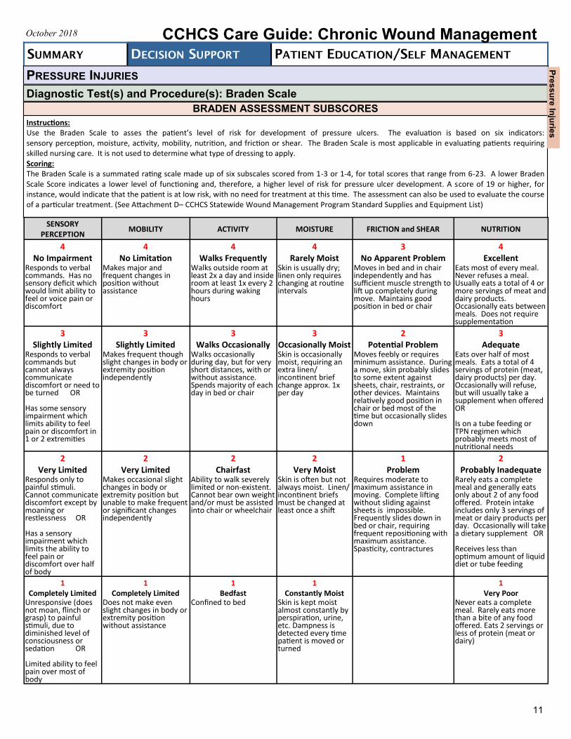
Diagnostic Test(s) and Procedure(s): Braden Scale
BRADEN ASSESSMENT SUBSCORES
Instructions: Use the Braden Scale to asses the patient’s level of risk for development of pressure ulcers. The evaluation is based on six indicators: sensory perception, moisture, activity, mobility, nutrition, and friction or shear. The Braden Scale is most applicable in evaluating patients requiring skilled nursing care. It is not used to determine what type of dressing to apply. Scoring: The Braden Scale is a summated rating scale made up of six subscales scored from 1-3 or 1-4, for total scores that range from 6-23. A lower Braden Scale Score indicates a lower level of functioning and, therefore, a higher level of risk for pressure ulcer development. A score of 19 or higher, for instance, would indicate that the patient is at low risk, with no need for treatment at this time. The assessment can also be used to evaluate the course of a particular treatment. (See Attachment D– CCHCS Statewide Wound Management Program Standard Supplies and Equipment List)
11
PRESSURE INJURIES
Pre
ss
ure
Inju
ries
SENSORY PERCEPTION
MOBILITY ACTIVITY MOISTURE FRICTION and SHEAR NUTRITION
4 No Impairment
4 No Limitation
4 Walks Frequently
4 Rarely Moist
3 No Apparent Problem
4 Excellent
Responds to verbal commands. Has no sensory deficit which would limit ability to feel or voice pain or discomfort
Makes major and frequent changes in position without assistance
Walks outside room at least 2x a day and inside room at least 1x every 2 hours during waking hours
Skin is usually dry; linen only requires changing at routine intervals
Moves in bed and in chair independently and has sufficient muscle strength to lift up completely during move. Maintains good position in bed or chair
Eats most of every meal. Never refuses a meal. Usually eats a total of 4 or more servings of meat and dairy products. Occasionally eats between meals. Does not require supplementation
3 Slightly Limited
3 Slightly Limited
3 Walks Occasionally
3 Occasionally Moist
2 Potential Problem
3 Adequate
Responds to verbal commands but cannot always communicate discomfort or need to be turned OR Has some sensory impairment which limits ability to feel pain or discomfort in 1 or 2 extremities
Makes frequent though slight changes in body or extremity position independently
Walks occasionally during day, but for very short distances, with or without assistance. Spends majority of each day in bed or chair
Skin is occasionally moist, requiring an extra linen/incontinent brief change approx. 1x per day
Moves feebly or requires minimum assistance. During a move, skin probably slides to some extent against sheets, chair, restraints, or other devices. Maintains relatively good position in chair or bed most of the time but occasionally slides down
Eats over half of most meals. Eats a total of 4 servings of protein (meat, dairy products) per day. Occasionally will refuse, but will usually take a supplement when offered OR Is on a tube feeding or TPN regimen which probably meets most of nutritional needs
2 Very Limited
2 Very Limited
2 Chairfast
2 Very Moist
1 Problem
2 Probably Inadequate
Responds only to painful stimuli. Cannot communicate discomfort except by moaning or restlessness OR Has a sensory impairment which limits the ability to feel pain or discomfort over half of body
Makes occasional slight changes in body or extremity position but unable to make frequent or significant changes independently
Ability to walk severely limited or non-existent. Cannot bear own weight and/or must be assisted into chair or wheelchair
Skin is often but not always moist. Linen/incontinent briefs must be changed at least once a shift
Requires moderate to maximum assistance in moving. Complete lifting without sliding against sheets is impossible. Frequently slides down in bed or chair, requiring frequent repositioning with maximum assistance. Spasticity, contractures
Rarely eats a complete meal and generally eats only about 2 of any food offered. Protein intake includes only 3 servings of meat or dairy products per day. Occasionally will take a dietary supplement OR Receives less than optimum amount of liquid diet or tube feeding
1 Completely Limited
1 Completely Limited
1 Bedfast
1 Constantly Moist
1 Very Poor
Unresponsive (does not moan, flinch or grasp) to painful stimuli, due to diminished level of consciousness or sedation OR Limited ability to feel pain over most of body
Does not make even slight changes in body or extremity position without assistance
Confined to bed
Skin is kept moist almost constantly by perspiration, urine, etc. Dampness is detected every time patient is moved or turned
Never eats a complete meal. Rarely eats more than a bite of any food offered. Eats 2 servings or less of protein (meat or dairy)
PRESSURE INJURIES
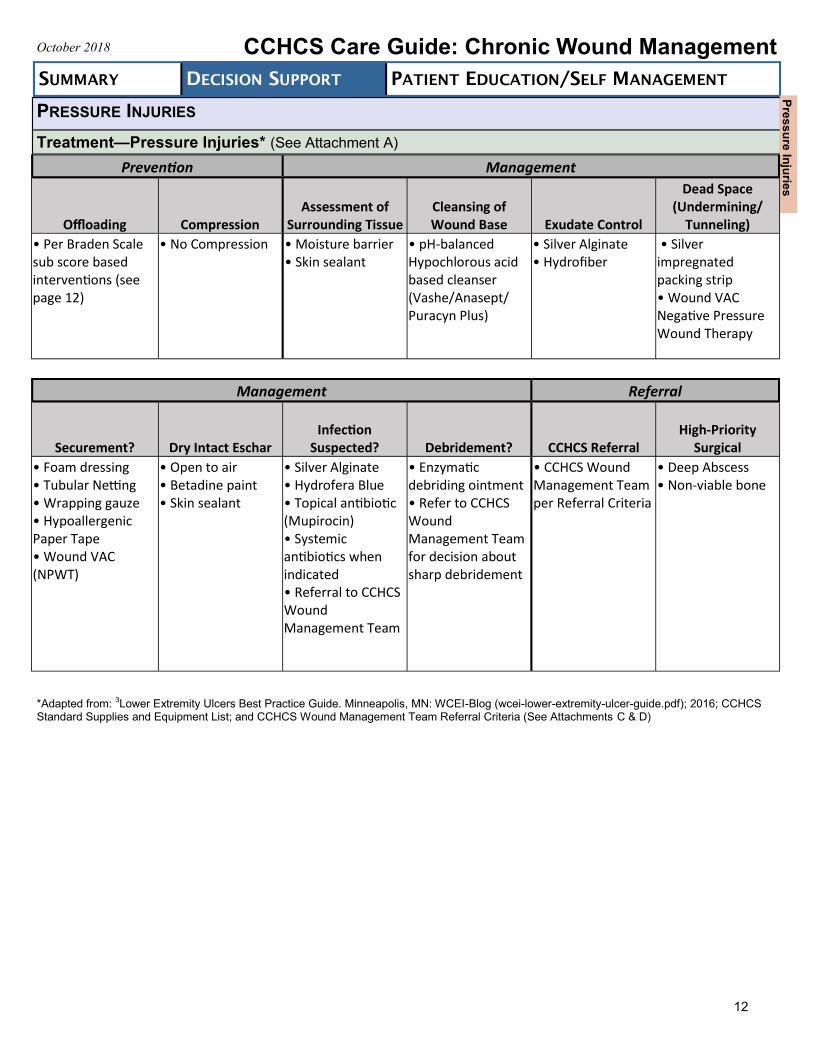
Treatment—Pressure Injuries* (See Attachment A)
SUMMARY DECISION SUPPORT PATIENT EDUCATION/SELF MANAGEMENT
CCHCS Care Guide: Chronic Wound Management October 2018
12
*Adapted from: 3Lower Extremity Ulcers Best Practice Guide. Minneapolis, MN: WCEI-Blog (wcei-lower-extremity-ulcer-guide.pdf); 2016; CCHCS Standard Supplies and Equipment List; and CCHCS Wound Management Team Referral Criteria (See Attachments C & D)
Pre
ss
ure
Inju
ries
Prevention Management
Offloading Compression Assessment of
Surrounding Tissue Cleansing of Wound Base Exudate Control
Dead Space (Undermining/
Tunneling)
• Per Braden Scale sub score based interventions (see page 12)
• No Compression • Moisture barrier • Skin sealant
• pH-balanced Hypochlorous acid based cleanser (Vashe/Anasept/ Puracyn Plus)
• Silver Alginate • Hydrofiber
• Silver impregnated packing strip • Wound VAC Negative Pressure Wound Therapy
Management Referral
Securement? Dry Intact Eschar Infection
Suspected? Debridement? CCHCS Referral High-Priority
Surgical
• Foam dressing • Tubular Netting • Wrapping gauze • Hypoallergenic Paper Tape • Wound VAC (NPWT)
• Open to air • Betadine paint • Skin sealant
• Silver Alginate • Hydrofera Blue • Topical antibiotic (Mupirocin) • Systemic antibiotics when indicated • Referral to CCHCS Wound Management Team
• Enzymatic debriding ointment • Refer to CCHCS Wound Management Team for decision about sharp debridement
• CCHCS Wound Management Team per Referral Criteria
• Deep Abscess • Non-viable bone
SUMMARY DECISION SUPPORT PATIENT EDUCATION/SELF MANAGEMENT
CCHCS Care Guide: Chronic Wound Management October 2018
Prevention
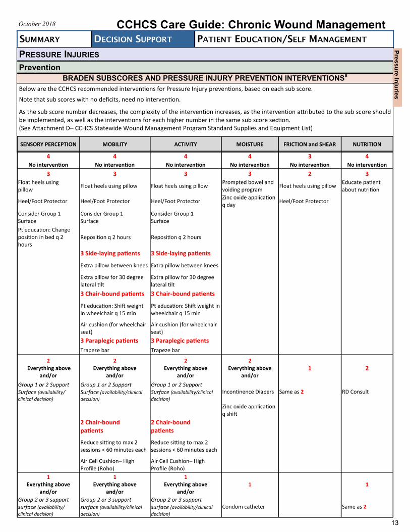
BRADEN SUBSCORES AND PRESSURE INJURY PREVENTION INTERVENTIONS8
Below are the CCHCS recommended interventions for Pressure Injury preventions, based on each sub score.
Note that sub scores with no deficits, need no intervention.
As the sub score number decreases, the complexity of the intervention increases, as the intervention attributed to the sub score should be implemented, as well as the interventions for each higher number in the same sub score section. (See Attachment D– CCHCS Statewide Wound Management Program Standard Supplies and Equipment List)
13
PRESSURE INJURIES
Pre
ss
ure
Inju
ries
SENSORY PERCEPTION MOBILITY ACTIVITY MOISTURE FRICTION and SHEAR NUTRITION
4 4 4 4 3 4 No intervention No intervention No intervention No intervention No intervention No intervention
3 3 3 3 2 3 Float heels using pillow
Float heels using pillow Float heels using pillow Prompted bowel and voiding program
Float heels using pillow Educate patient about nutrition
Heel/Foot Protector Heel/Foot Protector Heel/Foot Protector Zinc oxide application q day
Heel/Foot Protector
Consider Group 1 Surface
Consider Group 1 Surface
Consider Group 1 Surface
Pt education: Change position in bed q 2 hours
Reposition q 2 hours Reposition q 2 hours
3 Side-laying patients 3 Side-laying patients
Extra pillow between knees Extra pillow between knees
Extra pillow for 30 degree lateral tilt
Extra pillow for 30 degree lateral tilt
3 Chair-bound patients 3 Chair-bound patients
Pt education: Shift weight in wheelchair q 15 min
Pt education: Shift weight in wheelchair q 15 min
Air cushion (for wheelchair seat)
Air cushion (for wheelchair seat)
3 Paraplegic patients 3 Paraplegic patients
Trapeze bar Trapeze bar
2 Everything above
and/or
2 Everything above
and/or
2 Everything above
and/or
2 Everything above
and/or 1 2
Group 1 or 2 Support Surface (availability/
clinical decision)
Group 1 or 2 Support Surface (availability/clinical
decision)
Group 1 or 2 Support Surface (availability/clinical
decision)
Incontinence Diapers Same as 2 RD Consult
Zinc oxide application q shift
2 Chair-bound patients
2 Chair-bound patients
Reduce sitting to max 2 sessions < 60 minutes each
Reduce sitting to max 2 sessions < 60 minutes each
Air Cell Cushion– High Profile (Roho)
Air Cell Cushion– High Profile (Roho)
1 Everything above
and/or
1 Everything above
and/or
1 Everything above
and/or 1
1
Group 2 or 3 support surface (availability/
clinical decision)
Group 2 or 3 support surface (availability/clinical
decision)
Group 2 or 3 support surface (availability/clinical
decision)
Condom catheter Same as 2
PRESSURE INJURIES
Prevention (Cont.)
14
SUMMARY DECISION SUPPORT PATIENT EDUCATION/SELF MANAGEMENT
CCHCS Care Guide: Chronic Wound Management October 2018
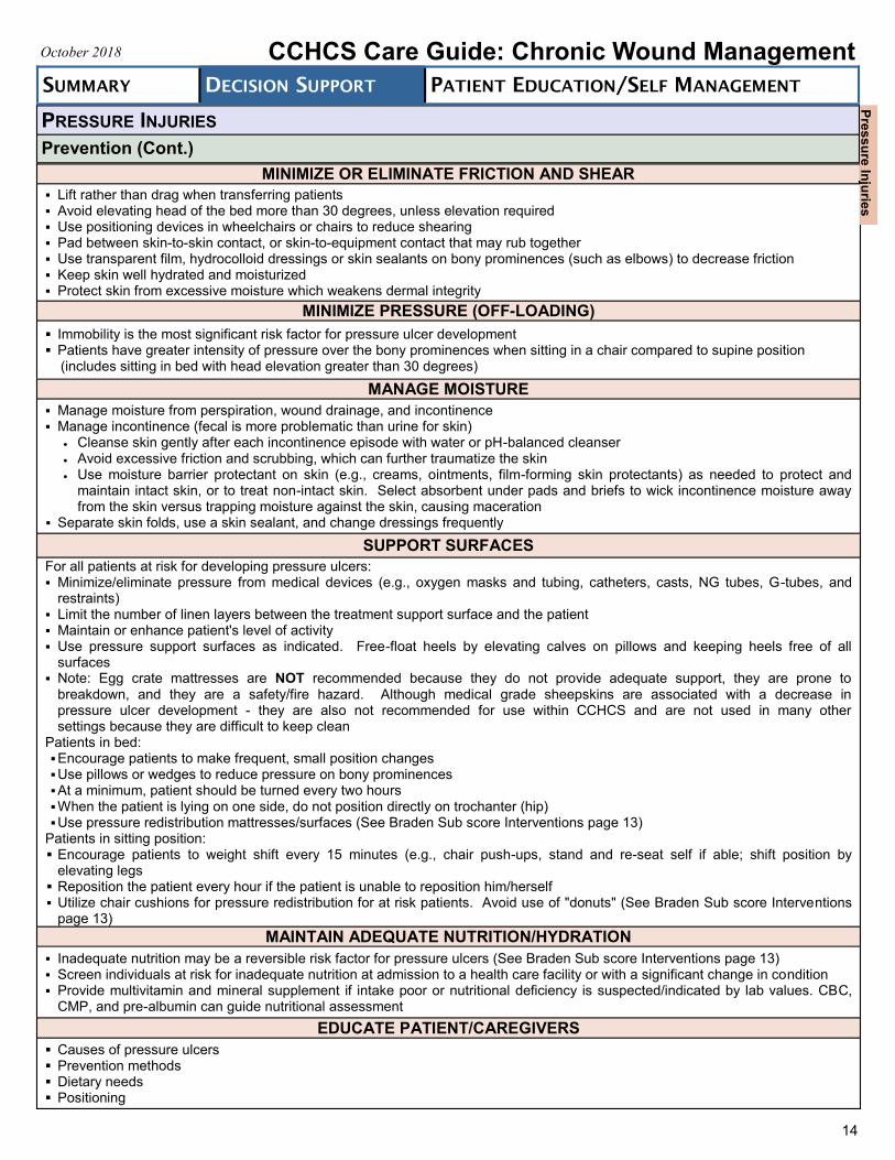
MINIMIZE OR ELIMINATE FRICTION AND SHEAR
Lift rather than drag when transferring patients Avoid elevating head of the bed more than 30 degrees, unless elevation required Use positioning devices in wheelchairs or chairs to reduce shearing Pad between skin-to-skin contact, or skin-to-equipment contact that may rub together Use transparent film, hydrocolloid dressings or skin sealants on bony prominences (such as elbows) to decrease friction Keep skin well hydrated and moisturized Protect skin from excessive moisture which weakens dermal integrity
MINIMIZE PRESSURE (OFF-LOADING)
Immobility is the most significant risk factor for pressure ulcer development Patients have greater intensity of pressure over the bony prominences when sitting in a chair compared to supine position (includes sitting in bed with head elevation greater than 30 degrees)
MANAGE MOISTURE
Manage moisture from perspiration, wound drainage, and incontinence Manage incontinence (fecal is more problematic than urine for skin)
Cleanse skin gently after each incontinence episode with water or pH-balanced cleanser Avoid excessive friction and scrubbing, which can further traumatize the skin Use moisture barrier protectant on skin (e.g., creams, ointments, film-forming skin protectants) as needed to protect and
maintain intact skin, or to treat non-intact skin. Select absorbent under pads and briefs to wick incontinence moisture away from the skin versus trapping moisture against the skin, causing maceration
Separate skin folds, use a skin sealant, and change dressings frequently
SUPPORT SURFACES
For all patients at risk for developing pressure ulcers: Minimize/eliminate pressure from medical devices (e.g., oxygen masks and tubing, catheters, casts, NG tubes, G-tubes, and
restraints) Limit the number of linen layers between the treatment support surface and the patient Maintain or enhance patient's level of activity Use pressure support surfaces as indicated. Free-float heels by elevating calves on pillows and keeping heels free of all
surfaces Note: Egg crate mattresses are NOT recommended because they do not provide adequate support, they are prone to
breakdown, and they are a safety/fire hazard. Although medical grade sheepskins are associated with a decrease in pressure ulcer development - they are also not recommended for use within CCHCS and are not used in many other settings because they are difficult to keep clean
Patients in bed: Encourage patients to make frequent, small position changes Use pillows or wedges to reduce pressure on bony prominences At a minimum, patient should be turned every two hours When the patient is lying on one side, do not position directly on trochanter (hip) Use pressure redistribution mattresses/surfaces (See Braden Sub score Interventions page 13)
Patients in sitting position: Encourage patients to weight shift every 15 minutes (e.g., chair push-ups, stand and re-seat self if able; shift position by
elevating legs Reposition the patient every hour if the patient is unable to reposition him/herself Utilize chair cushions for pressure redistribution for at risk patients. Avoid use of "donuts" (See Braden Sub score Interventions
page 13)
MAINTAIN ADEQUATE NUTRITION/HYDRATION
Inadequate nutrition may be a reversible risk factor for pressure ulcers (See Braden Sub score Interventions page 13) Screen individuals at risk for inadequate nutrition at admission to a health care facility or with a significant change in condition Provide multivitamin and mineral supplement if intake poor or nutritional deficiency is suspected/indicated by lab values. CBC,
CMP, and pre-albumin can guide nutritional assessment
EDUCATE PATIENT/CAREGIVERS
Causes of pressure ulcers Prevention methods Dietary needs Positioning
Pre
ss
ure
Inju
ries
15
SUMMARY DECISION SUPPORT PATIENT EDUCATION/SELF MANAGEMENT
CCHCS Care Guide: Chronic Wound Management October 2018
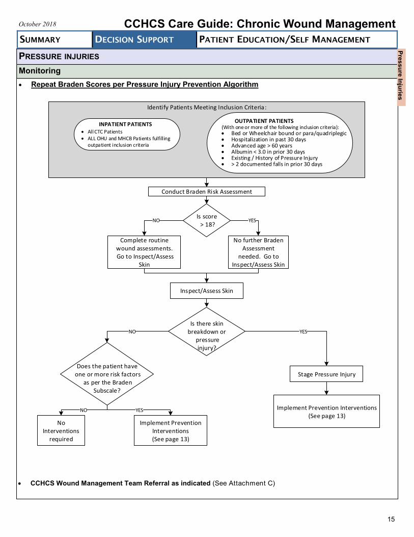
Repeat Braden Scores per Pressure Injury Prevention Algorithm
PRESSURE INJURIES
Monitoring
CCHCS Wound Management Team Referral as indicated (See Attachment C)
Identify Patients Meeting Inclusion Criteria:
OUTPATIENT PATIENTS(With one or more of the following inclusion criteria): Bed or Wheelchair bound or para/quadriplegic Hospitalization in past 30 days Advanced age > 60 years Albumin < 3.0 in prior 30 days Existing / History of Pressure Injury > 2 documented falls in prior 30 days
INPATIENT PATIENTS All CTC Patients ALL OHU and MHCB Patients fulfilling
outpatient inclusion criteria
Conduct Braden Risk Assessment
Is score > 18?
Complete routine wound assessments. Go to Inspect/Assess
Skin
No further Braden Assessment
needed. Go to Inspect/Assess Skin
Inspect/Assess Skin
Is there skin breakdown or
pressure injury?
Does the patient have one or more risk factors
as per the Braden Subscale?
Stage Pressure Injury
Implement Prevention Interventions (See page 13)
Implement Prevention Interventions (See page 13)
No Interventions
required
NO YES
YESNO
NO YES
Pre
ss
ure
Inju
ries
VENOUS ULCERS3,4,9,10
Pathophysiology
Normal Venous Function requires: Competent valves Normal functioning vessel walls and calf muscle pump
Valvular Incompetence Veins no longer fill normally via slow capillary inflow alone Retrograde transmission of pressure and volume flow into superficial venous system→ increased
venous hypertension→ increased valvular incompetence Valvular Dysfunction
Physical damage: splitting, tearing, thinning, adhesion to wall
Venous Insufficiency
Valvular incompetence and venous obstruction
Venous Hypertension Increased pressure at ankle → Swelling of the tissues → Widening endothelial gap junctions → Sequestration of
the RBCs, WBCs, Proteins → WBC entrapment in the capillary wall, setting up an inflammatory reaction, injuring the vein and valves
Pathophysiology of Venous Ulceration Fibrin cuff theory
Increased venous pressure
Loss of plasma proteins
Fibrinogen forms a cuff around the capillaries
Fibrin cuff interferes with the exchange of oxygen
Tissue breaks down Leukocyte migration theory
White cells migrate into the interstitial tissue
Breakdown of the WBCs→ release of cytokines and proteases
Loss of tissue integrity
Risk Factors*
Age Obesity Family history Pregnancy Female gender
Location and Appearance**
Locations:
On medial lower leg and ankle, superior to medial malleolus, seldom noted above the knee
Typically not seen on the foot or toes, which are common sites for arterial insufficiency ulcers and diabetic ulcers Shape:
Irregular wound margins
Typically saucer-shaped, initially with a shallow wound base Wound Base:
Shallow, partial-thickness, ruddy red, granular, necrotic tissue common Surrounding Tissue:
Scaling, pruritic, weepy, brown staining, dermatitis, firm edema
Pitting edema, induration, hemosiderosis, varicosities, lipodermatosclerosis, atrophie blanche, and/or stasis dermatitis
SUMMARY DECISION SUPPORT PATIENT EDUCATION/SELF MANAGEMENT
CCHCS Care Guide: Chronic Wound Management October 2018
Heart failure Hypertension Renal disease Greater height Physical inactivity
16
*Adapted from: 4Alquire PC, Mathes BM. Medical management of lower extremity chronic venous disease. November 7, 2017 ed. Waltham, MA: Up-to-date; 2017. 9Alguire PC, Scovell S. Overview and management of lower extremity chronic venous disease. May 29, 2018 ed. Waltham, MA: Up-to-date; 2018. 10Bryant RA, Nix DP. Acute & Chronic Wounds Current Management Concepts. 5th ed. St. Louis, MO: Elsevier; 2016.
**Adapted from: 3Lower Extremity Ulcers Best Practice Guide. Minneapolis, MN: WCEI-Blog (wcei-lower-extremity-ulcer-guide.pdf); 2016.
Personal ulcer history, parental history of ankle ulcers Previous varicose vein surgery Long hours standing, or sitting History of leg injury (fracture, burn, crush, penetrating injury,
phlebitis, DVT)
Ve
no
us
Ulc
ers
VENOUS ULCERS (CONTINUED)
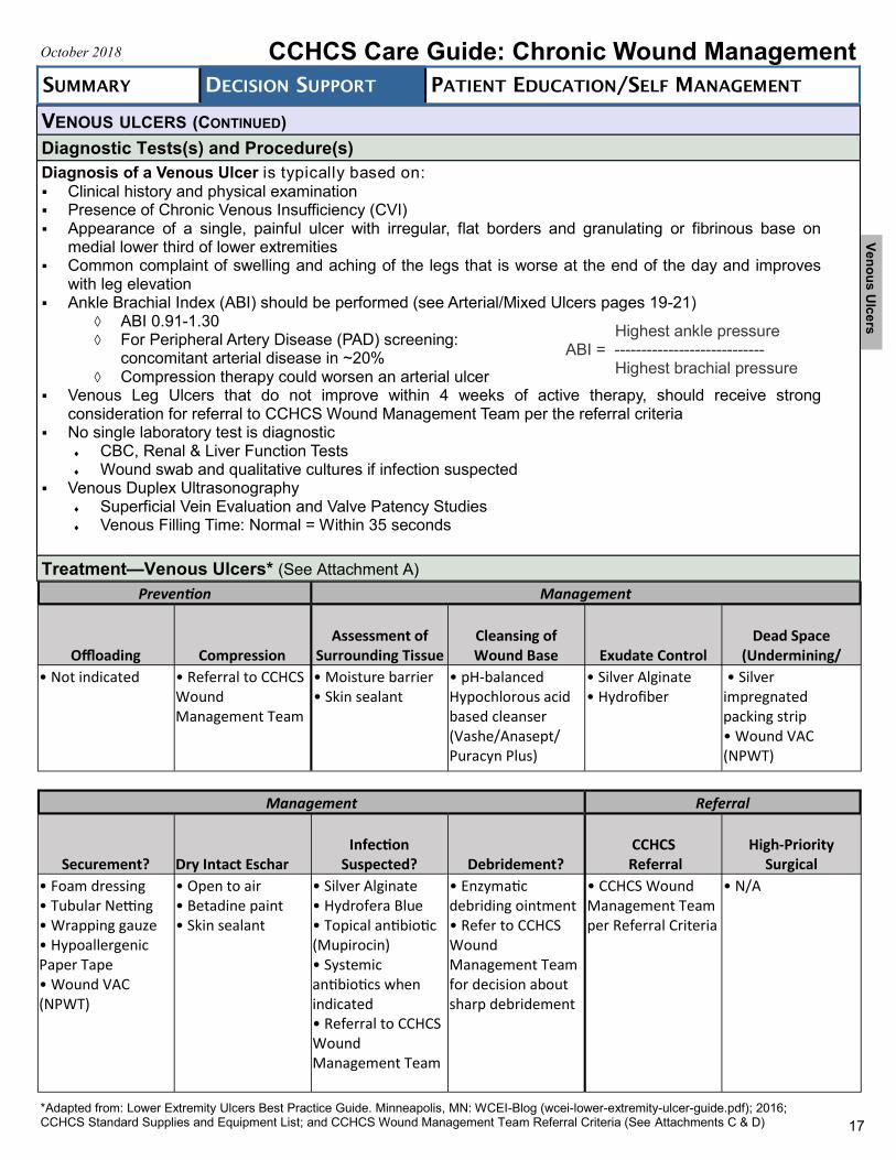
Diagnostic Tests(s) and Procedure(s)
Diagnosis of a Venous Ulcer is typically based on: Clinical history and physical examination Presence of Chronic Venous Insufficiency (CVI) Appearance of a single, painful ulcer with irregular, flat borders and granulating or fibrinous base on
medial lower third of lower extremities Common complaint of swelling and aching of the legs that is worse at the end of the day and improves
with leg elevation Ankle Brachial Index (ABI) should be performed (see Arterial/Mixed Ulcers pages 19-21)
ABI 0.91-1.30 For Peripheral Artery Disease (PAD) screening:
concomitant arterial disease in ~20% Compression therapy could worsen an arterial ulcer
Venous Leg Ulcers that do not improve within 4 weeks of active therapy, should receive strong consideration for referral to CCHCS Wound Management Team per the referral criteria
No single laboratory test is diagnostic CBC, Renal & Liver Function Tests Wound swab and qualitative cultures if infection suspected
Venous Duplex Ultrasonography Superficial Vein Evaluation and Valve Patency Studies Venous Filling Time: Normal = Within 35 seconds
Treatment—Venous Ulcers* (See Attachment A)
SUMMARY DECISION SUPPORT PATIENT EDUCATION/SELF MANAGEMENT
CCHCS Care Guide: Chronic Wound Management October 2018
Highest ankle pressure ABI = ---------------------------- Highest brachial pressure
17
*Adapted from: Lower Extremity Ulcers Best Practice Guide. Minneapolis, MN: WCEI-Blog (wcei-lower-extremity-ulcer-guide.pdf); 2016; CCHCS Standard Supplies and Equipment List; and CCHCS Wound Management Team Referral Criteria (See Attachments C & D)
Prevention Management
Offloading Compression Assessment of
Surrounding Tissue Cleansing of Wound Base Exudate Control
Dead Space (Undermining/
• Not indicated • Referral to CCHCS Wound Management Team
• Moisture barrier • Skin sealant
• pH-balanced Hypochlorous acid based cleanser (Vashe/Anasept/ Puracyn Plus)
• Silver Alginate • Hydrofiber
• Silver impregnated packing strip • Wound VAC(NPWT)
Management Referral
Securement? Dry Intact Eschar Infection
Suspected? Debridement? CCHCS
Referral High-Priority
Surgical
• Foam dressing • Tubular Netting • Wrapping gauze • Hypoallergenic Paper Tape • Wound VAC (NPWT)
• Open to air • Betadine paint • Skin sealant
• Silver Alginate • Hydrofera Blue • Topical antibiotic (Mupirocin) • Systemic antibiotics when indicated • Referral to CCHCS Wound Management Team
• Enzymatic debriding ointment • Refer to CCHCS Wound Management Team for decision about sharp debridement
• CCHCS Wound Management Team per Referral Criteria
• N/A
Ve
no
us
Ulc
ers
VENOUS ULCERS
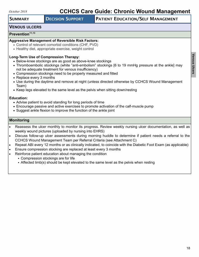
Prevention11,13
Aggressive Management of Reversible Risk Factors: Control of relevant comorbid conditions (CHF, PVD) Healthy diet, appropriate exercise, weight control
Long-Term Use of Compression Therapy:
Below-knee stockings are as good as above-knee stockings Thromboembolic stockings (white ”anti-embolism” stockings [6 to 19 mmHg pressure at the ankle] may
not be adequate treatment for venous insufficiency) Compression stockings need to be properly measured and fitted Replace every 3 months Use during the daytime and remove at night (unless directed otherwise by CCHCS Wound Management
Team) Keep legs elevated to the same level as the pelvis when sitting down/resting
Education:
Advise patient to avoid standing for long periods of time Encourage passive and active exercises to promote activation of the calf-muscle pump Suggest ankle flexion to improve the function of the ankle joint
Monitoring
Reassess the ulcer monthly to monitor its progress. Review weekly nursing ulcer documentation, as well as
weekly wound pictures (uploaded by nursing into EHRS)
Discuss follow-up ulcer assessments during morning huddle to determine if patient needs a referral to the
CCHCS Wound Management Team per Referral Criteria (see Attachment C)
Repeat ABI every 12 months or as clinically indicated, to coincide with the Diabetic Foot Exam (as applicable)
Ensure compression stocking are replaced at least every 3 months
Reinforce patient education about managing the condition
Compression stockings are for life Affected limb(s) should be kept elevated to the same level as the pelvis when resting
SUMMARY DECISION SUPPORT PATIENT EDUCATION/SELF MANAGEMENT
CCHCS Care Guide: Chronic Wound Management October 2018
18
Ve
no
us
Ulc
ers
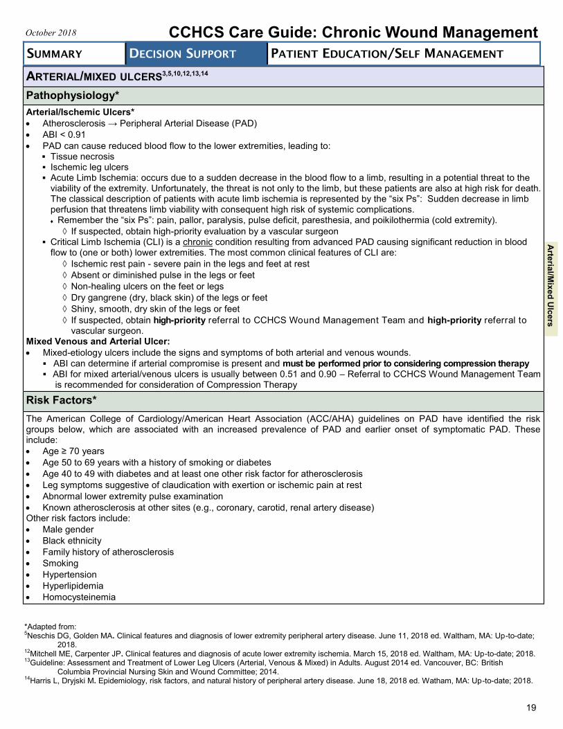
ARTERIAL/MIXED ULCERS3,5,10,12,13,14
Pathophysiology*
Arterial/Ischemic Ulcers*
Atherosclerosis → Peripheral Arterial Disease (PAD)
ABI < 0.91
PAD can cause reduced blood flow to the lower extremities, leading to: Tissue necrosis Ischemic leg ulcers Acute Limb Ischemia: occurs due to a sudden decrease in the blood flow to a limb, resulting in a potential threat to the
viability of the extremity. Unfortunately, the threat is not only to the limb, but these patients are also at high risk for death. The classical description of patients with acute limb ischemia is represented by the “six Ps”: Sudden decrease in limb perfusion that threatens limb viability with consequent high risk of systemic complications. Remember the “six Ps”: pain, pallor, paralysis, pulse deficit, paresthesia, and poikilothermia (cold extremity).
If suspected, obtain high-priority evaluation by a vascular surgeon Critical Limb Ischemia (CLI) is a chronic condition resulting from advanced PAD causing significant reduction in blood
flow to (one or both) lower extremities. The most common clinical features of CLI are:
Ischemic rest pain - severe pain in the legs and feet at rest
Absent or diminished pulse in the legs or feet
Non-healing ulcers on the feet or legs
Dry gangrene (dry, black skin) of the legs or feet
Shiny, smooth, dry skin of the legs or feet
If suspected, obtain high-priority referral to CCHCS Wound Management Team and high-priority referral to vascular surgeon.
Mixed Venous and Arterial Ulcer:
Mixed-etiology ulcers include the signs and symptoms of both arterial and venous wounds.
ABI can determine if arterial compromise is present and must be performed prior to considering compression therapy ABI for mixed arterial/venous ulcers is usually between 0.51 and 0.90 – Referral to CCHCS Wound Management Team
is recommended for consideration of Compression Therapy
Risk Factors*
The American College of Cardiology/American Heart Association (ACC/AHA) guidelines on PAD have identified the risk groups below, which are associated with an increased prevalence of PAD and earlier onset of symptomatic PAD. These include:
Age ≥ 70 years
Age 50 to 69 years with a history of smoking or diabetes
Age 40 to 49 with diabetes and at least one other risk factor for atherosclerosis
Leg symptoms suggestive of claudication with exertion or ischemic pain at rest
Abnormal lower extremity pulse examination
Known atherosclerosis at other sites (e.g., coronary, carotid, renal artery disease) Other risk factors include:
Male gender
Black ethnicity
Family history of atherosclerosis
Smoking
Hypertension
Hyperlipidemia
Homocysteinemia
19
SUMMARY DECISION SUPPORT PATIENT EDUCATION/SELF MANAGEMENT
CCHCS Care Guide: Chronic Wound Management October 2018
*Adapted from: 5Neschis DG, Golden MA. Clinical features and diagnosis of lower extremity peripheral artery disease. June 11, 2018 ed. Waltham, MA: Up-to-date;
2018. 12Mitchell ME, Carpenter JP. Clinical features and diagnosis of acute lower extremity ischemia. March 15, 2018 ed. Waltham, MA: Up-to-date; 2018. 13Guideline: Assessment and Treatment of Lower Leg Ulcers (Arterial, Venous & Mixed) in Adults. August 2014 ed. Vancouver, BC: British
Columbia Provincial Nursing Skin and Wound Committee; 2014. 14Harris L, Dryjski M. Epidemiology, risk factors, and natural history of peripheral artery disease. June 18, 2018 ed. Watham, MA: Up-to-date; 2018.
Arte
rial/M
ixe
d U
lce
rs
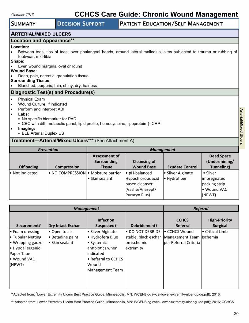
ARTERIAL/MIXED ULCERS Location and Appearance**
Location:
Between toes, tips of toes, over phalangeal heads, around lateral malleolus, sites subjected to trauma or rubbing of footwear, mid-tibia
Shape:
Even wound margins, oval or round Wound Base:
Deep, pale, necrotic, granulation tissue Surrounding Tissue:
Blanched, purpuric, thin, shiny, dry, hairless
Diagnostic Test(s) and Procedure(s)
Physical Exam
Wound Culture, if indicated
Perform and interpret ABI
Labs: No specific biomarker for PAD CBC with diff, metabolic panel, lipid profile, homocysteine, lipoprotein ↑, CRP
Imaging: BLE Arterial Duplex US
Treatment—Arterial/Mixed Ulcers*** (See Attachment A)
20
SUMMARY DECISION SUPPORT PATIENT EDUCATION/SELF MANAGEMENT
CCHCS Care Guide: Chronic Wound Management October 2018
**Adapted from: 3Lower Extremity Ulcers Best Practice Guide. Minneapolis, MN: WCEI-Blog (wcei-lower-extremity-ulcer-guide.pdf); 2016.
***Adapted from: Lower Extremity Ulcers Best Practice Guide. Minneapolis, MN: WCEI-Blog (wcei-lower-extremity-ulcer-guide.pdf); 2016; CCHCS
Prevention Management
Offloading Compression
Assessment of Surrounding
Tissue Cleansing of Wound Base Exudate Control
Dead Space (Undermining/
Tunneling)
• Not indicated • NO COMPRESSION • Moisture barrier • Skin sealant
• pH-balanced Hypochlorous acid based cleanser (Vashe/Anasept/ Puracyn Plus)
• Silver Alginate • Hydrofiber
• Silver impregnated packing strip • Wound VAC(NPWT)
Management Referral
Securement? Dry Intact Eschar Infection
Suspected? Debridement? CCHCS
Referral High-Priority
Surgical
• Foam dressing • Tubular Netting • Wrapping gauze • Hypoallergenic Paper Tape • Wound VAC (NPWT)
• Open to air • Betadine paint • Skin sealant
• Silver Alginate • Hydrofera Blue • Systemic antibiotics when indicated • Referral to CCHCS Wound Management Team
• DO NOT DEBRIDE stable, black eschar on ischemic extremity
• CCHCS Wound Management Team per Referral Criteria
• Critical Limb Ischemia
Arte
rial/M
ixe
d U
lce
rs
ARTERIAL/MIXED ULCERS
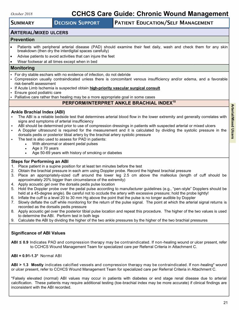
Prevention
Patients with peripheral arterial disease (PAD) should examine their feet daily, wash and check them for any skin breakdown (then dry the interdigital spaces carefully)
Advise patients to avoid activities that can injure the feet
Wear footwear at all times except when in bed
Monitoring
For dry stable eschars with no evidence of infection, do not debride Compression usually contraindicated unless there is concomitant venous insufficiency and/or edema, and a favorable
risk-benefit assessment If Acute Limb Ischemia is suspected obtain high-priority vascular surgical consult Ensure good podiatric care Palliative care rather than healing may be a more appropriate goal in some cases
21
SUMMARY DECISION SUPPORT PATIENT EDUCATION/SELF MANAGEMENT
CCHCS Care Guide: Chronic Wound Management October 2018
PERFORM/INTERPRET ANKLE BRACHIAL INDEX10
Ankle Brachial Index (ABI) The ABI is a reliable bedside test that determines arterial blood flow in the lower extremity and generally correlates with
signs and symptoms of arterial insufficiency ABI should be determined prior to use of compression dressings in patients with suspected arterial or mixed ulcers A Doppler ultrasound is required for the measurement and it is calculated by dividing the systolic pressure in the
dorsalis pedis or posterior tibial artery by the brachial artery systolic pressure
The test is also used to assess for PAD in patients: With abnormal or absent pedal pulses Age ≥ 70 years Age 50-69 years with history of smoking or diabetes
Steps for Performing an ABI 1. Place patient in a supine position for at least ten minutes before the test 2. Obtain the brachial pressure in each arm using Doppler probe. Record the highest brachial pressure 3. Place an appropriately-sized cuff around the lower leg 2.5 cm above the malleolus (length of cuff should be
approximately 20% bigger than circumstance of the extremity) 4. Apply acoustic gel over the dorsalis pedis pulse location 5. Hold the Doppler probe over the pedal pulse according to manufacturer guidelines (e.g., “pen-style” Dopplers should be
held at a 45-degree angle). Be careful not to occlude the artery with excessive pressure; hold the probe lightly! 6. Inflate the cuff to a level 20 to 30 mm Hg above the point that the pulse is no longer audible by Doppler 7. Slowly deflate the cuff while monitoring for the return of the pulse signal. The point at which the arterial signal returns is
recorded as the dorsalis pedis pressure 8. Apply acoustic gel over the posterior tibial pulse location and repeat this procedure. The higher of the two values is used
to determine the ABI. Perform test in both legs 9. Calculate the ABI by dividing the higher of the two ankle pressures by the higher of the two brachial pressures
Significance of ABI Values ABI ≤ 0.9 Indicates PAD and compression therapy may be contraindicated. If non -healing wound or ulcer present, refer
to CCHCS Wound Management Team for specialized care per Referral Criteria in Attachment C. ABI = 0.91-1.3* Normal ABI ABI > 1.3 Mostly indicates calcified vessels and compression therapy may be contraindicated. If non -healing* wound or ulcer present, refer to CCHCS Wound Management Team for specialized care per Referral Criteria in Attachment C. *Falsely elevated (normal) ABI values may occur in patients with diabetes or end stage renal disease due to arterial calcification. These patients may require additional testing (toe-brachial index may be more accurate) if clinical findings are inconsistent with the ABI recorded.
Arte
rial/M
ixe
d U
lce
rs
DIABETIC FOOT ULCERS3,6,10
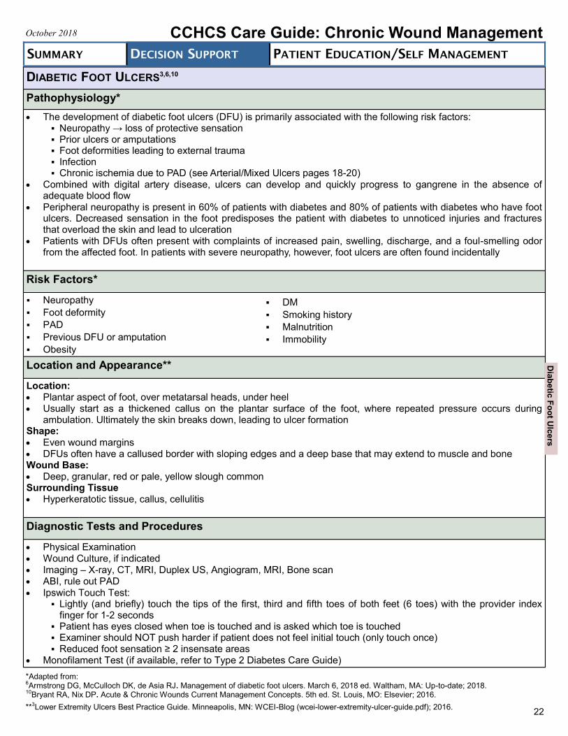
Pathophysiology*
The development of diabetic foot ulcers (DFU) is primarily associated with the following risk factors: Neuropathy → loss of protective sensation Prior ulcers or amputations Foot deformities leading to external trauma Infection Chronic ischemia due to PAD (see Arterial/Mixed Ulcers pages 18-20)
Combined with digital artery disease, ulcers can develop and quickly progress to gangrene in the absence of adequate blood flow
Peripheral neuropathy is present in 60% of patients with diabetes and 80% of patients with diabetes who have foot ulcers. Decreased sensation in the foot predisposes the patient with diabetes to unnoticed injuries and fractures that overload the skin and lead to ulceration
Patients with DFUs often present with complaints of increased pain, swelling, discharge, and a foul-smelling odor from the affected foot. In patients with severe neuropathy, however, foot ulcers are often found incidentally
Risk Factors*
Neuropathy
Foot deformity
PAD
Previous DFU or amputation
Obesity
Location and Appearance**
Location: Plantar aspect of foot, over metatarsal heads, under heel Usually start as a thickened callus on the plantar surface of the foot, where repeated pressure occurs during
ambulation. Ultimately the skin breaks down, leading to ulcer formation Shape: Even wound margins DFUs often have a callused border with sloping edges and a deep base that may extend to muscle and bone Wound Base: Deep, granular, red or pale, yellow slough common Surrounding Tissue Hyperkeratotic tissue, callus, cellulitis
Diagnostic Tests and Procedures
Physical Examination
Wound Culture, if indicated Imaging – X-ray, CT, MRI, Duplex US, Angiogram, MRI, Bone scan ABI, rule out PAD Ipswich Touch Test:
Lightly (and briefly) touch the tips of the first, third and fifth toes of both feet (6 toes) with the provider index finger for 1-2 seconds
Patient has eyes closed when toe is touched and is asked which toe is touched Examiner should NOT push harder if patient does not feel initial touch (only touch once) Reduced foot sensation ≥ 2 insensate areas
Monofilament Test (if available, refer to Type 2 Diabetes Care Guide)
SUMMARY DECISION SUPPORT PATIENT EDUCATION/SELF MANAGEMENT
CCHCS Care Guide: Chronic Wound Management October 2018
DM
Smoking history
Malnutrition
Immobility
22
*Adapted from: 6Armstrong DG, McCulloch DK, de Asia RJ. Management of diabetic foot ulcers. March 6, 2018 ed. Waltham, MA: Up-to-date; 2018. 10Bryant RA, Nix DP. Acute & Chronic Wounds Current Management Concepts. 5th ed. St. Louis, MO: Elsevier; 2016.
**3Lower Extremity Ulcers Best Practice Guide. Minneapolis, MN: WCEI-Blog (wcei-lower-extremity-ulcer-guide.pdf); 2016.
Dia
be
tic F
oo
t Ulc
ers
DIABETIC FOOT ULCERS
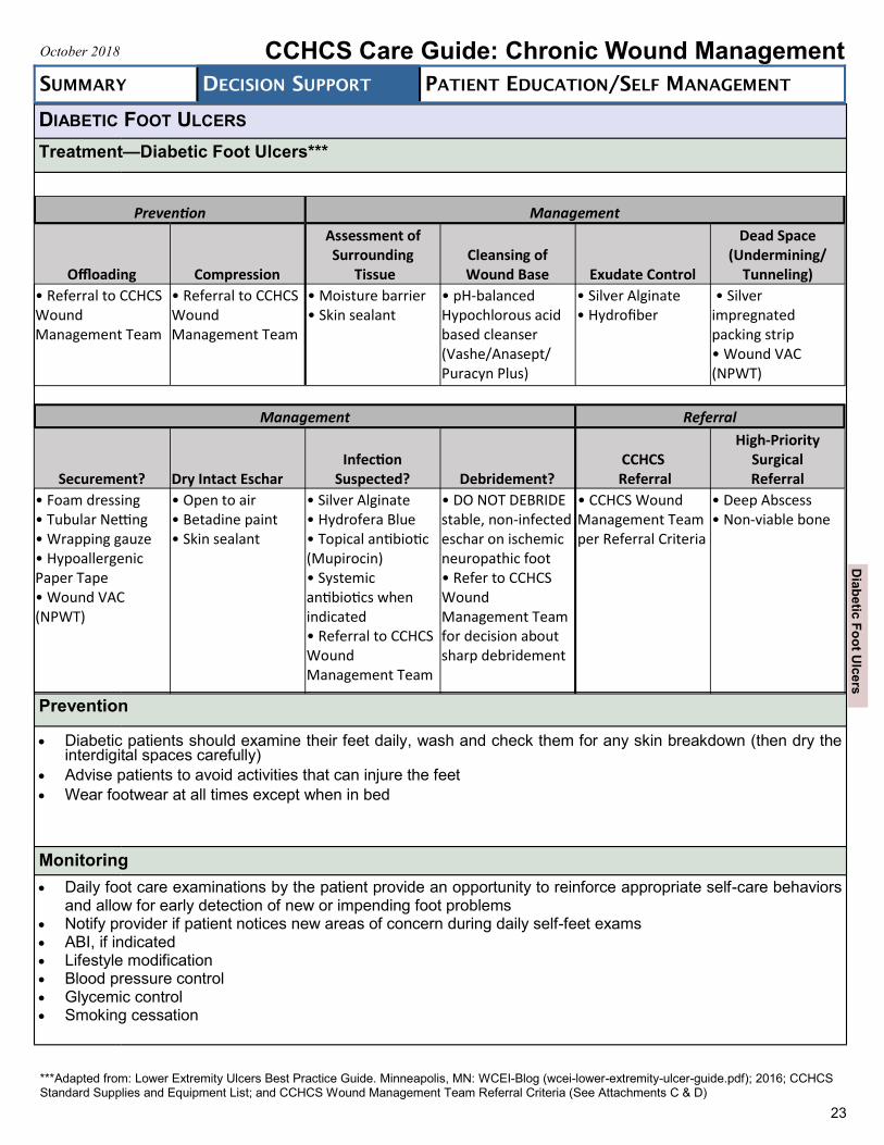
Treatment—Diabetic Foot Ulcers***
Prevention
Diabetic patients should examine their feet daily, wash and check them for any skin breakdown (then dry the interdigital spaces carefully)
Advise patients to avoid activities that can injure the feet
Wear footwear at all times except when in bed
Monitoring
Daily foot care examinations by the patient provide an opportunity to reinforce appropriate self-care behaviors and allow for early detection of new or impending foot problems
Notify provider if patient notices new areas of concern during daily self-feet exams ABI, if indicated Lifestyle modification Blood pressure control Glycemic control Smoking cessation
SUMMARY DECISION SUPPORT PATIENT EDUCATION/SELF MANAGEMENT
CCHCS Care Guide: Chronic Wound Management October 2018
23
***Adapted from: Lower Extremity Ulcers Best Practice Guide. Minneapolis, MN: WCEI-Blog (wcei-lower-extremity-ulcer-guide.pdf); 2016; CCHCS Standard Supplies and Equipment List; and CCHCS Wound Management Team Referral Criteria (See Attachments C & D)
Prevention Management
Offloading Compression
Assessment of Surrounding
Tissue Cleansing of Wound Base Exudate Control
Dead Space (Undermining/
Tunneling)
• Referral to CCHCS Wound Management Team
• Referral to CCHCS Wound Management Team
• Moisture barrier • Skin sealant
• pH-balanced Hypochlorous acid based cleanser (Vashe/Anasept/ Puracyn Plus)
• Silver Alginate • Hydrofiber
• Silver impregnated packing strip • Wound VAC (NPWT)
Management Referral
Securement? Dry Intact Eschar Infection
Suspected? Debridement? CCHCS
Referral
High-Priority Surgical Referral
• Foam dressing • Tubular Netting • Wrapping gauze • Hypoallergenic Paper Tape • Wound VAC (NPWT)
• Open to air • Betadine paint • Skin sealant
• Silver Alginate • Hydrofera Blue • Topical antibiotic (Mupirocin) • Systemic antibiotics when indicated • Referral to CCHCS Wound Management Team
• DO NOT DEBRIDE stable, non-infected eschar on ischemic neuropathic foot • Refer to CCHCS Wound Management Team for decision about sharp debridement
• CCHCS Wound Management Team per Referral Criteria
• Deep Abscess • Non-viable bone
Dia
be
tic F
oo
t Ulc
ers
NON-HEALING SURGICAL WOUNDS
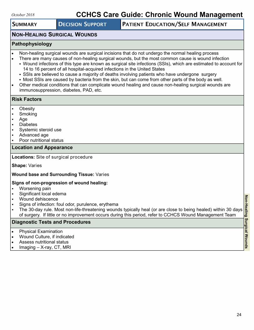
Pathophysiology
Non-healing surgical wounds are surgical incisions that do not undergo the normal healing process There are many causes of non-healing surgical wounds, but the most common cause is wound infection Wound infections of this type are known as surgical site infections (SSIs), which are estimated to account for
14 to 16 percent of all hospital-acquired infections in the United States SSIs are believed to cause a majority of deaths involving patients who have undergone surgery Most SSIs are caused by bacteria from the skin, but can come from other parts of the body as well.
Other medical conditions that can complicate wound healing and cause non-healing surgical wounds are immunosuppression, diabetes, PAD, etc.
Risk Factors
Obesity Smoking Age Diabetes Systemic steroid use Advanced age Poor nutritional status
Location and Appearance
Locations: Site of surgical procedure
Shape: Varies
Wound base and Surrounding Tissue: Varies
Signs of non-progression of wound healing:
Worsening pain Significant local edema Wound dehiscence Signs of infection: foul odor, purulence, erythema The 30-day rule. Most non-life-threatening wounds typically heal (or are close to being healed) within 30 days
of surgery. If little or no improvement occurs during this period, refer to CCHCS Wound Management Team
Diagnostic Tests and Procedures
Physical Examination Wound Culture, if indicated Assess nutritional status Imaging – X-ray, CT, MRI
SUMMARY DECISION SUPPORT PATIENT EDUCATION/SELF MANAGEMENT
CCHCS Care Guide: Chronic Wound Management October 2018
24
No
n-H
ea
ling
Su
rgic
al W
ou
nd
s
NON-HEALING SURGICAL WOUNDS
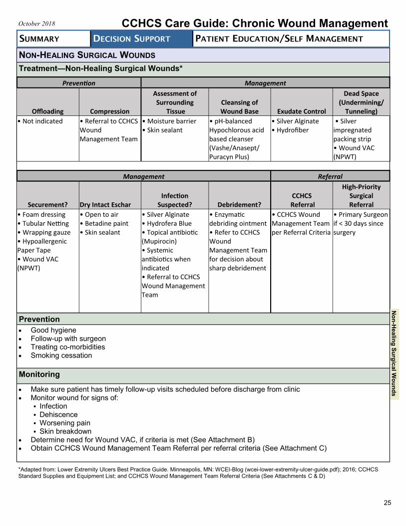
Treatment—Non-Healing Surgical Wounds*
Prevention
Good hygiene Follow-up with surgeon Treating co-morbidities Smoking cessation
Monitoring
Make sure patient has timely follow-up visits scheduled before discharge from clinic Monitor wound for signs of:
Infection Dehiscence Worsening pain Skin breakdown
Determine need for Wound VAC, if criteria is met (See Attachment B) Obtain CCHCS Wound Management Team Referral per referral criteria (See Attachment C)
SUMMARY DECISION SUPPORT PATIENT EDUCATION/SELF MANAGEMENT
CCHCS Care Guide: Chronic Wound Management October 2018
*Adapted from: Lower Extremity Ulcers Best Practice Guide. Minneapolis, MN: WCEI-Blog (wcei-lower-extremity-ulcer-guide.pdf); 2016; CCHCS Standard Supplies and Equipment List; and CCHCS Wound Management Team Referral Criteria (See Attachments C & D)
25
Prevention Management
Offloading Compression
Assessment of Surrounding
Tissue Cleansing of Wound Base Exudate Control
Dead Space (Undermining/
Tunneling)
• Not indicated • Referral to CCHCS Wound Management Team
• Moisture barrier • Skin sealant
• pH-balanced Hypochlorous acid based cleanser (Vashe/Anasept/ Puracyn Plus)
• Silver Alginate • Hydrofiber
• Silver impregnated packing strip • Wound VAC (NPWT)
Management Referral
Securement? Dry Intact Eschar Infection
Suspected? Debridement? CCHCS
Referral
High-Priority Surgical Referral
• Foam dressing • Tubular Netting • Wrapping gauze • Hypoallergenic Paper Tape • Wound VAC (NPWT)
• Open to air • Betadine paint • Skin sealant
• Silver Alginate • Hydrofera Blue • Topical antibiotic (Mupirocin) • Systemic antibiotics when indicated • Referral to CCHCS Wound Management Team
• Enzymatic debriding ointment • Refer to CCHCS Wound Management Team for decision about sharp debridement
• CCHCS Wound Management Team per Referral Criteria
• Primary Surgeon if < 30 days since surgery
No
n-H
ea
ling
Su
rgic
al W
ou
nd
s
SUMMARY DECISION SUPPORT PATIENT EDUCATION/SELF MANAGEMENT
CCHCS Care Guide: Chronic Wound Management October 2018
26
1. Berlowitz D. Epidemiology, pathogenesis, and risk assessment of pressure-induced skin and soft tissue injury. June 11, 2018 ed. Waltham, MA: Up-to-date; 2018.
2. Berlowitz D. Clinical staging and management of pressure-induced skin and soft tissue injury. April 17, 2017
ed. Waltham, MA: Up-to-date; 2017. 3. Lower Extremity Ulcers Best Practice Guide. Minneapolis, MN: WCEI-Blog
(wcei-lower-extremity-ulcer-guide.pdf); 2016. 4. Alquire PC, Mathes BM. Medical management of lower extremity chronic venous disease. November 7, 2017
ed. Waltham, MA: Up-to-date; 2017. 5. Neschis DG, Golden MA. Clinical features and diagnosis of lower extremity peripheral artery disease.
June 11, 2018 ed. Waltham, MA: Up-to-date; 2018. 6. Armstrong DG, McCulloch DK, de Asia RJ. Management of diabetic foot ulcers. March 6, 2018 ed. Waltham,
MA: Up-to-date; 2018. 7. NPUAP 2016; Pages. Accessed at NPUAP at
http://www.npuap.org/resources/educational-and-clinical-resources/npuap-pressure-injury-stages/ on June 27, 2018.
8. Guideline: Braden Scale for Predicting Pressure Ulcer Risk in Adults & Children/Infants. December 2014 ed.
Vancouver, BC: British Columbia Provincial Nursing Skin and Wound Committee; 2014. 9. Alguire PC, Scovell S. Overview and management of lower extremity chronic venous disease. May 29, 2018
ed. Waltham, MA: UpToDate; 2018. 10. Bryant RA, Nix DP. Acute & Chronic Wounds Current Management Concepts. 5th ed. St. Louis, MO: Elsevier;
2016. 11. Armstrong DG, Meyr AJ. Compression therapy for the treatment of chronic venous insufficiency.
January 29, 2018 ed. Waltham, MA: UpToDate; 2018. 12. Mitchell ME, Carpenter JP. Clinical features and diagnosis of acute lower extremity ischemia. March 15, 2018
ed. Waltham, MA: UpToDate; 2018. 13. Guideline: Assessment and Treatment of Lower Leg Ulcers (Arterial, Venous & Mixed) in Adults. August 2014
ed. Vancouver, BC: British Columbia Provincial Nursing Skin and Wound Committee; 2014. 14. Harris L, Dryjski M. Epidemiology, risk factors, and natural history of peripheral artery disease. June 18, 2018
ed. Watham, MA: UpToDate; 2018.
REFERENCES
“BED SORES” (PRESSURE INJURIES):
WHAT YOU SHOULD KNOW
WHAT IS A PRESSURE INJURY?
A pressure injury or bed sore is an injury to the skin (and sometimes muscle or fat under the skin) that is caused by sitting or lying down in one place for too long
Pressure injuries are given a “grade” (called Stage) of 1 to 4 depending on how deep they are
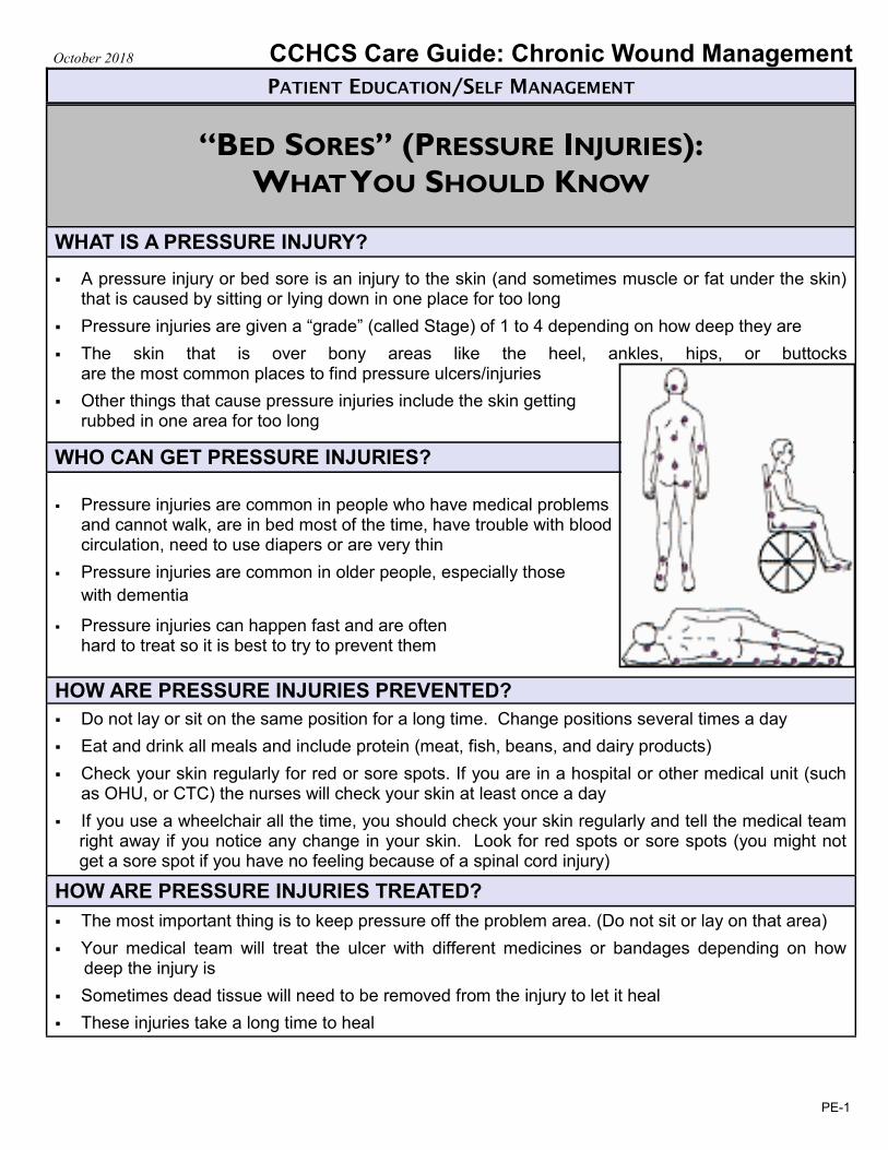
The skin that is over bony areas like the heel, ankles, hips, or buttocks are the most common places to find pressure ulcers/injuries
Other things that cause pressure injuries include the skin getting rubbed in one area for too long
WHO CAN GET PRESSURE INJURIES?
Pressure injuries are common in people who have medical problems and cannot walk, are in bed most of the time, have trouble with blood circulation, need to use diapers or are very thin
Pressure injuries are common in older people, especially those
with dementia
Pressure injuries can happen fast and are often hard to treat so it is best to try to prevent them
HOW ARE PRESSURE INJURIES PREVENTED?
Do not lay or sit on the same position for a long time. Change positions several times a day
Eat and drink all meals and include protein (meat, fish, beans, and dairy products)
Check your skin regularly for red or sore spots. If you are in a hospital or other medical unit (such as OHU, or CTC) the nurses will check your skin at least once a day
If you use a wheelchair all the time, you should check your skin regularly and tell the medical team right away if you notice any change in your skin. Look for red spots or sore spots (you might not get a sore spot if you have no feeling because of a spinal cord injury)
HOW ARE PRESSURE INJURIES TREATED?
The most important thing is to keep pressure off the problem area. (Do not sit or lay on that area)
Your medical team will treat the ulcer with different medicines or bandages depending on how deep the injury is
Sometimes dead tissue will need to be removed from the injury to let it heal
These injuries take a long time to heal
PE-1
PATIENT EDUCATION/SELF MANAGEMENT
October 2018 CCHCS Care Guide: Chronic Wound Management
PATIENT EDUCATION/SELF MANAGEMENT
VENOUS LEG ULCERS: WHAT YOU SHOULD KNOW
WHAT IS A VENOUS (VEE-NUS) LEG ULCER?
A sore in the skin of the leg caused by leg swelling and problems with your veins
It may start with a small injury to your skin
In people with bad veins or a lot of leg swelling, the skin sore may not heal and get bigger. This is a chronic venous leg ulcer
WHAT ARE THE SYMPTOMS?
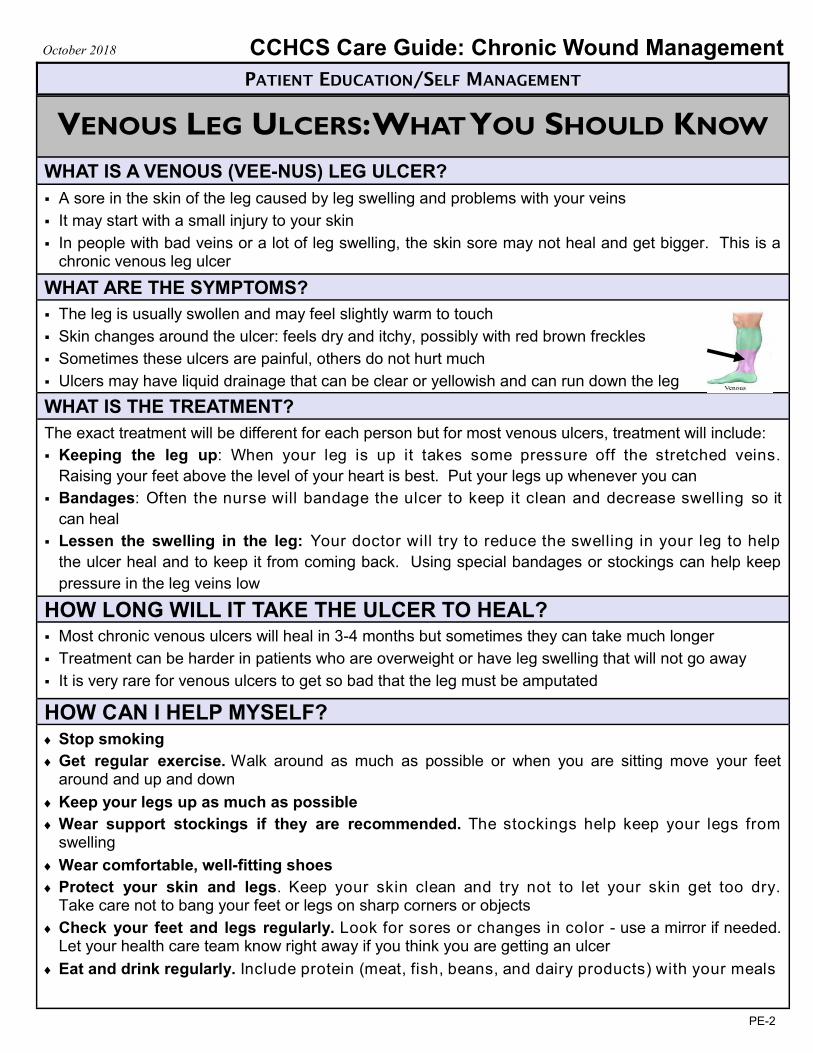
The leg is usually swollen and may feel slightly warm to touch
Skin changes around the ulcer: feels dry and itchy, possibly with red brown freckles
Sometimes these ulcers are painful, others do not hurt much
Ulcers may have liquid drainage that can be clear or yellowish and can run down the leg
WHAT IS THE TREATMENT?
The exact treatment will be different for each person but for most venous ulcers, treatment will include:
Keeping the leg up: When your leg is up it takes some pressure off the stretched veins.
Raising your feet above the level of your heart is best. Put your legs up whenever you can
Bandages: Often the nurse will bandage the ulcer to keep it clean and decrease swelling so it
can heal
Lessen the swelling in the leg: Your doctor will try to reduce the swelling in your leg to help
the ulcer heal and to keep it from coming back. Using special bandages or stockings can help keep
pressure in the leg veins low
HOW LONG WILL IT TAKE THE ULCER TO HEAL? Most chronic venous ulcers will heal in 3-4 months but sometimes they can take much longer
Treatment can be harder in patients who are overweight or have leg swelling that will not go away
It is very rare for venous ulcers to get so bad that the leg must be amputated
HOW CAN I HELP MYSELF? Stop smoking
Get regular exercise. Walk around as much as possible or when you are sitting move your feet around and up and down
Keep your legs up as much as possible
Wear support stockings if they are recommended. The stockings help keep your legs from swelling
Wear comfortable, well-fitting shoes
Protect your skin and legs. Keep your skin clean and try not to let your skin get too dry. Take care not to bang your feet or legs on sharp corners or objects
Check your feet and legs regularly. Look for sores or changes in color - use a mirror if needed. Let your health care team know right away if you think you are getting an ulcer
Eat and drink regularly. Include protein (meat, fish, beans, and dairy products) with your meals
PE-2
October 2018 CCHCS Care Guide: Chronic Wound Management
PATIENT EDUCATION/SELF MANAGEMENT
October 2018
ISCHEMIC (ARTERIAL) ULCERS:
WHAT YOU SHOULD KNOW
WHAT CAUSES ISCHEMIC (IS-KEE-MIC) ULCERS?
Ischemic ulcers can happen when blood flow to your leg is limited. This is called ischemic disease
Being cold, smoking, and high blood pressure can slow the blood flow to your lower legs
WHAT IS ISCHEMIC DISEASE? Ischemic disease is when you have restricted blood flow to your legs
You may have all or some of these symptoms:
Pain in the lower leg with walking that is relieved by rest
Pain in the lower leg while lying in bed
Loss of hair on the leg and foot
Cold feet
WHAT WILL INCREASE THE RISK OF GETTING ISCHEMIC ULCERS?
Diabetes
High blood pressure
Hardening of the arteries
Smoking
WHEN SHOULD I SEE MY HEALTH CARE PROVIDER?
If you have: Numbness in one or both of your feet
Loss of movement in your legs or feet
Color change in your lower legs
Redness, blisters or sores on either foot
If you have an ulcer, you should see your health care provider if there is: Increased pain at your ulcer site
Bad smell coming from the wound
Change in color or amount of drainage from wound
Fever or chills
You feel sick to your stomach or are vomiting
WHAT CAN I DO TO HELP PREVENT ISCHEMIC ULCERS?
Do not walk barefoot
Wear shoes and socks that protect your feet from the cold
Protect your legs and feet from injury
Inspect your lower legs and feet daily to find problems early
Don’t smoke
Exercise as directed
Follow recommended diet
PE-3
CCHCS Care Guide: Chronic Wound Management
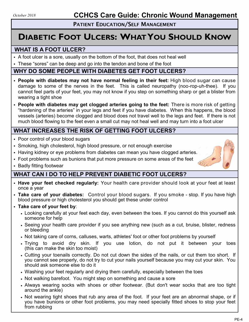
DIABETIC FOOT ULCERS: WHAT YOU SHOULD KNOW
WHAT IS A FOOT ULCER?
A foot ulcer is a sore, usually on the bottom of the foot, that does not heal well
These “sores” can be deep and go into the tendon and bone of the foot
WHY DO SOME PEOPLE WITH DIABETES GET FOOT ULCERS?
People with diabetes may not have normal feeling in their feet: High blood sugar can cause damage to some of the nerves in the feet. This is called neuropathy (noo-rop-uh-thee). If you cannot feel parts of your feet, you may not know if you step on something sharp or get a blister from wearing a tight shoe
People with diabetes may get clogged arteries going to the feet: There is more risk of getting “hardening of the arteries” in your legs and feet if you have diabetes. When this happens, the blood vessels (arteries) become clogged and blood does not travel well to the legs and feet. If there is not much blood flowing to the feet even a small cut may not heal well and may turn into a foot ulcer
WHAT INCREASES THE RISK OF GETTING FOOT ULCERS?
Poor control of your blood sugars
Smoking, high cholesterol, high blood pressure, or not enough exercise
Having kidney or eye problems from diabetes can mean you have clogged arteries.
Foot problems such as bunions that put more pressure on some areas of the feet
Badly fitting footwear
WHAT CAN I DO TO HELP PREVENT DIABETIC FOOT ULCERS?
Have your feet checked regularly: Your health care provider should look at your feet at least once a year
Take care of your diabetes: Control your blood sugars. If you smoke - stop. If you have high blood pressure or high cholesterol you should get these under control
Take care of your feet by:
Looking carefully at your feet each day, even between the toes. If you cannot do this yourself ask someone for help
Seeing your health care provider if you see anything new (such as a cut, bruise, blister, redness or bleeding
Not taking care of corns, calluses, warts, athletes' foot or other foot problems by yourself
Trying to avoid dry skin. If you use lotion, do not put it between your toes (this can make the skin too moist)
Cutting your toenails correctly. Do not cut down the sides of the nails, or cut them too short. If you cannot see properly, do not try to cut your nails yourself because you may cut your skin. You should ask someone else to do it
Washing your feet regularly and drying them carefully, especially between the toes
Not walking barefoot. You might step on something and cause a sore
Always wearing socks with shoes or other footwear. (But don't wear socks that are too tight around the ankle)
Not wearing tight shoes that rub any area of the foot. If your feet are an abnormal shape, or if you have bunions or other foot problems, you may need specially fitted shoes to stop your feet from rubbing
PE-4
PATIENT EDUCATION/SELF MANAGEMENT
October 2018 CCHCS Care Guide: Chronic Wound Management
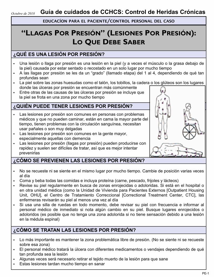
“LLAGAS POR PRESIÓN” (LESIONES POR PRESIÓN):
LO QUE DEBE SABER
¿QUÉ ES UNA LESIÓN POR PRESIÓN?
Una lesión o llaga por presión es una lesión en la piel (y a veces el músculo o la grasa debajo de la piel) causada por estar sentado o recostado en un solo lugar por mucho tiempo
A las llagas por presión se les da un “grado” (llamado etapa) del 1 al 4, dependiendo de qué tan profundas sean
La piel sobre las zonas huesudas como el talón, los tobillos, la cadera o los glúteos son los lugares donde las úlceras por presión se encuentran más comúnmente
Entre otras de las causas de las úlceras por presión se incluye que la piel se frota en una zona por mucho tiempo
¿QUIÉN PUEDE TENER LESIONES POR PRESIÓN?
Las lesiones por presión son comunes en personas con problemas médicos y que no pueden caminar, están en cama la mayor parte del tiempo, tienen problemas con la circulación sanguínea, necesitan usar pañales o son muy delgadas
Las lesiones por presión son comunes en la gente mayor, especialmente aquellas con demencia
Las lesiones por presión (llagas por presión) pueden producirse con rapidez y suelen ser difíciles de tratar, así que es mejor intentar prevenirlas
¿CÓMO SE PREVIENEN LAS LESIONES POR PRESIÓN?
No se recueste ni se siente en el mismo lugar por mucho tiempo. Cambie de posición varias veces al día
Coma y beba todas las comidas e incluya proteína (carne, pescado, frijoles y lácteos) Revise su piel regularmente en busca de zonas enrojecidas o adoloridas. Si está en el hospital o
en otra unidad médica (como la Unidad de Vivienda para Pacientes Externos [Outpatient Housing Unit, OHU], el Centro de Tratamiento Correccional [Correctional Treatment Center, CTC], las enfermeras revisarán su piel al menos una vez al día
Si usa una silla de ruedas en todo momento, debe revisar su piel con frecuencia e informar al personal médico de inmediato si nota algún cambio en su piel. Busque lugares enrojecidos o adoloridos (es posible que no tenga una zona adolorida si no tiene sensación debido a una lesión en la médula espinal)
¿CÓMO SE TRATAN LAS LESIONES POR PRESIÓN?
Lo más importante es mantener la zona problemática libre de presión. (No se siente ni se recueste sobre esa zona)
El personal médico tratará la úlcera con diferentes medicamentos o vendajes dependiendo de qué tan profunda sea la lesión
Algunas veces será necesario retirar el tejido muerto de la lesión para que sane Estas lesiones tardan mucho tiempo en sanar
PE-1
EDUCACÍON PARA EL PACIENTE/CONTROL PERSONAL DEL CASO
Octubre de 2018 Guía de cuidados de CCHCS: Control de Heridas Crónicas
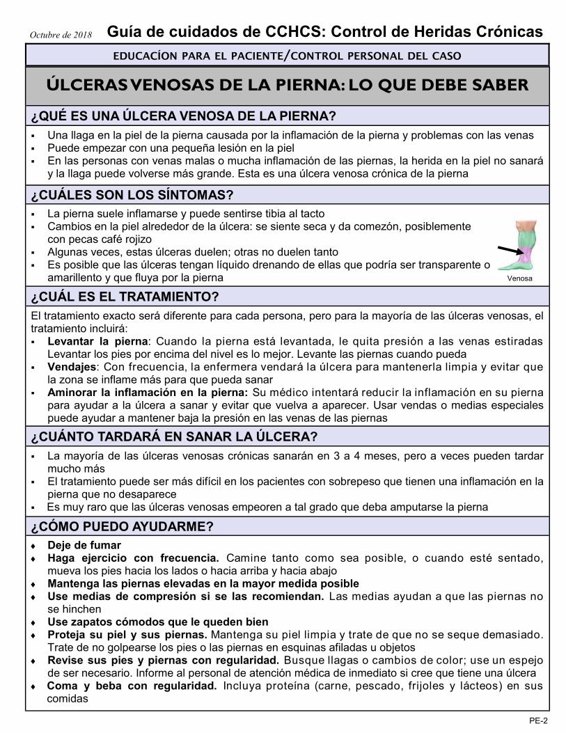
EDUCACÍON PARA EL PACIENTE/CONTROL PERSONAL DEL CASO
ÚLCERAS VENOSAS DE LA PIERNA: LO QUE DEBE SABER
¿QUÉ ES UNA ÚLCERA VENOSA DE LA PIERNA?
Una llaga en la piel de la pierna causada por la inflamación de la pierna y problemas con las venas Puede empezar con una pequeña lesión en la piel En las personas con venas malas o mucha inflamación de las piernas, la herida en la piel no sanará
y la llaga puede volverse más grande. Esta es una úlcera venosa crónica de la pierna
¿CUÁLES SON LOS SÍNTOMAS?
La pierna suele inflamarse y puede sentirse tibia al tacto Cambios en la piel alrededor de la úlcera: se siente seca y da comezón, posiblemente
con pecas café rojizo Algunas veces, estas úlceras duelen; otras no duelen tanto Es posible que las úlceras tengan líquido drenando de ellas que podría ser transparente o
amarillento y que fluya por la pierna
¿CUÁL ES EL TRATAMIENTO?
El tratamiento exacto será diferente para cada persona, pero para la mayoría de las úlceras venosas, el tratamiento incluirá: Levantar la pierna: Cuando la pierna está levantada, le quita presión a las venas estiradas
Levantar los pies por encima del nivel es lo mejor. Levante las piernas cuando pueda Vendajes: Con frecuencia, la enfermera vendará la úlcera para mantenerla limpia y evitar que
la zona se inflame más para que pueda sanar Aminorar la inflamación en la pierna: Su médico intentará reducir la inflamación en su pierna
para ayudar a la úlcera a sanar y evitar que vuelva a aparecer. Usar vendas o medias especiales puede ayudar a mantener baja la presión en las venas de las piernas
¿CUÁNTO TARDARÁ EN SANAR LA ÚLCERA?
La mayoría de las úlceras venosas crónicas sanarán en 3 a 4 meses, pero a veces pueden tardar mucho más
El tratamiento puede ser más difícil en los pacientes con sobrepeso que tienen una inflamación en la pierna que no desaparece
Es muy raro que las úlceras venosas empeoren a tal grado que deba amputarse la pierna
¿CÓMO PUEDO AYUDARME?
Deje de fumar Haga ejercicio con frecuencia. Camine tanto como sea posible, o cuando esté sentado,
mueva los pies hacia los lados o hacia arriba y hacia abajo Mantenga las piernas elevadas en la mayor medida posible Use medias de compresión si se las recomiendan. Las medias ayudan a que las piernas no
se hinchen Use zapatos cómodos que le queden bien Proteja su piel y sus piernas. Mantenga su piel limpia y trate de que no se seque demasiado.
Trate de no golpearse los pies o las piernas en esquinas afiladas u objetos Revise sus pies y piernas con regularidad. Busque llagas o cambios de color; use un espejo
de ser necesario. Informe al personal de atención médica de inmediato si cree que tiene una úlcera Coma y beba con regularidad. Incluya proteína (carne, pescado, frijoles y lácteos) en sus
comidas
PE-2
Octubre de 2018 Guía de cuidados de CCHCS: Control de Heridas Crónicas
Venosa
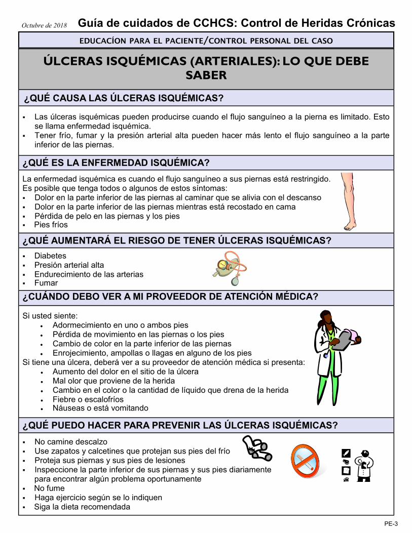
EDUCACÍON PARA EL PACIENTE/CONTROL PERSONAL DEL CASO
Octubre de 2018
ÚLCERAS ISQUÉMICAS (ARTERIALES): LO QUE DEBE
SABER
¿QUÉ CAUSA LAS ÚLCERAS ISQUÉMICAS?
Las úlceras isquémicas pueden producirse cuando el flujo sanguíneo a la pierna es limitado. Esto se llama enfermedad isquémica.
Tener frío, fumar y la presión arterial alta pueden hacer más lento el flujo sanguíneo a la parte inferior de las piernas.
¿QUÉ ES LA ENFERMEDAD ISQUÉMICA?
La enfermedad isquémica es cuando el flujo sanguíneo a sus piernas está restringido. Es posible que tenga todos o algunos de estos síntomas: Dolor en la parte inferior de las piernas al caminar que se alivia con el descanso Dolor en la parte inferior de las piernas mientras está recostado en cama Pérdida de pelo en las piernas y los pies Pies fríos
¿QUÉ AUMENTARÁ EL RIESGO DE TENER ÚLCERAS ISQUÉMICAS?
Diabetes Presión arterial alta Endurecimiento de las arterias Fumar
¿CUÁNDO DEBO VER A MI PROVEEDOR DE ATENCIÓN MÉDICA?
Si usted siente: Adormecimiento en uno o ambos pies Pérdida de movimiento en las piernas o los pies Cambio de color en la parte inferior de las piernas Enrojecimiento, ampollas o llagas en alguno de los pies
Si tiene una úlcera, deberá ver a su proveedor de atención médica si presenta: Aumento del dolor en el sitio de la úlcera Mal olor que proviene de la herida Cambio en el color o la cantidad de líquido que drena de la herida Fiebre o escalofríos Náuseas o está vomitando
¿QUÉ PUEDO HACER PARA PREVENIR LAS ÚLCERAS ISQUÉMICAS?
No camine descalzo Use zapatos y calcetines que protejan sus pies del frío Proteja sus piernas y sus pies de lesiones Inspeccione la parte inferior de sus piernas y sus pies diariamente
para encontrar algún problema oportunamente No fume Haga ejercicio según se lo indiquen Siga la dieta recomendada
PE-3
Guía de cuidados de CCHCS: Control de Heridas Crónicas
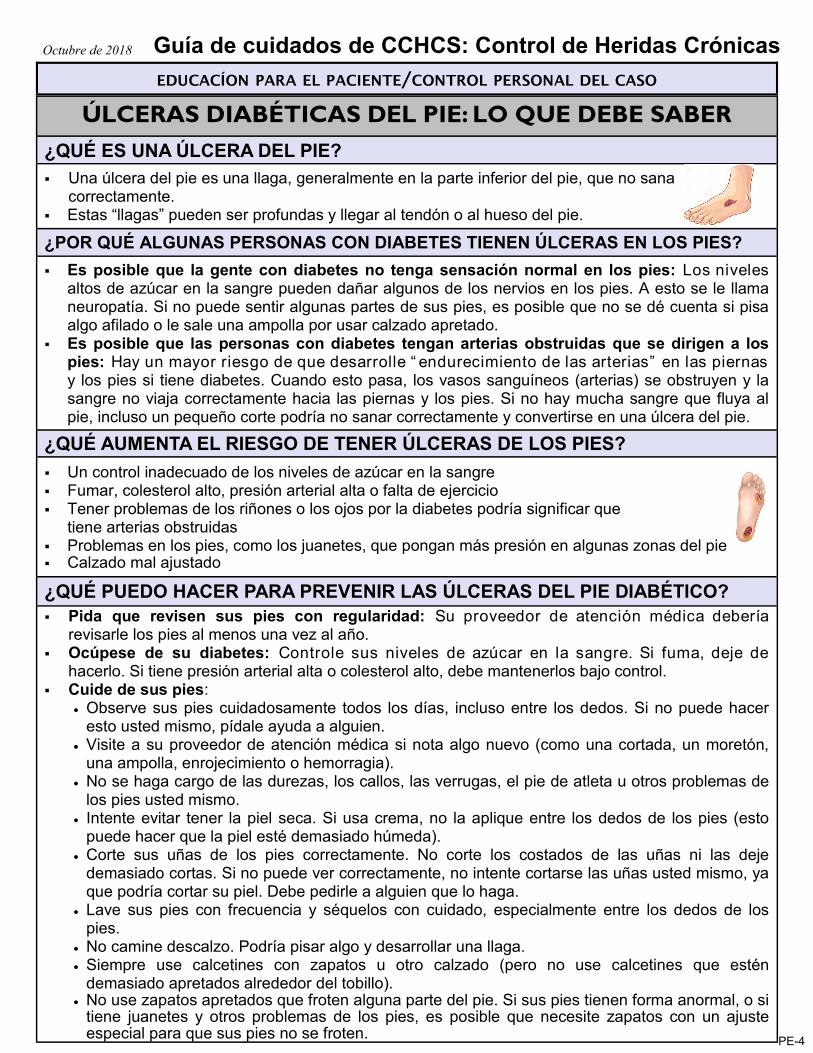
ÚLCERAS DIABÉTICAS DEL PIE: LO QUE DEBE SABER
¿QUÉ ES UNA ÚLCERA DEL PIE?
Una úlcera del pie es una llaga, generalmente en la parte inferior del pie, que no sana correctamente.
Estas “llagas” pueden ser profundas y llegar al tendón o al hueso del pie.
¿POR QUÉ ALGUNAS PERSONAS CON DIABETES TIENEN ÚLCERAS EN LOS PIES?
Es posible que la gente con diabetes no tenga sensación normal en los pies: Los niveles altos de azúcar en la sangre pueden dañar algunos de los nervios en los pies. A esto se le llama neuropatía. Si no puede sentir algunas partes de sus pies, es posible que no se dé cuenta si pisa algo afilado o le sale una ampolla por usar calzado apretado.
Es posible que las personas con diabetes tengan arterias obstruidas que se dirigen a los pies: Hay un mayor riesgo de que desarrolle “ endurecimiento de las arterias” en las piernas y los pies si tiene diabetes. Cuando esto pasa, los vasos sanguíneos (arterias) se obstruyen y la sangre no viaja correctamente hacia las piernas y los pies. Si no hay mucha sangre que fluya al pie, incluso un pequeño corte podría no sanar correctamente y convertirse en una úlcera del pie.
¿QUÉ AUMENTA EL RIESGO DE TENER ÚLCERAS DE LOS PIES?
Un control inadecuado de los niveles de azúcar en la sangre Fumar, colesterol alto, presión arterial alta o falta de ejercicio Tener problemas de los riñones o los ojos por la diabetes podría significar que
tiene arterias obstruidas Problemas en los pies, como los juanetes, que pongan más presión en algunas zonas del pie Calzado mal ajustado
¿QUÉ PUEDO HACER PARA PREVENIR LAS ÚLCERAS DEL PIE DIABÉTICO?
Pida que revisen sus pies con regularidad: Su proveedor de atención médica debería revisarle los pies al menos una vez al año.
Ocúpese de su diabetes: Controle sus niveles de azúcar en la sangre. Si fuma, deje de hacerlo. Si tiene presión arterial alta o colesterol alto, debe mantenerlos bajo control.
Cuide de sus pies: Observe sus pies cuidadosamente todos los días, incluso entre los dedos. Si no puede hacer
esto usted mismo, pídale ayuda a alguien. Visite a su proveedor de atención médica si nota algo nuevo (como una cortada, un moretón,
una ampolla, enrojecimiento o hemorragia). No se haga cargo de las durezas, los callos, las verrugas, el pie de atleta u otros problemas de
los pies usted mismo. Intente evitar tener la piel seca. Si usa crema, no la aplique entre los dedos de los pies (esto
puede hacer que la piel esté demasiado húmeda). Corte sus uñas de los pies correctamente. No corte los costados de las uñas ni las deje
demasiado cortas. Si no puede ver correctamente, no intente cortarse las uñas usted mismo, ya que podría cortar su piel. Debe pedirle a alguien que lo haga.
Lave sus pies con frecuencia y séquelos con cuidado, especialmente entre los dedos de los pies.
No camine descalzo. Podría pisar algo y desarrollar una llaga. Siempre use calcetines con zapatos u otro calzado (pero no use calcetines que estén
demasiado apretados alrededor del tobillo). No use zapatos apretados que froten alguna parte del pie. Si sus pies tienen forma anormal, o si
tiene juanetes y otros problemas de los pies, es posible que necesite zapatos con un ajuste especial para que sus pies no se froten.
PE-4
EDUCACÍON PARA EL PACIENTE/CONTROL PERSONAL DEL CASO
Octubre de 2018 Guía de cuidados de CCHCS: Control de Heridas Crónicas
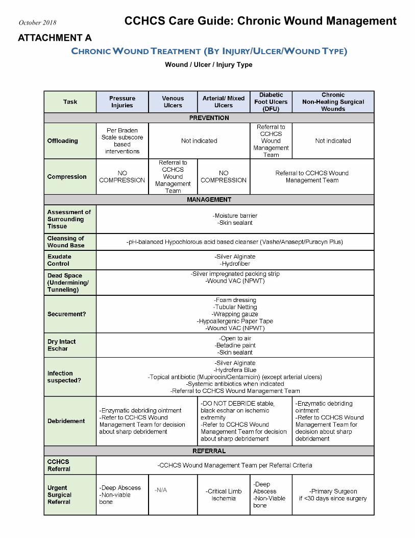
Wound / Ulcer / Injury Type
CCHCS Care Guide: Chronic Wound Management October 2018
ATTACHMENT A
CHRONIC WOUND TREATMENT (BY INJURY/ULCER/WOUND TYPE)
-N/A
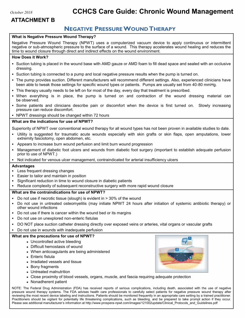
NEGATIVE PRESSURE WOUND THERAPY What is Negative Pressure Wound Therapy?
Negative Pressure Wound Therapy (NPWT) uses a computerized vacuum device to apply continuous or intermittent negative or sub-atmospheric pressure to the surface of a wound. This therapy accelerates wound healing and reduces the time to wound closure through direct and indirect effects on the wound environment.
How Does it Work?
Suction tubing is placed in the wound base with AMD gauze or AMD foam to fill dead space and sealed with an occlusive dressing.
Suction tubing is connected to a pump and local negative pressure results when the pump is turned on.
The pump provides suction. Different manufacturers will recommend different settings. Also, experienced clinicians have been able to tweak those settings for specific wound types or patients. Pumps are usually set from 40-80 mmHg.
This therapy usually needs to be left on for most of the day, every day that treatment is prescribed.
When everything is in place, the pump is turned on and contraction of the wound dressing material can be observed.
Some patients and clinicians describe pain or discomfort when the device is first turned on. Slowly increasing pressure can reduce discomfort.
NPWT dressings should be changed within 72 hours
What are the indications for use of NPWT?
Superiority of NPWT over conventional wound therapy for all wound types has not been proven in available studies to date.
Utility is suggested for traumatic acute wounds especially with skin grafts or skin flaps, open amputations, lower extremity fasciotomy, open abdomen, etc.
Appears to increase burn wound perfusion and limit burn wound progression
Management of diabetic foot ulcers and wounds from diabetic foot surgery (important to establish adequate perfusion prior to use of NPWT.)
Not indicated for venous ulcer management, contraindicated for arterial insufficiency ulcers
Advantages
Less frequent dressing changes
Easier to tailor and maintain in position
Significant reduction in time to wound closure in diabetic patients
Reduce complexity of subsequent reconstructive surgery with more rapid wound closure
What are the contraindications for use of NPWT?
Do not use if necrotic tissue (slough) is evident in > 30% of the wound
Do not use in untreated osteomyelitis (may initiate NPWT 24 hours after initiation of systemic antibiotic therapy) or other wound infections
Do not use if there is cancer within the wound bed or its margins
Do not use on unexplored non-enteric fistulas
DO NOT place suction catheter dressing directly over exposed veins or arteries, vital organs or vascular grafts
Do not use in wounds with inadequate perfusion
What are the precautions for use of NPWT?
Uncontrolled active bleeding
Difficult hemostasis of wound
When anticoagulants are being administered
Enteric fistula
Irradiated vessels and tissue
Bony fragments
Untreated malnutrition
Close proximity of blood vessels, organs, muscle, and fascia requiring adequate protection
Nonadherent patient
NOTE: The Federal Drug Administration (FDA) has received reports of serious complications, including death, associated with the use of negative pressure wound therapy systems. The FDA advises health care professionals to carefully select patients for negative pressure wound therapy after reviewing the most recent device labeling and instructions. Patients should be monitored frequently in an appropriate care setting by a trained practitioner. Practitioners should be vigilant for potentially life threatening complications, such as bleeding, and be prepared to take prompt action if they occur. Please see additional manufacturer’s information at http://www.prospera-npwt.com/images/121002update/Clinical_Protocols_and_Guidelines.pdf
CCHCS Care Guide: Chronic Wound Management October 2018
ATTACHMENT B
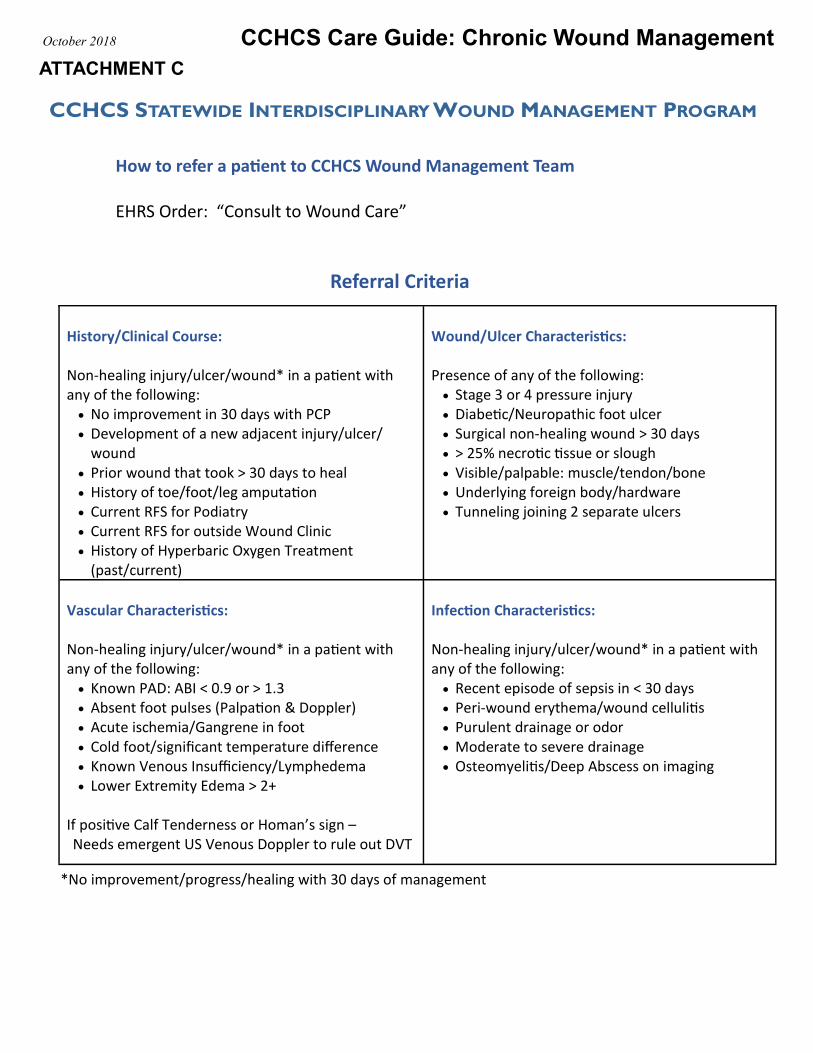
CCHCS Care Guide: Chronic Wound Management October 2018
ATTACHMENT C
CCHCS STATEWIDE INTERDISCIPLINARY WOUND MANAGEMENT PROGRAM
How to refer a patient to CCHCS Wound Management Team
EHRS Order: “Consult to Wound Care”
Referral Criteria
History/Clinical Course: Non‐healing injury/ulcer/wound* in a patient with any of the following:
No improvement in 30 days with PCP Development of a new adjacent injury/ulcer/
wound Prior wound that took > 30 days to heal History of toe/foot/leg amputation Current RFS for Podiatry Current RFS for outside Wound Clinic History of Hyperbaric Oxygen Treatment
(past/current)
Wound/Ulcer Characteristics: Presence of any of the following:
Stage 3 or 4 pressure injury Diabetic/Neuropathic foot ulcer Surgical non-healing wound > 30 days > 25% necrotic tissue or slough Visible/palpable: muscle/tendon/bone Underlying foreign body/hardware Tunneling joining 2 separate ulcers
Vascular Characteristics: Non‐healing injury/ulcer/wound* in a patient with any of the following:
Known PAD: ABI < 0.9 or > 1.3 Absent foot pulses (Palpation & Doppler) Acute ischemia/Gangrene in foot Cold foot/significant temperature difference Known Venous Insufficiency/Lymphedema Lower Extremity Edema > 2+
If positive Calf Tenderness or Homan’s sign – Needs emergent US Venous Doppler to rule out DVT
Infection Characteristics: Non‐healing injury/ulcer/wound* in a patient with any of the following:
Recent episode of sepsis in < 30 days Peri-wound erythema/wound cellulitis Purulent drainage or odor Moderate to severe drainage Osteomyelitis/Deep Abscess on imaging
*No improvement/progress/healing with 30 days of management
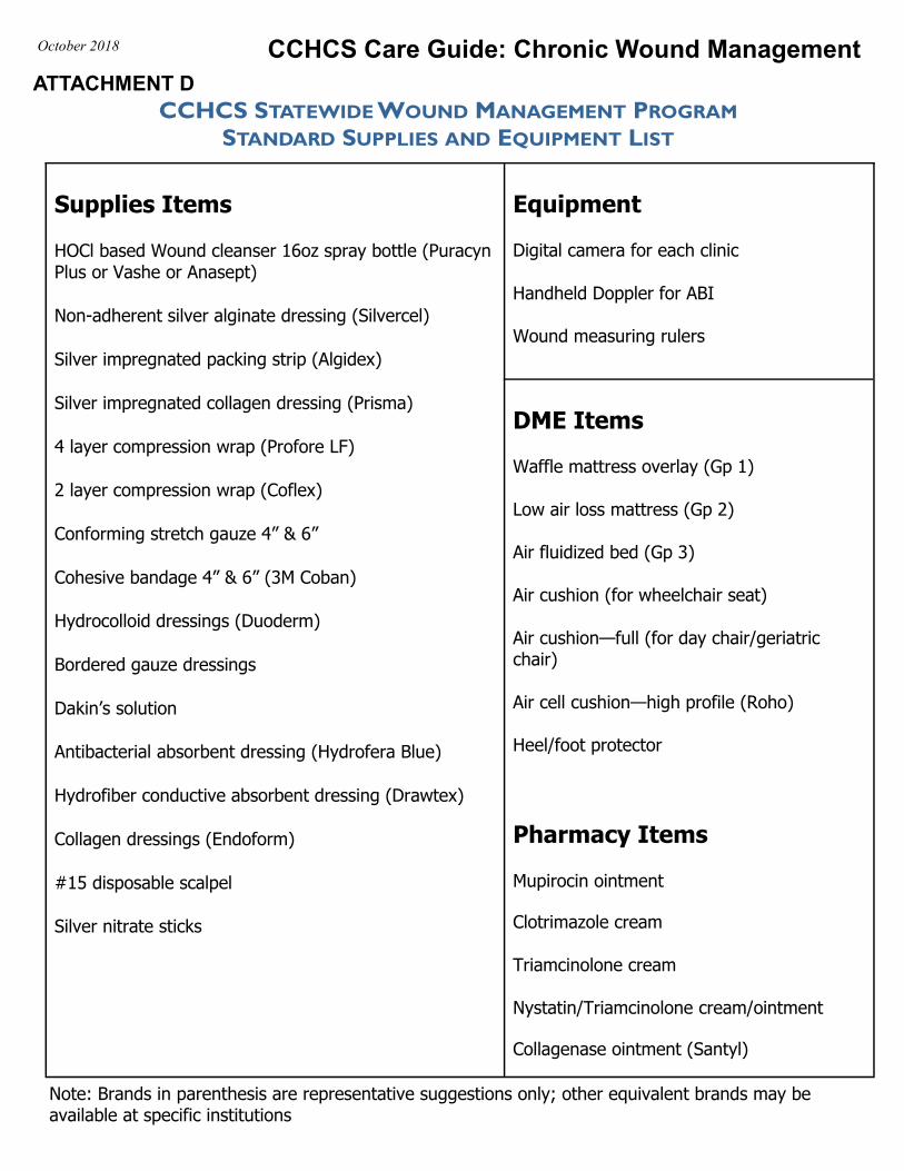
CCHCS Care Guide: Chronic Wound Management October 2018
ATTACHMENT D
CCHCS STATEWIDE WOUND MANAGEMENT PROGRAM
STANDARD SUPPLIES AND EQUIPMENT LIST
Supplies Items HOCl based Wound cleanser 16oz spray bottle (Puracyn Plus or Vashe or Anasept)
Non-adherent silver alginate dressing (Silvercel)
Silver impregnated packing strip (Algidex)
Silver impregnated collagen dressing (Prisma)
4 layer compression wrap (Profore LF)
2 layer compression wrap (Coflex)
Conforming stretch gauze 4” & 6”
Cohesive bandage 4” & 6” (3M Coban)
Hydrocolloid dressings (Duoderm)
Bordered gauze dressings
Dakin’s solution
Antibacterial absorbent dressing (Hydrofera Blue)
Hydrofiber conductive absorbent dressing (Drawtex)
Collagen dressings (Endoform)
#15 disposable scalpel
Silver nitrate sticks
Equipment
Digital camera for each clinic
Handheld Doppler for ABI
Wound measuring rulers
DME Items Waffle mattress overlay (Gp 1)
Low air loss mattress (Gp 2)
Air fluidized bed (Gp 3)
Air cushion (for wheelchair seat)
Air cushion—full (for day chair/geriatric chair)
Air cell cushion—high profile (Roho)
Heel/foot protector
Pharmacy Items Mupirocin ointment
Clotrimazole cream Triamcinolone cream
Nystatin/Triamcinolone cream/ointment
Collagenase ointment (Santyl)
Note: Brands in parenthesis are representative suggestions only; other equivalent brands may be available at specific institutions
Recommended