2/15/2013
1
Recent Advances in Neurology15 Feb 2013
Presenter: Neel SinghalDiscussant: Vanja Douglas
Neuropathology: Andrew Bollen
CPC: Recurrent ptosis and visual disturbances
Case History� 45 year old woman with diagnoses of multiple sclerosis and myasthenia gravis admitted to a local hospital for 2 weeks of progressive weakness, ptosis, and visual disturbances
� Symptoms preceded by URI
� Similar symptoms have occurred numerous times in the past
Case History� Ocular� Ptosis�Visual scene-skipping, ‘jitter’� Weakness� Fatigue�Difficulty with ambulation, stairs, household tasks� Systemic�Daily worsened migraine� Joint pains, abdominal pain, nausea
Case History� Family notes 2 years of poor memory manifested as
misplacing objects, forgetting appointments, and repetitive questioning
� ROS:�Chills, malaise, myalgias, fatigue, palpitations, heartburn, nausea, dysuria, diffuse pain in limbs/back

2/15/2013
2
Local Hospital Course� Started on antibiotics for suspected PNA� After neurologic consultation started on 5-day course of IVIG� Started on stress-dose steroids given history of prednisone use� No clear improvement in symptoms� Of note, patient demonstrated single episode of 3 second pause on telemetry
Past Medical History� Hypothyroidism� Diabetes� Anemia� Multiple Sclerosis: previously on interferon-β1a from 2008-2009� Myasthenia Gravis: maintained on steroids intermittently since 2009
Medications on Transfer� Levothyroxine 100 mcg PO daily� Pyridostigmine 30 mg PO TID� Hydrocortisone 100 mg IV q6hrs� Pantoprazole 40 mg PO daily� Amitriptyline 30 mg PO nightly� Doxyclycline 100 mg PO BID� Cefazolin 1 g IV q8hrs� Colace 200 mg PO BID
Family History� Father with history of brain tumor
� Mother with thyroid disease and arthritis
� 4 healthy siblings
� 4 healthy children
� 1 grandson died in neonatal period with multiple systemic complaints

2/15/2013
3
Social History� Lives in Stockton, CA with her husband
� Family has moved 4 times in last 10 years due to work obligations
� Patient has been on disability for the last 3 years
� No drugs, alcohol, or tobacco
Physical Exam� HR 72 BP 138/75 RR 16 Sat 100% T36.8
� Gen: Well-appearing, sitting up in bed� Cor: RRR NL S1 S2 � Chest: CTA bilaterally� Abd: soft, mild tenderness to palpation at LLQ� Ext: no edema, intact pulses� Skin: No rashes� Psych: Occasional lability, irritability. Normal thought
content, occasionally tangential.
Neurological Exam� MS: Alert, oriented. Fluent language. MOCA 22/30.� CN: VFFTC, acuity NL, discs without pallor or edema. PERRL. Severe bilateral ophthalmoplegia. L > R ptosis. Facial sensation intact. Mild facial diplegia. Nasal voicewithout dysarthria. NL tongue strength.� Motor: Bulk, tone NL. Neck flexion weakness. No pyramidal weakness. Power testing normal.� Reflexes: symmetric 2+ throughout� Coord/Gait: FTN/HKS intact. Negative Romberg. Narrow-based gait.� Sensory: intact to PP/Vib/LT/prop
Basic Laboratories
� PT 12.5 PTT 14.7 INR 0.9
� Tbili 0.7 AST 32 ALT 32 Alk Phos 63
� CK 18; B12: 1035; TSH: 1.38 T4 15
� RPR negative; HIV negative
1373.9 30
98.447 187 2.3
8.8
4.37.7 11632.2

2/15/2013
4
MRI Brain� Obtained prior to transfer
MRI Brain
MRI Brain MRI Brain

2/15/2013
5
MRI Brain MRI Brain
Additional History� Initial onset of visual symptoms
� Symptom recurrence led to extensive work-up without clear diagnosis
� Several weeks of worsened ptosis/visual jitter. MRI performed and diagnosed with MS. Started on Avonex.
� Diagnosed with myasthenia and started on prednisone, pyridostigmine
2001
2004
20082009
Additional History� 2004 Discharge Summary:� Laboratories�CSF profile noted to be normal� -AchRAb & MuSKAb negative� Imaging� Brain MRI: Greater 10 small, scattered white matter T2/FLAIR hyperintensities� Spine MRI: Normal�VEPs normal�CT Chest normal

2/15/2013
6
Case Discussion
AKG Images/Erich Lessing
Key Questions� Are the diagnoses the patient was previously
given correct?
Robert Fishman 1924 - 2012“The smartest neurologist is always the last neurologist.”
Key Questions� Is the diagnosis the patient was previously given
correct?
� Is there a critical symptom or sign on which to anchor this case?
�What additional clues does the history provide or what additional history do we need to obtain?
2/15/2013
7
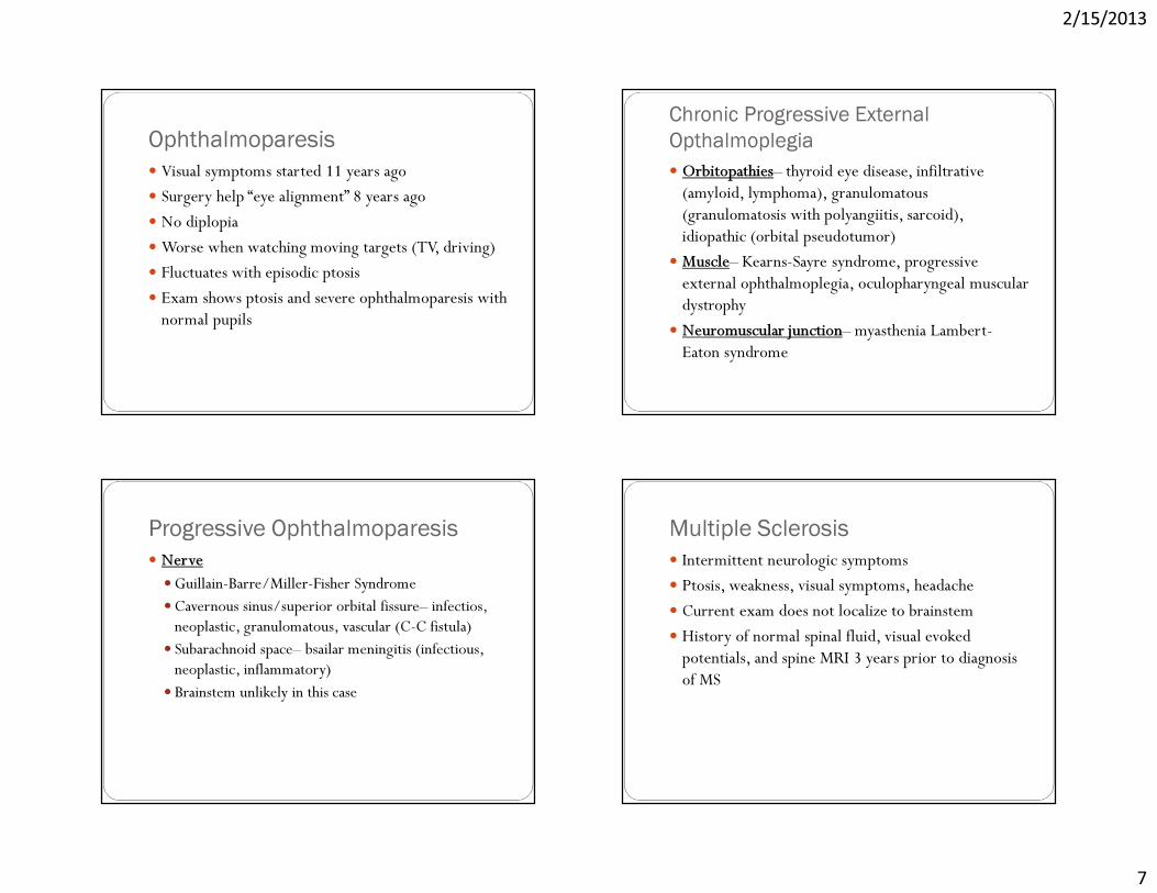
Ophthalmoparesis� Visual symptoms started 11 years ago� Surgery help “eye alignment” 8 years ago� No diplopia�Worse when watching moving targets (TV, driving)� Fluctuates with episodic ptosis� Exam shows ptosis and severe ophthalmoparesis with normal pupils
Chronic Progressive External Opthalmoplegia� Orbitopathies– thyroid eye disease, infiltrative (amyloid, lymphoma), granulomatous (granulomatosis with polyangiitis, sarcoid), idiopathic (orbital pseudotumor)� Muscle– Kearns-Sayre syndrome, progressive external ophthalmoplegia, oculopharyngeal muscular dystrophy� Neuromuscular junction– myasthenia Lambert-Eaton syndrome
Progressive Ophthalmoparesis� Nerve�Guillain-Barre/Miller-Fisher Syndrome�Cavernous sinus/superior orbital fissure– infectios, neoplastic, granulomatous, vascular (C-C fistula)� Subarachnoid space– bsailar meningitis (infectious, neoplastic, inflammatory)� Brainstem unlikely in this case
Multiple Sclerosis� Intermittent neurologic symptoms� Ptosis, weakness, visual symptoms, headache� Current exam does not localize to brainstem� History of normal spinal fluid, visual evoked potentials, and spine MRI 3 years prior to diagnosis of MS

2/15/2013
8
NMJ and Myasthenia Gravis?� Intermittent ptosis, opthalmoparesis, and weakness triggered by viral infection� Exam shows bifacial weakness, lower motor neuron speech, and neck flexion weakness� Cognitive decline and lack of diplopia are unusual for myasthenia� Normal AchR, MuSK, and EMG/NCS in 2004� Botulism and LEMS: normal reflexes, normal pupils, no autonomic symptoms, slow pace
Orbitopathies?� Thyroid eye disease� Infiltrative processes– amyloidosis, lymphoma� Granulomatous– granulomatosis with polyangiitis, sarcoid� Orbital pseudotumor� Infectious processes—cellulitis, fungal infections
Myopathies with PEO� Oculopharyngeal muscular dystrophy� Oculopharyngodistal myopathy� Myotonic dystrophy type 1� Progressive external ophthalmoplegia and Kearns-Sayre Syndrome� Other mitochondrial syndromes with PEO– POLG, MNGIE, Optic atrophy type 1, TWINKLE
Myopathies with PEO—AD � Oculopharyngeal muscular dystrophy� Oculopharyngodistal myopathy� Myotonic dystrophy type 1� Progressive external ophthalmoplegia and Kearns-Sayre Syndrome� Other mitochondrial syndromes with PEO– POLG, MNGIE, Optic atrophy type 1, TWINKLE

2/15/2013
9
Myopathies with PEO—AR � Oculopharyngeal muscular dystrophy� Oculopharyngodistal myopathy� Myotonic dystrophy type 1� Progressive external ophthalmoplegia and Kearns-Sayre Syndrome� Other mitochondrial syndromes with PEO– POLG, MNGIE, Optic atrophy type 1, TWINKLE
PEO & KSS� KSS�Onset < 20 years, PEO, pigmentary retinopathy, cardiac conduction block, high CSF protein, or cerebellar ataxia�Can also have dysphagia, hearing loss, migraine headaches, cognitive decline, endocrinopathies
� PEO—PEO plus limb weakness� PEO plus– PEO plus other features of KSS
Diagnostic Work-up� Orbital MRI: T1 Fat Saturated
Diagnostic Work-up� Orbital MRI: T1 Fat Saturated
ON

2/15/2013
10
Diagnostic Work-up� EMG/NCS
� Normal electrodiagnostic study
�Motor and sensory nerve conduction studies were within normal limits
�Repetitive nerve stimulation of the right facial nerve did not demonstrate defect
Diagnostic Work-up� Advanced Laboratories�Ach-R Ab negative
� Opthalmology consultation�Normal exam without retinal degeneration
� Muscle Biopsy
Andrew Bollen MDProfessor of Neuropathology
Muscle Biopsy

2/15/2013
11

2/15/2013
12

2/15/2013
13
Diagnostic Work-up Summary� EMG/NCS and serological testing did not support diagnosis of myasthenia� MRI suggested significant extraocular muscle atrophy� Ophthalmology evaluation was normal without evidence of retinal degeneration� Muscle biopsy demonstrated mitochondrial pathology
Final Diagnosis?
http://www.the-athenaeum.org/people/detail.php?ID=368

2/15/2013
14
CPEO� Clinical Features:� Onset in 30s� Insidious progressive symmetric ophthalmoplegiaoften without diplopia� Bilateral ptosis� Failure to respond to pyridostigmine� Approximately 50% of patients with systemic features
(McFarland et al., 2010)http://en.wikipedia.org/wiki/Albrecht_von_Graefe
CPEO
Pfeffer et al., 2011
� Associated non-ocular features:
CPEO� Significant psychosocial impact (Smits et al., 2011)
� Fatigue (67.9%)� Pain (96.2%)� Depression (32.1%)� Dependency in daily life (46.4%)
CPEO� Genetics:� Sporadic�Most often involve large mtDNA mutations�POLG mutations�Mitochondrial tRNA associated mutations
�Multiple dominant and recessive mutations identified�Associated with variant syndromes

2/15/2013
15
Progressive external ophthalmoplegia (PEO)- syndromes� Chronic PEO (CPEO)� PEO Plus� Kearns-Sayre Syndrome (KSS)
Taylor & Turnbull (2005)
Progressive external ophthalmoplegia (PEO)- syndromes
Taylor & Turnbull (2005)
PEO Plus Syndromes� PEO + Sensory Neuropathy:� Earlier onset from 10-30� Sensory ataxia, dysarthria, mild proximal weakness, and
arreflexia� Associated with POLG mutations
� tRNA-mutation associated PEO� Variable age of onset of PEO and ptosis� Associate features vary but can include migraine and short
stature
Kearns-Sayre Syndrome� Clinical Features� Onset < 20 (later onset extremely rare)� PEO, ptosis, pigmentary retinal degeneration� Proximal myopathy (90%)� Dysphagia (50%)� CNS: Ataxia (90%), dementia (85%)� Systemic: heart block and endocrine abnormalities
� Genetics� Sporadic large scale mtDNA mutations

2/15/2013
16
CPEO� Treatment and Surveillance�No clear benefit to CoQ10, creatine, L-carnitine�Often recommended given low risk of side effects�Moderate exercise may increase ratio of normal
mitochondria relative to mutant mitochondria within cells� Surgical correction of ptosis controversial� Strabismus surgery or botulinum toxin can be useful
in select patients� Yearly EKG and echocardiograms every 3-5 years� Periodic neuropsychological evaluation for cognitive
deficits
Follow-up� Weaned off steroids and pyridostigmine
� Persistent headache, malaise, and fatigue gradually improved
� Patient established care with local PMD for diabetes
� Involved in regular program of exercise
� Increased family support due to recognition of cognitive decline
Summary� Suspect PEO in patients with:� Onset of ophthalmoparesis and ptosis in 30-40s� Negative serological/electrophysiological studies � Poor response to typical myasthenia gravis therapies
� There is wide genotypic overlap and phenotypic variation in PEO, PEO+, and KSS
� Presence of systemic features may support mitochondrial etiology
� Useful diagnostic testing include: Brain/Orbital MRI, limb muscle biopsy with genetic analysis
Acknowledgments� Clinical Care Team� Dr. S. Andy Josephson� Dr. Nancy Oberheim-Bush� Dr. Kathryn Crozier� Shirin Golkar (UCSF MS3)� Dr. Catherine Lomen-Hoerth� Dr. Neil Simon� 8 South and Long nursing & rehab staff

2/15/2013
17
MRI Brain
MRI Brain MRI Brain

2/15/2013
18
MRI Brain CPEO� Associated non-ocular features:� Ataxia� Neuropathy� Generalized myopathy� Cognitive impairment� Deafness� Endocrinopathy (Hypothyroidism/T2DM)� Cardiac conduction defects
Diagnostic Work-up� MRI: Sagittal FLAIR

2/15/2013
19
Diagnostic Work-up� MRI: Sagittal FLAIR
Diagnostic Work-up� MRI: Axial T1 Post-gad
Recommended