By: Dr. Shkar R. Saeed
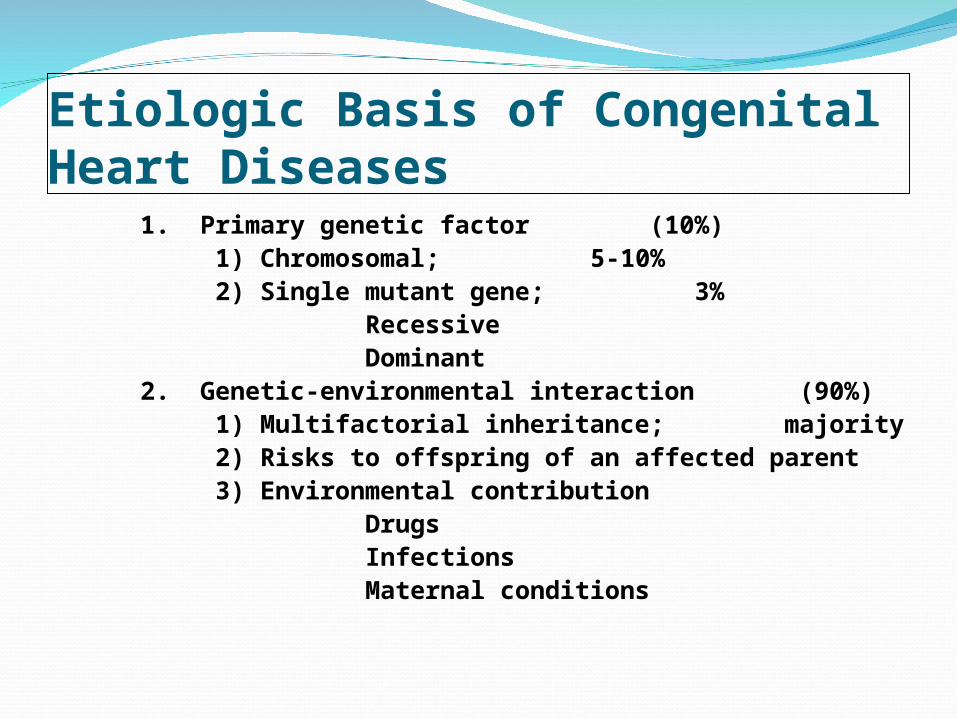
Etiologic Basis of Congenital Heart Diseases
1. Primary genetic factor (10%) 1) Chromosomal; 5-10% 2) Single mutant gene; 3% Recessive Dominant 2. Genetic-environmental interaction (90%) 1) Multifactorial inheritance; majority 2) Risks to offspring of an affected parent 3) Environmental contribution Drugs Infections Maternal conditions
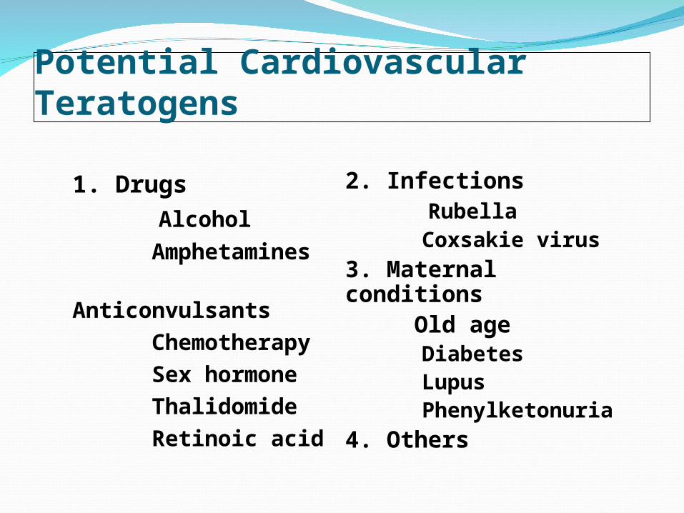
Potential Cardiovascular Teratogens
1. Drugs Alcohol Amphetamines
Anticonvulsants Chemotherapy Sex hormone Thalidomide Retinoic acid
2. Infections Rubella Coxsakie virus 3. Maternal
conditions Old age Diabetes Lupus Phenylketonuria 4. Others

Maternal Risk FactorsFactors Malformation Advanced age Trisomy 21
Maternal CHD Various Diabetes mellitus VSD, TGA,
cardiomyopathy SLE Heart block Phenylketonuria TOF, VSD, COA, HLHS Viruses Teratogenic,
myocarditis (*cytomegalovirus, herpes, coxsacki B, parvovirus)

Maternal Drug ExposuresDrug Malformation• Diphenylhydantoin PS, AS• Trimetadione VSD, TOF, TGA,
HLHS• Thalidomode TOF, Truncus
arteriosus • Lithium Ebstein anomalies• Alcohol VSD, ASD, PDA, TOF• Amphetamine VSD, ASD, PDA, TGA• Birth control pills VSD, TOF, TGA

classification of Congenital Heart Diseasesclassification of Congenital Heart Diseases
1. Lt to Rt Shunt ( 53 % ) PDA 17 % ASD 16.5 % VSD 13 % AVSD 3.5 %
2. Rt to Lt Shunt (11 % ) TOF 4.5 % TA 3 % PA+VSD 2.5 % PA+IVS 0.5 %
3. Admixture Lesion ( 15 % )
TGA 5 % Univ. Ht. 5 % DORV < 2 % Truncus 0.8 % Corrected TGA < 0.5 %
4. Obstructive Lesion ( 15 % )
Coarctation 9.5 % PS 2 % MS etc. 1.5 % LVOTO 1.3 % HLHS 0.9 % IAA 0.6 % 5. Valvular Lesion Ebstein < 1 % AR < 0.5 % MR < 0.5 % 6. Miscellaneous Arrhythmia 5 % Vascular ring 0.5 %

Evaluation of CHD by History Taking
1. Infants 1) Murmur 2) Symptoms of CHF poor feeding, low weight gain, tachypnea, tachycardia, sweating, anxiety, irritability, frequent
URI 3) Symptoms of
hypoxemia cyanosis, hypoxic spell
2. Children 1) Murmur 2) Symptoms of CHF exercise intolerance, dyspnea on exertion, frequent URI, palpitation 3) Syncope, chest pain 4) Symptoms of
Hypoxemia cyanosis, hypoxic
spell,clubbing
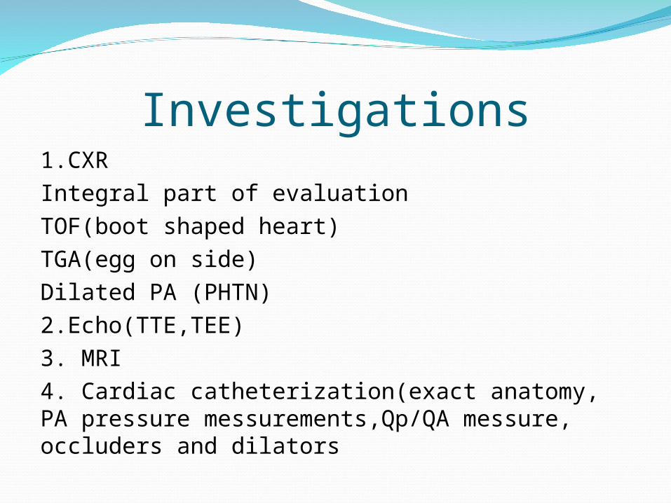
Investigations1.CXRIntegral part of evaluationTOF(boot shaped heart)TGA(egg on side)Dilated PA (PHTN)2.Echo(TTE,TEE)3. MRI4. Cardiac catheterization(exact anatomy, PA pressure messurements,Qp/QA messure, occluders and dilators

To Be Corrected in Neonate
Critical ASHypoplastic left heart syndrome Interrupted aortic archSymptomatic COATGATruncus ArteriosusOther symptomatic complex heart diseases

To Be Corrected in Infancy Cardiac anomalies with pulmonary
outflow tract obstruction
• Critical PS• Tricuspid atresia• TGA• TOF• PA with or without VSD• Corrected TGA


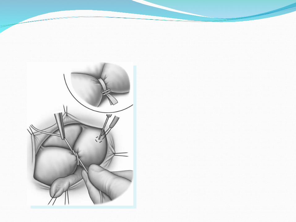
Palliative SurgerySystemic – pulmonary artery shunt Blalock-Taussig shunt
Cavopulmonary shunt (BCPS) Hemifontan & Fontan proceduresRVOT reconstruction Valvotomy
Patch widening Valved conduitPulmonary artery bandingAtrial septectomy

Systemic–Pulmonary Artery Shunt ( Qp<Qs i.e cyanosis)
Systemic–pulmonary artery shunt is indicated due to age, size, anatomy or other conditions when:
Complex anomaly with severe cyanosis, irritability, hypoxic episode
Critically ill neonates or infants due to decreased pulmonary flow
Facilitating growth of hypoplastic pulmonary artery

Pulmonary Artery BandingQp>Qs i.e more Pulm. flow
Pulmonary artery banding is indicated to decrease pulmonary blood flow & protect vascular disease
when:• Control of congestive heart failure Complex or multiple VSD (+/- coarctation) CPB medically contraindicated• Protection of pulmonary vascular bed
Atrial Septectomy For the increasing of effective pulmonary flow and systemic oxygen saturation
Indication of atrial septectomy : TGA Tricuspid atresia Pulmonary atresia MV and LV hypoplasiaDecreasing tendency of indication due to
early total correction or intervention

Reparative Surgery Non-open heart surgeryOpen heart surgery
Palliative procedure Corrective procedure
Anatomic correction Physiologic correction
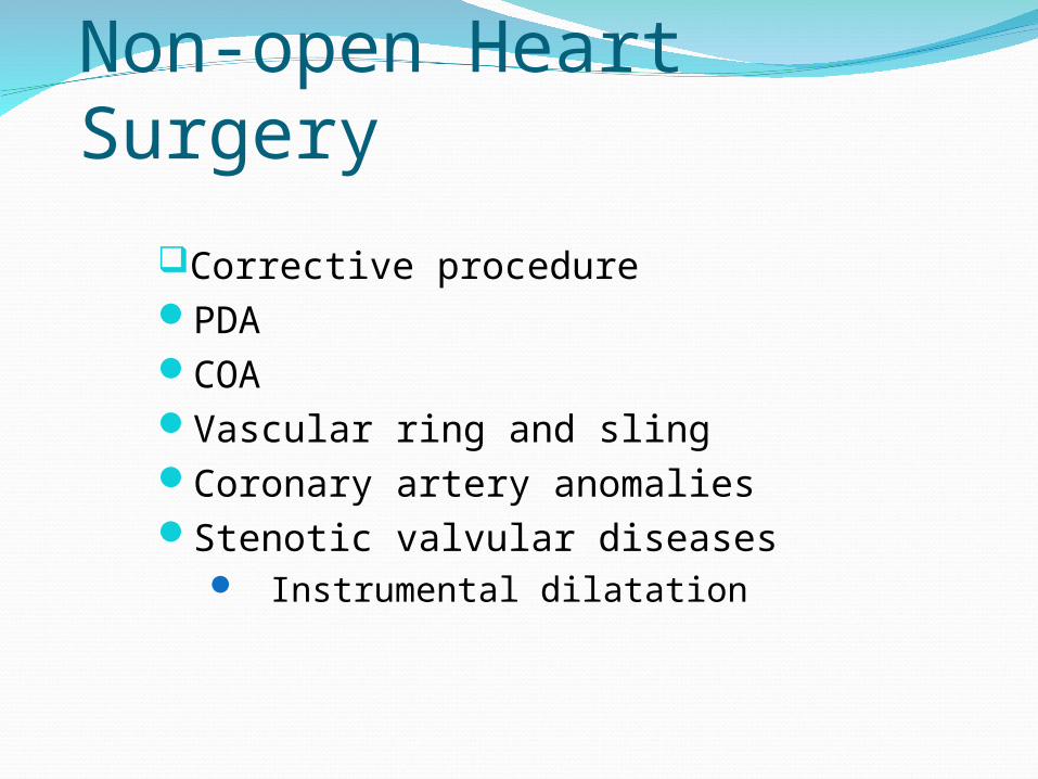
Non-open Heart Surgery
Corrective procedurePDACOAVascular ring and slingCoronary artery anomaliesStenotic valvular diseases
Instrumental dilatation

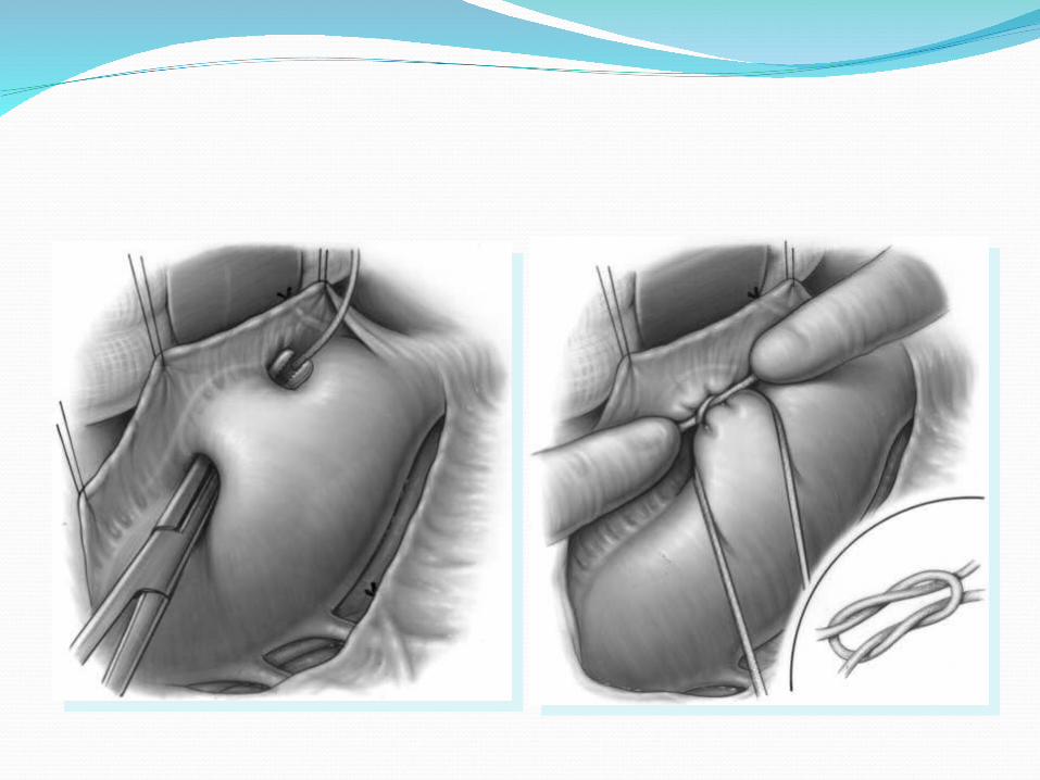
Patent Ductus ArteriosusOpen communication usually between upper
descending Aorta and proximal portion of LPA
Significant PDA : indicated after 1st monthProphylactic closure : 6-12 monthsSymptoms of heart failure or failure to
thrive: indicated at any timeSevere pulmonary vascular disease:
contraindicated


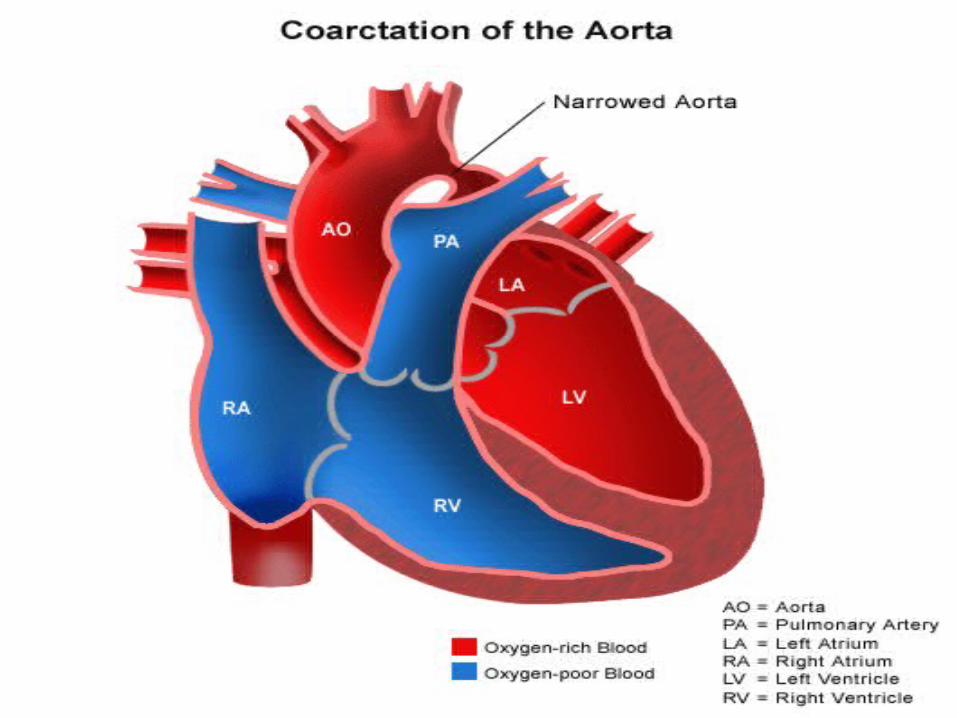
Coarctation of the AortaCongenital narrowing of upper thoracic aorta
adjacent to the ductus arteriosus
Operation is indicated when : Reduction of luminal diameter > 50% Upper body HT > 150mmHg in young
infant With CHF at any age COA with VSD COA with other important intracardiac defects One stage repair


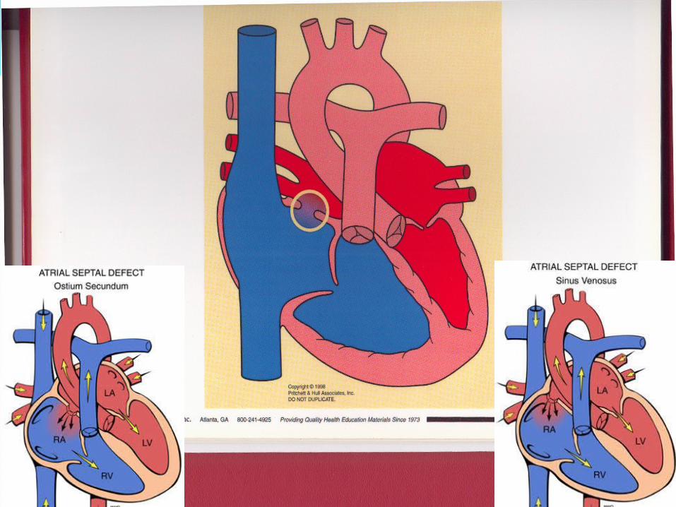
ASD with or without PAPVRA hole of variable size in the atrial septum and ismost common cardiac malformation with variouslocation of defect, fossa ovalis, posterior, ostium, primum, coronary sinus, subcaval (sinus venosus)
Uncomplicated ASD or of PAPVC with RV volume overload (Qp/Qs>1.5 or 2.0) : is an indication of
surgery.• Optimal age : under 5 years but recently 1-2 years
to avoid RV volume overload


Total Anomalous Pulmonary Venous Connection
These are no direct connection between anypulmonary vein and the LA. But rather, all thepulmonary veins connect to the RA or one of itstributaries
Diagnosis is an indication of operation• Immediate repair with Diagnosis in any ill neonate : Preoperative preparation is not needed• Repair should be done nearly always before 6 months• Diagnosis at 6-12 months: prompt repair is indicated
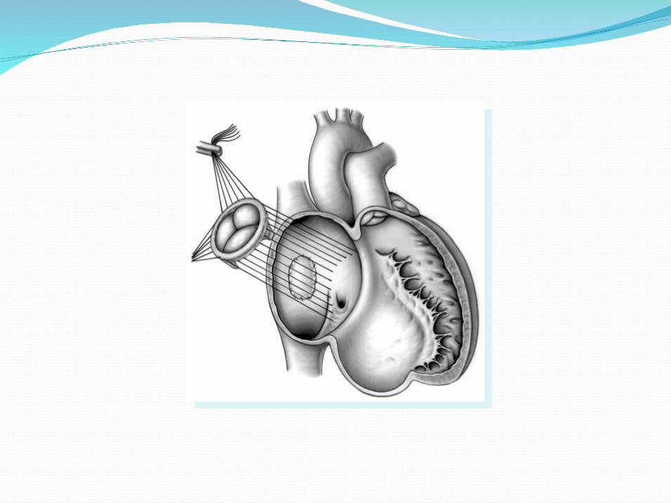
Ventricular Septal Defect A hole (or multiple holes) between Lt & Rt
ventricle
• Symptomatic large VSD : an indication of operation
• Before 3 months: indicated in large VSDs with CHF or respiratory symptoms
• Moderate sized VSDs (Qp/Qs < 3.0) with few symptoms : observation during infancy
• Small VSDs (Qp/Qs < 1.5) : not indicated, risk of bacterial endocarditis• Subarterial type : early repair is indicated before
childhood


Atrioventricular Septal DefectAbnormalities of atrioventricular valve form
& function and interatrial & interventricular communication from maldevelopment of the endocardial cushions
Presence of AVSD : indicated with Diagnosis• Partial AVSD : 1-2 years of age except CHF or
growth failure• Complete AVSD with good condition : 3-6
months
• Development of pulmonary vascular obstructive disease : not indicated

Congenital Aortic StenosisThe various forms of LVOTO occur in combination with
other cardiac lesions (IAA, COA, MV anomalies, LV hypoplasia) and obstructive types are supravalvular, valvular, subvalvular, intraventricular
• Critical AS in neonates : urgent (severe CHF, LV dilatation, hypertrophy)
• Infants and children Pressure gradient > 75mmHg Symtoms of angina, syncope, exercise intolerance,
LVH, pressure gradient > 50mmHg Pressure gradient over 40mmHg in subvalvular lesion to prevent progression

Ebsteins anomaly A congenital defect of tricuspid valve in which the
origin of septal and posterior leaflets or both are displaced downward into the right ventricle and the leaflets are variably deformed
Symptomatic Ebstein’s anomaly is an indication • Valve repair and ASD closure : with important TR, moderate and severe
cyanosis• WPW syndromes : ablation of accessory conduction pathway

Pulmonary Stenosis A form of RV outflow obstruction in which
stenosis is usually valvar or both valvar & subvalvularor only subvalvular
• Critical PS in neonate : indicated with Dx
Percutaneous balloon valvotomy Valvotomy with CPB Transannular RVOT patch widening
• PS in infants and children : indicated with Symptoms & Pr gradient over 50mmHg
• Surgical treatment is not indicated with mild stenosis

Tetralogy of FallotCharacterized by underdevelopment of RV
outflow
Diagnosis is an indication of operation• Symptomatic complicated in early life : Early total correction or Shunt (1-2 months) and total correction (1
year)• Asymptomatic uncomplicated : Total correction at 3-24 months• Multiple VSDs, LAD from RCA : Initial shunt and total correction
Double Outlet Right Ventricle A congenital cardiac anomalies which both great
arteries rise wholly or in large part from the RV. It is, then, a type of ventriculoarterial connection.
Dx is an indication of operation• Simple DORV with subaortic VSD : repair by 6
mo with PS --- repair like TOF• DORV with subpulmonic VSD (Taussig-Bing
heart) : arterial switch operation within 1 mo
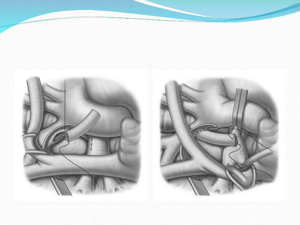
Transposition of Great Arteries A cardiac anomaly in which the Aorta arises
entirely or in large part from the RV, and PA from LV (atrioventricular concordant connection and ventriculoarterial discordant connection)
• Simple TGA in neonate : arterial switch operation within 1 months• Simple TGA beyond 30 days :
atrial switch operation (Mustard, Senning)• TGA with VSD :
arterial switch operation as early

Tricuspid Atresia A cardiac anomaly in which RV fails to open
into a ventricle through a AV valve. There is thus a univentricular AV connection PVR is an important indicator
> 4 unit -- contraindication2-4 unit -- BCPS< 2 unit -- Fontan operation
• Symptomatic in early life early shunt or PAB
BCPS or hemi-Fontan at 6-12 monthsFontan at 12-24 months
• AsymptomaticFontan candidate : 12-30 months

Congenitally Corrected TGA A cardiac anomaly with ventriculoarterial
discordant connection & atrioventricular discordant connection. The circulatory pathways are therefore in series
The presence of CCTGA per se is not an indication.• With VSD : indications for VSD• With VSD + PS : indications for TOF• With complete heart block : pacing• Double Switch operation : anatomic correction
Recommended