1
Brain Tumor Detection Using CT and MRI
Image Fusion
1ANJITHA P, 2Dr.SMITHA DHARAN
1M.Tech Student, Department of Computer Engineering,College of Engineering,Chengannur, Alapuzha ,Kerala
2Head of the Department, Department of Computer Engineering,College of Engineering,Chenagannur,Alapuzha,Kerala
Email: [email protected], [email protected]
Contact: 18547258203, 29947061826
Abstract: Image fusion is the integration of information from multiple images into a single image that gives more complete and accurate description about the object. Computed Tomography (CT), Magnetic Resonance Imaging (MRI), PET, X-ray are different medical imaging techniques. MRI gives best information about soft tissues and CT gives information about hard tissues. Therefore, the fusion of these images helps the physicians for better diagnosis. This paper presents a method of brain tumor detection based on image fusion. First, fusion is done based on NSCT domain. Second, segmentation is done based on SOM (Self Organization Map) clustering. Then, classification is performed.
Index terms: ComputedTomography(CT), Magnetic Resonance Imaging (MRI), PET, X-ray, NSCT (Non-
subsampled Contourlet Transform).
I. INTRODUCTION
One of the serious kinds of disease in the medical
field is considered to be the brain tumor. So it must to
have the fast and accurate detection. Different
algorithms are provided for the tumour detection and
segmentation. The important approach in the brain
tumor segmentation is to identify the various stages
includes benign, malignant and the normal. The brain
tumor are generally named and classified according
to either of the following: The type of brain cells in
which they originate The type of location in which
the cancer develops. Primary tumors are those which
develop in the brain. The initial growth of the
abnormal and the unwanted tissues in the brain is
called as the primary tumor.
Depending on the concentration the primary tumor
are classified into two types. Benign tumor is a tumor
where they are having their boundaries or the edges
in which they does not spread over the other parts of
the body. Benign tumor is considerably quite serious
if they are meant to be in the vital areas of the brain.
On another hand, benign tumor can step in to the
disability and even it lead to the death. In malignant
tumor are considered to be the most serious one and
they develop rapidly. They affect the various vital
organs which may leads to the death. About 80 of the
malignant tumors are referred to as the gliomas.
Gliomas refer to the tumors which have been
originated from the glial cells of the brain. Secondary
brain tumor is a tumor where the tumor in the brain is
arisen from the other tumor in the body. They are
mainly formed from the cells that have broken away
from the primary tumor and have spread in the
bloodstream to the brain. The primary source for the
secondary tumor is the lung or the blood cancer.
Currently, MRI and CT are the standard diagnosing
techniques to detect brain tumor. CT provides best
information about denser tissues. MRI offers better
information about soft tissues. Combining these two
images will generate an image that can offer more
information than each other separate. The final
obtained image can be useful in diagnosis process.
That„s why image fusion has become an important
research field.
Figure 1:CT Image
International Journal of Scientific & Engineering Research Volume 10, Issue 1, January-2019 ISSN 2229-5518
555
IJSER © 2019 http://www.ijser.org
IJSER
2
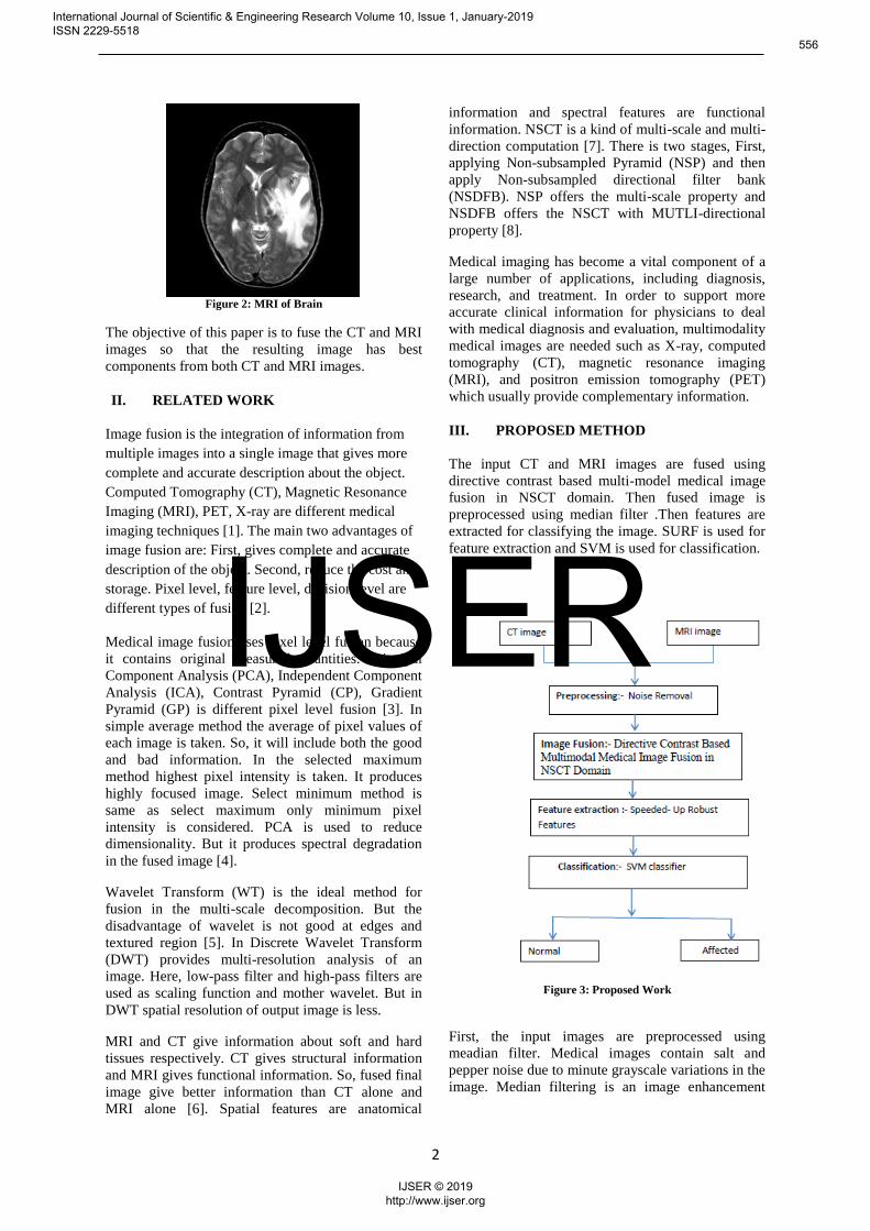
Figure 2: MRI of Brain
The objective of this paper is to fuse the CT and MRI
images so that the resulting image has best
components from both CT and MRI images.
II. RELATED WORK
Image fusion is the integration of information from
multiple images into a single image that gives more
complete and accurate description about the object.
Computed Tomography (CT), Magnetic Resonance
Imaging (MRI), PET, X-ray are different medical
imaging techniques [1]. The main two advantages of
image fusion are: First, gives complete and accurate
description of the object. Second, reduce the cost and
storage. Pixel level, feature level, decision level are
different types of fusion [2].
Medical image fusion uses pixel level fusion because
it contains original measured quantities. Principal
Component Analysis (PCA), Independent Component
Analysis (ICA), Contrast Pyramid (CP), Gradient
Pyramid (GP) is different pixel level fusion [3]. In
simple average method the average of pixel values of
each image is taken. So, it will include both the good
and bad information. In the selected maximum
method highest pixel intensity is taken. It produces
highly focused image. Select minimum method is
same as select maximum only minimum pixel
intensity is considered. PCA is used to reduce
dimensionality. But it produces spectral degradation
in the fused image [4].
Wavelet Transform (WT) is the ideal method for
fusion in the multi-scale decomposition. But the
disadvantage of wavelet is not good at edges and
textured region [5]. In Discrete Wavelet Transform
(DWT) provides multi-resolution analysis of an
image. Here, low-pass filter and high-pass filters are
used as scaling function and mother wavelet. But in
DWT spatial resolution of output image is less.
MRI and CT give information about soft and hard
tissues respectively. CT gives structural information
and MRI gives functional information. So, fused final
image give better information than CT alone and MRI alone [6]. Spatial features are anatomical
information and spectral features are functional
information. NSCT is a kind of multi-scale and multi-
direction computation [7]. There is two stages, First,
applying Non-subsampled Pyramid (NSP) and then
apply Non-subsampled directional filter bank
(NSDFB). NSP offers the multi-scale property and
NSDFB offers the NSCT with MUTLI-directional
property [8].
Medical imaging has become a vital component of a
large number of applications, including diagnosis,
research, and treatment. In order to support more
accurate clinical information for physicians to deal
with medical diagnosis and evaluation, multimodality
medical images are needed such as X-ray, computed
tomography (CT), magnetic resonance imaging
(MRI), and positron emission tomography (PET)
which usually provide complementary information.
III. PROPOSED METHOD
The input CT and MRI images are fused using
directive contrast based multi-model medical image
fusion in NSCT domain. Then fused image is
preprocessed using median filter .Then features are
extracted for classifying the image. SURF is used for
feature extraction and SVM is used for classification.
Figure 3: Proposed Work
First, the input images are preprocessed using
meadian filter. Medical images contain salt and
pepper noise due to minute grayscale variations in the
image. Median filtering is an image enhancement
International Journal of Scientific & Engineering Research Volume 10, Issue 1, January-2019 ISSN 2229-5518
556
IJSER © 2019 http://www.ijser.org
IJSER
3
techniques to remove impulse noise without affecting
image sharpness.
The proposed fusion is done based on two fusion
rules. Low- frequency coefficients are combined
using phase congruency. High frequency coefficients
are combined using directive contrast. Phase
congruency selects contrast-and-brightness invariant
components in the low frequency coefficient.
Directive contrast selects the texture and edge
information from the high frequency coefficients.
NSCT is a kind of multi-scale and multi-direction
computation. There is two stages, First, applying
Non-subsampled Pyramid (NSP) and then apply Non-
subsampled directional filter bank (NSDFB). NSP
offers the multi-scale property and NSDFB offers the
NSCT with MUTLI-directional property.
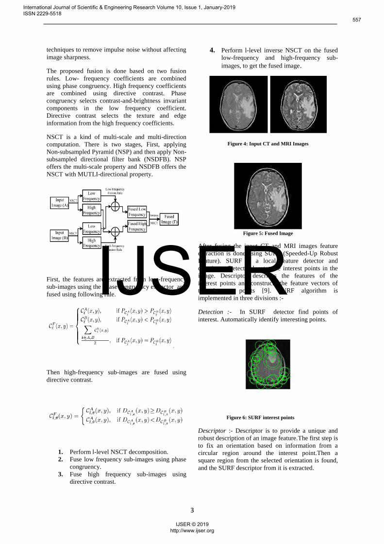
First, the features are extracted from low-frequency
sub-images using the phase congruency extractor and
fused using following rule.
Then high-frequency sub-images are fused using
directive contrast.
1. Perform l-level NSCT decomposition.
2. Fuse low frequency sub-images using phase
congruency.
3. Fuse high frequency sub-images using
directive contrast.
4. Perform l-level inverse NSCT on the fused
low-frequency and high-frequency sub-
images, to get the fused image.
Figure 4: Input CT and MRI Images
Figure 5: Fused Image
After fusing the input CT and MRI images feature
extraction is done using SURF (Speeded-Up Robust
Feature). SURF is a local feature detector and
descriptor. Detector locates the interest points in the
image. Descriptor describes the features of the
interest points and constructs the feature vectors of
the interest points [9]. SURF algorithm is
implemented in three divisions :-
Detection :- In SURF detector find points of
interest. Automatically identify interesting points.
Figure 6: SURF interest points
Descriptor :- Descriptor is to provide a unique and
robust description of an image feature.The first step is
to fix an orientation based on information from a
circular region around the interest point.Then a
square region from the selected orientation is found,
and the SURF descriptor from it is extracted.
International Journal of Scientific & Engineering Research Volume 10, Issue 1, January-2019 ISSN 2229-5518
557
IJSER © 2019 http://www.ijser.org
IJSER

4
Figure 7: Key points of interest points.
Matching:- Matching pairs are found using the
comparison of descriptors obtained from various
images.
Finally, classification using svm classifier is done.It
is a machine learning approach used for classification
and regression analysis. Supervised learning model
and trained by learning algorithm. Analyses large
amount of data and find pattern from them. Divides
into two categories by a clear gap.( partitioning by a
plane called hyper plane) [10]. SVM creates hyper
planes that have the largest margin in a high
dimensional space. Larger margin implies lower error
of the classifier.
IV. EXPERIMENTAL ANALYSIS
In this paper, for the purpose of detection of brain
tumor, we have chosen medical cases with patients
who have suffered from different brain tumors, where
in the tumor is diagnosed in fused image of CT and
MRI of a same patient. The proposed algorithm is
developed using MATLAB.
Figure 8: Final Result .
Figure 7: Performance Comparison
V. CONCLUSION
Medical imaging techniques became crucial for
medical diagnosis. CT and MRI image fusion
technique is more precise than either by CT alone or
by MRI. MRI-CT fusion can reduce the uncertainty
of brain tumor detection. The proposed system helps
in easy diagnosis of normal and abnormal brain.
REFERENCES [1] Deepak Kumar Sahu, M.P.Parsai, ”Different Image Fusion Techniques – A Critical review“,International journal of Modern
Engineering Research (IJMER) Vol.2, Issue. 5, Sep.-Oct.2012pp-
4298-4301.
[2] A.P.James, B.V. Dasarathy, “Medical Image Fusion – A
Survey of the State of the Art”, Information Fusion,2014.
[3] Bhavana.V, Krishnappa H.K, "Multi-Modality Medical Image Fusion using Discrete Wavelet Transform", 4th International
Conference on Eco-friendly Computing and Communication Systems, ICECCS 2015, Procedia Computer Science,pp.625-
631,2015
[4] Shutao Li, “Image Fusion with Guided Filtering”, IEEE
Transactions On Image Processing, Vol. 22, No. 7, July 2013
[5] Yufeng Zheng , Edward A. Essock et, “An Advanced Image
Fusion Algorithm Based on Wavelet Transform – Incorporation
with PCA and Morphological Processing”,SPIE5298,image processing:
algorithms systems
III,Vol.5298,2004.
[6] K Sharmila, S Rajkumar, V Vij ayaraj an, "Hybrid method for multimodality medical image fusion using Discrete Wavelet
Transform and Entropy concepts with Quantitative Analysis",
International conference on Communication and Signal Processing, pp.489-493, April 2013.
[7] S. Singh, D. Gupta, R. S. Anand and V. Kumar, “Nonsubsampled Shearlet based CT and MR Medical Image
Fusion using Biologically Inspired Spiking Neural Network,”
Biom. Signal Processing and Control, Vol. 18, pp. 91-101, April 2015.
[8] L. Yang, B. L. Guo, and W. Ni, “Multimodality medical
image fusion based on multiscale geometric analysis of contourlet
transform,” Neurocomputing, vol. 72, pp. 203–211, 2008.
[9] H. Bay, A. Ess, T. Tuytelaars, L. Van Gool, ”Speeded-up
robust features (SURF)”, Comput. Vis. Image Underst., 110(3),
346-359 (2008).
International Journal of Scientific & Engineering Research Volume 10, Issue 1, January-2019 ISSN 2229-5518
558
IJSER © 2019 http://www.ijser.org
IJSER
5
[10] Chih-Chung Chang, Chih-Wei Hsu, and Chih-Jen Lin. The
analysis of decomposition methods for support vector machines. IEEE Transactions on Neural Networks, 11(4):10031008, 2000.
International Journal of Scientific & Engineering Research Volume 10, Issue 1, January-2019 ISSN 2229-5518
559
IJSER © 2019 http://www.ijser.org
IJSER
Recommended