BONES
OF THE
FOOT
& ANKLE
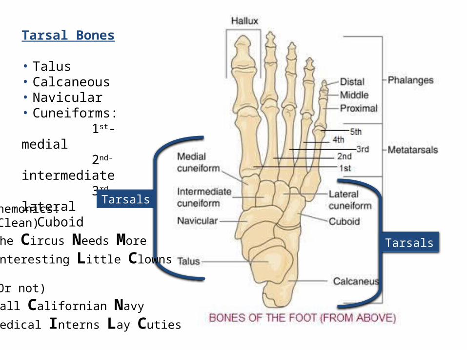
Tarsal Bones
• Talus• Calcaneous• Navicular• Cuneiforms: 1st-medial 2nd-intermediate 3rd- lateral Cuboid
Mnemonics:(Clean)
The Circus Needs More
Interesting Little Clowns
(Or not)
Tall Californian Navy
Medical Interns Lay Cuties
Tarsals
Tarsals
Calcaneus
Talus
Navicular Cuboid
1 32Metatarsals 4 5
Cuneiforms
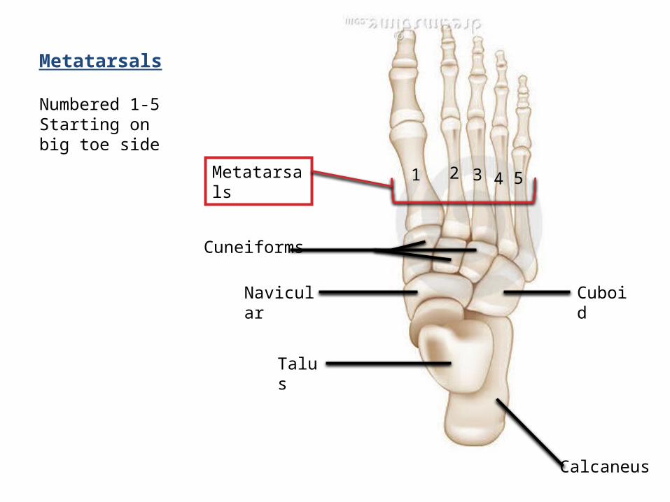
Metatarsals
Numbered 1-5Starting on big toe side

Metatarsals
Proximal phalangeal bones
Distal phalangeal bones
Phalangeal bones 14 totalAlso numbered 1-5 with big toe being #1
Proximal- (5) closest to metatarsals
Middle-(4) between proximal and distal phalanxOnly 4--like the thumb on the hand the big toe doesn’t have a middle phalanx
Distal-(5)tip of the toe
Middle phalangeal bones
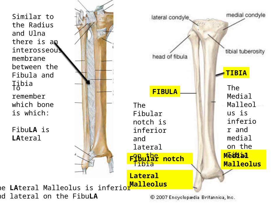
Lateral Malleolus
Fibular notch Medial Malleolus
The Fibular notch is inferior and lateral on the Tibia
The Medial Malleolus is inferior and medial on the Tibia
Similar to the Radius and Ulna there is an interosseous membrane between the Fibula and Tibia
The LAteral Malleolus is inferior and lateral on the FibuLA
To remember which bone is which:
FibuLA is LAteral
FIBULA
TIBIA
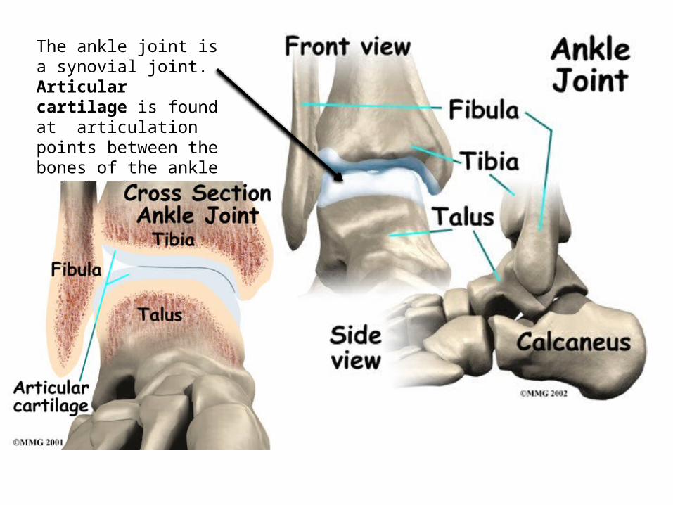
The ankle joint is a synovial joint. Articular cartilage is found at articulation points between the bones of the ankle and the foot
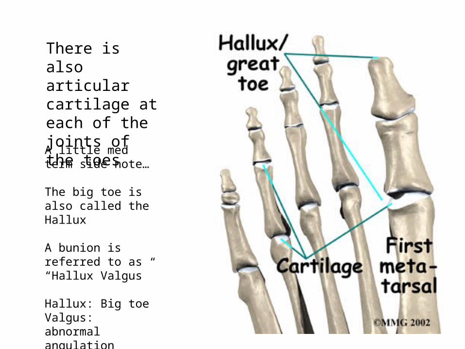
There is also articular cartilage at each of the joints of the toes
A little med term side note…
The big toe is also called the Hallux
A bunion is referred to as “Hallux Valgus”
Hallux: Big toeValgus: abnormal angulation
MetatarsophalangealMTP
Proximal interphalangeal PIPDistal interphalangeal
DIP
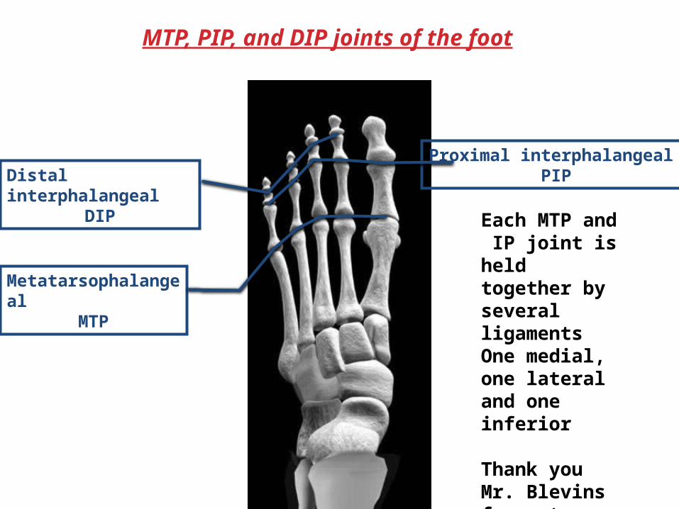
MTP, PIP, and DIP joints of the foot
Each MTP and IP joint is held together by several ligamentsOne medial, one lateral and one inferior
Thank you Mr. Blevins for not putting them all on our list!!
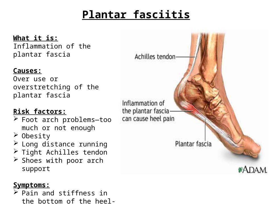
Plantar fasciitis
What it is:Inflammation of the plantar fascia
Causes:Over use or overstretching of the plantar fascia
Risk factors: Foot arch problems—too much or
not enough Obesity Long distance running Tight Achilles tendon Shoes with poor arch support
Symptoms: Pain and stiffness in the bottom of
the heel-may be dull or sharp Bottom of foot may ache or burn
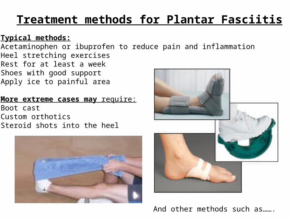
Treatment methods for Plantar FasciitisTypical methods:Acetaminophen or ibuprofen to reduce pain and inflammationHeel stretching exercisesRest for at least a weekShoes with good supportApply ice to painful area
More extreme cases may require:Boot castCustom orthoticsSteroid shots into the heel
And other methods such as…….
The redneck treatment method
To each his own…..
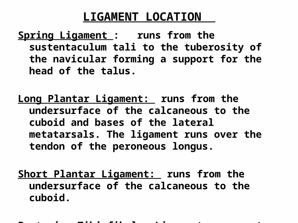
LIGAMENT LOCATION
Spring Ligament : runs from the sustentaculum tali to the tuberosity of the navicular forming a support for the head of the talus.
Long Plantar Ligament: runs from the undersurface of the calcaneous to the cuboid and bases of the lateral metatarsals. The ligament runs over the tendon of the peroneous longus.
Short Plantar Ligament: runs from the undersurface of the calcaneous to the cuboid.
Posterior Tibiofibular Ligament: connects the tibia and fibula posteriorly. It is located on the lateral aspect of the ankle.
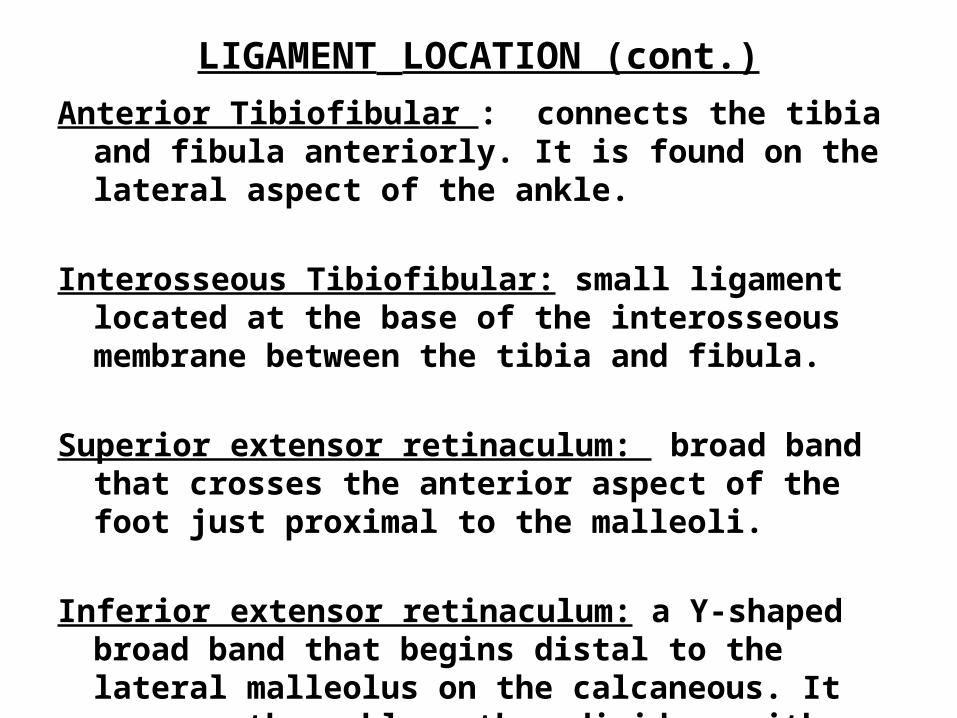
LIGAMENT LOCATION (cont.)Anterior Tibiofibular : connects the tibia and fibula anteriorly.
It is found on the lateral aspect of the ankle.
Interosseous Tibiofibular: small ligament located at the base of the interosseous membrane between the tibia and fibula.
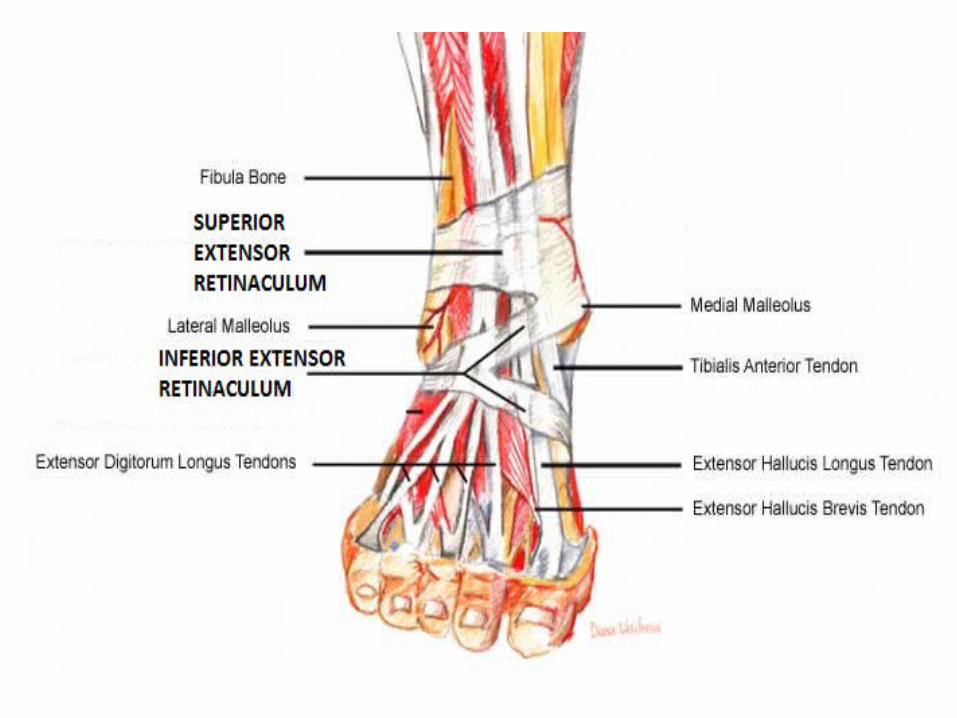
Superior extensor retinaculum: broad band that crosses the anterior aspect of the foot just proximal to the malleoli.
Inferior extensor retinaculum: a Y-shaped broad band that begins distal to the lateral malleolus on the calcaneous. It crosses the ankle , then divides, with one end attaching at the medial malleolus. The other fork connects to the navicular.
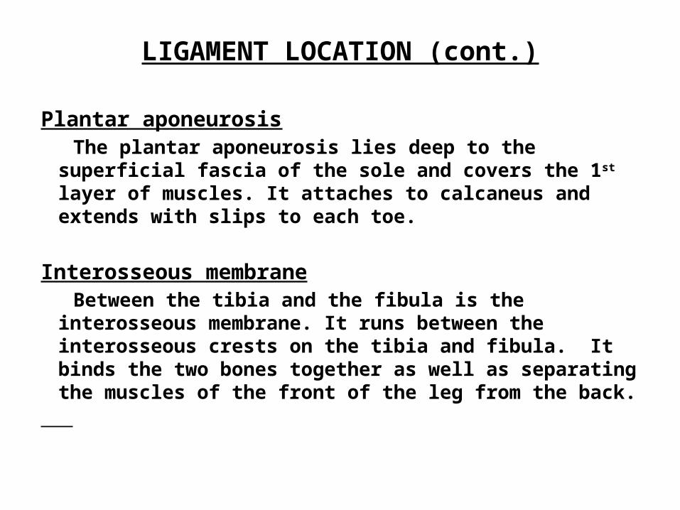
LIGAMENT LOCATION (cont.)
Plantar aponeurosis The plantar aponeurosis lies deep to the superficial fascia of
the sole and covers the 1st layer of muscles. It attaches to calcaneus and extends with slips to each toe.
Interosseous membrane Between the tibia and the fibula is the interosseous
membrane. It runs between the interosseous crests on the tibia and fibula. It binds the two bones together as well as separating the muscles of the front of the leg from the back.
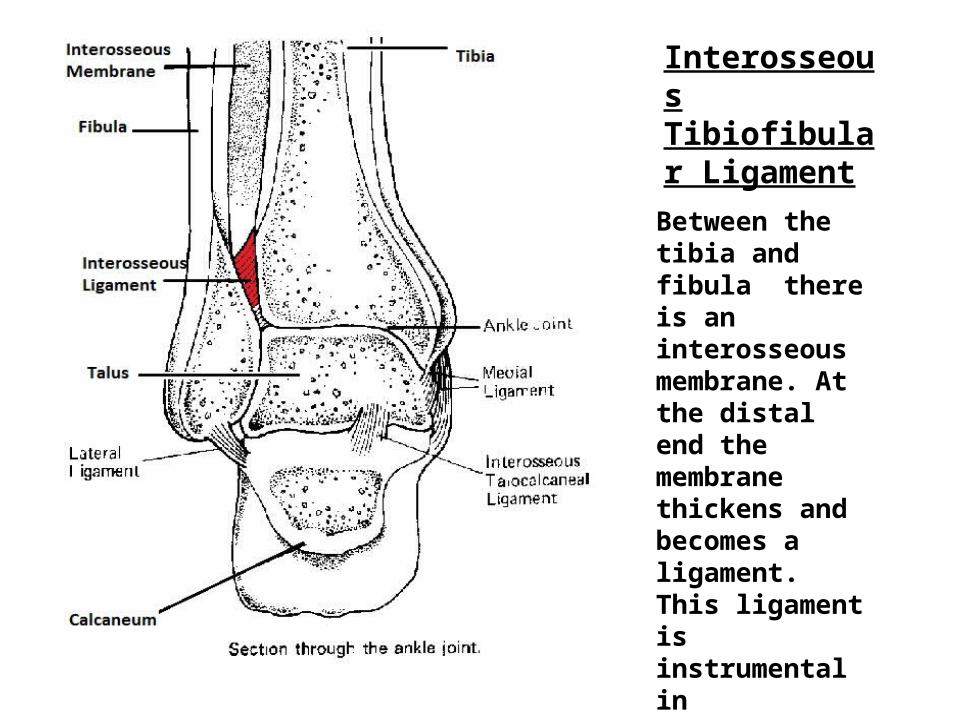
Interosseous Tibiofibular Ligament
Between the tibia and fibula there is an interosseous membrane. At the distal end the membrane thickens and becomes a ligament.This ligament is instrumental inholding these two bones together.
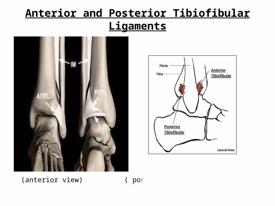
Anterior and Posterior Tibiofibular Ligaments
(anterior view) ( posterior view)
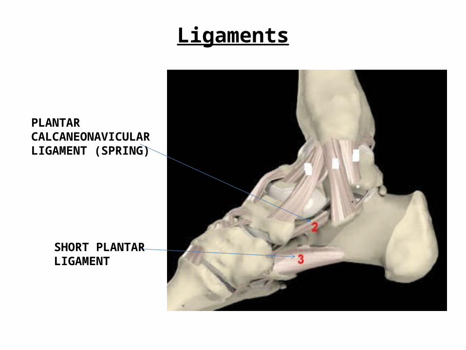
Ligaments
SHORT PLANTARLIGAMENT
PLANTARCALCANEONAVICULARLIGAMENT (SPRING)
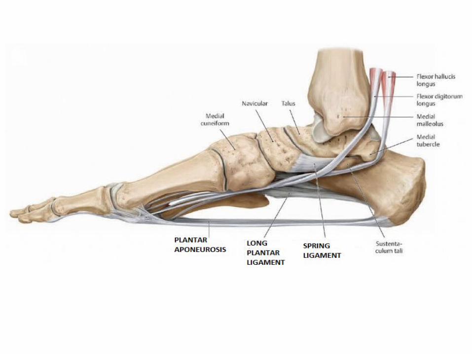
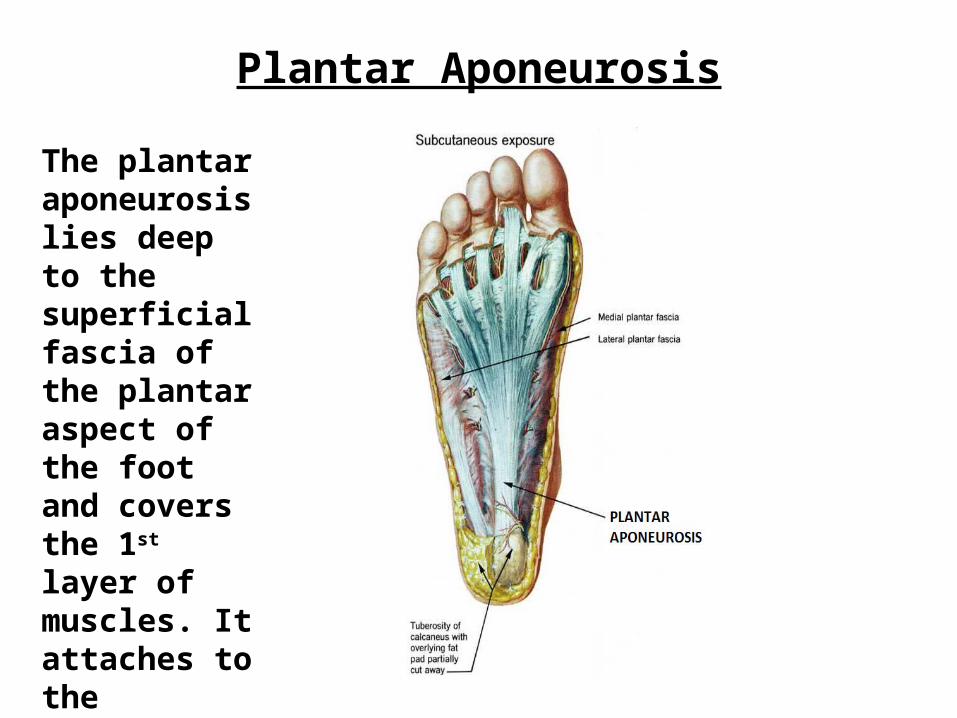
Plantar Aponeurosis
The plantar aponeurosislies deep to the superficial fascia of the plantar aspect of the foot and covers the 1st layer of muscles. It attaches to the calcaneus and sends a deep slip to each toe.
Bursae
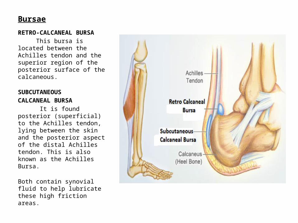
RETRO-CALCANEAL BURSA This bursa is located between the Achilles tendon and the superior region of the posterior surface of the calcaneous.
SUBCUTANEOUSCALCANEAL BURSA It is found posterior (superficial) to the Achilles tendon, lying between the skin and the posterior aspect of the distal Achilles tendon. This is also known as the Achilles Bursa.
Both contain synovial fluid to help lubricate these high friction areas.
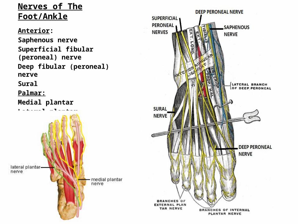
Nerves of The Foot/Ankle
Anterior:Saphenous nerveSuperficial fibular (peroneal) nerveDeep fibular (peroneal) nerveSuralPalmar:Medial plantarLateral plantar
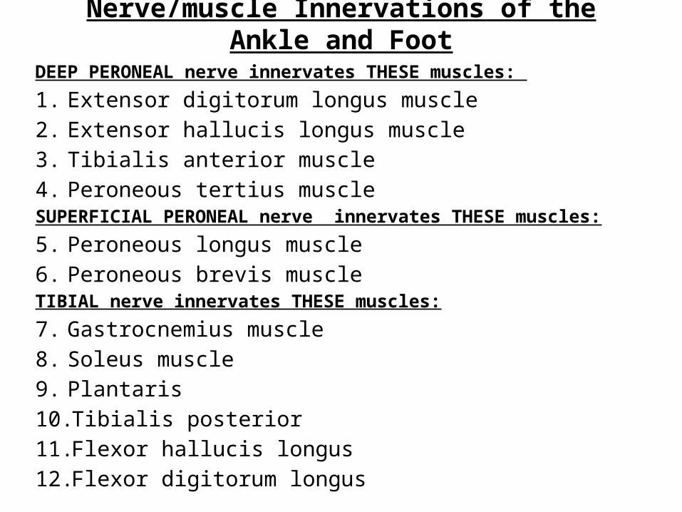
Nerve/muscle Innervations of the Ankle and Foot
DEEP PERONEAL nerve innervates THESE muscles:
1. Extensor digitorum longus muscle2. Extensor hallucis longus muscle3. Tibialis anterior muscle4. Peroneous tertius muscleSUPERFICIAL PERONEAL nerve innervates THESE muscles:
5. Peroneous longus muscle6. Peroneous brevis muscleTIBIAL nerve innervates THESE muscles:
7. Gastrocnemius muscle8. Soleus muscle9. Plantaris10. Tibialis posterior11. Flexor hallucis longus12. Flexor digitorum longus
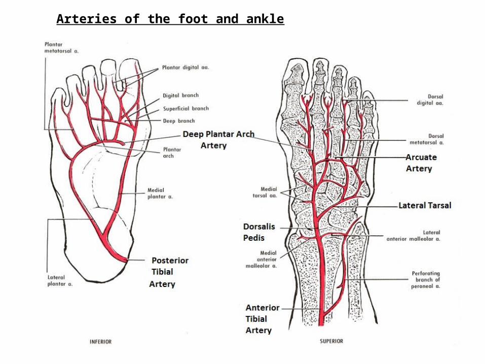
Arteries of the foot and ankle
Arteries of the Ankle and Foot
Anterior Tibial ArteryThis artery originates as the popliteal artery and becomes the anterior tibial artery. It descends the tibia on the anteriomedial aspect of the tibia and becomes the dorsalis pedis at the ankle.Dorsalis PedisThis is a continuation of the anterior tibial artery. It begins at the ankle and runs anteriomedially. The dorsalis pedis is a main blood supply to the foot.Posterior Tibial ArteryThis artery is a continuation of the popliteal artery. It begins at the inferior border of the popliteus and descends medially through the calf of the leg. It enters the foot at the medial side of the calcaneus .
Arteries of the Ankle and Foot (cont.)Arcuate Artery One of the branches of the dorsalis pedis is this artery. It runs
anteriorly and transversely at the base of the metatarsal bones towards the lateral aspect of the foot.
Deep Plantar Arch Artery (NOTE: Vein of same name accompanies)
Both the arcuate and deep planter arteries branch at the base of the metatarsal of the big toe. The deep plantar artery runs into the plantar aspect of the foot . It travels transversly across the remaining metatarsals towards the lateral side of the foot.
Lateral Tarsal ArteryA branch of the dorsalis pedis, this artery begins at the midline of
the talus anteriorly and runs at an angle towards the lateral aspect of the foot.
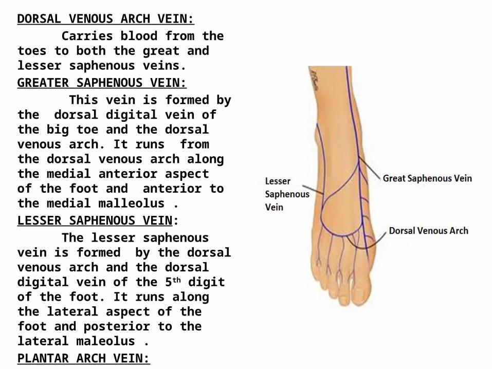
DORSAL VENOUS ARCH VEIN: Carries blood from the toes to both the great and lesser saphenous veins. GREATER SAPHENOUS VEIN: This vein is formed by the dorsal digital vein of the big toe and the dorsal venous arch. It runs from the dorsal venous arch along the medial anterior aspect of the foot and anterior to the medial malleolus .LESSER SAPHENOUS VEIN: The lesser saphenous vein is formed by the dorsal venous arch and the dorsal digital vein of the 5th digit of the foot. It runs along the lateral aspect of the foot and posterior to the lateral maleolus .PLANTAR ARCH VEIN: This vein is not pictured here, however, with each artery there is a co-ordinating vein. SEE NEXT SLIDE. It is located on the plantar side of the foot, running next to the plantar arch artery.
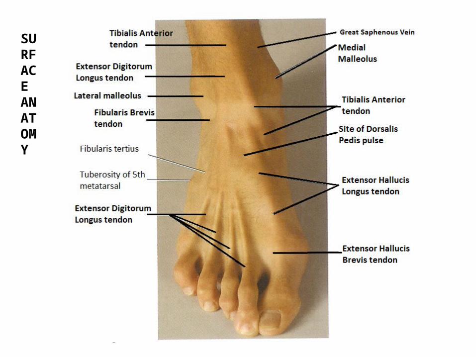
SURFACE ANATOMY
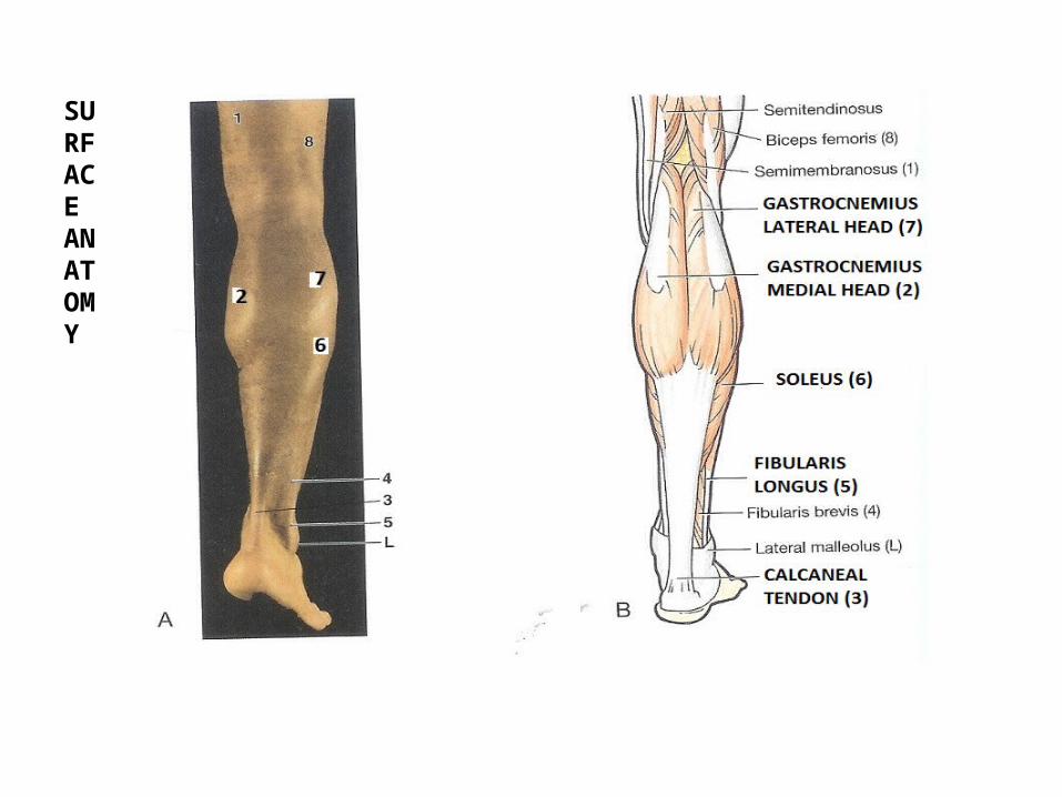
SURFACE ANATOMY
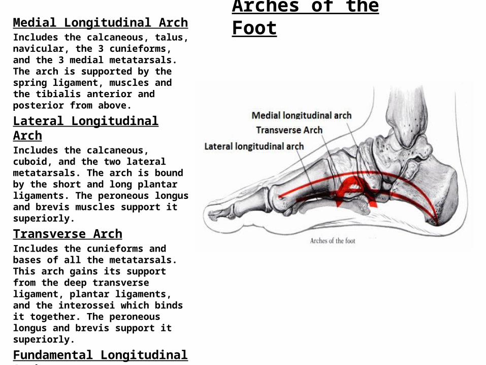
Arches of the FootMedial Longitudinal ArchIncludes the calcaneous, talus, navicular, the 3 cunieforms, and the 3 medial metatarsals. The arch is supported by the spring ligament, muscles and the tibialis anterior and posterior from above.
Lateral Longitudinal ArchIncludes the calcaneous, cuboid, and the two lateral metatarsals. The arch is bound by the short and long plantar ligaments. The peroneous longus and brevis muscles support it superiorly.
Transverse ArchIncludes the cunieforms and bases of all the metatarsals. This arch gains its support from the deep transverse ligament, plantar ligaments, and the interossei which binds it together. The peroneous longus and brevis support it superiorly.
Fundamental Longitudinal ArchThis arch is comprised of the calcaneus, cuboid, 3rd cunieform, and 3rd metatarsal. These bones run through the center of the foot and form the basic longitudinal arch of the foot.
A Little “Footnote”
The human foot and ankle is a strong and complex mechanical structure containing more than 26 bones, 33 joints (20 of which are actively articulated), and more than a hundred muscles, tendons, and ligaments.
Let’s all be grateful they are NOT all on the test!
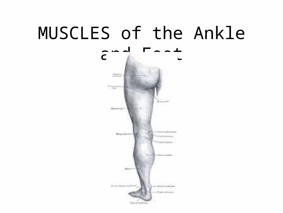
MUSCLES of the Ankle and Foot
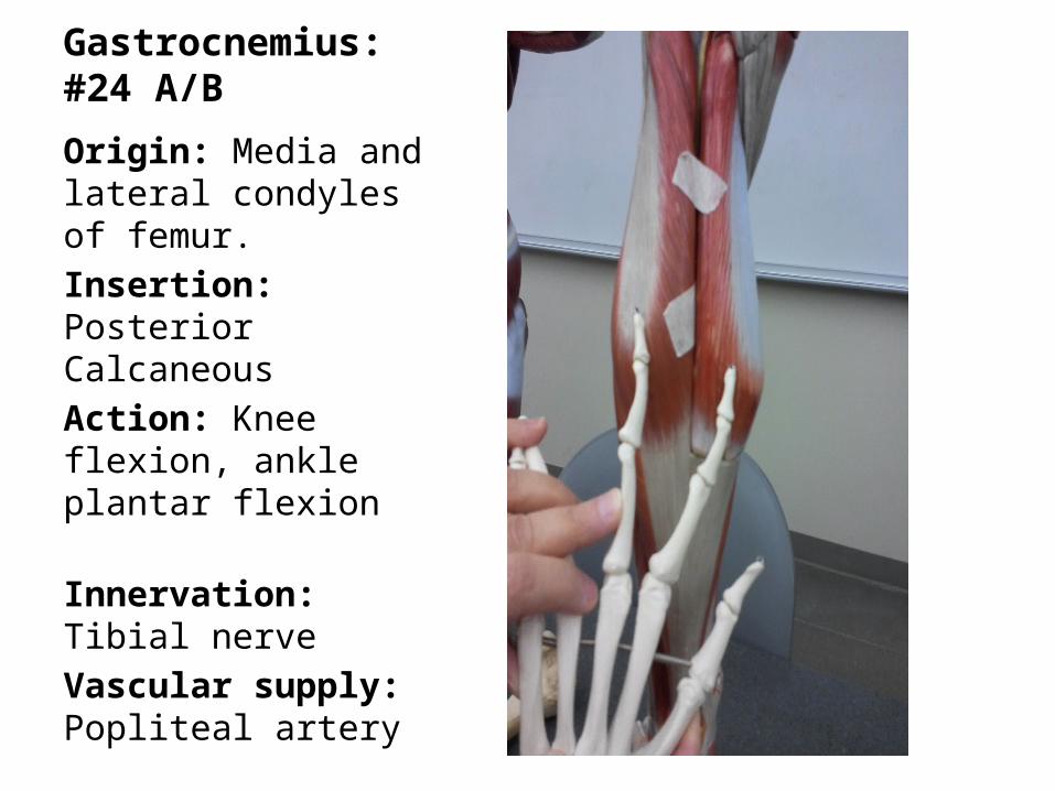
Gastrocnemius: #24 A/B
Origin: Media and lateral condyles of femur.Insertion: Posterior CalcaneousAction: Knee flexion, ankle plantar flexion Innervation: Tibial nerveVascular supply: Popliteal artery
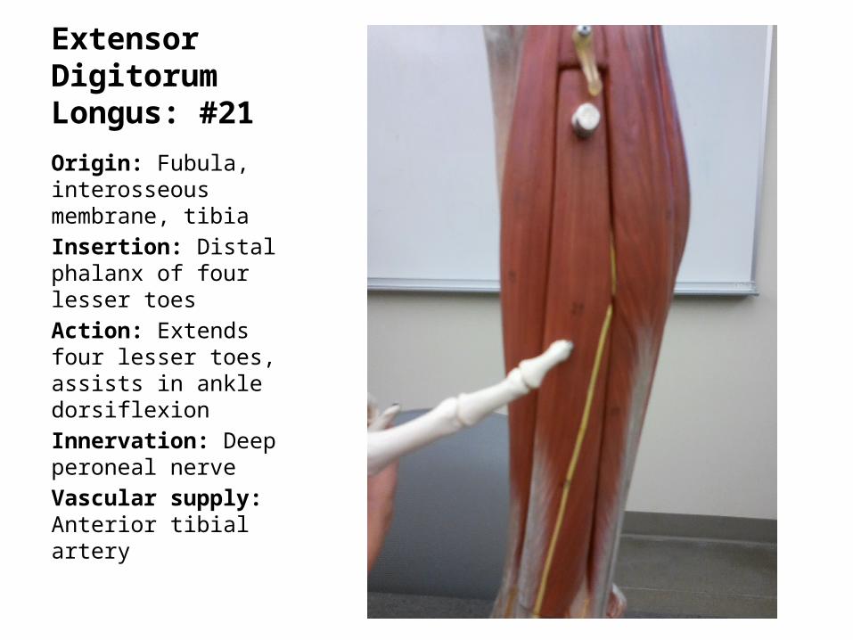
Extensor Digitorum Longus: #21
Origin: Fubula, interosseous membrane, tibiaInsertion: Distal phalanx of four lesser toesAction: Extends four lesser toes, assists in ankle dorsiflexionInnervation: Deep peroneal nerveVascular supply: Anterior tibial artery
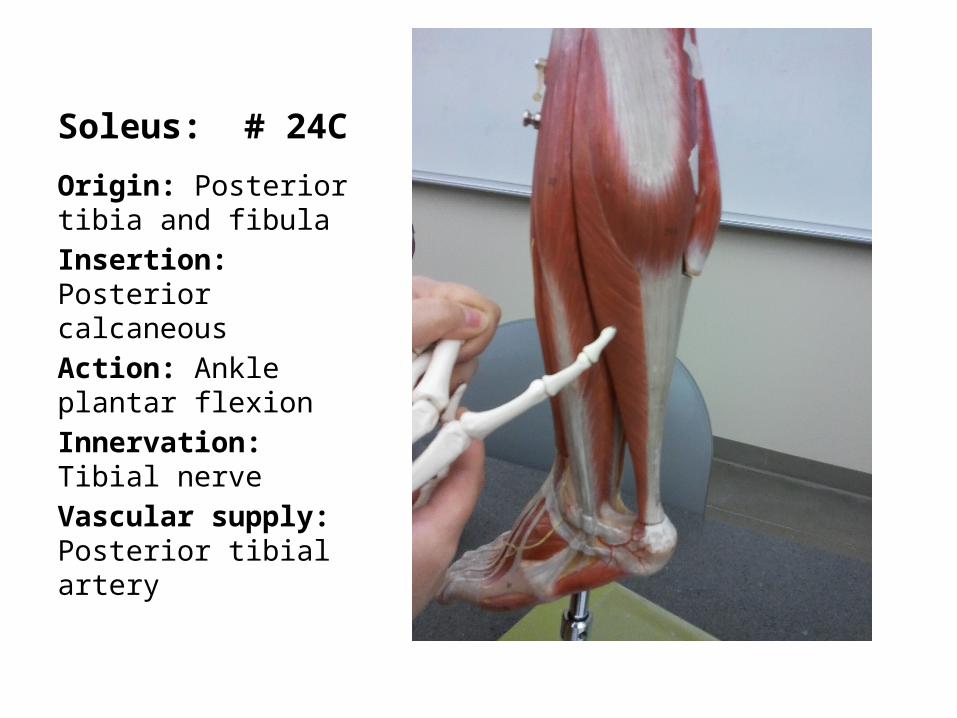
Soleus: # 24C
Origin: Posterior tibia and fibulaInsertion: Posterior calcaneousAction: Ankle plantar flexion Innervation: Tibial nerveVascular supply: Posterior tibial artery
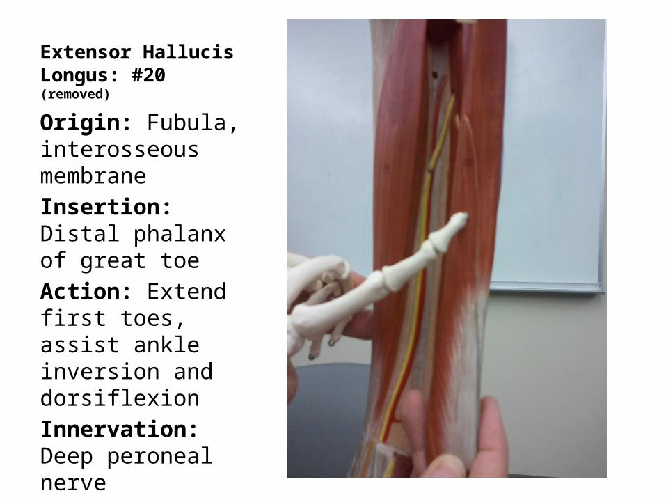
Extensor Hallucis Longus: #20(removed)
Origin: Fubula, interosseous membraneInsertion: Distal phalanx of great toeAction: Extend first toes, assist ankle inversion and dorsiflexionInnervation: Deep peroneal nerveVascular supply: Anterior tibial artery
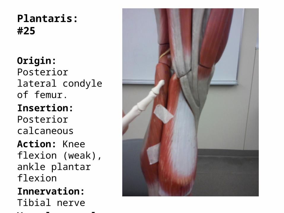
Plantaris: #25
Origin: Posterior lateral condyle of femur.Insertion: Posterior calcaneousAction: Knee flexion (weak), ankle plantar flexion Innervation: Tibial nerveVascular supply: Popliteal artery
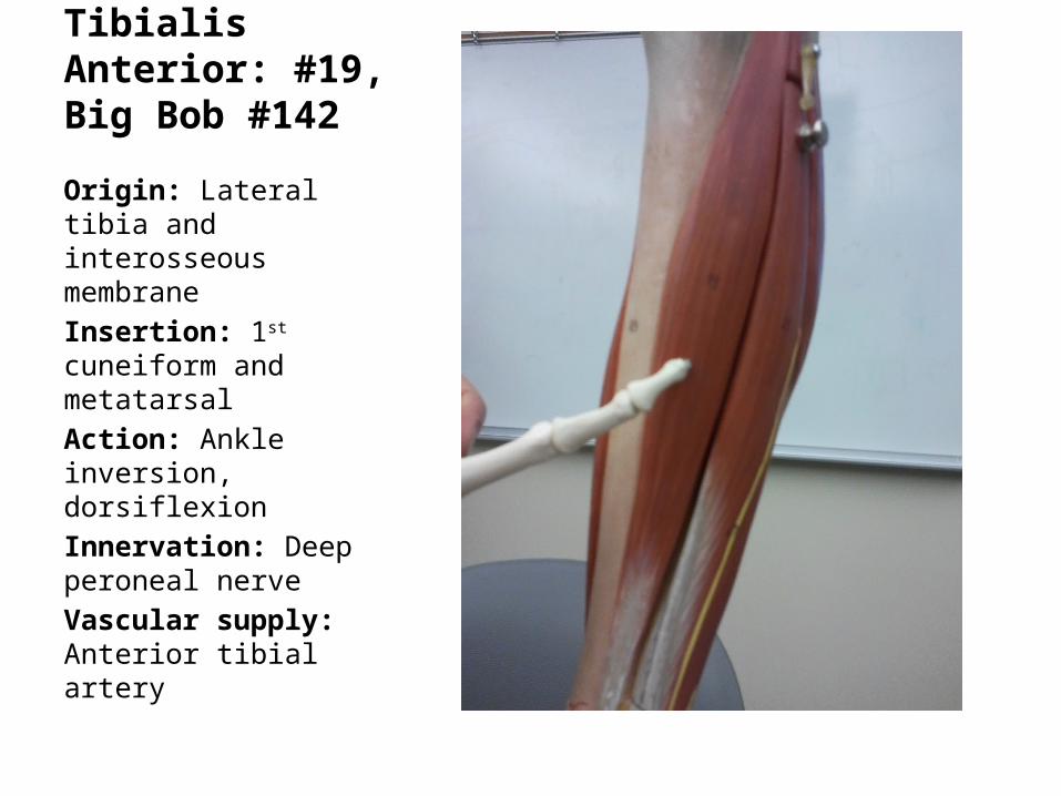
Tibialis Anterior: #19, Big Bob #142
Origin: Lateral tibia and interosseous membraneInsertion: 1st cuneiform and metatarsalAction: Ankle inversion, dorsiflexionInnervation: Deep peroneal nerveVascular supply: Anterior tibial artery
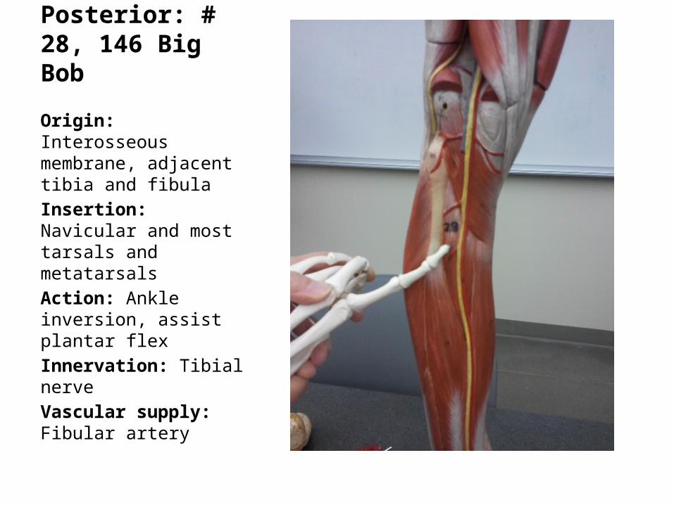
Tibialis Posterior: # 28, 146 Big Bob
Origin: Interosseous membrane, adjacent tibia and fibulaInsertion: Navicular and most tarsals and metatarsalsAction: Ankle inversion, assist plantar flexInnervation: Tibial nerveVascular supply: Fibular artery
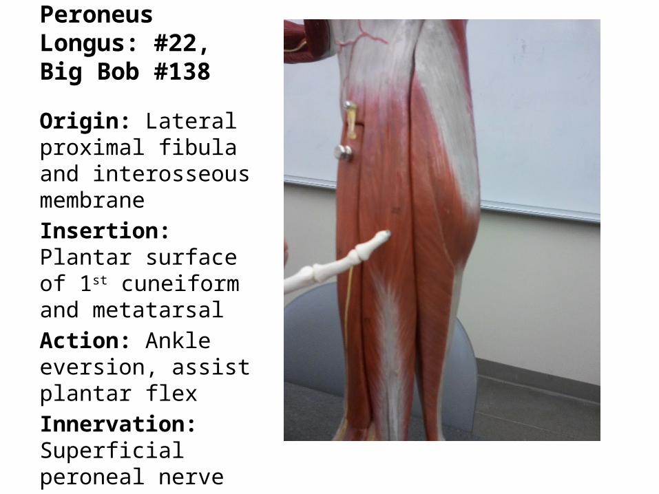
Peroneus Longus: #22, Big Bob #138
Origin: Lateral proximal fibula and interosseous membraneInsertion: Plantar surface of 1st cuneiform and metatarsalAction: Ankle eversion, assist plantar flexInnervation: Superficial peroneal nerveVascular supply: Fibular artery
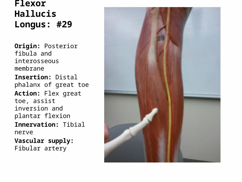
Flexor Hallucis Longus: #29
Origin: Posterior fibula and interosseous membraneInsertion: Distal phalanx of great toeAction: Flex great toe, assist inversion and plantar flexionInnervation: Tibial nerveVascular supply: Fibular artery
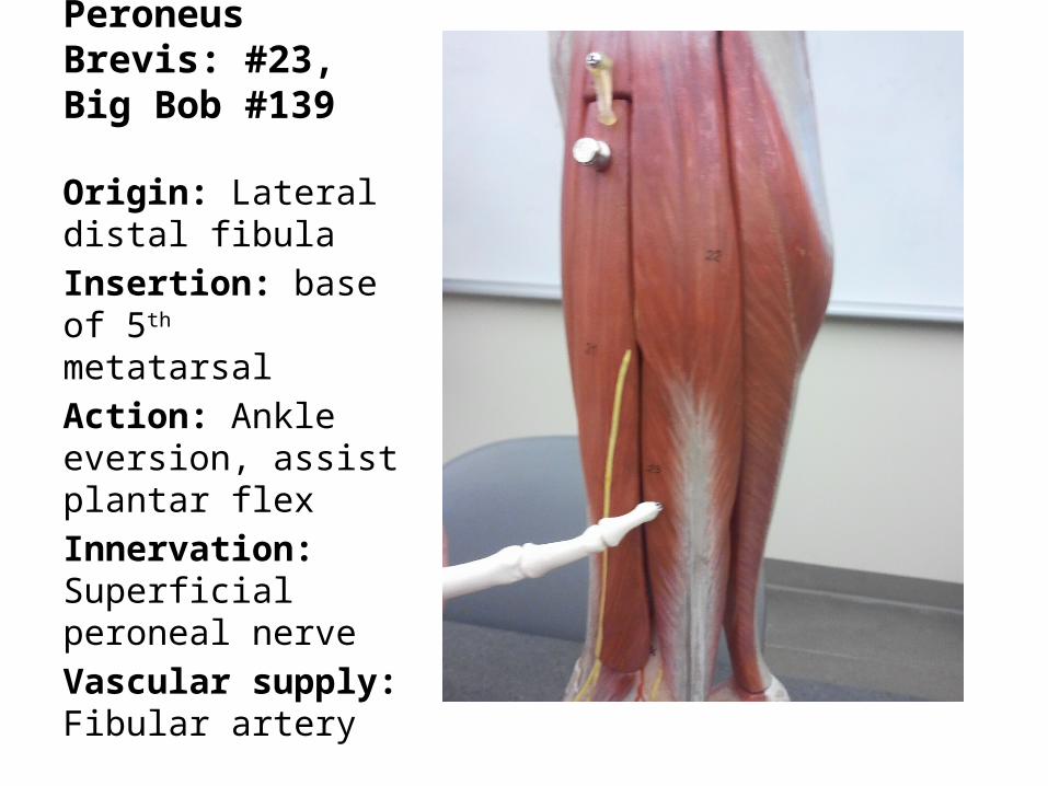
Peroneus Brevis: #23, Big Bob #139
Origin: Lateral distal fibulaInsertion: base of 5th metatarsalAction: Ankle eversion, assist plantar flexInnervation: Superficial peroneal nerveVascular supply: Fibular artery
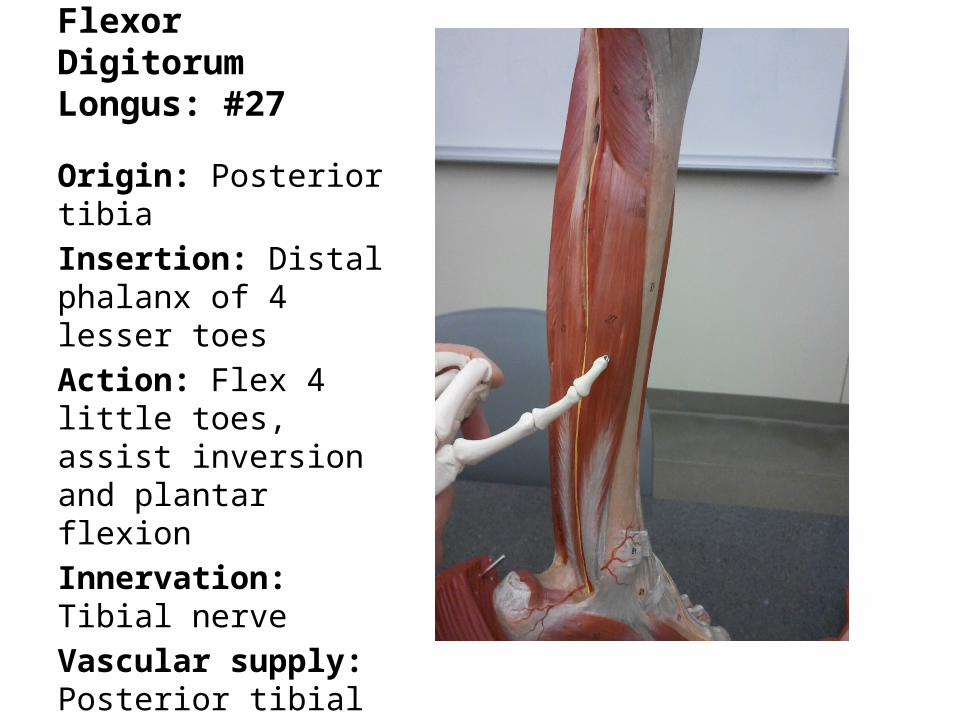
Flexor Digitorum Longus: #27
Origin: Posterior tibiaInsertion: Distal phalanx of 4 lesser toesAction: Flex 4 little toes, assist inversion and plantar flexionInnervation: Tibial nerveVascular supply: Posterior tibial artery
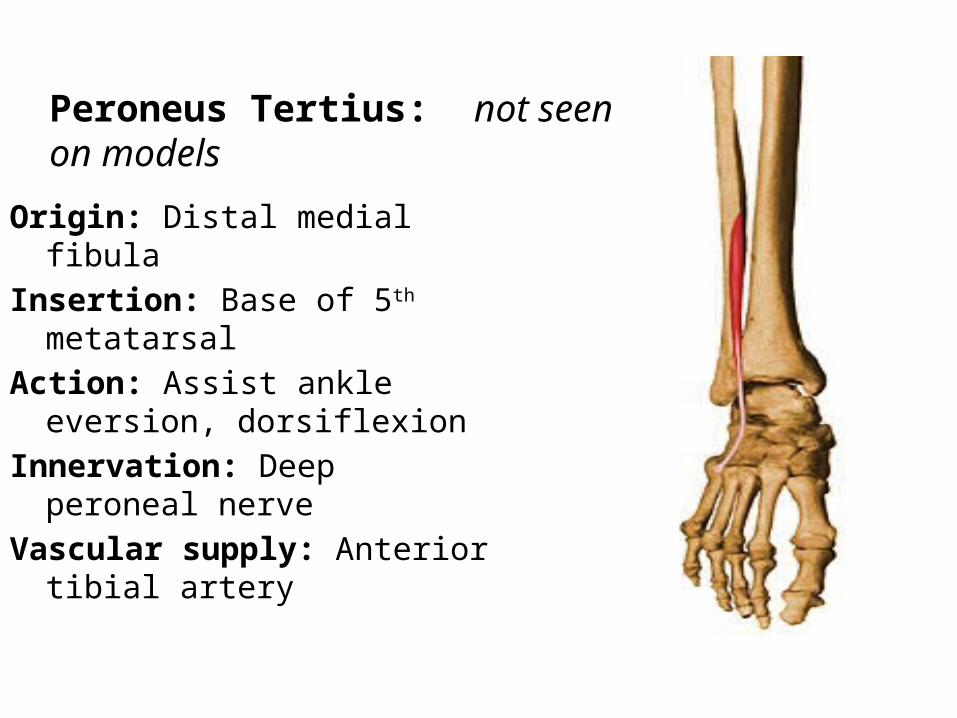
Origin: Distal medial fibulaInsertion: Base of 5th metatarsalAction: Assist ankle eversion,
dorsiflexionInnervation: Deep peroneal nerveVascular supply: Anterior tibial
artery
Peroneus Tertius: not seen on models
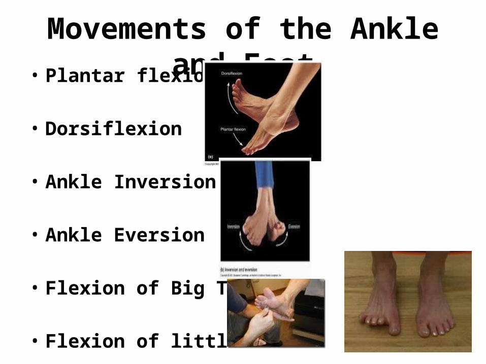
Movements of the Ankle and Foot• Plantar flexion
• Dorsiflexion
• Ankle Inversion
• Ankle Eversion
• Flexion of Big Toe
• Flexion of little toes
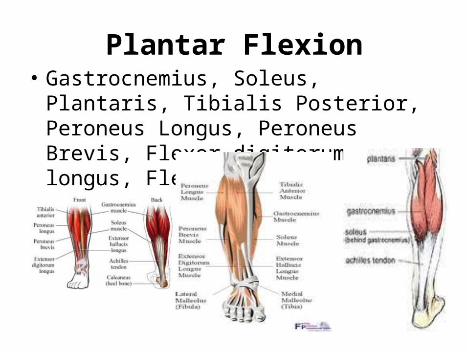
Plantar Flexion• Gastrocnemius, Soleus, Plantaris, Tibialis
Posterior, Peroneus Longus, Peroneus Brevis, Flexor digitorum longus, Flexor Hallucis longus,
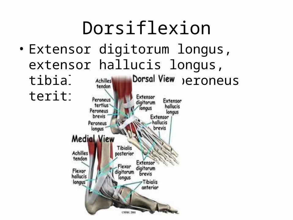
Dorsiflexion• Extensor digitorum longus, extensor hallucis
longus, tibialis anterior, peroneus teritius
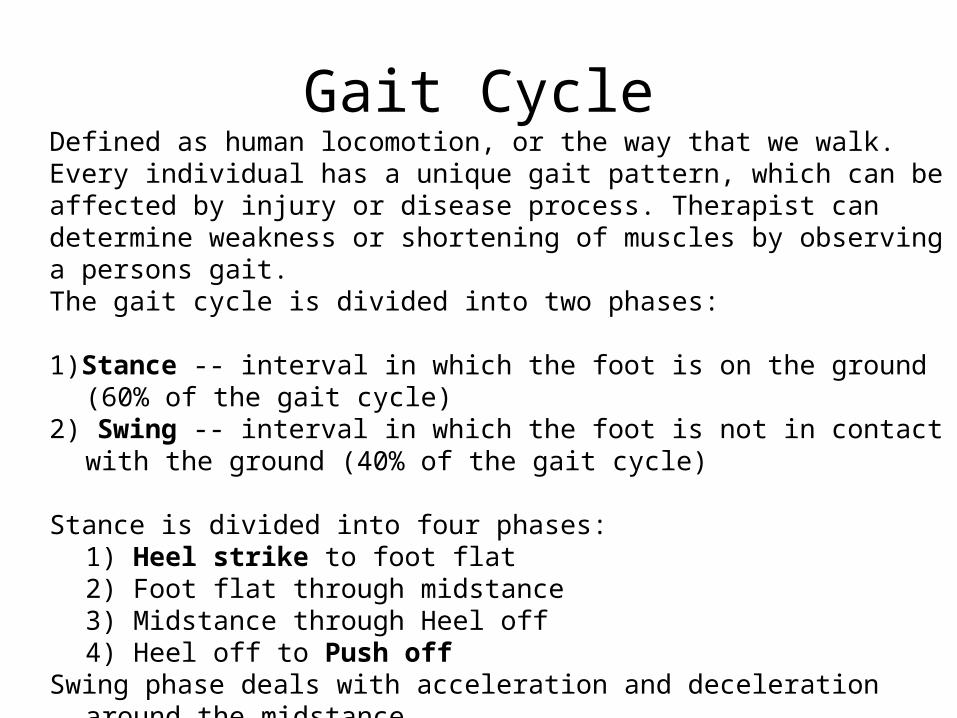
Gait CycleDefined as human locomotion, or the way that we walk. Every individual has a unique gait pattern, which can be affected by injury or disease process. Therapist can determine weakness or shortening of muscles by observing a persons gait.The gait cycle is divided into two phases:
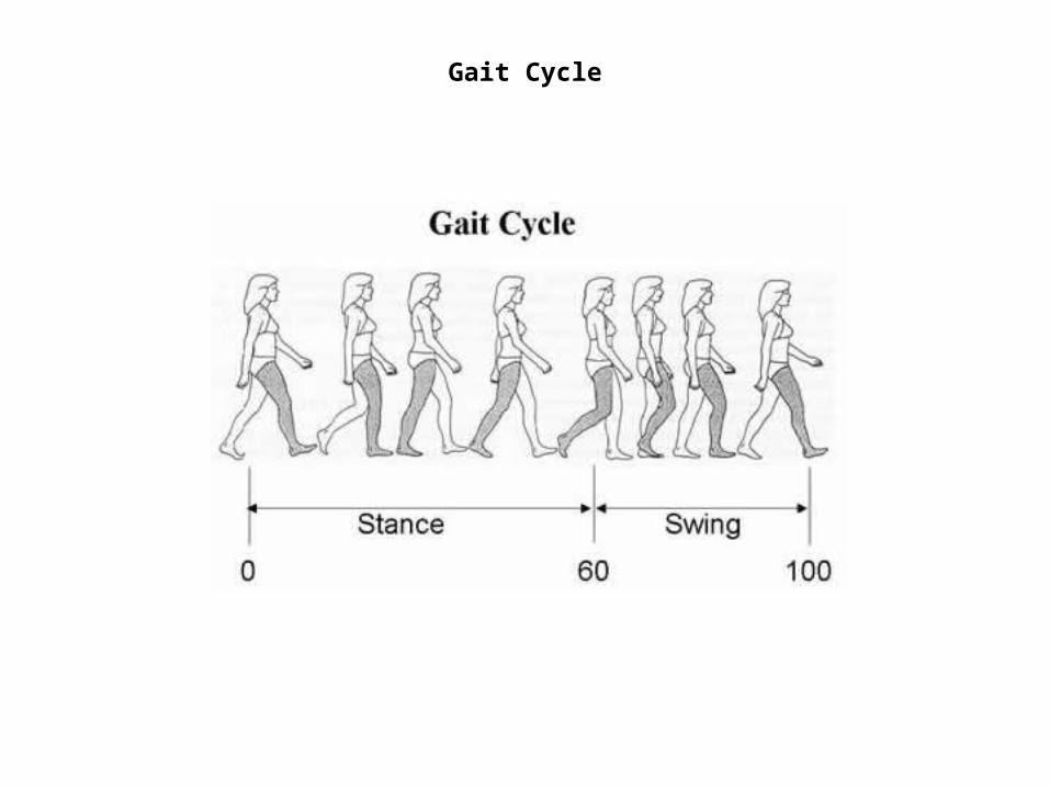
1)Stance -- interval in which the foot is on the ground (60% of the gait cycle)2) Swing -- interval in which the foot is not in contact with the ground (40% of the
gait cycle)
Stance is divided into four phases:1) Heel strike to foot flat2) Foot flat through midstance3) Midstance through Heel off4) Heel off to Push off
Swing phase deals with acceleration and deceleration around the midstance.
Gait Cycle
References• The Gait Cycle From Laura Inverarity, D.O.
, former About.com Guide,Updated August 03, 2007
• www.dreamstime.com• tibia: human tibia and fibula. [Art]. In
Encyclopædia Britannica. Retrieved from http://www.britannica.com/EBchecked/media/101354/Anterior-view-of-the-bones-of-the-lower-right-leg
• http://mikescottdpt.com/2011/02/03/revisiting-plantar-fasciitis
• Google images
Recommended