Blood Transfusions WWW.RN.ORG®
Reviewed September 2017, Expires September 2019 Provider Information and Specifics available on our Website
Unauthorized Distribution Prohibited
©2017 RN.ORG®, S.A., RN.ORG®, LLC By Wanda Lockwood, RN, BA, MA
Purpose The purpose of this course is to explain types of blood donations, types
of blood components used for transfusions, the procedure for transfusions, and adverse reactions.
Goals Upon completion of this course, the health professional should be able
to: • Give a brief description of the history of blood transfusions.
• Describe the criteria for eligibility for donating blood. • Describe 4 types of donations.
• Explain ABO and Rh blood typing. • List at least 8 infective agents for which donated blood is tested.
• List and describe at least 8 types of blood components. • Explain the volume of a unit of whole blood, packed red blood
cells, platelets, and fresh frozen plasma.
• Explain leukoreduction, irradiation, and apheresis, intraoperative blood salvage, and hemodilution.
• Discuss procedures for administration of packed red blood cells, platelets, and fresh frozen plasma.
• Describe 11 types of transfusion reactions, including symptoms
and treatment.
Introduction Approximately 4.5 million Americans need blood transfusions each
year, including 1 out of every 7 people hospitalized, with most people

on average requiring 3 units of whole blood or red blood cells. For much of early history, people were bled instead of transfused. The first
transfusion was given in 1765 by Phillp Syng Physick in Baltimore. Other physicians also tried transfusions, with mixed results because
they didn’t have knowledge of blood typing. As a result, survival was a matter of luck.
The discovery of blood types by Karl Landsteiner in 1901 and routine
typing of blood by 1907 changed the outlook for patients although blood supply was often insufficient to meet needs. A major turning
point in medicine came in 1939 when Dr. Charles Drew developed a method to separate plasma from red blood cells and freeze them
separately although transfusions still posed the risk of infections. Since the 1960s, tests have been available to detect infection, such as
hepatitis B and HIV, in the blood supply, and new tests are developed
as new pathogens emerge.
Blood donations
N. Gervacio, US Navy
An adequate blood supply is critical to providing transfusions to those in need. However, blood donors are unpaid volunteers. Blood given in
return for money cannot be used to transfuse people in the United States. About 43,000 people donate pint of blood each day in response
to blood drives or requests for blood. Donation requires completion of

a questionnaire to establish the person meets the criteria for donation, a brief physical assessment, donation of blood, and then a snack.
Standard donations usually involve removal
of about 450 mL of blood and take approximately 15 minutes. The donor remains
recumbent for a few minutes and then is given fluids and a snack and observed for about 15 minutes. A pressure dressing should be left in
place for several hours and the donor advised to avoid heavy lifting. The donor should not smoke for an hour or drink alcoholic beverages
for 3 hours.
Apheresis is a process in which blood is
removed from a patient or donor, passed
through a centrifuge or filter to remove components, and then the remaining blood is returned to the person.
The system is closed so there is little risk of bacterial contamination.
Donors may provide apheresis donations instead of donating whole blood in order to provide larger quantities of blood components, such
as platelets, than can normally be provided from a single donor. The procedure takes about 2 hours. Donors may be given medication to
take in the days before the procedure to increase production of certain blood cells. Apheresis is also used to obtain stem cells from peripheral
blood for peripheral blood stem cell transplant (PBSCT).
For example, if platelets are needed, apheresis is carried out and the platelets separated from the blood and saved. An apheresis collection
of platelets contains approximately 200 to 400 mL of plasma. To
reduce the chance of ABO antibodies, the unit is volume reduced (concentrated). When platelets are collected from a number of
different donors, these units are referred to as random apheresis platelets.
Donations by family or friends for a specific
person are directed donations. Some people feel safer getting blood and blood components
from people they know although there is no evidence that directed donations are safer than random donations. Additionally, in emergency
situations, obtaining and testing the blood may delay treatment.
Sometimes patients donate their own blood prior to elective surgeries, especially when
Apheresis donors
Directed donation
Standard donation
Autologous donation

the potential for blood loss is high, such as in orthopedic surgery. Usually, donations are made 4 to 6 weeks preoperatively and iron
supplements or erythropoietin may be given to stimulate blood cell production. People with rare blood types may store blood in case of
emergency.
While autologous donations can be frozen and stored for extended periods, the reality is that hospitals and blood banks do not have
facilities to save large quantities of blood, so autologous donations are routinely discarded after surgery. Once blood is designated for a
particular individual, it cannot then be used as a random donation for other people.
Criteria for blood donors Age 16-17 (depending on state).
Weight 110 pounds
Health Good
Medications Most do not affect donation, but the donor must provide a complete list of recent and current
medications and vaccinations as some may be contradicted and others (such as Accutane®)
may require a waiting period after drug cessation. Diabetics who have ever used bovine insulin
derived from cattle in the UK cannot donate.
Interval Generally people can only donate 1 unit of whole blood every 56 days.
Medical contraindications
• Clotting disorders, including use of warfarin or heparin.
• Chronic fatigue syndrome. • Cancer (depends on the type of cancer and
status). • Dura mater (brain covering) transplant or
human pituitary growth hormone.
• First-degree blood relative with Creutzfeld-Jacob disease.
• Viral hepatitis since age 11. • AIDS or history of positive HIV test.
• Chagas disease or babesiosis. • Sickle cell disease.
• Active TB. • IV drug use.
Waiting period • Organ transplants—12 months. • Heart disease—6 months with no heart-related

symptoms. • Syphilis and gonorrhea—12 months after
treatment. • Previous transfusion—12 months.
• Sex with person with viral hepatitis—12 months.
• Human bite—12 months. • Malaria—3 years after treatment or living in
endemic area, 12 months after visiting endemic area.
• Pregnancy—6 weeks after delivery.
BP Must be at least 80/50 and no higher than 180/100.
Hemoglobin 12.5.
Travel Some restrictions may be in place, such as 12-month waiting periods for travel from malaria
endemic areas and Iraq (because of leishmaniasis). People who were born or lived in
some West African countries may not be eligible to donate because a rare type of HIV (type O) is
found there.
Blood typing and testing

Once blood is received from a donor, it must be typed
and labeled and tested for a number of infectious diseases. The most commonly used system for typing
blood is ABO, or Landsteiner. The different blood groups are classified according to the antigens found on the surface of the red blood cells.
Cells may have A antigens, B antigens, A and B antigens, or no antigens (O). People spontaneously develop antibodies (also called
agglutinins) against antigens they lack. If the recipient receives a transfusion that is not correctly matched, the antibodies in the blood
will attack the antigens in the transfused blood.
Typing for RH antigens is also done. There are about 50 Rh antigens, but the D antigen is most important
because it can cause a systemic immune response in Rh
– (negative) individuals. People who are Rh- rarely have anti-D antibodies because they are not produced by contact with the
environment. However, people can produce these antibodies in response to a sensitizing event, such as a pregnancy or transfusion
with Rh + blood. If transfused with Rh+ blood, the Rh– person could experience a transfusion reaction, but this rarely occurs because of
blood typing.
Of bigger concern is the possibility of mother-fetus incompatibility, which occurs when the mother is Rh- and the father Rh+. Problems
usually don’t arise during the first pregnancy, but during delivery some blood may cross into the mother’s blood system, which causes the
mother to develop anti-D antibodies. During a second pregnancy, these antibodies pass to the baby, resulting in the fetal red blood cells
agglutinating or bursting. The baby can develop erythroblastosis fetalis
(“blue baby”). Rh- mothers with Rh+ mates now receive RhoGAM®, which contains anti-Rh+ antibodies, so the mother does not develop
antibodies that could pose a threat to the fetus.
Blood types are not distributed evenly among humans, and percentages vary
according to country of origin. For example, B type blood worldwide accounts for about 16% of people
but only 10% in the United States. It is most common in Asia and parts of Africa.
US Distribution of ABO/Rh blood types
ABO types Rh type Percentage Total
O + 37.4%
ABO typing
Rh typing
Blood type distribution
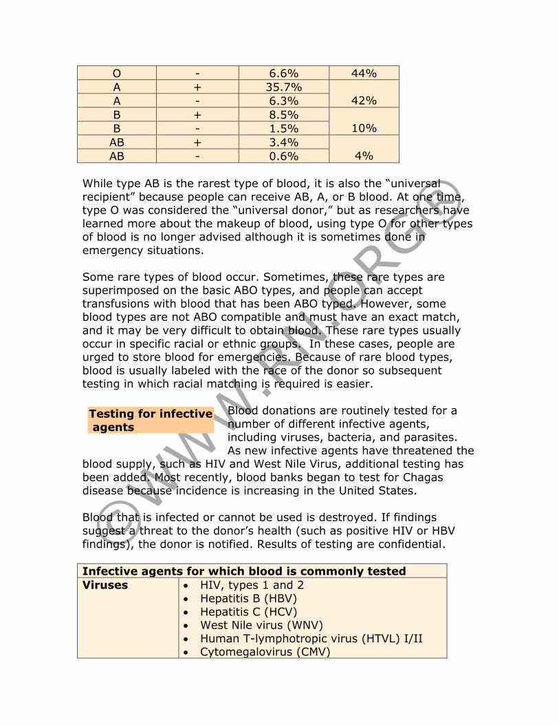
O - 6.6% 44%
A + 35.7%
42% A - 6.3%
B + 8.5% 10% B - 1.5%
AB + 3.4%
4% AB - 0.6%
While type AB is the rarest type of blood, it is also the “universal
recipient” because people can receive AB, A, or B blood. At one time, type O was considered the “universal donor,” but as researchers have
learned more about the makeup of blood, using type O for other types of blood is no longer advised although it is sometimes done in
emergency situations.
Some rare types of blood occur. Sometimes, these rare types are superimposed on the basic ABO types, and people can accept
transfusions with blood that has been ABO typed. However, some blood types are not ABO compatible and must have an exact match,
and it may be very difficult to obtain blood. These rare types usually
occur in specific racial or ethnic groups. In these cases, people are urged to store blood for emergencies. Because of rare blood types,
blood is usually labeled with the race of the donor so subsequent testing in which racial matching is required is easier.
Blood donations are routinely tested for a
number of different infective agents, including viruses, bacteria, and parasites.
As new infective agents have threatened the blood supply, such as HIV and West Nile Virus, additional testing has
been added. Most recently, blood banks began to test for Chagas disease because incidence is increasing in the United States.
Blood that is infected or cannot be used is destroyed. If findings
suggest a threat to the donor’s health (such as positive HIV or HBV
findings), the donor is notified. Results of testing are confidential.
Infective agents for which blood is commonly tested
Viruses • HIV, types 1 and 2
• Hepatitis B (HBV)
• Hepatitis C (HCV) • West Nile virus (WNV)
• Human T-lymphotropic virus (HTVL) I/II • Cytomegalovirus (CMV)
Testing for infective
agents

• Epstein-Barr virus (EBV)
Bacteria • Treponema pallidum (Syphilis)
Parasites • Trypanosoma cruzi (Chagas)
Transfusions, blood components
Whole blood is rarely transfused.
Instead, whole blood is fractionated into components, such as red blood
cells, white blood cells, platelets, and plasma. Plasma is further fractionated
into a number of blood components,
including albumin, gamma globulins, and clotting factors.
There are a number of reasons for the movement away from whole
blood and toward components: • Whole blood is more likely to carry disease.
• One unit of whole blood can be fractionated and provided to a number of individual instead of just one. For example, one unit of
whole blood can be divided into a unit or packed cells, a unit of platelets, and a unit of fresh frozen plasma. Then, the plasma can
be further fractionated. • Most people require only one component.
• Components generally have greater shelf life than whole blood. • Many blood products can be infused regardless of ABO blood type.
When blood is separated, the erythrocytes or red blood cells (about 40-50% of total volume) settle on the bottom, the leukocytes and
platelets settle above the red blood cells in a band called the buffy coat, and the plasma (about 55% of total volume) settles at the top.

Blood and blood components Whole blood Storage: Varies depending on intended
use/separation. Uses:
• Significant bleeding (>25% total blood volume) for volume replacement.
• Usually separated into components (erythrocytes, plasma, platelets) rather than
transfused as whole blood.
Packed red blood cells
Storage: • Refrigerated: 42 days.
• Frozen: 10 years. Uses:
• Chronic anemia. • Acute loss of blood.
• Low Hgb and Hct. • Sickle cell disease.
Considerations: • Some non-functioning platelets, plasma, and
WBCs remain. • WBCs may cause reaction, but this risk reduces
with leukoreduction. Irradiation reduces risk of
transfusion-associated graft vs host disease (TA-GVHD).
• Must be ABO/Rh compatible. • (155-pound adult) 1 unit should raise

hematocrit 3% and hemoglobin by 1 gm/dL. • Washed blood cells must be used within 24
hours from the start of the washing procedure. Washed RBCs should not be considered
leukocyte-reduced.
Platelets Storage: 5 days at room temperature (with
agitation to prevent clumping).
Uses: • Leukemia, other malignancies.
• Control of bleeding from low platelet count. • Prevent bleeding from thrombocytopenia
(<5000 -10,000). Considerations:
• ABO/Rh compatibility is desired but some substitutions can be made as the number of
RBCs is usually too low to cause reaction although an Rh- person can become sensitized.
• Some non-functioning RBCs and WBCs remain. • Alloimmunization decreased if single donor is
used rather than random donors.
• Single donor platelets preferred for repeated treatment.
• 1 unit of single donor platelets equals 6 to 8 units of random platelets.
• Platelets have increased risk of infection, so they should be tested with rapid culturing
before administration.
Plasma/Fresh
frozen plasma
Storage: Frozen: 1 year. (When thawed, it is
referred to as fresh frozen plasma and must be used within 24 hours of thawing.)
Uses:
• Control of bleeding related to low levels of some clotting factors.
• Plasma replacement (blood loss). • Plasmapheresis.
Considerations: • Must be ABO/Rh compatible.
• A standard dose of random platelets is equivalent to 4 units of pooled platelets.
• A large dose of random platelets is equivalent to 6 units of pooled platelets.
• Dose is not specified for an HLA matched apheresis directed donation.
Cryoprecipitate Storage: Frozen: 1 year.

(Fibrinogen, AHF, von
Willebrand factor,
fibronectin)
Uses: • Bleeding associated with hemophilia A and von
Willebrand’s disease (if other clotting factors unavailable).
• Hypofibrinogenemia. • Use generally supplanted by pure factor VII or
factor IX. Considerations:
• ABO/Rh compatibility is not necessary.
Granulocytes (Neutrophils)
Storage: Used as soon as possible but can be stored up to 24 hours at 20-24C without
agitation. Irradiated prior to administration. Uses: Severe neutropenia. (Use controversial.)
Considerations: • Must be ABO/Rh compatible.
• Some lymphocytes, RBCs and platelets remain. • May cause febrile transfusion reaction.
• May transmit infectious diseases, such as cytomegalovirus.
• Has been generally supplanted by use of colony
stimulating factors to stimulate the body to produce its own neutrophils:
o G-CSF or GM-CSF.
Lymphocytes/
Buffy coat
Storage: Used fresh immediately or frozen for
later use. Uses: Stimulate graft-versus-disease-effect for
treatment of leukemia after ablative and non-ablative stem cell transplantation with apheresis of
lymphoctyes from original stem cell donor to prevent treatment failure.
Considerations: Lymphocytes may be collected
and stored at the time of the original stem cell donation.
Antihemophilic factor (AHF)
(Factor VIII)
Storage: 12 months at -18C or 4 hours at 20-
24C. Only brief storage is permitted at room
temperature (25C).
Uses: Hemophilia A.
Considerations: ABO/Rh compatibility is not necessary.
Factor IX
concentrate
Storage: Refrigerate at 2-8C away from light and
moisture, but do not freeze. Uses: Hemophilia B.
Considerations: ABO/Rh compatibility is not necessary.
Factor IX complex
(Factors II, VII, IX, & S)
Storage: Refrigerate both dry medicine and diluent at 2-8C. Use within 3 hours of preparation.
Uses: • Factor VII, IX, X deficiencies.
• Hemophilia A with factor VII inhibitors. Considerations:
• ABO/Rh compatibility is not necessary.
Albumin (5%, 25%)
Storage: Room temperature 20-25C with
excursions to 15-30C permitted.
Uses: • Hypoproteinemia.
• Burns, • Volume expansion by 5% to increase blood
volume. • Volume expansion by 25% to decrease
hematocrit. Considerations:
ABO/Rh compatibility is not necessary.
Gamma globulin (IV)
Storage: Refrigerate at 2-8C, but do not freeze.
Uses:
• Hypogammaglobulinemia associated with chronic lymphocytic leukemia, ITP, and primary
immunodeficiency. Considerations:
ABO/Rh compatibility is not necessary.
Antithrombin
III concentrate
(AT III)
Storage: Refrigerate at 2-8C, but do not freeze.
Uses: AT III deficiency with thrombosis or increased risk of thrombosis.
Considerations:
ABO/Rh compatibility is not necessary.
Blood is usually ordered in units, but the volume varies depending on the type of component.
• Whole blood is usually about 450 mL, but anticoagulant is added to the blood, so the total volume is usually
about 500 to 520 mL. The volume of components may vary. • Packed red blood cells are typically about 350 mL and include
approximately 150-210 mL of red blood cells, 100 mL of Optisol®
(a crystalloid solution that extends shelf life) and 30 mL plasma. Hematocrit is about 57% but is less with washed or leukoreduced.
Pediatric/Divided RBC units are prepared by separating a standard unit (containing no Optisol®) into 4 parts. Each individual bag then
contains about 45 to 50 mL of RBCs and 15 mL of plasma. Divided units are generally irradiated. Hematocrit is about 72%
Volume
• Platelet concentrate is usually about 50mL and contains some WBCs, RBC, and about 50 mL of plasma.
• Platelet pheresis donation is about 300 mL and includes some WBCs and RBCs.
• Fresh frozen plasma is about 225 mL and contains plasma proteins, all coagulation factors, and complement.
• Cryoprecipitate is about 15 mL and contains 150mg fibrinogen, 80 units of factor VII, von Willebrand factor, factor XIII, and
fibronectin.
A variety of studies have been conducted
regarding the efficacy of leukoreduction/leukodepletion on whole blood,
red blood cells, or platelets to remove remaining leukocytes (white
blood cells). This reduction is achieved through centrifugation or filtration. One study showed a 50% reduction in post-transfusion
infections with the use of leukodepleted blood, but other studies have failed to replicate these findings. Other studies indicate that
leukoreduction also reduces transmission of viruses, such as CMV, herpesviruses, and Epstein-Barr virus and may decrease transmission
of Chagas disease as the filters used bind the Trypanosoma cruzi parasite.
However, about 10% of red blood cells are lost in the leukoreduction
process, and the process adds additional cost to each unit. Most developed countries now utilize universal leukoreduction of blood
components. However, the United States has not adopted universal leukoreduction although some hospitals and medical centers have
done so. Luekodepleted blood is commonly used for certain patient
populations, such as those who are immunocompromised.
Blood products may also be irradiated to inactivate T-lymphocytes. Irradiation is effective in preventing
transfusion-associated graft versus host disease (TA-GVHD). Gamma radiation (2500 rads) is used on whole blood, red
blood cells, and granulocytes. Irradiation destroys the lymphocytes’ ability to divide. Some red blood cells are lost in this process, but
irradiation does not affect platelet function. Irradiation also increases the rate of efflux of intracellular potassium. Cryoprecipitate or fresh
frozen plasma does not require irradiation, but fresh plasma should be irradiated.
Leukoreduction
Irradiation

In some cases, apheresis is done to reduce one type of blood component, such as white blood cells or platelets.
The effect is temporary, but may be used to give time for suppressive medications to work. Apheresis is often referred to in
relation to the type of blood component being removed:
Apheresis Plasmapheresis Removes plasma proteins for hyperviscosity
syndromes and treatment of some renal and neurological diseases (Guillain-Barré,
myasthenia gravis).
Plateletpheresis: Removes platelets for severe thrombocytosis, essential thrombocytopenia, and single donor
or random donor platelet transfusions.
Leucopheresis or
leukapheresis
Removes white blood cells and can be specific
to neutrophils or lymphocytes for extreme leukocytosis associated with acute or chronic
leukemia (AML, CML) and to separate WBCs
for transfusion.
Erythropheresis or
erythrocytapheresis:
Removes red blood cells for RBC dyscrasias
(such as sickle cell disease) and for replacement of RBCs.
Stem cell harvest Removes stem cells circulating in peripheral
blood for transplantation.
Plasma may be removed instead of blood cells or platelets when abnormal proteins are present, in conjunction with long-term therapy.
Plasmapheresis is used in autoimmune disorders, such as Guillain-Barré or myasthenia gravis, to remove disease-producing
autoantibodies. In some cases, plasma may be completely removed and replaced with fresh frozen plasma.
Red blood cells may be “washed” with
0.9% saline in a centrifuge or blood cell processor to remove plasma, plasma
protein, microaggregates, cytokines, and unwanted antibodies. Washing is done to decrease incidence of allergic and anaphylactic
reactions although the procedure may not remove all proteins implicated in allergic reactions. The washed RBCs may be stored at 1-
6 C but must be used within 24 hours of initiation of the washing
procedure because preparation is an open system. If stored at 20-24C,
the cells must be used within 4 hours.
Apheresis
Washed red blood cells

Disadvantages to this procedure include increased cost, reduced shelf life, and loss of 10 to 20% of RBCs, so washed red blood cells will raise
the hematocrit less that standard PRBCs. In some cases, such as to prevent febrile nonhemolytic reaction, centrifugation followed by
filtration through micro-aggregate or standard blood filters is almost as efficient and is less expensive and does not impair shelf life.
Blood can be salvaged from the
operative site to be reinfused into the patient. This type of salvage is
especially valuable in trauma situations where large volumes of blood are needed or with surgeries that involve excessive blood loss.
Equipment, such as cell savers, cleanse the blood.
During the procedure, blood is suctioned
from a sterile cavity, such as the hip joint or abdomen, through dual-channel
tubing so that anticoagulant is mixed with the blood, which is collected in a
reservoir and then pumped into a centrifuge for concentration.
The concentrated blood is washed with an isotonic electrolyte solution
(usually saline). The concentrated solution of red blood cells is pumped into a bag for reinfusion. Modern equipment can provide 225 ml of
washed and saline-suspended red blood cells (hematocrit 50%) in 3
minutes, or the equivalent of 12 units of blood per hour.
This procedure may be acceptable to Jehovah Witnesses, who
otherwise shun blood transfusions.
In the hemodilution procedure, 1 or 2 units of
blood are withdrawn prior to surgery and simultaneously replaced with a colloid or crystalloid
solution to dilute the blood so that fewer red blood cells are lost during the operative procedure. Then, the withdrawn blood is reinfused
postoperatively. This procedure is indicated for surgeries that entail excessive blood loss and contraindicated in those at risk of myocardial
infarction
Administration Protocol for administration of blood products should always be followed,
as there may be slight variations in procedures from one institution to
Intraoperative blood salvage
Hemodilution
another. However, the initial steps in any delivery of blood products should always include:
• Verifying the physician’s order. • Checking patient’s type and cross match.
• Verifying that the patient has signed a consent for a transfusion.
The patient should be educated about the transfusion procedure and indications of transfusion reactions. Before beginning a transfusion,
the patient’s cardinal signs (temperature, BP, pulse, and respirations) should be taken and recorded so they can serve as a baseline to
evaluate possible reactions.
The intravenous line should be started with normal saline prior to obtaining the blood product from the blood bank or blood storage area.
PRBCs are usually given through a 20 gauge or larger needle into a
large vein while platelets and fresh frozen plasma require a 22 gauge or larger needle.
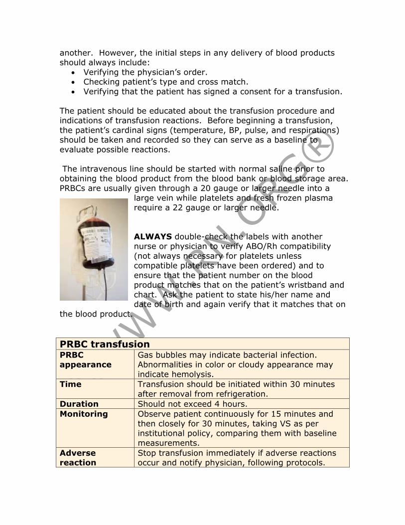
ALWAYS double-check the labels with another nurse or physician to verify ABO/Rh compatibility
(not always necessary for platelets unless compatible platelets have been ordered) and to
ensure that the patient number on the blood product matches that on the patient’s wristband and
chart. Ask the patient to state his/her name and date of birth and again verify that it matches that on
the blood product.
PRBC transfusion PRBC appearance
Gas bubbles may indicate bacterial infection. Abnormalities in color or cloudy appearance may
indicate hemolysis.
Time Transfusion should be initiated within 30 minutes
after removal from refrigeration.
Duration Should not exceed 4 hours.
Monitoring Observe patient continuously for 15 minutes and
then closely for 30 minutes, taking VS as per institutional policy, comparing them with baseline
measurements.
Adverse reaction
Stop transfusion immediately if adverse reactions occur and notify physician, following protocols.

Tubing Ensure that tubing is specific for use with PRBCs and contains a blood filter for fibrin clots and other
particulate material. Change tubing after every 2 units to decrease change of contamination.
Platelets or fresh frozen plasma (FFP) Appearance Note excessive redness, which may indicate
excessive contamination with red blood cells.
Time Should be administered immediately after being obtained.
Duration Infuse as fast as patient can tolerate. Platelets may
begin to clump if transfusion is prolonged.
Monitoring Observe patient continuously for 15 minutes and
note signs of circulatory overload that may require slowing infusion
Adverse
reaction
Stop transfusion immediately if adverse reactions
occur and notify physician, following protocols.
Tubing Ensure that tubing is appropriate for blood
component. Flush tubing with saline on completion of transfusion.
Transfusion reactions In most cases, the first step when a reaction occurs is to stop the transfusion, change the tubing to avoid further infusion of the blood
component, and maintain the IV with normal saline. Both the physician and the blood bank should be notified.
People may develop an allergic reaction to
foreign plasma proteins. These reactions may range from mild to severe anaphylaxis.
Symptoms of mild reactions include: • Pruritis.
• Urticaria. • Flushing.
The transfusion should be stopped and an antihistamine, typically diphenhydramine (Benadryl®), administered. If the person responds
to the antihistamine and symptoms subside, the transfusion can
usually be resumed slowly, monitoring the patient carefully. Pre-transfusion antihistamine may reduce incidence.
Allergic reaction,
mild
If fever, shock, or respiratory distress, including bronchospasm and laryngeal edema, develops,
the transfusion should not be resumed because this allergic reaction can be life threatening. Anaphylactic shock may
occur in IgA deficient patients that have developed Anti-IgA antibodies.
With severe allergic reactions, treatment may include: • Epinephrine (usually 0.4 ml of 1:1000 solution sc or 0.1 mL of
1:1000 solution diluted to 10 mL with NS), • Corticosteroids,
• Pressor support. • CPR if indicated.
Preventive measures for allergic responses include administration of an
antihistamine prior to transfusion and the use of extensively washed
RBCs and platelets from which all plasma has been removed.
Because some white blood cells remain in
packed red blood cells (PRBC), the person’s antibodies may react, causing a febrile
nonhemolytic reaction (NHR). A NHR occurs in about 1% of PRBC transfusions and 20% of platelet transfusions.
The NHR is responsible for approximately 90% of transfusion reactions, and 10% of people who have repeated transfusions for chronic
conditions develop NHR. It most commonly occurs in people who have had previous transfusions or Rh- women who have had Rh+ children.
Onset is usually sudden and symptoms include:
• Chills (may be mild to severe).
• Fever (>1C elevation), usually 2 hours after initiation of
transfusion.
• Headache. • Flushing.
• Anxiety. • Muscle aches.
Patients rarely exhibit hypotension or respiratory distress. Patients
must be monitored carefully to rule out bacterial infection or hemolytic reaction. The transfusion should be stopped until the physician orders
it to be restarted. Treatment is usually with non-aspirin antipyretics (acetaminophen, ibuprofen). The transfusion is usually resumed if
acute hemolytic reaction is excluded. Leukocyte-depleted red blood cell
Febrile nonhemolytic
reaction
Allergic reaction, severe
transfusions may reduce incidence and should be considered for those with a history of NHR.
TA-GVHD occurs when lymphocytes in
the transfused blood cause disease in the donor, usually 10-14 days after a
transfusion. Symptoms include • Fever
• Rash • Hepatitis
• Diarrhea • Pancytopenia.
• TA-GVHD is fatal in 90% of cases. TA-GVHD is rare in people who are
not immunocompromised. Those at risk include people who are
immunocompromised and those receiving directed donations from a relative or human leukocyte antigen (HLA) identical donors. HLA are
proteins (antigens) found on the surface of white blood cells and other tissues in the body. There are 3 general groups with many variations
within each group. When two people share the same HLAs, there tissues are compatible. HLA matching is important for people
undergoing hemopoietic stem cell or organ transplant. The ABO blood type and HLA type are inherited independently.
If the donor’s blood is incompatible
with the recipients, a severe life-threatening acute hemolytic reaction
can occur. Antibodies present in the recipient’s blood combine with
antigens on donor red blood cells and hemolyze, or destroy, them. This occurs most rapidly with ABO incompatibility, sometimes with as
little as 10 mL of PRBCs. Rh incompatibility causes a less severe reaction. This reaction almost always results from errors in labeling the
blood or identifying the patient.
Transfusion-associated
graft vs host disease
Acute hemolytic reaction
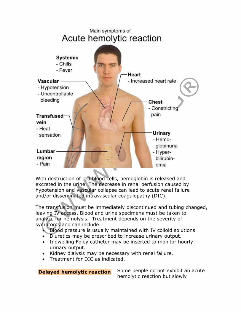
With destruction of red blood cells, hemoglobin is released and excreted in the urine. The decrease in renal perfusion caused by
hypotension and vascular collapse can lead to acute renal failure and/or disseminated intravascular coagulopathy (DIC).
The transfusion must be immediately discontinued and tubing changed,
leaving IV access. Blood and urine specimens must be taken to analyze for hemolysis. Treatment depends on the severity of
symptoms and can include: • Blood pressure is usually maintained with IV colloid solutions.
• Diuretics may be prescribed to increase urinary output. • Indwelling Foley catheter may be inserted to monitor hourly
urinary output.
• Kidney dialysis may be necessary with renal failure. • Treatment for DIC as indicated.
Some people do not exhibit an acute
hemolytic reaction but slowly Delayed hemolytic reaction

increase antibodies to blood products, resulting in a delayed reaction, usually between 3 to 14 days after transfusion although the reaction
can be delayed for months. Extravascular hemolysis occurs gradually. Symptoms include:
• Fever. • Anemia.
• Increased bilirubin. • Decreased or absent haptoglobin.
• Jaundice. • Decreased hematocrit.
In most people, this delayed reaction does not require treatment
although some may need further transfusions if hemolysis is severe enough. Of bigger concern is the possibility that people may develop a
more severe acute hemolytic reaction after further transfusions
because of the antibodies present.
In rare cases, the antibodies in the donor’s plasma stimulate leukocytes in the recipient.
These leukocytes form aggregates in the microvasculature of the lungs or cause damage to the epithelium,
resulting in pulmonary edema within about 4 hours of transfusion.
Symptoms include: • Fever.
• Chills. • Acute respiratory distress (without indications of left ventricular
failure). • Bilateral pulmonary infiltrates.
The transfusion must be stopped immediately. Aggressive treatment may be necessary to prevent death, including oxygen, intubation and
ventilation, and diuretics.
Hypervolemia can occur if blood is transfused too quickly or in too great a
volume. People with heart failure are especially at risk. In these patients, PRBCs are safer to use than whole
blood because the volume is smaller, but infusion rates should be slow and patients monitored carefully. Patients can also develop circulatory
overload from fresh frozen plasma or platelets.
Symptoms include: • Cough.
Transfusion-related acute lung injury
Circulatory overload
• Dyspnea. • Orthopnea.
• Pulmonary congestion with basilar crackles. • Headache.
• Hypertension. • Tachycardia,
• Jugular vein distention. • Indications of pulmonary edema (pink, frothy sputum and severe
dyspnea).
If fluid overload is mild, the transfusion may be continued at a slower rate with administration of diuretics. If overload is severe, patient is
placed in upright position with feet dependent and transfusion discontinued, but IV line kept open with normal saline or heparin lock
in case IV medications are needed. Treatment usually includes
diuretics, oxygen, and morphine. In some cases, phlebotomy may be indicated.
Blood products are rarely contaminated,
and most contamination derives from the donor’s skin. Many organisms don’t
survive cold temperatures. Platelets are especially at risk because they are stored at room temperature. Whole blood and PRBCs should be
administered within 4 hours because bacterial growth increases in a warm temperature.
Onset is usually rapid, and symptoms include:
• Chills. • High fever.
• Diarrhea.
• Marked hypotension. • Shock.
• Increased leukocyte count.
The transfusion should be discontinued but the IV line kept open with normal saline to facilitate administration of IV medications. Both the
physician and the blood bank must be notified. A blood culture should be obtained from the patient. The remaining blood product, including
the bag and tubing, should be sent to the lab for cultures. Treatment usually begins with a broad-spectrum antibiotic while awaiting cultures.
Treatment depends on the severity of symptoms but may include antibiotics, IV fluids, and vasopressors.
Bacterial contamination/
Sepsis

Despite screening, blood products can be contaminated with pathogenic organisms
from the donor. Even the best screening tools do not identify 100% of infectious agents. Diseases transmitted
by blood transfusion can include: • Hepatitis B.
• Hepatitis C. • HIV.
• HTVL. • Cytomegalovirus.
• Graft vs host disease. • Creutzfeldt-Jakob Disease.
• Chagas disease.
Unfortunately, disease transmission is usually identified weeks,
months, or years after the transfusion when symptoms occur. Treatment depends on the disease.
Because blood products are often frozen and thawed
or refrigerated prior to administration, patients may suffer from hypothermia, particularly with rapid
infusion of a large volume of blood. Infants and small children are especially vulnerable to hypothermia.
Symptoms may vary but can include decrease body temperature,
shivering, tachycardia, vasoconstriction, hypertension, and tachypnea. Treatment includes the use of blood warmers to increase the
temperature blood components before infusion, especially when large multiple units of blood are transfused.
Patients who require long-term transfusion therapy, such as those with
sickle cell disease, myelodysplastic syndromes, aplastic anemia, thalassemia, and leukemia with PRBCs
are at increased risk for complications. These complications may include increased risk of:
• Disease transmission. • Transfusion reactions.
• Iron overload: One unit of PRBCs contains 250 mg of iron, and the excess may accumulate in body organs, including the heart,
liver, testes, and pancreas. Iron chelation therapy is indicated to prevent permanent organ damage.
Conclusion
Disease transmission
Complications related to
long-term PRBC therapy
Hypothermia

The demand for blood transfusions will continue to remain high because, in many cases, there is no viable alternative. However, there
are alternatives to blood transfusions that can be used in some specific and/or non-emergent situations:
• Volume expanders: Crystalloids, such as normal saline and lactated Ringer’s solution, as well as colloids, such as
hydroxyethyl starch solution, provide volume to maintain circulation but do not carry blood cells.
• Growth factors: Recombinant hematopoietic growth factors can
increase production of blood cells in the bone marrow if the bone marrow is not impaired. However, they may take many weeks to
improve blood counts.
• Erythropoietin: This medication stimulates erythropoiesis and
is indicated for those who produce inadequate amounts, such as with chronic renal failure. Erythropoietin is also used to treat
myelodysplastic syndrome (MDS) and anemia secondary to chemotherapy or AZT therapy. It can also be used to stimulate
production of red blood cells for autologous donations.
• Granulocyte-colony stimulating factor: G-CSF stimulates production of neutrophils and is used to treat neutropenia
related to chemotherapy and some forms of MDS. G-CSF is especially valuable in fighting bacterial infection.
• Granulocyte-macrophage colony stimulating factor: GM-
CSF stimulates the bone marrow to produce blood cells, including not only neutrophils but also red blood cells and
platelets. GM-CSF is most effective in fighting against fungal
infections.
• Thrombopoietin: This medication is used to stimulate increased production of platelets for apheresis collection.
Research continues on blood substitute, but some have already been
withdrawn from the market after studies indicated increased mortality rates. The FDA has not approved any oxygen-carrying blood substitute.
References • Blood donation. (2008, May 21). JAMA 299 (19): 3250.
Retrieved April 4, 2011, from http://jama.ama-
assn.org/content/299/19/2350.full.pdf

• Blood product donation and transfusion. (2010, August 6). American Cancer Society. Retrieved April 4, 2010, from
http://www.cancer.org/Treatment/TreatmentsandSideEffects/TreatmentTypes/BloodProductDonationandTransfusion/blood-
product-donation-and-transfusion-toc
• Blood transfusion. (2009, July). National Heart, Lung, and Blood Institute. Retrieved April 4, 2011, from
http://www.nhlbi.nih.gov/health/dci/Diseases/bt/bt_whatis.html
• Blood transfusions: Knowing your options. (2011). America’s Blood Centers. Retrieved April 4, 2011, from
http://www.americasblood.org/go.cfm?do=Page.View&pid=247
• Cardigan, R. & Maclennan, S. (2008, November 12). Allogeneic
blood components. Medscape Nurses. Retrieved April 4, 2011, from http://www.medscape.com/viewarticle/583145
• Donating blood. (2011). American Red Cross. Retrieved April 4, 2011, from http://www.redcrossblood.org/donating-blood
• Facts about blood. (2011). AABB. Retrieved April 4, 2011, from
http://www.aabb.org/resources/bct/bloodfacts/Pages/default.aspx
• [Fifty Six] facts about blood. (2011). America’s Blood Centers.
Retrieved April 4, 2011, from http://www.americasblood.org/go.cfm?do=Page.View&pid=12
• Intraoperative cell saver. (2010, September). Ebme. Retrieved April 4, 2011, from http://www.ebme.co.uk/arts/cell_salvage/
• Kardon, E.M. (2009, December 10). Transfusion reactions in
emergency medicine. Medscape. Retrieved April 4, 2011, from http://emedicine.medscape.com/article/780074-overview
• Learn about blood. (2011). American Red Cross. Retrieved April
4, 2011, from http://www.redcrossblood.org/learn-about-blood
• Need for blood and organ donors continues to grow. (2011, February 11). HealthDay. Retrieved April 4, 2011, from
http://www.nlm.nih.gov/medlineplus/news/fullstory_108734.html
• Smeltzer, SC, Bare, BG, Hinkle, JL, & Cheever, KH. (2008).
Brunner & Suddarth’s Textbook of Medical-Surgical Nursing, 11 ed., Philadelphia: Wolters Kluwer/Lippincott, Williams, & Wilkins.
• Stoppler, MC. (2008, December 29). Apheresis (Hemapheresis,
Pheresis). MedicineNet. Retrieved April 4, 2011, from http://www.medicinenet.com/hemapheresis/article.htm
• Transfusion safety. (2010, October). National Institute of Health.
Retrieved April 4, 2011, from http://report.nih.gov/NIHfactsheets/Pdfs/TransfusionSafety(NHL
BI).pdf
Recommended