278 Surv Ophthalmol 56 (3) May--June 2011 LETTERS
6. Miller NR. Walsh & Hoyt’s Clinical Neuro-Ophthalmology, 5th
ed, Vol 1. Philadelphia, PA, Williams & Wilkins, 1998, pp 15717. Ross AH, Elston JS, Marion MH, et al. Review and update of
involuntary facial movement disorders presenting in theophthalmological setting. Surv Ophthalmol. 2011;56:54--67
doi:10.1016/j.survophthal.2011.02.005
Author’s Response:
We would like to thank Professor Miller for hisexcellent contribution and recommend that hiscomments are read as a postscript to our article.
With many thanks and kind regards,
Adam Ross, MBChB, MRCOphth, CertMedEdJohn S. Elston, BSc, MD, FRCS, FRCOphth
Raman Malhotra, MBChB, FRCOphth
doi:10.1016/j.survophthal.2011.02.006
Hyalinization of Choroidal Vessels inPolypoidal Choroidal Vasculopathy
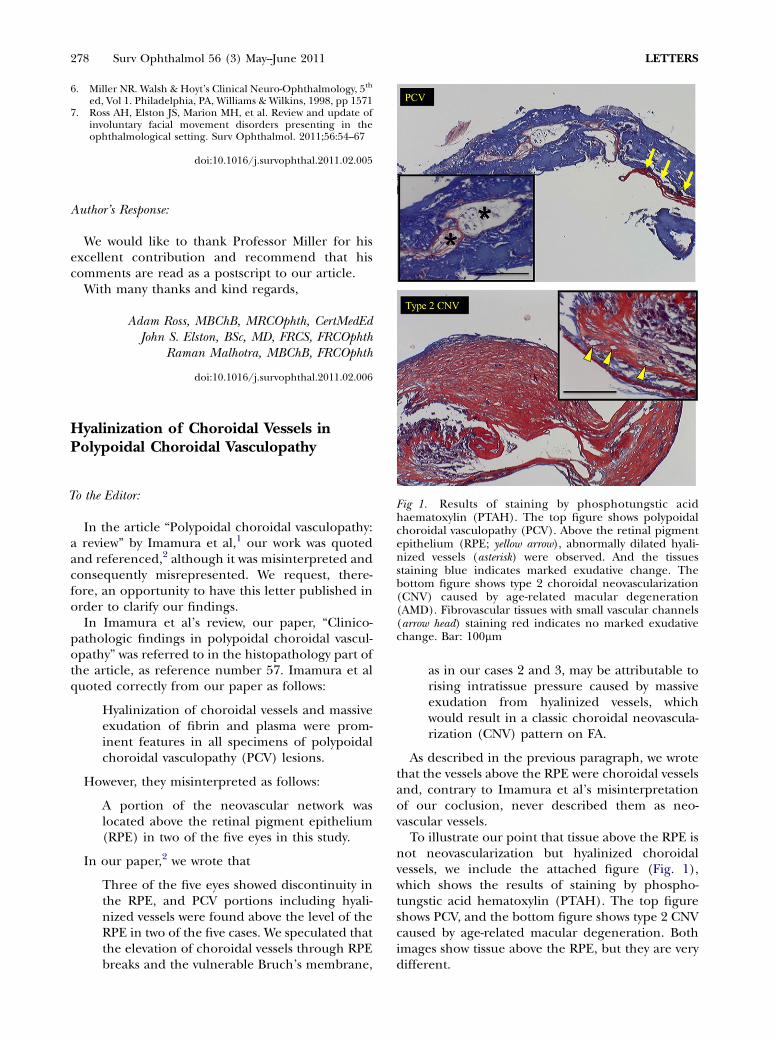
Fig 1. Results of staining by phosphotungstic acidhaematoxylin (PTAH). The top figure shows polypoidalchoroidal vasculopathy (PCV). Above the retinal pigmentepithelium (RPE; yellow arrow), abnormally dilated hyali-nized vessels (asterisk) were observed. And the tissuesstaining blue indicates marked exudative change. Thebottom figure shows type 2 choroidal neovascularization(CNV) caused by age-related macular degeneration(AMD). Fibrovascular tissues with small vascular channels(arrow head) staining red indicates no marked exudativechange. Bar: 100mm
To the Editor:
In the article “Polypoidal choroidal vasculopathy:a review” by Imamura et al,1 our work was quotedand referenced,2 although it was misinterpreted andconsequently misrepresented. We request, there-fore, an opportunity to have this letter published inorder to clarify our findings.
In Imamura et al’s review, our paper, “Clinico-pathologic findings in polypoidal choroidal vascul-opathy” was referred to in the histopathology part ofthe article, as reference number 57. Imamura et alquoted correctly from our paper as follows:
Hyalinization of choroidal vessels and massiveexudation of fibrin and plasma were prom-inent features in all specimens of polypoidalchoroidal vasculopathy (PCV) lesions.
However, they misinterpreted as follows:
A portion of the neovascular network waslocated above the retinal pigment epithelium(RPE) in two of the five eyes in this study.
In our paper,2 we wrote that
Three of the five eyes showed discontinuity inthe RPE, and PCV portions including hyali-nized vessels were found above the level of theRPE in two of the five cases. We speculated thatthe elevation of choroidal vessels through RPEbreaks and the vulnerable Bruch’s membrane,
as in our cases 2 and 3, may be attributable torising intratissue pressure caused by massiveexudation from hyalinized vessels, whichwould result in a classic choroidal neovascula-rization (CNV) pattern on FA.
As described in the previous paragraph, we wrotethat the vessels above the RPE were choroidal vesselsand, contrary to Imamura et al’s misinterpretationof our coclusion, never described them as neo-vascular vessels.
To illustrate our point that tissue above the RPE isnot neovascularization but hyalinized choroidalvessels, we include the attached figure (Fig. 1),which shows the results of staining by phospho-tungstic acid hematoxylin (PTAH). The top figureshows PCV, and the bottom figure shows type 2 CNVcaused by age-related macular degeneration. Bothimages show tissue above the RPE, but they are verydifferent.
Recommended
![· Web view[Author’s name]’s Research Proposal – [release date] – version # [Author’s name]’s Research Proposal – [release date] – version # [Author’s name](https://img.pdfslide.us/doc/110x75/5e3737bc34490a15c57bdf50/web-view-authoras-nameas-research-proposal-a-release-date-a-version.jpg)