DOMAIN ARCHAEA
Table of Contents
1. Glossary of terms
2. Discovery of domain Archaea
3. Structural attributes• Cell shape & size• Cell multiplication• Presence & type of cell wall structure• Membrane composition
4. Functional attributes• Habitat & ecology• Nutrition, physiology & metabolism
5. Molecular attributes• Genomes• Gene organisation in genomes• DNA replication• Transcription• Translation• Cloning and expression of archael genes• Phage and plasmids
6. Kingdom Crenarchaeota Section I. Thermophilic and hyperthermophilic crenarchaeotes
• Order "Igneococcales"• Order Sulfolobales• Order Thermoproteales
Section II. Cold dwelling crenarchaeotes
7. Kingdom Euryarchaeota• Order Halobacteriales, the extreme halophiles• Methanogens and it’s five orders• Thermoplasmatales, the cell wall-less order• Archaeoglobales, the sulfate reducers• Thermococcales, the sulfur respirers

8. Kingdom Korarchaeota
9. Evolution and Life at High Temperatures
10. The Limits to microbial existence:
11. Hyperthermophiles Archaea and Microbial Evolution:
12. Comparative genomics of Archaea:
13. Some Useful References

1. Glossary:• Acetotroph: A methanogen which consumes acetate and splits acetate to methane
and carbon dioxide during growth.• Acetyl-CoA (Ljungdahl-Wood) pathway: A autotrophic CO2 fixing pathway
widespread in strict anerobes (e.g. methanogens, homoacetogens & sulfate-reducing bacteria).
• Compatable Solute: Organic or inorganic substances that accumulate in halophiliccytoplasm for maintaining ionic pressure.
• Crenarchaeota: A kingdom of Archaea that contains hyperthermophiles and colddwelling organisms.
• Euryarchaeota: A kingdorm of Archaea that contains mainly methanogens, theextreme halophiles and Thermoplasma
• Extreme halophile: An organism whose growth is obligately dependent on highconcentrations (> 10%) NaCl.
• Hyperthermophile: A microbe that grows optimally with temperatures > 80oC.• Korarchaeota: A kingdom of Archeae that branches close to the archeal root.• Reverse DNA gyrases: An enzyme present in hyperthermophiles that introduces
positive super coiling into circular DNA.• Solfatara: A hot, sulfur-rich but generally acidic environment.• Thermosome: A type of heat shock chaperonin that refolds partially denatured
proteins in hyperthermophiles.
2. Discovery of domain Archaea:Until 1977, methanogens were regarded as bacteria. Based on 16S and 18S rRNAsequence data, Woese proposed a third kingdom to encompass them [Woese, (1977)PNAS 74:5088-5090]. In 1990, Woese concluded from further 16S rRNA and 18SrRNA sequences that Halobacterium regarded previously as a halophilicpseudomonad and Sulfolobus regarded as a gram-positive bacterium, were membersof domain Archaea. He proposed that life on earth is made of 3 primary lineageswhich he referred to as domains [Woese,(1990) PNAS 87:4576-4579].• Eubacteria (Eu = good or true)• Archeae (Archeae = ancient) and• Eukarya (Carya = nut or kernel)
The evolutionary history of life can be traced to the earliest common ancestor(progenote)for the three domains to be some 3.5 to 4 billion years (Brown andDoolittle (1995) PNAS 1995, 92: 2441-2445; Keeling and Doolittle (1995) PNAS92:5761-5764; Doolittle (1999) Science 284:2124-2128] and it is expected that as weunravel the mysteries surrounding the evolution of life, the descriptions of life willchange.
Members of the domain Archaea (aka as Empire Archaea) at the time of writing (June2000) are phylogenetically divided into three kingdoms, namely, Euryarchaeotoa,Crenarchaeota and Korarchaeota (Fig 1)

Figure 1 Phylogeny of domain Archaea based on comparision of the 16S rRNAsequences. Representatives of Euryarchaeota and Crenarchaeota have been culturedbut members of the Korarchaeotas have yet to be cultured. Genomes of 4euryarchaeotes and 1 crenarchaeote have been sequenced. The bar indicatesnucleotide divergence. Greek Archaios = ancient, primitive; Greek Eurus = wide(wide distribution); Greek Crene = spring, fount (primary habitat).
3. Structural Attributes:Shape and sizeMembers of domain Archaea are morphologically diverse and include spheres, spiral,rods, lobed, plate-shaped, irregular-shaped or pleomorphic. They may exist as singlecells, as aggregates or form filaments. The diameter range is 0.1 to 15 µm and thelength can be up to 200 µm.
Cell multiplicationUsually by binary fission, but some multiple by budding, fragmentation and as yetunknown mechanisms.
Cell WallsSome Archaea such as Thermoplasma species do not contain a cell wall whereas mostothers do contain cell walls. The cell wall-containing Archaea can stain Gram-positive or Gram-negative and ultrastructurally are similar to that of members ofdomain Bacteria.

Schematic representations and electron micrograph of (a) a gram-positive archaeum(e.g. Methanobacterium) and a gram-negative archaeum (e.g. Thermoproteus). CW =cell wall, CM = cytoplasmic membrane, CPL = cytoplasm and SL = surface layer.However, the chemistries are very different. In general, Archaea also posses morechemical variation in their cell walls than members of domain Bacteria do.
(a) Gram-positive archael cell walls: The ultrastructure structure of Archael Gram-positive shows a thick layer which is similar to the ultrastructure of Gram-positiveBacteria.
• Methanobacterium, Methanothermus and Methanopyrus containpseudomurein (glycans [sugars] and peptides in their cell walls). Glycans aremodified sugars viz, N-acetyl talosaminouronic acid (NAT or T) & N-acetlyglucose amine (NAG or G) T and G are linked to each other by a beta 1, 3glycosidic bond & alternate to form the cell wall backbone. Lysozyme (anenzyme produced by organisms that consume bacteria, and normal bodysecretions such as tears, saliva, & egg white = protect against would-bepathogenic bacteria) cannot digest beta 1,3 glycosidic bonds. Peptides areshort amino acid chains attached to T. The amino acids are only of the L-type.Penicillin is ineffective in inhibiting the cell wall peptide bridge formation.
• Some cell walls contain polysaccharides:� Methanosarcina are non-sulfated polysaccharide. These complex
polysaccharides are similar to chondroitin sulfate (aka methanochondrotin)of animal connective tissue.
� Halococcus are sulfated polysaccharides
(b) Gram-negative archael cell walls lack the outer membrane and complexlipopolysaccharide found in Gram-negative members of the domain Bacteria butinstead consists of either a surface glycoprotein or protein subunits.
• Halobacterium are made of glycoproteins but also contain negatively chargedacidic amino acids which counteract the positive charges of the high Na+
environment. Therefore, cells lyse in NaCl concentrations below 15%.

• The cell walls of Methanolobus, Sulfolobus, Thermoproteus, Desulfurococcusand Pyrodictium are made up of glycoproteins (Glycoprotein S-layer)
• Methanomicrobium, Methanococcus, Methanogenium and Thermococcus cellwalls are exclusively made up of protein subunits (protein S-layer).
• Methanospirillum cell wall consists of a protein sheath.
Lipids and Cell MembranesThe chemistry of lipids is very different to that of members of domains Bacteria andArchaea and is perhaps the most distinctive feature of archael cells.
Archael glycerol molecules may be linked:• to a phosphate group (similar to bacteria & eucaryotes) and / or• to a sulfate and carbohydrates (unlike bacteria & eucaryotes) & therefore
phospholipids are not regarded as universal structural lipids.
Archael lipids are hydrocarbons (isoprenoid hydrocarbons) not fatty acids, arebranched (straight chain in bacteria & eucaryotes) and linked to glycerol by etherbonds (ester linked in bacterial & eucaryotes).

Figure: Bacterial lipids are made of phospholipids - a phosphate group joined to 2fatty acids by glycerol (glycerol diester) (A) but archael lipids are composed ofphosphate, sulfate or carbohydrate joined to branched C20 and / or C40 hydrocarbonchains by glycerol diethers (B and C respectively).
Archael lipids are diverse in structure:• Glycerol diether (Glycerol + C20 hydrocarbons)- Bilayered membrane• Glycerl tetraether (Glycerol + C40 hydrocarbons)- Monolayered membrane• Mixture of di- & tetra- Mono /Bi layered membraneCyclic tetraethers (Glycerol + > C40)- maintain the 4-5nm membrane thickness
FigureXX. Membranes of archeae posses integral proteins and a bilayer composed ofC20 diethers (a) a rigid monolayer composed of C40 diethers (b) a mono / bilayerconsisting of a mixture of C20 and C40 diethers (c).
Diversity of membrane rigidity requirements is related to the diverse habitats thatArchaea live in and the changes are necessary to maintain membrane fluidity andstability:• Sulfolobus (90oC, pH 2)- branched chain C40 hydrocarbons. Branched chains
increase membrane fluidity (unbranched & saturated fatty acids limit sliding of
A
B
C

fatty acid molecules past one another) and is required for growth at hightemperatures (upto 110oC, hyperthermophies)
• Halobacterium cope with saturated salts.• Thermoplasma cope with high temperature without cell walls
4. Functional AttributesHabitat and ecologyThey are found in extreme environments and include anaerobic, saline to hypersaline,high temperature and cold temperature habitats (constitutes 34% of microbial biomassin the Antarctic surface waters). Some are symbionts in animal digestive tracts.
Nutrition, Physiology and MetabolismSome are aerobes, some strict anaerobes and some facultative anaerobes. Some arechemolithoautotrophs, others chemoorganotrophs and some can grow by changingfrom one to the other nutritional mode. The growth temperature varies beweenmesophilic to the hyperthermophilic. The pH growth range is between very acidic (<0.5) to slightly over neutral. Some are obligate halophiles yet others are halotolerant.
Chemoorganaotrophs: Use organic substrates as energy source for growth. Catabolismof glucose occurs via modified Entner-Doudoroff (E-D) pathway. Oxidation ofacetate to CO2 proceeds via the citric acid cycle or by the Acetyl-CoA pathway.Amino acid biosynthesis pathways unknown. Electron transport chains includingcytochromes of type a, b and exist in some Archeae. Consequently, electrons fromorganic electron donors enter the electron transport chain leading to the reduction ofO2, So with concurrent establishment of PMF to drive ATP synthesis throughmembrane-bound ATPases.
Autotrophy: Widespread and occurs by several different means. Methanogens useAcetyl-CoA or some modification to fix CO2. In some Archeae (e.g. Thermoproteusand Sulfolobus), CO2 fixation occurs via the reverse citric acid cycle and is incommon with the Green sulfur bacteria and of the genus Chlorobium and Aquifex ofdomain Bacteria or via the Calvin Cycle the most common autotrophic pathway inBacteria and Eucarya. Very thermostable RubisCO enzyme which catalyses the firststep in the Calvin Cycle is found the methanogen Methanococcus jannaschii and aPyrococcus species.
In summary, catabolic and anabolic processes are somewhat similar for Bacteria andArchaea but methanogens are very different. More will be said when archaelkingdoms as well as their genomes are discussed.
5. Molecular Attributes:Genomes• Archaeal chromosome is a single, circular DNA molecule & extrachromosomal
elements (eg plasmids) are found in Archaea. However, they are smaller thanbacterial chromosomes.

• The genomes can vary in the G+C content of their DNA between 21 to 68 mol%indicating a marked genotypic diversity.
• The typical genome of bacterial genome is a single circular DNA molecule.Plasmids are also common. Streptomyces spp. and Borrelia spp have linearchromosomes, and Rhodobacter sphaeroides has two chromosomes.
• A typical eucaryote genome consists of multiple linear DNA molecules (coveredin histones and organized in nucleosomes, chlorplast DNA, and mitochondrialDNA. (note the eukaryote dinoflagellate algae has no histones associated with itsDNA)
• Archael DNA binding proteins (aka Histone-like proteins similar to Eucarya):Methanosarcinaceae MC1 and Methanobacteriales HMf share amino acidhomology to eucaryal histone proteins
• Organization of DNA in chromatin-like structurehistone + Eucarya DNA = negative supercoiling & nucleosomeHMf + Archaea DNA = positive supercoilingHTa: ThermoplasmaHTa-like: Sulfolobus
• Genomic resistance to thermal denaturation & genomic structural intergrity inextreme halophiles is related to high intracellular salt concentrations (solutes)
Gene Organisation in genomes• Functionally related genes are often organised in operon like structures though the
primary sequences of archael proteins more often resemble eukaryotichomologues rather than bacterial ones
• Introns have been found in archael 23S and 16S rRNA and tRNA genes
• Varying arrangements of genes can be seen in Archaea
• Methyl coenzyme M reductase, RNAP and bacteriopsin genes are good Examples
DNA Modyfing Enzymes• Archael DNA polymerases involved in DNA replication have been identified. The
primary protein sequences of these enzymes resemble the DNA polymerases fromeukaryotes, eukaryal viruses and E.coli. Some posses 3’-5’ exonuclease (orproofreading) activity
• A Halobacterium halobium DNA polymerase / primase has been identified withreverse transcriptase activity
• Topoisomerases, gyrase and restriction endonucleases have also been identified inArchaea

Transcription• Bacteria have only one type of RNA polymerase, Eukaryotes have three types
namely, RNA polymerases POL I, II and III. and Archaea also has one type but itis similar to the eukaryote RNA polymerase POL II. Comparative sequencehomology of genes encoding subunits suggests that the RNA polymerase is moreclosely related to eukaryal polymerases than the bacterial counterpart.
• Archael RNA polymerases are complex, consisting of up to 14 subunits (c.f. 5 inE. coli)
• Unlike, E.coli RNA polymerase, archael RNA polymerases are unable to initiatetranscription in vitro. This is also seen in eukaryotes where general transcriptionfactors are required for initiation
• Archael promoters have an A-T rich sequence at -32 to -25 bp upstream of thetranscriptional start: the consensus sequence resembles a eukaryotic TATA box.
Translation• Translation signals resemble those found in bacteria (i.e. there are short regions of
complementarity between the 5’ end of the mRNA and the 3’ end of the 16SrRNA)
• Complementary 5’ mRNA to 3’ 16S rRNA similar to bacteria
• Lack of formylmethionine
Detailed understanding of Transcription and translation (You may also havecovered this in some other subjects)The translational machinery in Archaea is generally like those of Bacteria with 70S(bacterial-sized) ribosomes. Genes are arranged in co-transcribed clusters calledoperons. Ribosomes recongnize translational start sites and bind to the mRNAsdirectly at ’Shine-Dalgarno’ (SD) sequences just like Bacteria. Also like in Bacteria,transcription and tranlation are linked - that is, they occur simultaneously, and failureof an mRNA to be translated causes the RNA polymerase to abort transcription.

Eukaryal genes generally are transcribed separately rather than in clusters. In plantsand animals, most genes are segmented into ’exons’ separated by ’introns’ - introns arespliced out of the mRNAs after transcription. In addition, the 5’ end of the mRNA iscapped with a modified nucleoside, and the 3’ end is cleaved and polyadenylated.Ribosomes generally bind the 5’ ’cap’ of the mRNA & scan downstream for AUGcodons to start translation. Because transcription occurs in the nucleoplasm andtranslation occurs in the cytoplasm, the mRNA has to be exported through the nuclearmembrane after transcription before it can be translated, so transcription andtranslation are not linked.
However, in many ways translation in Archaea is like it is in Eukarya. Translation isinitiated with methionine (like Eukarya), not formyl-methionine (Bacteria).Translation is inhibited by diphtheria toxin, as are eukaryal ribosomes, but is notinhibited by most bacterial-translation-inhibiting antibiotics. Chimericarchaeal/eukarya ribosomes are functional - bacterial/archael & bacteria/eukarya arenot functional.
Example - RNA polymeraseA typical example of how Archaea resemble eukaryotes in some ways and Bacteria inothers is RNA polymerase:
Bacteria contain a single RNA polymerase that transcribes all genes in the cell. Theholoenzyme contains 5 polypeptides - 2 copies of alpha, and one each beta, betaprime, and sigma. The sigma subunit of RNA polymerase provides promoterspecificity by directing binding to signal sequences 10 and 35 base-pairs upstream ofthe transcription initiation site. Different sigmas regulate developmental pathways,e.g. sporulation.
Bacterial promoters:

Eucarya contain 3 nuclear RNA polymerases. Each is specialized for transcription ofspecific gene types. These enzymes contain many more subunits than do bacterialRNA polymerase: 3 or 4 large, ~9-14 small. Promoter recognition is not a function ofthe RNA polymerase, but is provided by transcription factors (TF) that bind directlyto the promoter (not RNA polymerase), and the DNA:TF complex is recognized bythe RNApolymerase.
RNA polymerase I transcribes ribosomal RNA genes, and is confined to thenucleolus. Promoter sequences surround the initiation site, defining a binding site fora transcription factor complex.
RNA polymerase II transcribes primarily mRNAs. Promoter sequences varywidely, but generally have a -35 TATA-box that is a binding site for a general RNApolymersase II transcription factor TFIID.
RNA polymerase III transcribes primarily 5S rRNAs and tRNAs. The promoterelements are downstream of the transcription initiation site, and are binding sites forTFIIIC or B and TFIIID.
Archaea, like Bacteria, have a single RNA polymerase that transcribes all genes.However, archaeal RNA polymerases are like those of eukaryotes in that they contain3 or 4 large and many small subunits. Archaeal RNA polymerases are similar insequence and in antigenicity to eukaryal RNA polymerase II. Promoters in Archaeaare a -30 TATA-like sequence that is a binding site for a transcription factor (TFB),not sigma-like subunits.
Comparison of RNA polymerase gene sequences:

Archaeal and eukaryal RNA polymerae II are primitive RNA polymerases, whereasbacterial RNA polymerase is the most highly evolved. This is typical of bacterial vseukaryal vs archaeal evolution; Bacteria keep the mechanisms basically unchangedbut hone the parts to perfection, whereas eukaryotes duplicate genes are specialized,each for separate functions. Archaea, on the other hand, appear to have remainedunchanged.
Archaea as missing links between eukaryotes and BacteriaIn many ways, Archaea are a ’missing link’ between Bacteria & Eukarya. Archaealfeatures generally resemble Eucarya & Bacteria features more than they do eachother, a real sign of their primitiveness. Having a third evolutionary group, especiallya primitive one, allows the identification of primitive traits, i.e traits of the LastCommon Ancestor (LAC). With only two groups, it’s impossible to tell which versionof a trait, if either, is primitive. It used to be generally assumed that the bacterialversion was primitive, but this is rarely the case.
It has also become clear that the Archaea share a common ancestry with the Eucaryato the exclusion of the Bacteria - in other words, the Archaea and Eucarya are’sibling’ groups, whereas the Bacteria are ’cousins’ to the Archaea and Eucarya.Archaea, then, are primitive relatives of Eucarya, and as such are ideal organisms toshed light on the complexity of eukaryotic organisms - for example the RNApolymerase we just discussed.
Cloning and expression• Cloning usually follows protein-purification using standard molecular biology
techniques
• Expression in heterologous hosts may be complicated by the altered environmentin which expression is occuring and differences in translation and post-translational mechanisms
Phage and plasmidsA few reports on the occurrence of plasmids and phage have been reported inmembers of the Archaea. However, studies in these areas are progressing slowly dueto the very specialised techniques required for culturing especially for thehyperthermophilies.

6. Kingdom Crenarchaeota:This kingdom is divided into 2 sections in order to make the discussion easier. Thefirst section deals with the thermophilic and hyperthermophilic crenarchaeotes and thesecond section deals with the uncultured cold dwelling crenarchaeotes.
Section I: Thermophilic & hyperthermophilic CrenarchaeotaNot all crenarchaeaotes have been cultured and those that have been cultured are allthermophilic or extremely thermophilic (aka hyperthermophiles), with optimal growthtemperatures above 80oC. Some of the crenarchaeotes are the most thermophilicorganisms known. Most are also acidophilic and autotrophic. This phenotype is alsoshared by the deepest branches of kingdom Euryarchaea and domain Bacteria, andtherefore it has been suggested that these traits are probably the primitive phenotypeof the Last Common Ancestor (LCA).
Most crenarchaeotes, not surprisingly, have been isolated from volcanic geothermalenvironments rich in elemental sulfur (called solfataras). Volcanic activity is found onthe land mass (terrestrial) or on the ocean floor formed along tectonic plates(hydrothermal vents) (Figure 2, Map of volcanic areas of the world, also the tectonicplates). ALSO FIGURE FROM MY PHOTO COLLECTION).
Sulfur metabolism as a key trait: Crenarchaeotes oxidise and / or reduce sulfur by oneof 3 biochemical processes. In most cases sulfur compounds such as thiosulfate arealso usable in place of sulfur.
1. Sulfur reduction: These organisms are autotrophic anaerobes that fix carbon fromCO2. Hydrogen is the electron donor for electron transport and elemental sulfur(or sulfur compounds such as thiosulfate) is the terminal electron acceptor.
Sulfur + H2 ---> H2S + protons
2. Sulfur respiration: These organisms are heterotrophic anaerobes. Both carbon andenergy are extracted from organic compounds. Organics are the electron donor forelectron transport and sulfur (or sulfur compounds) is the terminal electron acceptor.This process is much like ’regular’ respiration, except that sulfur compounds take theplace of O2
Sulfur + organics ---> CO2 + H2S
3. Sulfur oxidation: These organisms can usually grow heterotrophically, getting fixedcarbon from low concentrations of organics in the medium. Most can also be grownautotrophically, fixing carbon from CO2 via the reverse TCA cycle. All are aerobes,of course, since it is the terminal electron acceptor (sulfur is the electron donor) forelectron transport.
Sulfur + O2 ---> H2SO4
Taxonomy: At the time of writing, at least 12 genera belonging to the two orders,namely, Sulfolobales and Thermoproteales have been validly described, and a third,order “Igneococcales”, has been proposed (Figure 3).

Figure 2. Members of kingdom Crenarchaeota are thermophilic to hyperthermophilicmicrobes and are divided into 3 orders.
The most studied genera include Thermoproteus and Sulfolobus. The characteristicsof some of the members from each of the order are described as examples below.
Order "Igneococcales":Pyrodictium: Most isolates have been cultured from ocean floor volcanoes
(hydrothermal vents). All are strict anaerobes. Some species are organotrophs andsome are lithotrophs growing on H2 and So. P. occultum is the most thermophilic(hyperthermophile) organisms known to date. The optimum temperature for growth is105 oC (requires a bar of pressure in culture tubes to avoid medium from boiling) witha minimum growth temperature of 82 oC and a maximum growth temperature of 115oC.
Desulfurococcus: These are cocci shaped and have been isolated fromterrestrial and marine volcanic environments. Some members are motile and some arenon-motile. Strict anaerobes that reduce sulfur and are also able to respire (See figureXX below)

Figure XX. A Desulfurococcus species shows extensive glycocalx used in attachment.In this case polycarbonate membrane was immersed for 5 hours in a New Zealand hotspring with a temperature of 90 oC and the sample processed for electron microscopy.The chemical composition of glycocalax is unknown.
Order Sulfolobales:Sulfolobus: Several species have been described and all belong to the order
Sulfolobales. The cells gram-negative aerobes and have an irregular spherical lobedshape (see Figure XX below). The temperature optimum is between 70 to 80 oC witha pH optimum of between 2 to 3. Some species are able to grow at pH 0 (equal to0.5M H2S04) Sulfolobus is sometimes referred to as a thermoacidophile. The cellwalls lack peptdioglycan but contain lipoprotein and carbohydrates. Oxygen is thenormal electron acceptor but ferric iron can also be used. Some strains aremicroaerophilic
Figure XX. Sulfolobus is not spherical but is lobe-shaped and can be isolated fromacidic hot springs.
Some strains grow lithotrophically by oxidising So producing sulfuric acid whereassome other strains grow on sugars and organic acids (eg glutamate) as a carbon andenergy source. These organisms, like most acidophiles, are oligotrophic ie highconcentrations of organics, especially organic acids, are toxic.
The reason for this is that organic acids are protonated in the external growth medium(pH < 3.5), & hence remain uncharged, enabling them to diffuse freely into thecytoplasm through the lipid membrane. The internal cytoplasmic pH of the cells is pH>5.5 and under this condition, the organic acid ionizes, releasing H+. Cytoplasmacidification leads to decomposition of the proton gradient force (i.e. highconcentrations of organic acids act as uncouplers). See Fig XX below.
Figure XX: High concentrations of organic acids act as decouplers as they areprotonated in the external acidic environment of the growth medium (pH < 3) and are

ionized releasing protons inside the cytoplasm (pH > 5.5). The net result of this is adisruption to the proton gradient leading to death of the cells.
Sulfolobus acidocaldarius can be easily isolated from volcanic acid environments(soil and springs). The genome of Sulfolobus acidocaldarius has been partiallysequenced (REFERENCE TO THE WEB SITE).
Order Thermoproteales:Thermoproteus: These are members of the order Thermoproteales. Cells are
long and thin and can be identified by the presence of occasional branching of thecells and presence of golf ball-like terminal structures. Cell walls are made ofglycoprotein subunits. Strict anaerobes which grow between 70 to 90 oC (optimum 85oC). Lithotrophic growth occurs by So reduction (with H2) and CO2 (or CO) as a solecarbon source. In addition, they can also grow organotrophically by oxidising glucose,amino acids, alcohols and organic acids by anaerobic So respiration. Isolated fromneutral volcanic terrestrial and marine hot springs rich in sulfur.
Figure XX. A variety of thermophilic Archeae thrive as mixed populations interrestrial hot springs and can be visualised using electron microscopy techniques.The crenarchaeotes Thermofilum are thin filament, Thermoproteus are rod tofilaments with a larger diameter whereas Desulfurococcus are cocci.
Thermofilum: The genus Thermofilum is member of the orderThermoproteales and two species have so far been described. They require as yetunidentified factors from Thermoproteus (by either co-culturing with Thermoproteusor with Thermoproteus extracts) without which they are unable to grow. The cells areextremely filamentous and very thin (<100 x 0.15-0.3 µm) and can be easilydistinguished from Thermoproteus based on this trait.
Section II. The cold dwelling CrenarchaeotaCold dwelling crenarchaeotes have not as yet been cultured but there presence hasbeen demonstrated by fluorescent phylogenetic staining in marine waters world wideand have also been found in frigid marine waters of the Antarctic. High crenarchaeoteconcentrations (around 10,000 cells / ml) have been observed in these cold andnutrient deficient waters.

The metabolism and physiology of these organisms remains a mystery, though thecells which were collected after membrane filtration were found to contain etherlinked lipids of the diphytanyl tetraether type which have so far been known to occuronly in hyperthermophiles. The diphytanyl tetraethers were thought to be themolecular secret behind hyperthermophiles surviving high temperatures but theirpresence in cold dwellers, leaves this hypothesis to refinement.
7. Kingdom Euryarchaeota:Phenotypic traits and evolution: Most members of kingdom Euryarchaea arepredominantly methanogens, but there are 2 other phenotypes found in this group -sulfur-metabolizing thermophiles and extreme halophiles. One group of sulfur-metabolizing thermophiles, Thermococcus & relatives, seem to have retained thatphenotype from the common ancestry of Euryarchea & Crenarchaea (and Bacteria,for that matter), whereas the other sulfur-metabolizers and halophiles evolved thesephenotypes from methanogenic ancestry (see figure below).
Diversity and Taxonomy: This is an extremely diverse kingdom and is characterisedby 5 distinct orders (SEE FIGURE BELOW):(a) Halobacteriales (the extreme halophiles)(b) Methanogens which are divided into 5 orders(c) Thermoplasmatales(d) Archaeoglobales (sulfate reducers) and(e) Thermococcales

Figure 3. Dendrogram showing the five distinct groups consisting of the ordersHalobacteriales (the halophiles) Thermoplasmales (lacking cell walls),Archaeoglobales (sulfate reducers), Thermococcales and methanogens (which aredivided into 5 orders) within the Kingdom Euryarchaeota.
1. Order Halobacteriales, the extreme halophilesThe extremely halophilic Archaea require at least 2M NaCl or equivalent ionicstrength for growth - most grow in saturated or near-saturated brines. They are theprimary, or only, inhabitants of salt lakes. Red pigments make it obvious when largenumbers of these organisms are present - blooms often occurs after a rain carries
organic material into a salt lake, and the Red Sea gets its name from such blooms. Sodoes the ’Red Herring’, from foul-smelling but hound-decoying salted fish beingspoiled by Halobacterium. They are common in hypersaline seas, salt evaporationpools, salted meats, dry soil, salt marshes, etc. They are also found in subterraneansalt deposits, where micropockets of saturated water ’diffuse’ around in the otherwisesolid salt.
Other halophilic organisms (e.g. fungi, brine shrimp) have normal cytoplasmic saltconcentrations, expending energy to continuously pump salt out of the cell and waterinto the cell, and contain organic osmolytes like glycerol or sugars. HalophilicArchaea grow at much higher salt concentrations, and don’t fight back at all - theinternal salt concentrations are as high as they are outside! For this reason, there islittle or no net osmotic pressure on the cell wall, and some organisms take advantageof this by adopting high surface-area shapes that are not possible for organisms in’normal’ ionic strength. One example is Haloarcula, which comes in squares andtriangles with straight edges, sharp corners, and is very flat. Other halophiles are rodsor cocci.
Halophiles are mesophilic facultative aerobes. Aerobically, they growheterotrophically, via respiration, using O2 as the terminal electron acceptor.Anaerobically, they grow photochemotrophically - they get energy (ATP) from light,but still need organics for carbon.
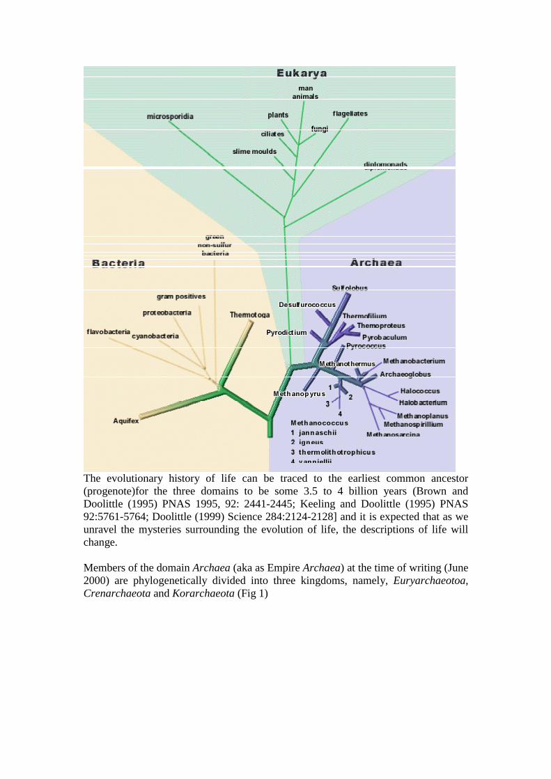
They do not contain the usual photosystems or electron transport chain for gatheringenergy from light. Phototrophy is driven by a single protein, bacteriorhodopsin, that isa light-driven proton pump.
This proton pump generates a proton gradient used to make ATP via ATPase, just likein other organisms. It’s not nearly as efficient as the bacterial photosystems, but lightis rarely limiting for growth in the desert salt lakes where they predominate.
Some halophiles grow at high pH (up to pH10-10.5) i.e. Natronobacterium in sodalakes. This is a problem for them (or at least for us, trying to understand how they getaway with it). At that pH, any protons pumped to the outside, by eletron transport orrhodopsin, are gone forever. Even though the resulting electric potential is still there,it can’t be harvested by an ATPase unless it can get protons from the outside. Howthey get around this is not known, and it is probably this issue that limits the upper pHrange or life.
The salient features of the extreme halophiles is shown in the table below.

2. Methanogens and it’s 5 orders:Methanogens make methane (CH4) via a unique metabolic pathway with uniqueenzymes & cofactors, incuding F420, that makes the cells fluorescent.
All methanogens are obligate lithotrophs, that is, they can make energy only bymethanogenesis. All can make methane from CO2 + H2, some can also use other onecarbon compounds (e.g. CO, formic acid), and a very few can use acetate andmethylamines. At least one ATP is generated with the reduction of CO2 to CH4 withH2.
One carbon compounds (formate, CO2, CO) are reduced with hydrogen and attachedto methanofuran (MF) in the form of a formyl group. This formyl is transfered totmethanopterin (MP) and sequentially reduced through methenyl and methylene tomethyl, which is then transferred to coenzyme M, and finally reduced to free methane.Organisms that grow on acetate transfer the methyl group of acetate directly to MPand the carboxy group is released as CO2. Organisms growing on methanol transferthe methyl group (indirectly) to CoM.


The enzymes in the methanogenic pathway are very oxygen sensitive, somethanogens are extreme anaerobes.
Methanogens are common organisms, found in all types of anaerobic environments,and are certainly the most prevalent Archaea in the ’moderate’ world.
Sediments and soils - swamp gas is methane which, because of its low ignitiontemperature and low threshold concentration, is readily ignited and glows very faintlywhite in ’will-o-the-wisps’ visible at night in swamps. Methanogens are crucialcomponents of the micobial populations of the ’rhizosphere’, the plant rootenvironment. Animal guts - especially wood-eating insects and ruminants. Africantermite mounds are scruoulously aerated by the insects not for oxygen, but to keepmethane concentrations low. Termite mounds struck by lightening after a light rain(blocking aeration) can explode spectacularly! Cows also produce large amounts ofmethane.
Wastewater and landfills - The whole wastewater process works because organicsin the wastewater are converted first to biomass (in the early stages of treatment),thendigested anaerobically to H2, CO2, and acetate which in turn is converted bymethanogenesis into methane, which floats away. Landfills have to be carefullyvented to prevent very messy explosions! Several houses near older unvented landfillshave exploded because of the buildup of methane that seeped through the ground intotheir basements.
Oil deposits - natural gas is methane, produced not geochemically but bymethanogens living in the subterranean oil deposit.

Methanogens form a variety of symbioses with plants, animals and protists, butdespite these close associations there are no known pathogenic methanogens. None ofthe other Archaea are pathogens either, but considering the conditions under whichthey grow, this is not surprizing. Methanogens also form close syntrophic associationswith heterotrophic Bacteria that generate hydrogen (i.e. use protons as the terminalelectron acceptor). Hydrogen-generating heterotrophism is only energetically-favorable where the ambient concentration of hydrogen is extremely low.Methanogens associate with these organisms, utilizing the hydrogen they generate formethanogenesis, and keep the hydrogen concentrartion low enough for theheterotrophs to make a living. Neither of these organisms could persist in theenvironment alone, but together they are successful.
Other than the fact that they all make a living the same way, methanogens are adiverse phenotypic and ecological group. The methanogens are divided into 5 orders,the characteristics of which are given below.
1. Order MethanobacterialesThe order Methanobacteriales currently encompasses non-motile methanogens withpeudomurein cell walls and C20 and C40 isopranyl glycerol ethers in theirmembranes. The order contains two families namely Family I. Methanobacteriaceaeand family II. Methanothermaceae.
Family I Methanobacteriaceae. Family Methanobacteriaceae contains fourmorphologically distinct genera. (a) The 13 species of the genus Methanobacterium[58-,71] are rod to filamentous cells. Some species are thermophilic and a few arealcaliphilic and found in various freshwater habitats. Methanobacteriumsubterraneum, an isolate of a deep granitic groundwater is alkaliphilic, eurythermicand halotolerant methanogen [68]. Only six species (M. formicicum, M. defluvii, M.oryzae, M. palustre, M. subterraneum, M. thermoflexum) can use formate. Threespecies (M. bryantii, M. formicicum and M. palustre) can grow on 2-propanol/CO2.All species are able to grow on H2+CO2. Provided the G+C values were correctlydetermined, the broad range 29 to 62 mol% indicates that the genusMethanobacterium is still heterogenous and composed of more than one genus. Thetype species is M. formicicum [58-,60]. (b) The genus Methanothermobacter [8] wasproposed for the inclusion of thermophilic methanogens such as M.thermoautotrophicum [72] and M. wolfei [73]. M. thermoalcaliphilum [74,75] and M.thermoformicicum [76-,78] are considered as synonymous of M.thermoautotrophicum [72]. The proposal has now been accepted and a new genusMethanothermobacter created to include 3 species namely Methanothermobacterthermoautotrophicus comb. nov., Methanothermobacter wolfeii comb. nov. andMethanothermobacter marburgensis sp. nov. [79]. (c) The seven members of genusMethanobrevibacter are neutrophilic mesophilic short rods, often forming pairs orchains and the G+C content varies from between 28 to 32 mol% [80-,85]. Eachspecies inhabits a specialised habitat. M. ruminantium, the type species, is thepredominant methanogen in the bovine rumen [80]. It requires cofactors for growth,but like M. smithii and M. cuticularis can use formate. M. smithii is abundant insewage sludge and intestinal tracts of animals and man [85]. M. arboriphilus does notuse formate and was isolated from wetwood of living trees [81]. Methanobrevibactercurvatus, M. cuticularis [82], and M. filiformis [83] have been isolated from gut of asubterranean termite recently whereas M. oralis has been isolated from human
subgingival plaque [84]. (d) The two species of the genus Methanosphaera are Gram-positive spherical-shaped organisms which have been isolated from feces of man [86]and rabbit [87] and are generally observed in the digestive tracts of animals. The G+Ccontent is 23 to 26 mol%. Both species require both methanol and H2 as substrates formethanogenesis and are unable to use H2 plus CO2 or formate. Their inability toreduce CO2 to CH4 is due to the lack of an active or the presence of an inactive CO2
reductase system and methyltetrahydromethanopterin:coenzyme M methyltransferase[88]. The type species is M. stadtmaniae [86].
Family II Methanothermaceae. Family Methanothermaceae consists of the singlegenus Methanothermus and its 2 species [89,90]. Both the species are extremethermophiles and have been isolated from a specific habitat (volcanic springs). Thetemperature optimum is 80°C. The cells are rod shaped, contain a double-layered walland have a mol G+C content of 33-34%. As hydrogenotrophic methanogens, they useonly hydrogen and carbon dioxide with prototrophic growth. The type species is M.fervidus [89].
2. Order MethanococcalesBoone et al. [8] proposed a thorough reorganization of this order. The order nowcontains two families and four genera (Figure 1) of hydrogenotrophic methanogensisolated essentially from marine and coastal environments. All species are irregularcocci, contain proteinaceous cell walls and are motile by a polar tuft of flagella. Cellslyse quickly in detergents. C20 isopranyl glycerol ethers are abundant and C40 ethersare absent excepted in "Methanocaldococcus jannaschii". All species use both H2 andformate as electron donors, and are prototrophs, except the three species of"Methanocaldococcus" and "Methanoignis igneus" which are unable to utilizeformate. Growth is often stimulated by selenium.
Family I Methanococcaceae. Family Methanococcaceae contains two genera. (a)The genus Methanococcus includes five mesophilic species (including 1 synonymous)whose G+C content varies between 30 to 41 mol% [91-97]. The type species is M.vannielii [91]. Methanococcus deltae has been recognized as a synonym of M.maripaludis [96]. “Methanococcus aeolicus” was included in a genetic study but itscharacteristics have never been formally described [96]. (b) The genus"Methanothermococcus" has been proposed to include the thermophilic species M.thermolithotrophicus [8,97].
Family II "Methanocaldococcaceae". Family "Methanocaldococcaceae" has beenrecently proposed to include two thermophilic genera. The G+C ranges from 31 to 33mol%. (a) "Methanocaldococcus jannaschii" [98], an extreme thermophile isolatedfrom a hydrothermal vent on the East Pacific rise, is the fastest growing methanogenknown to date (generation time = 30 min). Two new species, M. fervens and M.vulcanius, have been recently described in genus Methanococcus but is to bereclassified in the genus "Methanocaldococcus" [99]. (b)"Methanoignis igneus"[100] is the only species in the new genus proposed by Boone et al. [8].
3. Order MethanomicrobialesThe order Methanomicrobiales comprises three families and 9 genera [8,101] ofhydrogenotrophic methanogens.

Family I Methanomicrobiaceae. Family Methanomicrobiaceae contains 7 generawith a variety of different morphologies which includes small rods, highly irregularcocci, and plane-shaped cells. The cell walls are proteinaceous and the lipids includeboth C20 and C40 isopranyl glycerol ethers. The G+C range of the family is 39 to 50mol%. Almost all strains can use formate and some secondary alcohols. (a) The genusMethanomicrobium includes the single mesophilic species, M. mobile whose G+Ccontent is 49 mol%. [102]. It is a slightly curved rod, sluggishly motile with a polarflagellum. It was isolated from bovine rumen and has a complex nutritionalrequirement which includes rumen fluid. An unidentified growth factor found inrumen fluid could be replaced by extracts of Methanobacterium thermoautotrophicum[103]. (b) The genus Methanolacinia has been created to include the reclassifiedspecies Methanomicrobium paynteri [104] as Methanolacinia paynteri [105].Methanolacinia paynteri a short and irregular non motile rod, isolated from marinesediments is unable to use formate. Cells lyse in detergents. The G+C content is 45mol%. (c) The genus Methanogenium contains five species isolated from variousenvironments [106-,109]. Morphologically they are highly irregular cocci, stainGram-negative and are nonmotile but do posses flagella. Cell walls are composed ofregular protein subunits. Cells readily lyse in dilute detergents. They require growthfactors and use formate. The G+C content varies from 47 to 52 mol%. Two speciescan use CO2 + secondary alcohols to form methane. Methanogenium frittonii is athermophilic species [108] whereas M. frigidum, which has an optimum temperaturefor growth of 15°C was isolated from Ace Lake in Antartica and is a psychrophile[107]. The type species is M. cariaci [106]. (d) The genus Methanoculleus [110]consists of five mesophilic species (including 1 synonymous) of highly irregular nonmotile cocci which stain Gram negative [110-,113] and one thermophilic species[114,115]. Formate is used by five species. The G+C content range is between 49 to62 mol%. The type species has been proposed as M. olentangyi [91,110] and M.bourgense [111] as a synonym of M. olentangyi [8]. (e) The genus Methanoplanuscomprises three species of plane-shaped organisms with polar tuft of flagella [116-,118]. The cell walls contain at least one major glycoprotein. Formate is used formethanogenesis. The type species is M. limicola [116]. One species is anendosymbiont of marine ciliates and is found in close association with microbodies,and is thought to provide hydrogen to the methanogen [117]. The methanogenfunctions as an electron sink in the oxidation steps of the carbon flow in the ciliates.These symbiotic relationship is thought to be responsible for a total conversion ofmetabolites to carbon dioxide and methane in marine sediments. Recently a newspecies, M. petrolearius, has been isolated from an oil well [118]. The G+C range ofthe family is 39 to 50 mol%. (f) Zellner et al. [119] have proposed to reclassifyMethanogenium tationis [120] and M. liminatans [121] in a new genus Methanofollis.These species use formate and have a G+C content of 54-60 mol%. (g)Methanocalculus is a newly described genus which encompasses the irregularcoccoid M. halotolerans, an isolate from an offshore oil well [122]. It is ahydrogenotrophic halotolerant methanogen which grows optimally at 5% andtolerates up to 12% NaCl. The 0 to 12% NaCl growth range is the widest reported todate for any hydrogenotrophic methanogen including members of the ordersMethanobacteriales, Methanococcales and Methanomicrobiales. Furtherinvestigation may lead to the reclassification of this genus to the familyMethanocorpusculaceae.

Family II Methanocorpusculaceae. Family Methanocorpusculaceae [123]contains one genus, Methanocorpusculum, and five species (including 1 synonymous)[123-,127] of mesophilic, small coccoid methanogens with monotrichous flagellation.They use H2/CO2 and formate and some species can use 2-propanol/CO2. The typespecies proposed by Boone et al. [8] is M. parvum, a tungsten requiring bacterium. Itis the first hydrogenotrophic methanogen to possess a cytochrome of b- or c-type,probably involved in the oxidation of 2-propanol. Methanocorpusculum aggregans[125] has been recently recognized as synonymous of M. parvum [8]. The mol% G+Cof this genus is 48 to 52.
Family III "Methanospirillaceae". The creation of family "Methanospirillaceae"has been proposed recently by Boone et al. [8] to include the single genusMethanospirillum. Members of the genus are mesophilic and have been reported fromvarious habitats. However, only one species, Methanospirillum hungatei, has beendescribed so far [128]. Cells are curved rods and often form filaments several hundredµm in length. Cells present polar, tufted flagella and are sheathed. The cell wallcomposition contains 70 % amino acids, 11 % lipids, and 6.6 % carbohydrates [129].The cytoplasmic membrane and cell sheath have also been isolated and theircomposition determined [130]. The type species uses H2+CO2 and formate, and somestrains are able to use 2-propanol and 2-butanol as hydrogen donors formethanogenesis from CO2 [131,132]. Methanospirillum hungatei gave a positivechemotactic response to acetate [133]. The G+C content is 45-49 mol%. Thishydrogenotrophic methanogen shows the best affinity to hydrogen and is alwaysutilized for isolation of syntrophic bacteria, when sulfate reducers are not used.
4. Order "Methanosarcinales"This new order proposed by Boone et al. [8] regroups all the acetotrophic and/ormethylotrophic methanogens into two families (Figure 1).
Family I Methanosarcinaceae [134]. Family Methanosarcinaceae contains sixgenera and 21 species (including 1 synonymous). (a) The genus Methanosarcinarepresents the acetotrophic methanogens which predominate in many anaerobicecosystems where organic matter is completely degraded to CH4 and CO2. They arefound in freshwater and marine mud, anoxic soils, animal-waste lagoons, andanaerobic digestors. Some are the most versatile methanogens, able to use H2-CO2,acetate and methyl compounds (methanol, methylamines), including six mesophilicspecies (including 1 synonymous), Methanosarcina barkeri, the type species [135-,137], M. acetivorans [138] M. mazei [139-,141], M. siciliae [142-,144], and M.vacuolata [145] and only one thermophilic species, M. thermophila [146]. They sharea characteristic pseudosarcina cell arrangement and morphology.
Several isolates have been described which use H2-CO2 and methyl compounds, andhave a coccoid morphology as M. frisia transferred from Methanococcus frisius[147,148] and recognized later as a synonym of M. mazei [149]. This intermediateform, M. mazei, has a morphology similar to both pseudosarcina and the cocci duringdifferent phases of its life cycle [150-,152]. A complex life cycle involving the releaseof single cells may provide a mechanism for cell dispersal during unfavorable growthconditions, whereas a limited cycle facilitates colony division during growth infavorable conditions. With strain LYC, there is a production of a disaggregataseenzyme that hydrolyses the matrix holding the colony together [151]. Xun et al. [152]

have shown that the life cycle of strain S-6 can be controled by manipulation ofgrowth conditions (magnesium, calcium, and substrate concentration), and byinoculum size.
Methanosarcina vacuolata shows the presence of large vacuoles containing gasvesicles but is unable to float in the liquid medium [145]. Methanosarcina siciliaewas transferred from genus Methanolobus. It uses only methyl compounds [142,143]and is similar in this trait to M. semesiae [153] but recently an aceticlastic strain of M.siciliae was isolated from marine canyon sediments [144]. Methanosarcinaacetivorans is the only marine species in this genus [138]. Methanosarcina barkeri isthe most studied acetoclastic methanogen and one of the earliest species ofmethanogen isolated in axenic culture by Schnellen in 1947 [154], but lost andisolated again by Bryant in 1966 (strain MST) and described as the type strain of thespecies [135-,137]. Cells are pseudosarcinae, mostly in small aggregates butsometimes in large masses visible to the unaided eye. They are nonmotile and stainGram-positive.
The cell wall polymer contains N-acetyl-D-galactosamine and D-glucuronic (or D-galacturonic) acid at a molar ratio of 2:1, as well as a minor amount of D-glucose andtraces of D-mannose. Partial hydrolysis of cell wall material yields a dissacharideidentical with chondrosine, the N-acetylated and sulfated form of which is known asthe repeating unit of animal chondroitin [155]. This unique polymer ofMethanosarcina is another example of the various eucaryotic resemblances found inArchaea and may be termed "methanochondroitin" [155].
Species of Methanosarcina contain only C20 isopranyl glycerol ethers. Nutritionalrequirements vary between species. The hydrogen metabolism during methanogenesisfrom acetate has been extensively studied [156,157]. H2 production by the cellsappears to be linked to several intracellular redox processes which follow the cleavageof acetate. Belay and Daniels [158] have described the formation of ethane by M.barkeri during growth in ethanol supplemented medium; ethanol is converted toethane using terminal portion of the methanol-to-methane pathway.
In the order "Methanosarcinales", the following five remaining genera areobligatory methylotrophic methanogens. These methylotrophs are nonmotile, mostlymesophilic, irregular cocci, using only methanol and methylamines as substrates formethanogenesis. Most biotypes have been isolated from environments with high saltconcentrations and some of these are regarded as true hyperhalophilic methanogens.
(b) The genus Methanolobus [159] contains five species [160-,164]. The typespecies, M. tindarius is an irregular mesophilic coccus isolated from coastalsediments, with a single flagellum, based on electron micrographs [160]. The optimalconcentration of NaCl is about 0.5 M. This concentration can reach 1.5 M for M.oregonensis [162]. The G+C content range is 39-46 mol%.
(c) The genus Methanococcoides includes two species with M. methylutens as thetype species [165]. Cells lyse readily in SDS. The optimal concentration of NaCl is0.2-0.6 M, and high concentrations of magnesium (50 mM) are also required.Methanococcoides burtonii is a psychrophilic methanogen isolated from Ace Lake in
Antartica; the optimum temperature is 23°C [166]. The mol% G+C of the genus is 40-42.
(d) The genus Methanohalophilus encloses four mesophilic, hyperhalophilic species[167-,172]. The type species, M. mahii has been isolated from the sediments of theGreat Salt Lake, Utah. The optimum salinity for growth is 1-2.5 M NaCl.Methanohalophilus euhalobius has been recently transferred from the genusMethanococcoides [168,169] and M. halophilus from the genus Methanococcus[170,171]. The G+C content range of the genus is 38-49 mol%.
(e) The genus "Methanosalsus" has been recently proposed to reclassifyMethanohalophilus zhilinae as "Methanosalsus zhilinae" [8,173], an alkaliphilic,halophilic species of methanogen isolated from an Egyptian lake, and able tocatabolize dimethylsulfide [173,174]. The mol% G+C is 38.
(f) The genus Methanohalobium is represented by only one extremely halophilicspecies, M. evestigatum [175] growing at 25% NaCl and at 50°C.
Family II "Methanosaetaceae". Family "Methanosaetaceae" includes all theobligatory acetotrophic methanogens grouped into the genus Methanosaeta currentlyconsists of two species. The type species, M. concilii, forms an immunologicallycohesive group [176-,179]. The rod-shaped cells, 0.8 x 2 µm, form long sheathedfilaments that often form floc-like aggregates. The outer layer of the cell wall consistsof proteins. Only C20 isopranyl glycerol ethers are present. Acetate is the solesubstrate for methanogenesis, with a doubling time of 4-7 days at 37°C. Formate issplit to H2-CO2. The bacterium was first described as Methanothrix soehngenii [180-,183] but the cultures were recognized as non-axenic [177-,179] and as a consequencethis name has been rejected [184]. A thermophilic gas-vacuolated species, M.thermoacetophila, has been described but culture again was found not to be axenic[185]. Another thermophilic strain has been isolated and characterized [186]. Recentlythe name M. thermoacetophila was rejected and replaced with M. thermophila[184,187]. The mol% G+C of the genus is 50-61.
5. Order "Methanopyrales"Boone et al. [8] have proposed to include the genus Methanopyrus into a new order,"Methanopyrales". This order currently represents a novel group of methanogensgrowing at 110°C and unrelated to all other known methanogens [188-,191]. Thesingle family, "Methanopyraceae", includes only one species, Methanopyrus kandleri(Figure 1). Methanopyrus kandleri is a hydrogenotrophic, hyperthermophilicarchaeum which stains Gram positive. It has been isolated from a hydrothermallyheated deep sea sediment and from a shallow marine hydrothermal system. In thepresence of sulfur, H2S is formed and cells tend to lyse. The cell wall consists of anew type of pseudomurein which contains ornithine and lysine but not N-acetylglucosamine. The pseudomurein is covered by a detergent-sensitive proteinsurface layer. The core lipid consists exclusively of phytanyl diether. The G+Ccontent is 60 mol%.
3. Order Archaeoglobales, the sulfate reducers.Members of the genus Archaeoglobus have been isolated only from deep-seahydrothermal vents & heated marine sediments.

It is a thermophilic (85oC) coccus; some species are motile with tufted flagella (muchlike Thermococcus) and others are nonmotile.
Archaeglobus can be grown either of two ways. It can grow heterotrophically by aunique pathway - it uses the methanogenic pathway in reverse! lactate or acetate ---> H2 + CO2
It also grows autotrophically by sulfate reduction: H2 + SO4=---> H2S (carbon fixed from CO2)
4. The sulfur-metabolizing order ThermococcalesThis order includes members of the genera Thermococcus & Pyrococcus.T.celer is the most primitive organism known, i.e. it is closer to the ’root’ of theuniversal tree than any other known living organism. Thermococcus is a neutral pHheterotroph, and is thermophilic (75 - 90). Pyrococcus is similar but grows at highertemperatures, about 100oC.
These organisms grow by anaerobic sulfur respiration S + organics ---> H2S + CO2
They are common in hot marine sediments, especially in deep-sea hydrothermal ventareas. They are motile, via a distinctive tuft of polar flagella.
5. The cell wall-less order ThermoplasmatalesThese organisms are thermoacidophilic heterotrophs. They are facultatively anaerobic- O2 or sulfur can serve as terminal electron acceptors.
They are acidophilic, most isolates growing best at pH2, but some grow as low as pH0.8! (>0.1M HCl). They are also moderately thermophilic, preferring around 60oC forgrowth.
Thermoplasma has been isolated exclusively from smouldering coal refuse piles, andit is presumed that subterranean coal deposits are their natural habitat. All isolates aremonotrichously flagellated motile cocci and lack any cell wall. Crosslinking of thecarbohydrate chain of membrane glycoproteins provide what little cell rigidity andosmotic tolerance they have.
Like Archaeoglobus, they reveal their methanogenic ancestry by containing F420 (themajor methanogenic cofactor) & other components of the methanogenic pathway, butit is not known what use, if any, they make of these.
8. Kingdom Korarchaeota:There is very little information on this kingdom as the cells have only just beingcultured. The studies on this kingdom will be very important as they fall very close tothe root of the tree of life. Therefore the molecular information contained in them maycontribute to our knowledge and understanding of ancient organisms.
9. Evolution and Life at high temperatures

How do cells cope with high temperatures and what is the maximum temperatures atwhich life could exist? These questions have yet to be answered conclusively butsome pointers are already available for discussion.
(a) Heat stability of Biomolecules: Most proteins from hyperthermophiles have thesame structural features. For example the amino acids around the active sites ofthermostable enzymes and their mesophilic counterpart are the same. However,thermostable proteins do tend to have highly hydrophobic cores (probablyincreases internal "sticking" and in general have more "salt bridges" (ionicinteractions between amino acids) proteins and vice versa. on the surface.Therefore, it is now thought that subtle changes which is reflected in proteinfolding, rather than drastic gross changes, are sufficient to render heat labileproteins to heat resistant and vice versa.
Chaperonins (heat shock proteins, aka HSP) refold partially denatured proteinsback to "life". In Pyrococcus, the HSP, called thermosome, increases inconcentration dramatically (to 80% of the cell content) when the culture is grownat 108oC (which is at the temperature limit for growth). This is a type of protectionmechanism. The culture with thermosome can withstand autoclaving (121oC) for1 hour!!!
(b) DNA stability: (i) High temperature depurination of DNA is decreased due to thepresence of potassium cyclic 2.3-diphosphoglycerate (K+ is actually responsiblefor this) but only in some hyperthermophiles. (ii) Positive supercoiling of DNA(rather than negative coiling) by reverse gyrase stabilises DAN againstdenaturation at high temperatures. (iii) A minor grove DNA binding protein,termed Sac7d, found in Sulfolobus, (crenarchaeotes) increases the meltingtemperature of DNA by 40oC. Sac7d also kinks the DNA and is therefore thoughtto play a role in gene expression.The euryarchaeotes contain highly basic histone-like proteins which have homology with eucaryotic histone proteins. They windand compact DNA into a nucleosome-like structure and thereby protects the DNAfrom heat denaturation. The presence of Sac7d and histone-like proteins mayexplain why the transcriptional apparatus is more in line with the eucaryoticsystem.
(c) Lipid Stability: Contain heat resistant dibiphytanyl diether lipid membranes whichforms a monolyaer rather than a bilayer (see above). Stops the cell membranefrom being pulled apart.
(d) Satbility of monomers: The thermostability of monomers is of more significancethan macromolecules- dictate the upper temperature of life. (1) ATP and NAD+
hydrolyse rapidly- half life of 30 mins at 120oC.
10. The Limits to microbial existence:Extrapolate and hypothesise from the knowledge given above about the limits to lifeat high temperatures:a. Cells require water for life but water above 100oC is steam. Life therefore at
temperatures > 100oC will be restricted to hydrothermal vents of the sea floor.Most Archaea cultured in the laboratory above 100oC are from suchenvironments:-
� Black smokers- 250 – 350oC form upright metallic sulfide structures calledchimneys. The temperature gradient is 250 oC (inside) to 2 oC outside. Archaeahave been isolated from chimney walls but water >250 oC appears to be sterile.
b. Laboratory experiments suggest that life can live at the limits of 140 to 150 oCbeyond which biomolecules become heat labile. For example, ATP will not be anenergy currency but will have to be in some other form.
11. Hyperthermophiles Archaea and Microbial Evolution:Have Archaea adapted to extreme environments or co-evolved and flourished withsuch extremes during earth’s formation, that is, relics of ancient life. Their studieswill reveal intersetin principles of early life.� 16S rRNA sequencing and sequence analysis suggests the later. Aquifex and
Thermotogales, domain Bacteria which are also extreme (hyperthermophiles) andhave slow evolutionary clocks. Their molecular clocks are slower and thereforethey evolved at a slower rate than did Eucarya and Bacteria – a fast clock meansadaptation. Phylogenetic trees shows shorter branches.
� Life under extremes are under strong evolutionary pressure to maintain their genesessential for their survival and therefore beyond a certain point additional geneticchanges will not be of any further benefits.
� Koraracheaota clocks are the slowest and found in volcanic hot springs.� H2 as an electron acceptor with S0, NO3
- and Fe3+ as electron donors underanaerobic, high temperature and dark subsurface (protected from UV radiation)conditions (primodial early earth conditions) by Archaea may be an ancient relicof metabolism.
12. Comparative genomics of Archaea:Refer to the attached published article
13. Some Useful ReferencesScience 283 p 1476 -5th March 1999 "Mitochondrial Evolution" contains a discussionof the Endosymbiotic theory of eukaryote evolution.
Nature 399 p 323 27 May 1999 Presents evidence for lateral gene transfer betweenArchaea and Bacteria. Microbiology and Molecular Biology Reviews December 1998p1436-1491.
Woese, (1977) PNAS 74:5088-5090].
[Woese,(1990) PNAS 87:4576-4579].
Takai-K; Sako-YA molecular view of archaeal diversity in marine and terrestrial hotwater environments FEMS-MICROBIOLOGY-ECOLOGY. FEB 1999; 28 (2) : 177-188
Molecular phylogenetic survey of naturally occurring archaeal diversities in hot waterenvironments was carried out by using the PCR-mediated small subunit rRNA gene(SSU rDNA) sequencing. Mixed population DNA was directly extracted from theeffluent hot water or sediment of a shallow marine hydrothermal vent at TachibanaBay, or the acidic hot water of hot spring pools at Mt. Unzen, in Nagasaki Prefecture,

Japan. Based on the partial rDNA sequences amplified with an Archaea-specificprimer set, the archaeal populations of hot water environments consisted ofphylogenetically and physiologically diverse group of microorganisms. The archaealpopulations were varied in each sample and subject to its environmental conditions. Inaddition, the large number of archaeal rDNA sequences recovered from hot waterenvironments revealed the distant relationship not only to the rDNA sequences of thecultivated thermophilic archaea, but also to the sequences of unidentified archaealrDNA clones found in other hot water environments. The findings extend our view ofarchaeal diversity in hot water environments and phylogenetic organization of theseorganisms. (C) 1999 Federation of European Microbiological Societies. Published by
Kardinahl-S; Schmidt-CL; Hansen-T; Anemuller-S; Petersen-A; Schafer-G RP:Schafer, G . The strict molybdate-dependence of glucose-degradation by thethermoacidophile Sulfolobus acidocaldarius reveals the first crenarchaeoticmolybdenum containing enzyme - an aldehyde oxidoreductase. EUROPEAN-JOURNAL-OF-BIOCHEMISTRY. MAR 1999; 260 (2) : 540-548
In order to investigate the effects of trace elements on different metabolic pathways,the thermoacidophilic Crenarchaeon Sulfolobus acidocaldarius (DSM 639) has beencultivated on various carbon substrates in the presence and absence of molybdate,When grown on glucose (but neither On glutamate nor casein hydrolysate) as solecarbon source, the lack of molybdate results in serious growth inhibition. Byanalysing cytosolic fractions of glucose adapted cells for molybdenum containingcompounds, an aldehyde oxidoreductase was detected that is present in the cytosol toat least 0.4% of the soluble protein. with Cl(2)Ind (2,6-diuhlorophenolindophenol) asartificial electron acceptor, the enzyme exhibits oxidizing activity towardsglyceraldehyde, glyceraldehyde-3-phosphate, isobutyraldehyde, formaldehyde,acetaldehyde and propionaldehyde. At its pH-optimum (6.7, close to the intracellularpH of Sulfolobus, the glyceraldehyde-oxidizing activity is predominant. The proteinhas an apparent molecular mass of 177 kDa and consists of three subunits of 80.5 kDa(alpha), 32 kDa (beta) and 19.5 kDa (gamma). It contains close to one Mo, four Fe,four acid-labile sulphides and four phosphates per protein molecule. Methanolextraction revealed the existence of 1 FAD per molecule and 1 molybdopterin permolecule, which was identified as molybdopterin guanine dinucleotide on the basis ofperchloric acid cleavage and thin layer chromatography. EPR-spectra of theaerobically prepared enzyme exhibit the so-called ’desulpho-inhibited‘-signal. knownfrom chemically modified forms of molybdenum containing proteins. Anaerobicallyprepared samples show bath, the signals arising from the active molybdenum-cofactoras well as from the two [2Fe-2S]-clusters. According to metal-, cofactor-, andsubunit-composition, the enzyme resembles the members of the xanthine oxidasefamily. Nevertheless, the melting point and long-term thermostability of the proteinare outstanding and perfectly in tune with the growth temperature of S. acidocaldarius(80 degrees C).The findings suggest the enzyme to function as a glyceraldehydeoxidoreductase in the course of the nonphosphorylated Entner-Doudoroff pathwayand thereby may attribute a new physiological role to this class of enzyme.
Hopfner-KP; Eichinger-A; Engh-RA; Laue-F; Ankenbauer-W; Huber-R; Angerer-BRP: Hopfner, KP Crystal structure of a thermostable type B DNA polymerase fromThermococcus gorgonarius. PROCEEDINGS-OF-THE-NATIONAL-ACADEMY-

OF-SCIENCES-OF-THE-UNITED-STATES-OF-AMERICA. MAR 30 1999; 96 (7) :3600-3605
Most known archaeal DNA polymerases belong to the type B family, which alsoincludes the DNA replication polymerases of eukaryotes, but maintain high fidelity atextreme conditions. We describe here the 2.5 Angstrom resolution crystal structure ofa DNA polymerase from the Archaea Thermococcus gorgonarius and identifystructural features of the fold and the active site that are likely responsible for itsthermostable function. Comparison with the mesophilic B type DNA polymerasegp43 of the bacteriophage RB69 highlights thermophilic adaptations, which includethe presence of two disulfide bonds and an enhanced electrostatic complementarity atthe DNA-protein interface. In contrast to gp43, several loops in the exonuclease andthumb domains are more closely packed; this apparently blocks primer binding to theexonuclease active site. A physiological role of this "closed" conformation isunknown but may represent a polymerase mode, in contrast to an editing mode withan open exonuclease site. This archaeal 13 DNA polymerase structure provides astarting point for structure-based design of polymerases or ligands with applicationsin biotechnology and the development of antiviral or anticancer agents.
The most well studied similarity between archaea and eukaryotes is transcription.Zillig EMBO J. 2 1291-1294 1983.
Madigan, M.T., Martinko, J.M. and Parker, J. Brock, Biology of Microorganisms,Prentice Hall, 8th Edition, 1997
M.Ciaramella et al, Molecular biology of extremophiles, World Journal ofMicrobiology and Biotechnology, Vol 11, pp 71-84, 1995
Danson et al., Archaebacteria, Biochemistry and Biotechnology, London:Biochemical Society, 1992
Brown, J.W. et al, Gene Structure, Organisation and Expression in Archaebacteria.CRC Critical Reviews in Microbiology, Vol 16, No. 4, 1989
Questions for thought:1. How do you suppose that organisms like Halobacterium were studied for so longwithout it being realized that it really wasn’t a lot like it’s supposed relatives,Pseudomonas?
2. If Archaea are specifically related to eukaryotes to the exclusion of Bacteria, whydon’t we consider Archaea to be eukaryotes (even if primitive ones)?

Genome Biology 2003, 4:115
com
ment
reviews
reports
deposited research
interactions
inform
ation
refereed research
OpinionComparative genomics of archaea: how much have we learned insix years, and what’s next?Kira S Makarova and Eugene V Koonin
Address: National Center for Biotechnology Information, National Library of Medicine, National Institutes of Health, Bethesda, MD 20894, USA.
Correspondence: Eugene V Koonin. E-mail: [email protected]
Published: 16 July 2003
Genome Biology 2003, 4:115
The electronic version of this article is the complete one and can befound online at http://genomebiology.com/2003/4/8/115
© 2003 BioMed Central Ltd
“A phylogenetic analysis based upon ribosomal RNA sequence
characterization reveals that living systems represent one of
three aboriginal lines of descent: (i) the eubacteria, compris-
ing all typical bacteria; (ii) the archaebacteria, containing
methanogenic bacteria; and (iii) the urkaryotes, now repre-
sented in the cytoplasmic component of eukaryotic cells.”
CR Woese and GE Fox, 1977 [1]
Archaea before and after genomes The quotation above neatly summarizes what is arguably
one of the most important scientific discoveries of the twen-
tieth century (rather remarkably, this quote is the entire
abstract of Woese and Fox’s groundbreaking article [1]). So
profound are its implications that the debate rages to this
day: did Carl Woese and George Fox really discover a new
domain of life, which is equal in status to bacteria and
eukaryotes [2,3], or is it ‘merely’ an unusual branch of bac-
teria [4-7]? This debate is reflected even in the different
names that, 25 years after their description as a distinct,
third line of the evolution of life, are still applied to this
group of organisms: on the one hand, archaea, in adherence
with the three-domain interpretation, and on the other
archaeabacteria, emphasizing the purported affinity with
bacteria. Of course, Woese and Fox did not actually discover
these unusual organisms; some of the would-be archaea
have been known for decades and their unusual properties,
such as extreme halophilic and extreme thermophilic
phenotypes, have been described in considerable detail
(see, for example, [8-10]). The revolutionary aspect of
Woese and Fox’s work was subtler and more profound: by
comparing certain parts of the genomic sequences of
various organisms, they came up with a three-domain clas-
sification of life, in which a group of prokaryotes they desig-
nated archaebacteria has been accorded the status of a
distinct domain (subsequently renamed archaea, to empha-
size the fundamental separation from other domains), on
an equal footing with bacteria and eukaryotes. Numerous
microbiologists had seen archaea before, but without Woese
and Fox’s foray into genome analysis no-one recognized
these organisms for what they really were. Their way of
comparing genome sequences was, by today’s standards,
extremely crude, as they analyzed not even sequences but
oligonucleotide catalogues of rRNA genes. It is all the more
astounding that the principal conclusion achieved with this
‘primitive’ approach stands to this day, 25 years and 16
complete (and several more nearly complete) archaeal
genome sequences later (Table 1).
In the years following Woese and Fox’s breakthrough [1],
many unique features of archaea have become apparent. To
Abstract
Archaea comprise one of the three distinct domains of life (with bacteria and eukaryotes). With 16complete archaeal genomes sequenced to date, comparative genomics has revealed a conservedcore of 313 genes that are represented in all sequenced archaeal genomes, plus a variable ‘shell’ thatis prone to lineage-specific gene loss and horizontal gene exchange. The majority of archaeal geneshave not been experimentally characterized, but novel functional pathways have been predicted.

115.2 Genome Biology 2003, Volume 4, Issue 8, Article 115 Makarova and Koonin http://genomebiology.com/2003/4/8/115
Genome Biology 2003, 4:115
Table 1
Completely sequenced archaeal genomes
Species Abbreviation Optimal Lifestyle Number Number (%) Date of Referencegrowth and other of proteins in genome
temperature features proteins* COGs release(°C)
Euryarchaeota
Archaeoglobus Afu 83 Anaerobic, sulfate-reducing chemolito- 2,420 1,953 (81%) 1997 [124]fulgidus DSM or chemorgano-autotroph,
motile
Halobacterium Hsp 37 Aerobic chemorganotroph, obligate 2,622 1,809 (69%) 2000 [125]sp. NRC-1 halophile, with a cell envelope;
motile; two extrachromosomal elements
Methanocaldococcus Mja 85 Chemolitoautotroph, strict anaerobe, 1,758 1,448 (82%) 1996 [27]jannaschii methanogen, motile; two
extrachromosomal elements
Methanopyrus Mka 110 Chemolitoautotroph, strict anaerobe, 1,691 1,253 (74%) 2002 [45]kandleri AV19 methanogen, with high cellular salt
concentration
Methanosarcina Mac 37 Chemolitoautotroph, anaerobe possibly 4,540 3,142 (69%) 2002 [55]acetivorans C2A capable of aerobic growth; nitrogen-fixing,
versatile methanogen; motile, and able to form multicellular structures
Methanosarcina Mma 37 As for Mac 3,371 N/A 2002 [54]mazei Goe1
Methanothermobacter Mth 65 Chemolitoautotroph, strict anaerobe, 1,873 1,500 (80%) 1997 [126]thermoautotrophicus nitrogen-fixing, methanogendelta H
Pyrococcus horikoshii Pho 96 Anaerobic heterotroph, sulfur 1,801 1,425 (79%) 1998 [127]enhances growth; motile
Pyrococcus abyssi Pab 96 As for Pho 1,769 1,506 (85%) 2001 [128]
Pyrococcus furiosus Pfu 96 As for Pho 2,065 N/A 2001 [129]DSM 3638
Thermoplasma Tac 59 Facultative anaerobe, chemorganotroph, 1,482 1,261 (85%) 2000 [96]acidophilum thermoacidophilic, anaerobically able to
metabolize sulfur; motile, with a plasma membrane
Thermoplasma Tvo 60 As for Tac 1,499 1,277 (85%) 2000 [130]volcanium
Crenarchaeota
Pyrobaculum Pae 100 Facultative nitrate-reducing anaerobe 1,840 1,236 (67%) 2002 [131]aerophilum
Aeropyrum pernix Ape 90 Aerobic chemorganotroph; sulfur 2,605 1,529 (59%) 1999 [132]enhances growth
Sulfolobus Sso 80 Aerobe metabolizing sulfur; thermo- 2,977 2,207 (74%) 2001 [97]solfataricus acidophilic chemorganotroph;
motile
Sulfolobus Sto 80 As for Sso 2,826 N/A 2001 [133]tokodaii
*According to the original genome annotation.

begin with, many of these organisms thrive under conditions
that, by the usual standards of biology, seem unimaginable,
such as in the water in the vicinity of the hydrothermal vents
called ‘black smokers’ heated to over-boiling temperatures
and saturated with hydrogen sulfide, or in extreme salinity
[11-13]. In the most extreme hyperthermophilic habitats,
archaea are, in fact, the only detectable life forms. In more
moderate environments, archaea coexist with bacteria and
eukaryotes, and their ecological importance is being increas-
ingly recognized [14]. The first molecular biological studies
showed that archaea are highly unusual and clearly distinct
from bacteria at the molecular level. In particular, the struc-
ture of the membrane glycerolipids in archaea is different
from that of bacterial and eukaryal cells, and archaea do not
contain murein, the predominant component of bacterial
cell walls [15,16].
But the most striking differences between archaea and bacteria
are seen in the organization of their information-processing
systems. The structures of ribosomes and chromatin, the
presence of histones, and sequence similarity between pro-
teins involved in translation, transcription, replication and
DNA repair all point to a closer relationship between archaea
and eukaryotes than between either of these and bacteria [17-
21]. Moreover, the key components of the DNA replication
machinery - such as the polymerases involved in elongation
and initiation and the replicative helicases - are not homolo-
gous, or at least not orthologous, in archaea and eukaryotes
on the one hand, and bacteria on the other [17,22]. This
observation led to the hypothesis that replication of double-
stranded DNA as the principal form of replication of the
genetic material was ‘invented’ twice, independently: once in
bacteria and once in the ancestor of archaea and eukaryotes
[22,23]. In contrast many - although not all - of the metabolic
pathways of archaea more closely resemble their bacterial
rather than eukaryotic counterparts [24-26]. These studies
support the status of archaea as a distinct domain of life with
specific connections to eukaryotes, and emphasize the
unusual and unique nature of archaeal genomes.
The new age of archaea began in 1996 with the whole-
genome shotgun sequencing of the first archaeal genome,
that of Methanococcus (now Methanocaldococcus) jan-
naschii [27]. The Methanococcus ‘genomescape’ at first
looked largely mysterious, with clear functional assignments
produced for only 38% of the genes [27]. A more detailed
computational analysis that pushed the methodology avail-
able at the time to its limits yielded general functional pre-
dictions for up to 70% of the genes, showing that a solid
connection between the genomes of archaea and those of
other, better known forms of life did exist [24]. Nevertheless,
the fact remained that, more than anything, the first
sequenced archaeal genome revealed the depth of our igno-
rance of the biology of this remarkable group of organisms.
Subsequent genome sequencing, while certainly less exten-
sive than the devoted ‘archaeologists’ would wish, produced
a rich sampling of genomes of taxonomically diverse archaea
(Table 1). This set of completely sequenced genomes
includes multiple representatives of the two major divisions
of the archaea established by phylogenetic analysis of rRNA,
namely the Euryarchaeota and the Crenarchaeota [3], as well
as the principal ecological types of archaea, such as hyper-
thermophiles, moderate thermophiles, and mesophiles, as
well as halophiles and methanogens; autotrophic and het-
erotrophic forms, and anaerobes and aerobes are also repre-
sented by multiple species (Table 1).
Some potentially important branches of archaea are still
missing from sequence databases, however, such as the mys-
terious Korachaeota, which might have branched off the
trunk of the phylogenetic tree prior to the divergence of the
remainder of the archaea [28], and the equally intriguing
Nanoarchaea that so far seem to have the smallest genomes
of all known cellular life forms [29,30]. These lacunae
notwithstanding, the available sampling of archaeal
genomes is substantial and is complemented by an even
greater diversity of bacterial and eukaryotic genomes that
are available for comparative analysis. This article critically
assesses the contribution of comparative genomics to our
understanding of the functional systems of archaeal cells
and their evolution. We pose the following question: what
have we learned from comparisons of archaeal genomes that
could not easily have been learned by other, more traditional
approaches? We suggest some tentative answers, as we see
them. What follows is a viewpoint from behind a computer
terminal; we realize that, from the experimenter’s bench, the
perspective might be somewhat different.
Evolutionary archaeogenomics From the beginning of comparative genomics, it has been
obvious that genome comparisons will yield valuable func-
tional and evolutionary information only within a frame-
work of the rational classification of genes and proteins. In
our view, perhaps the most natural form of such a classifica-
tion is a system of orthologous gene sets, which allows a
researcher to analyze the evolutionary fate of each individ-
ual gene [31]. Orthologs are homologous genes that evolved
from a single ancestral gene in the last common ancestor of
the compared genomes, whereas paralogs are genes related
via duplication within a genome [32-34]. When duplica-
tion(s) succeeds speciation, a family of paralogs in one
species should be considered orthologous to the corre-
sponding family in the other species [34]. Insomuch as
orthologous relationships are correctly defined, phyletic (or
phylogenetic) patterns of orthologous gene sets help in the
prediction of gene functions and provide clues to the pre-
vailing trends in genome evolution (a phyletic pattern is
defined, simply, as the pattern of representation of genomes
in each orthologous set) [26,31,35,36]. These phyletic pat-
terns are captured in the database of Clusters of Orthologous
Groups of proteins (COGs) [37], and here we use COGs for a
com
ment
reviews
reports
deposited research
interactions
inform
ation
refereed research
http://genomebiology.com/2003/4/8/115 Genome Biology 2003, Volume 4, Issue 8, Article 115 Makarova and Koonin 115.3
Genome Biology 2003, 4:115

115.4 Genome Biology 2003, Volume 4, Issue 8, Article 115 Makarova and Koonin http://genomebiology.com/2003/4/8/115
Table 2
The top 15 phyletic patterns in proteins from archaea
Pattern* Number of COGs Comments and examplesAHMMMMTTPPPSA (and of the fbatjkavhaasp complementary uschaacoobeoe pattern, CP)
+++++++++++++ 313 (0) Archaeal core, including 200 COGs present in both B† and E, 34 present in at least one B, 63 presentin at least one E, 16 unique for ACP: Only COG0564, pseudouridylate synthase, 23S RNA-specific pseudouridylate synthase present in all E (in which it has an apparently mitochondrial origin) and B, but not in A. In all A another specific pseudouridylate synthase is present (COG1258)
--+---------- 163 (3) This pattern reflects a large number of genes acquired via HGT† in Mac (see [55]), including F0F1-type ATP synthase and NADH:ubiquinone oxidoreductase, and a specific signal transduction system based on several apoptosis-related domainsCP: The small number of such COGs indicates that the archaeal core is almost fully conserved in Mac
-+----------- 79 (14) This pattern reflects a substantial amount of HGT in Hsp; see [125]
+-++++------- 47 (7) This pattern consists of COGs including four methanogens and Afu; these organisms specifically share several metabolic pathways (see [45]). The set includes subunits of coenzyme F420-reducing hydrogenase,formylmethanofuran dehydrogenase, CO dehydrogenase/acetyl-CoA synthase and other enzymes of energymetabolism. These might have originally evolved in methanogens and subsequently transferred to AfuCP: Sugar ABC transporter and some fatty acid biosynthesis enzymes are missing from methanogens and Afu
--++++------- 40 (2) This pattern is specific for four methanogens, including unique pathways for coenzyme M biosynthesis and reduction and 14 uncharacterized proteins, many of which are likely to be unique enzymes involved in biosynthesis of other specific coenzymes and their utilizationCP: COG2096, cob(I)alamin adenosyltransferase and COG1058, predicted nucleotide-utilizing enzymerelated to molybdopterin-biosynthesis enzyme MoeA, for which functional substitutes remain to be identified
---+++------- 33 (16) A pattern specific for thermophilic methanogens (Mth, Mja and Mka), comprising mostly uncharacterized COGs, it includes a specific membrane complex EhaA-EhaP (approximately 18 components) involved in hydrogen production and possibly electron transfer [45,134]CP: Specific gene loss: peptide ABC-type transporter, NADH:ubiquinone oxidoreductase, malic enzyme (COG0281), and cysteinyl-tRNA synthetase (COG0215; see text)
-----------+- 28 (6) This pattern reflects a substantial amount of HGT in Sso, including several enzymes of carbohydrate metabolism (beta-glucosidase, alpha-L-fucosidase, and malto-oligosyl trehalose synthase) [97]
+------------ 27 (1) This reflects a substantial amount of HGT in AfuCP: COG0449, glucosamine 6-phosphate synthetase, which catalyzes the first step in hexosamine metabolism. A functional substitute remains to be identified
-++---------- 25 (4) A pattern specific for two mesophilic archaea, probably resulting from independent HGT
----+-------- 23 (7) This pattern includes genes that might have been acquired via HGT in Mja, in particular three enzymesof biotin biosynthesis: pimeloyl-CoA synthetase (COG1424), dethiobiotin synthetase (COG0132), and adenosylmethionine-8-amino-7-oxononanoate aminotransferase (COG0161)
----------+++ 21 (13) A crenarchaea-specific pattern, including 11 COGs that do not have orthologs outside this lineage. Among genes shared with bacteria but not euryarchaeota are three subunits of aerobic-type CO dehydrogenase and CO dehydrogenase maturation factor. Genes specifically shared with eukaryotes are three ribosomal proteins (S30, S25 and L13E)CP: Euryarchaea-specific pattern, including two subunits of archaeal DNA polymerase II and ERCC4-like helicase, division GTPase FtsZ (COG0206) and ATP-dependent protease LonB (COG1067) plus six COGs that do not have orthologs outside this lineage
+-+---------- 20 (0) Apparent independent HGT to Mac and Afu
++++++--+++++ 19 (16) Apparent specific gene loss in the Thermoplasma lineage: two subunits of topoisomerase VI (COG1389,1697), adenylate cyclase of class 2 (COG1437), and predicted exosome subunits (COG1325, COG1931).CP: genes apparently acquired via HGT in Thermoplasma, including bacterial nucleoid DNA-binding protein HU (COG0776). See also [96]
++++++++++++- 18 (6) Apparent gene loss in Ape, including 9 enzymes of purine biosynthesis [135]
--------++--- 17 (11) Apparent HGT in Pyrococci. Includes two subunits of allophanate hydrolase (COG1984, 2049), two enzymes of carbohydrate metabolism, �-galactosidase (COG1874) and endoglucanase (COG2730)CP: Specific gene loss in the Pyrococcus lineage includes five enzymes of heme biosynthesis
*The pattern of appearance within the 13 sequenced archaeal species currently available in the COG database. Species abbreviations are as given inTable 1 and are written vertically. †Abbreviations: A, archaea; B, bacteria; E, eukaryotes; CP, complementary pattern; HGT, horizontal gene transfer.
Genome Biology 2003, 4:115

systematic survey of archaeal genomes (most of the phyletic
pattern analyses can be done directly on the COG website by
using the phyletic pattern search tool [38]).
The most common phyletic patterns found in archaea are
shown in Table 2. Not unpredictably, the top pattern con-
sists of the 313 COGs that are represented in all archaeal
genomes sequenced so far. What is more remarkable is that
this apparent conserved core of archaeal genomes has
undergone only limited shrinkage since the time it was first
defined by comparative analysis of four archaeal genomes
[39] (Figure 1). Extrapolating from the effect (or rather the
near lack thereof) of the latest additions to the collection of
archaeal genomes on the size of the conserved core of
archaeal genes, we are compelled to conclude that around
300 genes are shared by all archaea, encode essential func-
tions and have not been subject to non-orthologous gene dis-
placement during archaeal evolution (non-orthologous gene
displacement is a widespread phenomenon whereby a gene
responsible for an essential function is displaced by an unre-
lated or distantly related gene responsible for the same func-
tion [40]).
Of the COGs represented in all archaea, 16 so far have no
members from other domains of life and comprise a unique
archaeal genomic signature, whereas 61 are exclusively
archaeo-eukaryotic. The majority of the pan-archaeal genes
are known to be involved in, or are implicated in, informa-
tion processing, particularly translation and RNA modifica-
tion (Figure 2). Strikingly, among the 61 COGs that are
uniquely shared by archaea and eukaryotes, only two do not,
technically, belong to the information-processing machinery
(COG1936, a nucleotide kinase, and COG3642, a protein
kinase typically fused to a metalloprotease domain); the 10
uncharacterized COGs in this category consist of proteins
whose predicted biochemical activity (GTPase, methyltrans-
ferase or RNA-binding protein) suggests a role in translation
or RNA modification.
Thus, phyletic pattern analysis strongly supports the identity
of archaea as a distinct group of organisms with a stable, con-
served core of genes that primarily encodes proteins involved
in the replication and expression of the genome. Further-
more, there is clearly a subset of genes, again primarily asso-
ciated with information processing, that is shared by archaea
and eukaryotes, to the exclusion of bacteria; this is compati-
ble with the archaeo-eukaryotic affinity suggested by phylo-
genetic analyses of rRNA and proteins involved in
translation, transcription and replication. The fact that this
archaeo-eukaryotic component is quantitatively small,
however, shows that the process of evolution has been more
complex than simple vertical inheritance and has involved
extensive horizontal gene transfer (HGT) between archaea
and bacteria, at least outside the core gene set [24,25,41]. An
intensely mixing pool of genes coding for metabolic enzymes,
structural components of the cell and other proteins outside
the central information-processing machinery might have
existed after the divergence of bacteria and archaea but prior
to the separation of the major archaeal and bacterial lineages.
More recent HGT, which has emerged as a major aspect of
prokaryotic evolution in general [26,42-44], was apparently
prominent in all archaea, although gene exchange with bac-
teria seems to have been much less extensive in hyperther-
mophiles than in mesophiles such as Methanosarcina or
even Halobacterium [44,45]. Apparent preferential HGT
has been noticed between archaea and hyperthermophilic
com
ment
reviews
reports
deposited research
interactions
inform
ation
refereed research
http://genomebiology.com/2003/4/8/115 Genome Biology 2003, Volume 4, Issue 8, Article 115 Makarova and Koonin 115.5
Figure 1The archaeal gene core: changes resulting from the appearance of newgenome sequences. Black bars indicate the current set of pan-archaealgenes (313 COGs); gray indicates COGs that are not part of the currentpan-archaeal core but are seen to be conserved after the addition of thegiven genome sequence. The genomes are listed from left to right inchronological order of release of the complete sequence; species nameabbreviations are as in Table 1.
0100200300
Afu/M
ja/M
thPho Ape Pab Tac Tvo Hbs Sso
Mka Pya
Mac
400
Gen
es
500600700800
Figure 2Functional breakdown of genes within the conserved archaeal core.‘Universal’ indicates genes with orthologs in both bacteria and eukaryotes;‘eukaryotic’, genes with orthologs only in eukaryotes; ‘bacterial’, geneswith orthologs only in bacteria; ‘archaeal’, genes without non-archaealorthologs. The data on orthology and functional classification are derivedfrom the COGs.
Information processingCellular processesMetabolismPoorly characterized
0102030405060708090
Universal Eukaryotic Bacterial Archaeal
Genome Biology 2003, 4:115

bacteria, such as Aquifex and Thermotoga; when compared
to bacterial mesophiles these bacteria have many more pro-
teins with greater similarity to archaeal than to bacterial
homologs [46,47]. With HGT, or more precisely the pivotal
role of HGT in evolution, remaining a controversial subject
[48], this conclusion has been disputed on the grounds that
Aquifex and Thermotoga might be early-branching bacteria
retaining ancestral features in many protein sequences [49].
But this argument seems untenable simply because of the
obvious split of the gene complements of these bacteria into
‘garden variety’ bacterial genes and ‘archaeal’ genes [50].
The reality of horizontal gene flow from archaea to ther-
mophilic bacteria becomes even more tangible upon exami-
nation of the proteins encoded in the genome of
Thermoanaerobacter tengcongensis [51,52], which contains
many more ‘archaeal’ genes than appear in other bacteria of
the Bacillus-Clostridium group and to which the early-
branching argument would not apply.
Although archaeal hyperthemophiles do not appear to have
many genes acquired via HGT from bacteria, at least after
the divergence of the archaeal lineages, horizontal gene
exchange between archaea themselves might have been
extensive. Strikingly, even within the conserved core of
archaeal genes, major diversity of phylogenetic tree topolo-
gies has been observed ([53] and Y.I. Wolf and E.V.K.,
unpublished observations). As noted by Nesbo and
coworkers [53], “the notion that there is a core of non-
transferable genes…has not been proven and may be
unprovable”. These findings do not invalidate the notion of
a core of indispensable genes that are conserved across
archaea but suggest a wide spread of xenologous gene dis-
placement, whereby an essential gene is displaced by an
ortholog from a distant lineage, typically via an intermedi-
ate stage of redundancy [44].
Other phyletic patterns that are common among archaea
seem primarily to reflect HGT or gene loss prevalent in indi-
vidual archaeal lineages (Table 2). Thus, Methanosarcina, a
mesophile with by far the largest genome among the
sequenced archaeal genomes, is represented in numerous
COGs that have no other archaeal members but are present
in various groups of bacteria. This organism, which coexists
with a diverse bacterial biota, appears to be a veritable sink
for horizontally acquired bacterial genes [54,55]. Similar, if
less dramatic, evidence of apparent horizontal gene transfer
was seen in Halobacterium, Sulfolobus, and A. fulgidus
(Table 2; [44]). Of further note are the patterns of genes that
are ubiquitous in one of the major branches of archaea,
namely Euryarchaeota or Crenarchaeota, but are missing
from the other branch. While quantitatively small, the set of
euryarchaea-specific genes includes those for several crucial
cellular functions, such as the two subunits of DNA poly-
merase II and the FtsZ GTPase that is required for cell divi-
sion in Euryarchaeota and bacteria but missing from
Crenarchaeota and eukaryotes.
Phyletic patterns can be used for interesting and potentially
useful forays into functional genomics - more specifically for
the identification of the genomic cognates of particular phe-
notypes. The most dramatic phenotypic characteristic of
archaea is hyperthermophily, and attempts have been made
to use the phyletic pattern approach to identify a gene set
typical of hyperthermophiles. Strikingly, there is only one
COG that is represented in all hyperthermophiles (both bac-
teria and archaea) but not in any other sequenced genomes,
the reverse gyrase ([56]; COG1110). Reverse gyrase consists
of a topoisomerase and a helicase domain and functions to
introduce negative supercoiling into DNA; this activity is
apparently required for DNA replication and gene expres-
sion at extreme high temperatures [57]. But ‘clean’ phyletic
patterns that have an unequivocal association with a given
phenotype are an exception rather than the rule, so flexible
pattern selection approaches have been employed. Our recent
analysis of phyletic patterns enriched in archaeal and bacter-
ial hyperthermophiles yielded around 60 COGs potentially
related to this phenotype [58]. About one quarter of these
COGs encode parts of a predicted DNA repair system that is
largely characteristic of thermophiles ([59] and see below).
The remaining COGs in this set suggest the existence of a
transcriptional regulator that might be involved in adaptation
to hyperthermal environments, and a distinct class of
enzymes, the S-adenosyl methionine (SAM)-radical enzymes,
whose chemistry is likely to be particularly efficient under
these conditions [58]. Finally, a substantial number of COGs
are specific for methanogens or shared by the methanogens
and A. fulgidus (Table 2 and [45]). Many of these include
known or predicted enzymes involved in methanogenesis
and associated metabolic pathways [45,60]; others remain
to be characterized and are likely to encode additional com-
ponents of these pathways.
Further functional and evolutionary information can be
extracted from complementary phyletic patterns, which are
the signature of non-orthologous gene displacement
[26,61]. Although the complementarity is, most often, only
partially due to redundancy in some species, several cases
of near-perfect complementarity among archaea are
notable, such as the two classes of unrelated lysyl-tRNA
synthetases [62,63], and two forms of thymidylate synthase
that are also unrelated to each other [61,64]. Below, when
discussing functional genomics of the archaea, we return to
the use of conserved and complementary phyletic patterns
for functional prediction.
Genome-wide phylogeny of archaea andreconstruction of archaeal ancestorsComparative genomics nowadays includes a new variety of
phylogenetic analysis, which for short has been dubbed
genome-tree construction. Under this approach, phyloge-
netic trees are built not from the sequences of a single gene
(such as an rRNA) but from concatenated sequences of
115.6 Genome Biology 2003, Volume 4, Issue 8, Article 115 Makarova and Koonin http://genomebiology.com/2003/4/8/115
Genome Biology 2003, 4:115

multiple genes (proteins), from other, integral measures of
the evolutionary distance between genomes (for example,
the median of the distribution of evolutionary rates between
orthologs), or from non-sequence-based measures such as
the similarity of gene repertoire and gene orders [65].
Generally, it appears that trees produced from concatenated
alignments of gene products that are not particularly prone
to HGT yield the best resolution [66-68]. All genome-tree
analyses unequivocally supported the monophyly of archaea
and the monophyly of Crenarchaeota. Beyond that,
however, the genome-tree topology is not necessarily com-
patible with that of rRNA-based trees. Thus, genome-tree
analysis cast doubt on the bifurcation of Euryarchaeota and
Crenarchaeota being the first split in archaeal evolution; in
some of these analyses, Halobacterium and Thermoplasma
branch off first, suggesting that Crenarchaeota are a highly
derived lineage that evolved from within Euryarchaeota
[66]. The same versions of genome-trees strongly suggest
monophyly of methanogens, which is compatible with their
distinct gene repertoire and life style [45]; but alternative
trees constructed from concatenated multiple alignments of
a different assortment of translation machinery compo-
nents support the original divergence of Crenarchaeota and
Euryarchaeota but reject the monophyly of methanogens
[21,69]. It appears that a robust phylogeny of archaea will
require many additional genome sequences and perhaps
also further refinement of phylogenetic methods dealing
with long branches and with large amounts of data. The
reconstruction of the best approximation of archaeal phylo-
geny is of interest not so much in and of itself, but more in
terms of clarifying the tempo and mode of evolution of this
remarkable group of organisms. A definitive tree topology
will help answer fundamental questions, such as whether
methanogenesis evolved only once or several times,
whether the role of histones in chromatin formation is
ancestral or derived in the archaeo-eukaryotic lineage, and
even the exact evolutionary relationship between archaea
and eukaryotes.
Phylogenetic trees can also be employed for reconstruction
of the gene sets of ancestral life forms. Given a species tree
topology and phyletic patterns of the maximum possible
number of orthologous gene sets (or COGs), the most parsi-
monious evolutionary scenario, which includes the
minimum possible number of elementary events, can be
reconstructed using various parsimony algorithms [70,71].
The elementary events included in this type of analysis are
gene gain and gene loss. Gene gain in a given lineage may
occur either as emergence of new genes (COGs), primarily via
duplication with subsequent radical divergence, or as HGT
from other lineages. The relative likelihood of gene loss and
gene gain (the gain penalty) substantially affects the recon-
structed evolutionary scenario and the gene composition of
the reconstructed ancestral genomes - but this parameter is a
major unknown. Nevertheless, examination of the gene sets
for the last universal common ancestor (LUCA) derived with
different gain penalties showed, perhaps rather unexpectedly,
that the assumption of equal probabilities of gains and losses
(a gain penalty of 1) yields a reasonable reconstruction of the
main functional systems of the cell [71].
We therefore applied our version of the weighted parsimony
algorithm [70], with that assumption, to the updated set of
bacterial, archaeal and eukaryotic genomes (also assuming
the dichotomy of Euryarchaeota and Crenarchaeota sug-
gested by rRNA trees and some of the genome-trees) and the
results are schematically shown in Figure 3 (see also addi-
tional data file). This reconstruction suggests that the
common ancestor of archaea could have had around 900
genes, with substantial gene gain but only minimal gene loss
compared to the more ancient common ancestor of the
archaeo-eukaryotic lineage. Obviously, the conserved core of
the pan-archaeal genes is a subset of the reconstructed
ancestral gene set, but it seems striking that approximately
two thirds of the ancestral genes have been lost from at least
one of the sequenced archaeal genomes (Figure 3).
From genome comparisons to functional andstructural genomics of the archaeaIn the era of comparative genomics, experimental studies on
a genomic scale lag woefully behind computational studies.
The great majority of the genes in most species will never be
studied experimentally, and our understanding of the bio-
chemistry and physiology of the respective organisms therefore
com
ment
reviews
reports
deposited research
interactions
inform
ation
refereed research
http://genomebiology.com/2003/4/8/115 Genome Biology 2003, Volume 4, Issue 8, Article 115 Makarova and Koonin 115.7
Genome Biology 2003, 4:115
Figure 3The most parsimonious scenario for the evolution of the main lineages oflife. The red numbers in ovals near the internal nodes show the size ofthe reconstructed gene sets of the respective ancestral forms. Greennumbers show gene gains and brown numbers gene losses assigned toeach of the branches in the tree. LUCA, last universal common ancestor.
LUCA
392
162
359 97
233 30
158 11
129 36
929 987963897
667
870
505
Bacteria Euryarchaeota Crenarchaea Eukaryota

models from which the predictions are inferred (Table 3). A
good example is the archaeal ortholog of the bacterial DNA
primase (DnaG), which is a highly conserved protein present
in all archaea [24]. The discovery of a predicted bacterial-
type primase in archaea was unexpected, given that the
archaeal replication system is orthologous to that of eukary-
otes and, in particular, archaea encode the two subunits of
the eukaryotic-type primase (COG1467 and COG2219; it
should be noted parenthetically that detection of the large
primase subunit itself required extremely careful sequence
analysis due to the low similarity to the eukaryotic ortholog
[22]). Given that the niche of the replicative primase seems
to be occupied by the eukaryotic-type enzyme [74,75], the
DnaG ortholog is likely to have a critical role in repair, but
beyond this general idea its function has yet to be deter-
mined by direct experimentation; such experiments have
the potential to reveal completely new repair systems and
pathways. Other proteins implicated in repair as a result of
exhaustive sequence analysis, such as the putative nucleases
encoded by COG1833 and COG1628 (Table 3), illustrate the
same point: the biochemical activities are predicted but the
biology remains to be investigated experimentally.
Some of the other functional predictions inferred from
sequence analysis directly help filling glaring gaps in otherwise
well-characterized pathways of archaeal metabolism. A good
example of such focused prediction is the identification of an
archaeal fructose-1,6-bisphophate aldolase, an indispensable
glycolytic enzyme, which was first predicted computationally
to be a member of the DhnA family of aldolases by our group
[76] and subsequently identified experimentally [77]. In the
same vein, during work for this article, we predicted the
missing archaeal aconitase, an essential enzyme of the tri-
carboxylic acid cycle (Table 3; K.S.M. and E.V.K., unpub-
lished observations).
The identities of a considerable number of proteins respon-
sible for essential functions in archaea remain a mystery.
Perhaps the most notable case is the missing cysteinyl-
tRNA synthetase of thermophilic methanogens. Cysteine is
incorporated into the proteins of these organisms as readily
as in any others, but they lack an ortholog of cysteinyl-tRNA
synthetase. Two different solutions for this paradox have
been proposed, one involving an uncharacterized protein
that has been proposed to be a ‘third class’ of aminoacyl-
tRNA synthetases [78], and the other based on the apparent
ability of the archaeal prolyl-tRNA synthetase to couple
tRNACys with cysteine [79]. The first hypothesis has been
refuted by our group upon more detailed sequence analysis
[80], however, and the second did not seem to be compati-
ble with subsequent structural studies [81]. The real cys-
teinyl-RNA synthetase of methanogens seems still to be
hiding among uncharacterized proteins. Gaping holes also
remain in archaeal pathways of isoleucine biosynthesis
[82], heme biosynthesis [83], biotin biosynthesis [26], and
several others.
115.8 Genome Biology 2003, Volume 4, Issue 8, Article 115 Makarova and Koonin http://genomebiology.com/2003/4/8/115
Genome Biology 2003, 4:115
depends on the transfer of information from functionally
characterized orthologs [26,72]. For both bacteria and
eukaryotes, such transfer is facilitated by the availability of
a vast body of experimental data on model organisms,
such as Escherichia coli, Bacillus subtilis, the yeast Sac-
charomyces cerevisiae or the fruit fly Drosophila
melanogaster. The situation is quite different for archaea
because, some genetic studies of mesophilic archaeal
species notwithstanding [73], there is, so far, no satisfac-
tory model system; this results primarily from the fact that
most of these organisms grow slowly and are hard to culti-
vate. The functions of most of the archaeal genes have
therefore been predicted by sequence analysis. Moreover,
on many occasions the similarity between an archaeal
protein and its functionally characterized homolog is so
low that computational methods for sequence analysis
have to be extended to the limit of their power.
A substantial fraction of the functional predictions for
archaeal proteins appear ‘trivial’ in the sense that the
respective proteins are highly conserved orthologs of well-
characterized proteins from model organisms and, for all
practical purposes, the validity of the prediction is beyond
reasonable doubt (which is not to say that there are no
important details of the functions of these proteins that can
be uncovered only by experiment). For many other proteins,
however, the prediction remains only a pointer to the proba-
ble biochemical function while the biology remains a
mystery. A rough breakdown of the state of functional char-
acterization of several archaea with sequenced genomes is
given in Figure 4. The substantial fraction of genes for
which only general, typically biochemical, prediction is
available, is testimony to the current limited understanding
of archaeal biology. Moreover, even some of the more defin-
itive predictions only serve to emphasize the biological dif-
ferences between archaea and the bacterial or eukaryotic
Figure 4Functional breakdown of genes in each of the sequenced archaealgenomes. The data are from COGs; species name abbreviations are as inTable 1.
Afu Hsp Mac Mth Mja Mka Tac Tvo Pho Pab Pae Sso Ape0
500
1,000
Gen
es
1,500
2,000
2,500
3,000
3,500
4,000
4,500
5,000
Not in COGs
Uncharacterized
General function predicted
Function assigned

Beyond straightforward (even if highly sensitive) sequence
analysis, a powerful approach to the prediction of functions
involves analysis of various forms of genomic context, or
establishing ‘guilt by association’ [26,84-87]. The associa-
tions employed to infer gene functions may be manifest at
different levels, including the phyletic patterns discussed
above, juxtaposition of domains in multidomain proteins,
clustering of genes in (predicted) operons, co-expression,
and protein-protein interaction. The last two of these types
of data, obtained through transcriptomic and proteomic
efforts, are becoming increasingly important in the func-
tional genomics of eukaryotes and, to a somewhat lesser
com
ment
reviews
reports
deposited research
interactions
inform
ation
refereed research
http://genomebiology.com/2003/4/8/115 Genome Biology 2003, Volume 4, Issue 8, Article 115 Makarova and Koonin 115.9
Genome Biology 2003, 4:115
Table 3
Examples of computational and experimental discovery of unexpected functions in archaea
COG numbers [37,38] Function and comments References
Computational predictions
0012, 1325, 1603, 1369, 0638, 1500, 1097, Archaeal exosome. Orthologs of eukaryotic exosome subunits form the [88]689, 2123, 1996, 2136, 2892, 0618, 1782, largest conserved superoperon in archaea, after the ribosomal 1096, 3286, 1761 and more superoperon, suggesting the existence of a physical complex
1769, 1336, 3337, 1583, 1367, 1604, 1517, DNA repair system represented primarily in thermophiles [59]1857, 1688, 1203, 1468, 1518, 2254, 1343, 1353, 1421, 1337, 1567, 1332, 4343
0358 Bacterial-type DNA primase (DnaG orthologs) [24]
1311 Small subunit of euryarchaeal DNA polymerase II, predicted PHP family [123]phosphohydrolase (probably phosphatase); eukaryotic homologs appear to be inactivated
1833 Uri superfamily endonuclease [136]
1628 Endonuclease V homologs K.S.M. and E.V.K., unpublished observations
1679,1786 Aconitase catalytic core and an interacting ‘swiveling domain’ K.S.M. and E.V.K., unpublished observations
1711 Possible subunit of the DNA replication machinery K.S.M. and E.V.K., unpublished observations
1310 Zn2+-dependent hydrolase homologous to the eukaryotic ubiquitin [137,138]isopeptidase contained in the proteasome and COP9 signalosome
Computational predictions validated by experiments
1708 ‘Minimal’ nucleotidyltransferases [100,139]
1830 Fructose-1,6-bisphosphate aldolases (DhnA family) [76,77]
1351 Thymidylate synthase [61,64]
1685 Shikimate kinase (predicted on the basis of operon organization) [140]
3635 Phosphoglycerate mutase [24,141]
Experimental discovery of unexpected protein functions in archaea
1384 Class I lysyl-tRNA synthetase [62]
1933 DNA polymerase II [104]
1980 Fructose 1,6-bisphosphatase [142]
1630 NurA, a novel 5�-3� nuclease encoded next to Rad50 and Mre11 [143] and K.S.M. orthologs; present in all sequenced archaeal genomes and some bacteria and E.V.K., unpublished
observations
1812 S-adenosylmethionine synthetase, was identified by mass tags [144]
1591 Holliday junction resolvase [101]
1581 Alba, a major DNA-binding chromatin protein in Crenarchaeota [106]
1945 Pyruvoyl-dependent arginine decarboxylase (PvlArgDC), involved in [145]polyamine biosynthesis

extent, bacteria, but are so far unavailable for archaea. The
main type of context information in archaea has therefore
been obtained by analyzing conserved elements of gene
order and multidomain proteins. Only a relatively small frac-
tion (10-15%) of each archaeal genome is covered by evolu-
tionarily conserved gene strings that can be predicted to
form operons [87]. Nevertheless, by comparing gene orders
in multiple genomes, partially conserved gene neighbor-
hoods can be reconstructed and examination of some of
these leads to predictions of functional systems whose exis-
tence has not previously been suspected (Table 3).
The most notable illustrations of this approach (both from
our own group) are the prediction of the archaeal exosome
[88] and a potential new repair system typical of archaeal
and bacterial thermophiles [59]. The eukaryotic exosome is a
multisubunit complex that consists of RNAses, helicases and
RNA-binding proteins and is involved in the exonucleolytic
degradation of various classes of RNA [89-91]. During com-
parative analysis of gene order in prokaryotic genomes, it
was observed that a distinct set of genes, some of which
encode orthologs of eukaryotic exosome components, form a
partially conserved predicted superoperon, which includes
in total over 15 genes (although none of the archaeal
genomes contains every one of these within the predicted
superoperon). In addition to RNAses and RNA-binding pro-
teins (with an RNA helicase apparently encoded in a sepa-
rate operon), the exosomal superoperon also encodes a
proteasome subunit and a subunit of prefoldin, a co-transla-
tional molecular chaperone ([88] and Figure 5a). Thus, these
observations point to the existence of a multifunctional
macromolecular complex that could couple post-transla-
tional protein folding with regulated, ATP-dependent degra-
dation of RNA and proteins. This complex remains to be
discovered experimentally, and the potential implications
for new functional and physical interactions in eukaryotes
are also open to experimental study.
A more sophisticated comparison of gene orders, which
required special algorithms for delineation of partially con-
served genomic neighborhoods [92], led us to predict a dis-
tinct DNA repair system that is most prevalent in
thermophiles and includes genes for a predicted novel DNA
polymerase, a helicase, two nucleases and several uncharac-
terized genes, at least one of which could encode a novel
nuclease ([59] and Figure 5b). Furthermore, this neighbor-
hood contains multiple, diverged versions of a gene coding
for a protein with a probable structural role dubbed RAMP
(repair-associated mysterious protein). The proliferation of
RAMP genes (Figure 5b) is an example of a potentially adap-
tive lineage-specific expansion of a gene family; such expan-
sions are discussed below in greater detail.
Additional, simpler cases of functional prediction via ‘guilt by
association’ are illustrated in Figure 5c-e. The gene for the
uncharacterized protein represented by COG1711 (Figure 5c)
forms an evolutionarily highly conserved gene pair with the
gene for the clamp subunit of DNA polymerase (ortholog of
the eukaryotic PCNA). The orthologs of COG1711 proteins are
conserved in all eukaryotes, and this protein might be an
essential but still uncharacterized component of the archaeo-
eukaryotic DNA replication machinery (K.S.M. and E.V.K.,
unpublished observations). The gene represented by unchar-
acterized COG1909 is squeezed between genes for RNA poly-
merase subunits and that for a ribosomal protein (Figure 5d).
Examination of the multiple alignments that lead to this COG
shows conservation of polar residues compatible with an
enzymatic function (K.S.M. and E.V.K., unpublished observa-
tions). There are no readily detectable eukaryotic orthologs
for this protein, which is therefore likely to be an archaea-
specific enzyme with a house-keeping function.
Finally, uncharacterized COG1545 consists of genes encod-
ing putative zinc-ribbon-containing proteins that form a
stable gene pair with the gene for acetyl-CoA acetyltrans-
ferase, a central enzyme of fatty acid biosynthesis
(Figure 5e). Both these genes show remarkable paralogous
expansion in several archaea, probably as a result of a series
of duplications of the gene doublet. It appears likely that
proteins from COG1545 form a complex with acetyl-CoA
acetyltransferase, with the zinc-ribbon protein regulating
and/or stabilizing the enzyme. The predictions depicted in
Figure 5c-e and other similar ones ([87]; and K.S.M. and
E.V.K., unpublished observations) are not particularly
precise, even in terms of the biochemical activity of the
respective proteins. Nevertheless, guilt by association impli-
cates each of these proteins in specific biological functions,
and the evolutionary conservation of both the proteins
themselves and the gene order all but proves that their func-
tions are essential. Thus, these proteins appear to be excel-
lent targets for experimental studies, which have the potential
to reveal new facets of central cellular processes in archaea.
Comparative-genomic analysis of prokaryotes and eukaryotes
points to lineage-specific expansion (proliferation) of par-
alogous gene families as a major means by which organisms
adapt to their specific environment and lifestyle [93-95]. A
number of such expansions are seen in archaea but in most
cases we have, at best, only a vague understanding of the
associated biology; several examples are given in Figure 6.
The expansion of two groups of permeases in Thermo-
plasma and Sulfolobus (Figure 6a) clearly reflects the
heterotrophic metabolism of the former [96] and the chemo-
organotrophic lifestyle of the latter [97]. The specific prolif-
eration of ferredoxin in methanogens (Figure 6b) is also
easily explained by the role of these proteins in the oxido-
reduction reactions of methanogenesis [98]. The remaining
two cases in Figure 6(c,d) are much more enigmatic. The
congruent proliferation of the transcription-initiation factors
TFIIB and TFIID in Halobacterium (Figure 6c) might point
to unusual aspects of transcription regulation in this
archaeon but the details remain obscure. The proliferation of
115.10 Genome Biology 2003, Volume 4, Issue 8, Article 115 Makarova and Koonin http://genomebiology.com/2003/4/8/115
Genome Biology 2003, 4:115

two subunits of a predicted nucleotidyltransferase in several
archaea [99,100] (Figure 6d) is of special interest and might
have something to do with thermal adaptation, but the actual
functions and even the substrates of these enzymes remain a
mystery. Other lineage-specific expansions, such as that of
distinct families of predicted ATPases in Methanocaldococ-
cus and Pyrococcus, or a specific family of RadA(RecA)-like
ATPases and the UspA-family of NTP-binding proteins in
several archaeal species [39], suggest the existence of unusual
pathways, perhaps involved in stress response and signal
transduction, but the actual biology associated with these
expansions can only be uncovered experimentally.
Archaeal comparative genomics is a young field and so far, as
we have seen, largely predictive. But a few experimental
studies have already been instigated as a result of compara-
tive-genomic predictions. The discovery of the archaeal fruc-
tose-1,6-bisphosphate aldolase mentioned above [76,77] is a
case in point, and several other examples of experimental val-
idation of predictions are given in Table 3. It does not seem to
com
ment
reviews
reports
deposited research
interactions
inform
ation
refereed research
http://genomebiology.com/2003/4/8/115 Genome Biology 2003, Volume 4, Issue 8, Article 115 Makarova and Koonin 115.11
Genome Biology 2003, 4:115
Figure 5Prediction of gene functions in archaea by genomic context analysis. (a) The superoperon coding for the predicted archaeal exosome (see [88]). (b) Thepartially conserved gene neighborhood coding for the predicted repair system found in archaeal and bacterial thermophiles (see [59] for details). (c-e)Predicted operons containing uncharacterized genes in the neighborhood of genes from the following COGs: COG1594, DNA-directed RNApolymerase, subunit M, and transcription elongation factor TFIIS (RPB9); COG0592, encoding a DNA polymerase sliding clamp subunit (PCNA ortholog);COG1631, ribosomal protein L44E; COG1095, DNA-directed RNA polymerase, subunit E� (RPB7); COG2093, DNA-directed RNA polymerase, subunitE�� (RPE2); COG2004, ribosomal protein S24E; COG1709, transcriptional regulator; COG3425, 3-hydroxy-3-methylglutaryl CoA synthase (PksG);COG0183, acetyl-CoA acetyltransferase (Fad A/PaaJ orthologs). UC, uncharacterized, shown by white arrows. Species abbreviations are as in Table 1.Genes are shown not to scale and are denoted by their respective genes names (some are discussed further in the text); arrows indicate the direction oftranscription. A solid line connects genes in a predicted operon. Species that have the same operon organization as the listed species are indicated inparentheses. Orthologous genes are aligned. Genes with similar general functions are shown by the same shading. Broken lines show that genes are inthe same predicted operon but are not adjacent. Small arrows indicate the presence of additional functionally related genes in the same predictedoperon; these genes are not shown for lack of space.
VNG0678g
L15ECOG1632
UCCOG1325
Rpp30COG1603
Rpp14COG1369
PS subunitCOG0638
UCCOG1500
Rrp4COG1097
RNAse PHCOG0689
Rrp42COG2123
L37ECOG1997
RPC10COG1996
IMP4COG2136
UCCOG2892
PrefoldinCOG1382
MTH690 MTH689 MTH688 MTH687 MTH686 MTH685 MTH684 MTH683 MTH682 MTH681 MTH680.a MTH680 MTH679 MTH678 Mth
SSO0742 SSO0741 SSO0740 SSO0739 SSO0738 SSO0737 SSO0736 SSO0735 SSO0732 SSO6418 SSO5865 SSO0731 SSO0730.a SSO0730 Sso
PAB0575 PAB1135 PAB1136 PAB0467 PAB0417 PAB0418 PAB0419 PAB0420 PAB0421 PAB7067 PAB3072 PAB2357 PAB3073 PAB1364 Pab
Ta1286 Ta1288 Ta1291 Ta1292 Ta1293 Ta1294 Ta1295 Ta1137 Tac
PolymeraseCOG1353
UCCOG1421
RAMPCOG1337
RAMPCOG1567
RAMPCOG1332
UCCOG1517
MTH1082 MTH1081 MTH1080 MTH1079 MTH1078
RAMPCOG1583
Mth
TM1811 TM1812 TM1813 TM1814 TM1815TM1809
TVN0112 TVN0111 TVN0110 TVN0109 TVN0108 TVN0114TVN0113
PH0163 PH0164 PH0165 PH0166 PH0167 PH0168PH0162
MTH1083
Tvo
Pab
Thermotogamaritima
TM1805 TM1806 TM1807 TM1808
TVN0106 TVN0105
PH1248 PH1247 PH1246 PH1245 PH0915PH1249
MJ0983 MJ0213 MJ1139 MJ0494 MJ0591 MJ0592 MJ0593 MJ0593.a MJ0594 MJ0594.a MJ0978 Mja
MTH1088 MTH1087 MTH1086 MTH1085 MTH1084
TM1810
MTH1089
PH0916
UCCOG1343
Nuclease?COG1518
RecBnucleaseCOG1468
HelicaseCOG1203
RAMP?COG1688
UCCOG1857
HDnucleaseCOG2254
MK1314 MK1315 MK1316 MK1317MK1308 MK1310 MkaMK1318
PAB1465 PAB1466
MTH1310
UCCOG1711
PCNACOG0592
RPB9COG1594
L44ECOG1631
MTH1312 MTH1311
PAB1464
MTH1314
MJ0249MJ0248 MJ0248
Mth
Mja
MJ0248 MJ0248 Mka
MK1453MK1451 MK1452 MK1454
UCCOG1909
RPE2COG2093
RPB7COG1095
S24ECOG2004
PAE3284PAE3562 PAE3563 PAE2434
Mka,mostothers
SSO0416SSO0415 SSO5798 Sso(Ape)
SSO0435
Pae
MK1378MK1380 MK1379 MK1377
UCCOG1545
PksGCOG3425
Transcriptionalregulator
COG1709FadA/PaaJCOG0183
PH0675PH1471 PH0677 PH0676 Pho
Mka(Afu)
VNG0676cVNG1616c VNG1615g Hsp
TA0031 TA0030TA1282m TA1455Tac,mostothers
Pho
(a)
(b)
(c) (d) (e)
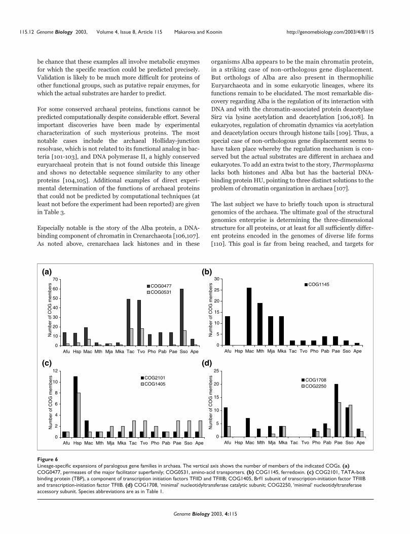
be chance that these examples all involve metabolic enzymes
for which the specific reaction could be predicted precisely.
Validation is likely to be much more difficult for proteins of
other functional groups, such as putative repair enzymes, for
which the actual substrates are harder to predict.
For some conserved archaeal proteins, functions cannot be
predicted computationally despite considerable effort. Several
important discoveries have been made by experimental
characterization of such mysterious proteins. The most
notable cases include the archaeal Holliday-junction
resolvase, which is not related to its functional analog in bac-
teria [101-103], and DNA polymerase II, a highly conserved
euryarchaeal protein that is not found outside this lineage
and shows no detectable sequence similarity to any other
proteins [104,105]. Additional examples of direct experi-
mental determination of the functions of archaeal proteins
that could not be predicted by computational techniques (at
least not before the experiment had been reported) are given
in Table 3.
Especially notable is the story of the Alba protein, a DNA-
binding component of chromatin in Crenarchaeota [106,107].
As noted above, crenarchaea lack histones and in these
organisms Alba appears to be the main chromatin protein,
in a striking case of non-orthologous gene displacement.
But orthologs of Alba are also present in thermophilic
Euryarchaeota and in some eukaryotic lineages, where its
functions remain to be elucidated. The most remarkable dis-
covery regarding Alba is the regulation of its interaction with
DNA and with the chromatin-associated protein deacetylase
Sir2 via lysine acetylation and deacetylation [106,108]. In
eukaryotes, regulation of chromatin dynamics via acetylation
and deacetylation occurs through histone tails [109]. Thus, a
special case of non-orthologous gene displacement seems to
have taken place whereby the regulation mechanism is con-
served but the actual substrates are different in archaea and
eukaryotes. To add an extra twist to the story, Thermoplasma
lacks both histones and Alba but has the bacterial DNA-
binding protein HU, pointing to three distinct solutions to the
problem of chromatin organization in archaea [107].
The last subject we have to briefly touch upon is structural
genomics of the archaea. The ultimate goal of the structural
genomics enterprise is determining the three-dimensional
structure for all proteins, or at least for all sufficiently differ-
ent proteins encoded in the genomes of diverse life forms
[110]. This goal is far from being reached, and targets for
115.12 Genome Biology 2003, Volume 4, Issue 8, Article 115 Makarova and Koonin http://genomebiology.com/2003/4/8/115
Genome Biology 2003, 4:115
Figure 6Lineage-specific expansions of paralogous gene families in archaea. The vertical axis shows the number of members of the indicated COGs. (a)COG0477, permeases of the major facilitator superfamily; COG0531, amino-acid transporters. (b) COG1145, ferredoxin. (c) COG2101, TATA-boxbinding protein (TBP), a component of transcription initiation factors TFIID and TFIIIB; COG1405, Brf1 subunit of transcription-initiation factor TFIIIBand transcription-initiation factor TFIIB. (d) COG1708, ‘minimal’ nucleotidyltransferase catalytic subunit; COG2250, ‘minimal’ nucleotidyltransferaseaccessory subunit. Species abbreviations are as in Table 1.
0
10
20
30
40
50
60
70
Afu Hsp Mac Mth Mja Mka Tac Tvo Pho Pab Pae Sso Ape
COG0477COG0531
0
2
4
6
8
10
12
Afu Hsp Mac Mth Mja Mka Tac Tvo Pho Pab Pae Sso Ape
COG2101COG1405
0
5
10
15
20
25
30
Afu Hsp Mac Mth Mja Mka Tac Tvo Pho Pab Pae Sso Ape
COG1145
0
5
10
15
20
25
Afu Hsp Mac Mth Mja Mka Tac Tvo Pho Pab Pae Sso Ape
COG1708COG2250
Num
ber
of C
OG
mem
bers
Num
ber
of C
OG
mem
bers
Num
ber
of C
OG
mem
bers
Num
ber
of C
OG
mem
bers
(a) (b)
(c) (d)

structural determination have been prioritized by different
researchers on the basis of different principles, from nearly
random choice to relatively elaborate strategies, including
the use of the COG database [111-115]. The development of
structural genomics so far has been a mixture of success,
when informative and interesting structures have been
solved, and mild disappointment in cases when the structure
determination did not seem to shed any light on a protein’s
function. Structural genomics could be particularly impor-
tant in the case of archaea, for which a miniscule number of
structures had been solved prior to the launch of structural
genomic initiatives, and in which proteins often show low
similarity to bacterial or eukaryotic homologs, making
homology modeling difficult.
Notable developments that illustrate both the benefits and
the pitfalls of structural genomics, are the concerted effort
on ‘structural proteomics’ of Methanothermobacter thermo-
autotrophicus [116] and a similar project on M. jannaschii
[117]. The elucidation of the structure of the M. jannaschii
protein MJ0577 [117] is an excellent case for the power of
structural genomics. Analysis of this structure and accompa-
nying biochemical experiments revealed a distinct
nucleotide-binding domain that is distantly related to the
catalytic domains of class I aminoacyl-tRNA synthetases and
belongs to the so-called HUP fold of nucleotide-binding
domains [118]. Together with comprehensive sequence
analysis, the determination of this structure provided the
structural, functional and evolutionary context for the UspA
protein family, which is specifically expanded in archaea
[39]. The exact function(s) of these proteins remains
unknown but, in this case, structural genomics ensured a
substantial functional insight. On several other occasions,
however, determination of the structures of archaeal pro-
teins has failed to provide clear functional clues; these
remain structures in search of a function.
What’s around the corner?The first sequenced archaeal genome was a veritable terra
incognita. Six years after that sequence appeared, the
archaeal genomescape looks quite different. The principal
landmarks have been mapped and now, when a new archaeal
genome is released, we largely know what to expect from it.
Computational approaches to comparative genomics, com-
bining in-depth sequence and structure comparison with
genome context analysis, have led to the reconstruction of the
central functional systems of archaeal cells. But these
approaches have also produced numerous isolated predic-
tions of biochemical activities of archaeal proteins that
remain to be fitted into a general picture, and this can be
done only through ‘wet’ experiments, although new genome
sequences will substantially help by enriching the genomic
context. A shrinking but still notable set of archaeal genes
includes those that encode highly conserved proteins without
any clue to function; solving these mysteries has the potential
to bring out truly new biology. Furthermore, in this article we
have not even touched upon important aspects of archaeal
genomics, such as the in-depth studies of the translation
system, which have revealed several highly unusual, remark-
able mechanisms and enzymatic systems [63,119] or the
identification of regulatory sites in DNA and patterns of
transcription regulation [120,121]. The latter avenue of
research is still in its infancy but will certainly grow in scale
once more archaeal genomes, and in particular closely
related ones, are sequenced.
Because of the lack of established model systems for
archaeal experimental biology and the resulting difficulty
with large-scale experimentation, clues from genome com-
parison are even more crucial for archaeal functional
genomics than they are in the case of bacteria or eukaryotes.
So far, the input of comparative genomics into actual experi-
ments has been less prominent than we would hope. Simply
put, it is not often that experimenters rush to test predic-
tions produced by in silico genome comparison and, further-
more, it is even rarer that targets for functional
characterization are carefully prioritized on the basis of how
unusual and fundamental the predictions are. As discussed
above, however, the few cases when such tests have been
performed are encouraging. It is our hope that the future
belongs to a much tighter integration of comparative, struc-
tural and functional genomics.
Beyond functional studies, archaeal genomics is fundamen-
tal to our understanding of two critical transitions in the
evolution of life. The first is the primary split between the
bacterial and archaeo-eukaryotic lineages, which might have
involved the origin of the DNA-replication machinery and of
the large, double-stranded DNA genomes themselves
[22,23], and the second is the origin of eukaryotes [122].
With regard to the latter problem, archaea are a particularly
valuable source of information because, on many occasions,
they seem to have retained primitive traits while eukaryotes
have undergone major changes. A characteristic example is
the small DNA polymerase subunit, which has all the hall-
marks of an active phosphatase in archaea, but not in
eukaryotes, in which the phosphatase activity is predicted to
be inactivated [123]. Indubitably, archaea resemble the
common ancestor of the archaeo-eukaryotic line of descent
more closely than eukaryotes do, so archaeal genomics is our
best chance to reconstruct this critical intermediate in the
evolution of life. We are confident that comparative
archaeogenomics has a bright future, with major progress in
both the functional and the evolutionary avenues of research
expected within the next few years.
Additional data fileThe list of genes in the reconstructed gene set of the last
common ancestor of archaea is available with the complete
version of this article, online.
com
ment
reviews
reports
deposited research
interactions
inform
ation
refereed research
http://genomebiology.com/2003/4/8/115 Genome Biology 2003, Volume 4, Issue 8, Article 115 Makarova and Koonin 115.13
Genome Biology 2003, 4:115

AcknowledgementsWe thank Boris Mirkin for producing the data used for Figure 3 andStephen Bell, Michael Galperin, Dieter Söll and Yuri Wolf for usefuldiscussions.
References1. Woese CR, Fox GE: Phylogenetic structure of the prokaryotic
domain: the primary kingdoms. Proc Natl Acad Sci USA 1977,74:5088-5090.
2. Fox GE, Stackebrandt E, Hespell RB, Gibson J, Maniloff J, Dyer TA,Wolfe RS, Balch WE, Tanner RS, Magrum LJ, et al.: The phylogenyof prokaryotes. Science 1980, 209:457-463.
3. Woese CR, Kandler O, Wheelis ML: Towards a natural systemof organisms: proposal for the domains Archaea, Bacteria,and Eucarya. Proc Natl Acad Sci USA 1990, 87:4576-4579.
4. Woese CR, Gupta R: Are archaebacteria merely derived‘prokaryotes’? Nature 1981, 289:95-96.
5. Mayr E: Two empires or three? Proc Natl Acad Sci USA 1998,95:9720-9723.
6. Woese CR: Default taxonomy: Ernst Mayr’s view of themicrobial world. Proc Natl Acad Sci USA 1998, 95:11043-11046.
7. Gupta RS: Life’s third domain (Archaea): an established factor an endangered paradigm? Theor Popul Biol 1998, 54:91-104.
8. Kushner DJ: Lysis and dissolution of cells and envelopes of anextremely halophilic bacterium. J Bacteriol 1964, 87:1147-1156.
9. Langworthy TA, Smith PF, Mayberry WR: Lipids of Thermoplasmaacidophilum. J Bacteriol 1972, 112:1193-1200.
10. Brock TD, Brock KM, Belly RT, Weiss RL: Sulfolobus: a newgenus of sulfur-oxidizing bacteria living at low pH and hightemperature. Arch Mikrobiol 1972, 84:54-68.
11. Stetter KO: Extremophiles and their adaptation to hot envi-ronments. FEBS Lett 1999, 452:22-25.
12. Segerer AH, Burggraf S, Fiala G, Huber G, Huber R, Pley U, StetterKO: Life in hot springs and hydrothermal vents. Orig Life EvolBiosph 1993, 23:77-90.
13. DeLong EF: A phylogenetic perspective on hyperthermophilicmicroorganisms. Methods Enzymol 2001, 330:3-11.
14. DeLong EF, Pace NR: Environmental diversity of bacteria andarchaea. Syst Biol 2001, 50:470-478.
15. Hanford MJ, Peeples TL: Archaeal tetraether lipids: unique struc-tures and applications. Appl Biochem Biotechnol 2002, 97:45-62.
16. Engelhardt H, Peters J: Structural research on surface layers: afocus on stability, surface layer homology domains, andsurface layer-cell wall interactions. J Struct Biol 1998, 124:276-302.
17. Edgell DR, Doolittle WF: Archaea and the origin(s) of DNAreplication proteins. Cell 1997, 89:995-998.
18. Sandman K, Pereira SL, Reeve JN: Diversity of prokaryotic chro-mosomal proteins and the origin of the nucleosome. Cell MolLife Sci 1998, 54:1350-1364.
19. Sandman K, Bailey KA, Pereira SL, Soares D, Li WT, Reeve JN:Archaeal histones and nucleosomes. Methods Enzymol 2001,334:116-129.
20. Lecompte O, Ripp R, Thierry JC, Moras D, Poch O: Comparativeanalysis of ribosomal proteins in complete genomes: anexample of reductive evolution at the domain scale. NucleicAcids Res 2002, 30:5382-5390.
21. Forterre P, Brochier C, Philippe H: Evolution of the Archaea.Theor Popul Biol 2002, 61:409-422.
22. Leipe DD, Aravind L, Koonin EV: Did DNA replication evolvetwice independently? Nucleic Acids Res 1999, 27:3389-3401.
23. Forterre P: The origin of DNA genomes and DNA replicationproteins. Curr Opin Microbiol 2002, 5:525-532.
24. Koonin EV, Mushegian AR, Galperin MY, Walker DR: Comparisonof archaeal and bacterial genomes: computer analysis ofprotein sequences predicts novel functions and suggests achimeric origin for the archaea. Mol Microbiol 1997, 25:619-637.
25. Jain R, Rivera MC, Lake JA: Horizontal gene transfer amonggenomes: the complexity hypothesis. Proc Natl Acad Sci USA1999, 96:3801-3806.
26. Koonin EV, Galperin MY: Sequence - Evolution - Function. Computa-tional Approaches in Comparative Genomics. New York: Kluwer Acade-mic; 2002.
27. Bult CJ, White O, Olsen GJ, Zhou L, Fleischmann RD, Sutton GG,Blake JA, FitzGerald LM, Clayton RA, Gocayne JD, et al.: Complete
genome sequence of the methanogenic archaeon,Methanococcus jannaschii. Science 1996, 273:1058-1073.
28. Pace NR: A molecular view of microbial diversity and thebiosphere. Science 1997, 276:734-740.
29. Huber H, Hohn MJ, Rachel R, Fuchs T, Wimmer VC, Stetter KO: Anew phylum of Archaea represented by a nanosized hyper-thermophilic symbiont. Nature 2002, 417:63-67.
30. Huber H, Hohn MJ, Stetter KO, Rachel R: The phylum Nano-archaeota: present knowledge and future perspectives of aunique form of life. Res Microbiol 2003, 154:165-171.
31. Tatusov RL, Koonin EV, Lipman DJ: A genomic perspective onprotein families. Science 1997, 278:631-637.
32. Fitch WM: Distinguishing homologous from analogous pro-teins. Syst Zool 1970, 19:99-113.
33. Fitch WM: Homology: a personal view on some of the prob-lems. Trends Genet 2000, 16:227-231.
34. Sonnhammer EL, Koonin EV: Orthology, paralogy and proposedclassification for paralog subtypes. Trends Genet 2002, 18:619-620.
35. Gaasterland T, Ragan MA: Microbial genescapes: phyletic andfunctional patterns of ORF distribution among prokaryotes.Microb Comp Genomics 1998, 3:199-217.
36. Pellegrini M, Marcotte EM, Thompson MJ, Eisenberg D, Yeates TO:Assigning protein functions by comparative genome analy-sis: protein phylogenetic profiles. Proc Natl Acad Sci USA 1999,96:4285-4288.
37. Tatusov RL, Natale DA, Garkavtsev IV, Tatusova TA, ShankavaramUT, Rao BS, Kiryutin B, Galperin MY, Fedorova ND, Koonin EV:The COG database: new developments in phylogenetic clas-sification of proteins from complete genomes. Nucleic AcidsRes 2001, 29:22-28.
38. Prokaryotic COGs project phyletic pattern search[http://www.ncbi.nlm.nih.gov/COG/new/release/phylox.cgi]
39. Makarova KS, Aravind L, Galperin MY, Grishin NV, Tatusov RL,Wolf YI, Koonin EV: Comparative genomics of the Archaea(Euryarchaeota): evolution of conserved protein families,the stable core, and the variable shell. Genome Res 1999,9:608-628.
40. Koonin EV, Mushegian AR, Bork P: Non-orthologous gene dis-placement. Trends Genet 1996, 12:334-336.
41. Doolittle WF, Logsdon JM, Jr.: Archaeal genomics: do archaeahave a mixed heritage? Curr Biol 1998, 8:R209-R211.
42. Doolittle WF: Phylogenetic classification and the universaltree. Science 1999, 284:2124-2129.
43. Doolittle WF: Lateral genomics. Trends Cell Biol 1999, 9:M5-M8.44. Koonin EV, Makarova KS, Aravind L: Horizontal gene transfer in
prokaryotes - quantification and classification. Annu Rev Micro-biol 2001, 55:709-42.
45. Slesarev AI, Mezhevaya KV, Makarova KS, Polushin NN, ShcherbininaOV, Shakhova VV, Belova GI, Aravind L, Natale DA, Rogozin IB, etal.: The complete genome of hyperthermophile Methano-pyrus kandleri AV19 and monophyly of archaealmethanogens. Proc Natl Acad Sci USA 2002, 99:4644-4649.
46. Aravind L, Tatusov RL, Wolf YI, Walker DR, Koonin EV: Evidencefor massive gene exchange between archaeal and bacterialhyperthermophiles. Trends Genet 1998, 14:442-444.
47. Nelson KE, Clayton RA, Gill SR, Gwinn ML, Dodson RJ, Haft DH,Hickey EK, Peterson JD, Nelson WC, Ketchum KA, et al.: Evidencefor lateral gene transfer between Archaea and bacteriafrom genome sequence of Thermotoga maritima. Nature 1999,399:323-329.
48. Brown JR: Ancient horizontal gene transfer. Nat Rev Genet2003, 4:121-132.
49. Kyrpides NC, Olsen GJ: Archaeal and bacterial hyperther-mophiles: horizontal gene exchange or common ancestry?Trends Genet 1999, 15:298-299.
50. Aravind L, Tatusov RL, Wolf YI, Walker DR, Koonin EV: Reply.Archaeal and bacterial hyperthermophiles: horizontal geneexchange or common ancestry? Trends Genet 1999, 15:299-300.
51. Bao Q, Tian Y, Li W, Xu Z, Xuan Z, Hu S, Dong W, Yang J, Chen Y,Xue Y, et al.: A complete sequence of the T. tengcongensisgenome. Genome Res 2002, 12:689-700.
52. Thermoanaerobacter tengcongensis proteins[http://www.ncbi.nlm.nih.gov/sutils/taxik.cgi?gi=237]
53. Nesbo CL, Boucher Y, Doolittle WF: Defining the core of non-transferable prokaryotic genes: the euryarchaeal core. J MolEvol 2001, 53:340-350.
115.14 Genome Biology 2003, Volume 4, Issue 8, Article 115 Makarova and Koonin http://genomebiology.com/2003/4/8/115
Genome Biology 2003, 4:115

54. Deppenmeier U, Johann A, Hartsch T, Merkl R, Schmitz RA, Mar-tinez-Arias R, Henne A, Wiezer A, Baumer S, Jacobi C, et al.: Thegenome of Methanosarcina mazei: evidence for lateral genetransfer between bacteria and archaea. J Mol Microbiol Biotech-nol 2002, 4:453-461.
55. Galagan JE, Nusbaum C, Roy A, Endrizzi MG, Macdonald P, FitzHughW, Calvo S, Engels R, Smirnov S, Atnoor D, et al.: The genome ofM. acetivorans reveals extensive metabolic and physiologicaldiversity. Genome Res 2002, 12:532-542.
56. Forterre P: A hot story from comparative genomics: reversegyrase is the only hyperthermophile-specific protein. TrendsGenet 2002, 18:236-237.
57. Forterre P, Bergerat A, Lopez-Garcia P: The unique DNA topol-ogy and DNA topoisomerases of hyperthermophilicarchaea. FEMS Microbiol Rev 1996, 18:237-248.
58. Makarova KS, Wolf YI, Koonin EV: Potential genomic determi-nants of hyperthermophily. Trends Genet 2003, 19:172-176.
59. Makarova KS, Aravind L, Grishin NV, Rogozin IB, Koonin EV: ADNA repair system specific for thermophilic archaea andbacteria predicted by genomic context analysis. Nucleic AcidsRes 2002, 30:482-496.
60. White RH: Biosynthesis of the methanogenic cofactors. VitamHorm 2001, 61:299-337.
61. Galperin MY, Koonin EV: Who’s your neighbor? New computa-tional approaches for functional genomics. Nat Biotechnol 2000,18:609-613.
62. Ibba M, Morgan S, Curnow AW, Pridmore DR, Vothknecht UC,Gardner W, Lin W, Woese CR, Soll D: A euryarchaeal lysyl-tRNA synthetase: resemblance to class I synthetases. Science1997, 278:1119-1122.
63. Praetorius-Ibba M, Ibba M: Aminoacyl-tRNA synthesis inarchaea: different but not unique. Mol Microbiol 2003, 48:631-637.
64. Myllykallio H, Lipowski G, Leduc D, Filee J, Forterre P, Liebl U: Analternative flavin-dependent mechanism for thymidylatesynthesis. Science 2002, 297:105-107.
65. Wolf YI, Rogozin IB, Grishin NV, Koonin EV: Genome trees andthe tree of life. Trends Genet 2002, 18:472-479.
66. Wolf YI, Rogozin IB, Grishin NV, Tatusov RL, Koonin EV: Genometrees constructed using five different approaches suggestnew major bacterial clades. BMC Evol Biol 2001, 1:8.
67. Clarke GD, Beiko RG, Ragan MA, Charlebois RL: Inferringgenome trees by using a filter to eliminate phylogeneticallydiscordant sequences and a distance matrix based on meannormalized BLASTP scores. J Bacteriol 2002, 184:2072-2080.
68. Korbel JO, Snel B, Huynen MA, Bork P: SHOT: a web server forthe construction of genome phylogenies. Trends Genet 2002,18:158-162.
69. Matte-Tailliez O, Brochier C, Forterre P, Philippe H: Archaeal phy-logeny based on ribosomal proteins. Mol Biol Evol 2002, 19:631-639.
70. Snel B, Bork P, Huynen MA: Genomes in flux: the evolution ofarchaeal and proteobacterial gene content. Genome Res 2002,12:17-25.
71. Mirkin BG, Fenner TI, Galperin MY, Koonin EV: Algorithms forcomputing parsimonious evolutionary scenarios for genomeevolution, the last universal common ancestor and domi-nance of horizontal gene transfer in the evolution ofprokaryotes. BMC Evol Biol 2003, 3:2.
72. Wilson CA, Kreychman J, Gerstein M: Assessing annotationtransfer for genomics: quantifying the relations betweenprotein sequence, structure and function through tradi-tional and probabilistic scores. J Mol Biol 2000, 297:233-249.
73. Luo Y, Wasserfallen A: Gene transfer systems and their appli-cations in Archaea. Syst Appl Microbiol 2001, 24:15-25.
74. Liu L, Komori K, Ishino S, Bocquier AA, Cann IK, Kohda D, Ishino Y:The archaeal DNA primase: biochemical characterizationof the p41-p46 complex from Pyrococcus furiosus. J Biol Chem2001, 276:45484-45490.
75. Bocquier AA, Liu L, Cann IK, Komori K, Kohda D, Ishino Y:Archaeal primase: bridging the gap between RNA and DNApolymerases. Curr Biol 2001, 11:452-456.
76. Galperin MY, Aravind L, Koonin EV: Aldolases of the DhnAfamily: a possible solution to the problem of pentose andhexose biosynthesis in archaea. FEMS Microbiol Lett 2000,183:259-264.
77. Siebers B, Brinkmann H, Dorr C, Tjaden B, Lilie H, van der Oost J,Verhees CH: Archaeal fructose-1,6-bisphosphate aldolases
constitute a new family of archaeal type class I aldolase. J BiolChem 2001, 276:28710-28718.
78. Fabrega C, Farrow MA, Mukhopadhyay B, de Crecy-Lagard V, OrtizAR, Schimmel P: An aminoacyl tRNA synthetase whosesequence fits into neither of the two known classes. Nature2001, 411:110-114.
79. Stathopoulos C, Li T, Longman R, Vothknecht UC, Becker HD, IbbaM, Soll D: One polypeptide with two aminoacyl-tRNA syn-thetase activities. Science 2000, 287:479-482.
80. Iyer LM, Aravind L, Bork P, Hofmann K, Mushegian AR, Zhulin IB,Koonin EV: Quod erat demonstrandum? The mystery of experi-mental validation of apparently erroneous computationalanalyses of protein sequences. Genome Biol 2001,2:research0051.1-0051.11
81. Kamtekar S, Kennedy WD, Wang J, Stathopoulos C, Soll D, SteitzTA: The structural basis of cysteine aminoacylation oftRNAPro by prolyl-tRNA synthetases. Proc Natl Acad Sci USA2003, 100:1673-1678.
82. Xie G, Forst C, Bonner C, Jensen RA: Significance of two distincttypes of tryptophan synthase beta chain in Bacteria, Archaeaand higher plants. Genome Biol 2002, 3:research0004.1-0004.13
83. Panek H, O’Brian MR: A whole genome view of prokaryotichaem biosynthesis. Microbiology 2002, 148:2273-2282.
84. Huynen M, Snel B, Lathe W, Bork P: Exploitation of genecontext. Curr Opin Struct Biol 2000, 10:366-370.
85. Huynen M, Snel B, Lathe W 3rd, Bork P: Predicting protein func-tion by genomic context: quantitative evaluation and quali-tative inferences. Genome Res 2000, 10:1204-1210.
86. Aravind L: Guilt by association: contextual information ingenome analysis. Genome Res 2000, 10:1074-1077.
87. Wolf YI, Rogozin IB, Kondrashov AS, Koonin EV: Genome align-ment, evolution of prokaryotic genome organization, andprediction of gene function using genomic context. GenomeRes 2001, 11:356-372.
88. Koonin EV, Wolf YI, Aravind L: Prediction of the archaealexosome and its connections with the proteasome and thetranslation and transcription machineries by a comparative-genomic approach. Genome Res 2001, 11:240-252.
89. Decker CJ: The exosome: a versatile RNA processingmachine. Curr Biol 1998, 8:R238-R240.
90. van Hoof A, Parker R: The exosome: a proteasome for RNA?Cell 1999, 99:347-350.
91. Mitchell P, Petfalski E, Shevchenko A, Mann M, Tollervey D: Theexosome: a conserved eukaryotic RNA processing complexcontaining multiple 3��—>5�� exoribonucleases. Cell 1997,91:457-466.
92. Rogozin IB, Makarova KS, Murvai J, Czabarka E, Wolf YI, Tatusov RL,Szekely LA, Koonin EV: Connected gene neighborhoods inprokaryotic genomes. Nucleic Acids Res 2002, 30:2212-2223.
93. Lespinet O, Wolf YI, Koonin EV, Aravind L: The role of lineage-specific gene family expansion in the evolution of eukary-otes. Genome Res 2002, 12:1048-1059.
94. Jordan IK, Makarova KS, Spouge JL, Wolf YI, Koonin EV: Lineage-specific gene expansions in bacterial and archaeal genomes.Genome Res 2001, 11:555-565.
95. Remm M, Storm CE, Sonnhammer EL: Automatic clustering oforthologs and in-paralogs from pairwise species compar-isons. J Mol Biol 2001, 314:1041-1052.
96. Ruepp A, Graml W, Santos-Martinez ML, Koretke KK, Volker C,Mewes HW, Frishman D, Stocker S, Lupas AN, Baumeister W: Thegenome sequence of the thermoacidophilic scavenger Ther-moplasma acidophilum. Nature 2000, 407:508-513.
97. She Q, Singh RK, Confalonieri F, Zivanovic Y, Allard G, Awayez MJ,Chan-Weiher CC, Clausen IG, Curtis BA, De Moors A, et al.: Thecomplete genome of the crenarchaeon Sulfolobus solfatari-cus P2. Proc Natl Acad Sci USA 2001, 98:7835-7840.
98. Deppenmeier U: The unique biochemistry of methanogenesis.Prog Nucleic Acid Res Mol Biol 2002, 71:223-283.
99. Aravind L, Koonin EV: DNA polymerase beta-like nucleotidyl-transferase superfamily: identification of three new families,classification and evolutionary history. Nucleic Acids Res 1999,27:1609-1618.
100. Lehmann C, Lim K, Chalamasetty VR, Krajewski W, Melamud E,Galkin A, Howard A, Kelman Z, Reddy PT, Murzin AG, Herzberg O:The HI0073/HI0074 protein pair from Haemophilus influen-zae is a member of a new nucleotidyltransferase family:structure, sequence analyses, and solution studies. Proteins2003, 50:249-260.
com
ment
reviews
reports
deposited research
interactions
inform
ation
refereed research
http://genomebiology.com/2003/4/8/115 Genome Biology 2003, Volume 4, Issue 8, Article 115 Makarova and Koonin 115.15
Genome Biology 2003, 4:115

101. Komori K, Sakae S, Shinagawa H, Morikawa K, Ishino Y: A Hollidayjunction resolvase from Pyrococcus furiosus: functional simi-larity to Escherichia coli RuvC provides evidence for con-served mechanism of homologous recombination inBacteria, Eukarya, and Archaea. Proc Natl Acad Sci USA 1999,96:8873-8878.
102. Aravind L, Makarova KS, Koonin EV: Survey and summary: Holli-day junction resolvases and related nucleases: identificationof new families, phyletic distribution and evolutionary tra-jectories. Nucleic Acids Res 2000, 28:3417-3432.
103. Daiyasu H, Komori K, Sakae S, Ishino Y, Toh H: Hjc resolvase is adistantly related member of the type II restriction endonu-clease family. Nucleic Acids Res 2000, 28:4540-4543.
104. Ishino Y, Komori K, Cann IK, Koga Y: A novel DNA polymerasefamily found in Archaea. J Bacteriol 1998, 180:2232-2236.
105. Ishino Y, Ishino S: DNA polymerases from euryarchaeota.Methods Enzymol 2001, 334:249-260.
106. Bell SD, Botting CH, Wardleworth BN, Jackson SP, White MF: Theinteraction of Alba, a conserved archaeal chromatinprotein, with Sir2 and its regulation by acetylation. Science2002, 296:148-151.
107. White MF, Bell SD: Holding it together: chromatin in theArchaea. Trends Genet 2002, 18:621-626.
108. Wardleworth BN, Russell RJ, Bell SD, Taylor GL, White MF: Struc-ture of Alba: an archaeal chromatin protein modulated byacetylation. EMBO J 2002, 21:4654-4662.
109. Kurdistani SK, Grunstein M: Histone acetylation and deacetyla-tion in yeast. Nat Rev Mol Cell Biol 2003, 4:276-284.
110. Vitkup D, Melamud E, Moult J, Sander C: Completeness in struc-tural genomics. Nat Struct Biol 2001, 8:559-566.
111. Elofsson A, Sonnhammer EL: A comparison of sequence andstructure protein domain families as a basis for structuralgenomics. Bioinformatics 1999, 15:480-500.
112. Gaasterland T: Structural genomics: bioinformatics in the dri-ver’s seat. Nat Biotechnol 1998, 16:625-627.
113. Brenner SE: Target selection for structural genomics. NatStruct Biol 2000, 7 Suppl:967-969.
114. Gerstein M: Integrative database analysis in structuralgenomics. Nat Struct Biol 2000, 7 Suppl:960-963.
115. Koonin EV, Wolf YI, Aravind L: Protein fold recognition usingsequence profiles and its application in structural genomics.Adv Protein Chem 2000, 54:245-275.
116. Christendat D, Yee A, Dharamsi A, Kluger Y, Savchenko A, Cort JR,Booth V, Mackereth CD, Saridakis V, Ekiel I, et al.: Structural pro-teomics of an archaeon. Nat Struct Biol 2000, 7:903-909.
117. Zarembinski TI, Hung LW, Mueller-Dieckmann HJ, Kim KK, YokotaH, Kim R, Kim SH: Structure-based assignment of the bio-chemical function of a hypothetical protein: a test case ofstructural genomics. Proc Natl Acad Sci USA 1998, 95:15189-15193.
118. Aravind L, Anantharaman V, Koonin EV: Monophyly of class Iaminoacyl tRNA synthetase, USPA, ETFP, photolyase, andPP-ATPase nucleotide-binding domains: implications forprotein evolution in the RNA. Proteins 2002, 48:1-14.
119. Woese CR: Translation: in retrospect and prospect. RNA2001, 7:1055-1067.
120. Gelfand MS, Koonin EV, Mironov AA: Prediction of transcriptionregulatory sites in Archaea by a comparative genomicapproach. Nucleic Acids Res 2000, 28:695-705.
121. Rodionov DA, Mironov AA, Gelfand MS: Conservation of thebiotin regulon and the BirA regulatory signal in eubacteriaand archaea. Genome Res 2002, 12:1507-1516.
122. Dacks JB, Doolittle WF: Reconstructing/deconstructing theearliest eukaryotes: how comparative genomics can help.Cell 2001, 107:419-425.
123. Aravind L, Koonin EV: Phosphoesterase domains associatedwith DNA polymerases of diverse origins. Nucleic Acids Res1998, 26:3746-3752.
124. Klenk HP, Clayton RA, Tomb JF, White O, Nelson KE, Ketchum KA,Dodson RJ, Gwinn M, Hickey EK, Peterson JD, et al.: The completegenome sequence of the hyperthermophilic, sulphate- reduc-ing archaeon Archaeoglobus fulgidus. Nature 1997, 390:364-370.
125. Ng WV, Kennedy SP, Mahairas GG, Berquist B, Pan M, Shukla HD,Lasky SR, Baliga NS, Thorsson V, Sbrogna J, et al.: Genomesequence of Halobacterium species NRC-1. Proc Natl Acad SciUSA 2000, 97:12176-12181.
126. Smith DR, Doucette-Stamm LA, Deloughery C, Lee H, Dubois J,Aldredge T, Bashirzadeh R, Blakely D, Cook R, Gilbert K, et al.:
Complete genome sequence of Methanobacterium ther-moautotrophicum deltaH: functional analysis and compara-tive genomics. J Bacteriol 1997, 179:7135-7155.
127. Kawarabayasi Y, Sawada M, Horikawa H, Haikawa Y, Hino Y,Yamamoto S, Sekine M, Baba S, Kosugi H, Hosoyama A, et al.: Com-plete sequence and gene organization of the genome of ahyper-thermophilic archaebacterium, Pyrococcus horikoshiiOT3. DNA Res 1998, 5:55-76.
128. Cohen GN, Barbe V, Flament D, Galperin M, Heilig R, Lecompte O,Poch O, Prieur D, Querellou J, Ripp R, et al.: An integrated analy-sis of the genome of the hyperthermophilic archaeon Pyro-coccus abyssi. Mol Microbiol 2003, 47:1495-1512.
129. Robb FT, Maeder DL, Brown JR, DiRuggiero J, Stump MD, Yeh RK,Weiss RB, Dunn DM: Genomic sequence of hyperthermophile,Pyrococcus furiosus: implications for physiology and enzy-mology. Methods Enzymol 2001, 330:134-157.
130. Kawashima T, Amano N, Koike H, Makino S, Higuchi S, Kawashima-Ohya Y, Watanabe K, Yamazaki M, Kanehori K, Kawamoto T, et al.:Archaeal adaptation to higher temperatures revealed bygenomic sequence of Thermoplasma volcanium. Proc Natl AcadSci USA 2000, 97:14257-14262.
131. Fitz-Gibbon ST, Ladner H, Kim UJ, Stetter KO, Simon MI, Miller JH:Genome sequence of the hyperthermophilic crenarchaeonPyrobaculum aerophilum. Proc Natl Acad Sci USA 2002, 99:984-989.
132. Kawarabayasi Y, Hino Y, Horikawa H, Yamazaki S, Haikawa Y, Jin-noK, Takahashi M, Sekine M, Baba S, Ankai A, et al.: Completegenome sequence of an aerobic hyper-thermophilic crenar-chaeon, Aeropyrum pernix K1. DNA Res 1999, 6:83-101.
133. Kawarabayasi Y, Hino Y, Horikawa H, Jin-no K, Takahashi M, SekineM, Baba S, Ankai A, Kosugi H, Hosoyama A, et al.: Completegenome sequence of an aerobic thermoacidophilic crenar-chaeon, Sulfolobus tokodaii strain7. DNA Res 2001, 8:123-140.
134. Tersteegen A, Hedderich R: Methanobacterium thermo-autotrophicum encodes two multisubunit membrane-bound[NiFe] hydrogenases. Transcription of the operons andsequence analysis of the deduced proteins. Eur J Biochem 1999,264:930-943.
135. Natale DA, Shankavaram UT, Galperin MY, Wolf YI, Aravind L,Koonin EV: Towards understanding the first genomesequence of a crenarchaeon by genome annotation usingclusters of orthologous groups of proteins (COGs). GenomeBiol 2000, 1:research0009.1-0009.19.
136. Aravind L, Walker DR, Koonin EV: Conserved domains in DNArepair proteins and evolution of repair systems. Nucleic AcidsRes 1999, 27:1223-1242.
137. Cope GA, Suh GS, Aravind L, Schwarz SE, Zipursky SL, Koonin EV,Deshaies RJ: Role of predicted metalloprotease motif ofJab1/Csn5 in cleavage of Nedd8 from Cul1. Science 2002,298:608-611.
138. Verma R, Aravind L, Oania R, McDonald WH, Yates JR, 3rd, KooninEV, Deshaies RJ: Role of Rpn11 metalloprotease in deubiquiti-nation and degradation by the 26S proteasome. Science 2002,298:611-615.
139. Aravind L, Koonin EV: DNA polymerase beta-like nucleotidyl-transferase superfamily: identification of three new families,classification and evolutionary history. Nucleic Acids Res 1999,27:1609-1618.
140. Daugherty M, Vonstein V, Overbeek R, Osterman A: Archaealshikimate kinase, a new member of the GHMP-kinasefamily. J Bacteriol 2001, 183:292-300.
141. van der Oost J, Huynen MA, Verhees CH: Molecular characteri-zation of phosphoglycerate mutase in archaea. FEMS MicrobiolLett 2002, 212:111-120.
142. Rashid N, Imanaka H, Kanai T, Fukui T, Atomi H, Imanaka T: Anovel candidate for the true fructose-1,6-bisphosphatase inarchaea. J Biol Chem 2002, 277:30649-30655.
143. Constantinesco F, Forterre P, Elie C: NurA, a novel 5��-3�� nucle-ase gene linked to rad50 and mre11 homologs of ther-mophilic Archaea. EMBO Rep 2002, 3:537-542.
144. Graham DE, Bock CL, Schalk-Hihi C, Lu ZJ, Markham GD: Identifi-cation of a highly diverged class of S-adenosylmethioninesynthetases in the archaea. J Biol Chem 2000, 275:4055-4059.
145. Graham DE, Xu H, White RH: Methanococcus jannaschii uses apyruvoyl-dependent arginine decarboxylase in polyaminebiosynthesis. J Biol Chem 2002, 277:23500-23507.
115.16 Genome Biology 2003, Volume 4, Issue 8, Article 115 Makarova and Koonin http://genomebiology.com/2003/4/8/115
Genome Biology 2003, 4:115

com
ment
reviews
reports
deposited research
interactions
inform
ation
refereed research
http://genomebiology.com/2003/4/8/115 Genome Biology 2003, Volume 4, Issue 8, Article 115 Makarova and Koonin 115.17
Genome Biology 2003, 4:115
Recommended