REVIEW
APP transgenic modeling of Alzheimer’s disease: mechanismsof neurodegeneration and aberrant neurogenesis
Leslie Crews • Edward Rockenstein •
Eliezer Masliah
Received: 22 September 2009 / Accepted: 11 November 2009 / Published online: 29 November 2009
� The Author(s) 2009. This article is published with open access at Springerlink.com
Abstract Neurodegenerative disorders of the aging
population affect over 5 million people in the US and
Europe alone. The common feature is the progressive
accumulation of misfolded proteins with the formation of
toxic oligomers. Alzheimer’s disease (AD) is characterized
by cognitive impairment, progressive degeneration of
neuronal populations in the neocortex and limbic system,
and formation of amyloid plaques and neurofibrillary tan-
gles. Amyloid-b (Ab) is the product of proteolysis of
amyloid precursor protein (APP) by b and c-secretase
enzymes. The neurodegenerative process in AD initiates
with axonal and synaptic damage and is associated with
progressive accumulation of toxic Ab oligomers in the
intracellular and extracellular space. In addition, neurode-
generation in AD is associated with alterations in neuro-
genesis. Ab accumulation is the consequence of an altered
balance between protein synthesis, aggregation rate, and
clearance. Identification of genetic mutations in APP
associated with familial forms of AD and gene polymor-
phisms associated with the more common sporadic variants
of AD has led to the development of transgenic (tg) and
knock out rodents as well as viral vector driven models of
AD. While APP tg murine models with mutations in the N-
and C-terminal flanking regions of Ab are characterized by
increased Ab production with plaque formation, mutations
in the mid-segment of Ab result in increased formation of
oligomers, and mutations toward the C-terminus (E22Q)
segment results in amyloid angiopathy. Similar to AD, in
APP tg models bearing familial mutations, formation of Aboligomers results in defective plasticity in the perforant
pathway, selective neuronal degeneration, and alterations
in neurogenesis. Promising results have been obtained
utilizing APP tg models of AD to develop therapies
including the use of b- and c-secretase inhibitors, immu-
nization, and stimulating neurogenesis.
Keywords Transgenic � Neurodegenerative disease �Aging � Alzheimer � APP � Synapse loss � Neurogenesis
Introduction
Alzheimer’s disease (AD) is the most common neurode-
generative disorder in the aging population. It is charac-
terized by the progressive and irreversible deafferentation
of the limbic system, association neocortex, and basal
forebrain (Perry et al. 1977; Hyman et al. 1984; Wilcock
et al. 1988; Hof et al. 1990; Palmer and Gershon 1990;
Masliah et al. 1993), accompanied by the formation of
neuritic amyloid plaques, amyloid angiopathy, neurofi-
brillary tangles (NFTs), and neuropil threads (Terry et al.
1994). This neurodegenerative process is followed by
reactive astrogliosis (Dickson et al. 1988) and microglial
cell proliferation (Rogers et al. 1988; Masliah et al. 1991).
Loss of synapses (DeKosky and Scheff 1990; Masliah
2001; Scheff and Price 2001) and axonal pathology (Raff
et al. 2002) are probably key neuropathological features
leading to dementia in these neurodegenerative disorders.
In addition, recent evidence suggests that alterations in the
niches for neurogenesis in the adult brain might also
contribute to the neurodegenerative process (Haughey
et al. 2002; Tatebayashi et al. 2003; Dong et al. 2004;
L. Crews � E. Masliah
Department of Pathology, University of California,
San Diego, La Jolla, CA, USA
E. Rockenstein � E. Masliah (&)
Department of Neurosciences, University of California,
San Diego, 9500 Gilman Drive, La Jolla, CA 92003-0624, USA
e-mail: [email protected]
123
Brain Struct Funct (2010) 214:111–126
DOI 10.1007/s00429-009-0232-6
Jin et al. 2004; Wen et al. 2004; Chevallier et al. 2005;
Donovan et al. 2006). The unique patterns of cognitive
impairment that characterize AD, in turn, depend on the
neural circuitry specifically affected (Hof and Morrison
1994), the extent of the synapto-dendritic damage, and the
speed with which the injury propagates (Terry et al. 1991;
DeKosky et al. 1996). Recent evidence supports the con-
tention that neuronal cell death might occur later in the
progression of neurodegeneration and that damage to the
synapto-dendritic apparatus might be one of the earliest
pathological alterations (Masliah and Terry 1993, 1994;
Masliah 1998, 2001; Honer 2003; Scheff and Price 2003).
This is accompanied by the abnormal accumulation of
neuronal proteins in the extracellular space (e.g., plaques,
cerebral amyloid angiopathy [CAA]) or in intracellular
compartments (e.g., tangles and Lewy bodies [LBs]).
Abnormal accumulation and misfolding (toxic conversion)
of these synaptic and cytoskeletal proteins are being
explored as key pathogenic events leading to neurodegen-
eration (Koo et al. 1999; Ramassamy et al. 1999; Ferrigno
and Silver 2000).
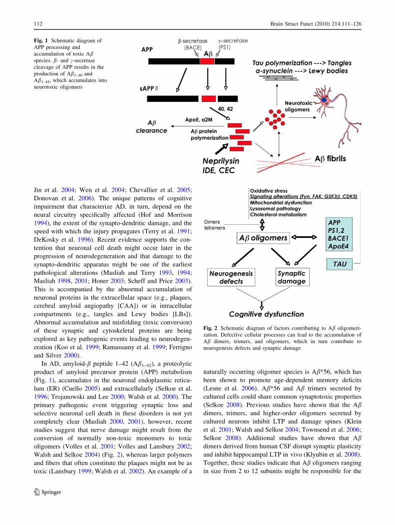
In AD, amyloid-b peptide 1–42 (Ab1–42), a proteolytic
product of amyloid precursor protein (APP) metabolism
(Fig. 1), accumulates in the neuronal endoplasmic reticu-
lum (ER) (Cuello 2005) and extracellularly (Selkoe et al.
1996; Trojanowski and Lee 2000; Walsh et al. 2000). The
primary pathogenic event triggering synaptic loss and
selective neuronal cell death in these disorders is not yet
completely clear (Masliah 2000, 2001), however, recent
studies suggest that nerve damage might result from the
conversion of normally non-toxic monomers to toxic
oligomers (Volles et al. 2001; Volles and Lansbury 2002;
Walsh and Selkoe 2004) (Fig. 2), whereas larger polymers
and fibers that often constitute the plaques might not be as
toxic (Lansbury 1999; Walsh et al. 2002). An example of a
naturally occurring oligomer species is Ab*56, which has
been shown to promote age-dependent memory deficits
(Lesne et al. 2006). Ab*56 and Ab trimers secreted by
cultured cells could share common synaptotoxic properties
(Selkoe 2008). Previous studies have shown that the Abdimers, trimers, and higher-order oligomers secreted by
cultured neurons inhibit LTP and damage spines (Klein
et al. 2001; Walsh and Selkoe 2004; Townsend et al. 2006;
Selkoe 2008). Additional studies have shown that Abdimers derived from human CSF disrupt synaptic plasticity
and inhibit hippocampal LTP in vivo (Klyubin et al. 2008).
Together, these studies indicate that Ab oligomers ranging
in size from 2 to 12 subunits might be responsible for the
Fig. 1 Schematic diagram of
APP processing and
accumulation of toxic Abspecies. b- and c-secretase
cleavage of APP results in the
production of Ab1–40 and
Ab1–42, which accumulates into
neurotoxic oligomers
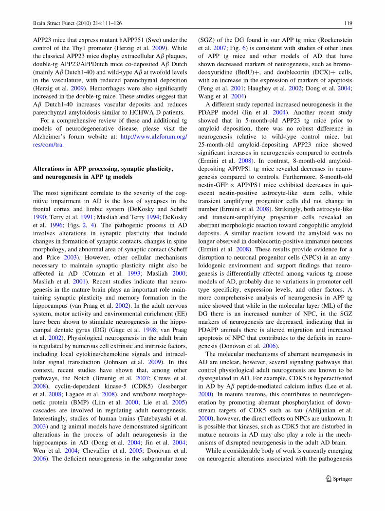
Fig. 2 Schematic diagram of factors contributing to Ab oligomeri-
zation. Defective cellular processes can lead to the accumulation of
Ab dimers, trimers, and oligomers, which in turn contribute to
neurogenesis defects and synaptic damage
112 Brain Struct Funct (2010) 214:111–126
123

synaptic damage and memory deficits associated with AD
(Lacor et al. 2007).
Thus, when developing transgenic (tg) models of AD it
is important to consider the following outcome features:
(1) targeting the selective neuronal populations in the
neocortex and limbic system involved in learning and
memory; (2) favoring production of high levels of Ab1–42
over Ab1–40; (3) promoting accumulation of Ab oligomers;
(4) inducing post-transcriptional modifications (e.g., pyro-
glutamate Ab); (5) evaluating abnormal accumulation of
intracellular Ab; (6) evaluating development of amyloid
plaques and CAA; (7) evaluating neurodegeneration with
synaptic loss, axonal damage, and defects in neurogenesis;
and (8) behavioral and electrophysiological impairments
that correspond to the pattern of neurodegeneration.
Experimental modeling of Alzheimer’s disease
Although dramatic progress has been made in under-
standing the pathogenesis of neurodegenerative condi-
tions of the aged population, such as AD, Parkinson’s
disease (PD), and Lewy body disease (LBD), most of
these disorders remain incurable. Because of the near
epidemic proportion in the aging population, these dis-
orders pose a serious challenge to the health care system.
Identification of new targets and development of bio-
logical mouse models holds the promise of better
understanding their pathogenesis and discovering and
testing new treatments.
Experimental models of AD could mimic individual or
multiple alterations found in AD; however, to date not a
single model mimics all the alterations observed in AD.
The best model is probably the aged monkey (Price et al.
1994), however, because of the time and cost involved in
utilizing this model, most studies have been focused on
developing murine models. Most of the tg animal models
of AD are based on the targeted overexpression of single
or multiple mutant molecules associated with familial AD
(FAD) (Table 1, Fig. 3). Currently, mutations in three
genes have been described, namely APP, presenilin (PS)1,
and PS2 (Hutton and Hardy 1997; Cruts and Van
Broeckhoven 1998; Rocchi et al. 2003; Bertoli-Avella
et al. 2004; Pastor and Goate 2004). Other APP and PS tg
models have been developed in rats, drosophila, and
C. elegans, either using constitutively active or regulat-
able promoters, or viral vectors. The main focus of this
review will be dedicated to murine APP tg models and the
corresponding neurodegenerative pathology and altera-
tions in neurogenesis, however, it is important to
emphasize that this represents one aspect of the disease;
other components, such as NFT pathology will be
addressed by others.
Experimental APP transgenic murine models
of Alzheimer’s disease
In AD, APP mutations as well as mutations in PS1 and 2
and polymorphisms in apolipoprotein E (ApoE) have also
been linked with AD (Fig. 2) and as such are important
targets. Most efforts toward developing tg models have
been focused on overexpression of mutant APP (Table 1)
in combination with mutant PS1. A summary of the FAD
mutations reproduced in tg mouse models is presented in
Fig. 3. Previously developed tg animal models have shown
that it is possible to reproduce certain aspects of AD
pathology over a shorter period of time (Masliah et al.
1996; Games et al. 1997; Price et al. 2000). In this model,
the platelet-derived growth factor (b chain) (PDGF-b)
promoter drives an alternatively spliced human APP
(hAPP) minigene (PDAPP) encoding mutated (Indiana,
V717F) hAPP695, 751, and 770 (Games et al. 1995;
Rockenstein et al. 1995). This confers a high ratio of
mRNA encoding mutated hAPP versus wild-type mouse
APP (Rockenstein et al. 1995), which promotes develop-
ment of typical amyloid plaques, dystrophic neurites, loss
of presynaptic terminals, astrocytosis and microgliosis
(Games et al. 1995, 1997; Masliah et al. 1996).
Other models have expressed mutant hAPP under the
regulatory control of either the human or murine (m)Thy1
promoter (Andra et al. 1996; Sturchler-Pierrat et al. 1997;
Moechars et al. 1999; Bornemann and Staufenbiel 2000) or
the prion protein (PrP) promoter (Hsiao et al. 1996;
Borchelt et al. 1997). Amyloid deposition begins at
12 months of age; however, co-expression of mutant PS1
accelerates amyloid deposition, beginning at 4 months of
age (Borchelt et al. 1996, 1997; Holcomb et al. 1998).
Another previously developed model, where hAPP is also
expressed under the control of the PrP promoter, displays
even earlier onset of amyloid deposition, starting at
3 months and progressing to mature plaques and neuritic
pathology from 5 months of age, accompanied by high
levels of Ab1–42 (Chishti et al. 2001). While the PrP pro-
moter has provided several models that mimic aspects of
FAD, other promoters targeting expression of APP to
neurons provide alternative models demonstrating pathol-
ogy that recapitulate similar and additional aspects of FAD.
In this regard, we have generated lines of tg mice
expressing hAPP751 cDNA containing the London (Lon,
V717I) and Swedish (Swe, K670N/M671L) mutations
under the regulatory control of the mThy1 gene (mThy1-
hAPP751) (Rockenstein et al. 2001; Fig. 4). While
expression of mutant hAPP under the PDGF-b promoter
results in the production of diffuse (and some mature)
plaques (Games et al. 1995; Mucke et al. 2000), tg
expression of mutant hAPP under the mThy1 (Andra et al.
1996) and PrP (Hsiao et al. 1996; Borchelt et al. 1997)
Brain Struct Funct (2010) 214:111–126 113
123

Ta
ble
1R
epre
sen
tati
ve
anim
alm
od
els
of
AD
pat
ho
log
y
Mo
del
Gen
e/M
uta
tio
nS
pec
ies
Str
ateg
yP
rom
ote
rP
hen
oty
pe
Ref
eren
ces
PD
AP
Ptg
hA
PP
69
5,
75
1,
77
0M
ou
seO
ver
exp
ress
ion
PD
GF
-bIn
crea
sed
exp
ress
ion
of
AP
P,
accu
mu
lati
on
of
dif
fuse
amy
loid
pla
qu
es,
syn
apti
c
dam
age,
and
astr
o/
mic
rog
lio
sis
Gam
eset
al.
(19
95
),
Ro
cken
stei
net
al.
(19
95
),
Mu
cke
etal
.(2
00
0)
V7
17
F(I
nd
)
Th
y1
-AP
P7
51
Sw
e/In
dtg
hA
PP
75
1M
ou
seO
ver
exp
ress
ion
Th
y1
Am
ylo
idd
epo
siti
on
(6m
os)
,
gli
osi
san
dta
u
hy
per
ph
osp
ho
ryla
tio
n
An
dra
etal
.(1
99
6),
Stu
rch
ler-
Pie
rrat
etal
.(1
99
7)
K6
70
N/M
67
1(S
we)
?V
71
7F
(In
d)
Th
y1
-AP
P6
95
Sw
e/L
on
tgh
AP
P6
95
Mo
use
Ov
erex
pre
ssio
nT
hy
1A
my
loid
dep
osi
tio
n(1
2m
os)
,
hig
hle
vel
so
fA
b 1–42
accu
mu
lati
on
Mo
ech
ars
etal
.(1
99
9)
K6
70
N/M
67
1(S
we)
?V
71
7I
(Lo
n)
Th
y1
-AP
P7
51
Sw
e/L
on
tgh
AP
P7
51
Mo
use
Ov
erex
pre
ssio
nT
hy
1A
my
loid
dep
osi
tio
n(3
mo
s)
and
mat
ure
pla
qu
efo
rmat
ion
inh
ipp
oca
mp
us
and
neo
cort
ex
Ro
cken
stei
net
al.
(20
01
)
K6
70
N/M
67
1(S
we)
?V
71
7I
(Lo
n)
Tg
25
76
hA
PP
69
5M
ou
seO
ver
exp
ress
ion
PrP
Am
ylo
idd
epo
siti
on
(9–
12
mo
s),
som
ev
ascu
lar
Ab
dep
osi
tio
n
Hsi
aoet
al.
(19
96
),B
orc
hel
t
etal
.(1
99
7)
K6
70
N/M
67
1(S
we)
Tg
25
76
/PS
1m
ut
hA
PP
69
5M
ou
seO
ver
exp
ress
ion
PrP
-hA
PP
Ear
ly(4
mo
s)am
ylo
id
dep
osi
tio
n,
elev
atio
no
f
Ab4
2(4
3)
lev
els
Bo
rch
elt
etal
.(1
99
7),
Ho
lco
mb
etal
.(1
99
8)
K6
70
N/M
67
1(S
we)
PD
GFb
-PS
1
PS
1M
14
6L
Tg
CR
ND
8h
AP
P6
95
Mo
use
Ov
erex
pre
ssio
nP
rPE
arly
(3–
5m
os)
amy
loid
dep
osi
tio
n,
hig
hle
vel
so
f
Ab 1
–42
accu
mu
lati
on
Ch
ish
tiet
al.
(20
01
)
K6
70
N/M
67
1(S
we)
?V
71
7F
(In
d)
3x
Tg
-AD
hA
PP
69
5M
ou
seO
ver
exp
ress
ion
of
hA
PP
,h
Tau
Th
y1
Am
ylo
idd
epo
siti
on
(9m
os)
,
som
ein
tran
euro
nal
Ab
accu
mu
lati
on
,ta
ng
le-l
ike
pat
ho
log
yin
the
hip
po
cam
pu
s,b
ehav
iora
l
defi
cits
Od
do
etal
.(2
00
3a,
b)
K6
70
N/M
67
1K
no
ckin
PS
1
(Sw
e)
hT
au4
R0
NP
30
1L
PS
1M
14
6V
Rat
PD
GFb-
AP
P7
51
tgh
AP
P7
51
Rat
Ov
erex
pre
ssio
nP
DG
F-b
Intr
acel
lula
ram
ylo
id
accu
mu
lati
on
,C
RE
B
acti
vat
ion
and
tau
pat
ho
log
y
Ech
ever
ria
etal
.(2
00
4a,
b)
K6
70
N/M
67
1(S
we)
?V
71
7F
(In
d)
PS
1M
14
6L
Rat
Ub
C-A
PP
swe
tgh
AP
P6
95
Rat
Ov
erex
pre
ssio
nU
bC
Ex
trac
ellu
lar
amy
loid
dep
osi
tio
nat
15
–1
8m
os,
som
ece
reb
rov
ascu
lar
Ab
accu
mu
lati
on
Fo
lkes
son
etal
.(2
00
7)
K6
70
N/M
67
1(S
we)
114 Brain Struct Funct (2010) 214:111–126
123

Ta
ble
1co
nti
nu
ed
Mo
del
Gen
e/M
uta
tio
nS
pec
ies
Str
ateg
yP
rom
ote
rP
hen
oty
pe
Ref
eren
ces
Rat
Ub
C-A
PP
Sw
e/In
dtg
hA
PP
69
5R
atO
ver
exp
ress
ion
Ub
CH
igh
lev
els
of
AP
Pex
pre
ssio
n,
Ab
det
ecte
din
seru
m
Ag
caet
al.
(20
08
)
K6
70
N/M
67
1(S
we)
?V
71
7F
(In
d)
Rat
Sy
nap
sin
-AP
P/P
S1
tgh
AP
P6
95
Rat
Ov
erex
pre
ssio
nS
yn
apsi
n1
Am
ylo
idd
epo
siti
on
at7
mo
sin
do
ub
letg
Flo
od
etal
.(2
00
9)
K6
70
N/M
67
1(S
we)
PS
1M
14
6V
TB
A2
tgA
b3Q
-42
Mo
use
Ov
erex
pre
ssio
no
f
py
rog
luta
mat
eA
bT
hy
1A
ccu
mu
lati
on
of
intr
aneu
ron
al
py
rog
luta
mat
eA
b,
exte
nsi
ve
neu
ron
allo
ss,
no
tan
gle
so
r
hip
po
cam
pal
deg
ener
atio
n
Wir
ths
etal
.(2
00
9)
Tet
-AP
Ptg
Ch
imer
icm
o/h
AP
P6
95
Mo
use
Reg
ula
tab
le
ov
erex
pre
ssio
n
tetO
Ab
un
dan
tp
laq
ues
rem
ain
afte
r
abo
liti
on
of
AP
Pex
pre
ssio
n
Jan
ko
wsk
yet
al.
(20
05
)
K6
70
N/M
67
1(S
we)
?V
71
7F
(In
d)
AP
PS
we
KI
Hu
man
ized
Ab
inm
oA
PP
Mo
use
Kn
ock
inE
nd
og
eno
us
Nin
efo
ldg
reat
erA
ble
vel
sth
an
inn
orm
alh
um
anb
rain
,
spat
ial
and
tem
po
ral
exp
ress
ion
pat
tern
so
fh
um
an
Ab
are
rep
rod
uce
d
Rea
um
eet
al.
(19
96
)
K6
70
N/M
67
1(S
we)
Arc
tic
mu
tA
btg
hA
PP
K6
70
N/M
67
1
(Sw
e)?
V7
17
F(I
nd
)
Mo
use
Ov
erex
pre
ssio
no
f
hA
PP
,g
ener
atio
no
f
mu
tA
b
PD
GF
-bA
ccu
mu
lati
on
of
oli
go
mer
ic
and
fib
rill
arm
ut
Ab,
exte
nsi
ve
pla
qu
efo
rmat
ion
Ch
eng
etal
.(2
00
4)
?A
bE
22
G(A
rc)
AP
PD
utc
htg
hA
PP
75
1M
ou
seO
ver
exp
ress
ion
of
hA
PP
,g
ener
atio
no
f
mu
tA
b
Th
y1
Mo
del
of
CA
A;
accu
mu
lati
on
of
pri
mar
ily
cere
bro
vas
cula
r
amy
loid
,li
ttle
par
ench
ym
al
amy
loid
Her
zig
etal
.(2
00
4)
Ab
E2
2Q
(Du
tch
)
Brain Struct Funct (2010) 214:111–126 115
123

promoters favors the formation of mature plaques in the
hippocampus and neocortex. This suggests that the differ-
ential patterns of Ab deposition might be dependent on the
specific neuronal populations selected by the promoter,
levels of expression and topographical distribution of the
transgene, and levels of Ab1–40 and Ab1–42. Consistent with
this, in FAD and Down syndrome, production of high
levels of Ab1–42 results in early plaque formation (Citron
et al. 1997). This suggests that early age of onset and
plaque formation depends on high levels of Ab1–42 pro-
duction (Rockenstein et al. 2001). Finally, of considerable
interest and wide attention is the triple tg mouse model
developed by LaFerla et al. (2007) (3xTg-AD) that
involves overexpression of mutant APP (Swe) and Tau
(P301L) under the Thy1.2 promoter in homozygous mutant
PS1 (M146V) knockin mice. These mice develop neuro-
logical deficits, amyloid deposition, and tangle-like
pathology in the hippocampus (Oddo et al. 2003a, b).
Models with accumulation of intracellular Ab have also
been developed. Among the most interesting ones is a rat tg
model that expresses Swe/Ind mutant hAPP751 (K670N/
M671L, V717F) alone or with mutant PS1 (M146L) under
the control of the PDGF-b promoter (Echeverria et al.
2004a, b). These animals are characterized by extensive
intracellular amyloid accumulation, CREB activation, and
tau pathology (Echeverria et al. 2004a, b). Other APP tg rat
models express mutant hAPP695 (K670N/M671L) under
the control of the ubiquitin-C (UbC) promoter, which
shows a similar ubiquitous expression pattern as the human
APP promoter (Folkesson et al. 2007); (Agca et al. 2008).
These animals have been shown to develop extracellular
amyloid deposits at 15–18 months. A third rat line carrying
both mutant APP (K670N/M671L) and a human PS1
transgene with the FAD M146V mutation under the control
of the rat synapsin 1 promoter develops plaques at
7 months of age (Flood et al. 2009). While these models
show promising results recapitulating the intracellular
accumulation of Ab that is observed in AD patients, more
detailed neuropathological examination is necessary to
fully characterize these models. Interestingly, the 3xTg-AD
model described earlier has also been shown to accumulate
intraneuronal Ab, and more recent studies have focused on
this as a mouse model of intracellular Ab deposition. In
these animals, accumulation of intraneuronal Ab leads to
progressive degeneration and death of neurons in the brains
of tg mice (LaFerla et al. 2007).
Remarkably, post-transcriptionally modified Ab also
accumulates in substantial quantities intraneuronally
(Fig. 5). In particular, pyroglutamate Ab3–42 triggers a
lethal phenotype with neurodegeneration in a mouse model
that expresses Ab3–42 peptides under the control of the
Thy1 promoter (TBA2 tg) (Wirths et al. 2009). This model
expresses Ab with N-terminal glutamine (Ab3Q-42) as a
fusion protein with the murine thyrotropin-releasing hor-
mone (mTRH), for transport via the secretory pathway
(Cynis et al. 2006). The levels of converted Ab (3(pE)-42)
in TBA2 mice are comparable to the APP/PS1 knockin
mouse model (Casas et al. 2004), with extensive neuron
loss and associated behavioral deficits (Wirths et al. 2009).
Eight weeks after birth, TBA2 mice develop neurological
Fig. 3 Diagram showing
common mutations in the APP
gene that are utilized in the
generation of animal models of
AD. Mutations in the N- and
C-terminal domains of APP
result in the accumulation of
intracellular and/or extracellular
Ab species, while mutations in
the Ab region lead to the
development of amyloid
angiopathy. Swe Swedish
mutation, Lon London mutation,
Ind Indiana mutation,
Arc Arctic mutation,
TM transmembrane domain
116 Brain Struct Funct (2010) 214:111–126
123

impairments together with abundant loss of Purkinje cells
(Wirths et al. 2009). Although the TBA2 model lacks
important AD-typical neuropathological features like tan-
gles and hippocampal degeneration, it clearly demonstrates
that intraneuronal Ab (3(pE)-42) is neurotoxic in vivo
(Wirths et al. 2009).
To further understand the mechanisms involved in Abdeposition and clearance, a unique model of neuronal APP
expression has been developed that expresses mutant (Swe/
Ind) hAPP695 under the control of a tetracycline-sensitive
promoter (Jankowsky et al. 2005). This approach allows
the instantaneous abolition (95%) of hAPP expression upon
treatment with doxycycline. Animals were treated with
antibiotic after plaques were already formed and in this
model, very little new hAPP—and subsequently, Ab—is
produced after antibiotic treatment, so the natural rate of
Ab clearance can be assessed. Notably, even after
12 months of suppression of APP production, amyloid
deposits in the brains of treated mice remained abundant
(Jankowsky et al. 2005). This indicates that, once formed,
amyloid plaques are highly resistant to dissolution in the
brain, and further suggests that therapeutic approaches
based solely on reducing amyloid production may not be
effective, and may need to be combined with clearance-
based treatments.
While most animal models of AD involve ectopic
expression or overexpression of FAD-related genes, some
pathological features of AD have been recapitulated in
mutant and knockin animals in which FAD-causing
mutations are targeted into their endogenous genes. One
advantage to gene-targeted and knockin models is that
developmental and tissue-specific expression patterns of
human Ab are relatively preserved because the gene
encoding the mutant precursor protein remains in its nor-
mal chromosomal location. One of the first models to
express mutant APP in its endogenous location in the
Fig. 4 Characterization of cognitive and neuropathological altera-
tions in the brains of mThy1-hAPP tg mice. a Structure of mutant
hAPP transgene under the control of the mThy-1 promoter. b Memory
portion of the water maze behavioral test where the platform was
removed (Probe test) to evaluate the number of entrances into the
target quadrant where the platform was previously located
(# Entrances), the number of times the animal passed over the
location, where the platform was (# Passes), and time spent (Time)
swimming in the target quadrant where the platform was previously
located. APP tg mice exhibited reduced performance compared to
non-tg controls in all three measures of memory retention in this
behavioral test. c Ab-immunoreactive deposits in the cortex of
an APP tg mouse. Scale bar 50 lm. d Reduced synaptophysin
immunoreactivity in the brain of an APP tg mouse. Scale bar 0.2 mm.
e Degeneration of the MAP2-immunoreactive dendritic arbor in
the cortex of an APP tg mouse. Scale bar 10 lm. f hAPP
immunoreactivity in the dentate gyrus (DG) of an APP tg mouse.
Scale bar 1 mm (left panel), 20 lm (right panel). *p \ 0.05
compared to non-tg controls by Student’s t-test (n = 4 mice per
group)
Brain Struct Funct (2010) 214:111–126 117
123

mouse genome used site-specific mutagenesis to transform
murine APP into ‘‘humanized’’ APP bearing the Swe
mutation (K670N/M671L) (Reaume et al. 1996). In this
model, amyloidogenic processing was somewhat enhanced
and measurable increases in Ab production were detected
(Siman et al. 2000), however, the effects were not as robust
as in APP tg models. Interestingly, in the gene-targeted
mutant APP animals, the pathological process is markedly
enhanced upon introduction of a PS1 P264L knockin
mutation in these mutant APP animals, characterized by a
large increase in Ab1–42 levels and an acceleration of age-
dependent onset of amyloid pathology (Siman et al. 2000).
It is still controversial which Ab species is responsible
for the neurodegenerative process in AD. While most
recent studies support a role for small oligomers (dimers,
trimers, up to dodecamers) (Walsh and Selkoe 2004; Glabe
2005; Glabe and Kayed 2006), others emphasize an
important role for the accumulation of intracellular Ab and
for post-transcriptional modifications, such as pyrogluta-
mate changes (Cynis et al. 2008; Schilling et al. 2008).
Models reproducing the formation of Ab oligomers include
those expressing APP bearing the Arctic mutation (E22G)
under the control of the PDGF-b promoter (Cheng et al.
2004). This mutation is highly fibrillogenic in vivo, and
these mice rapidly develop extensive plaque formation
(Cheng et al. 2004). Mutations that increase oligomers and
protofibril formation include the E22G (Arctic), E22K
(Italian), E22Q (Dutch), and the D23N (Iowa) amino acid
substitutions (Demeester et al. 2001; Lashuel et al. 2003;
Betts et al. 2008; Fig. 3). Interestingly, all of these muta-
tions are located within the Ab-containing sequence of
APP, and in addition to the apparent oligomer-promoting
effect of these mutations, it has been proposed that the
pathogenic effect may also be related to a resistance in
proteolysis of Ab (Tsubuki et al. 2003). In support of this
possibility, another familial APP mutation, A21G (Flem-
ish), exhibits a significantly reduced rate of proteolysis by
the Ab-degrading enzyme Neprilysin (Betts et al. 2008).
Most emphasis has been placed on the amyloid depo-
sition in the intracellular compartment and in the plaques.
However, patients with AD also develop amyloid deposi-
tion around blood vessels, a process that ultimately leads to
a condition known as cerebral amyloid angiopathy (CAA).
This can cause vascular fragility and hemorrhages
(reviewed in Pezzini et al. (2009)), and it is important to
note that therapeutic anti-Ab vaccination strategies may
increase susceptibility to developing CAA (Boche et al.
2008). Patients who have hereditary cerebral hemorrhage
with amyloidosis-Dutch type (HCHWA-D) generate both
wild-type Ab and E22Q-mutant Ab (Ab Dutch) (van
Duinen et al. 1987; Levy et al. 1990). The brains of
patients with HCHWA-D show CAA with very little
parenchymal amyloid deposition (Herzig et al. 2006). To
date, there are several mouse models that deposit amyloid
in the blood vessels, including some models more widely
used to study parenchymal Ab accumulation, such as the
Tg2576 mouse (Hsiao et al. 1996; Frackowiak et al. 2003).
The only mouse model that develops significant cerebro-
vascular amyloid with virtually no parenchymal amyloid is
the APPDutch mouse, which overexpresses hAPP751
bearing the E693Q (E22Q in Ab) mutation under the
control of the Thy1 promoter (Herzig et al. 2004). More
recently, the same group has also developed a new model
of CAA by crossing the APPDutch mutant mice with
Fig. 5 Increased intracellular
pyroglutamate-Abimmunoreactivity and synaptic
deterioration in mThy1-hAPP tg
mice. a–c Sections from the
brains of non-tg and APP tg
mice were immunolabeled with
an antibody against
pyroglutamate Ab3–42 and
developed with DAB. Increased
intraneuronal immunoreactivity
was detected in the cortex of
APP tg mice compared to
non-tg controls. Scale bar20 lm. d–f Reduced
synaptophysin (SY38)
immunoreactivity in the
neuropil of APP tg mice
compared to non-tg controls.
Scale bar 20 lm. *p \ 0.05
compared to non-tg controls by
Student’s t-test (n = 4 mice
per group)
118 Brain Struct Funct (2010) 214:111–126
123
APP23 mice that express mutant hAPP751 (Swe) under the
control of the Thy1 promoter (Herzig et al. 2009). While
the classical APP23 mice display extracellular Ab plaques,
double-tg APP23/APPDutch mice co-deposited Ab Dutch
(mainly Ab Dutch1-40) and wild-type Ab at twofold levels
in the vasculature, with reduced parenchymal deposition
(Herzig et al. 2009). Hemorrhages were also significantly
increased in the double-tg mice. These studies suggest that
Ab Dutch1-40 increases vascular deposits and reduces
parenchymal amyloidosis similar to HCHWA-D patients.
For a comprehensive review of these and additional tg
models of neurodegenerative disease, please visit the
Alzheimer’s forum website at: http://www.alzforum.org/
res/com/tra.
Alterations in APP processing, synaptic plasticity,
and neurogenesis in APP tg models
The most significant correlate to the severity of the cog-
nitive impairment in AD is the loss of synapses in the
frontal cortex and limbic system (DeKosky and Scheff
1990; Terry et al. 1991; Masliah and Terry 1994; DeKosky
et al. 1996; Figs. 2, 4). The pathogenic process in AD
involves alterations in synaptic plasticity that include
changes in formation of synaptic contacts, changes in spine
morphology, and abnormal area of synaptic contact (Scheff
and Price 2003). However, other cellular mechanisms
necessary to maintain synaptic plasticity might also be
affected in AD (Cotman et al. 1993; Masliah 2000;
Masliah et al. 2001). Recent studies indicate that neuro-
genesis in the mature brain plays an important role main-
taining synaptic plasticity and memory formation in the
hippocampus (van Praag et al. 2002). In the adult nervous
system, motor activity and environmental enrichment (EE)
have been shown to stimulate neurogenesis in the hippo-
campal dentate gyrus (DG) (Gage et al. 1998; van Praag
et al. 2002). Physiological neurogenesis in the adult brain
is regulated by numerous cell extrinsic and intrinsic factors,
including local cytokine/chemokine signals and intracel-
lular signal transduction (Johnson et al. 2009). In this
context, recent studies have shown that, among other
pathways, the Notch (Breunig et al. 2007; Crews et al.
2008), cyclin-dependent kinase-5 (CDK5) (Jessberger
et al. 2008; Lagace et al. 2008), and wnt/bone morphoge-
netic protein (BMP) (Lim et al. 2000; Lie et al. 2005)
cascades are involved in regulating adult neurogenesis.
Interestingly, studies of human brains (Tatebayashi et al.
2003) and tg animal models have demonstrated significant
alterations in the process of adult neurogenesis in the
hippocampus in AD (Dong et al. 2004; Jin et al. 2004;
Wen et al. 2004; Chevallier et al. 2005; Donovan et al.
2006). The deficient neurogenesis in the subgranular zone
(SGZ) of the DG found in our APP tg mice (Rockenstein
et al. 2007; Fig. 6) is consistent with studies of other lines
of APP tg mice and other models of AD that have
shown decreased markers of neurogenesis, such as bromo-
deoxyuridine (BrdU)?, and doublecortin (DCX)? cells,
with an increase in the expression of markers of apoptosis
(Feng et al. 2001; Haughey et al. 2002; Dong et al. 2004;
Wang et al. 2004).
A different study reported increased neurogenesis in the
PDAPP model (Jin et al. 2004). Another recent study
showed that in 5-month-old APP23 tg mice prior to
amyloid deposition, there was no robust difference in
neurogenesis relative to wild-type control mice, but
25-month-old amyloid-depositing APP23 mice showed
significant increases in neurogenesis compared to controls
(Ermini et al. 2008). In contrast, 8-month-old amyloid-
depositing APP/PS1 tg mice revealed decreases in neuro-
genesis compared to controls. Furthermore, 8-month-old
nestin-GFP 9 APP/PS1 mice exhibited decreases in qui-
escent nestin-positive astrocyte-like stem cells, while
transient amplifying progenitor cells did not change in
number (Ermini et al. 2008). Strikingly, both astrocyte-like
and transient-amplifying progenitor cells revealed an
aberrant morphologic reaction toward congophilic amyloid
deposits. A similar reaction toward the amyloid was no
longer observed in doublecortin-positive immature neurons
(Ermini et al. 2008). These results provide evidence for a
disruption to neuronal progenitor cells (NPCs) in an amy-
loidogenic environment and support findings that neuro-
genesis is differentially affected among various tg mouse
models of AD, probably due to variations in promoter cell
type specificity, expression levels, and other factors. A
more comprehensive analysis of neurogenesis in APP tg
mice showed that while in the molecular layer (ML) of the
DG there is an increased number of NPC, in the SGZ
markers of neurogenesis are decreased, indicating that in
PDAPP animals there is altered migration and increased
apoptosis of NPC that contributes to the deficits in neuro-
genesis (Donovan et al. 2006).
The molecular mechanisms of aberrant neurogenesis in
AD are unclear, however, several signaling pathways that
control physiological adult neurogenesis are known to be
dysregulated in AD. For example, CDK5 is hyperactivated
in AD by Ab peptide-mediated calcium influx (Lee et al.
2000). In mature neurons, this contributes to neurodegen-
eration by promoting aberrant phosphorylation of down-
stream targets of CDK5 such as tau (Ahlijanian et al.
2000), however, the direct effects on NPCs are unknown. It
is possible that kinases, such as CDK5 that are disturbed in
mature neurons in AD may also play a role in the mech-
anisms of disrupted neurogenesis in the adult AD brain.
While a considerable body of work is currently emerging
on neurogenic alterations associated with the pathogenesis
Brain Struct Funct (2010) 214:111–126 119
123

of AD, it is important to keep the role of neurogenesis in
perspective given the relatively limited number of brain
regions that are neurogenic. It is an exciting prospect that
new neurons from progenitor cells in the hippocampus
could replace cells damaged during the pathogenesis of AD.
However, the extent to which progenitor cells might inte-
grate into already-established neural circuitry is unclear,
and the problem of re-establishing long inter-regional
connections is considerable. Furthermore, taking into
account the distribution of AD pathology among various
non-neurogenic brain regions, such as the entorhinal cortex
and neocortex, it is possible that neurogenesis may not be an
ideal target for repairing global neuronal damage in the
brain. However, given the critical role of the hippocampus
in the processes of learning and memory, and the recent
studies showing that neurogenesis plays an important part in
these functions (van Praag et al. 2002), it is possible that
modulation of neurogenesis in this area might have thera-
peutic potential. Moreover, it is important to consider the
interconnected circuitry that characterizes the brain’s
architecture. Although AD pathology in the hippocampus
primarily affects the non-neurogenic pyramidal cell layers,
the granular cells extend processes into the pyramidal
layers, and additionally affect the connectivity of the
entorhinal cortex via the perforant pathway. Taken together,
much further work will be necessary to fully understand the
role of neurogenesis in the pathogenesis of AD and the
therapeutic potential of harnessing the regenerative capacity
of progenitor cells.
Alterations in synaptic plasticity in AD might involve
not only direct damage to the synapses but also interference
with adult neurogenesis (Fig. 2). The mechanisms of syn-
aptic pathology in AD are the subject of intense investi-
gation. Studies of experimental models of AD and in
human brain support the notion that aggregation of Ab,
resulting in the formation of toxic oligomers rather than
fibrils, might be ultimately responsible for the synaptic
damage that leads to cognitive dysfunction in patients with
AD (Walsh and Selkoe 2004; Glabe 2005; Glabe and
Kayed 2006; Fig. 1). Supporting this notion, it has been
shown that Ab oligomers reduce synaptic transmission and
dendritic spine movement (Lacor et al. 2004; Moolman
et al. 2004; Walsh and Selkoe 2004), and interfere with
axoplasmic flow and activate signaling pathways that
might lead to synaptic dysfunction, tau hyperphosphory-
lation, and cell death. Moreover, a dodecameric Ab com-
plex denominated *56 has been recently characterized
(Lesne et al. 2006) in brains from APP tg mice and shown
Fig. 6 Reduced markers of
neurogenesis and increased
apoptosis in the hippocampus of
APP tg mice. a–c Reduced
BrdU immunoreactivity in the
hippocampal dentate gyrus of
APP tg mice treated with BrdU
compared to non-tg controls
treated with BrdU. d–e Reduced
doublecortin (DCX)
immunoreactivity in the
hippocampal dentate gyrus
of APP tg mice compared to
non-tg controls. g–i Reduced
proliferating cell nuclear
antigen (PCNA)
immunoreactivity in the
hippocampal dentate gyrus
of APP tg mice compared to
non-tg controls. j–l Increased
TUNEL-positive cells in the
hippocampal dentate gyrus of
APP tg mice compared to non-
tg controls. Scale bar 50 lm for
all panels. *p \ 0.05 compared
to non-tg controls by Student’s
t-test (n = 4 mice per group)
120 Brain Struct Funct (2010) 214:111–126
123

to contribute to the behavioral alterations in these animals.
The differential effects of this and other toxic oligomeric
species of Ab in mature and developing neurons and syn-
apses awaits further investigation.
The accumulation of Ab in the CNS and the formation
of toxic oligomers most likely depend on the rate of Abaggregation, synthesis, and clearance (Fig. 7). Although
most effort has been concentrated at elucidating the
mechanisms of Ab production and aggregation (Luo et al.
1999; Sinha et al. 1999; Vassar et al. 1999; Selkoe 2000;
Cai et al. 2001), less is known about the mechanisms of Abclearance. As previously discussed in the section on tg
models, the conditional APP tg model showed that
enhancing clearance of Ab may actually be more critical
than preventing its production when considering potential
therapies. This is also important because while most
familial forms of AD might result from mutations that
affect the rate of Ab synthesis and aggregation, sporadic
AD might be the result of alterations in Ab clearance
(Fig. 7). Pathways involved in Ab clearance include
LDLR-related protein (LRP) ligands, such as apolipopro-
tein E (ApoE) (Holtzman et al. 1995, 1999), lysosomal
degradation (Nixon et al. 1992, 2005) and cleavage by
proteolytic enzymes, such as Neprilysin, insulin-degrading
enzyme (IDE), endothelin-converting enzyme (ECE),
angiotensin converting enzyme (ACE), and matrix
metalloproteinase-9 (MMP9) (Iwata et al. 2000, 2001;
Selkoe 2001; Carson and Turner 2002; Leissring et al.
2003; Marr et al. 2004; Eckman and Eckman 2005; Saito
et al. 2005; Fig. 7).
Functional correlations to the structural pathology
in APP tg mice
The relationship among the patterns of Ab production,
amyloid deposition, neurodegeneration, and behavioral
deficits in the tg models of AD is complex. One important
finding common to several of these APP tg models is that
there is no obvious neuronal dropout in early stages of
pathogenesis (Irizarry et al. 1997a, b). In fact, the earliest
neuronal pathology before amyloid deposition is the loss of
synapses and dendrites in the limbic system and neocortex
(Hsia et al. 1999; Mucke et al. 2000). This is accompanied
by neurophysiological deficits and alterations in long-term
potentiation (LTP) and field potential (Chapman et al.
1999; Hsia et al. 1999). Since the synaptic damage in these
mice correlates better with the levels of soluble Ab1–42 than
with plaques, it has been proposed that neurodegeneration
might be associated more with the neurotoxic effects of Aboligomers than with fibrillar amyloid (Mucke et al. 2000).
In agreement with this possibility, studies of brain slices
have shown that Ab oligomers rather than fibrillar amyloid
are toxic to synapses and depress LTP, leading to cognitive
impairment (Walsh et al. 2002).
Other recently developed models have also focused on
defining the effects of APP and its products on functional
markers, including behavioral performance and LTP.
These studies have shown that overexpression of the
C-terminal APP fragment C100 under the control of neu-
rofilament (NF) promoter results in amyloid-like produc-
tion and electrophysiological alterations (Nalbantoglu et al.
1997). Although these, as well as the other APP tg animal
models, have been shown to be of significant interest, the
basic principle for their success rests in the ability to
overexpress high levels of mutant APP, which is in a way
rather non-physiological event. With the exception of
Down syndrome, in sporadic AD there is no evidence for
upregulation of APP expression but rather a shift in the
ratio between APP770 and 751 to APP695. In this regard, a
previous study (van Leeuwen et al. 1998) has shown frame
shift mutations in APP and ubiquitin genes are present in
some AD patients. It is conceivable that future tg models
might utilize this mechanism in an attempt to reflect the
more common sporadic forms of the disease.
Conclusions
In summary, a number of known mutations in the APP gene
associated with familial forms of amyloid disorders have
made it possible to recapitulate several features of amyloid
pathology in the brains of tg animal models. While other
genes, such as PS1 and tau have been manipulated alone or
in combination with APP mutations, a large portion of the
current literature describing the AD-like neurodegenerative
phenotypes in animal models has been based on animal
models expressing high levels of mutant APP. While APP
tg murine models with mutations in the N- and C-terminal
flanking regions of Ab are characterized by increased Abproduction with plaque formation, mutations in the mid-
segment of Ab result in increased formation of oligomers,
and mutations toward the C-terminus (E22Q) segment
results in amyloid angiopathy. Several lines of evidence
Fig. 7 Schematic diagram showing several factors involved in the
regulation of Ab accumulation into oligomers, including production,
aggregation, and clearance
Brain Struct Funct (2010) 214:111–126 121
123

suggest that formation of Ab oligomers reduces neuro-
plasticity and contributes to neuronal degeneration and
alterations in neurogenesis. Specifically targeting these
toxic oligomeric species of Ab with b- and c-secretase
inhibitors, immunization, and neurogenesis-stimulating
therapies may provide individual or combination treatments
that ameliorate multiple features of AD pathology.
Acknowledgments This work was supported by National Institutes
of Health grants AG5131, AG11385, AG10435, NS44233 and
AG18440.
Open Access This article is distributed under the terms of the
Creative Commons Attribution Noncommercial License which per-
mits any noncommercial use, distribution, and reproduction in any
medium, provided the original author(s) and source are credited.
References
Agca C, Fritz JJ, Walker LC, Levey AI, Chan AW, Lah JJ, Agca Y
(2008) Development of transgenic rats producing human beta-
amyloid precursor protein as a model for Alzheimer’s disease:
transgene and endogenous APP genes are regulated tissue-
specifically. BMC Neurosci 9:28
Ahlijanian MK, Barrezueta NX, Williams RD, Jakowski A, Kowsz
KP, McCarthy S, Coskran T, Carlo A, Seymour PA, Burkhardt
JE, Nelson RB, McNeish JD (2000) Hyperphosphorylated tau
and neurofilament and cytoskeletal disruptions in mice over-
expressing human p25, an activator of cdk5. Proc Natl Acad Sci
USA 97:2910–2915
Andra K, Abramowski D, Duke M, Probst A, Wiederholt K, Burki K,
Goedert M, Sommer B, Staufenbiel M (1996) Expression of APP
in transgenic mice: a comparison of neuron-specific promoters.
Neurobiol Aging 17:183–190
Bertoli-Avella AM, Oostra BA, Heutink P (2004) Chasing genes in
Alzheimer’s and Parkinson’s disease. Hum Genet 114:413–438
Betts V, Leissring MA, Dolios G, Wang R, Selkoe DJ, Walsh DM
(2008) Aggregation and catabolism of disease-associated intra-
Abeta mutations: reduced proteolysis of AbetaA21G by nepri-
lysin. Neurobiol Dis 31:442–450
Boche D, Zotova E, Weller RO, Love S, Neal JW, Pickering RM,
Wilkinson D, Holmes C, Nicoll JA (2008) Consequence of
Abeta immunization on the vasculature of human Alzheimer’s
disease brain. Brain 131:3299–3310
Borchelt D, Thinakaran G, Eckman C, Lee M, Davenport F,
Ratovitsky T, Prada C, Kim G, Seekins S, Yager D (1996)
Familial Alzheimer’s disease-linked presenilin 1 variants elevate
Ab1–42/1–40 ratio in vitro and in vivo. Neuron 17:1005–1013
Borchelt D, Ratovitski T, van Lare J, Lee M, Gonzales V, Jenkins N,
Copeland N, Price D, Sisodia S (1997) Accelerated amyloid
deposition in the brains of transgenic mice coexpressing mutant
presenilin 1 and amyloid precursor proteins. Neuron 19:939–945
Bornemann KD, Staufenbiel M (2000) Transgenic mouse models of
Alzheimer’s disease. Ann N Y Acad Sci 908:260–266
Breunig JJ, Silbereis J, Vaccarino FM, Sestan N, Rakic P (2007)
Notch regulates cell fate and dendrite morphology of newborn
neurons in the postnatal dentate gyrus. Proc Natl Acad Sci USA
104:20558–20563
Cai H, Wang Y, McCarthy D, Wen H, Borchelt DR, Price DL, Wong
PC (2001) BACE1 is the major beta-secretase for generation of
Abeta peptides by neurons. Nat Neurosci 4:233–234
Carson JA, Turner AJ (2002) Beta-amyloid catabolism: roles for
neprilysin (NEP) and other metallopeptidases? J Neurochem
81:1–8
Casas C, Sergeant N, Itier JM, Blanchard V, Wirths O, van der Kolk
N, Vingtdeux V, van de Steeg E, Ret G, Canton T, Drobecq H,
Clark A, Bonici B, Delacourte A, Benavides J, Schmitz C,
Tremp G, Bayer TA, Benoit P, Pradier L (2004) Massive CA1/2
neuronal loss with intraneuronal and N-terminal truncated
Abeta42 accumulation in a novel Alzheimer transgenic model.
Am J Pathol 165:1289–1300
Chapman PF, White GL, Jones MW, Cooper-Blacketer D, Marshall
VJ, Irizarry M, Younkin L, Good MA, Bliss TV, Hyman BT,
Younkin SG, Hsiao KK (1999) Impaired synaptic plasticity and
learning in aged amyloid precursor protein transgenic mice. Nat
Neurosci 2:271–276
Cheng IH, Palop JJ, Esposito LA, Bien-Ly N, Yan F, Mucke L (2004)
Aggressive amyloidosis in mice expressing human amyloid
peptides with the Arctic mutation. Nat Med 10:1190–1192
Chevallier NL, Soriano S, Kang DE, Masliah E, Hu G, Koo EH (2005)
Perturbed neurogenesis in the adult hippocampus associated with
presenilin-1 A246E mutation. Am J Pathol 167:151–159
Chishti MA, Yang DS, Janus C, Phinney AL, Horne P, Pearson J,
Strome R, Zuker N, Loukides J, French J, Turner S, Lozza G,
Grilli M, Kunicki S, Morissette C, Paquette J, Gervais F, Bergeron
C, Fraser PE, Carlson GA, George-Hyslop PS, Westaway D
(2001) Early-onset amyloid deposition and cognitive deficits in
transgenic mice expressing a double mutant form of amyloid
precursor protein 695. J Biol Chem 276:21562–21570
Citron M, Westaway D, Xia W, Carlson G, Diehl T, Levesque G,
Johnson-Wood K, Lee M, Subert P, Davis A, Kholodenko D,
Motter R, Sherrington R, Perry B, Hong Y, Strome R,
Lieberburg I, Rommens J, Kim S, Schenk D, Fraser P,
St. George Hyslop P, Selkoe D (1997) Mutant presenilins of
Alzheimer’s disease increase production of 42-residue amyloid
beta-protein in both transfected cells and transgenic mice. Nature
Med 3:67–72
Cotman C, Cummings B, Pike C (1993) Molecular cascades in
adaptive versus pathological plasticity. In: Gorio A (ed)
Neurodegeneration. Raven Press, New York, pp 217–240
Crews L, Mizuno H, Desplats P, Rockenstein E, Adame A, Patrick C,
Winner B, Winkler J, Masliah E (2008) Alpha-synuclein alters
Notch-1 expression and neurogenesis in mouse embryonic stem
cells and in the hippocampus of transgenic mice. J Neurosci
28:4250–4260
Cruts M, Van Broeckhoven C (1998) Molecular genetics of Alzhei-
mer’s disease. Ann Med 30:560–565
Cuello AC (2005) Intracellular and extracellular Abeta, a tale of two
neuropathologies. Brain Pathol 15:66–71
Cynis H, Schilling S, Bodnar M, Hoffmann T, Heiser U, Saido TC,
Demuth HU (2006) Inhibition of glutaminyl cyclase alters
pyroglutamate formation in mammalian cells. Biochim Biophys
Acta 1764:1618–1625
Cynis H, Scheel E, Saido TC, Schilling S, Demuth HU (2008)
Amyloidogenic processing of amyloid precursor protein:
evidence of a pivotal role of glutaminyl cyclase in generation of
pyroglutamate-modified amyloid-beta. Biochemistry 47:7405–
7413
DeKosky S, Scheff S (1990) Synapse loss in frontal cortex biopsies in
Alzheimer’s disease: correlation with cognitive severity. Ann
Neurol 27:457–464
DeKosky ST, Scheff SW, Styren SD (1996) Structural correlates of
cognition in dementia: quantification and assessment of synapse
change. Neurodegeneration 5:417–421
Demeester N, Mertens C, Caster H, Goethals M, Vandekerckhove J,
Rosseneu M, Labeur C (2001) Comparison of the aggregation
properties, secondary structure and apoptotic effects of wild-
122 Brain Struct Funct (2010) 214:111–126
123

type, Flemish and Dutch N-terminally truncated amyloid beta
peptides. Eur J Neurosci 13:2015–2024
Dickson D, Farlo J, Davies P, Crystal H, Fuld P, Yen S (1988)
Alzheimer disease. A double immunohistochemical study of
senile plaques. Am J Pathol 132:86–101
Dong H, Goico B, Martin M, Csernansky CA, Bertchume A,
Csernansky JG (2004) Modulation of hippocampal cell prolif-
eration, memory, and amyloid plaque deposition in APPsw
(Tg2576) mutant mice by isolation stress. Neuroscience
127:601–609
Donovan MH, Yazdani U, Norris RD, Games D, German DC, Eisch
AJ (2006) Decreased adult hippocampal neurogenesis in the
PDAPP mouse model of Alzheimer’s disease. J Comp Neurol
495:70–83
Echeverria V, Ducatenzeiler A, Alhonen L, Janne J, Grant SM,
Wandosell F, Muro A, Baralle F, Li H, Duff K, Szyf M, Cuello
AC (2004a) Rat transgenic models with a phenotype of
intracellular Abeta accumulation in hippocampus and cortex.
J Alzheimers Dis 6:209–219
Echeverria V, Ducatenzeiler A, Dowd E, Janne J, Grant SM, Szyf M,
Wandosell F, Avila J, Grimm H, Dunnett SB, Hartmann T,
Alhonen L, Cuello AC (2004b) Altered mitogen-activated
protein kinase signaling, tau hyperphosphorylation and mild
spatial learning dysfunction in transgenic rats expressing the
beta-amyloid peptide intracellularly in hippocampal and cortical
neurons. Neuroscience 129:583–592
Eckman EA, Eckman CB (2005) Abeta-degrading enzymes: modu-
lators of Alzheimer’s disease pathogenesis and targets for
therapeutic intervention. Biochem Soc Trans 33:1101–1105
Ermini FV, Grathwohl S, Radde R, Yamaguchi M, Staufenbiel M,
Palmer TD, Jucker M (2008) Neurogenesis and alterations of
neural stem cells in mouse models of cerebral amyloidosis. Am J
Pathol 172:1520–1528
Feng R, Rampon C, Tang YP, Shrom D, Jin J, Kyin M, Sopher B,
Miller MW, Ware CB, Martin GM, Kim SH, Langdon RB,
Sisodia SS, Tsien JZ (2001) Deficient neurogenesis in forebrain-
specific presenilin-1 knockout mice is associated with reduced
clearance of hippocampal memory traces. Neuron 32:911–926
Ferrigno P, Silver P (2000) Polyglutamine expansions: proteolysis,
chaperones, and the dangers of promiscuity. Neuron 26:9–12
Flood DG, Lin YG, Lang DM, Trusko SP, Hirsch JD, Savage MJ,
Scott RW, Howland DS (2009) A transgenic rat model of
Alzheimer’s disease with extracellular Abeta deposition. Neu-
robiol Aging 30:1078–1090
Folkesson R, Malkiewicz K, Kloskowska E, Nilsson T, Popova E,
Bogdanovic N, Ganten U, Ganten D, Bader M, Winblad B,
Benedikz E (2007) A transgenic rat expressing human APP with
the Swedish Alzheimer’s disease mutation. Biochem Biophys
Res Commun 358:777–782
Frackowiak J, Miller DL, Potempska A, Sukontasup T, Mazur-
Kolecka B (2003) Secretion and accumulation of Abeta by brain
vascular smooth muscle cells from AbetaPP-Swedish transgenic
mice. J Neuropathol Exp Neurol 62:685–696
Gage FH, Kempermann G, Palmer TD, Peterson DA, Ray J (1998)
Multipotent progenitor cells in the adult dentate gyrus. J Neuro-
biol 36:249–266
Games D, Adams D, Alessandrini R, Barbour R, Berthelette P,
Blackwell C, Carr T, Clemes J, Donaldson T, Gillespie F, Guido
T, Hagopian S, Johnson-Wood K, Khan K, Lee M, Leibowitz P,
Lieberburg I, Little S, Masliah E, McConlogue L, Montoya-
Zavala M, Mucke L, Paganini L, Penniman E, Power M, Schenk
D, Seubert P, Snyder B, Soriano F, Tan H, Vitale J, Wadsworth
S, Wolozin B, Zhao J (1995) Alzheimer-type neuropathology in
transgenic mice overexpressing V717F b-amyloid precursor
protein. Nature 373:523–527
Games D, Masliah E, Lee M, Johnson-Wood K, Schenk D (1997)
Neurodegenerative Alzheimer-like pathology in PDAPP 717V–
[F transgenic mice. In: Hyman B, Duyckaerts C, Christen Y
(eds) Connections, cognition and Alzheimer’s disease. Springer,
Berlin, pp 105–119
Glabe CC (2005) Amyloid accumulation and pathogenesis of
Alzheimer’s disease: significance of monomeric, oligomeric
and fibrillar Abeta. Subcell Biochem 38:167–177
Glabe CG, Kayed R (2006) Common structure and toxic function of
amyloid oligomers implies a common mechanism of pathogen-
esis. Neurology 66:S74–S78
Haughey NJ, Nath A, Chan SL, Borchard AC, Rao MS, Mattson MP
(2002) Disruption of neurogenesis by amyloid beta-peptide, and
perturbed neural progenitor cell homeostasis, in models of
Alzheimer’s disease. J Neurochem 83:1509–1524
Herzig MC, Winkler DT, Burgermeister P, Pfeifer M, Kohler E,
Schmidt SD, Danner S, Abramowski D, Sturchler-Pierrat C,
Burki K, van Duinen SG, Maat-Schieman ML, Staufenbiel M,
Mathews PM, Jucker M (2004) Abeta is targeted to the
vasculature in a mouse model of hereditary cerebral hemorrhage
with amyloidosis. Nat Neurosci 7:954–960
Herzig MC, Van Nostrand WE, Jucker M (2006) Mechanism of
cerebral beta-amyloid angiopathy: murine and cellular models.
Brain Pathol 16:40–54
Herzig MC, Eisele YS, Staufenbiel M, Jucker M (2009) E22Q-mutant
Abeta peptide (AbetaDutch) increases vascular but reduces
parenchymal Abeta deposition. Am J Pathol 174:722–726
Hof P, Morrison J (1994) The cellular basis of cortical disconnection
in Alzheimer disease and related dementing conditions. In: Terry
R, Katzman R, Bick K (eds) Alzheimer disease. Raven Press,
New York, pp 197–230
Hof P, Cox K, Morrison J (1990) Quantitative analysis of a vulnerable
subset of pyramidal neurons in Alzheimer’s disease: I. Superior
frontal and inferior temporal cortex. J Comp Neurol 301:44–54
Holcomb L, Gordon M, McGowan E, Yu X, Benkovic S, Jantzen P,
Wright K, Saad I, Mueller R, Morgan D, Sanders P, Zehr C,
O’Campo K, Hardy J, Prada C-M, Eckman C, Younkin S, Hsiao
K, Duff K (1998) Accelerated Alzheimer-type phenotype in
transgenic mice carrying both mutant amyloid precursor protein
and presenilin 1 transgenes. Nature Med 4:97–100
Holtzman DM, Pitas RE, Kilbridge J, Nathan B, Mahley RW, Bu G,
Schwartz AL (1995) Low density lipoprotein receptor-related
protein mediates apolipoprotein E-dependent neurite outgrowth
in a central nervous system-derived neuronal cell line. Proc Natl
Acad Sci USA 92:9480–9484
Holtzman DM, Bales KR, Wu S, Bhat P, Parsadanian M, Fagan AM,
Chang LK, Sun Y, Paul SM (1999) Expression of human
apolipoprotein E reduces amyloid-ß deposition in a mouse model
of Alzheimer’s disease. J Clin Invest 103:R15–R21
Honer WG (2003) Pathology of presynaptic proteins in Alzheimer’s
disease: more than simple loss of terminals. Neurobiol Aging
24:1047–1062
Hsia AY, Masliah E, McConlogue L, Yu G-Q, Tatsuno G, Hu K,
Kholodenko D, Malenka RC, Nicoll RA, Mucke L (1999)
Plaque-independent disruption of neural circuits in Alzheimer’s
disease mouse models. Proc Natl Acad Sci USA 96:3228–3233
Hsiao K, Chapman P, Nilsen S, Eckman C, Harigaya Y, Younkin S,
Yang F, Cole G (1996) Correlative memory deficits, Abeta
elevation, and amyloid plaques in transgenic mice. Science
274:99–102
Hutton M, Hardy J (1997) The presenilins and Alzheimer’s disease.
Hum Mol Genet 6:1639–1646
Hyman B, VanHoesen G, Damasio A, Barnes C (1984) Alzheimer’s
disease: Cell-specific pathology isolates the hippocampal for-
mation. Science 225:1168–1170
Brain Struct Funct (2010) 214:111–126 123
123

Irizarry M, Soriano F, McNamara M, Page K, Schenk D, Games D,
Hyman B (1997a) Abeta deposition is associated with neuropil
changes, but not with overt neuronal loss in the human amyloid
precursor protein V717F (PDAPP) transgenic mouse. J Neurosci
17:7053–7059
Irizarry MC, McNamara M, Fedorchak K, Hsiao K, Hyman BT
(1997b) APPSw transgenic mice develop age-related A beta
deposits and neuropil abnormalities, but no neuronal loss in
CA1. J Neuropathol Exp Neurol 56:965–973
Iwata N, Tsubuki S, Takaki Y, Watanabe K, Sekiguchi M, Hosoki E,
Kawashima-Morishima M, Lee HJ, Hama E, Sekine-Aizawa Y,
Saido TC (2000) Identification of the major Abeta1–42-degrad-
ing catabolic pathway in brain parenchyma: suppression leads to
biochemical and pathological deposition. Nat Med 6:143–150
Iwata N, Tsubuki S, Takaki Y, Shirotani K, Lu B, Gerard NP, Gerard
C, Hama E, Lee HJ, Saido TC (2001) Metabolic regulation of
brain Abeta by neprilysin. Science 292:1550–1552
Jankowsky JL, Slunt HH, Gonzales V, Savonenko AV, Wen JC,
Jenkins NA, Copeland NG, Younkin LH, Lester HA, Younkin
SG, Borchelt DR (2005) Persistent amyloidosis following
suppression of Abeta production in a transgenic model of
Alzheimer disease. PLoS Med 2:e355
Jessberger S, Aigner S, Clemenson GD Jr, Toni N, Lie DC, Karalay
O, Overall R, Kempermann G, Gage FH (2008) Cdk5 regulates
accurate maturation of newborn granule cells in the adult
hippocampus. PLoS Biol 6:e272
Jin K, Galvan V, Xie L, Mao XO, Gorostiza OF, Bredesen DE,
Greenberg DA (2004) Enhanced neurogenesis in Alzheimer’s
disease transgenic (PDGF-APPSw, Ind) mice. Proc Natl Acad
Sci USA 101:13363–13367
Johnson MA, Ables JL, Eisch AJ (2009) Cell-intrinsic signals that
regulate adult neurogenesis in vivo: insights from inducible
approaches. BMB Rep 42:245–259
Klein WL, Krafft GA, Finch CE (2001) Targeting small Abeta
oligomers: the solution to an Alzheimer’s disease conundrum?
Trends Neurosci 24:219–224
Klyubin I, Betts V, Welzel AT, Blennow K, Zetterberg H, Wallin A,
Lemere CA, Cullen WK, Peng Y, Wisniewski T, Selkoe DJ,
Anwyl R, Walsh DM, Rowan MJ (2008) Amyloid beta protein
dimer-containing human CSF disrupts synaptic plasticity: pre-
vention by systemic passive immunization. J Neurosci 28:4231–
4237
Koo E, Lansbury PJ, Kelly J (1999) Amyloid diseases: abnormal
protein aggregation in neurodegeneration. Proc Natl Acad Sci
USA 96:9989–9990
Lacor PN, Buniel MC, Chang L, Fernandez SJ, Gong Y, Viola KL,
Lambert MP, Velasco PT, Bigio EH, Finch CE, Krafft GA, Klein
WL (2004) Synaptic targeting by Alzheimer’s-related amyloid
beta oligomers. J Neurosci 24:10191–10200
Lacor PN, Buniel MC, Furlow PW, Clemente AS, Velasco PT, Wood
M, Viola KL, Klein WL (2007) Abeta oligomer-induced
aberrations in synapse composition, shape, and density provide
a molecular basis for loss of connectivity in Alzheimer’s disease.
J Neurosci 27:796–807
LaFerla FM, Green KN, Oddo S (2007) Intracellular amyloid-beta in
Alzheimer’s disease. Nat Rev Neurosci 8:499–509
Lagace DC, Benavides DR, Kansy JW, Mapelli M, Greengard P, Bibb
JA, Eisch AJ (2008) Cdk5 is essential for adult hippocampal
neurogenesis. Proc Natl Acad Sci USA 105:18567–18571
Lansbury PT Jr (1999) Evolution of amyloid: what normal protein
folding may tell us about fibrillogenesis and disease. Proc Natl
Acad Sci USA 96:3342–3344
Lashuel HA, Hartley DM, Petre BM, Wall JS, Simon MN, Walz T,
Lansbury PT Jr (2003) Mixtures of wild-type and a pathogenic
(E22G) form of Abeta40 in vitro accumulate protofibrils,
including amyloid pores. J Mol Biol 332:795–808
Lee MS, Kwon YT, Li M, Peng J, Friedlander RM, Tsai LH (2000)
Neurotoxicity induces cleavage of p35 to p25 by calpain. Nature
405:360–364
Leissring MA, Farris W, Chang AY, Walsh DM, Wu X, Sun X,
Frosch MP, Selkoe DJ (2003) Enhanced proteolysis of beta-amy-
loid in APP transgenic mice prevents plaque formation, second-
ary pathology, and premature death. Neuron 40:1087–1093
Lesne S, Koh MT, Kotilinek L, Kayed R, Glabe CG, Yang A,
Gallagher M, Ashe KH (2006) A specific amyloid-beta protein
assembly in the brain impairs memory. Nature 440:352–357
Levy E, Carman MD, Fernandez-Madrid IJ, Power MD, Lieberburg I,
van Duinen SG, Bots GT, Luyendijk W, Frangione B (1990)
Mutation of the Alzheimer’s disease amyloid gene in hereditary
cerebral hemorrhage, Dutch type. Science 248:1124–1126
Lie DC, Colamarino SA, Song HJ, Desire L, Mira H, Consiglio A,
Lein ES, Jessberger S, Lansford H, Dearie AR, Gage FH (2005)
Wnt signalling regulates adult hippocampal neurogenesis.
Nature 437:1370–1375
Lim DA, Tramontin AD, Trevejo JM, Herrera DG, Garcia-Verdugo
JM, Alvarez-Buylla A (2000) Noggin antagonizes BMP
signaling to create a niche for adult neurogenesis. Neuron
28:713–726
Luo JJ, Wallace W, Riccioni T, Ingram DK, Roth GS, Kusiak JW
(1999) Death of PC12 cells and hippocampal neurons induced by
adenoviral-mediated FAD human amyloid precursor protein
gene expression. J Neurosci Res 55:629–642
Marr RA, Guan H, Rockenstein E, Kindy M, Gage FH, Verma I,
Masliah E, Hersh LB (2004) Neprilysin regulates amyloid Beta
peptide levels. J Mol Neurosci 22:5–11
Masliah E (1998) The role of synaptic proteins in neurodegenerative
disorders. Neurosci News 1:14–20
Masliah E (2000) The role of synaptic proteins in Alzheimer’s
disease. Ann N Y Acad Sci 924:68–75
Masliah E (2001) Recent advances in the understanding of the role of
synaptic proteins in Alzheimer’s disease and other neurodegen-
erative disorders. J Alzheimers Dis 3:121–129
Masliah E, Terry R (1993) The role of synaptic proteins in the
pathogenesis of disorders of the central nervous system. Brain
Pathol 3:77–85
Masliah E, Terry R (1994) The role of synaptic pathology in the
mechanisms of dementia in Alzheimer’s disease. Clin Neurosci
1:192–198
Masliah E, Mallory M, Hansen L, Alford M, Albright T, Terry R,
Shapiro P, Sundsmo M, Saitoh T (1991) Immunoreactivity of
CD45, a protein phosphotyrosine phosphatase, in Alzheimer
disease. Acta Neuropathol 83:12–20
Masliah E, Mallory M, Hansen L, Alford M, DeTeresa R, Terry R
(1993) An antibody against phosphorylated neurofilaments
identifies a subset of damaged association axons in Alzheimer’s
disease. Am J Pathol 142:871–882
Masliah E, Sisk A, Mallory M, Mucke L, Schenk D, Games D (1996)
Comparison of neurodegenerative pathology in transgenic mice
overexpressing V717F b-amyloid precursor protein and Alzhei-
mer’s disease. J Neurosci 16:5795–5811
Masliah E, Mallory M, Alford M, DeTeresa R, Hansen LA, McKeel
DW Jr, Morris JC (2001) Altered expression of synaptic proteins
occurs early during progression of Alzheimer’s disease. Neurol-
ogy 56:127–129
Moechars D, Dewachter I, Lorent K, Reverse D, Baekelandt V, Naidu
A, Tesseur I, Spittaels K, van den Haute C, Checler F, Godaux E,
Cordell B, Van Leuven F (1999) Early phenotypic changes in
transgenic mice that overexpress different mutants of amyloid
precursor protein in brain. J Biol Chem 274:6483–6492
Moolman DL, Vitolo OV, Vonsattel JP, Shelanski ML (2004)
Dendrite and dendritic spine alterations in Alzheimer models.
J Neurocytol 33:377–387
124 Brain Struct Funct (2010) 214:111–126
123

Mucke L, Masliah E, Yu GQ, Mallory M, Rockenstein EM, Tatsuno
G, Hu K, Kholodenko D, Johnson-Wood K, McConlogue L
(2000) High-level neuronal expression of abeta 1–42 in wild-
type human amyloid protein precursor transgenic mice: syna-
ptotoxicity without plaque formation. J Neurosci 20:4050–4058
Nalbantoglu J, Tirado-Santiago G, Lahsaini A, Poirier J, Goncalves
O, Verge G, Momoli F, Welner S, Massicotte G, Julien J (1997)
Impaired learning and LTP in mice expressing the carboxy
terminus of the Alzheimer amyloid precursor protein. Nature
387:500–505
Nixon RA, Cataldo AM, Paskevich PA, Hamilton DJ, Wheelock TR,
Kanaley-Andrews L (1992) The lysosomal system in neurons.
Involvement at multiple stages of Alzheimer’s disease patho-
genesis. Ann N Y Acad Sci 674:65–88
Nixon RA, Wegiel J, Kumar A, Yu WH, Peterhoff C, Cataldo A,
Cuervo AM (2005) Extensive involvement of autophagy in
Alzheimer disease: an immuno-electron microscopy study.
J Neuropathol Exp Neurol 64:113–122
Oddo S, Caccamo A, Kitazawa M, Tseng BP, LaFerla FM (2003a)
Amyloid deposition precedes tangle formation in a triple
transgenic model of Alzheimer’s disease. Neurobiol Aging
24:1063–1070
Oddo S, Caccamo A, Shepherd JD, Murphy MP, Golde TE, Kayed R,
Metherate R, Mattson MP, Akbari Y, LaFerla FM (2003b)
Triple-transgenic model of Alzheimer’s disease with plaques and
tangles: intracellular Abeta and synaptic dysfunction. Neuron
39:409–421
Palmer A, Gershon S (1990) Is the neuronal basis of Alzheimer’s
disease cholinergic or glutamatergic? FASEB J 4:2745–2752
Pastor P, Goate AM (2004) Molecular genetics of Alzheimer’s
disease. Curr Psychiatry Rep 6:125–133
Perry E, Perry R, Blessed G, Tomlinson B (1977) Neurotransmitter
enzyme abnormalities in senile dementia: CAT and GAD
activities in necropsy tissue. J Neurol Sci 34:247–265
Pezzini A, Del Zotto E, Volonghi I, Giossi A, Costa P, Padovani A
(2009) Cerebral amyloid angiopathy: a common cause of
cerebral hemorrhage. Curr Med Chem 16:2498–2513
Price D, Martin L, Sisodia S, Walker L, Voytko M, Wagster M, Cork
L, Koliatsos V (1994) The aged nonhuman primate. A model for
the behavioral and brain abnormalities occurring in aged
humans. In: Terry R, Katzman R, Bick K (eds) Alzheimer
disease. Raven Press, New York, pp 231–245
Price DL, Wong PC, Markowska AL, Lee MK, Thinakaren G,
Cleveland DW, Sisodia SS, Borchelt DR (2000) The value of
transgenic models for the study of neurodegenerative diseases.
Ann N Y Acad Sci 920:179–191
Raff MC, Whitmore AV, Finn JT (2002) Axonal self-destruction and
neurodegeneration. Science 296:868–871
Ramassamy C, Averill D, Beffert U, Bastianetto S, Theroux L,
Lussier-Cacan S, Cohn JS, Christen Y, Davignon J, Quirion R,
Poirier J (1999) Oxidative damage and protection by antiox-
idants in the frontal cortex of Alzheimer’s disease is related to
the apolipoprotein E genotype. Free Rad Biol Med 27:544–553
Reaume A, Howland D, Trusko S, Savage M, Lang D, Greenberg B,
Siman R, Scott R (1996) Enhanced amyloidogenic processing
of the beta-amyloid precursor protein in gene-targeted mice
bearing the Swedish familial Alzheimer’s disease mutations
and a ‘‘humanized’’ Abeta sequence. J Biol Chem
271:23380–23388
Rocchi A, Pellegrini S, Siciliano G, Murri L (2003) Causative and
susceptibility genes for Alzheimer’s disease: a review. Brain Res
Bull 61:1–24
Rockenstein E, McConlogue L, Tan H, Power M, Masliah E, Mucke
L (1995) Levels and alternative splicing of amyloid b protein
precursor (APP) transcripts in brains of APP transgenic mice
and humans with Alzheimer’s disease. J Biol Chem 270:28257–
28267
Rockenstein E, Mallory M, Mante M, Sisk A, Masliah E (2001) Early
formation of mature amyloid-b proteins deposits in a mutant
APP transgenic model depends on levels of Ab1–42. J Neurosci
Res 66:573–582
Rockenstein E, Mante M, Adame A, Crews L, Moessler H, Masliah E
(2007) Effects of Cerebrolysintrade mark on neurogenesis in an
APP transgenic model of Alzheimer’s disease. Acta Neuropathol
(Berl) 113:265–275
Rogers J, Luber-Narod J, Styren S, Civin W (1988) Expression of
immune system-associated antigens by cells of the human
central nervous system: relationship to the pathology of Alzhei-
mer’s disease. Neurobiol Aging 9:339–349
Saito T, Iwata N, Tsubuki S, Takaki Y, Takano J, Huang SM,
Suemoto T, Higuchi M, Saido TC (2005) Somatostatin regulates
brain amyloid beta peptide Abeta42 through modulation of
proteolytic degradation. Nat Med 11:434–439
Scheff SW, Price DA (2001) Alzheimer’s disease-related synapse loss
in the cingulate cortex. J Alzheimers Dis 3:495–505
Scheff SW, Price DA (2003) Synaptic pathology in Alzheimer’s
disease: a review of ultrastructural studies. Neurobiol Aging
24:1029–1046
Schilling S, Zeitschel U, Hoffmann T, Heiser U, Francke M, Kehlen
A, Holzer M, Hutter-Paier B, Prokesch M, Windisch M, Jagla W,
Schlenzig D, Lindner C, Rudolph T, Reuter G, Cynis H, Montag
D, Demuth HU, Rossner S (2008) Glutaminyl cyclase inhibition
attenuates pyroglutamate Abeta and Alzheimer’s disease-like
pathology. Nat Med 14:1106–1111
Selkoe DJ (2000) The genetics and molecular pathology of Alzhei-
mer’s disease: roles of amyloid and the presenilins. Neurol Clin
18:903–922
Selkoe DJ (2001) Clearing the brain’s amyloid cobwebs. Neuron
32:177–180
Selkoe DJ (2008) Soluble oligomers of the amyloid beta-protein
impair synaptic plasticity and behavior. Behav Brain Res
192:106–113
Selkoe DJ, Yamazaki T, Citron M, Podlisny MB, Koo EH, Teplow
DB, Haass C (1996) The role of APP processing and trafficking
pathways in the formation of amyloid beta-protein. Ann N Y
Acad Sci 777:57–64
Siman R, Reaume AG, Savage MJ, Trusko S, Lin YG, Scott RW,
Flood DG (2000) Presenilin-1 P264L knock-in mutation: differ-
ential effects on abeta production, amyloid deposition, and
neuronal vulnerability. J Neurosci 20:8717–8726
Sinha S, Anderson JP, Barbour R, Basi GS, Caccavello R, Davis D,
Doan M, Dovey HF, Frigon N, Hong J, Jacobson-Croak K,
Jewett N, Keim P, Knops J, Lieberburg I, Power M, Tan H,
Tatsuno G, Tung J, Schenk D, Seubert P, Suomensaari SM,
Wang S, Walker D, John V et al (1999) Purification and cloning
of amyloid precursor protein beta-secretase from human brain.
Nature 402:537–540
Sturchler-Pierrat C, Abramowski D, Duke M, Wiederholt K, Mistl C,
Rothacher S, Ledermann B, Burki K, Frey P, Paganetti P,
Waridel C, Calhoun M, Jucker M, Probst A, Staufenbiel M,
Sommer B (1997) Two amyloid precursor protein transgenic
mouse models with Alzheimer disease-like pathology. Proc Natl
Acad Sci USA 94:13287–13292
Tatebayashi Y, Lee MH, Li L, Iqbal K, Grundke-Iqbal I (2003) The
dentate gyrus neurogenesis: a therapeutic target for Alzheimer’sdisease. Acta Neuropathol (Berl) 105:225–232
Terry RD, Masliah E, Salmon DP, Butters N, DeTeresa R, Hill R,
Hansen LA, Katzman R (1991) Physical basis of cognitive
alterations in Alzheimer’s disease: synapse loss is the major
correlate of cognitive impairment. Ann Neurol 30:572–580
Brain Struct Funct (2010) 214:111–126 125
123

Terry R, Hansen L, Masliah E (1994) Structural basis of the cognitive
alterations in Alzheimer disease. In: Terry R, Katzman R (eds)
Alzheimer disease. Raven Press, New York, pp 179–196
Townsend M, Shankar GM, Mehta T, Walsh DM, Selkoe DJ (2006)
Effects of secreted oligomers of amyloid beta-protein on
hippocampal synaptic plasticity: a potent role for trimers.
J Physiol 572:477–492
Trojanowski JQ, Lee VM (2000) ‘‘Fatal attractions’’ of proteins A
comprehensive hypothetical mechanism underlying Alzheimer’s
disease and other neurodegenerative disorders. Ann N Y Acad
Sci 924:62–67
Tsubuki S, Takaki Y, Saido TC (2003) Dutch, Flemish, Italian, and
Arctic mutations of APP and resistance of Abeta to physiolog-
ically relevant proteolytic degradation. Lancet 361:1957–1958
van Duinen SG, Castano EM, Prelli F, Bots GT, Luyendijk W,
Frangione B (1987) Hereditary cerebral hemorrhage with
amyloidosis in patients of Dutch origin is related to Alzheimer
disease. Proc Natl Acad Sci USA 84:5991–5994
van Leeuwen F, de Kleijn D, van den Hurk H, Neubauer A,
Sonnemans M, Sluijs J, Keoyceau S, Ramdjielal R, Salehi A,
Martens G, Grosveld F, Peter J, Burbach H, Hol E (1998)
Frameshift mutants of beta amyloid precursor protein and
ubiquitin-B in Alzheimer’s and Down patients. Science
279:242–247
van Praag H, Schinder AF, Christie BR, Toni N, Palmer TD, Gage FH
(2002) Functional neurogenesis in the adult hippocampus.
Nature 415:1030–1034
Vassar R, Bennett BD, Babu-Khan S, Kahn S, Mendiaz EA, Denis P,
Teplow DB, Ross S, Amarante P, Loeloff R, Luo Y, Fisher S,
Fuller J, Edenson S, Lile J, Jarosinski MA, Biere AL, Curran E,
Burgess T, Louis JC, Collins F, Treanor J, Rogers G, Citron M
(1999) Beta-secretase cleavage of Alzheimer’s amyloid precur-
sor protein by the transmembrane aspartic protease BACE.
Science 286:735–741
Volles MJ, Lansbury PT Jr (2002) Vesicle permeabilization by
protofibrillar alpha-synuclein is sensitive to Parkinson’s disease-
linked mutations and occurs by a pore-like mechanism.
Biochemistry 41:4595–4602
Volles MJ, Lee SJ, Rochet JC, Shtilerman MD, Ding TT, Kessler JC,
Lansbury PT Jr (2001) Vesicle permeabilization by protofibrillar
alpha-synuclein: implications for the pathogenesis and treatment
of Parkinson’s disease. Biochemistry 40:7812–7819
Walsh DM, Selkoe DJ (2004) Oligomers on the brain: the emerging
role of soluble protein aggregates in neurodegeneration. Protein
Pept Lett 11:213–228
Walsh D, Tseng B, Rydel R, Podlisny M, Selkoe D (2000) The
oligomerization of amyloid beta-protein begins intracellularly in
cells derived from human brain. Biochemistry 39:10831–10839
Walsh DM, Klyubin I, Fadeeva JV, Rowan MJ, Selkoe DJ (2002)
Amyloid-beta oligomers: their production, toxicity and thera-
peutic inhibition. Biochem Soc Trans 30:552–557
Wang R, Dineley KT, Sweatt JD, Zheng H (2004) Presenilin 1
familial Alzheimer’s disease mutation leads to defective asso-
ciative learning and impaired adult neurogenesis. Neuroscience
126:305–312
Wen PH, Hof PR, Chen X, Gluck K, Austin G, Younkin SG, Younkin
LH, DeGasperi R, Gama Sosa MA, Robakis NK, Haroutunian V,
Elder GA (2004) The presenilin-1 familial Alzheimer disease
mutant P117L impairs neurogenesis in the hippocampus of adult
mice. Exp Neurol 188:224–237
Wilcock GK, Esiri MM, Bowen DM, Hughes AO (1988) The
differential involvement of subcortical nuclei in senile dementia
of Alzheimer’s type. J Neurol Neurosurg Psychiatry 51:842–849
Wirths O, Breyhan H, Cynis H, Schilling S, Demuth HU, Bayer TA
(2009) Intraneuronal pyroglutamate-Abeta 3-42 triggers neuro-
degeneration and lethal neurological deficits in a transgenic
mouse model. Acta Neuropathol 118(4):487–496
126 Brain Struct Funct (2010) 214:111–126
123
Recommended