Acute Stress Alters Auditory Selective Attention inHumans Independent of HPA: A Study of EvokedPotentialsLudger Elling1*, Christian Steinberg1, Ann-Kathrin Brockelmann1, Christan Dobel1, Jens Bolte2, Markus
Junghofer1
1 Institute for Biomagnetism and Biosignalanalysis, University Hospital Munster, Munster, Germany, 2 Institute for Psychology, University of Munster, Munster, Germany
Abstract
Background: Acute stress is a stereotypical, but multimodal response to a present or imminent challenge overcharging anorganism. Among the different branches of this multimodal response, the consequences of glucocorticoid secretion havebeen extensively investigated, mostly in connection with long-term memory (LTM). However, stress responses compriseother endocrine signaling and altered neuronal activity wholly independent of pituitary regulation. To date, knowledge ofthe impact of such ‘‘paracorticoidal’’ stress responses on higher cognitive functions is scarce. We investigated the impactof an ecological stressor on the ability to direct selective attention using event-related potentials in humans. Based onresearch in rodents, we assumed that a stress-induced imbalance of catecholaminergic transmission would impair thisability.
Methodology/Principal Findings: The stressor consisted of a single cold pressor test. Auditory negative difference (Nd) andmismatch negativity (MMN) were recorded in a tonal dichotic listening task. A time series of such tasks confirmed anincreased distractibility occuring 4–7 minutes after onset of the stressor as reflected by an attenuated Nd. Salivary cortisolbegan to rise 8–11 minutes after onset when no further modulations in the event-related potentials (ERP) occurred, thusprecluding a causal relationship. This effect may be attributed to a stress-induced activation of mesofrontal dopaminergicprojections. It may also be attributed to an activation of noradrenergic projections. Known characteristics of the modulationof ERP by different stress-related ligands were used for further disambiguation of causality. The conjuncture of anattenuated Nd and an increased MMN might be interpreted as indicating a dopaminergic influence. The selective effect onthe late portion of the Nd provides another tentative clue for this.
Conclusions/Significance: Prior studies have deliberately tracked the adrenocortical influence on cognition, as it has provenmost influential with respect to LTM. However, current cortisol-optimized study designs would have failed to detect thepresent findings regarding attention.
Citation: Elling L, Steinberg C, Brockelmann A-K, Dobel C, Bolte J, et al. (2011) Acute Stress Alters Auditory Selective Attention in Humans Independent of HPA: AStudy of Evoked Potentials. PLoS ONE 6(4): e18009. doi:10.1371/journal.pone.0018009
Editor: Olivier Manzoni, Institut National de la Sante et de la Recherche Medicale, France
Received October 1, 2010; Accepted February 22, 2011; Published April 5, 2011
Copyright: � 2011 Elling et al. This is an open-access article distributed under the terms of the Creative Commons Attribution License, which permitsunrestricted use, distribution, and reproduction in any medium, provided the original author and source are credited.
Funding: This research was funded by the Deutsche Forschungsgemeinschaft, grant number FOR 751 (http://gepris.dfg.de/gepris/octopus/gepris/).The funders had no role in study design, data collection and analysis, decision to publish, or preparation of the manuscript.
Competing Interests: The authors have declared that no competing interests exist.
* E-mail: [email protected]
Introduction
The impact of acute stress on cognition and sensory processing
has been investigated to a lesser extent compared to the
consequences of chronic stress. The distinction between the two
is, however, important. In some respects, the only aspect shared
between both states is the term stress [1–3]. Even within the acute
stress response, the temporal dynamics of its various aspects should
not be neglected [4–6]. A coarse subdivision of these aspects may
be based on their relative temporal inertness and divided into a
first (fast) wave and a second (slow) wave of reactions involved in
the entire acute stress response [7].
The term ‘‘second wave’’ mainly refers to altered levels of
gonadal and adrenocortical steroid hormones. Among these, the
rising secretion of the glucocorticoids cortisol and corticosterone is
salient to the extent of providing definitions for the medical
concept of stress. The signaling pathway of substances specifically
involved in glucocorticoid regulation is commonly referred to as
the hypothalamic–pituitary–adrenal axis (HPA). The impact of
these hormones on brain function may not occur faster than their
rise time. The onset of this rise exceeds several minutes at a
minimum. Depending on their diverse mechanisms of cellular
action, their effects can outlast their decay, which can take more
than an hour. The first wave comprises up- or downregulation of a
number of signaling substances that, in turn, regulate the secretion
of the aforementioned steroids. Many of these substances cross the
blood-brain barrier and exert direct actions on cerebral functions
by themselves, bypassing the additional impact of subsequent
steroids. These peptides and hormones not only have a relatively
fast rise time and half-life, but their actions tend to be rather
PLoS ONE | www.plosone.org 1 April 2011 | Volume 6 | Issue 4 | e18009
ionotropic and thus instantaneous as opposed to ligands of the
second wave, which predominantly have delayed transcriptional
effects [4]. The first wave further comprises activation of the
neuronal sympathetic-adrenal-medullary system and the associat-
ed secretion of peripheral adrenaline and noradrenaline. Although
it does not cross the blood-brain barrier, adrenaline is still
suspected to mediate cerebral actions [8,9]. Independent of these
first and second waves of blood-borne neuroactive ligands, acute
stress induces specific patterns of neuronal activation within the
brain as a third branch of reactions. We will term these patterns
intracerebral stress responses and discuss them in more detail
below.
In summary, a complete stress response comprises a heteroge-
neous orchestra of processes, most being capable of influencing
cognitive functions. Nevertheless, by far, the majority of research
efforts have been devoted to the cerebral backpropagation of the
HPA, in particular of glucocorticoids. This mainly pertains to
cortisol and, to a lesser extent, adrenocorticotropic hormone
(ACTH) as a direct cortisol secretagogue. By comparison, other
‘‘paracorticoidal’’ processes have been fairly neglected. Little is
known about the influence of these factors on higher cognitive
functions.
Despite the above oblivion, a convincing body of evidence has
demonstrated that acute stress elicits an excess of transmission in
catecholaminergic systems. Regarding our distinction of fast, slow
and intracerebral responses, this falls into the last category. This
excess occurs mainly in dopaminergic projections from the ventral
tegmental area into prefrontal and anterior cingulate cortices. As
opposed to this mesofrontal dopaminergic turnover (MDT),
dopamine turnover in nigrostriatal pathways is much less affected.
The same holds for other catecholaminergic systems [10–16].
Although prefrontal glutamatergic turnover is also increased after
acute stress [17], this effect seems to be a secondary consequence
of glucocorticoid action [18]. Interestingly, [13] supposed a
narrow functionally optimal range of dopaminergic transmission
in the prefrontal target regions of MDT. Analogous to a Yerkes-
Dodson inverted U-shaped function [19], leaving the optimal
MDT range might compromise performance. Taking this
together, it is tempting to assume that higher cognitive functions
depending on prefrontal integrity, such as working memory,
executive functions or attention allocation, are sensitive to
disturbance by acute stress. Besides the MDT, similar consider-
ations hold for ascending noradrenergic afferences from the
pontine locus coeruleus (LC-NE). Here again, stress-related
activation is supposed to affect higher cognitive functions.
The present article focuses particularly on the impact of stress-
related catecholaminergic imbalance on selective attention in
humans via systems such as MDT and LC-NE. We will extend
upon the former as a working hypothesis and address the latter in
the Discussion. In a broader scope, this article also aims at
promoting scientific interest in causal relationships besides the
HPA, as we can exclude HPA to explain our findings.
Stress-related MDT imbalance has already been suspected to
impair human selective attention before, as for instance suggested in
a review by Arnsten [20]. Currently, however, no data have been
used to test the validity of this assumption (however, see the same
author’s contribution on nonhuman primates [13]). Conversely, an
alternative review claimed that stress enhances the attentional focus
[2]. The assumption in question comprises two interrelated but
separate proposals both awaiting confirmation: first, that acute stress
alters the MDT in humans and second the deduction that this
alteration impairs the ability to direct selective attention.
The primary proposal regarding the immediate impact of stress
on prefrontal dopamine efflux has predominantly been demon-
strated using invasive techniques such as microdialysis or
intracranial recordings in rodents [12,14,21,17,22,23]. This
evidence is now undisputed. The validity of rodent animal models
for the human prefrontal cortex (PFC) is limited, however, and
there have been no subsequent investigations in humans. To a
larger extent than other brain structures, the PFC exhibits
profound phylogenetic changes between these species [24,25].
Interspecies differences between rodents and primates also pertain
to the mesofrontal dopaminergic system itself [26,27]. A structure
homologous to the human dorsolateral prefrontal cortex (DLPFC)
is lacking in rodents [28]. In humans, this area is not notably
affected by dopaminergic input from the ventral tegmental area, as
opposed to the medial prefrontal cortex (MPFC) [29]. It is this
DLPFC, however, that is apparently implicated in cognitive
functions such as working memory or attention rather than the
MPFC (see [30,31], for a review). Thus, the second of the above
proposals remains questionable: provided that a stress-induced
MDT imbalance also occurs in humans, does it impair these
cognitive functions?
Current studies using according behavioral tests in humans have
reported and reviewed inconsistent findings. Concerning the
impact of ecological stressors on working memory and/or selective
attention, the general picture also includes examples of improved
performance or null results (as in [32,33] ; but see [34–36]). All of
these contributions used what might be termed a ‘‘lagged design’’.
That is, subjects were first exposed to a stressor. Subsequent
recordings were then deliberately postponed by the estimated
cortisol rise time in order to catch the peak. The delayed post-
stressor offset varied between 10 and 30 minutes. This approach is
neither uncommon nor invalid and reflects the widely accepted
definition of stress as threat-related HPA activation. Note that,
from a theoretical viewpoint, stress-induced MDT or LC-NE
reactions are unlikely to outlast such a delay and to account for
these observations. A less frequent suggestion is that a synergistic
influence of both the fast and inert wave of the stress response is a
prerequisite for the effect under study [34,37,38]. Other authors
have concluded that both the fast and slow waves could account
for their findings independently [39]. In two examples of
immediate post-exposition testing, acute stress was found to
diminish selective attention as reflected by negative priming [40]
and latent inhibition paradigms [41]. In a rare comparison of both
immediate and lagged testing, working memory capacity was
significantly reduced during the presence of a stressor 15 minutes
after its onset, but the effect was no more present 15 minutes after
its offset [37]. This is particularly noteworthy as it is a first clue for
the influences of fast stress reactions independent of the second
wave.
Thus far, we have elaborated on the second proposal that stress
impairs selective attention. In the case of evidence for this, it is
then a reverse conclusion that remains to be confirmed: impaired
performance must convincingly be causally related to the MDT
imbalance. Plausible alternative causes are addressed in the
Discussion section.
In the present study, we pursued three graded goals. First, we
aimed to confirm that acute stress impairs selective attention in
humans. We primarily investigated the auditory negative differ-
ence (Nd) in a tonal dichotic listening paradigm (DL, [42]). This
evoked potential underwent extensive functional validation and
may be considered as an electrophysiological indicator of selective
attention. Compromising the ability to selectively attend to task-
relevant stimuli would reduce the Nd area amplitude (see also the
Discussion section).
Second, after the application of a single transient stressor, we
sampled a close-meshed, equidistant time series of evoked
Acute Stress Alters Auditory Selective Attention
PLoS ONE | www.plosone.org 2 April 2011 | Volume 6 | Issue 4 | e18009
potentials. Given the short-lived nature of central arousal after
stressor offset, electrophysiological indicators of selective attention
should decay and recover during a brief period relative to the
stress induction. In turn, influences of more inert adrenocortical
activity would appear to be sustained after a delayed onset.
Third, provided that the above transient time course emerges,
the ascription to a particular fast-acting process among several
suspects still has to be made. To this end, we will compare the
characteristics of our ERP with ERP in studies having used
pharmacological challenges (i.e., dopaminergic agonists) and
demonstrate morphological similarities in both the MMN
component and the Nd component.
Various pharmacological challenges in humans modulate
auditory-evoked potentials related to attention. The Nd has been
functionally and morphologically subdivided into an early and a
late deflection. There is some consensus that, while the early Nd
seems to originate in temporal regions, the later phase also
involves frontal sources [43,44]. Low single oral doses of the D2-
antagonists haloperidol or droperidol markedly reduce the Nd,
which is an effect that is largely restricted to its late phase
[45,46,43,47]. Haloperidol also reduces glucose metabolism in
prefrontal and anterior cingulate cortices (e.g., [48]). Note that
these frontal, but not temporal, sources are affected regarding
stress-induced MDT. Whereas the modulation of the Nd
components could also be demonstrated for pharmacological
challenges of adrenocorticotropic hormone analogues (ACTH 4–
10 and ACTH 4–9), no such dissociation of the early and late
component was observed here [49–52]. These ACTH analogues
do not have a corticotropic impact [53], and with respect to
corticoids, no effects on the Nd were observed even during
continued infusion of 16 mg hydrocortisone in the low point of the
HPA circadian cycle [54].
Based on the above three-step line of reasoning, we hypothe-
sized acute stress to first, attenuate the Nd; second, to do so only
immediately after application of a stressor; and third, to have a
preponderance of the effect in the later and less in the early Nd
time range. These phenomena may be interpreted as being
progressively indicative of an altered MDT.
Besides an evaluation of the Nd, the dichotic listening paradigm
also permits a reanalysis of the same data for the MMN. The
MMN was increased by the same haloperidol challenge that
already proved to reduce the Nd [45]. An attenuated rather than
increased MMN due to ACTH 4–10 intranasal application was
reported by [49] (but see [52], for a null result). Hydrocortisone
infusion also considerably attenuated the MMN amplitude [54].
As reviewed above, in the same study, this did not modulate the
Nd. The synopsis of these findings permits a further distinction of
influences in our data. Besides a selective decreasing effect on the
late Nd, a further clue for MDT would be an increased MMN. A
trend in terms of MMN reduction, in turn, would rather point to
consequences of HPA-related ligands. This reduction being
temporally coincident with a reduced Nd would further allow us
to track the critical aspect of the HPA to ACTH rather than to
glucocorticoids. Under the premise of MDT as a working
hypothesis, a conceivable outcome was an initial MMN amplifi-
cation that eventually gives way to decline, as the post-stressor
central arousal decays and adrenocortical output rises over time.
Methods
Forty-three adult subjects were recruited via local advertise-
ments. Screening criteria comprised drugs of abuse including
nicotine, current medication with hormone preparations, beta-
adrenergic antagonists or psychopharmaceuticals and diagnosis of
impaired hearing or psychiatric conditions. As counterindications
for cold pressor stress induction, cases of epilepsy, cardiovascular
diseases, hypertension or diabetes mellitus were also excluded. To
avoid influences of the ovarian cycle on adrenocortical reactivity,
only men were included (see [55,56], for review). Subjects were
instructed to refrain from caffeine on the day of recording and
from ample meals, juice or candy one hour beforehand. Nine
subjects were discarded post-hoc due to equivocal statements on
substance abuse, self-determined abort during the stressor
application, task default or artifactual recordings. In one additional
subject, only the recordings of electrodermal activity (EDA) failed.
The findings below are based on N = 34, aged MN = 23.8 years
(SD = 3.5) or N = 33 for EDA.
Ethics statementThe subjects gave written informed consent prior to participa-
tion and were individually debriefed thereafter. Approval from the
ethics committee was granted at the University of Konstanz.
General time courseAfter the receipt of instructions, the completion of a consent
form and a health declaration and the technical preparation,
subsequent steps were then conducted at a schedule-pace. Timing
relative to the stressor was thus aligned for each subject. In an
initial relaxation phase, a soothing piano sonata and a video were
presented. A series of ten DL-task runs followed, which were
interrupted by a control procedure and a stress induction after
runs 3 and 5, respectively. Each of these steps (DL-tasks, stress
induction, control) lasted for 180 seconds and was followed by a
break of 60-seconds. The counterbalancing of condition orders
was waived in order to prevent crosstalk from endocrine reactions
in the control condition (cf. [57]). Instead, three introductory runs
permitted control for habituation or sensitization trends and
permitted such trends to saturate. Refer to Figure 1A for a timing
scheme of the experimental steps.
StressorThe stress induction protocol consisted of a single cold pressor
task (see [58], for a review of validations). Subjects were aware of
potential cold and warm foot baths to come, but did not know the
number, order or duration prior to the recordings. During the
stress induction, subjects were then prompted to submerge their
bare feet up to the ankles in cold water (3uC) and, after 180
seconds, to withdraw them. In the case of apparently unresponsive
subjects, the water was additionally stirred. The control procedure
was similar with warm water.
Dichotic listening taskThe subjects were dichotically presented with two independent
streams of standard tones (800 and 1500 Hz) with interspersed
deviants (840 and 1560 Hz) at a ratio of 1:9 per stream. The tones
were at 60 dB (SPL) with a ramp time of 2 times 5 ms and a 40-ms
plateau. The common inter stimulus-interval (ISI) for both streams
was equally distributed within [250:1250] ms (thus, a mean ISI of
750 ms). Subjects were instructed to silently count the occurrence
of deviants in one stream while ignoring the other side and to
report the sum at the end of the run. The performance verified the
proper compliance of all subjects involved, sparing further
rejections. The behavioral data are provided in Table S1. The
frequency (800 Hz/1500 Hz) and the side to attend to (left/right)
was permuted over four blocks within each run, and the respective
block order was counterbalanced over subjects in a Latin square
scheme. Per run, the average number of trials per subject and
Acute Stress Alters Auditory Selective Attention
PLoS ONE | www.plosone.org 3 April 2011 | Volume 6 | Issue 4 | e18009
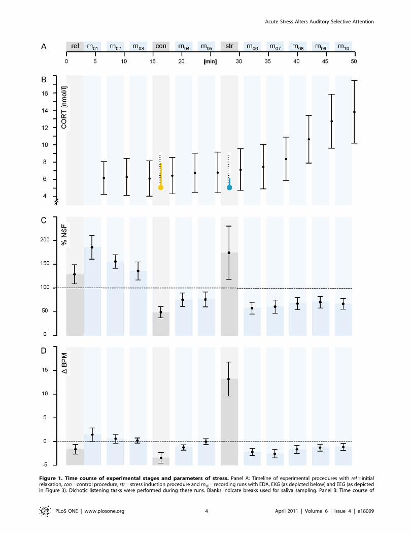
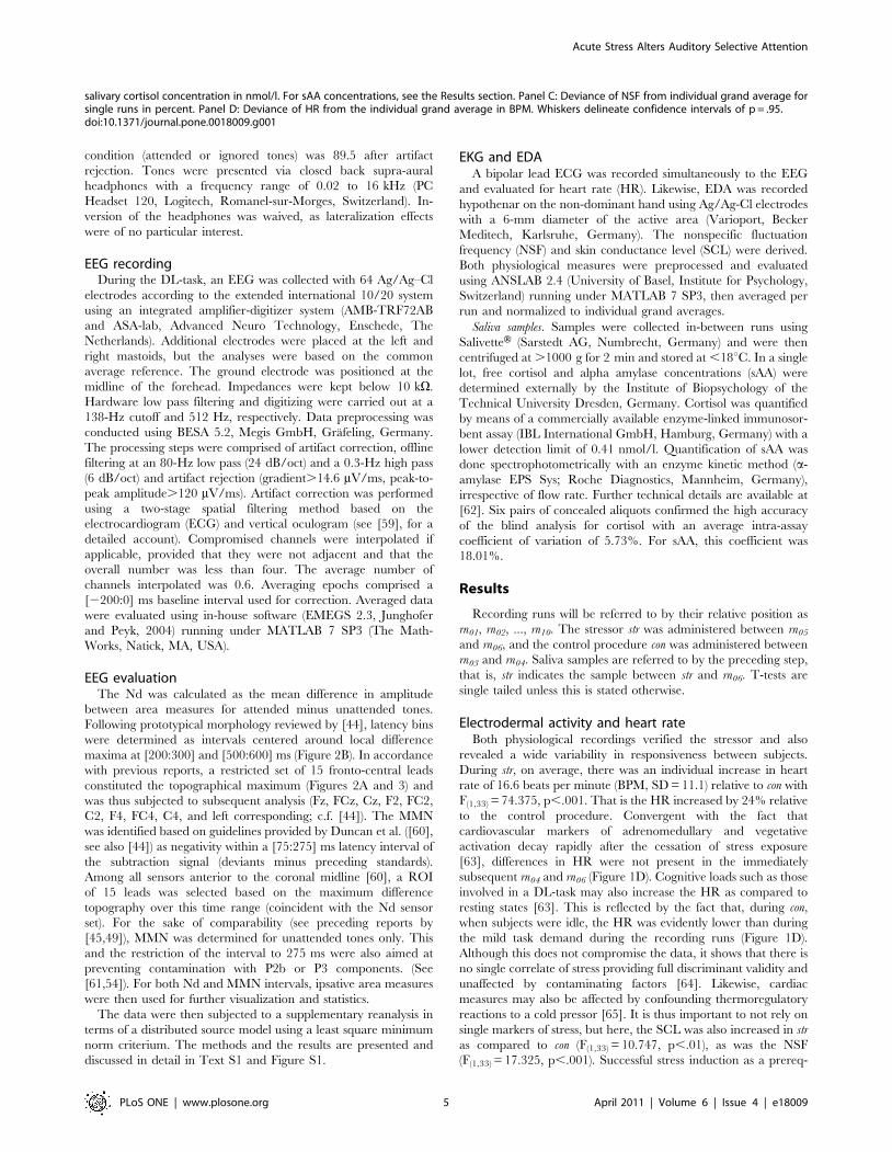
Figure 1. Time course of experimental stages and parameters of stress. Panel A: Timeline of experimental procedures with rel = initialrelaxation, con = control procedure, str = stress induction procedure and rn# = recording runs with EDA, EKG (as depicted below) and EEG (as depictedin Figure 3). Dichotic listening tasks were performed during these runs. Blanks indicate breaks used for saliva sampling. Panel B: Time course of
Acute Stress Alters Auditory Selective Attention
PLoS ONE | www.plosone.org 4 April 2011 | Volume 6 | Issue 4 | e18009
condition (attended or ignored tones) was 89.5 after artifact
rejection. Tones were presented via closed back supra-aural
headphones with a frequency range of 0.02 to 16 kHz (PC
Headset 120, Logitech, Romanel-sur-Morges, Switzerland). In-
version of the headphones was waived, as lateralization effects
were of no particular interest.
EEG recordingDuring the DL-task, an EEG was collected with 64 Ag/Ag–Cl
electrodes according to the extended international 10/20 system
using an integrated amplifier-digitizer system (AMB-TRF72AB
and ASA-lab, Advanced Neuro Technology, Enschede, The
Netherlands). Additional electrodes were placed at the left and
right mastoids, but the analyses were based on the common
average reference. The ground electrode was positioned at the
midline of the forehead. Impedances were kept below 10 kV.
Hardware low pass filtering and digitizing were carried out at a
138-Hz cutoff and 512 Hz, respectively. Data preprocessing was
conducted using BESA 5.2, Megis GmbH, Grafeling, Germany.
The processing steps were comprised of artifact correction, offline
filtering at an 80-Hz low pass (24 dB/oct) and a 0.3-Hz high pass
(6 dB/oct) and artifact rejection (gradient.14.6 mV/ms, peak-to-
peak amplitude.120 mV/ms). Artifact correction was performed
using a two-stage spatial filtering method based on the
electrocardiogram (ECG) and vertical oculogram (see [59], for a
detailed account). Compromised channels were interpolated if
applicable, provided that they were not adjacent and that the
overall number was less than four. The average number of
channels interpolated was 0.6. Averaging epochs comprised a
[2200:0] ms baseline interval used for correction. Averaged data
were evaluated using in-house software (EMEGS 2.3, Junghofer
and Peyk, 2004) running under MATLAB 7 SP3 (The Math-
Works, Natick, MA, USA).
EEG evaluationThe Nd was calculated as the mean difference in amplitude
between area measures for attended minus unattended tones.
Following prototypical morphology reviewed by [44], latency bins
were determined as intervals centered around local difference
maxima at [200:300] and [500:600] ms (Figure 2B). In accordance
with previous reports, a restricted set of 15 fronto-central leads
constituted the topographical maximum (Figures 2A and 3) and
was thus subjected to subsequent analysis (Fz, FCz, Cz, F2, FC2,
C2, F4, FC4, C4, and left corresponding; c.f. [44]). The MMN
was identified based on guidelines provided by Duncan et al. ([60],
see also [44]) as negativity within a [75:275] ms latency interval of
the subtraction signal (deviants minus preceding standards).
Among all sensors anterior to the coronal midline [60], a ROI
of 15 leads was selected based on the maximum difference
topography over this time range (coincident with the Nd sensor
set). For the sake of comparability (see preceding reports by
[45,49]), MMN was determined for unattended tones only. This
and the restriction of the interval to 275 ms were also aimed at
preventing contamination with P2b or P3 components. (See
[61,54]). For both Nd and MMN intervals, ipsative area measures
were then used for further visualization and statistics.
The data were then subjected to a supplementary reanalysis in
terms of a distributed source model using a least square minimum
norm criterium. The methods and the results are presented and
discussed in detail in Text S1 and Figure S1.
EKG and EDAA bipolar lead ECG was recorded simultaneously to the EEG
and evaluated for heart rate (HR). Likewise, EDA was recorded
hypothenar on the non-dominant hand using Ag/Ag-Cl electrodes
with a 6-mm diameter of the active area (Varioport, Becker
Meditech, Karlsruhe, Germany). The nonspecific fluctuation
frequency (NSF) and skin conductance level (SCL) were derived.
Both physiological measures were preprocessed and evaluated
using ANSLAB 2.4 (University of Basel, Institute for Psychology,
Switzerland) running under MATLAB 7 SP3, then averaged per
run and normalized to individual grand averages.
Saliva samples. Samples were collected in-between runs using
SalivetteH (Sarstedt AG, Numbrecht, Germany) and were then
centrifuged at .1000 g for 2 min and stored at ,18uC. In a single
lot, free cortisol and alpha amylase concentrations (sAA) were
determined externally by the Institute of Biopsychology of the
Technical University Dresden, Germany. Cortisol was quantified
by means of a commercially available enzyme-linked immunosor-
bent assay (IBL International GmbH, Hamburg, Germany) with a
lower detection limit of 0.41 nmol/l. Quantification of sAA was
done spectrophotometrically with an enzyme kinetic method (a-
amylase EPS Sys; Roche Diagnostics, Mannheim, Germany),
irrespective of flow rate. Further technical details are available at
[62]. Six pairs of concealed aliquots confirmed the high accuracy
of the blind analysis for cortisol with an average intra-assay
coefficient of variation of 5.73%. For sAA, this coefficient was
18.01%.
Results
Recording runs will be referred to by their relative position as
rn01, rn02, ..., rn10. The stressor str was administered between rn05
and rn06, and the control procedure con was administered between
rn03 and rn04. Saliva samples are referred to by the preceding step,
that is, str indicates the sample between str and rn06. T-tests are
single tailed unless this is stated otherwise.
Electrodermal activity and heart rateBoth physiological recordings verified the stressor and also
revealed a wide variability in responsiveness between subjects.
During str, on average, there was an individual increase in heart
rate of 16.6 beats per minute (BPM, SD = 11.1) relative to con with
F(1,33) = 74.375, p,.001. That is the HR increased by 24% relative
to the control procedure. Convergent with the fact that
cardiovascular markers of adrenomedullary and vegetative
activation decay rapidly after the cessation of stress exposure
[63], differences in HR were not present in the immediately
subsequent rn04 and rn06 (Figure 1D). Cognitive loads such as those
involved in a DL-task may also increase the HR as compared to
resting states [63]. This is reflected by the fact that, during con,
when subjects were idle, the HR was evidently lower than during
the mild task demand during the recording runs (Figure 1D).
Although this does not compromise the data, it shows that there is
no single correlate of stress providing full discriminant validity and
unaffected by contaminating factors [64]. Likewise, cardiac
measures may also be affected by confounding thermoregulatory
reactions to a cold pressor [65]. It is thus important to not rely on
single markers of stress, but here, the SCL was also increased in str
as compared to con (F(1,33) = 10.747, p,.01), as was the NSF
(F(1,33) = 17.325, p,.001). Successful stress induction as a prereq-
salivary cortisol concentration in nmol/l. For sAA concentrations, see the Results section. Panel C: Deviance of NSF from individual grand average forsingle runs in percent. Panel D: Deviance of HR from the individual grand average in BPM. Whiskers delineate confidence intervals of p = .95.doi:10.1371/journal.pone.0018009.g001
Acute Stress Alters Auditory Selective Attention
PLoS ONE | www.plosone.org 5 April 2011 | Volume 6 | Issue 4 | e18009
uisite for the intended principal analysis is thus confirmed, and we
have no further questions addressing the physiological data.
Salivary samplesFrom str to the final rn10, the individual rise in cortisol
concentration was MN = 6.42 nnol/l (SD = 8.17). The timing
was in line with the known kinetic profile (Figure 1B and [66]). A
linear trend over str to rn10 resulted in F(1,30) = 22.014, p,.001.
The sAA concentration remained stable throughout the experi-
ment without any noticeable trends. The sAA peak concentration
after stress induction occurred at str with MN = 85.9 U/ml
(SD = 62.4) whereas the grand average of rn01:10 was 83.5, which
is a difference that is clearly below the error in the assay (see
Methods). Although this timing of the peak concentration meets
our expectations, a planned contrast of str vs. all other runs
rn01:03;con;04:12 was not significant with F(1,25) = 0.015, p = .904. The
same holds for a test of the post-stress vs. post-control samples rn04
vs. rn06 with F(1,30) = 1.355, p = .254.
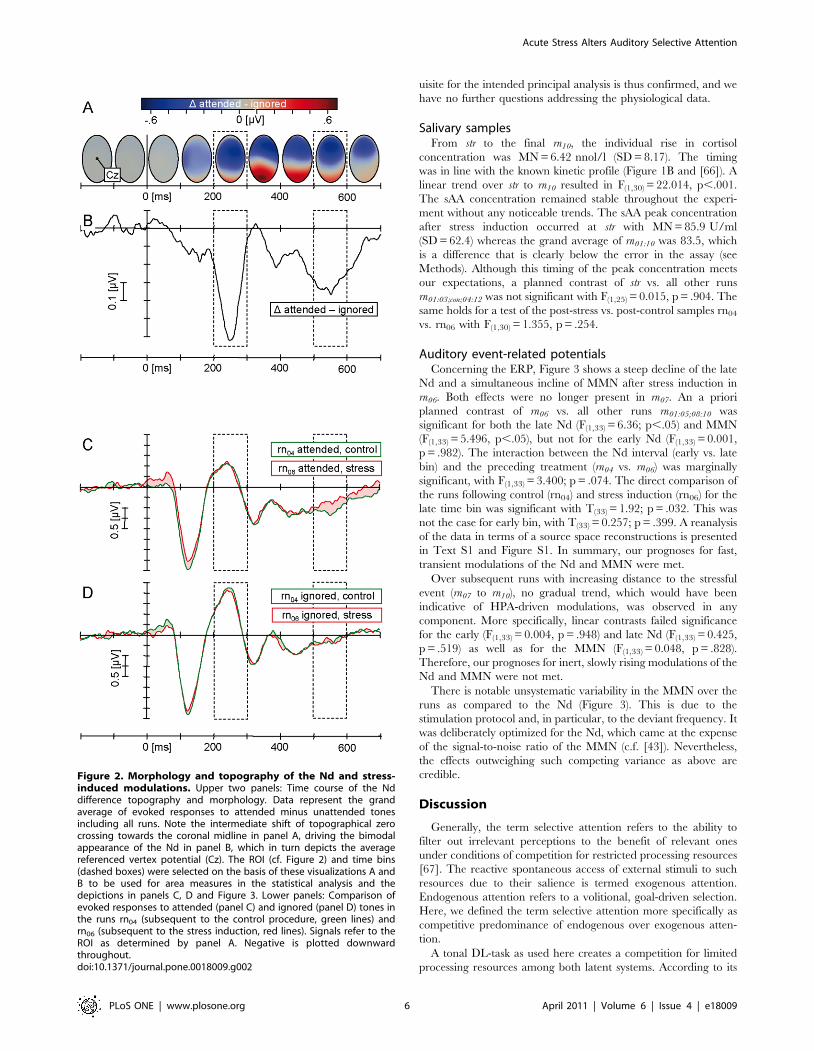
Auditory event-related potentialsConcerning the ERP, Figure 3 shows a steep decline of the late
Nd and a simultaneous incline of MMN after stress induction in
rn06. Both effects were no longer present in rn07. An a priori
planned contrast of rn06 vs. all other runs rn01:05;08:10 was
significant for both the late Nd (F(1,33) = 6.36; p,.05) and MMN
(F(1,33) = 5.496, p,.05), but not for the early Nd (F(1,33) = 0.001,
p = .982). The interaction between the Nd interval (early vs. late
bin) and the preceding treatment (rn04 vs. rn06) was marginally
significant, with F(1,33) = 3.400; p = .074. The direct comparison of
the runs following control (rn04) and stress induction (rn06) for the
late time bin was significant with T(33) = 1.92; p = .032. This was
not the case for early bin, with T(33) = 0.257; p = .399. A reanalysis
of the data in terms of a source space reconstructions is presented
in Text S1 and Figure S1. In summary, our prognoses for fast,
transient modulations of the Nd and MMN were met.
Over subsequent runs with increasing distance to the stressful
event (rn07 to rn10), no gradual trend, which would have been
indicative of HPA-driven modulations, was observed in any
component. More specifically, linear contrasts failed significance
for the early (F(1,33) = 0.004, p = .948) and late Nd (F(1,33) = 0.425,
p = .519) as well as for the MMN (F(1,33) = 0.048, p = .828).
Therefore, our prognoses for inert, slowly rising modulations of the
Nd and MMN were not met.
There is notable unsystematic variability in the MMN over the
runs as compared to the Nd (Figure 3). This is due to the
stimulation protocol and, in particular, to the deviant frequency. It
was deliberately optimized for the Nd, which came at the expense
of the signal-to-noise ratio of the MMN (c.f. [43]). Nevertheless,
the effects outweighing such competing variance as above are
credible.
Discussion
Generally, the term selective attention refers to the ability to
filter out irrelevant perceptions to the benefit of relevant ones
under conditions of competition for restricted processing resources
[67]. The reactive spontaneous access of external stimuli to such
resources due to their salience is termed exogenous attention.
Endogenous attention refers to a volitional, goal-driven selection.
Here, we defined the term selective attention more specifically as
competitive predominance of endogenous over exogenous atten-
tion.
A tonal DL-task as used here creates a competition for limited
processing resources among both latent systems. According to its
Figure 2. Morphology and topography of the Nd and stress-induced modulations. Upper two panels: Time course of the Nddifference topography and morphology. Data represent the grandaverage of evoked responses to attended minus unattended tonesincluding all runs. Note the intermediate shift of topographical zerocrossing towards the coronal midline in panel A, driving the bimodalappearance of the Nd in panel B, which in turn depicts the averagereferenced vertex potential (Cz). The ROI (cf. Figure 2) and time bins(dashed boxes) were selected on the basis of these visualizations A andB to be used for area measures in the statistical analysis and thedepictions in panels C, D and Figure 3. Lower panels: Comparison ofevoked responses to attended (panel C) and ignored (panel D) tones inthe runs rn04 (subsequent to the control procedure, green lines) andrn06 (subsequent to the stress induction, red lines). Signals refer to theROI as determined by panel A. Negative is plotted downwardthroughout.doi:10.1371/journal.pone.0018009.g002
Acute Stress Alters Auditory Selective Attention
PLoS ONE | www.plosone.org 6 April 2011 | Volume 6 | Issue 4 | e18009
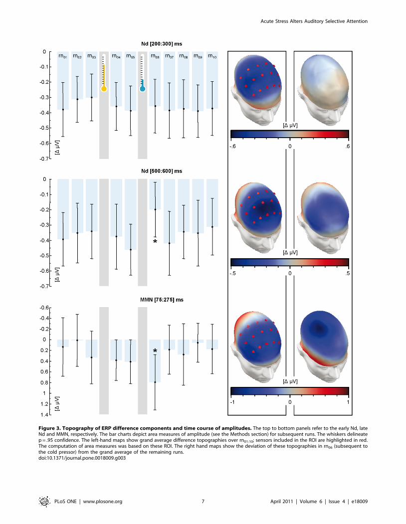
Figure 3. Topography of ERP difference components and time course of amplitudes. The top to bottom panels refer to the early Nd, lateNd and MMN, respectively. The bar charts depict area measures of amplitude (see the Methods section) for subsequent runs. The whiskers delineatep = .95 confidence. The left-hand maps show grand average difference topographies over rn01:10; sensors included in the ROI are highlighted in red.The computation of area measures was based on these ROI. The right hand maps show the deviation of these topographies in rn06 (subsequent tothe cold pressor) from the grand average of the remaining runs.doi:10.1371/journal.pone.0018009.g003
Acute Stress Alters Auditory Selective Attention
PLoS ONE | www.plosone.org 7 April 2011 | Volume 6 | Issue 4 | e18009
extensive functional validation, the manifest Nd is a correlate of
endogenous attention [68,44]. The MMN reflects processes
involved in the initiation of exogenous attention [69,61]. A
reduced relative prevalence of endogenous attention should be
reflected in an attenuation of the Nd. Our findings clearly point to
this posited and evident increase in distractibility. The time course
of a quick rise and decay also matches the prior model-based
expectancies.
Another more detailed claim has to be considered more
tentatively, however: We interpret the driving latent process
behind this as stress-induced activation of dopaminergic projec-
tions from the ventral tegmental area into prefrontal and anterior
cingulate cortices, which we coined MDT. Indeed, the observed
modulations of the evoked potentials exhibit the pattern known
from drugs acting as dopaminergic ligands with respect to Nd as
well as MMN. Similarly, we can preclude ACTH and corticoids
based on their known characteristic ERP modulations. This
reasoning, however, depends on the reliability of premises derived
from pharmacological studies. Admittedly, our deductions are
based on a small body of literature. In particular, the selective
functional sensitivity of the late Nd to dopaminergic challenges
needs further confirmation.
Furthermore, there are several alternative causal attributions that
cannot be discarded at this point. Let us now consider these
potential confounding factors. The most important is a substantial
change in discharge patterns of noradrenergic projections emanat-
ing from the locus coeruleus (LC-NE). This change is another
important aspect of the intracerebral stress response [70–72]. In
fact, the LC is among the most stress-sensitive brain structures [73].
In an exemplary study, Alexander et al. [74] attributed cognitive
effects to stress-induced activation of LC-NE by means of a
propranolol challenge. [75] also interpreted their findings on
emotional attention under acute stress as related to LC-NC activity.
Moreover, stress-related LC-NE activity is regulated by extra-
hypothalamic corticotropin releasing factor (CRF), which also exerts
more direct actions on limbic structures [76–78]. As opposed to this
extrahypothalamic CRF, neurocrine CRF in the pituitary portal
circulation is not directly neuroactive, as it does not pass through the
blood-brain barrier [1,79]. However, other stress-related neurocrine
peptides do and also affect cognition [80,81]. Hence, a number of
interpretations of our findings that involve processes other than
MDT are viable, provided they have a comparable temporal
dynamic. This remains beyond the scope of this study.
More distinctly, the present data rule out the influence of
downstream stages of the HPA on the basis of effect latency. This,
as the authors themselves point out, is not covered in the otherwise
conclusive evidence of Alexander et al. [74]. Although ACTH is
secreted quickly after the advent of a stressor, its effects on ERP
arise much later. Regarding the temporal dynamics of ACTH 4–
10 action with some temporal resolution, [53] found a time lag of
10–30 minutes for the modulation to develop after a single bolus of
1 mg, which may be interpreted in terms of delayed metabotropic
or transcriptional signaling pathways. Thus, HPA activation does
not account for our volatile effects.
The fact that no effects of ACTH, even in our later recording
runs, were observed is somewhat unexpected. We offer the
explanation that the latest recordings were terminated 20 minutes
after the stressor. The electrophysiological effects of the ACTH 4–
10 challenges reviewed above stem from studies using a lagged
design of consecutive substance administration and testing. Delays
ranged between 30, 40 and 60 minutes after intranasal [49],
intravenous [50] and oral administration [51], respectively.
However, our explanation remains speculative, as there is a
shortage of investigations using high-resolution time series.
Given the absolute increase in cortisol concentrations, it is also
possible that the respective ACTH response did not reach some
speculative critical limit in our case. The preponderance of
sympathico-adrenocortical reactions to a CP has already been
discussed by Schwabe et al. [82] (see also [83]). Although the
cortisol reaction to our CP is about three times as high as in
comparable studies (e.g., [82,84], see also [83]), it falls short of
more potent social evaluative stressors. Here, a common finding
could be about twice this amount [85,86,66]. Pharmacological
studies, as reviewed above, even tend to exceed physiological doses
(but see [52]). This might explain why their findings do not agree
with out data.
To summarize, the current study’s outcomes are threefold.
There is sufficient evidence for the supposition that stress impairs
selective attention. Furthermore, there is tentative evidence that
MDT causes the resulting effects among all of the candidate
factors outlined above. Importantly, there is marked evidence that
a causal role of HPA is unlikely.
As reviewed above, the long-term effects of HPA activation
have attracted major interest in the present research and debate.
Given their latency, prior investigations commonly use a time-
lagged design of consecutive stressor exposition and data
recording. It is evident that the present findings would have
escaped such an approach. Besides the immediate question
under study, our report also aims at stimulating the discussion
with a different methodological scope. For future studies on the
impact of stress on cognitive functioning, we offer three
suggestions. First, we deem attentive consideration of both
HPA and non-HPA causality equally important. Second, the
temporal dynamic of stress-related cognitive changes deserves
particular interest. These may not only differ in latency for
different substances, but they may even be inversed for single
substances over time [87]. Third, such non-monotonic response
curves also pertain to the topic of dose dependency [88,87],
which we addressed only superficially. Inconsistent findings in
current research might be explained in terms of these topics. As
the term stress refers to a heterogeneous construct, differentiated
investigations seem promising.
Supporting Information
Figure S1 All depictions show subtractions of attended minus
unattended stimulation. Whiskers delineate confidence intervals of
p = .95. Panel A: Topography of the difference source activity in a
time range of [100:600] ms. A bilateral temporal and a frontopolar
dipole cluster were selected based on this topography as ROI.
These are the basis of the below panels and subsequent analyses.
Panel B: Global Power of the Nd source activity in the temporal
(green) and frontal (blue) ROI. Time bins of [100:200] ms and
[400:550] ms were selected based on the bimodal maxima of the
joint activity of both ROI (dashed boxes). Influences of stress do
not differentially affect the early and late Nd. Thus, Panels C and
D depict unweighted mean activity over both bins. Panel C:
Activity of the frontal Nd generator during consecutive runs. Note
the drop of Nd amplitude after stress exposition. By comparison,
the activity of the temporal generator (Panel D) remains constant.
This pattern occurs without great difference in both latency
intervals (Panel E).
(TIF)
Table S1 Itemization of the individual performance in the
dichotic listening task. The numbers indicate the deviation
between the correct solution and the subject’s reply.
(PDF)
Acute Stress Alters Auditory Selective Attention
PLoS ONE | www.plosone.org 8 April 2011 | Volume 6 | Issue 4 | e18009
Text S1 A reanalysis of the EEG data using a pseudoinverse
calculation.
(PDF)
Acknowledgments
We would like to thank Dipl. Psych. Gerrit Hirschfeld (Department of
Psychology, WWU, Munster, Germany), Dipl. Psych. Ida Wessing (Child
and Adolescent Psychiatric Clinic, UKM, Munster) and Prof. Harald
Schupp (Department of Psychology, University of Konstanz, Konstanz,
Germany) for their valuable contributions.
Author Contributions
Conceived and designed the experiments: LE AKB MJ. Performed the
experiments: CD CS JB. Analyzed the data: CS AKB LE. Contributed
reagents/materials/analysis tools: MJ. Wrote the paper: LE MJ CD CS JB.
References
1. Chrousos GP (2009) Stress and disorders of the stress system. Nat Rev
Endocrinol 5(7): 374–381.
2. Chrousos GP, Gold PW (1992) The concepts of stress and stress system disorders
- overview of physical and behavioral homeostasis. Jama-J Am Med Assoc267(9): 1244–1252.
3. Lightman SL (2008) The neuroendocrinology of stress: A never ending story.
J Neuroendocrinol 20(6): 880–884.
4. Joels M, Baram TZ (2009) The neuro-symphony of stress. Nat Rev Neurosci
10(6): 459–466.
5. de Kloet ER, de Jong IEM, Oitzi MS (2008) Neuropharmacology of
glucocorticoids: Focus on emotion, cognition and cocaine. Eur J Pharmacol585(2–3): 473–482.
6. de Kloet ER, Karst H, Joels M (2008) Corticosteroid hormones in the centralstress response: Quick-and-slow. Front Neuroendocrin 29(2): 268–272.
7. de Kloet ER, Joels M, Holsboer F (2005) Stress and the brain: From adaptationto disease. Nat Rev Neurosci 6(6): 463–475.
8. McGaugh JL, Roozendaal B (2002) Role of adrenal stress hormones in forminglasting memories in the brain. Curr Opin Neurobiol 12(2): 205–210.
9. Roozendaal B, McEwen BS, Chattarji S (2009) Stress, memory and theamygdala. Nat Rev Neurosci 10(6): 423–433.
10. Finlay JM, Zigmond MJ (1997) The effects of stress on central dopaminergic
neurons: Possible clinical implications. Neurochem Res 22(11): 1387–1394.
11. Mantz J, Thierry AM, Glowinski J (1989) Effect of Noxious Tail Pinch on the
Discharge Rate of Mesocortical and Mesolimbic Dopamine Neurons - Selective
Activation of the Mesocortical System. Brain Res 476(2): 377–381.
12. Abercrombie ED, Keefe KA, Difrischia DS, Zigmond MJ (1989) Differentialeffect of stress on in vivo dopamine release in striatum, nucleus accumbens, and
medial frontal-cortex. J Neurochem 52(5): 1655–1658.
13. Arnsten AFT, Goldman-Racic PS (1998) Noise stress impairs prefrontal cortical
cognitive function in monkeys: Evidence for a hyperdopaminergic mechanism.
Arch Gen Psychiatry 55(4): 362–368.
14. Cenci MA, Kalen P, Mandel RJ, Bjorklund A (1992) Regional differences in theregulation of dopamine and noradrenaline release in medial frontal-cortex,
nucleus-accumbens and caudate-putamen - a microdialysis study in the rat.
Brain Res 581(2): 217–228.
15. Deutch AY, Clark WA, Roth RH (1990) Prefrontal cortical dopamine depletion
enhances the responsiveness of mesolimbic dopamine neurons to stress. BrainRes 521(1–2): 311–315.
16. Thierry AM, Tassin JP, Blanc G, Glowinski J (1976) Selective activation of
mesocortical DA system by stress. Nature 263(5574): 242–244.
17. Steciuk M, Kram M, Kramer GL, Petty F (2000) Immobilization-induced
glutamate efflux in medial prefrontal cortex: blockade by (+)-Mk-801, a selective
NMDA receptor antagonist. Stress 3(3): 195–199.
18. Yuen EY, Liu WH, Karatsoreos IN, Feng J, McEwen BS, et al. (2009) Acutestress enhances glutamatergic transmission in prefrontal cortex and facilitates
working memory. Proc Natl Acad Sci U S A 106(33): 14075–14079.
19. Robbins TW (2005) Role of cortical and striatal dopamine in cognitive function.
In: Dunnett SB, ed. Dopamine. Amsterdam, Boston: Elsevier. pp 395–434.
20. Arnsten AFT (2009) Stress signalling pathways that impair prefrontal cortex
structure and function. Nat Rev Neurosci 10(6): 410–422.
21. Gresch PJ, Sved AF, Zigmond MJ, Finlay JM (1994) Stress-induced sensitization
of dopamine and norepinephrine efflux in medial prefrontal cortex of the rat.J Neurochem 63(2): 575–583.
22. Yoshioka M, Matsumoto M, Togashi H, Saito H (1996) Effect of conditionedfear stress on dopamine release in the rat prefrontal cortex. Neurosci Lett 209(3):
201–203.
23. Morrow BA, Roth RH, Elsworth JD (2000) TMT, a predator odor, elevates
mesoprefrontal dopamine metabolic activity and disrupts short-term workingmemory in the rat. Brain Res Bull 52(6): 519–523.
24. Butler AB, Hodos W (1996) Comparative vertebrate neuroanatomy. Evolutionand adaptation. New York, NY: Wiley-Liss.
25. Striedter GF (2005) Principles of brain evolution. Sunderland, Mass.: SinauerAssociates.
26. Williams SM, Goldman-Racic PS (1998) Widespread origin of the primate
mesofrontal dopamine system. Cereb Cortex 8(4): 321–345.
27. Berger B, Gaspar P, Verney C (1991) Dopaminergic innervation of the cerebral
cortex: unexpected differences between rodents and primates. Trends Neurosci
14(1): 21–27.
28. Preuss TM (1995) Do rats have prefrontal cortex - the rose-woolsey-akertprogram reconsidered. J Cognitive Neurosci 7(1): 1–24.
29. Goldman-Racic PS, Bergson C, Krimer LS, Lidow MS, Williams SM, et al.
(1999) The primate mesocortical dopamine system. In: Bloom FE, Bjorklund A,
Hokfelt T, Bloom FE, eds. The primate nervous system III. Vol. 15. Amsterdam,
New York: Elsevier. pp 403–428.
30. Drevets WC, Raichle ME (1998) Reciprocal suppression of regional cerebral
blood flow during emotional versus higher cognitive processes: Implications for
interactions between emotion and cognition. Cognition Emotion 12(3): 353–385.
31. Corbetta M, Shulman GL (2002) Control of goal-directed and stimulus-driven
attention in the brain. Nat Rev Neurosci 3(3): 201–215.
32. Al’Absi M, Hugdahl K, Lovallo WR (2002) Adrenocortical stress responses and
altered working memory performance. Psychophysiology 39(1): 95–99.
33. Hoffman R, Al’Absi M (2004) The effect of acute stress on subsequent
neuropsychological test performance. Arch Clin Neuropsych 19(4): 497–506.
34. Oei NY, Everaerd WT, Elzinga BM, van Well S, Bermond B (2006)
Psychosocial stress impairs working memory at high loads: an association with
cortisol levels and memory retrieval. Stress 9(3): 133–141.
35. Schoofs D, Preuss D, Wolf OT (2008) Psychosocial stress induces working
memory impairments in an n-back paradigm. Psychoneuroendocrino 33(5):
643–653.
36. Skosnik PD, Chatterton RT, Jr., Swisher T, Park S (2000) Modulation of
attentional inhibition by norepinephrine and cortisol after psychological stress.
Int J Psychophysiol 36(1): 59–68.
37. Elzinga BM, Roelofs K (2005) Cortisol-induced impairments of working
memory require acute sympathetic activation. Behav Neurosci 119(1): 98–103.
38. Erickson K, Drevets W, Schulkin J (2003) Glucocorticoid regulation of diverse
cognitive functions in normal and pathological emotional states. Neurosci
Biobehav Rev 27(3): 233–246.
39. Qin SZ, Hermans EJ, van Marle HJ, Luo J, Fernandez G (2009) Acute
Psychological Stress Reduces Working Memory-Related Activity in the
Dorsolateral Prefrontal Cortex. Biol Psychiatry 66(1): 25–32.
40. Braunstein-Bercovitz H (2003) Does stress enhance or impair selective attention?
The effects of stress and perceptual load on negative priming. Anxiety Stress
Copin 16(4): 345–357.
41. Braunstein-Bercovitz H, Dimentman-Ashkenasi I, Lubow RE (2001) Stress
affects the selection of irrelevant stimuli. Emotion 1(2): 182–192.
42. Hillyard SA, Hink RF, Schwent VL, Picton TW (1973) Electrical signs of
selective attention in human brain. Science 182(4108): 177–180.
43. Ahveninen J, Jaaskelainen IP, Pennanen S, Liesivuori J, Ilmoniemi RJ, et al.
(2003) Auditory selective attention modulated by tryptophan depletion in
humans. Neurosci Lett 340(3): 181–184.
44. Jemel B, Oades RD, Oknina L, Achenbach C, Ropcke B (2003) Frontal and
temporal lobe sources for a marker of controlled auditory attention: the negative
difference (Nd) event-related potential. Brain Topogr 15(4): 249–262.
45. Kahkonen S, Ahveninen J, Jaaskelainen IP, Kaakkola S, Naatanen R, et al.
(2001) Effects of haloperidol on selective attention: a combined whole-head
MEG and high-resolution EEG study. Neuropsychopharmacol 25(4): 498–504.
46. Kahkonen S, Ahveninen J (2002) Combination of magneto- and electroenceph-
alography in studies of monoamine modulation on attention. Methods Find Exp
Clin Pharmacol 24(Suppl C): 27–34.
47. Shelley AM, Catts SV, Ward PB, Andrews S, Mitchell P, et al. (1997) The effect
of decreased catecholamine transmission on ERP indices of selective attention.
Neuropsychopharmacol 16(3): 202–210.
48. Bartlett EJ, Brodie JD, Simkowitz P, Dewey SL, Rusinek H, et al. (1994) Effects
of haloperidol challenge on regional cerebral glucose utilization in normal
human subjects. Am J Psychiatry 151(5): 681–686.
49. Smolnik R, Molle M, Fehm HL, Born J (1999) Brain potentials and attention
after acute and subchronic intranasal administration of ACTH 4-10 and
desacetyl-alpha-MSH in humans. Neuroendocrinology 70(1): 63–72.
50. Molle M, Albrecht C, Marshall L, Fehm HL, Born J (1997) Adrenocorticotropin
widens the focus of attention in humans. A nonlinear electroencephalographic
analysis. Psychosom Med 59(5): 497–502.
51. Born J, Fehm-Wolfsdorf G, Schiebe M, Birbaumer N, Fehm HL, et al. (1985) An
ACTH-4-9 Analog Impairs Selective Attention in Man. Life Sci 36(22):
2117–2125.
52. Born J, Brauninger W, Fehm-Wolfsdorf G, Voigt KH, Pauschinger P, et al.
(1987) Dose-dependent influences on electrophysiological signs of attention in
humans after neuropeptide ACTH 4-10. Exp Brain Res 67(1): 85–92.
53. Born J, Unseld U, Pietrowsky R, Bickel U, Voigt KH, et al. (1990) Time course
of ACTH 4-10 effects on human attention. Neuroendocrinology 52(2): 169–174.
Acute Stress Alters Auditory Selective Attention
PLoS ONE | www.plosone.org 9 April 2011 | Volume 6 | Issue 4 | e18009
54. Born J, Kern W, Fehm-Wolfsdorf G, Fehm HL (1987) Cortisol effects on
attentional processes in man as indicated by event-related potentials.Psychophysiology 24(3): 286–292.
55. Kirschbaum C, Kudielka BM, Gaab J, Schommer NC, Hellhammer DH (1999)
Impact of gender, menstrual cycle phase, and oral contraceptives on the activityof the hypothalamus-pituitary-adrenal axis. Psychosom Med 61(2): 154–162.
56. Kudielka BM, Kirschbaum C (2005) Sex differences in HPA axis responses tostress: A review. Biol Psychol 69(1): 113–132.
57. Simoens VL, Istok E, Hyttinen S, Hirvonen A, Naatanen R, et al. (2007)
Psychosocial stress attenuates general sound processing and duration changedetection. Psychophysiology 44(1): 30–38.
58. Biondi M, Picardi A (1999) Psychological stress and neuroendocrine function inhumans: The last two decades of research. Psychother Psychosom 68(3):
114–150.59. Ille N, Berg P, Scherg M (2002) Artifact correction of the ongoing EEG using
spatial filters based on artifact and brain signal topographies. J Clin
Neurophysiol 19(2): 113–124.60. Duncan CC, Barry RJ, Connolly JF, Fischer C, Michie PT, et al. (2009) Event-
related potentials in clinical research: Guidelines for eliciting, recording, andquantifying mismatch negativity, P300, and N400. Clin Neurophysiol 120(11):
1883–1908.
61. Naatanen R, Paavilainen P, Rinne T, Alho K (2007) The mismatch negativity(MMN) in basic research of central auditory processing: A review. Clinical
Neurophysiology 118(12): 2544–2590.62. Strahler J, Mueller A, Rosenloecher F, Kirschbaum C, Rohleder N (2010)
Salivary alpha-amylase stress reactivity across different age groups. Psychophys-iology 47(3): 587–595.
63. Herd JA (1991) Cardiovascular Response to Stress. Physiol Rev 71(1): 305–330.
64. Cox T (1985) The nature and measurement of stress. Ergonomics 28(8):1155–1163.
65. Streff A, Kuehl LK, Michaux G, Anton F (2010) Differential physiological effectsduring tonic painful hand immersion tests using hot and ice water. Eur J Pain
14(3): 266–272.
66. Kirschbaum C, Hellhammer DH (1994) Salivary cortisol in psychoneuroendo-crine research - recent developments and applications. Psychoneuroendocrino
19(4): 313–333.67. Lavie N, Tsal Y (1994) Perceptual load as a major determinant of the locus of
selection in visual-attention. Percept Psychophys 56(2): 183–197.68. Kok A (2000) Age-related changes in involuntary and voluntary attention as
reflected in components of the event-related potential (ERP). Biol Psychol 54(1–
3): 107–143.69. Herrmann CS, Knight RT (2001) Mechanisms of human attention: event-
related potentials and oscillations. Neurosci Biobehav Rev 25(6): 465–476.70. Valentino RJ, van Bockstaele E (2008) Convergent regulation of locus coeruleus
activity as an adaptive response to stress. Eur J Pharmacol 583(2–3): 194–203.
71. Sara SJ (2009) The locus coeruleus and noradrenergic modulation of cognition.Nat Rev Neurosci 10(3): 211–223.
72. Bouret S, Sara SJ (2005) Network reset: a simplified overarching theory of locus
coeruleus noradrenaline function. Trends Neurosci 28(11): 574–582.
73. Herman JP, Cullinan WE (1997) Neurocircuitry of stress: Central control of the
hypothalamo-pituitary-adrenocortical axis. Trends Neurosci 20(2): 78–84.
74. Alexander JK, Hillier A, Smith RM, Tivarus ME, Beversdorf DQ (2007) Beta-
adrenergic modulation of cognitive flexibility during stress. J Cognitive Neurosci
19(3): 468–478.
75. van Marle HJ, Hermans EJ, Qin SZ, Fernandez G (2009) From Specificity to
Sensitivity: How Acute Stress Affects Amygdala Processing of Biologically
Salient Stimuli. Biol Psychiatry 66(7): 649–655.
76. Cook CJ (2004) Stress induces CRF release in the paraventricular nucleus, and
both CRF and GABA release in the amygdala. Physiol Behav 82(4): 751–762.
77. Curtis AL, Lechner SM, Pavcovich LA, Valentino RJ (1997) Activation of the
locus coeruleus noradrenergic system by intracoerulear microinfusion of
corticotropin-releasing factor: effects on discharge rate, cortical norepinephrine
levels and cortical electroencephalographic activity. J Pharmacol Exp Ther
281(1): 163–172.
78. Koob GF (1999) Corticotropin-releasing factor, norepinephrine, and stress. Biol
Psychiatry 46(9): 1167–1180.
79. Martins JM, Kastin AJ, Banks WA (1996) Unidirectional specific and modulated
brain to blood transport of corticotropin-releasing hormone. Neuroendocrinol-
ogy 63(4): 338–348.
80. De Wied D (1980) Behavioural Actions of Neurohypophysial Peptides. Proc R
Soc Lond B (1178): 183–194.
81. Zlocovic BV, Hyman S, McComb JG, Lipovac MN, Tang G, et al. (1990)
Kinetics of Arginine-Vasopressin Uptake at the Blood-Brain-Barrier. Biochim
Biophys Acta 1025(2): 191–198.
82. Schwabe L, Haddad L, Schachinger H (2008) HPA axis activation by a socially
evaluated cold-pressor test. Psychoneuroendocrino 33(6): 890–895.
83. Duncko R, Johnson L, Merikangas K, Grillon C (2009) Working memory
performance after acute exposure to the cold pressor stress in healthy volunteers.
Neurobiol Learn Mem 91(4): 377–381.
84. van Stegeren AH, Wolf OT, Kindt M (2008) Salivary alpha amylase and cortisol
responses to different stress tasks: Impact of sex. Int J Psychophysiol 69(1):
33–40.
85. Rohleder N, Wolf JM, Maldonado EF, Kirschbaum C (2006) The psychosocial
stress-induced increase in salivary alpha-amylase is independent of saliva flow
rate. Psychophysiology 43(6): 645–652.
86. Nater UM, Rohleder N, Gaab J, Berger S, Jud A, et al. (2005) Human salivary
alpha-amylase reactivity in a psychosocial stress paradigm. Int J Psychophysiol
55(3): 333–342.
87. Joels M (1997) Steroid hormones and excitability in the mammalian brain. Front
Neuroendocrin 18(1): 2–48.
88. Sapolsky RM, Romero LM, Munck AU (2000) How do glucocorticoids
influence stress responses? Integrating permissive, suppressive, stimulatory, and
preparative actions. Endocr Rev 21(1): 55–89.
Acute Stress Alters Auditory Selective Attention
PLoS ONE | www.plosone.org 10 April 2011 | Volume 6 | Issue 4 | e18009
Recommended