5/14/2020
1
REGISTER NOW…ACG’s IBD School & Eastern Regional!
Are now VIRTUAL events, withOn‐Demand Presentations and LIVE Webcast Q&A sessions!
Visit meetings.gi.org to register for both today!
NEW!! ACG 2020 ABSTRACT SUBMISSION DEADLINE
EXTENDED 2 WEEKS!
NEW!! DEADLINE: JUNE 15, 2020 11:59pm Eastern
1
2
American College of Gastroenterology

5/14/2020
2
3
3
4
American College of Gastroenterology

5/14/2020
3
Participating in the Webinar
All attendees will be muted and will remain in Listen Only Mode.
Type your questions here so that the moderator can see them. Not all questions will be answered but we will get to as many as possible.
How to Receive CME and MOC Points
LIVE VIRTUAL GRAND ROUNDS WEBINAR
ACG will send a link to a CME & MOC evaluation to all attendees on the live webinar.
ABIM Board Certified physicians need to complete their MOC activities by December 31, 2020 in order for the MOC points to count toward any MOC requirements that are due by the end of the year. No MOC credit may be awarded after March 1, 2021 for this activity.
ACG will submit MOC points on the first of each month. Please allow 3‐5 business days for your MOC credit to appear on your ABIM account.
5
6
American College of Gastroenterology
5/14/2020
4
MOC QUESTION
If you plan to claim MOC Points for this activity, you will be asked to: Please list specific changes you will make in your
practice as a result of the information you received from this activity.
Include specific strategies or changes that you plan to implement.THESE ANSWERS WILL BE REVIEWED.
8
7
8
American College of Gastroenterology
5/14/2020
5
ACG Virtual Grand RoundsJoin us for upcoming Virtual Grand Rounds!
Week 9: Positioning of Old and New Therapies in IBDDavid T. Rubin, MD, FACG May 21, 2020 at Noon EDT
Visit gi.org/ACGVGR to Register
Week 10: Colorectal Cancer Screening in a Post Covid World Renee L. Williams, MD, MHPE, FACGMay 28, 2020 at Noon EDT
REGISTER NOW…ACG’s IBD School & Eastern Regional!
Are now VIRTUAL events, withOn‐Demand Presentations and LIVE Webcast Q&A sessions!
Visit meetings.gi.org to register for both today!
9
10
American College of Gastroenterology

5/14/2020
6
NEW!! ACG 2020 ABSTRACT SUBMISSION DEADLINE
EXTENDED 2 WEEKS!
NEW!! DEADLINE: JUNE 15, 2020 11:59pm Eastern
Disclosures:
Moderator:
Brooks D. Cash, MD, FACG Consultant: Allergan, QOL Medical, Salix, Takeda Speakers Bureau: Allergan, QOL Medical, Salix, Takeda
Speaker: Off Label Use:
Carol A. Burke, MD, FACG NoneConsultant: Aries, Ferring, FreenomeResearch Grant: Cancer Prevention Pharmaceuticals, Janssen
11
12
American College of Gastroenterology

5/14/2020
7
Serrated Polyps and Serrated Polyposis Syndrome:
Cancer Risk and Appropriate Surveillance
Carol A. Burke, MD, FACGDepartment of Gastroenterology, Hepatology and Nutrition
Cleveland Clinic, Cleveland Ohio
Objectives
• Understand the nomenclature of serrated polypsand diagnostic criteria for serrated polyposissyndrome
• Identify endoscopic features of serrated lesions• Recognize CRC risk and formulate strategies to
manage patients with serrated polyps andserrated polyposis syndrome
13
14
American College of Gastroenterology
5/14/2020
8
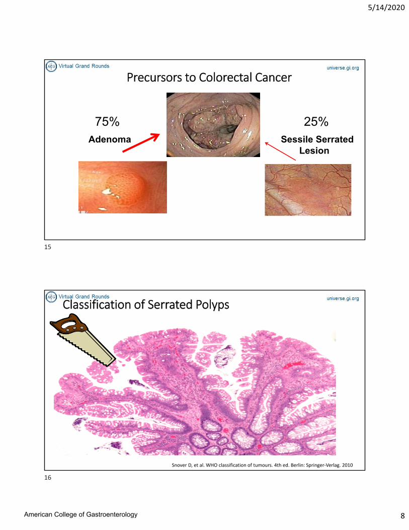
Precursors to Colorectal Cancer
Adenoma Sessile SerratedLesion
75% 25%
Classification of Serrated Polyps
Snover D, et al. WHO classification of tumours. 4th ed. Berlin: Springer‐Verlag. 2010
15
16
American College of Gastroenterology

5/14/2020
9
Nomenclature of Serrated Polyps
• Hyperplastic Polyp (HP)
• Sessile Serrated Lesion (SSL)• Formerly: sessile serrated adenoma / sessile serrated polyp
• Traditional Serrated Adenoma (TSA)
Snover D, et al. WHO 2010
What Pathology Criteria Distinguish SSL from HP?
17
18
American College of Gastroenterology
5/14/2020
10
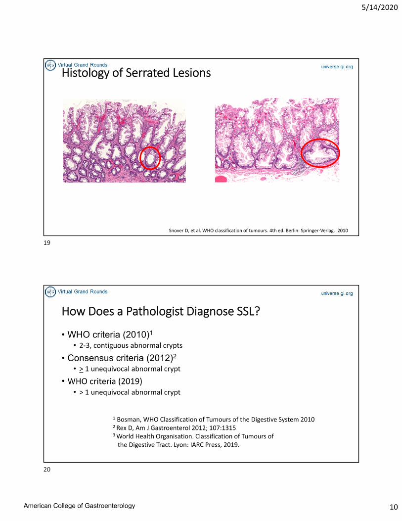
Histology of Serrated Lesions
Snover D, et al. WHO classification of tumours. 4th ed. Berlin: Springer‐Verlag. 2010
How Does a Pathologist Diagnose SSL?
• WHO criteria (2010)1
• 2‐3, contiguous abnormal crypts
• Consensus criteria (2012)2
• > 1 unequivocal abnormal crypt
• WHO criteria (2019)• > 1 unequivocal abnormal crypt
1 Bosman, WHO Classification of Tumours of the Digestive System 2010 2 Rex D, Am J Gastroenterol 2012; 107:13153 World Health Organisation. Classification of Tumours ofthe Digestive Tract. Lyon: IARC Press, 2019.
19
20
American College of Gastroenterology
5/14/2020
11
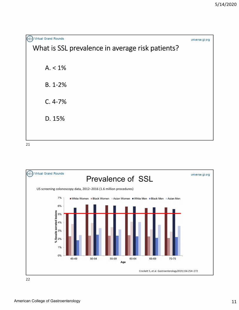
What is SSL prevalence in average risk patients?
A. < 1%
B. 1‐2%
C. 4‐7%
D. 15%
Prevalence of SSL
Crockett S, et al. Gastroenterology2019;156:254–272
US screening colonoscopy data, 2012–2016 (1.6 million procedures)
21
22
American College of Gastroenterology

5/14/2020
12
Detection Rate of SSL
Sarvepalli S, et al. JAMA Surg 2019 Jul 1;154(7):627‐63556 endoscopists
Overall(N=16.089)
Men(N=7749)
Women(N=8339)
ADR 32.3% 36.9% 26%
SSLDR 4.6% 4.6% 4.6%
Screening colonoscopies; single center, 2015‐2017
ADR CSSDR (SSL, TSA, HP > 10 mm)Median (IQR)
< 15% 1.3% (0.9‐3.1)
15‐25% 3.5% (2.5‐4.8)
25‐<35% 6.3% (3.0‐7.2)
> 35% 10.0% (8.5‐13.1)
77 endoscopists, 28 centers;2009‐2014
Correlation CoefficientBetween ADR and SSL
P= .69
Anderson J, et al GIE 2017;85:1188‐94
Prevalence and Location of Serrated Lesions
IJspeert JE. Endoscopy.2016;48:740‐6
• 3364 patients, 2011‐2015, Single center• 25 endoscopists, ADR 38.5% (22.5%–53.9%)• SSL detection rate: 7.3% (2.5% ‐13.6%); 2 dilated crypts and/or hyper‐serrations in crypt base
23
24
American College of Gastroenterology

5/14/2020
13
Prevalence and Location of Serrated Lesions
IJspeert JE. Endoscopy.2016;48:740‐6
• 3364 patients, 2011‐2015, Single center• 25 endoscopists, ADR 38.5% (22.5%–53.9%)• SSL detection rate: 7.3% (2.5% ‐13.6%); 2 dilated crypts and/or hyper‐serrations in crypt base
SSL76 % in proximal colon
> 80% SSL with dysplasia or > 10 mm in proximal colon.
Factors Associated with SSL DetectionAssociated of SSL with Synchronous Advanced Neoplasia
IJspeert JE. Endoscopy.2016;48:740‐6
Factor Detection of SSL (95% CI)
Age, yrs 1.01 (1.00‐1.03)
Male gender 1.04 (0.81‐1.34)
Colonoscopy Indication
Symptoms Reference
FIT + 1.08 (0.75‐1.54)
Family History CRC 1.52 (1.05‐2.22)
Surveillance 1.73 (1.20‐2.49)
SSPDR > 7.3% vs < 7.3% 2.65 (2.00‐3.50)
SSL Feature Risk of Synchronous Advanced Neoplasia
(95% CI)
> 1 SSL 1.71 (1.25‐2.34)
Proximal 1.69 (1.20‐2.39)
> 10 mm 2.78 (1.56‐4.96)
> 10 mm/with dysplasia 2.65 (1.56‐4.67)
25
26
American College of Gastroenterology
5/14/2020
14
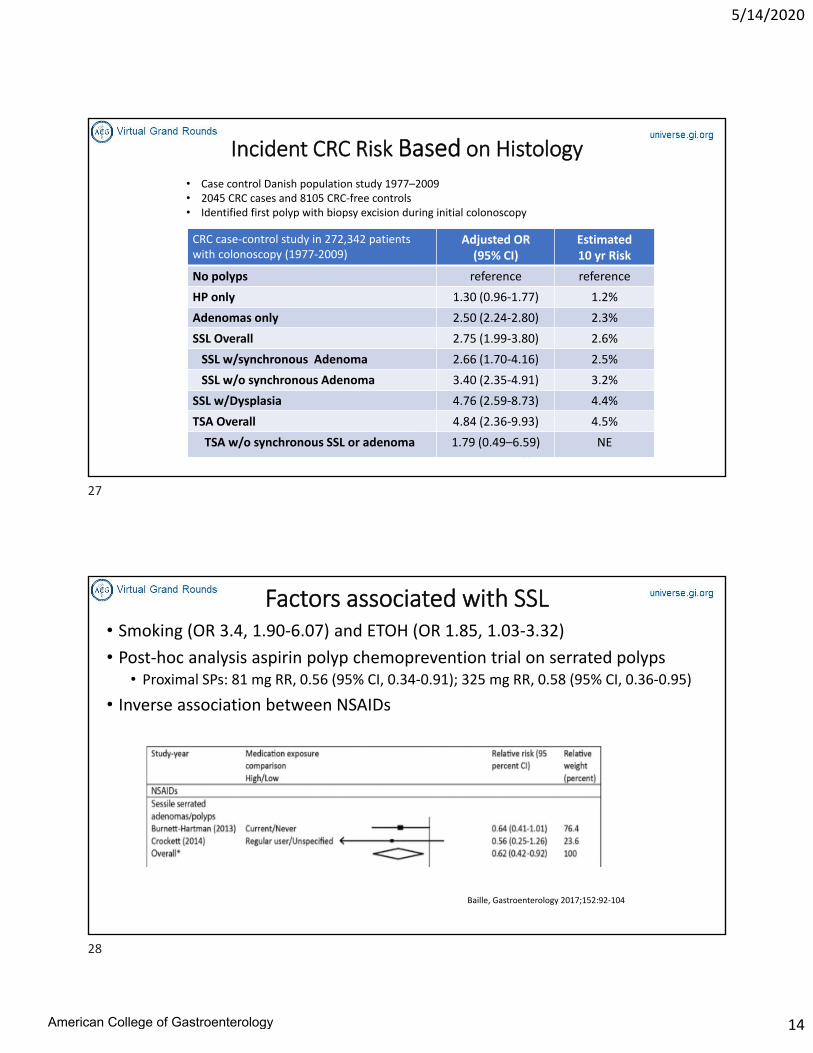
Incident CRC Risk Based on Histology
Erichsen R, Gastroenterology 2016;150:895–902
CRC case‐control study in 272,342 patients with colonoscopy (1977‐2009)
Adjusted OR(95% CI)
Estimated10 yr Risk
No polyps reference reference
HP only 1.30 (0.96‐1.77) 1.2%
Adenomas only 2.50 (2.24‐2.80) 2.3%
SSL Overall 2.75 (1.99‐3.80) 2.6%
SSL w/synchronous Adenoma 2.66 (1.70‐4.16) 2.5%
SSL w/o synchronous Adenoma 3.40 (2.35‐4.91) 3.2%
SSL w/Dysplasia 4.76 (2.59‐8.73) 4.4%
TSA Overall 4.84 (2.36‐9.93) 4.5%
TSA w/o synchronous SSL or adenoma 1.79 (0.49–6.59) NE
• Case control Danish population study 1977–2009• 2045 CRC cases and 8105 CRC‐free controls • Identified first polyp with biopsy excision during initial colonoscopy
Factors associated with SSL• Smoking (OR 3.4, 1.90‐6.07) and ETOH (OR 1.85, 1.03‐3.32)
• Post‐hoc analysis aspirin polyp chemoprevention trial on serrated polyps• Proximal SPs: 81 mg RR, 0.56 (95% CI, 0.34‐0.91); 325 mg RR, 0.58 (95% CI, 0.36‐0.95)
• Inverse association between NSAIDs
Baille, Gastroenterology 2017;152:92‐104
27
28
American College of Gastroenterology
5/14/2020
15
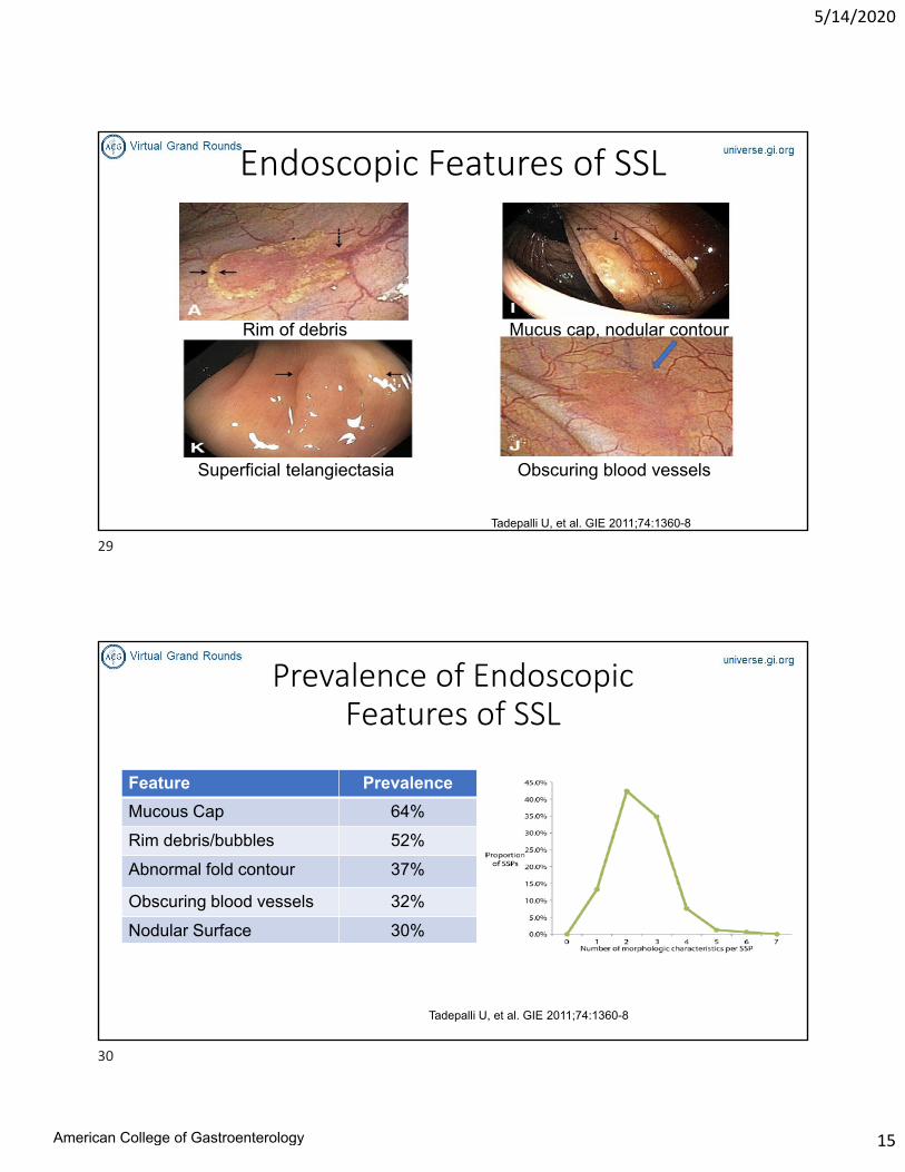
Tadepalli U, et al. GIE 2011;74:1360-8
Rim of debris Mucus cap, nodular contour
Superficial telangiectasia Obscuring blood vessels
Endoscopic Features of SSL
Prevalence of Endoscopic Features of SSL
Feature Prevalence
Mucous Cap 64%
Rim debris/bubbles 52%
Abnormal fold contour 37%
Obscuring blood vessels 32%
Nodular Surface 30%
Tadepalli U, et al. GIE 2011;74:1360-8
29
30
American College of Gastroenterology
5/14/2020
16
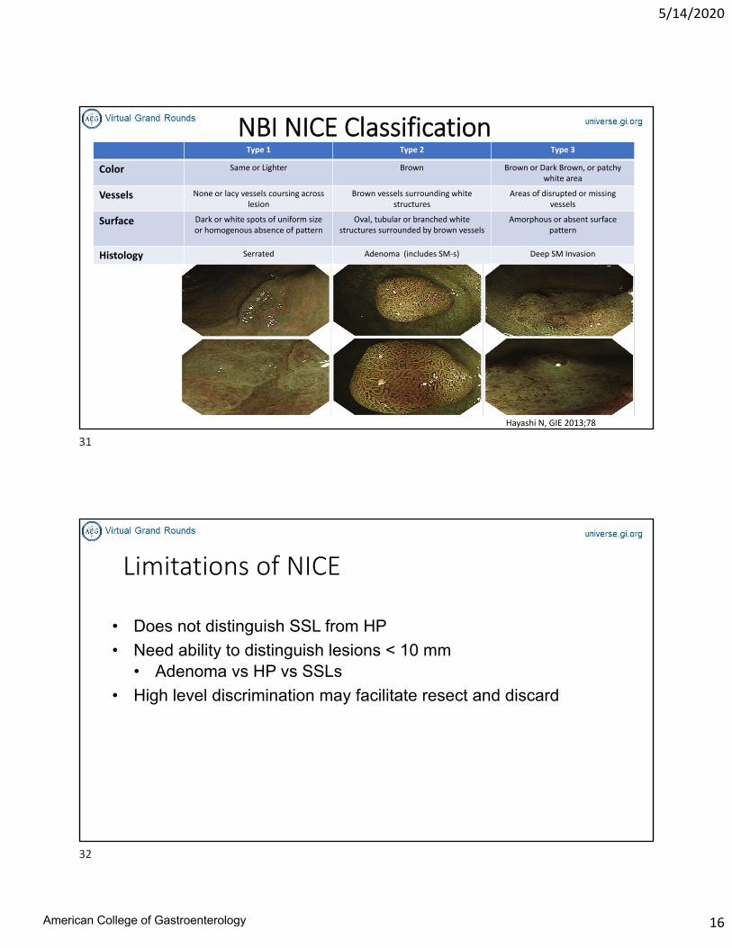
NBI NICE ClassificationType 1 Type 2 Type 3
Color Same or Lighter Brown Brown or Dark Brown, or patchy white area
Vessels None or lacy vessels coursing across lesion
Brown vessels surrounding white structures
Areas of disrupted or missing vessels
Surface Dark or white spots of uniform size or homogenous absence of pattern
Oval, tubular or branched white structures surrounded by brown vessels
Amorphous or absent surface pattern
Histology Serrated Adenoma (includes SM‐s) Deep SM Invasion
Hayashi N, GIE 2013;78
Limitations of NICE
• Does not distinguish SSL from HP
• Need ability to distinguish lesions < 10 mm• Adenoma vs HP vs SSLs
• High level discrimination may facilitate resect and discard
31
32
American College of Gastroenterology
5/14/2020
17
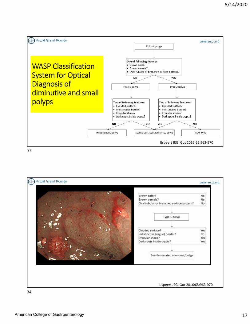
IJspeert JEG. Gut 2016;65:963‐970
WASP Classification System for Optical Diagnosis of diminutive and small polyps
IJspeert JEG. Gut 2016;65:963‐970
33
34
American College of Gastroenterology

5/14/2020
18
Impact of WASP training
IJspeert JEG. Gut 2016;65:963‐970
Effectiveness of Polyp Resection• Prospective 2 center study
• Snare resection blended coagulation
• Polyps 5‐20mm
• Attestation of complete polyp removal
• Biopsies from the resection margin
• 2 from opposing sides of 5–9 mm polyps
• 4 quadrant from 10–20 mm polyps
Pohl H, et al. Care study. Gastroenterology 2013:144;74
Incomplete ResectionAdenoma: 7%
SSL: 31%
35
36
American College of Gastroenterology
5/14/2020
19
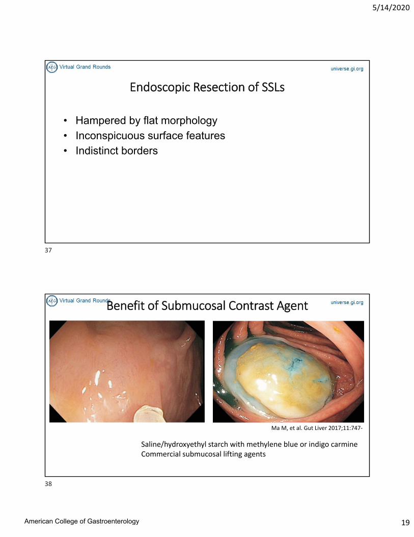
Endoscopic Resection of SSLs
• Hampered by flat morphology• Inconspicuous surface features• Indistinct borders
Benefit of Submucosal Contrast Agent
Ma M, et al. Gut Liver 2017;11:747‐
Saline/hydroxyethyl starch with methylene blue or indigo carmineCommercial submucosal lifting agents
37
38
American College of Gastroenterology
5/14/2020
20
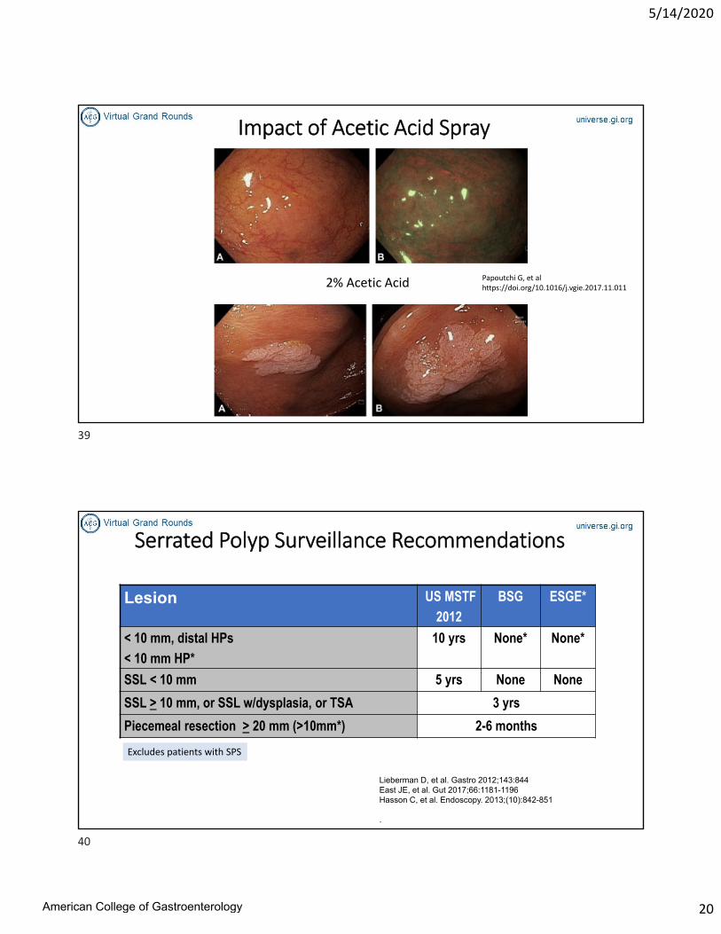
Impact of Acetic Acid Spray
2% Acetic Acid Papoutchi G, et alhttps://doi.org/10.1016/j.vgie.2017.11.011
Serrated Polyp Surveillance Recommendations
Lesion US MSTF
2012
BSG ESGE*
< 10 mm, distal HPs
< 10 mm HP*
10 yrs None* None*
SSL < 10 mm 5 yrs None None
SSL > 10 mm, or SSL w/dysplasia, or TSA 3 yrs
Piecemeal resection > 20 mm (>10mm*) 2-6 months
Lieberman D, et al. Gastro 2012;143:844East JE, et al. Gut 2017;66:1181-1196Hasson C, et al. Endoscopy. 2013;(10):842-851
.
Excludes patients with SPS
39
40
American College of Gastroenterology

5/14/2020
21
USMSTF Recommendations for Surveillance in Individuals with Serrated Polyps
Baseline Colonoscopy FindingInterval for
ColonoscopyStrength of
RecommendationQuality of Evidence
< 20, HPs < 10 mm in rectosigmoid< 20, HPs < 10 mm above sigmoid
10 yrs10 yrs
StrongWeak
ModerateVery Low
1 to 2 SSPs < 10 mm 5 -10 yrs Weak Very low
3 to 4 SSPs < 10 mm, HP > 10 mm* 3 - 5 yrs Weak Very low
5 to 10 SSPs, SSP > 10 mm or w/dysplasia, TSA 3 yrs Weak Very low
Piecemeal resection of SSP > 20 mm 6 mos Strong Moderate
Gupta et al. Gastroenterol 2020;158:1131‐1153
Serrated Polyposis Syndrome
• Under‐recognized
• Predisposition Syndrome
• Basis uncertain• Environmental
• Familial
• Hereditary: RNF43mutation
41
42
American College of Gastroenterology

5/14/2020
22
Do you have patients with SPS?
Serrated Polyposis Syndrome
• WHO Criteria 2010 1. > 5 SPs proximal to sigmoid & >
2 are > 10 mm; or
2. > 1 SP proximal to sigmoid with Family Hx of SPS; or
3. > 20 SPs throughout colon
• WHO Criteria 20191. > 5 SPs proximal to rectum, all
> 5mm, with > 2, > 10 mm or
2. > 20 SPs throughout colon with at least 5 proximal to rectum
Rosty C, Brosens LA, Dekker E, Nagtegaal ID. Serrated Polyposis. In: WHO Classificationof tumours of the digestive system, 5th Edition. Lyon: International Agency for Researchon Cancer, Forthcoming 2019
43
44
American College of Gastroenterology
5/14/2020
23
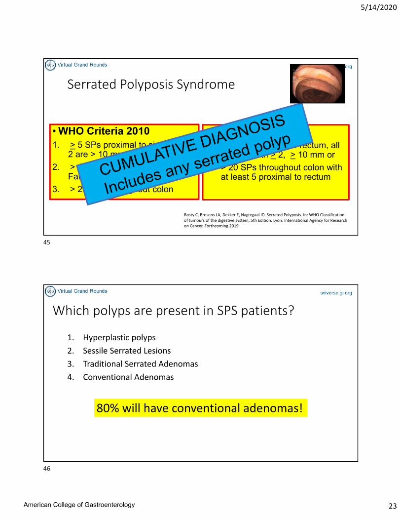
Serrated Polyposis Syndrome
• WHO Criteria 2010 1. > 5 SPs proximal to sigmoid & >
2 are > 10 mm; or
2. > 1 SP proximal to sigmoid with Family Hx of SPS; or
3. > 20 SPs throughout colon
• WHO Criteria 20191. > 5 SPs proximal to rectum, all
> 5mm, with > 2, > 10 mm or
2. > 20 SPs throughout colon with at least 5 proximal to rectum
Rosty C, Brosens LA, Dekker E, Nagtegaal ID. Serrated Polyposis. In: WHO Classificationof tumours of the digestive system, 5th Edition. Lyon: International Agency for Researchon Cancer, Forthcoming 2019
Which polyps are present in SPS patients?
1. Hyperplastic polyps
2. Sessile Serrated Lesions
3. Traditional Serrated Adenomas
4. Conventional Adenomas
80% will have conventional adenomas!
45
46
American College of Gastroenterology

5/14/2020
24
SPS Frequently Undiagnosed
• 529 patients referred for removal polyp > 20 mm
• 4% (20) met the WHO criteria
• 9 at index examination
• 11 during surveillance
• Only 1 (5%) suspected by referring MD
• 50% (10) diagnosed by endoscopist
• Failure to detect attributed to lack of systematic application of WHO criteria
Vemulapalli KC, GIE 2012;75:1206‐1210
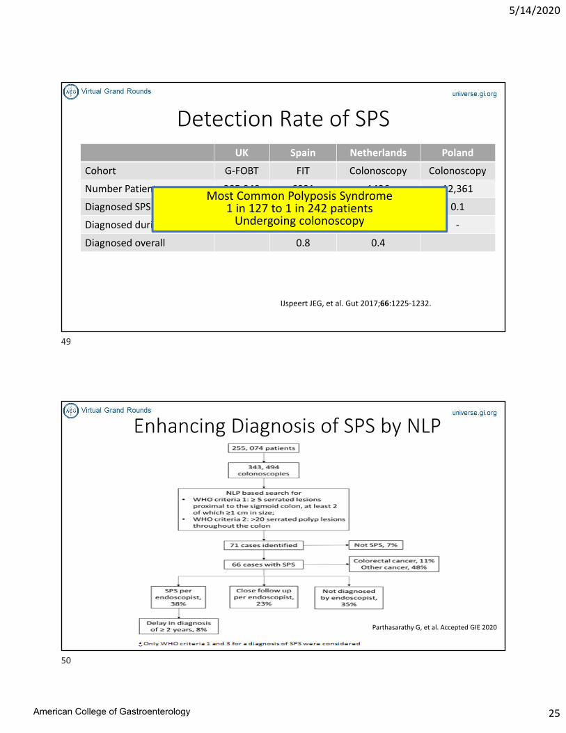
Detection Rate of SPS
IJspeert JEG, et al. Gut 2017;66:1225‐1232.
UK Spain Netherlands Poland
Cohort G‐FOBT FIT Colonoscopy Colonoscopy
Number Patients 205,949 6091 1426 12,361
Diagnosed SPS 1st exam 0.03 0.5 0 0.1
Diagnosed during FU ‐ 0.3 0.4 ‐
Diagnosed overall 0.8 0.4
47
48
American College of Gastroenterology
5/14/2020
25
Detection Rate of SPS
IJspeert JEG, et al. Gut 2017;66:1225‐1232.
UK Spain Netherlands Poland
Cohort G‐FOBT FIT Colonoscopy Colonoscopy
Number Patients 205,949 6091 1426 12,361
Diagnosed SPS 1st exam 0.03 0.5 0 0.1
Diagnosed during FU ‐ 0.3 0.4 ‐
Diagnosed overall 0.8 0.4
Most Common Polyposis Syndrome1 in 127 to 1 in 242 patientsUndergoing colonoscopy
Enhancing Diagnosis of SPS by NLP
Parthasarathy G, et al. Accepted GIE 2020
49
50
American College of Gastroenterology

5/14/2020
26
CRC in SPS• Reported in up to 70% of SPS patients
• Nearly all occurs before or at SPS diagnosis
• Mean age: 50-60 years• 50% in recto‐sigmoid colon
• Risks for CRC• Fulfilling WHO 1 and 3 (OR 1.60, 1.04 ‐ 2.51)
• SP with dysplasia (OR 2.07, 1.28 ‐ 3.33)
• Advanced adenoma (OR 2.30, 1.47 ‐ 3.67)• 80% of SPS patients have coexistent adenomas
IJspeert JEG, et al. Gut 2017;66:278-284
Extra‐colonic Cancer Risk
• None known• No additional surveillance indicated outside colon
• *Except when SPS coexists with other genetic syndrome
BSG: East JE, et al. Gut 2017;66:1181‐1196
51
52
American College of Gastroenterology

5/14/2020
27
CRC Risk in FDR of SPS patients
• Boparai: (RR 5.4, 3.7 to 7.8)• Gut 2010 Sep;59(9):1222‐5
• Win: (SIR 5.16, 3.7-7.3)• Am J Gastroenterol 2012;107(5):770‐778
CRC in SPS vs Multiple Serrated Polyps
Egoavil C, Juarez M, Gastro 2017:153;06-112
SPS Patient
MSPPatient
SPS FDR
MSP FDR
Prevalent CRC 22.6% 28.3% 12.2% 10.4%
Cumulative CRC 2.7% 4.1%
53 SPS pts & 145 pts w/ > 10 polyps with > 50% serratedCompared CRC risk in patients and their FDRs
53
54
American College of Gastroenterology

5/14/2020
28
Lymphoma and SPS
HLN= 101
No HLN=1426
P value
Advanced Neoplasia
25% 12% < .001
Advanced Serrated Polyp
12% 4% < .001
6% 0% < .001
N =102 patients with SPS Hx lymphoma=10; preceded SPS dx by 21 yrs
Lymphoma Prevalence SPS Prevalence
Rigter LS, et al. Cancer 2018;0:1‐10.
Lymphoma and SPS
HLN= 101
No HLN=1426
P value
Advanced Neoplasia
25% 12% < .001
Advanced Serrated Polyp
12% 4% < .001
6% 0% < .001
N =102 patients with SPS Hx lymphoma=10; preceded SPS dx by 21 yrs
Lymphoma Prevalence SPS Prevalence
Rigter LS, et al. Cancer 2018;0:1‐10.
Consider colonoscopy in HL survivorstreated with abdominal radiotherapy and/or
procarbazine
55
56
American College of Gastroenterology

5/14/2020
29
Mankaney G, Rouphael C, Burke CA. Clin Gastroenterol Hepatol. 2020;18(4):777‐779
Surveillance Recommendations in SPS
• ESGE and BSG
• Every 1-2 years
ESGE: Kaminski MF, et al. Endoscopy 2014:46:435‐449MSTF: Lieberman DA, et al. Gastroenterology 2012;143:844‐857BSG: East JE, et al. Gut 2017;66:1181‐1196NCCN: Genetic/Familial High Risk Assessment‐Colorectal. NCCN.org;V3 2018
USMSTFEvery 1 year
NCCNEvery 1-3 years
57
58
American College of Gastroenterology
5/14/2020
30
CRC Incidence After Clearing Colonoscopy
• 3.1% at 3 years, 6.4% at 5 years1
• 152 patients, surveillance interval 1‐3 years
• 1.4% at median follow-up of 3.2 years 2
• 434 patients, median surveillance interval 1.2 years
• 1.3% at 5 years3
• 271 SPS patients, individualized interval 1 vs 2‐years
• 0% at median follow up of 3.1 years4
• 41 patients, median surveillance interval of 1 year
1Rodriguez-Alcade D, et al. Endoscopy 2019;51:1422IJspeert JEG, et al. Gut 2017;66:278-2843 Bleijenberg AGC, et al. Gut 2019;0:1–9. doi:10.1136/gutjnl-2018-3181344Hazewinkel Y, et al. Gastroenterology 2014;147:88-95
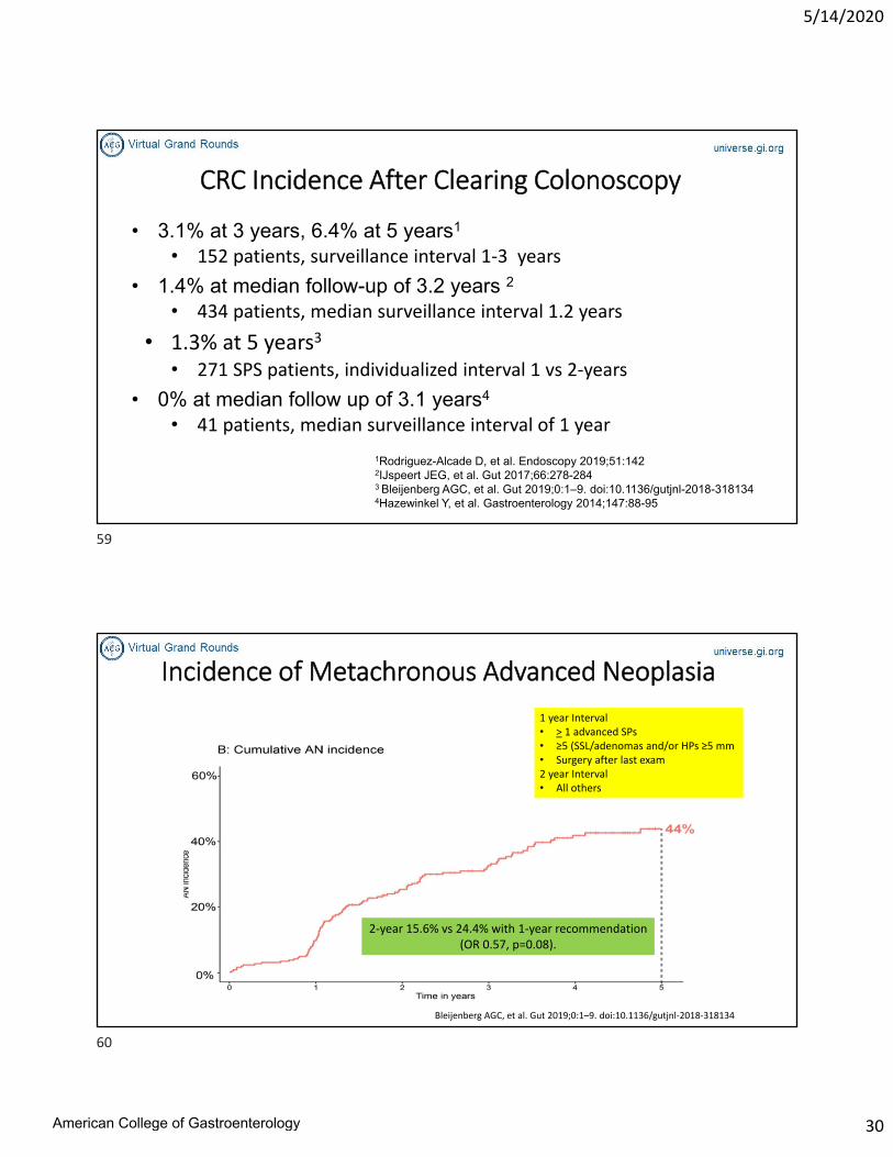
Incidence of Metachronous Advanced Neoplasia
Bleijenberg AGC, et al. Gut 2019;0:1–9. doi:10.1136/gutjnl‐2018‐318134
1 year Interval • > 1 advanced SPs• ≥5 (SSL/adenomas and/or HPs ≥5 mm• Surgery after last exam2 year Interval • All others
2‐year 15.6% vs 24.4% with 1‐year recommendation(OR 0.57, p=0.08).
59
60
American College of Gastroenterology

5/14/2020
31
Colonoscopy Surveillance in FDRs
Begin colonoscopy every 5 years at earliest of:
• Age 40• Same age as youngest diagnosis of SPS• 10 years younger than earliest CRC complicating SPS
NCCN: Genetic/Familial High Risk Assessment‐Colorectal. NCCN.org;V1 2018
Surgery in SPS
•Treatment for CRC
• When endoscopically unmanageable• Remove segments with all lesions not amenable to endoscopic resection
Lieberman D, et al. Gastro 2012;143:844East JE, et al. Gut 2017;66:1181–1196.
NCCN guidelines 2016
61
62
American College of Gastroenterology

5/14/2020
32
Conclusions• Variety of serrated colorectal lesions exist
• SSLs and SPS underdiagnosed and CRC risk factor
• Characteristic endoscopic features of SSPs established
• Patients with SPS and their FDR are high risk group, surveillance warranted
63
64
American College of Gastroenterology
Recommended