Abstract
Characterizing the tolerance of near infrared fluorescent bacterial phytochromes to
random backbone fission and circular permutation
by
Naresh Pandey
Protein fission, fusion, and circular permutation have been used to convert green
fluorescent protein (GFP) family members into biosensors that dynamically report on
cellular processes, ranging from protein expression and metabolite concentrations to
protein solubility, protein-protein interactions, and ligand-binding. Unfortunately, GFP
are unsuitable for deep tissue reporting in animal models because the wavelengths of
light used with these reporters are highly absorbed by tissues. In contrast, near infrared
fluorescent proteins (IFP and iRFP) derived from bacterial phytochrome proteins (BphP)
are excited by light in the near-infrared spectrum (~700 nm, less absorptive) and are
better suited for probing cellular processes within tissues.
To better understand the tolerance of BphP to various mutational lesions (fission,
fusion, and circular permutation), I have subjected IFP to random backbone
fragmentation and iRFP to circular permutation using transposase mutagenesis. Screening
a library of split IFP for fluorescent variants yielded thirteen unique fragmented IFP and
with parent like spectral properties. These two-fragment IFP all required assistance from
associating proteins for maximal fluorescence. In all cases, the split IFP displayed AND
gate logic behavior when the open reading frames (ORFs) encoding the different
fragment were placed under distinct transcriptional regulation. In addition, screening a

library of circularly permuted iRFP led to the discovery of twenty seven variants with
near infrared fluorescence. These variants arose from backbone fission in both PAS and
GAF domains, although the brightest permuted iRFP variants initiated translation at
residues near the domain linker and termini. Biochemical analysis revealed that permuted
iRFP display similar oligomerization, quantum yield, and stability as native iRFP. These
proteins also retained sufficient BV affinity to serve as reporters of gene expression in
mammalian cells without the addition of exogenous BV.
These results demonstrate that knotted BphP retain the ability to fold as their contact
order changes, suggesting that these proteins can be further developed as reporters of
biological processes. The split IFP represent a suite of assays that will be useful for
monitoring the dynamics of protein-protein interactions under conditions where split GFP
do not yield strong signals. In addition, the circularly permuted iRFP should be useful for
building molecular switches through domain insertion.

iv
Acknowledgements
I would like to thank my advisor and mentor, Dr. Jonathan (Joff) Silberg, for giving
me opportunity to work in his lab to pursue my Ph.D. I would also like to thank Silberg
Lab, Matthews Lab members and Shirley Liu for helping me along the way. I thank my
committee members (Dr. Shamoo, Dr. Nikonwicz and Dr. Segatori) for their insight and
guidance through the years. Thanks to my collaborators Dr. Christopher Nobles, Dr.
Anthony Maresso and Dr. Lynn Zechiedrich at Baylor College of Medicine for their help
and letting me work in their labs. Special thanks to Barbara Nassif, Brianna Kuypers,
Emily Thomas and Razan Alnahhas for helping to generate data for this thesis. I also
thank my family for supporting me throughout this journey.

v
TABLE OF CONTENTS
List of tables...................................................................................................................... ix
List of illustrations .............................................................................................................x
List of abbreviations ...................................................................................................... xiii
Chapter 1. Introduction ....................................................................................................1
1.1 Genetically-encoded fluorescent proteins ..................................................................1
1.2 The GFP protein family ..............................................................................................1
1.2.1 Discovery of GFP ..................................................................................................1
1.2.2 Applications of GFP as simple reporters ..............................................................2
1.2.3 Discovery and engineering of additional colors ....................................................3
1.2.4 Introducing biosensing functions into GFP ...........................................................4
1.2.5 Limitations of GFP-type reporters ........................................................................6
1.3 The IFP family ............................................................................................................6
1.3.1 Development of the first IFP ..................................................................................6
1.3.2 Discovery of iRFP ..................................................................................................7
1.3.3 Advantages of IFP and iRFP ..................................................................................9
1.3.4 Bacterial phytochromes have a knotted topology ................................................10
1.4 IFP/iRFP have potential as biosensors ......................................................................13
1.5 Designing proteins using combinatorial experiments ...............................................13
1.5.1 Creating libraries of randomly fragmented proteins ............................................15
1.5.2 Creating libraries of randomly circularly permuted proteins ...............................17
1.6 Summary of my thesis research .................................................................................19

vi
Chapter 2. Materials and Methods ................................................................................21
2.1 Library construction .................................................................................................21
2.1.1 Library of split IFP variants ................................................................................21
2.1.2 Library of circularly permuted iRFP variants .....................................................24
2.2 High-throughput library screening ...........................................................................26
2.3 Whole cell fluorescence measurements ....................................................................27
2.4 Vector construction ..................................................................................................28
2.5 BV-dependence assay ...............................................................................................29
2.6 Western immunoblot ................................................................................................29
2.7 Protein production and purification ..........................................................................30
2.8 Gel filtration chromatography ..................................................................................31
2.9 Native gel electrophoresis ........................................................................................31
2.10 Quantum yield determination .................................................................................32
2.11 Extinction coefficient determination ......................................................................32
2.12 Equilibrium unfolding ............................................................................................32
2.13 Mammalian tissue culture and flow cytometry ......................................................33
Chapter 3. IFP tolerance to random fission and rational fusion .................................34
3.1 Abstract .....................................................................................................................34
3.2 Introduction ..............................................................................................................35
3.3 Results ......................................................................................................................39
3.3.1 Combining IFP fission with fusion to peptides ...................................................39
3.3.2 IFP tolerates fission when fused to peptides .......................................................40
3.3.3 Split IFP fluoresce better at 23°C .......................................................................40

vii
3.3.4 Split IFP have similar BV dependence ...............................................................42
3.3.5 Split IFP function as AND gates .........................................................................46
3.3.6 Fragmented IFP require assistance for fluorescence ...........................................48
3.3.7 Combining CheA/CheY fusion with IFP fission ................................................58
3.4 Discussion .................................................................................................................63
Chapter 4. iRFP tolerance to random circular permutation .......................................68
4.1 Abstract .....................................................................................................................68
4.2 Introduction ..............................................................................................................69
4.3 Results ......................................................................................................................72
4.3.1 Generation of permuted iRFP .............................................................................72
4.3.2 Screening the library identified multiple variants ...............................................72
4.3.3 Permuted variants display low contact order ......................................................74
4.3.4 A majority of permuted iRFP fluoresce better at 37°C .......................................77
4.3.5 Permuted iRFP have similar BV dependence as iRFP ........................................79
4.3.6 Relationship between translation initiation and fluorescence .............................79
4.3.7 In vitro analysis of circularly permuted iRFP .....................................................85
4.3.8 Fluorescence in mammalian cells .......................................................................94
4.4 Discussion .................................................................................................................96
Chapter 5. Conclusions and future directions .............................................................101
5.1 Knotted IFP are sensitive to backbone fission .......................................................101
5.2 Split IFP should be useful for studying protein-protein interactions in situ............101
5.3 A generalizable method for discovering split protein that depend on protein-protein
interactions ...................................................................................................................103

viii
5.4 Split proteins will be useful for studying DNA minivector delivery in situ ...........104
5.5 Circularly permuted iRFP will be useful for domain insertion ...............................106
5.6 Conclusions and Outlook .......................................................................................108
References .......................................................................................................................110

ix
List Of Tables
Table 3.1 Spectral properties of fragmented IFP ...............................................................44
Table 4.1 Spectral properties of circularly permuted iRFP ...............................................75
Table 4.2 In vitro properties of circularly permuted iRFP ................................................89

x
List Of Illustrations
Figure 1.1 Reporting capabilities of GFP-type reporters .....................................................5
Figure 1.2 Structural comparison of GFP and IFP ..............................................................8
Figure 1.3 Amino acid sequences alignment of IFP and iRFP ..........................................11
Figure 1.4 IFP contains a knot ...........................................................................................12
Figure 1.5 IFP has limited reporting capabilities ...............................................................14
Figure 1.6 Schematic illustration of IFP and iRFP fission and permutation .....................18
Figure 2.1 Scheme for creating libraries of fragmented IFP .............................................22
Figure 2.2 Synthetic DNA used for the split IFP library construction ..............................23
Figure 2.3 Scheme for creating a library of circularly permuted iRFP .............................25
Figure 3.1 Fragmented IFP created by random fission and rational fusion .......................38
Figure 3.2 Fluorescent fragmented IFP from the EK library .............................................41
Figure 3.3 Fluorescence of fragmented IFP at 23°C .........................................................43
Figure 3.4 Effect of BV concentration on the fluorescence of fragmented IFP fused to
IAAL-E3 and IAAL-K3 peptides ......................................................................................45
Figure 3.5 Two-input transcriptional regulation of fragmented IFP..................................47
Figure 3.6 140-EK displayed similar fluorescence to full-length IFP from either inducible
promoter .............................................................................................................................49
Figure 3.7 IAAL-E3 and IAAL-K3 peptide removal diminishes IFP fragment
complementation ................................................................................................................50
Figure 3.8 Effect of biliverdin concentration on the fluorescence of fragmented IFP
lacking fusion to any peptides or proteins .........................................................................52

xi
Figure 3.9 Effect of changing protein fusion on the levels of IFP fragments within
cells ....................................................................................................................................53
Figure 3.10 Calculated translation initiation rates for C-terminal fragments containing or
lacking fusion to IAAL-K3 ................................................................................................54
Figure 3.11 Removal of IAAL-E3 decreases IFP fragment complementation ..................56
Figure 3.12 CheA and CheY rescue IFP fragment complementation ...............................57
Figure 3.13 Fluorescent fragmented IFP discovered in the AK library .............................60
Figure 3.14 Fragmented IFP discovered in the AY library ...............................................61
Figure 3.15 IFP displayed increased fluorescence at 23°C ...............................................65
Figure 4.1 Quality assessment of the circularly permuted iRFP library ............................73
Figure 4.2 Mapping of the iRFP tolerance to permutation and contact order ...................76
Figure 4.3 Comparison of circularly permuted iRFP variants fluorescence at 23°C and
37°C ...................................................................................................................................78
Figure 4.4 Effect of BV concentration on E. coli fluorescence expressing circularly
permuted iRFP ...................................................................................................................80
Figure 4.5 Trend of BV dependence among circularly permuted iRFP ............................81
Figure 4.6 Thermodynamic analysis of permuted iRFP translation initiation ...................83
Figure 4.7 Addition of an N-terminal affinity tag enhances whole cell fluorescence .......84
Figure 4.8 Immunoblot analysis of permuted protein expression......................................86
Figure 4.9 Protein-normalized fluorescence in E. coli ......................................................87
Figure 4.10 Absorbance of circularly permuted iRFP .......................................................90
Figure 4.11 Effect of permutation on iRFP structure ........................................................91
Figure 4.12 Equilibrium unfolding of circularly permuted iRFP ......................................93

xii
Figure 4.13 Flow cytometry analysis of tissue culture cells without exogenous BV ........95
Figure 5.1 Split IFP for gene delivery using DNA minvectors .......................................105
Figure 5.2 Domain insertion using permuted iRFP .........................................................107

xiii
List Of Abbreviations
APC-Cy7, Allophycocyanin conjugated with cyanine dye
ARA, Arabinose
AY, CheA and CheY proteins
BphP, Bacteriophytochrome
BV, Biliverdin
CO, Contact order
DHFR, Dihydrofolate reductase
E. coli, Escherichia coli
EK, IAAL-E3 and IAAL-K3 peptides
FRET, Fluorescence resonance energy transfer
GAF, cGMP phosphodiesterase/adenyl cyclase/FhlA
GFP, Green fluorescent protein
GndHCl, Guanidium hydrochloride
GST, Glutathione S-transferase
HA, Hemagglutinin
IFP, Near-infrared fluorescent protein (from Deinococcus radiodurans)
IPTG, Isopropyl β-D-1-thiogalactopyranoside
iRFP, Near-infrared fluorescent protein (from Rhodopseudomonas palustris)
KanR, Kanamycin nucleotidyl transferase
kb, Kilo base
kDa, Kilo dalton

xiv
LB, Luria broth
MuA, Bacteriophage Mu transposase
ORF, Open reading frame
Ori, Origin of replication
PAS, Per/Arndt/Sim
PBS, Phosphate-buffered saline
PERMUTE, PERmutation Using Transposase Engineering
R1R2, Transposase recognition sequence
R2R1, Transposase recognition sequence
RBS, Ribosomal binding site
SDS-PAGE, Sodium dodecyl sulfate-polyacrylamide gel electrophoresis
λem, Emission wavelength
λex, Excitation wavelength

1
Chapter 1
Introduction
1.1 Genetically-encoded fluorescent proteins
Fluorescent proteins, which emit light after absorbing electromagnetic radiation of
lower wavelength, are an important part of life sciences because their production can be
coupled to variety of biological processes like protein expression, protein solubility,
protein dynamics and localization (Cabantous et al., 2005; Lippincott-Schwartz et al.,
2001; Felmeier et al., 2000). Their discovery resulted in molecular tools that enabled
scientists to continuously monitor biological processes like cellular pH, metal
homeostasis, metabolite concentrations, protein solubility, and protein-protein
interactions (Kneen et al., 1998; Chapleau et al., 2008; Sakaguchi et al., 2009; Cabantous
et al., 2005; Hoff et al., 2009). These proteins were transformative for biology because
they emit light when excited by specific wavelength providing a powerful imaging tool
for spatial and temporal information on biological processes (Sun et al., 1999).
Fluorescent proteins either form their chromophore autocatalytically or bind to a ligand
that exhibits fluorescence (Hastings et al., 1996). These chromophores must reside inside
the protective protein scaffold to fluoresce.
1.2 The GFP protein family
1.2.1 Discovery of GFP
In 2008, the Nobel Prize committee awarded Prize in Chemistry "for the discovery
and development of the green fluorescent protein, GFP". The first fluorescent protein to

2
be discovered was Aequorea victoria GFP, which exhibits green fluorescence upon
excitation with blue light (Shimomura et al., 1962). GFP is composed of 238 residues and
forms its own chromophore through an oxidative reaction (Heim et al., 1994; Barondeau
et al., 2003). The chromophore is generated by cyclization of three residues (Ser, Tyr,
and Gly at positions 65-67), which form a p-hydroxybenzylidene-imidazolidinone (Heim
et al., 1994). Shimomura's group was the first to identify the presence of GFP in the
extracts of jellyfish (Aequorea) in 1962 (Shimomura et al., 1962). In 1992, Prasher and
group successfully cloned and sequence the GFP gene (Prasher et al., Gene 1992). In
1994, Chalfie's group demonstrated that the GFP can be used as a marker of gene
expression in E. coli and C. elegans (Chalfie et al., Science 1994), and Roger Tsien's
group demonstrated that oxygen is required for the chromophore maturation and
engineered another variant of GFP, with a blue colored variant paving the way for
multiple genes expression reporters (Heim et al., 1994).
1.2.2 Applications of GFP as simple reporters
GFP and its derivatives with different spectral properties (called GFP herein) have
been widely used as non-invasive reporters of different cellular behaviors (for review, see
Chaudakov et al., 2010). They represent a powerful analytical tool that can be used to
measure the expression of any gene in any organism where the GFP fluorescent signal
can be monitored. Expression detection was initially accomplished by tagging the mec-7
gene (codes for beta-tubulin) in C. elegans neurons (Chalfie et al., 1994). GFP has also
been used to provide information on cellular localization and transport mechanism of
different proteins in cells. For example, maltose binding protein was tagged with GFP to

3
study the export mechanism in periplasmic space of bacteria (Felmeier et al., 2000). In
addition, GFP has been used to identify cells expressing specific proteins at a single cell
level and to establish the variability in expression across a cell population (Longo et al.,
2006). Furthermore, to determine the localization of proteins, GFP has been fused to
either the N- and C-termini of proteins, and the cellular localization has been monitored
using microscopy (Palmer et al., 2004). Fluorescent proteins are also useful for labeling
specific organs and subcellular regions (Palmer et al., 2004). GFP can also be used as a
genetic screen to quantify the activation different promoters across varying cell types
(Ducrest et al., 2002).
1.2.3 Discovery and engineering of additional colors
The original GFP developed by Tsien and coworkers only allowed the study of a
single biological reaction at a time. To overcome this limitation, a palette of GFP
orthologs with different colors were developed that allow for the detection of multiple
cellular events simultaneously (Heim et al., 1996). This was accomplished using directed
evolution, where mutation and screening were used to identify mutations that alter the
spectral properties of GFP without disrupting folding (Heim et al., 1996; Nagai et al.,
2002). Since that time, GFP has been extensively engineered to create dozens of variants
with different colors (and spectral characteristics), including enhanced green fluorescent
protein (λex = 489 nm, λem = 509 nm), blue fluorescent protein (λex = 402 nm, λem = 457
nm), cyan fluorescent protein (λex = 434 nm, λem = 477 nm), and yellow fluorescent
protein or Venus (λex = 513 nm, λem = 527 nm). In addition, gene sequencing in
fluorescent organisms has revealed a palette of GFP orthologs with different colors,

4
including red variants from Corals (Matz et al., 1999; Fradkov et al., 2002; Bevis et al.,
2002). To date, the farthest red-shifted GFP is mKate2 (λex = 588 nm, λem = 633 nm).
1.2.4 Introducing biosensing functions into GFP
Protein reporters like GFP have transformed biological studies, although they are
limited in their applications as biosensors. They require some modifications to function
as reporters of pH, metal, and protein-protein interactions. Protein engineering has been
used to create GFP that report on these additional reactions (Figure 1.1). For example,
calcium signals can be dynamically monitored in cells through microscopy using changes
in a FRET (Fluorescence resonance energy transfer) signal arising from BFP and GFP,
which are covalently linked to calmodulin and the calmodulin binding peptide (Miyawaki
et al., 1997). The FRET signal changes whenever free Ca2+ binds with the fusion protein,
resulting in the localization and monitoring Ca2+ signal in the cell. A pH-sensitive GFP
was developed by creating point mutations (F64L, S65T, Y66H) near the chromophore
(Kneen et al., 1998; Llpois et al., 1998). Fluorescence and absorbance changes of the
mutant GFP can be used as a pH indicator when expressed in the cell organelles, which
can have different pH than cytosol. Metabolites can be sensed using GFP like inositol,
(Sakaguchi et al., 2009) and metal clusters (Hoff et al., 2009). The former required
insertion of a PH domain into the circularly permuted GFP while the latter fused
fragments of GFP to the glutaredoxins. The change in the fluorescence intensity after
expression of these proteins is attributed to metabolite binding. These are powerful tools
for studying dynamic biological reactions in cells, but they are limited to conditions
where GFP spectral probes can be used, which represents only a subset of cells in nature.

5
Figure 1.1 Reporting capabilities of GFP-type reporters. Examples of reporters include:
(A) a pH-sensitive mutant which can report on intracellular pH conditions, (B) a split
GFP that reports on protein-protein interactions when fused to interacting proteins like
glutaredoxins, (C) a metabolite-sensing biosensor made up of a PH domain fused to GFP
which reports on inositol concentrations, and (D) a metal sensing-GFP which is
composed of a calcium-binding calmodulin, calmodulin binding peptide, BFP, and GFP
fusion.

6
1.2.5 Limitations of GFP-type reporters
GFP-type reporters absorb and emit light within the visible spectrum range, which
allows their use in cells that do not significantly absorb light in this range. However, this
characteristic hinders our ability to use GFP for deep tissue imaging in animals (e.g., gut
or liver) and other complex conditions where the extinction of visible light is high (e.g.,
soils and sediments). This occurs because these more complex settings display a strong
absorption of visible light and exhibit autofluorescence (Jobsis et al., 1977; Shu et al.,
2009). To overcome this challenge, a new class of fluorescent proteins called infrared
fluorescent proteins (IFP or iRFP) was developed that absorb and emit light in near
infrared region (Shu et al., 2009; Filonov et al., 2011). These proteins are better suited for
animal tissues studies because tissues display lower autofluorescence at near infrared
wavelengths compared with visible wavelengths and absorb near infrared wavelengths of
light to a lesser extent than visible light (Weissleder et al., 2003).
1.3 The IFP family
1.3.1 Development of the first IFP
Phytochromes are red/far-red light photoreceptors that direct photosensory responses
across the bacterial, fungal and plant kingdoms (for review, see Piatkevich et al., 2013).
Early studies revealed that bacterial phytochromes display weak absorbance and
fluorescence in the near infrared region (Lamparter et al., 2002; Giraud et al., 2005;
Wagner et al., 2008). In 2005, a group from Wisconsin solved the structure of the
chromophore-binding region of a bacterial phytochrome BphP from Dienococcous
radiodurans (Wagner et al., 2005). This structure included the portion of the
7
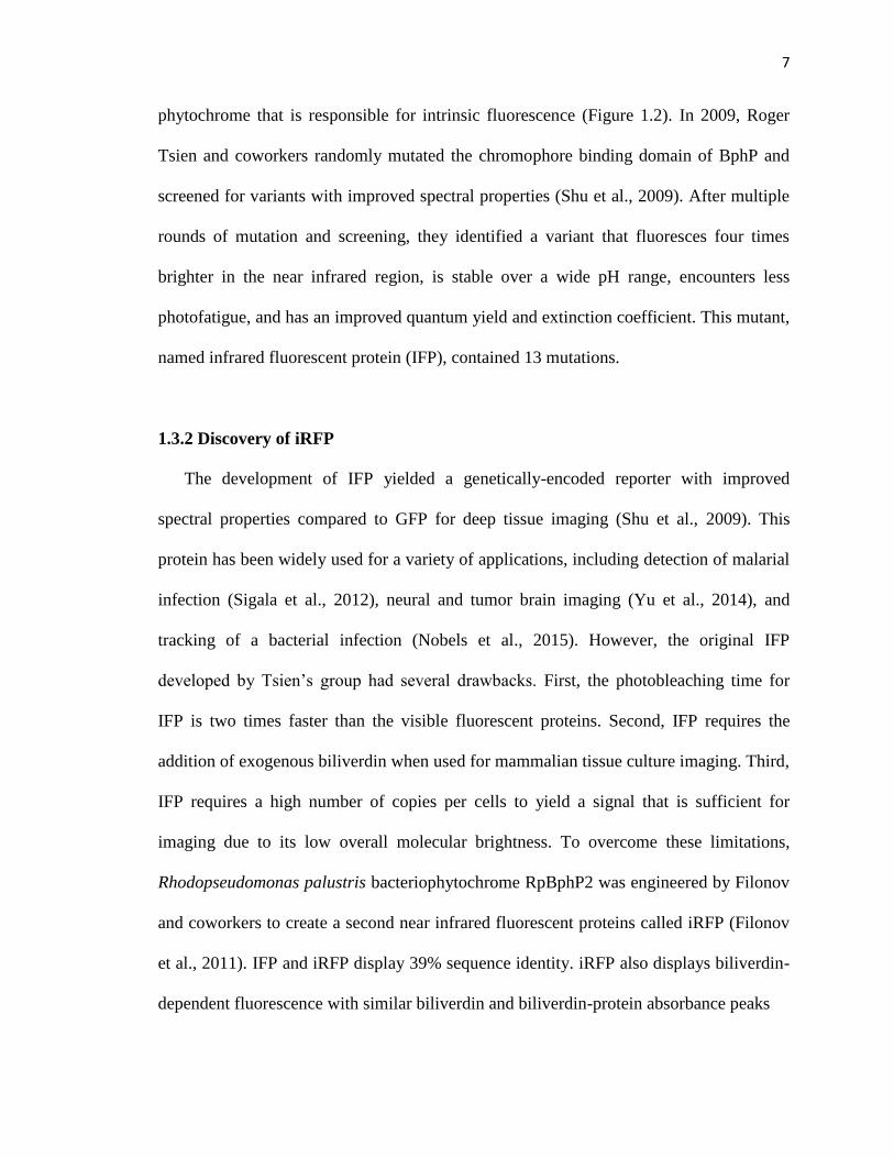
phytochrome that is responsible for intrinsic fluorescence (Figure 1.2). In 2009, Roger
Tsien and coworkers randomly mutated the chromophore binding domain of BphP and
screened for variants with improved spectral properties (Shu et al., 2009). After multiple
rounds of mutation and screening, they identified a variant that fluoresces four times
brighter in the near infrared region, is stable over a wide pH range, encounters less
photofatigue, and has an improved quantum yield and extinction coefficient. This mutant,
named infrared fluorescent protein (IFP), contained 13 mutations.
1.3.2 Discovery of iRFP
The development of IFP yielded a genetically-encoded reporter with improved
spectral properties compared to GFP for deep tissue imaging (Shu et al., 2009). This
protein has been widely used for a variety of applications, including detection of malarial
infection (Sigala et al., 2012), neural and tumor brain imaging (Yu et al., 2014), and
tracking of a bacterial infection (Nobels et al., 2015). However, the original IFP
developed by Tsien’s group had several drawbacks. First, the photobleaching time for
IFP is two times faster than the visible fluorescent proteins. Second, IFP requires the
addition of exogenous biliverdin when used for mammalian tissue culture imaging. Third,
IFP requires a high number of copies per cells to yield a signal that is sufficient for
imaging due to its low overall molecular brightness. To overcome these limitations,
Rhodopseudomonas palustris bacteriophytochrome RpBphP2 was engineered by Filonov
and coworkers to create a second near infrared fluorescent proteins called iRFP (Filonov
et al., 2011). IFP and iRFP display 39% sequence identity. iRFP also displays biliverdin-
dependent fluorescence with similar biliverdin and biliverdin-protein absorbance peaks

8
Figure 1.2 Structural comparison of GFP and IFP. (A) GFP is a barrel shaped protein
comprised mostly of beta sheets. The GFP chromophore is formed through an
autocatalytic process involving residues 65-67 in the middle of the barrel. (B) IFP/iRFP is
a globular shaped protein with mixed alpha helices and beta sheets. They are comprised
of two domains, including PAS and GAF domains. Biliverdin (green) is the
chromophore, and it makes a thioether bond with a conserved cysteine residue at N-
terminal. Images were created using PDB files 1GFL and 1ZTU.

9
near 391 nm and 700 nm, respectively. iRFP also displays a higher affinity for biliverdin,
such that it is able to fluoresce in mammalian cells without addition of exogenous
biliverdin. In addition, iRFP is a ten times brighter and more photostable than IFP. Since
its development, iRFP has been used for a wide variety of applications, including deep
tissue photoacoustic tomography (Filonov et al., 2012), localization of tumor margins in
cancer models (Zhu et al., 2013), and as a marker for tumor growth in mice (Hock et al.,
2014; Scherbakova et al., 2013).
1.3.3 Advantages of IFP and iRFP
Near-infrared light provides a window of opportunity for imaging in deep tissues
because endogenous biological molecules do not absorb strongly in near infrared
spectrum. Long wavelengths of light (650-950 nm) provide high signal to noise ratio in
deep tissues imaging due to reduced tissue autofluorescence and absorption (Weissleder
et al., 2003). Unfortunately, GFP type fluorescent reporters emit light within the visible
spectrum, which hinders our ability to use them for deep tissue imaging in animals (e.g.,
gut), due to the absorption of visible light (Jobsis et al., 1977). Fluorescent dyes were
developed for to use in deep tissue but require extensive chemical preparation and lack
efficient control of the fluorescence expression (Resch-Genger et al., 2008). To solve
those problems, genetically encoded near-infrared fluorescent proteins (IFP and iRFP)
were developed from bacterial phytochromes. These proteins allow for non-invasive
monitoring of gene expression or disease progression in intact animals for longer periods
of time. Despite their advantages in deep tissue imaging, both iRFP and IFP have low

10
quantum yields (6%) compared to GFP (60%). These proteins also dimerize when
expressed in high concentrations, which might impede their usage as a reporter.
1.3.4 Bacterial phytochromes have a knotted topology
The structure of BphP, which was mutated to create IFP, was solved using
crystallography in 2005 (Wagner et al., 2005). This structure revealed two distinct
domains, including PAS and GAF domains. The biliverdin chromophore is covalently
linked to the 24th residue (cysteine) of the IFP PAS domain through a thioether linkage
(Figure 1.3). Within the structure, this chromophore is covalently linked to the PAS
domain but buried within the GAF domain. BphP also contains a trefoil knot. This knot
(Figure 1.4) arises because the first 33 residues must pass through a C-terminal loop
(residues 225-257) to generate the native structure (Wagner, et al., 2005). Knotted
proteins are thought to have lengthy folding times due to topological barrier of a knot
during translation and folding (King et al., 2010). They have two major steps in folding.
First, they must form a twisted native loop. Second, they must thread one of the termini
through the loop (Sulkowska et al., 2012). In case of IFP, the loop region is located in the
C-terminal domain so threading can only happens after the protein is synthesized by the
ribosome, which could limit the rate at which this protein can mature. Knotted proteins
have been proposed to have advantages. There is evidence that knots can provide extra
mechanical stability to proteins (Sulkowska et al., 2008). In vitro experiment performed
with a knotted protein showed spontaneous formation of the knotted topology without the
any aid of molecular chaperones. However the rate of the knot formation was reduced in
the absence of molecular chaperones (Mallam et al., 2011). For IFP, no previous
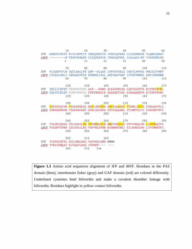
11
Figure 1.3 Amino acid sequences alignment of IFP and iRFP. Residues in the PAS
domain (blue), interdomain linker (gray) and GAF domain (red) are colored differently.
Underlined cysteines bind biliverdin and make a covalent thioether linkage with
biliverdin. Residues highlight in yellow contact biliverdin.

12
Figure 1.4 IFP contains a knot. (A) The plot shows knot features obtained from knotprot
(http://knotprot.cent.uw.edu.pl/). IFP contains a trefoil knot that forms when the two tails
interlace. The N-terminal tail is comprised of residues 1-33, the knot core made up of
residues 34-273 and the C-terminal tail is comprised of residues 274-321. (B) IFP
topology diagram showing a knot formation when first 33 residues (NH2 tail) of PAS
domain passed through C-terminal residues 225 to 257 (loop) of GAF domain.
13
experiments have been performed to probe the importance of the knot. Also, it is unclear
previous experiments have been performed to probe the importance of the knot. Also, it is
unclear how knot will tolerate to backbone fission and circular permutation. This thesis
provides the first insight into knotted protein tolerance to random fission and circular
permutation.
1.4 IFP/iRFP have potential as biosensors
GFP has been extensively engineered through directed evolution to create biosensors
for biotechnological applications. By combining different mutational strategies (point
mutation, recombination, splitting, fusion, and domain insertion) with screens, GFP have
been identified that are capable of pH sensing, protein complex sensing, metal sensing
and metabolite sensing (see Figure 1.1). In contrast, similar biosensors have not yet been
developed using fluorescent bacterial phytochromes. For this reason, there is a need to
engineer IFP to make new classes of biosensors for systems and synthetic biology that are
compatible with measurements in tissues where GFP biosensors yield poor signals.
However, prior to this thesis research, it was unclear if the knotted protein topology in
BphP would tolerate backbone fission and circular permutation to the same extent as
GFP. Generating split IFP and circularly permuted iRFP will allow researchers to create
biosensors or applications suited for deep tissue (Figure 1.5). Circularly permuted iRFP
can be used for domain insertion for generating allosteric switches and also can be used
as fusion protein for building sensors.

14
Figure 1.5 IFP has limited reporting capabilities. IFP has the potential to be engineered
into biosensors with a range of functions, including: (A) pH sensing, (B) protein complex
sensing, (C) metal sensing, and (D) metabolite sensing.

15
1.5 Designing proteins using combinatorial experiments
Laboratory evolution can be used to study where proteins tolerate different types of
mutational lesions. In laboratory evolution, a library of protein variants is created and
then screened (or selected) to examine the effects of sequence changes on protein
structure and function (for review, see Yuan et al., 2005). Sequence diversity in libraries
is created using a variety of approaches. These include random mutation, homologous
recombination, backbone fission, and circular permutation. Studies with random
backbone fission have shown that some proteins tolerate a large number of backbone
fission sites, i.e., the fragments arising from fission cooperatively self assemble to
function like a full-length protein (Segall-Shapiro et al., 2011). In addition, studies
examining domain insertion have revealed that hybrid proteins can be generated by
inserting one protein into the backbone of a second protein; these hybrid proteins can
retain the function of the parent proteins and/or display novel switching activity (Giraldez
et al., 2005). Studies examining protein tolerance to random circular permutation have
shown that some proteins retain parent-like function even though their contact order has
changed (Mehta et al., 2012). Laboratory evolution can also be used to create protein
variants with properties distinct from those observed in nature, which are useful for
biotechnology applications. Prior to the work described in this thesis, the extent to which
IFP tolerated random backbone fission and iRFP tolerated circular permutation had not
been reported.
1.5.1 Creating libraries of randomly fragmented proteins
Proteins are robust macromolecules, which can be quite tolerant to the mutational that

16
they experience in nature. Surprisingly, proteins that experience backbone fragmentation
frequently reassemble in vivo and function (Shiba et al., 1992; Landro et al., 1993). Using
laboratory evolution, various proteins have been subjected to fission for biosensor
purposes. For example split GFP has been used to study protein-protein interactions
(Barnard et al., 2008), where non-fluorescent GFP fragments were attached to the
proteins of interest. An interaction between the proteins of interest resulted in the
assembly of GFP fragments and a signal as GFP matured into a fluorescent protein. Split
DHFR (Dihydrofolate reductase) has also been used as a protein-fragment complement
assay (Remy et al., 2007). With DHFR, two proteins of interest were fused to DHFR
fragments, and the physical interaction between proteins of interest resulted in a
functional DHFR. Split adenylate kinase can also complement the bacterial growth
(Nguyen et al., 2008). These adenylate kinase fragments complemented a growth defect
of an E. coli strain whose adenylate kinase is non functional at high temperatures. T7
RNA polymerase can also function as a split protein AND gate (Shis et al., 2013). Cells
expressing fragments of T7 RNA polymerase transcribe GFP when it is placed under
control of the T7 promoter. The effects of stability on tolerance to fission has also been
examined and reveled that thermophilic proteins tolerate random backbone fission to a
greater extent than mesophilic proteins. In fact, the Silberg lab showed that more than
40% of all split variants of a thermophilic adenylate kinase retained parent-like function
(Segall-Shapiro et al., 2011), while only 6% of the possible split variants of a mesophilic
adenylate kinase retained detectable activity under similar assay conditions.
Although DNA synthesis has become cheap, it can still be arduous to use
oligonucleotide-directed mutagenesis to make all of the open reading frames that encode

17
the different possible two-fragment versions of a natural protein. Transposon mutagenesis
has appeared as a simple method for building libraries of randomly split proteins (Segall-
Shapiro et al., 2010). In this method, a transposon is randomly inserted into an expression
vector containing the gene of interest. The transposon is then replaced by a synthetic
construct to incorporate the regulatory elements needed to transcribe and translate the two
open reading frames created by gene fission. Using this approach, any protein can be
subjected to backbone fission and screened or selected for the functional variants. In this
thesis, I describe how IFP has been subjected to backbone fission using transposase
mutagenesis. Prior to this thesis, it was not known that if IFP could tolerate backbone
fission within the PAS and GAF domains or within the interdomain linker (Figure 1.6A),
because no previous studies had examined IFP tolerance to random backbone fission.
1.5.2 Creating libraries of randomly circularly permuted proteins
Circular permutation has been extensively used in protein engineering to study
protein folding (Haglund et al., 2008), improve the protein functions (Yu et al., 2010),
alter substrate or ligand binding properties (Maatta et al., 2008), alter oligomerization
(Yang et al., 1993), create allosteric regulation (Ostermeier et al., 2005) and develop
proteins with novel functions by domain insertion (Baird et al., 1999). Despite the
generation of a large number of circularly permuted protein variants, there is still lack of
rules governing this design. Often a rational approach is used to create permuted proteins
by targeting flexible regions or loops in protein structures (for proteins where structural
information is available). Library methods have the advantage over rational design that
they sample all of the possible sites to obtain the variants that are tolerant to circular

18
Figure 1.6 Schematic illustrations of IFP and iRFP fission and permutation. (A) Some
sites in the protein backbone are more tolerant to fission than others. During backbone
fission, IFP is randomly cleaved at different backbone locations to create two fragments.
Each fragment has its own N- and C-termini, and when translated they associate and form
a folded IFP. (B) Some sites in the protein backbone are predicted to be more tolerant to
circular permutation than others. During circular permutation, the original termini are
linked with flexible peptide linker and new peptide termini are created by fission
elsewhere in the backbone.

19
permutation. Methods have been developed to create library of random permuted proteins
(Graf et al., 1996; Hennecke et al., 1999; Guntas et al., 2004; Hida et al., 2010).
However, the libraries generated using those methods contain deletions and duplications.
The Silberg Lab developed a method to create circularly permuted proteins using
transposon-based mutagenesis, which can rapidly generate libraries of permuted proteins
without deletions and large duplications (Mehta et al., 2012). In this thesis, I describe an
improvement to this method, and I describe the first efforts to use this new approach to
study the effects of circular permutation on a knotted protein. The goal of this thesis was
to see where iRFP tolerates backbone fission upon circular permutation (Figure 1.6B).
1.6 Summary of my thesis research
Near infrared fluorescent proteins can be useful for whole body imaging in mammals.
However, the IFP toolbox only contains simple reporters of gene expression, which have
not yet been diversified into a toolkit of biosensors similar to GFP (Tainaka et al., 2010;
Lindenburg et al., 2014). I hypothesized that: (i) fragmented and circularly permuted IFP
can be generated using directed evolution and (ii) these variants can be used for
engineering novel IFP based biosensors. In Chapter 2, I describe the methods used to
perform my thesis research. In Chapter 3, I describe my studies examining IFP tolerance
to backbone fission and my efforts characterizing fragmented IFP dependence upon
protein-protein interactions. For these studies, I generated a library of split IFP using
transposase mutagenesis and screened for functional variants. These split variants were
tested with different protein-protein interactions to determine if there is a correlation
between binding affinity and fluorescence. In Chapter 4, I describe my studies examining
where iRFP tolerates backbone fission arising from circular permutation. Library

20
screening identified over two dozen variants that display near infrared fluorescence.
Purified permuted iRFP showed biochemical properties similar to wild type. In addition,
permuted iRFP displayed near infrared fluorescence in mammalian tissue culture without
the addition of exogenous biliverdin. In Chapter 5, I review my findings and describe
how my experiments have expanded the IFP/iRFP toolbox. In addition, I discuss
applications and future paths others can take that would build upon the discoveries
presented herein.

21
Chapter 2
Materials and Methods
2.1 Library construction
2.1.1 Library of split IFP variants
A library of randomly fragmented ifp genes was constructed as illustrated in Figure
2.1 using pGEX2TK-IFP1.4, a pGEX-2TK-derived vector that expresses IFP1.4 (Shu et
al., 2009) from a tac promoter with glutathione S-transferase (GST) fused to its N-
terminus and a hemagglutinin (HA) tag fused to its C-terminus. A transposon containing
NotI restriction sites proximal to both ends was inserted into pGEX2TK-IFP1.4 by
incubating 310 ng pGEX2TK-IFP1.4, 100 ng M1-KanR transposon (Thermo Scienctific),
and 1 U HyperMu MuA transposase (Epicentre Biotechnologies) in a 20 µL reaction
containing HyperMu buffer for 14 hours at 37°C. Reactions were terminated by adding
HyperMu Stop Solution (1 µL) and incubating each reaction at 70°C for 10 min. Total
DNA was purified, electroporated into Escherichia coli, spread onto multiple Luria Broth
(LB)-agar plates containing 25 µg/mL kanamycin, and grown at 37°C overnight. Total
plasmid DNA was purified from colonies obtained on plates to obtain the Mu-insertion
library. The ensemble of purified vectors was digested using restriction enzymes (BamHI
and EcoRI) that cut at sites flanking the IFP gene. Then, agarose electrophoresis was used
to purify the ifp-transposon hybrids (2.1 kb) away from the other DNA fragments (1 kb
ifp alone, 5 kb vector backbone, and 6.1 and 7.2 kb vector backbones containing one or
two 1.1 kb transposons). The ifp-transposon hybrids were cloned back into pGEX2TK to

22
Figure 2.1 Scheme for creating libraries of fragmented IFP. Library construction
involves four steps, including: (i) random insertion of transposons into the IFP genes
using MuA, (ii) purifying IFP genes with inserted transposons away from IFP genes
lacking inserts, (iii) subcloning the ensemble of IFP-transposon hybrid genes into an
expression vector, and (iv) subcloning synthetic DNA encoding different pairs of
interacting proteins in place of the transposons. The last step can be performed using
synthetic DNA that encode for interacting peptides (IAAL-E3 and IAAL-K3), proteins
(CheA and CheY), or lacking additional ORFs as shown in Figure 2.2.

23
Figure 2.2 Synthetic DNA used for the split IFP library construction. The architecture of
the four DNA used to create vectors that express fragmented IFP. Upon insertion into IFP
genes, all of these DNA terminate expression of the IFP fragment that precedes the
original transposon insertion site and initiate expression of the IFP fragment that follows
the ORF. f1-kanR does not amend associating peptides or proteins to either IFP fragment.
ek-kanR fuses IAAL-E3 to the end of the IFP fragments that precede the fragmentation
site and IAAL-K3 to the beginning of the IFP fragments that follow the fragmentation
sites. k-kanR fuses IAAL-K3 to the beginning of the IFP fragments that follow the
fragmentation sites and nothing to the fragment that precedes the fragmentation site. ay-
kanR fuses CheA to the end of the IFP fragments that precede the fragmentation site and
CheY to the beginning of the IFP fragments that follow the fragmentation sites.

24
create a size-selected library (pGEX2TK-IFP-SS). DNA inserts shown in Figure 2.2 (f1-
kanR, ek-kanR, and ay-kanR) were subcloned in place of the transposon within pGEX-IFP-
SS to create three different libraries of vectors that express randomly fragmented IFP,
including: (i) the split IFP library that expresses fragmented IFP without fusion to other
proteins, (ii) the EK library, which expresses the N- and C-terminal IFP fragments as
fusions to the IAAL-E3 and IAAL-K3 peptides, and (iii) the AY library, which expresses
the N- and C-terminal IFP fragments as fusions to Thermotoga maritima CheA (P2
domain; residues 175-264) and CheY. All steps involving bacterial transformations used
MegaX DH10B competent cells (Life Technologies) and yielded lawns of colonies
(>50,000 per transformation) whose numbers were always >10-fold larger than the
number of variants in each library. Each library contained similar sequence diversity
(1,924 variants), which is determined by the number of sites in the IFP gene where a NotI
site is inserted (962) by the transposase MuA and the number of orientations (2) that each
synthetic DNA (f1-kanR, ek-kanR, and ay-kanR) can be integrated into the IFP gene.
2.1.2 Library of circularly permuted iRFP variants
Circularly permuted iRFP genes were constructed with modification of a previously
described method (Mehta et al., 2012) as illustrated in Figure 2.3. NotI restriction sites
flanking iRFP genes were amplified using primers from pBAD/His-B-iRFP (addgene).
NotI flanked iRFP genes were digested with NotI, agarose gel purified and 60 ng of
purified DNA was self ligated for 16 hours at 16°C to obtained a circularized iRFP genes.
100 ng of BglII linearized transposon (containing tac promoter, pBR322 origin of
replication and kanamycin resistance cassette) was inserted into purified circularized

25
Figure 2.3 Scheme for creating a library of circularly permuted iRFP. (A) In this method,
(i) NotI flanked iRFP genes are self ligated after NotI restriction digestion to create a
circular iRFP genes containing twelve residues linker to join original N- and C-termini.
(ii) A synthetic permuteposon P4 (black open circle) is randomly inserted into the
circular iRFP genes by transposase (MuA) mutagenesis (iii) iRFP genes harboring an
integrated permuteposon are excised from vectors containing mutiple permuteposon
insertion using NotI digestion and (iv) these genes are self-ligated to create a library of
vectors that express the different circularly permuted iRFP variants. (B) The target gene
lacks a stop codon and is flanked by identical restriction sites (NotI) with six glycine rich
linkers (GGSGGS), which become fused after self ligation and encode the linker that
connects the original N- and C-termini of iRFP. An additional adenine was inserted
between the initial NotI site and the linker gene to keep the linker in frame upon
permutation. C) The permuteposon P4 contains a stop codon (stop), MuA recognition site
(R1R2), a terminator (term), a pBR322-derived origin of replication (ori), an IPTG
inducible tac promoter (Ptac), a kanR selectable marker, a hybrid MuA recognition site
(R2’R1’) with ribosomal binding site (RBS) and a start codon (start).

26
iRFP genes by incubating with 1 U HyperMu MuA transposase in a 20 µL reaction
containing HyperMu buffer for 16 hours at 37°C and terminated by incubating each
reaction at 70°C for 10 min. Purified DNA electroporated into E. coli and spread onto
LB-agar plates containing 25 µg/mL kanamycin, and grown at 37°C overnight. Plasmid
DNA was purified from colonies obtained on plates, linearized using NotI and run on
agarose electrophoresis to purify the iRFP-transposon hybrids (2.9 kb). The iRFP-
transposon hybrids were self ligated for 16 hours at 16°C to create a closed vector for
expression. DNA was purified, electroporated into E. coli, spread onto LB-agar plates
containing 25 µg/mL kanamycin, and grown at 37°C overnight. All bacterial
transformations involved use of MegaX DH10B competent cells and yielded lawns of
colonies (>50,000 per transformation) which were >20-fold larger than the number of
variants in each library.
2.2 High-throughput library screening
E. coli Rosetta 2 DE3 (Novagen) or BL21 Star DE3 (Life Technologies) or XL1 Blue
transformed with each library were spread onto LB-agar plates containing 100 µg/ml
ampicillin or 50 µg/mL kanamycin and incubated overnight at 37°C. Single colonies
from these plates were arrayed into 96-well deep well plates containing 200 µL LB and
100 µg/mL ampicillin (or 25 µg/mL kanamycin) and grown for 18 hours at 37°C while
shaking at 250 rpm. Stationary phase cultures were diluted 4x by adding LB containing
100 µg/mL ampicillin or (25 µg/mL kanamycin). After growing for 1 hour at 37°C,
isopropyl β-D-1-thiogalactopyranoside (IPTG) and BV (Frontier Scientific) were added
to final concentrations of 0.5 mM and 80 µM, respectively. Deep well plates were

27
incubated at 23°C while shaking at 250 rpm for 18 hours in the dark. A fraction (150 µL)
of each culture was transferred to clear polystyrene 96-well flat bottom plates (Corning)
or 96-well v bottom plates (Rainin), cells were pelleted by centrifuging plates at 3,000 x
g for 5 minutes, supernatant containing excess BV was removed, and whole cell
fluorescence (λex = 684 nm; λem = 695-720 nm or λex = 690 nm; λem = 700-725 nm) was
measured using a Tecan M1000 plate reader. IFP variants with fluorescence (703 to 714
nm) that was >3σ higher than the signal obtained from cells lacking the IFP gene were
sequenced and given names that correspond to the IFP residue at the end of the first
fragment followed by letters that describe the proteins fused to the termini created by
fragmentation, where EK designates IAAL-E3 and IAAL-K3 and AY designates CheA
and CheY. For iRFP, emission data was normalized to absorbance in each well and
variants with fluorescence (705 to 720) that was >5σ higher than the signal obtained from
cells lacking the iRFP gene were sequenced and given names that correspond to the iRFP
residue appeared after start codon.
2.3 Whole cell fluorescence measurements
Vectors encoding fragmented IFP or circularly permuted iRFP were transformed into
E. coli BL21 Star DE3 or XL1 Blue, and individual colonies were used to inoculate LB
cultures containing 100 µg/mL ampicillin or 50 µg/mL kanamycin. After 16 hour at 37°C
and 250 rpm, cells (1 mL) were harvested by centrifugation and used to inoculate a fresh
5 mL LB culture containing 0.5 mM IPTG, 80 µM BV, and 100 µg/mL ampicillin or 50
µg/mL kanamycin. Cells were grown for 5 hours at the indicated temperatures and 250
rpm in the dark, washed with 25% glycerol (1 mL), and resuspended in 25% glycerol (1

28
mL). Whole cell absorbance (600 nm) and fluorescence (λex = 684 nm, λem = 695-800 nm
or λex = 690 nm, λem = 700-800 nm) were acquired from samples arrayed in flat bottom
96-well plates using a Tecan M1000 plate reader. Emission data was normalized to
absorbance in each well, and data reported represent the average of three or more
colonies for each sample with four replicates measured of each within 96 well plates. The
vector that expresses full-length IFP (pGEX2TK-IFP1.4) and the same vector lacking the
IFP gene were used as frames of references for fragmented IFP signals. For iRFP, vector
that expresses full-length pBAD/His-B-iRFP and the transposon vector lacking the iRFP
gene were used as frames of references for permuted iRFP signals.
2.4 Vector construction
To create pairs of vectors for analyzing regulated expression of each fragmented IFP,
N-terminal IFP fragments were cloned into pQE80∆cmR, a plasmid with an IPTG-
inducible T5 promoter, and C-terminal IFP fragments were cloned in place of T7 RNA
polymerase in the plasmid pTara (Wycuff et al., 2000), which has an arabinose-inducible
PBAD promoter. pQE80∆cmR was created by deleting the chloramphenicol resistance gene
from pQE80 (Qiagen). All other expression vectors were generated by subcloning
different NotI-flanked DNA inserts in place of the NotI-flanked inserts used to build
vectors in each library. Expression vector for circularly permuted variants were created
by cloning into pBAD/His-B vector under arabad promoter with His-tag fused to N-
terminus of the circularly permuted variants. For mammalian expressions circularly
permuted variants were created by cloning into pEGFP-C1vector under cytomegalovirus
promoter with HA tag fused to C-terminus of the variants.

29
2.5 BV-dependence assay
The BV dependence of whole cell fluorescence for IFP split variants were measured
using protocol of section 2.3, except the concentration of BV was varied. For iRFP
variants, vectors encoding circularly permuted iRFP were transformed into E. coli (XL1
Blue), and individual colonies were used to inoculate LB cultures containing 100 µg/mL
ampicillin. After 16 hour at 37°C and 250 rpm, cells (2 mL) were harvested by
centrifugation and used to inoculate a fresh 8 mL LB culture containing 1 mM Arabinose,
and 100 µg/mL ampicillin. 1 mL culture added into each well in deep well plates.
Various amounts of BV were added to individual wells and incubated at 37°C while
shaking at 250 rpm for 5 hours in the dark. For each well, a fraction (200 µL) of each
culture was transferred to clear polystyrene 96-well V bottom plates (Rainin), cells were
pelleted by centrifuging plates at 3,000 x g for 5 minutes, supernatant containing excess
BV was removed, and whole cell absorbance (600 nm) and fluorescence (λex = 690 nm;
λem = 700-800 nm) was measured using a Tecan M1000 plate reader.
2.6 Western immunoblot
E. coli expressing the different fragmented IFP (or circularly permuted iRFP) were
grown as described for fluorescent analysis, harvested by centrifugation, and resuspended
to identical optical densities. Sodium dodecyl sulfate polyacrylamide gel electrophoresis
(SDS-PAGE) was carried out under reducing conditions using NuPAGE 12% Bis-Tris
Gels (Life Technologies) and MOPS SDS running buffer and transferred to Protran
nitrocellulose membrane (Whatman) using a TE 22 Mini Tank Transfer Unit (GE
Healthcare). After washing the nitrocellulose paper in TBST buffer (100 mM Tris pH 7.5,

30
150 mM NaCl, 0.1% Tween 20) for 5 minutes and blocking for 1 hour with 10% dry milk
in TBST, the membranes were incubated for 1 hour with either GST rabbit (Millipore) or
hemagglutinin Ab-1 (NeoMarkers) polyclonal antibodies at dilutions of 1:10,000 in
TBST. The nitrocellulose was then incubated for 1 hour in TBST with secondary
antibody, goat anti-rabbit IgG conjugated to peroxidase conjugate (Calbiochem), at a
dilution of 1:10,000. Signals were detected using the ECL western blotting substrate (GE
Healthcare) according to the manufacturer’s protocol. For iRFP His-tag polyclonal
antibody (Qiagen) at dilution of 1:1000 in TBST was used as primary antibody and goat
anti-rabbit IgG conjugated to peroxidase conjugate (Calbiochem) at dilution of 1:1000
was used as secondary antibody.
2.7 Protein production and purification
E. coli (JW2509-2) were cotransformed with hemeoxygenase plasmid (pSR34-Bvd)
and iRFP variants expressed from pBAD, spread onto LB-agar plates containing 100
µg/mL streptomycin and 100 µg/mL ampicillin and incubated overnight at 37°C. Single
colonies were used to inoculate fresh 50 mL LB containing 100 µg/mL l streptomycin
and 100 µg/mL ampicillin for overnight growth. A 1 mL overnight culture was used for
inoculating fresh 50 ml LB containing 100 µg/mL streptomycin and 100 µg/mL
ampicillin. After 3.5 hrs at 37°C and 250 rpm, cells (10 mL) were harvested by
centrifugation and used to inoculate a fresh 1 L LB culture containing 100 µg/mL
streptomycin and 100 µg/mL ampicillin. The culture was grown at 37°C and 250 rpm
until it reached OD600 of 0.5-0.6, and the culture was induced with 1.0 mM arabinose.
After 19 hours cultures were harvested by centrifuging at 4,000 g for 10 minutes at 4°C.

31
Cultures were resuspended in lysis buffer (50mM phosphate buffer pH 7.0, 300 mM
NaCl, 10 mM Imidazole, 1 mM MgCl2, lysozyme, and DNAase) and stored at -80°C for
overnight. The bacterial lysates were centrifuged at 45,000 g for 1 hour at 4°C, and the
proteins were purified from the supernatants by Nickel-nitrilotriacetic acid
chromatography (Qiagen) following the manufacturer’s protocol. Protein was further
purified using HiTrap Phenyl HP (GE Healthcare) on AKTA protein purification system
(GE Healthcare) following the manufacturer's protocol. All variants were dialyzed
against 1X phosphate buffered saline (PBS) pH 7.5 prior to biochemical and biophysical
characterizations.
2.8 Gel filtration chromatography
Purified proteins in 1X PBS pH 7.5 were analyzed using a Superdex 200 column (GE
Healthcare) and eluted with a flow rate of 0.5 ml/min with proteins detected using
absorbance at 280 nm. Standard curve was calculated using molecular weight markers kit
(Sigma Aldrich).
2.9 Native gel electrophoresis
Purified proteins in 1X PBS pH 7.5 were mixed with 2X sample buffer (62.5 mM
Tris-HCl pH 6.8, 25% glycerol, 1% bromophenol blue) loaded on native gel. Samples
were run under non denaturing running buffer (25 mM Tris, 192 mM glycine) with 30
mAmp for 45 minutes. The native gel was composed of a separating gel (10% acrylamide
pH 8.8) and a stacking gel (3.9% acrylamide pH 6.8).

32
2.10 Quantum yield determination
Purified iRFP variants were diluted in 1X PBS pH7.5 until the maximum absorbance
at any wavelength is less than 0.1. Nile blue dye was diluted in 200 proof ethanol and
diluted until the maximum absorbance at any wavelength is less than 0.1. Absorbance
spectrum was obtained using Cary spectrophotometer from 205 to 750 nm. Fluorescence
was measured using Tecan M1000 plate reader by exciting at 659 nm and emission from
669 to 800nm. Fluorescence area was calculated for both Nile blue and iRFP variants.
Quantum yield was calculated using the equation described by Fery-Forgues et al., 1999.
2.11 Extinction coefficient determination
Purified iRFP variants were diluted into 1X PBS pH7.5 until the maximum
absorbance at any wavelength was less than 0.1. As described by Shu et al., 2009, I
compared the absorbance values for the proteins at the main peak 694 nm with the
absorbance value at the 391 nm peak representing free BV whose extinction coefficient is
39,900 M-1cm-1.
2.12 Equilibrium unfolding
A 6 M stock of guanidinium hydrochloride (GndHCl) was mixed with PBS to make
solutions containing a range of GndHCl concentrations (0, 0.5, 1, 1.5, 2, 2.25, 2.5, 2.75,
3, 3.25, 3.5, 4 and 4.5 M). Aliquots of each protein were mixed with 325 μl of each
GndHCl solution, incubated at room temperature for 2 hours in dark, and transferred into
transparent flat-bottom 96-well plate (Corning) where fluorescence (λex = 690 nm and λem
= 700 to 800 nm) was measured. The fluorescence signal at 715 nm was used for

33
comparison. The free energy of folding (∆Gfolding) and concentration of GndHCl that
yields half maximal folding was calculated for each protein by fitting the unfolding data
using a linear extrapolation method (Pace et al., 2000).
2.13 Mammalian tissue culture and flow cytometry
HeLa cells (American Type Culture Collection) were cultured in Dulbecco modified
eagle medium, Lonza, supplemented with 10% fetal bovine serum (Sigma) and 1%
penicillin–streptomycin–glutamine (Hyclone) and maintained at 37 °C and 5% CO2. Cells
were plated in 12-well plates at 8x10⁴ cells per well. 24 hours after seeding, cells were
transiently transfected using JetPrime (Polyplus transfection) according to the
manufacturer’s protocol with 0.5 ug of plasmid expressing the iRFP variant along with
0.05 ug of pcDNA4-GFP plasmid as transfection control per well. The culture media of
transfected cells was replaced with fresh media 16 hours post transfection. Cells
were harvested for analysis at 48 hours post transfection by trypsinization (TrypLE,
GIBCO Invitrogen). Cells were analyzed with a FACSCanto II flow cytometer (BD, San
Jose, CA) to measure fluorescence intensity of iRFP variants in the APC-Cy7
(Allophycocyanin conjugated with cyanine dye) channel (633 nm laser, 780/60 nm
emission filter). At least 10,000 cells were recorded in each sample for analysis. For
transient transfection experiments, the APC-Cy7 signal changes were monitored within
GFP-positive cells (488 nm laser, 530/30 emission filter) to monitor changes within
transfected cells. The reported output signal was calculated by normalizing APC-Cy7
intensity by GFP intensity in order to eliminate differences arising from transfection
efficiencies.
34
Chapter 3
IFP tolerance to random fission and rational fusion
Modified and expanded from manuscript published in ACS Synthetic Biology, 20151
3.1 Abstract
Gene fission can convert monomeric proteins into two-piece catalysts, reporters, and
transcription factors for systems and synthetic biology. However, some proteins can be
challenging to fragment without disrupting function, such as near-infrared fluorescent
protein (IFP). I describe a directed evolution strategy that can overcome this challenge by
randomly fragmenting proteins and concomitantly fusing the protein fragments to pairs of
proteins or peptides that associate. I used this method to create libraries that express
fragmented IFP as fusions to a pair of associating peptides (IAAL-E3 and IAAL-K3) and
proteins (CheA and CheY) and screened for fragmented IFP with detectable near-infrared
fluorescence. Thirteen novel fragmented IFP were identified, all of which arose from
backbone fission proximal to the interdomain linker. Either the IAAL-E3 and IAAL-K3
peptides or CheA and CheY proteins could assist with IFP fragment complementation,
although the IAAL-E3 and IAAL-K3 peptides consistently yielded higher fluorescence.
These results demonstrate how random gene fission can be coupled to rational gene
fusion to create libraries enriched in fragmented proteins with AND gate logic that is
dependent upon a protein-protein interaction, and they suggest that these near-infrared
fluorescent protein fragments will be suitable as reporters for pairs of promoters and
protein-protein interactions within whole animals.
1Pandey, N. et al., 2015. Combining random gene fission with rational fusion to discover near-infrared
fluorescent protein fragments that report on protein-protein interactions. ACS Synth. Biol. 4, 615-624.

35
3.1 Introduction
Component limitations arise in synthetic biology because a restricted number of
biological parts can be reliably used to program cellular behaviors (Grunberg et al., 2010;
Marcheschi et al., 2013). Gene fragmentation represents a simple strategy to overcome
component limitations by converting proteins encoded by individual genes into proteins
encoded by two or more gene fragments (Grunberg et al., 2010; Marcheschi et al., 2013).
Protein fragments arising from fission display “AND” gate logic when the ORFs
encoding the different fragments are placed under distinct transcriptional regulation so
that protein fragment complementation only occurs when the promoter driving
transcription of the first ORF is active AND the promoter controlling transcription of the
second ORF is also active (Shis et al., 2013). Unfortunately, researchers often cannot
predict a priori where genes (and proteins) can be fragmented into pieces to construct
AND gates. In these cases, laboratory evolution can be used to identify which of the
many possible fission sites within a protein are least disruptive to function (Ostermeier et
al., 1999). This combinatorial approach has the advantage that it samples all of the
possible designs for constructing fragmented proteins and provides information on the
backbone fission sites that yield fragments with the strongest functional
complementation. In addition, comprehensive information on protein tolerance to fission
can be used to guide the construction of more complex logic gates such as three-input
AND gates (Segall-Shapiro et al., 2014; Cabantous et al., 2013).
Fragmented proteins often display decreased activity compared with their natural
counterparts encoded by single polypeptides, which can limit their utility for synthetic
biology applications (Burbaum et al., 1991). One way to create fragmented proteins with

36
improved activity is to introduce non-disruptive backbone fission sites into homologs
having enhanced stability, such as a thermophilic homolog of a fragmented mesophilic
protein (Nguyen et al., 2008). Highly stable proteins can also be used as starting points
for laboratory evolution experiments to increase the fraction of fragmented proteins that
retain function within a combinatorial library (Segall-Shapiro et al., 2011), provided that
a highly stable homolog is available as a starting point for laboratory evolution. An
alternative way to enhance the cooperative function of protein fragments is to fuse
protein fragments to a pair of proteins that form a stable complex and promote
complementation (Michnick et al., 2007). Assisted protein-fragment complementation
has been used to create a variety of genetically-encoded devices whose molecular outputs
report on a protein-protein interaction, including devices that generate metabolic (Remy
et al., 1999), visual (Rossi et al., 1997; Luker et al., 2004; Hu et al., 2003), transcriptional
(Fields et al., 1989; Russ et al., 1999; Karimova et al., 1998), proteolytic (Johnsson et al.,
1994; Williams et al., 2009), and antibiotic resistance (Galarneau et al., 2002; Paschon et
al., 2005) outputs. The design of fragmented proteins that report on molecular
interactions can be arduous (Paschon et al., 2004; Paschon et al., 2005; Choe et al., 2005),
and no laboratory evolution method has been described to simplify the discovery of
protein fragments which require assistance for complementation.
The Silberg Lab recently described a transposon mutagenesis approach for
constructing libraries of vectors that express different fragmented variants of a protein
(Segall-Shapiro et al., 2011). Libraries created using this method can be coupled to
screens and selections to convert any protein into a genetically-encoded AND gate,
provided that one or more pairs of fragments derived from that protein cooperatively

37
function. Because previous studies have shown that interacting proteins can assist with
the complementation of protein fragments (Michnick et al., 2007), I sought to explore
whether this library approach could be extended so that it generates each possible two-
fragment variant of a protein as fusions to a pair of proteins that associate to form a
complex (Figure 3.1). I hypothesized that fragmented proteins mined from this type of
library would require associating proteins for maximal activity. I focused my efforts on
the near-infrared fluorescent protein (IFP), a mutant of the Deinococcus radiodurans
bacteriophytochrome BphP which displays spectral properties that are compatible with
whole body imaging in animals (Shu et al., 2009). This protein uses BV as a
chromophore and folds into a structure containing a knot (Wagner et al., 2005; Wagner et
al., 2007). The extent to which IFP can be fragmented to create bimolecular fluorescence
complementation assays has not been explored using laboratory evolution, although
fragmented IFP have been discovered through rational design which require assistance
from a protein-protein interaction for fragment complementation (Filonov et al., 2013;
Tchekanda et al., 2014).
To better understand IFP tolerance to fragmentation and the best architectures for
building bimolecular near-infrared fluorescence complementation devices, I developed a
simple method to construct libraries encoding the different possible fragmented IFP as
fusions to pairs of associating peptides (and proteins) and screened for IFP that display
near-infrared fluorescence. I examined whether the fluorescent fragmented IFP required
assistance from protein-protein interactions for complementation, and I investigated how
the biochemical properties of fragmented IFP differed from full-length IFP.

38
Figure 3.1 Fragmented IFP created by random fission and rational fusion. (A)
Transposon mutagenesis creates clones that use two promoters (Px and Py) to control the
expression of IFP fragments (F1 and F2) fused at their termini to a pair of proteins (P1
and P2) that associate. When both fusion proteins (F1-P1 and P2-F2) are expressed, they
associate to form a folded two-fragment IFP that binds BV and emits in the near infrared.
(B) Truth table for a two-fragment protein AND gate that requires fusion to associating
proteins for an output.

39
3.3 Results
3.3.1 Combining IFP fission with fusion to peptides
To establish where IFP can be fragmented into polypeptides that cooperatively fold
without assistance, we first created a library of vectors that express fragmented IFP using
transposon mutagenesis (Segall-Shapiro et al., 2011) and screened this library for variants
that fluoresce like IFP in the near-infrared upon expression within E. coli. Screening this
library did not identify any two-fragment IFP that retained fluorescence, suggesting that
the IFP structure is easily disrupted by fragmentation. Because previous studies have
shown that interacting proteins can assist with the complementation of some fragmented
proteins (Michnick et al., 2007), I hypothesized that combining random IFP fission with
fusion to associating proteins would enrich the library in variants whose fragments
associate into an IFP-like structure, bind the BV chromophore, and fluoresce in the near-
infrared.
To test the combined effects of random fission and rational fusion on IFP
fluorescence, I constructed a library that fuses a pair of peptides (IAAL-E3 and IAAL-
K3) designed to associate strongly (KD = 70 nM) to the IFP fragment termini (Litowski et
al., 2002). In this EK library, the IAAL-E3 peptide was added to the C-terminus of IFP
fragment that precede the backbone fission site, whereas the IAAL-K3 peptide was added
to the N-terminus of the IFP fragment that follows the fission site. The IAAL-E3 and
IAAL-K3 peptides were chosen for initial library construction because they are small (21
residues each), form a heterodimeric coiled-coil (Litowski et al., 2002), and have been
previously used to assist with fragment complementation (Nguyen et al., 2008).
Structural studies have shown that the N- and C-termini of IAAL-E3 and IAAL-K3 that

40
become fused to IFP fragment termini have a physical separation of ~30 Å (Lindhout et
al., 2004). To minimize IFP structural disruption upon fusion of fragments to termini
having this separation, twelve residue glycine-rich linkers were used for library
construction.
3.3.2 IFP tolerates fission when fused to peptides
Screening 1,760 clones from the EK library identified multiple colonies with λem >
700 nm, and sequencing vectors from these colonies identified seven unique fragmented
IFP having their peptide backbone cleaved distal (>25 residues) from the termini.
Mapping the locations of these backbone cleavage sites onto the Deinococcus
radiodurans BphP chromophore-binding domain (Wagner et al., 2005), the protein that
was mutated to create IFP (Shu et al., 2009), revealed that all of the fission sites are
proximal to the linker that connects the PAS and GAF domains within IFP (Figure 3.2A).
These fission sites occur at the end of the second β-sheet within PAS domain (117- and
118-EK), the end of the third β-sheet within PAS domain (129-EK and 131-EK), and the
beginning of first helix within GAF domain (140-EK, 142-EK, 144-EK).
3.3.3 Split IFP fluoresce better at 23°C
The finding that peptide fusion was required to discover fragmented IFP with near-
infrared fluorescence suggested that these proteins may be marginally stable and display
fluorescence that is sensitive to increasing temperature. To test this idea, I compared the
near-infrared fluorescence of each variant with full-length IFP at 23 and 37°C.

41
Figure 3.2 Fluorescent fragmented IFP from the EK library. (A) For those variants that
displayed near infrared fluorescence, the IFP backbone fission sites (spheres) were
mapped onto the PAS (blue) and GAF (red) domains of BphP, PDB = 1ZTU,25 using
Pymol. Each site represents the last IFP residue in the N-terminal fragment. The domain
linker that is disordered in the BphP structure is shown as a dashed line. (B) Fluorescence
of each fragmented IFP upon expression in E. coli is shown relative to that of cells
expressing full-length IFP and lacking an IFP (-ctrl). Whole cell fluorescence (λex = 684
nm; λem = 710 nm) was measured at 37°C and normalized to cell density. The
fluorescence intensity obtained with cells expressing each split variant was significantly
different from with cells lacking an IFP (two tailed t test; p<0.01). (C) Western blot
detection of N-terminal IFP fragments fused to IAAL-E3 (IFP-F1 +E3), which have a
GST tag and the C-terminal IFP fragments fused to IAAL-K3 (IFP-F2 +K3), which have
a HA tag. Error bars represent ±1σ calculated using three or more replicates.

42
A comparison of E. coli expressing each fragmented IFP revealed similar emission
intensity as full-length IFP when fluorescence was analyzed at 23°C (Figure 3.3). In
contrast, greater variability was observed when measurements were performed at 37°C
(Figure 3.2B), the temperature where IFP biosensors must function to be useful within
animal models. Two fragmented IFP displayed <30% of the IFP emission intensity (117-
EK and 118-EK), one displayed higher fluorescence (140-EK), and all others displayed
intermediate signals.
3.3.4 Split IFP have similar BV dependence
To determine whether the varying fluorescence intensities of the mutants arose
because they had peak emission and excitation wavelengths that differed from IFP (Shu
et al., 2009), I measured the fluorescence spectrum of each variant. All of the fragmented
IFP displayed maximal excitation and emission within 4 nm of intact IFP (Table 3.1). To
investigate whether the variability in whole cell fluorescence arose because some
fragmented IFP required a different concentration of supplemental BV for maximal
fluorescence, I evaluated the dependence of whole cell fluorescence on the amount of BV
added to the cells when expressing each fragmented IFP (Figure 3.4). In all cases, the
concentration of BV required for half-maximal fluorescence in whole cells (2.3 to 15.5
µM) was lower than the concentration (80 µM) used for screening the libraries. The
amounts of BV required for half-maximal fluorescence with fragmented IFP was also
similar to the amount (11.2 µM) required for half-maximal fluorescence with IFP.
Furthermore, the concentrations required for half maximal fluorescence did not correlate
with the relative emission from each fragmented IFP. These findings suggest that the

43
Figure 3.3 Fluorescence of fragmented IFP at 23°C. The fluorescence (λex = 684 nm; λem
= 710 nm) of E. coli expressing fragmented IFP identified in the EK library was
measured at 23°C, normalized to cell density, and reported relative to the fluorescence of
cells expressing full-length IFP and lacking an IFP (-Ctrl). Error bars represent ±1σ. The
fluorescence intensity obtained with cells expressing each split variant was significantly
different from with cells lacking an IFP (two tailed t test; p<0.005).

44
Table 3.1 Spectral properties of fragmented IFP. Emission and excitation spectra were
measured using E. coli expressing each fragmented IFP to determine the wavelengths that
yield maximum emission (λem) and excitation (λex) at 37°C. For each variant discovered
in the EK and AY libraries, spectral analysis was performed with fragmented IFP fused
to IAAL-E3 and IAAL-K3 (+EK), CheA and CheY (+AY), or in the absence of any
fusions (-fusion). A subset of the fragmented IFPs were also analyzed with only one
fragment fused to the IAAL-K3 peptide (+K). ORF1 represents the IFP residues encoded
by the open reading frame that precedes the peptide backbone cleavage site, whereas
ORF2 represents the IFP residues encoded by the open reading frame that follows the
cleavage site. A sharp excitation spectrum was not detected (nd) with several of the
fragmented IFP upon removal of IAAL-E3 or IAAL-E3 and IAAL-K3 due to the weak
signal. Cells expressing full-length IFP yielded maximal emission upon excitation at 683
nm and maximal excitation when emission was monitored at 711 nm.

45
Figure 3.4 Effect of BV concentration on the fluorescence of fragmented IFP fused to
IAAL-E3 and IAAL-K3 peptides. The fluorescence (λex = 684 nm; λem = 710 nm) of E.
coli expressing different fragmented IFP discovered in the EK library was measured in
cells grown in medium supplemented with varying BV amounts (0, 5, 10, 20, 40, 80, and
160 µM). Fluorescence normalized to the maximum value obtained for each variant was
fit to a hyperbolic saturation function to obtain the concentration required for half
maximal signal (KM) within whole cells. Error bars represent ±1σ.

46
variability in emission does not arise because fragmented IFP differ in the fraction of
protein that contains bound BV. To test whether the variation in emission of the IFP
variants arose because the different fragment pairs accumulated to varying steady-state
levels, I used western blot analysis to evaluate IFP fragment stability (Figure 3.2C).
These experiments revealed that the different IFP fragments accumulated to similar
extents when expressed from the same promoters and RBS and did not reveal a
correlation between near-infrared emission and protein fragment expression. Taken
together, these findings suggests that the differences in fluorescence arise because of
structural and/or fragment association differences among the split IFP.
3.3.5 Split IFP function as AND gates
The IFP BV chromophore makes a large number of non-covalent interactions with
residues within the C-terminal GAF domain that reside within the C-terminal fragments
(Figure 1.3). This clustering of BV-interacting residues suggested that fragmented IFP
may only require BV association with one fragment to fluoresce. To test this idea, I
cloned each pair of IFP gene fragments into vectors that use different inducible promoters
(PT5 and PBAD) to control expression (Figure 3.5) and examined whether the maximum
signal required expression of both N- and C-terminal fragments. Whole cell fluorescence
measurements revealed that each of the fragmented IFP displayed the highest
fluorescence when E. coli harboring these vectors were grown in the presence of both
arabinose and IPTG. 140-EK displayed the highest fluorescence as observed with the
constructs discovered in the EK library, and similar whole cell fluorescence as cells

47
Figure 3.5 Two-input transcriptional regulation of fragmented IFP. The near infrared
fluorescence of E. coli (XL1-Blue) transformed with vectors that use IPTG- and
arabinose-inducible promoters to express the N- and C-terminal fragments of IFP,
respectively. The individual and combined effects of 5 mM arabinose (ARA) and 5 mM
IPTG on whole cell fluorescence (λex = 684 nm; λem = 710 nm) was measured at 37°C,
normalized to cell density, and reported relative to the fragmented IFP with the largest
signal. Error bars represent ±1σ.

48
expressing full-length IFP from either inducible promoter (Figure 3.6). In all cases, I
detected a low level of near-infrared fluorescence with cells grown only in the presence
of arabinose. This trend was interpreted as arising from basal expression of the fragments
under PT5 promoter regulation, which has been observed previously with the vectors used
for the measurements (Mahdavi et al., 2013). These results show that all seven of the
fragmented IFP exhibit AND gate genetic logic.
3.3.6 Fragmented IFP require assistance for fluorescence
The paucity of fluorescent proteins discovered when IFP was subjected to fission
alone suggested that the fragmented IFP mined out of the EK library might require
assistance from the IAAL-E3 and IAAL-K3 coils for maximal fluorescence. This idea
was tested by analyzing the fluorescence of cells expressing each fragmented IFP without
fusion to these associating peptides. All of the fragmented IFP lacking peptides displayed
<10% of the whole cell fluorescence observed with full-length IFP (Figure 3.7A) and
lower fluorescence than identical IFP fragments fused to the IAAL-E3 and IAAL-K3
peptides. Among the different fragmented IFPs that fluoresce, peptide removal led to a
loss of fluorescence that varied between 7- and 40-fold (Figure 3.7B).
To investigate why peptide removal decreased fluorescence of the fragmented IFP, I
analyzed the fluorescence spectra, fluorescence dependence on BV, and steady-state
expression of fragments lacking peptide fusions. Fragmented IFP lacking peptides
displayed similar excitation and emission maxima as homologous variants fused to the
IAAL-E3 and IAAL-K3 peptides (Table 3.1). In contrast, higher concentrations of BV
(25.3 to 76.4 µM) were required for half-maximal fluorescence with IFP fragments

49
Figure 3.6 140-EK displayed similar fluorescence to full-length IFP from either
inducible promoter. The near infrared fluorescence of E. coli (XL1-Blue) transformed
with vectors that use IPTG- and arabinose-inducible promoters to express the N- and C-
terminal fragments of 140 EK respectively and compared to vector that use same
promoters to express full-length IFP separately. The individual and combined effects of 5
mM arabinose (ARA) and 5 mM IPTG on whole cell fluorescence (λex = 684 nm; λem =
710 nm) was measured at 37°C, normalized to cell density, and reported relative to the
fragmented IFP with the largest signal. Error bars represent ±1σ. The fluorescence
intensity obtained with cells expressing each split variant or full-length without
inducer(s) were was significantly different from with cells with inducer(s) (two tailed t
test; p<0.005).

50
Figure 3.7 IAAL-E3 and IAAL-K3 peptide removal diminishes IFP fragment
complementation. (A) Whole cell fluorescence (λex = 684 nm; λem = 710 nm) of
fragmented IFP lacking peptide fusions is shown as the fraction of signal observed with
cells expressing full-length IFP. Fluorescence was measured at 37°C and normalized to
cell density. (B) The ratio of fluorescence measured for two-piece IFP peptide fusions
(+EK) to homologous fragmented IFP lacking peptides (–EK). Removal of the EK coils
from each split IFP led to a significant change in the fluorescence intensity of each
variant (two tailed t test; p<0.01 for all except variant 129 which had a p<0.02). (C)
Western blot detection of IFP fragments expressed in E. coli without IAAL-E3 and
IAAL-K3 peptides. N-terminal IFP fragments (IFP-F1) were detected using an antibody
against their GST tag, and C-terminal fragments (IFP-F2) were detected using an
antibody against their HA tag. Error bars represent ±1α.

51
lacking the IAAL-E3 and IAAL-K3 peptides (Figure 3.8). This finding indicates that
these variants only displayed a fraction of their maximal whole cell fluorescence under
the condition where library screening was performed (80 µM BV). Western blot analysis
revealed that the different IFP fragments lacking peptides accumulated to similar extents
(Figure 3.7C) and did not reveal a strong correlation between near-infrared emission and
the relative expression of the different fragmented variants lacking fusion to peptides. A
comparison of a subset of the fragmented IFP containing IAAL-E3 and IAAL-K3 and
homologous fragmented IFP lacking these peptides (Figure 3.9), however, revealed that
removal of the peptides from the fragmented IFP altered steady-state levels of each
homologous polypeptide fragment. Removal of IAAL-E3 and IAAL-K3 increased
accumulation of the N-terminal IFP fragments preceding the peptide backbone cleavage
site and decreased accumulation of the C-terminal fragments following the cleavage site.
I posited that the decreased expression of C-terminal fragments upon IAAL-K3 removal
arose in part because translation initiation of this fragment was decreased by changing the
context of the RBS driving expression. This idea was tested by using a thermodynamic
model to calculate the relative translation initiation rates from the RBS controlling IFP
fragment expression (Salis et al., 2009). All seven of the C-terminal fragments had lower
calculated translation initiation rates upon removal of the IAAL-K3 peptide (Figure
3.10). To test this prediction, I created vectors that expressed all of the IFP variants with
only one fragment (C-terminal) fused to a peptide (IAAL-K3). Thermodynamic
calculations predicted that all of these vectors would initiate translation of the IFP
fragments to the same extent as fragmented IFP fused to both IAAL-E3 and IAAL-K3. I
found that fragmented IFP fused to only one peptide displayed 1 to 7.5% of the whole

52
Figure 3.8 Effect of biliverdin concentration on the fluorescence of fragmented IFP
lacking fusion to any peptides or proteins. The fluorescence (λex = 684 nm; λem = 710 nm)
of E. coli expressing fragmented IFP that lack fusion to IAAL-E3 and IAAL-K3 peptides
was analyzed in medium supplemented with 0, 5, 10, 20, 40, 80, and 160 µM BV.
Fluorescence normalized to the maximum value obtained for each variant was fit to a
hyperbolic saturation function to obtain the concentration required for half maximal
signal (KM) within whole cells. Error bars represent ±1σ.

53
Figure 3.9 Effect of changing protein fusion on the levels of IFP fragments within cells.
Western blot analysis was used to analyze how changing the peptides and proteins fused
to different fragmented IFP affects the steady-state levels of the fragments under
conditions where fluorescence measurements were performed. For four of the fragmented
IFP discovered in the EK library, I compared the relative expression of the N- and C-
terminal fragments (IFP-F1 and IFP-F2, respectively) of each variant with IAAL-E3 and
IAAL-K3 (EK) fused to the different IFP fragments, CheA and CheY (AY) fused to the
different IFP fragments, IAAL-K3 (K) fused to only one of the fragments, and no
peptides or proteins (–) fused to either fragment. ImageJ was used to quantify the levels
of the N- and C-terminal fragments in each blot, which are reported relative to the levels
of the two-piece IFP fused to IAAL-E3 and IAAL-K3. For comparison, the bottom
panels illustrates the effects of peptide and protein fusion on fluorescence (λex = 684 nm;
λem = 710 nm).

54
Figure 3.10 Calculated translation initiation rates for C-terminal fragments containing or
lacking fusion to IAAL-K3. A thermodynamic model for translation initiation was used
to calculate the relative translation initiation rates of the RBS that controlled the
expression of IFP fragments containing (open bars) or lacking (closed bars) IAAL-K3
peptides. Translation initiation rates were normalized to the maximum value obtained
with IAAL-K3 peptides. All calculations were performed using the DNA sequence that
flanked the start codon of the ORFs encoding the IFP fragments, including 50 bases
before and after the predicted start codons.

55
cell fluorescence observed with full-length IFP (Figure 3.11A). This fluorescence
emission was similar to or lower than that observed with homologous IFP lacking both
peptides (Figure 3.11B). I also found that the excitation and emission spectrum of each
variant was comparable to IFP fragments fused to IAAL-E3 and IAAL-K3 (Table 3.1).
Western blot analysis did not reveal a strong correlation between near-infrared emission
and the relative expression of the different N- and C-terminal fragments (Figure 3.11C).
However, I found that the C-terminal fragments fused to the IAAL-K3 peptide
accumulated to greater levels than the same IFP fragments lacking the peptide (Figure
3.10) as predicted from RBS strength analysis.
To test whether other interacting proteins could support the complementation of IFP
fragments identified in the EK library, I created vectors that expressed each of the
fragmented IFP as fusions to T. maritima CheA (P2 domain) and CheY. CheA and CheY
were chosen because they associate to form a heterodimer (Park et al., 2009) and have
been used to assist with fragment complementation of other proteins (Nguyen et al.,
2010). I hypothesized that CheA and CheY would be able to substitute for IAAL-E3 and
IAAL-K3 without disrupting the fragmented IFP fluorescence because the distance
between the termini of CheA and CheY (~30 Å) is similar to the distance between the
IAAL-E3 and IAAL-K3 termini fused to IFP fragments (Lindhout et al., 2004; Park et al.,
2004).
I found that IFP fragments fused to CheA and CheY displayed between 15 and 71%
of the whole cell fluorescence observed with full-length IFP (Figure 3.12A). CheA and
CheY enhanced IFP fragment complementation to varying extents over IFP fragments
lacking fusions to interacting proteins, ranging from 4- to 22-fold (Figure 3.12B). The

56
Figure 3.11 Removal of IAAL-E3 decreases IFP fragment complementation. (A)
Fluorescence of E. coli (λex = 684 nm; λem = 710 nm) expressing fragmented IFP with
their C-terminal fragments fused to IAAL-K3 and their N-terminal fragments lacking a
peptide. Fluorescence is reported as the fraction of the signal observed with full-length
IFP. (B) The ratio of fluorescence measured for fragmented IFP having IAAL-K3 fused
at the terminus of one fragment (+K) to fragmented IFP homologs lacking peptide
fusions (–K). The fluorescence intensity of split variants containing one K coil was not
significantly different from variants lacking both coils, with the exception of variant 144
(two tailed t test; p<0.05). (C) Western blot detection of N-terminal IFP fragments (IFP-
F1), which have a GST tag, and the C-terminal fragments fused to IAAL-K3 (IFP-F2
+K3), which have an HA tag. Error bars represent ±1σ.

57
Figure 3.12 CheA and CheY rescue IFP fragment complementation. (A) Whole cell
fluorescence of E. coli expressing each pair of IFP fragments as fusions to CheA and
CheY. Fluorescence emission (λex = 684 nm; λem = 710 nm) measured at 37°C was
normalized to cell density and is shown relative to the signal from cells expressing full-
length IFP. (B) The ratio of fluorescence measured for fragmented IFP having CheA and
CheY fused at their termini (+AY) to homologous IFP fragments lacking CheA and
CheY (-AY). The fluorescence intensity obtained with each split variant +AY was
significantly different from homologous variants -AY (two tailed t test; p<0.005). (C)
The ratio of fluorescence for fragmented IFP having IAAL-E3 and IAAL-K3 fused at
their termini (+EK) to homologous IFP fragments having CheA and CheY fused at their
termini (+AY). Error bars represent ±1σ. Four variants (118, 140, 142, and 144)
displayed significantly higher fluorescence +EK compared with +AY (two tailed t test;
p<0.05).

58
relative effects of CheA and CheY on fragmented IFP emission were also similar to the
effects of IAAL-E3 and IAAL-K3. IFP fragmented after residues 117 and 144 displayed
the largest fluorescence enhancement upon fusion to the different pairs of associating
proteins, whereas IFP fragmented after residues 129 and 142 consistently exhibited the
smallest enhancement. On average, however, fragmented IFP fused to CheA and CheY
displayed lower whole cell fluorescence than the same IFP fragments fused to IAAL-E3
and IAAL-K3 (Figure 3.12C). These differences were not interpreted as arising from
changes in spectral properties or fragment expression. Fragmented IFP fused to CheA
and CheY displayed spectra with similar excitation and emission maxima as IFP (Table
3.1), and IFP fragments accumulated to a similar level when fused to CheA/CheY and
IAAL-E3/IAAL-K3 (Figure 3.9). Instead, these differences are thought to arise from the
varying strength of the protein-protein interactions used to assist with IFP fragment
complementation. The fragment complementation correlated with the relative affinities of
the CheA/CheY (KD = 200 nM) and IAAL-E3/IAAL-K3 (KD = 70 nM) complexes
(Litowski et al., 2002; Park et al., 2004).
3.3.7 Combining CheA/CheY fusion with IFP fission
I next sought to determine if combining random IFP fission with fusion to CheA and
CheY would yield distinct fragmented IFP from those discovered in the EK library or
fragmented IFP with a higher fluorescence. I hypothesized that screening for fragmented
IFP from a library created using an alternative protein-protein interaction might identify
different fragmented IFP because of differences in fragment expression, stability, and
association. To test these ideas, I created a library where CheA and CheY were fused to

59
the termini of each randomly fragmented IFP. Like the EK library, screening 1,760
clones from this AY library for variants with near-infrared fluorescence yielded multiple
variants with λem > 700 nm. Sequencing vectors from these cells identified nine IFP that
were fragmented distal from the termini (>25 residues). While three of these variants
(118-AY, 131-AY, and 140-AY) were identified when screening the EK library, six of
the variants had distinct fragmentation sites. Mapping the locations of these sites onto the
BphP structure (Wagner et al., 2005) revealed that they cluster near the boundary of the
PAS and GAF domain (Figure 3.13A). Two of the backbone fission sites are after the
first β-strand of the PAS domain (108-AY and 111-AY), one is within the linker that
connects the domains (135-AY), two are within the first helix of the GAF domain (141-
AY and 143-AY), and one is after the first helix of the GAF domain (147-AY).
Cells expressing the variants mined out of the AY library displayed only a fraction
(16 to 69%) of the fluorescence signal obtained with cells expressing full-length IFP
(Figure 3.14). The IFP variants arising from backbone cleavage proximal to the residues
linking the PAS and GAF domains showed the highest whole cell fluorescence, similar to
that observed with fragmented IFP discovered in the EK library. Analysis of the emission
and excitation spectrum of each variant also revealed maximal excitation and emission
similar to that of IFP (Table 3.1). I posited that the fluorescence of fragmented IFP
discovered in the AY library would depend on fusion to interacting proteins, like the
variants discovered from the EK library. To test this idea, I built constructs that expressed
each of these fragmented IFP without fusion to any proteins, and measured the near-
infrared fluorescence of cells transformed with these vectors. All of these cells expressing

60
Figure 3.13 Fluorescent fragmented IFP discovered in the AK library. (A) The backbone
fragmentation sites (spheres) of fragmented IFP discovered in the AY library were
mapped onto the PAS (blue) and GAF (red) domains of BphP using Pymol. (B) The ratio
of fluorescence measured for fragmented IFP having CheA and CheY fused at their
termini (+AY) to homologous variants lacking CheA and CheY (-AY). The fluorescence
intensity obtained +AY was significantly different from homologous variants -AY (two
tailed t test; p<0.01 for all except variant 143 which had a p<0.02). (C) The ratio of
fluorescence measured for fragmented IFP having IAAL-E3 and IAAL-K3 fused at their
termini (+EK) to variants lacking peptides (-EK). The fluorescence intensity obtained
+EK was significantly different from -AY (two tailed t test; p<0.01). (D) The ratio of
fluorescence measured for fragmented IFP having IAAL-E3 and IAAL-K3 fused at their
termini (+EK) to variants fused to CheA and CheY (+AY). Three variants (111, 141, and
147) displayed significantly higher fluorescence +EK compared with +AY (two tailed t
test; p<0.05). Error bars represent ±1α.
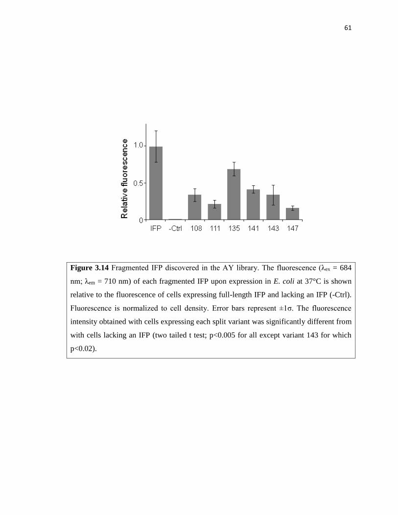
61
Figure 3.14 Fragmented IFP discovered in the AY library. The fluorescence (λex = 684
nm; λem = 710 nm) of each fragmented IFP upon expression in E. coli at 37°C is shown
relative to the fluorescence of cells expressing full-length IFP and lacking an IFP (-Ctrl).
Fluorescence is normalized to cell density. Error bars represent ±1σ. The fluorescence
intensity obtained with cells expressing each split variant was significantly different from
with cells lacking an IFP (two tailed t test; p<0.005 for all except variant 143 for which
p<0.02).

62
fragmented IFP displayed decreased fluorescence upon removal of CheA and CheY
(Figure 3.13B), as observed with variants discovered in the EK library. The fluorescence
of cells expressing IFP fragments fused to CheA and CheY was 4- to 21-fold higher than
cells expressing IFP fragments lacking CheA and CheY fusions. This finding provides
additional evidence that random protein fission can be combined with rational fusion to
discover near-infrared fluorescence protein fragments that report on protein-protein
interactions.
To establish if the variants discovered in the AY library were dependent upon CheA
and CheY for maximal fluorescence, I substituted the IAAL-E3 and IAAL-K3 peptides
for CheA and CheY, respectively, and analyzed IFP fragment complementation.
Substitution of the peptides for CheA and CheY enhanced fragment complementation
over that observed with fragmented IFP lacking fusion to any peptides or proteins (Figure
3.13C). As observed with fragmented IFP discovered in the EK library, fluorescence
complementation was enhanced to the greatest extent when IFP fragments were fused to
the IAAL-E3 and IAAL-K3 peptides (Figure 3.13D). These findings suggest that the
affinity of the protein-protein interaction used for library construction contributes to
protein-fragment complementation, because the higher affinity interaction (IAAL-
E3/IAAL-K3) consistently yielded stronger complementation with the fragmented IFP.

63
3.4 Discussion
The results described in Chapter 3 show how combining random gene fission with
fusion to ORFs encoding associating proteins can aid with the discovery of protein
fragments that cooperatively function (fluorescence) and display two-input AND gate
logic (two-promoter regulation). The EK and AY libraries yielded fragmented IFP that
displayed near-infrared fluorescence, while fluorescent variants were not discovered in
the library encoding IFP that had been subjected to random fission alone. The fragmented
IFP discovered in the EK and AY libraries provide positional information on the
combined effects of fission and fusion on IFPs retention of near-infrared fluorescence.
All of the IFP backbone fission sites discovered using laboratory evolution were distal
from the BV-binding site, suggesting that IFP fission and fusion near this chromophore
binding site is disruptive to fragment complementation. The backbone fragmentation sites
identified in this study (108, 111, 117, 118, 129, 131, 135, 140, 141, 142, 143, 144, and
147) were distinct from those evaluated in previous studies that used rational design to
sample a smaller number of backbone cleavage sites within IFP (Tchekanda et al., 2014)
and iRFP (Filonov et al., 2013), an IFP homolog that displays higher affinity for
biliverdin. These studies each identified backbone cleavage sites within the interdomain
linker that can be fragmented to build split protein assays that report on protein-protein
interactions. The sites clustered proximal to the best site (132) identified in the IFP study
and within the secondary structures (α-helixes, β-sheet, and loops) adjacent to this
domain linker. Whether or not these new split IFP have any advantages or disadvantages
over the rationally designed IFP/iRFP fragment complementation assays will require

64
additional experiments that directly compare their reversibility, self-association, and
signal within cellular models using different pairs of interacting proteins.
The decreased near infrared fluorescence consistently observed when the associating
peptides and proteins were removed from the thirteen fragmented IFP is interpreted as
arising because each of these IFPs require assistance from interacting proteins for
fragment complementation. Evidence for this idea comes from the finding that IAAL-E3
removal diminished fluorescence complementation dramatically (10- to 70-fold) while
only having minor effects on IFP fragment levels. Among the three libraries screened for
protein fragments displaying near-infrared fluorescence, only the IAAL-E3/IAAL-K3
and CheA/CheY libraries yielded fragmented IFP with strong signals. The dependence on
associating peptides (or proteins) for discovery of fragmented IFP can be contrasted with
past fission studies. Using laboratory evolution, larger numbers of non-disruptive
backbone fragmentation sites have been discovered within Thermotoga neapolitana
adenylate kinase (Segall-Shapiro et al., 2011), E. coli methionyl tRNA synthetases
(Mahdavi et al., 2013), and T7 phage RNA polymerase (Segall-Shapiro et al., 2014). I
believe paucity of fragmented IFP that retain strong near-infrared fluorescence without
assistance from interacting proteins could arise because the IFP screen could only reliably
identify fragmented proteins with near-native function, while previous studies used
assays that could detect a fraction of parental protein activity. In addition, IFP
fragmentation could decrease its affinity for BV, which is required for fluorescence, and
the stability of IFP under the screening conditions. Support for the latter idea comes from
the observation that full-length IFP displays almost 4-fold lower whole cell fluorescence
when expressed at 37°C compared with 23°C (Figure 3.15).

65
Figure 3.15 IFP displayed increased fluorescence at 23°C. The fluorescence (λex = 684
nm; λem = 710 nm) of IFP upon expression in E. coli at 23°C and 37°C is shown relative
to the fluorescence of cells lacking an IFP (-Ctrl). Fluorescence is normalized to cell
density. Error bars represent ±1σ. The fluorescence intensity obtained with cells
expressing IFP was significantly different from with cells lacking an IFP (two tailed t
test; p<0.005) and significantly different IFP fluorescence at 23°C vs. 37°C (two tailed t
test; p<0.005).

66
The transposon mutagenesis approach used to randomly fragment IFP and fuse the
fragments to interacting proteins differs from a combinatorial protein engineering method
previously described for this application, which uses incremental truncation for random
fission (Ostermeier et al., 1999) and blunt ligations for rational fusion (Paschon et al.,
2005). In contrast to transposon mutagenesis (Segall-Shapiro et al., 2011), incremental
truncation requires the creation of two vector libraries: one that encodes the different N-
terminal protein fragments and a second that encodes the C-terminal protein fragments
(Paschon et al., 2004). Genes encoding a pair of interacting proteins are then subcloned
into each of these libraries using blunt ligation, and the two vector ensembles are
cotransformed into cells and screened or selected for pairs of complementing fragments. I
expect that transposon mutagenesis will be easier for many synthetic biology labs to
implement, because transposon-mediated gene fission does not require time-dependent
sampling like incremental truncation (Paschon et al., 2004). In addition, gene fragments
created using transposon mutagenesis can be fused to any associating proteins by simply
creating an insert encoding a pair of interacting proteins (Figure 2.2) and cloning that
insert into the NotI restriction site created by transposon insertion (Figure 2.1), rather
than performing multiple low efficiency blunt ligations, which is required with libraries
created using incremental truncation. The sequence diversity created by transposon
mutagenesis [= protein length – 1] is also more constrained than that created by
incremental truncation [= (protein length – 1)·(protein length – 1)]. This occurs because
transposon mutagenesis cleaves a gene into two fragments within the context of a single
vector by inserting a unique NotI restriction site at different locations. In contrast,
incremental truncation generates two vector libraries encoding the different types of

67
proteins fragments, and screens all possible N-terminal fragments with all possible C-
terminal fragments by crossing the two libraries (Paschon et al., 2005). Whether or not
the more limited diversity created by transposon mutagenesis is more or less
advantageous than that created by incremental truncation will require further
experiments. The major advantage of creating limited diversity is that additional targeted
diversity can be included in combinatorial libraries, e.g., within the linker region, and
more thoroughly sampled using screens whose throughput is limited.

68
Chapter 4
iRFP tolerance to random circular permutation
4.1 Abstract
Near infrared fluorescent proteins have been generated by mutating bacterial
phytochromes, which contain a topological knot. To explore how rearrangements in the
sequence of the peptides that interlace into a knot affect iRFP folding, I subjected this
phytochrome to random circular permutation using transposase mutagenesis and screened
for variants that displayed near infrared fluorescence. I identified 27 circularly permuted
iRFP that display biliverdin-dependent fluorescence in E. coli. The variants with the
brightest whole cell fluorescence initiated translation at residues near the BphP domain
linker and knot tails, although fluorescent variants were discovered that initiate
translation within the PAS and GAF domains. Permuted iRFP retained sufficient
biliverdin affinity to fluoresce in tissue culture without the addition of biliverdin, and one
variant displayed enhanced protein-normalized fluorescence in bacteria and tissue
culture. This variant displayed a similar quantum yield as iRFP, although it exhibited
enhanced resistance to chemical denaturation suggesting that the increased signal arises
from more efficient knotted protein maturation. These results show how the contact order
of a knotted bacteriophytochrome can be altered without disrupting folding, chromophore
binding, and fluorescence. These permuted iRFP should be useful for engineering near-
infrared biosensors through domain insertion, and they should be useful for guiding the
construction of circularly permuted phytochromes that display light dependent
conformational switching.

69
4.2 Introduction
The first description of a knotted protein was controversial because of the entropic
penalty that is associated with interlacing a polypeptide chain (Liang et al., 1995;
Mansfield et al., 1994). Since that time, over one thousand knotted structures have been
deposited in the protein data bank (Jamroz et al., 2010), suggesting that this structural
feature is present in many macromolecules that support cellular reactions (Yeates et al.,
2007, Sulkowska et al., 2013, Viranu et al., 2006). Unfortunately, our understanding of
the benefits and challenges associated with the design and evolution of different knotted
topologies remains limited. A recent experimental study provided evidence that some
knotted proteins spontaneously fold, albeit with greatly reduced rates compared with the
rates observed in the presence of molecular chaperones (Mallam et al., 2012). In addition,
knots are thought to provide extra mechanical stability to proteins (Sulkowska et al.,
2008). However, this additional free energy is thought to come at the expense of
decreased folding rates, since introduction of a knot into an unknotted protein decreased
the folding rate by over an order of magnitude (King et al., 2010). While this study
demonstrated that knotted proteins can arise from an unknotted topology, our
understanding of knotted protein tolerance to sequence changes that alter protein contact
order (circular permutation and domain insertion) remains limited.
One of the simplest ways to study a protein’s tolerance to changes in contact order is
to create circularly permuted variants in which the original NH2 and COOH termini are
connected through a peptide linker and new termini are created elsewhere through
backbone fission (Yu et al., 2011). Previous studies have shown that systematic circular
permutation can be used to identify essential folding elements in proteins (Iwakura et al.,
70
2000). This approach has also been used to create proteins with improved activity (Yu et
al., 2010; Daugherty et al., 2013; Sheng et al., 2014), altered ligand binding (Dai et al.,
2014; Maatta et al., 2008; Stephen et al., 2012; Gauntas et al., 2012) and enhanced
stability (Whitehead et al., 2009). A method was recently described for building
permuted protein libraries called PERMutation Using Transposase Engineering
(PERMUTE), which simplifies the construction of circularly permuted protein libraries
for use in laboratory evolution experiments (Mehta et al., 2012). Application of
PERMUTE to a thermophilic adenylate kinase revealed multiple structure-function
correlations (Jones et al., 2016). Protein tolerance to circular permutation correlated with
the distance between the active site and the location of the new termini. Additionally, the
functional variants were enriched in structures with NH2 and COOH termini at sites that
display high sequence variability in a multiple sequence alignment and high positional
deviation in superpositions of structures. While these findings suggest that structural
correlations may exist in other proteins, it remains unclear how they will relate to
proteins whose native structures contain knots.
To better understand knotted protein tolerance to changes in contact order, I subjected
a phytochrome to random circular permutation and screened for variants that display
parent-like function. Phytochromes are photoreceptors found in many taxa (bacteria,
fungi, algae and plants) that use light-dependent conformational changes to alter the
activity of signaling domains that affect growth and development (Rockwell et al., 2006;
Burgie et al., 2014). In bacteriophytochrome, the photosensory core is made up of fused
PAS and GAF domains that interlace into a knot, which use the photochemical
isomerization of a covalently bound tetrapyrrole to interconvert between two

71
conformational states upon exposure to red and far-red light (Wagner et al., 2005;
Wagner et al., 2007). I chose the knotted BphP photosensory core as a model system
because mutants of the photosensory core have been described that exhibit strong
fluorescence upon folding, such as near infrared fluorescent protein (iRFP), which can be
readily monitored using measurements of whole cell fluorescence in E. coli (Pandey et
al., 2015). In addition, a better understanding of BphP tolerance to circular permutation is
currently needed to understand the extent to which knotted members of this protein
family can be used to create near-infrared biosensors or light-dependent switches.

72
4.3 Results
4.3.1 Generation of permuted iRFP
I created circularly permuted variants of iRFP (Filonov et al., 2011), a mutant of the
photosensory core from Rhodopseudomonas palustris BphP2 whose native structure
displays near infrared fluorescence upon BV binding. iRFP was chosen for these
experiments because it displays enhanced stability compared with the other
bacteriophytochromes (Filonov et al., 2011), a property that has been implicated as
increasing protein tolerance to different classes of mutational lesions (Bloom et al.,
2005). PERMutation Using Transposase Engineering (PERMUTE) was used to construct
a library encoding circularly permuted variants of iRFP (Mehta et al., 2012). With
PERMUTE, an artificial transposon containing all of the attributes of a vector (called a
permuteposon) is randomly inserted into a second vector harboring the gene of interest,
and a multistep procedure is used to convert the product of this reaction into a library
(Figure 2.3A). To allow for regulated protein expression in PERMUTE libraries, I
synthesized a new transposon, referred to as a permuteposon P4 (Figure 2.3C) and
randomly inserted P4 into a circular iRFP gene to create a library. Restriction digestion
analysis on the final library showed that the library contains an ensemble of vectors
(Figure 4.1).
4.3.2 Screening the library identified multiple variants
My vector library was transformed into E. coli, and individual colonies were screened
at either 23 or 37°C for near infrared fluorescence (λex = 690 nm; λem = 715 nm). Two

73
Figure 4.1 Quality assessment of the circularly permuted iRFP library. When digested
with NotI, the final library yields a single band that corresponds to the molecular weight
of P4 (1911 bp) and the iRFP gene (993 bp). When digested with NotI and NcoI, a smear
is observed, consistent with the library containing an ensemble of permuted iRFP.

74
temperatures were used for screening to increase the likelihood of identifying marginally
stable variants. Screening a total of 1760 colonies identified 38 variants with emission
>5σ over that observed with cells alone. Sequencing vectors from these colonies revealed
27 unique in frame variants that retained near infrared fluorescence characteristic of BV-
bound iRFP (Table 4.1). To visualize how these permuted proteins differ in primary
structure from iRFP, the first iRFP residue that is translated in each permuted variant was
mapped onto the structure of the Deinococcus radiodurans BphP chromophore-binding
domain (Figure 4.2), a bacteriophytochrome that displays 39% identity with iRFP. This
analysis revealed the highest density of variants arising from backbone fission proximal
to the domain linker and near the termini of the protein.
4.3.3 Permuted variants display low contact order
To determine how permutation tolerance relates to iRFP folding, the contact order
(CO) of permuted protein variants was evaluated. Previous studies have shown that
contact order is inversely correlated with protein folding rates (Plaxco et al., 1998),
suggesting that knotted proteins might display a low tolerance to increases in contact
order. This analysis revealed that fluorescent variants have a range of CO values,
including values that are higher and lower than that observed with native iRFP. The
highest density of permuted variants in the primary structure had new termini that
clustered in a region that yielded CO values that are lower than iRFP (Figure 4.2).

75
Table 4.1 Spectral properties of circularly permuted iRFP. Excitation and emission
spectra were measured using E. coli expressing each permuted iRFP to determine the
wavelengths that yield excitation (λex) and maximum emission (λem) at 23°C, 37°C and
pBAD vector. Variant represents the first residue of permuted iRFP that belongs to wild-
type iRFP sequence. σ represents the signal of the permuted iRFP compared to vector
lacking iRFP gene. A sharp excitation spectrum was not detected (nd) with several of the
permuted iRFP due to the weak signal. Cells expressing full-length iRFP yielded
maximal emission upon excitation at 694 nm and maximal excitation when emission was
monitored at 715 nm.

76
Figure 4.2 Mapping of the iRFP tolerance to permutation and contact order. In iRFP, a
knot is formed when the first 33 residues (NH2 tail) of the PAS domain pass through a
loop created by residues from the GAF domain. Relationship between domain structure,
contact order, and the location of the new termini created by permutation. The fission
sites in fluorescent cp-iRFP are mapped (with lines) onto BphP (PBD=1ZTU) to relate
how their location relate to the NH2 tail (cyan), PAS domain (blue), domain linker (grey),
loop from the GAF domain (black) and remainder of the GAF domain (orange). Each line
represents the first residue in the circularly permuted iRFP.

77
4.3.4 A majority of permuted iRFP fluoresce better at 37°C
Distinct sets of circularly permuted iRFP were discovered at the different
temperatures where screening was performed (23 and 37°C), suggesting that some of the
permuted variants may display fluorescence that varies with temperature. To test this
hypothesis, I compared the emission and excitation spectra of each permuted variant with
those of native iRFP. Initial spectroscopic measurements at 23°C revealed that my
permuted iRFP display similar spectral shapes but a range of peak intensities (Table 4.1).
In addition, five variants yielded whole cell emission that was greater than that observed
with iRFP, three variants yielded intensities that were comparable to iRFP, and the
remaining nineteen variants showed lower emission that was statistically significant
(Figure 4.3A). When measurements were performed at 37°C, most variants displayed a
lower signal, with only three variants yielded a signal that matched or exceeded the
intensity as iRFP. To determine which permuted proteins, similar to iRFP, display an
enhanced emission signal as temperature is raised from 23 to 37°C, I calculated the ratio
of fluorescence emission for each variant expressed in cells grown at these different
temperatures (Figure 4.3B). This analysis revealed that 52% of the circularly permuted
iRFP display higher emission at 37°C compared with 23°C like iRFP, 37% have similar
fluorescence ratios as iRFP, and 11% display decreased emission as temperature is
increased. Among the variants that displayed a decreased signal at 37°C (i.e.,
fluorescence emission ratios ≤1), 80% were discovered in my screen performed at that
23°C.

78
Figure 4.3 Comparison of circularly permuted iRFP variants fluorescence at 23°C and
37°C. (A) The fluorescence of cells expressing each variant was measured at 23°C and
37°C (λex = 690 nm; λem = 715 nm), normalized to cell density, and compared to the
fluorescence of cells expressing parental iRFP and cells lacking iRFP (-ctrl). All circular
permuted iRFP except variants 59, 182, 198 and 291 had significantly higher signal than
background at 37°C, and all variants except 59, 122, 129, 182 and 198 had signals that
were significantly higher than background 23°C (two-tailed t test; p<0.05). (B) The ratio
of the fluorescence signals acquired at 37°C to the signals observed at 23°C. Blue and red
symbols represent the variants identified in screens at 23°C and 37°C, respectively, while
white symbols represent variant discovered at both temperatures. Error bars represent
±1σ calculated using three or more independent measurements.

79
4.3.5 Permuted iRFP have similar BV dependence as iRFP
The variability observed with circularly permuted protein fluorescence intensity could
arise because permutation alters protein expression, folding, cofactor binding, and/or
stability and degradation. In a previous study, I found that backbone fission increased the
concentration of BV required for fluorescence in a related bacteriophytochrome (Pandey
et al., 2015), suggesting that backbone fission arising from permutation could have a
similar effect on iRFP. To test whether BV affinity is affected by permutation, I
evaluated whether near infrared fluorescence varied with the amount of BV added to the
cells that are expressing each circularly permuted iRFP (Figure 4.4). The BV
concentration (11 µM) required for half-maximal fluorescence in cells expressing iRFP
was ~7-fold lower than the concentration used in my screen for permuted variants (Figure
4.5). The majority of the permuted iRFP (85%) required a similar (or lower) amount of
BV for half-maximal fluorescence, 4 to 15 µM (Figure 4.5). The remaining variants,
which all arose from backbone fission in the GAF domain that binds BV, required
between 18 and 25 µM BV for half-maximal fluorescence. These findings suggest that
the large variability in whole cell fluorescence does not arise because circularly permuted
iRFP differ in their BV-binding affinity.
4.3.6 Relationship between translation initiation and fluorescence
Previous studies have shown that genetic context can alter the strength of translation
initiation from a single bacterial RBS by more than two orders of magnitude (Salis et al.,
2009; Lou et al., 2012; Mutalik et al., 2013), and vectors in my library express permuted
iRFP with sequences that vary adjacent to the same RBS. To examine whether permuted

80
Figure 4.4 Effect of BV concentration on E. coli fluorescence expressing circularly
permuted iRFP. The fluorescence (λex = 690 nm; λem = 715 nm) of E. coli XL1 Blue
expressing different circularly permuted iRFP was measured in cells grown in medium
supplemented with varying BV concentrations (0, 5, 10, 20, 40, and 80 µM) and 1 mM
arabinose. Fluorescence that had been normalized to the maximum signal obtained with
each variant was fit to a hyperbolic saturation function to obtain the apparent affinity
(KM) within whole cells.

81
Figure 4.5 Trend of BV dependence among circularly permuted iRFP. Half maximal
signal (KM) for each variant was plotted against variants. Black filled square and grey
filled squares represent iRFP and variants respectively. Variants below and above the
dash line represent having improved and decreased BV affinity respectively.

82
gene sequences influence protein expression from the RBS in my permuteposon, I
analyzed how permuted iRFP vary in their translation initiation rates in E. coli using a
thermodynamic model for translation initiation (Salis et al., 2009). These calculations
revealed that the calculated translation initiation rates for all possible circularly permuted
iRFP vary by over three orders of magnitude when expressed using the RBS present in
permuteposon P4 (Figure 4.6A). To test whether the fluorescent signals obtained with my
original clones correlates with calculated translation initiation, I compared the translation
initiation rates for each variant to the whole cell fluorescence signals. This analysis
revealed that variants with the smallest relative translation initiation rates also display the
weakest whole cell fluorescence when measured at 23°C and 37°C (Figure 4.6B). The
dispersion of fluorescence at low calculated rates was also smaller than that at higher
levels (e.g., above calculated relative values of 1000). These findings suggest that the
variability in the production rates of permuted iRFP contributes to the dispersion in
fluorescence signals observed in whole cell measurements. The large dispersion of
signals at higher calculated translation initiation rates suggests that protein folding and
maturation vary across the proteins with similar translation initiation rates.
The close proximity between the RBS and the first codon in each circularly permuted
gene within my permuteposon is expected to underlie the variability in translation
initiation because each permuted gene presents a different genetic context to the same
RBS. To minimize differences in protein synthesis that arise from this genetic context, I
created vectors that express each of my circularly permuted iRFP with the same large N-
terminal affinity tag (37 residues) that maintains the translation initiation site in a

83
Figure 4.6 Thermodynamic analysis of permuted iRFP translation initiation. (A)
Translation rates of all circularly permuted iRFP (line) are compared with the rates for
fluorescent variants (). (B) The relative whole cell fluorescent signals of circularly
permuted iRFP at 23°C and 37°C are compared with calculated rates. Spearman's rank
correlation coefficients (RSR) at 23°C (RSR = 0.403) and 37°C (RSR = 0.384) yielded
significant correlations (two-tailed t-test; P<0.05).

84
Figure 4.7 Addition of an N-terminal affinity tag enhances whole cell fluorescence. (A)
The fluorescence (λex = 690 nm; λem = 715 nm) of E. coli XL1 Blue expressing different
circularly permuted iRFP with His tags was measured at 37°C and normalized to cell
density. The signal from each variant is reported as the fraction of the signal obtained
with the variant displaying the highest fluorescence. (B) The ratio of fluorescence
measured for circularly permuted iRFP with a His tag (+tag) to same variant without a
tag (-tag). The dashed line represents the ratio expected if expression does not vary with
tag addition. Error bars represent ±1σ from three or more experiments.

85
constant genetic context. When expressed from these vectors, permuted variants with
new N- and C-termini proximal to the domain linker displayed the highest whole cell
fluorescence (Figure 4.7). The fluorescence of each variant was also compared in the
presence and absence of the affinity tag. A majority of the variants (85%) displayed an
increased signal in the presence of the N-terminal tag, indicating that this expression
strategy yielded higher steady-state levels of each fluorescent, BV-bound variant. To
better understand the signal variability observed under these expression conditions, I
analyzed the steady-state levels of a subset of these circularly permuted variants (Figure
4.8), focusing on the subset of circularly permuted iRFP that displayed enhanced
fluorescence at 37°C compared with that observed at 23°C. Immunoblot analysis
revealed that the steady-state levels of these circularly permuted varies up to 10-fold from
that observed with iRFP. When whole cell fluorescence was normalized to protein
expression in matched samples (Figure 4.9), a majority of the permuted variants
displayed a signal that was similar to that measured with native iRFP. However, iRFP-
198 displayed a signal that was 25-fold lower than iRFP, consistent with the low signal
obtained in bacterial cells. In addition, iRFP variants 12 and 102 exhibited signals that
were higher than iRFP, 1.94- and 4.38-fold respectively.
4.3.7 In vitro analysis of circularly permuted iRFP
My finding that permuted iRFP display a range of protein-normalized fluorescence
signals in whole cell measurements suggested that permutation affects the fraction of
iRFP that folds into a fluorescence-competent conformation and/or quantum yield. To
directly address the effects of permutation on quantum yield, I purified two circularly
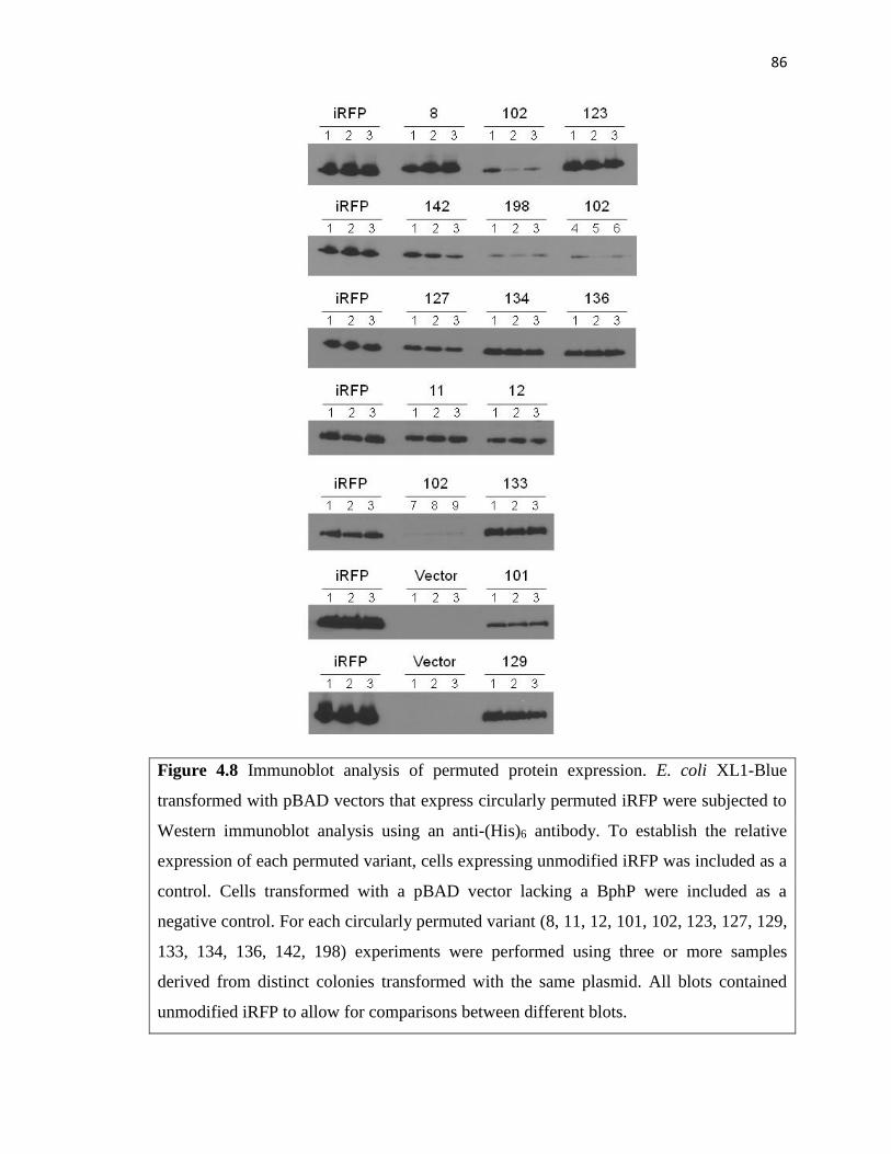
86
Figure 4.8 Immunoblot analysis of permuted protein expression. E. coli XL1-Blue
transformed with pBAD vectors that express circularly permuted iRFP were subjected to
Western immunoblot analysis using an anti-(His)6 antibody. To establish the relative
expression of each permuted variant, cells expressing unmodified iRFP was included as a
control. Cells transformed with a pBAD vector lacking a BphP were included as a
negative control. For each circularly permuted variant (8, 11, 12, 101, 102, 123, 127, 129,
133, 134, 136, 142, 198) experiments were performed using three or more samples
derived from distinct colonies transformed with the same plasmid. All blots contained
unmodified iRFP to allow for comparisons between different blots.

87
Figure 4.9 Protein-normalized fluorescence in E. coli. (A) Fluorescence of each
circularly permuted iRFP upon expression is shown relative to that of cells expressing
wild-type iRFP. Whole cell fluorescence was measured at 37°C and normalized to cell
density. (B) The relative expression of the circularly permuted iRFP variants of panel A.
ImageJ was used to quantify the levels of the variants in each blot, which are reported
relative to the level of the wild-type iRFP. (C) The ratio of fluorescence to the level of
each protein was calculated using data acquired from matched samples. Error bars
represent ±1σ calculated using three or more independent measurements.

88
permuted iRFP that displayed improved signals (variants 12 and 102), two with iRFP-like
signals (variants 129 and 133) and iRFP. Proteins were purified using a combination of
affinity and reverse phase chromatography. As observed with iRFP previously, I found
that the reverse phase column yielded two populations of proteins, one that eluted as a
monodisperse peak at low ammonium sulfate (~5%) and one that eluted when the column
was washed with buffer lacking ammonium sulfate. All of my biochemical
characterization was performed using the recombinant proteins that eluted in the presence
of ammonium sulfate as monodisperse peaks.
I first compared the spectral properties of each permuted protein with iRFP (Figure
4.10). Like iRFP, all four of the permuted variants displayed three absorbance peaks with
maxima at similar wavelengths (280, 391, and 693 nm). In addition, all four permuted
variants displayed maximal excitation and emission wavelengths that mirrored iRFP
(Table 4.2). Furthermore, all four variants displayed quantum yields that were not
significantly different from the value obtained with iRFP (Table 4.2). These findings
suggest that permutation does not significantly alter the chromophore environment.
I next sought to determine how permutation affects protein structure, since backbone
fission sites within the dimerization interface of Deinococcus radiodurans BphP could
disrupt oligomerization. Variants 129 and 133 introduce new termini that map onto the
dimer interface in the BphP crystal structure (Figure 4.11A), while variants 12 and 102
have termini that map to sites distal from the dimer interface. To test whether these
proteins display altered structures, I analyzed their electrophoretic mobility using native
PAGE (Figure 4.11B). This analysis revealed that three variants (102, 129 and 133)
display increased mobility compared with iRFP. To test if this altered mobility arises
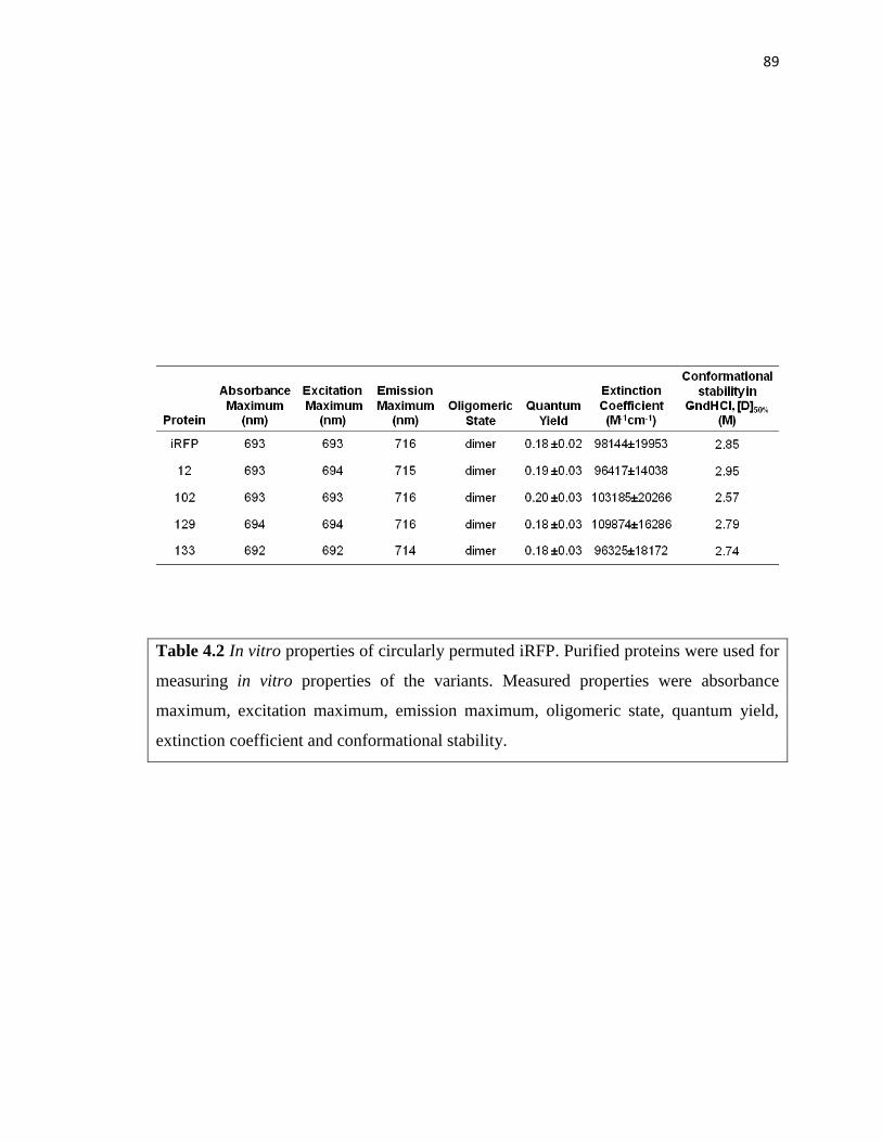
89
Table 4.2 In vitro properties of circularly permuted iRFP. Purified proteins were used for
measuring in vitro properties of the variants. Measured properties were absorbance
maximum, excitation maximum, emission maximum, oligomeric state, quantum yield,
extinction coefficient and conformational stability.

90
Figure 4.10 Absorbance of circularly permuted iRFP. Data acquired using purified
proteins yielded similar absorbance peaks as native iRFP. A comparison of the ratio of
the absorbance arising from BV (391 nm) and total protein (280 nm) yielded ratios for
each permuted variant (0.9-1.1), which were similar to unmodified iRFP (0.85).

91
Figure 4.11 Effect of permutation on iRFP structure. (A) Dimer interface diagrams
showing interaction of Bphp monomers (red and purple). Green represents variant 12 and
102 whereas yellow represents variant 129 and 133. (B) Native-PAGE of purified
circularly permuted iRFP variants (12, 102, 129 and 133) and wild-type iRFP (N). (C)
Gel filtration analysis of selected purified circularly permuted iRFP using Superdex 200
10/300 GL column chromatography using PBS buffer at 4°C. The dashed lines represent
the volumes where the standards beta amylase (200 kDa), alcohol dehydrogenase (150
kDa), bovine serum albumin (66 kda), carbonic anhydrase (29 kDa), cytochrome C (12.4
kDa) eluted. Number represents the first residue of the new circularly permuted iRFP.

92
because oligomerization is disrupted in these purified variants, I used an analytical gel
filtration column to compare the oligomeric state of iRFP with variants 12, 102, 129, and
133 (Figure 4.11C). As previously observed, iRFP elutes as a monodisperse peak with an
apparent MW (72,443 Da) that is 1.90 fold greater than the calculated MW of the iRFP
polypeptide. In addition, all of the recombinant permuted proteins elute at volumes
consistent with oligomerization. However, one variant (129) displayed an apparent
molecular weight that was greater than iRFP (91,201 Da). This permuted iRFP also
displayed the largest change in electrophoretic mobility with native PAGE. Taken
together, these findings suggest that circular permutation affects iRFP shape but not
oligomerization (Table 4.2).
Previous studies have shown that permutation can increase or decrease protein
stability (Hultqvist et al., 2012). To examine how permutation affects iRFP stability, I
compared the equilibrium unfolding of each purified permuted proteins with iRFP.
Unfolding was monitored by measuring near infrared fluorescence signal as a function of
the chemical denaturant guanidiniumhydrochloride (GndHCl). GndHCl-induced
equilibrium unfolding of iRFP displayed a single cooperative transition with a midpoint
at 2.85 M (Figure 4.12). The concentration of guanidine required for half-maximal
unfolding of variants 102, 129 and 133 was lower (2.57, 2.79, and 2.74 M, respectively),
indicating that these variants are destabilized compared with iRFP (Figure 4.12 and Table
4.2). In contrast, a higher concentration (2.95 M) of GndHCl was needed to achieve half
maximal unfolding of variant 12 (Figure 4.12).

93
Figure 4.12 Equilibrium unfolding of circularly permuted iRFP. The effect of GndHCl
concentrations on circularly permuted iRFP fluorescence () is compared with
unmodified iRFP (). For variant 12, GndHCl concentrations >1 M and <3.25 displayed
signals that were significantly higher than iRFP (two-tailed t-test; P<0.05). Error bars
represent ±1σ calculated using three or more measurements.

94
4.3.8 Fluorescence in mammalian cells
Although my initial analysis identified permuted iRFP with altered contact order that
fold and fluoresce in bacteria, it was unclear if these variants would fold in a mammalian
environment where the rate of translation is different. In addition, it wasn’t clear if my
variants would retain iRFP-like affinity for BV, which is sufficient to fluoresce in tissue
culture cells without addition of exogenous BV (Filonov et al., 2011). To test these
questions, I created mammalian constructs for constitutively expressing seven of my
permuted iRFP, transiently transfected these plasmids into HeLa cells with a green
fluorescent protein transfection control, and examined whole cell green and near infrared
fluorescence using flow cytometry (Figure 4.13). A comparison of GFP-normalized near
infrared fluorescence revealed that one of my permuted iRFP display enhanced whole
cell fluorescence compared with iRFP (Figure 4.13), one variant displayed similar
fluorescence as iRFP, four variants displayed ~25% of iRFP fluorescence, and one
variant lacked detectable near infrared fluorescence. While bacterial measurements
predicted that one variant should have low fluorescence (variant 198), the signals
obtained with the other variants did not correlate across bacterial and mammalian cells.
The brightest variants in tissue culture were variants 11 and 12, while the brightest
variant in bacteria at 37°C was variant 133.

95
Figure 4.13 Flow cytometry analysis of tissue culture cells without exogenous BV.
Green and near infrared fluorescence emission of HeLa cells transiently transfected with
a pair of vectors that express GFP and select circularly permuted iRFP variants. For each
variant near infrared fluorescence signal was normalized to the GFP transfection control
to obtain relative fluorescence. Cells were grown in the medium that had not been
supplemented with BV. Error bars represent ±1σ calculated from two independent
measurements. Variant 12 displayed a GFP-normalized signal that was significantly
higher than iRFP (two tailed t test; p<0.05).

96
4.4 Discussion
My results provide direct evidence that knotted BphP are tolerant to a wide range of
sequence rearrangements that change contact order. High-throughput screening revealed
that 8% of all possible circularly permuted iRFP display detectable near infrared
fluorescence. This number is thought to represent a lower bound on the fraction of
permuted iRFP that fluoresce, since my screening was only predicted to sample 58% of
the possible permuted iRFP. Because retention of fluorescence requires a bound cofactor
within a similar structural environment as iRFP, my findings suggest that many of these
permuted proteins display iRFP-like structures. However, it is not clear which of these
permuted variants acquire a knot like iRFP. My in vitro biochemical studies revealed
parent-like stability and spectral properties, suggesting that the environment of the BV
chromophore is not disrupted by permutation, while native gel analysis suggested that
some variants may have subtle structural differences from iRFP. Within the BphP family
there is evidence that some family members lack protein knots. Sequence comparisons
have shown that the cyanobacteriochromes AnPixJ and TePixJ lack the loop that serves as
the knot lasso (Narikawa et al., 2008; Ishizuka et al., 2007). In addition, a recent study
revealed that the BphP knot can be removed through truncation without disrupting
cofactor binding (Rumyantsev et al., 2015). However, unlike my circularly permuted
iRFP, which display parent-like excitation and emission wavelengths, truncated BphP
become blue shifted. These differences suggest that my permuted variants may still
contain a knot. However, structural studies will be required to determine how the
topology of my permuted proteins relate to iRFP, and if variants arising from backbone
97
fission in the domain linker are able to thread the full PAS domain through the loop to
mature into a folded and fluorescent protein.
My whole cell fluorescence measurements revealed that all of my permuted variants
display excitation and emission maxima that are similar to iRFP. However, at each
temperature, I observed a large variation in fluorescence intensities even though each
variant was transcribed and translated using the same promoter and RBS. In past studies,
circular permutation has been shown to alter protein folding (Lindberg et al., 2002;
Nobrega et al., 2014), ligand binding (Cheltsov et al., 2001; Qian et al., 2005), quantum
yield (Topell et al., 1999) and expression (Jones et al., 2016). My results suggest that
differences in permuted iRFP signal in E. coli do not arise from changes in BV binding
affinity. The BV concentration required for half-maximal fluorescence was uniformly
lower than the concentration used in my assays. However, expression did vary across
each permuted variant. Thermodynamic calculations analyzing the effects of different
permuted iRFP genes on the RBS in my permuteposon revealed a correlation between
fluorescence and calculated translation initiation rates. In addition, whole cell
fluorescence signals normalized to permuted protein accumulation revealed signals that
were more consistent with iRFP. This observation emphasizes the importance of
considering steady-state protein levels when using whole cell assays to analyze the
effects of permutation on a protein.
Among the fluorescent variants discovered in my screen, variant 12 displayed
significantly higher protein-normalized fluorescence compared with iRFP. Biochemical
analysis of variant 12 did not reveal any differences in spectral properties from iRFP.
This finding implicates improved maturation as the underlying cause of the enhanced

98
fluorescence. In my biochemical studies, I obtained evidence that only a fraction of the
iRFP translated in E. coli matures into a fluorescent protein. When I subjected
recombinant iRFP to reverse phase chromatography, I found that it eluted at two different
salt concentrations and noted that these differed in their fluorescent signals as previously
observed (Auldridge et al., 2012). Variant 12 also eluted at two different salt
concentrations suggesting that only a subset of translated variant 12 matures into a
fluorescent protein. However, my whole cell fluorescence measurements suggest that a
greater fraction of this variant fully matures. Mapping variant 12 onto the D. radiodurans
BphP structure (Wagner et al., 2005) suggests a mechanism for enhanced maturation.
Because this permuted protein arises from fission within the N-terminal tail, near the BV
attachment site at residue 15, it has fewer residues to thread through the loop that serves
as the iRFP knot lasso. In future studies, the effects of permutation on protein maturation
could be evaluated by analyzing protein folding and fluorescence within a cell free
transcription and translation system. A recent study demonstrated that this approach can
be used to study cofactor binding to a protein using the heme-binding myoglobin (Samuel
et al., 2015). Using myoglobin orthologs of varying stability, this study observed a
correlation between the levels of holo-myoglobin and the protein stability in the absence
of cofactor (Samuel et al., 2015). This trend is consistent with my finding that variant 12
displays the highest stability among the permuted proteins purified, although my analysis
was performed with BV-bound variants.
Although the permuted iRFP discovered in my screen did not yield variants with
dramatic improvements in stability and quantum yield, my results have implications for
protein design. Circularly permuted proteins are increasingly being used to design protein

99
switches through domain insertion, where an ensemble of permuted protein variants are
randomly inserted at different locations within the backbone of a second protein to create
new proteins (Ostermeier et al., 2009). Domain insertion studies have shown that a subset
of the variants created in such libraries display switching behavior, where ligand binding
within one domain modulates the structure and function of the second domain (Baird et
al., 1999; Guntas et al., 2005). In fact, high signal-to-noise switches that couple the
ligand-binding to the fluorescence of different GFP family members have been created by
inserting circularly permuted fluorescent proteins into periplasmic binding proteins
(Marvin et al., 2011). The circularly permuted iRFP discovered in my screens should be
useful for creating similar biosensors, albeit with emission in the near infrared region
which is better suited for tissue imaging (Filonov et al., 2011). All of my iRFP display
parent-like BV affinity, which is sufficient to fluoresce in tissue culture without the
addition of biliverdin. This suggests that biosensors created using these variants may
have sufficient BV affinity to fluoresce in mammalian cells without requiring the addition
of exogenous BV. The extent to which my permuted variants will fold, bind BV, and
fluoresce upon insertion into other proteins will require further studies. If formation of a
knot is required for permuted iRFP fluorescence, then it is possible that some of these
iRFP variants could experience folding challenges upon domain insertion.
By using a circular iRFP gene for library construction, I was able to create vectors
that express different permuted variants of iRFP using a single MuA reaction. This
modified approach is quicker than the original protocol described for PERMUTE because
it avoids manipulations required after the MuA reaction (Mehta et al., 2012), which take
another day to perform. This modified PERMUTE protocol will simplify future studies

100
that seek to create vectors libraries that express different circularly permuted variants of
other proteins. My results show that this simplified protocol can be used with
permuteposons that contain hybrid transposase and ribosomal binding sites that only
amend two extra amino acids to protein termini (Jones et al., 2016). This protocol should
also be compatible with other permuteposons that have been developed for PERMUTE,
including those that amend peptides of varying size to the N-termini of permuted proteins
(Mehta et al., 2012). Beyond simplifying the construction of permuted protein libraries,
the approach described herein has the advantage that it creates permuted proteins with
well-defined sequence diversity and avoids random deletions that arise with previously
described methods (Graf et al., 1996; Hennecke et al., 1999). Whether or not this
approach comprehensively samples all possible permuted protein variants will require
future studies that use deep sequencing to analyze library diversity, similar to that
described for libraries created using random mutation (Firnberg et al., 2012).

101
Chapter 5
Conclusions and future directions
5.1 Knotted IFP are sensitive to backbone fission
Knotted proteins are now widely accepted as naturally occurring proteins. However,
the structural and function role(s) for knot are still not fully appreciated. The work
described herein shows that knotted proteins are quite robust to mutational lesions that
cleave the backbone and alter contact order. In my study of random IFP fragmentation, I
found that this protein displays a low tolerance to fragmentation. However, addition of
the associating peptides to the termini of each fragment led to the discovery of 13
fragmented IFP. Removing the associating peptides from these 13 split proteins
uniformly diminished the fluorescence of the fragmented IFP. This could be due to the
fact that unassisted IFP fragment cannot form the proper knot, or because the fragments
simply do not associate strongly. Whether these split IFP still form a knot is not known.
My split protein results can be contrasted with previous studies in the Silberg lab.
Analysis of the tolerance of an adenylate kinase to fission revealed that a thermophilic
protein retains function 40% of the time, while a mesophilic ortholog retained function
~6% of the time (Segall-Shapiro et al., 2011). The frequency of fluorescent two fragment
IFP described in this thesis (4%) is similar to this latter value, although assistance was
required from associating proteins.
5.2 Split IFP should be useful for studying protein-protein interactions in situ
The dependence of my fragmented IFP on associating proteins for maximal

102
fluorescence suggests that these split proteins will be useful for reporting on other
protein-protein interactions like previously described bimolecular fluorescence
complementation assays (Hu et al., 2002; Wilson et al., 2004; Kerppola et al., 2009). The
longer wavelengths of light used to excite IFP penetrates tissues to a greater extent than
the wavelengths used to excite GFP-family members, suggesting that these split IFP
should be useful as reporters of molecular interactions within animal models like IFP
(Shu et al., 2009). These split proteins should also be useful in anaerobic environments
where GFP-family members cannot be used. Whether or not they are reversible will
require further investigation. In the case of split GFP, fragment association is irreversible
which hinder its use in aggregations studies. The fragmented IFP identified by laboratory
evolution will also be useful for studies examining the spatial and temporal distribution
of promoter activities within different cells within animals. Such measurements could be
used to analyze the cells in complex populations where pairs of promoters are active.
Such information can be coupled to emerging chemical biology tools, such as two-input
AND gates that control metabolic labeling of newly synthesized proteins (Mahdavi et al.,
2013). Such methods enable proteomic measurements with improved temporal and
spatial resolution (Yuet et al., 2014). IFP fragment complementation can be used to report
on the subset of cells within an organism (bacterial or eukaryotic) where a given pair of
promoters is active, information that cannot be obtained using pairs of near-infrared
fluorescent protein reporters because their excitation and emission spectra overlap
(Shcherbakova et al., 2013). However, IFP has a low quantum yield, and this property
could limit the application of the split IFP under conditions where fragments are
expressed at low levels. Whether or not simultaneous expression of both IFP fragments is

103
required to achieve optimal fluorescence will require future studies that examine how
each fragment degrades in the absence of other fragment.
5.3 A generalizable method for discovering split protein that depend on protein-
protein interactions
My results show that using laboratory evolution to discover split protein has
fundamental challenges associated with protein expression. In bacteria, translation
initiation is determined by the context of an RBS. With split protein, different gene
fragments adjacent to the same RBS change its context. To keep similar translation
initiation rate across multiple variants in a vector library, it is best to have a similar
sequence (~35 bp) next to RBS. In my split protein library approach, I found that protein
expression can be made consistent by using a two step procedure. My results suggest that
one should first screen for fragmented proteins that retain function upon fusion to a pair
of interacting proteins like IAAL-E3 and IAAL-K3. Upon discovery, one should rescreen
for loss-of-function upon removal of only one of the fused proteins, i.e., IAAL-E3,
because it is located at the end of the first fragment and removal will not affect the
context of any RBS. In contrast, removing both IAAL-E3 and IAAL-K3 could lead to
false negatives due to changes in translation initiation rates. Future applications of this
transposon mutagenesis approach are expected to simplify the discovery of fragmented
protein devices that couple the function of other proteins to protein-protein interactions
(Michnick et al., 2007). However, deep sequencing may be required for analyzing
libraries that contain and lack fusion proteins, since some fission sites may be non-
disruptive to protein function in the absence of protein fusions. To identify those that

104
require fusion to proteins that interact, it may be important to deep sequence libraries
±fusions before screening and after screening to find the backbone locations that are most
enriched +fusions. In cases where fused proteins have an interaction that is conditional,
e.g., dependent upon ligand (Remy et al., 1999) or cofactor (Hoff et al., 2009) binding,
fragmented proteins will display three-input AND gate logic. The first two inputs will be
the promoters controlling protein fragment expression, while a third input will be the
post-translational event that promotes the protein-protein interaction.
5.4 Split proteins will be useful for studying DNA minivector delivery in situ
The protein fission strategy described here is expected to be useful for synthetic
biology applications that involve DNA minivectors (and minicircles), small vectors that
display amazing stability. These small DNA are being developed for gene therapy
because of their ability to enter difficult-to-transfect mammalian cells and survive shear
forces often associated with DNA delivery (Zhao et al., 2011; Catanese et al., 2012;
Stenler et al., 2014). The beneficial properties of these small DNA are inversely
correlated with DNA length, and only a limited number of natural proteins can be
encoded by a single minivector at the sizes (<1,000 base pairs) where these small DNA
display the greatest stabilities. The benefits of minivectors could be leveraged with larger
proteins if those proteins were broken into smaller fragments that fit into minivectors
having sizes <1,000 base pairs, because pairs or trios of minivectors encoding these
fragments could be delivered to difficult-to-transfect mammalian cells to reconstitute a
functional protein through fragment complementation (Figure 5.1). Because my results
show that IFP can be fragmented into pieces that associate through the fusion of small

105
Figure 5.1 Split IFP for gene delivery using DNA minivectors. (A) Typical viral or
plasmid vectors with gene of interest (purple) for gene therapy are larger than 5 kb, which
gets degraded during delivery by shear force developed in blood or nebulization. (B)
Alternatively, supercoiled minivectors are smaller vectors (>1200 bp) which survive
shear forces but only deliver small genes. (C) To deliver larger genes (including IFP),
split genes system can be utilized by cloning fragments into multiple minivectors for
delivery. Using minivectors for delivering of split IFP one can test the dosage response of
the split protein delivery for hard to transfect tissues and in deep tissues.

106
coils, I expect that the approach described herein will be useful for breaking natural
proteins into pieces that can be encoded by minivectors, e.g., delivery protein
therapeutics in pieces.
5.5 Circularly permuted iRFP will be useful for domain insertion
Near infrared fluorescent proteins provide powerful imaging tools to decipher cellular
activity in tissue of live animals. The variants discovered in this thesis work are poised to
be used to create biosenors with a range of sensing functions. For example, the circularly
permuted iRFP discovered in my thesis work will be useful for domain insertion. With
domain insertion, a protein of interest is randomly inserted into the target protein, and
screening (or selection) is used to identify functional proteins that display switching
activity (Baird et al., 1999; Guntas et al., 2005). In some cases, domain insertion yields a
pair of domains that fold independently and occasionally the function of one domain is
regulated by the activity of the other protein. In domain insertion, the N- and C-termini of
the inserted protein must be nearby one another or they will disrupt the folding of the
other protein This design constraint has limited previous applications of IFP/iRFP in
domain insertion, since these proteins have N- and C-termini that are distant (>20A) from
one another. My circularly permuted proteins should be compatible with domain insertion
since permutation links the original distant N- and C-termini with flexible linker and
creates new proximal N- and C-termini by backbone fission elsewhere in the protein
(Figure 1.3 and Figure 5.2). In this study, I have generated 27 variants of circularly
permuted variants, all of which should be useful for domain insertion. These can be
inserted into any protein imaginable to couple IFP folding, cofactor binding and/or

107
Figure 5.2 Domain insertion using permuted iRFP. Domain insertion required closer N-
and C-termini of the original protein. For those protein (including iRFP) that do not fit
this criteria, circular permutation is performed to create closer N- and C-termini. In this
process original N- and C-termini are connected through flexible peptide linker (blue
line) and new N- and C-termini are created somewhere else in the protein. New N- and
C-termini are connected to the other protein or domain (green) to create a domain
inserted protein fusion which are useful for designing allosteric switches or sensors.

108
fluorescence to other proteins. For example, circularly permuted could be randomly
inserted into proteins that bind specific metabolites, and these libraries could be screened
for variants that display metabolite-dependent near infrared fluorescence.
5.6 Conclusions and Outlook
In this thesis, I provide the first evidence that a knotted protein can be split and
permuted without disrupting structure and function using laboratory evolution. I have
identified multiple split and permutation sites (13 and 27, respectively). Some of these
backbone fission sites cluster near the interdomain linker between PAS and GAF domain,
although some differences are observed in the sites discovered from backbone fission and
permutation. Many questions remain about knotted BphP. It is not clear if my split IFP
and permuted iRFP fold into a knot like full-length IFP and iRFP. Biophysical analysis
will be required to answer this question. X-ray crystallography could be potentially used
to directly probe the presence of the knot in the structure. However, it might be
challenging to obtain crystals for these proteins, especially the split proteins which could
be challenging to purify. NMR may be useful for structural analysis as well.
The stability of split IFP and permuted iRFP might be affected by backbone fission to
differing extents. IFP is less stable than iRFP (Filonov et al., 2011), and this could
explain why no two fragment IFP were discovered that displayed fluorescence in the
absence of protein fusions. A laboratory evolution experiment subjecting iRFP to random
fission might discover split variants that do not display the same dependence upon
protein-protein interactions. For permuted variants of the iRFP, GndHCl unfolding of the
purified variants did not show major difference in stability among the variants (Table

109
4.2), although one variant displayed a minor increase in stability. Whether the same is
true when IFP is circularly permuted is not known. Additional studies will be required to
determine if IFP or other BphP can be permuted at the same locations as iRFP without
disrupting folding, structure and function. It is tempting to speculate that BphP, which
sense light and use light dependent isomerization of their cofactor for signaling, could be
permuted like iRFP and used in domain insertion experiments to create novel light
switchable proteins.

110
References
Auldridge, M. E., Satyshur, K. A., Anstrom, D. M., and Forest, K. T. (2012). Structure-
guided engineering enhances a phytochrome-based infrared fluorescent protein. J.
Biol. Chem. 287, 7000-7009.
Baird, G. S., Zacharias, D. A., and Tsien, R. Y. (1999). Circular permutation and receptor
insertion within green fluorescent proteins. Proc. Natl. Acad. Sci. U.S.A. 96, 11241-
11246.
Barnard, E., McFerran, N.V., Trudgett, A., Nelson, J., and Timson, D.J. (2008). Detection
and localisation of protein–protein interactions in Saccharomyces cerevisiae using a
split-GFP method. Fungal Genetics and Biology, 45, 597-604.
Barondeau, D.P., Putnam, C.D., Kassmann, C.J., Tainer, J.A., and Getzoff, E.D. (2003).
Mechanism and energetics of green fluorescent protein chromophore synthesis
revealed by trapped intermediate structures. Proc. Natl. Acad. Sci. U.S.A. 100, 12111-
12116.
Bevis, B.J., and Glick, B.S. (2002). Rapidly maturing variants of the Discosoma red
fluorescent protein (DsRed). Nat. Biotech. 20, 83-87.
Bloom, J.D., Silberg, J.J., Wilke, C.O., Drummond, D.A., Adami, C., Arnold, F.H. (2005).
Thermodynamic prediction of protein neutrality. Proc. Natl. Acad. Sci. U.S.A. 102,
606-11.
Burbaum, J. J. and Schimmel, P. (1991) Assembly of a class I tRNA synthetase from
products of an artificially split gene. Biochemistry 30, 319-324.
Cabantous, S., Terwilliger, T.C., and Waldo, G.S. (2005). Protein tagging and detection with
engineered self-assembling fragments of green fluorescent protein. Nat. Biotech. 23,
102-107.
Cabantous, S., Nguyen, H.B., Pedelacq, J.D., Koraïchi, F., Chaudhary, A., Ganguly, K.,
Lockard, M. A., Favre, G., Terwilliger, T. C., and Waldo, G. S. (2013). A new
protein–protein interaction sensor based on tripartite split-GFP association. Sci. Rep.
3, 2854.
Catanese, D. J., Fogg, J. M., Schrock, D. E., Gilbert, B. E., and Zechiedrich, L. (2012).
Supercoiled minivector DNA resists shear forces associated with gene therapy
delivery. Gene Ther. 19, 94-100.
Chalfie, M., Tu, Y., Euskirchen, G., Ward, WW., and Prasher, D.C. (1994). Green
fluorescent protein as a marker for gene expression. Science 263, 802-805.

111
Chapleau, R.R., Blomberg, R., Ford, P.C., and Sagermann, M. (2008). Design of highly
specific and noninvasive biosensor for real-time in vivo imaging of mercury (II)
uptake. Protein Science 17, 614-622.
Cheltsov, A.V., Barber, M.J., and Ferreira, G.C. (2001). Circular permutation of 5-
aminolevulinate synthase. Mapping the polypeptide chain to its function. J. Biol.
Chem. 276, 19141-19149.
Choe, W., Chandrasegaran, S., and Ostermeier, M. (2005). Protein fragment
complementation in M.HhaI DNA methyltransferase. Biochem. Biophys. Res.
Commun. 334, 1233-1240.
Chudakov, D.M., Matz, M.V., Lukyanov, S., and Lukyanov, K.A. (2010). Fluorescent
proteins and their applications in imaging living cells and tissues. Physiology Rev. 90,
1103-1163.
Daugherty, A.B., Govindarajan, S., and Lutz, S. (2013). Improved Biocatalysts from a
Synthetic Circular Permutation Library of the Flavin-Dependent Oxidoreductase Old
Yellow Enzyme. J. Am. Chem. Soc. 135, 14425–14432.
Ducrest, A., Amacker, M., Linger, J., and Markus Nabholz. (2002). Detection of promoter
activity by flow cytometric analysis of GFP reporter expression. Nucleic Acids
Research 30, e65.
Feilmeier, B.J., Iseminger, G., Schroeder, D., Webber, H., and Phillips, G.J. (2000). Green
fluorescent protein functions as a reporter for protein localization in Escherichia coli.
J. Bacteriology 182, 4068-4076.
Fery-Forgues, S., and Lavabre, D. (1999). Are fluorescence quantum yields so tricky to
measure? A demonstration using familiar stationery products. J. Chem. Educ. 76,
1260-1264.
Fields, S. and Song, O. (1989). A novel genetic system to detect protein–protein
interactions. Nature 340, 245-246.
Filonov, G.S., Krumholz, A., Xia, J., Yao, J., Wang, L.V., and Verkhusha, V.V. (2012).
Deep-tissue photoacoustic tomography of genetically encoded iRFP probe. Angew.
Chem. Int. Ed. Engl. 51, 1448-1451.
Filonov, G.S., Piatkevich, K.D., Ting, L., Zhang, J., Kim, K., and Verkhusha V.V. (2011).
Bright and stable near-infrared fluorescent protein for in vivo imaging. Nat. Biotech.
29, 757-761.
Filonov, G. S. and Verkhusha, V. V. (2013). A near-infrared BiFC reporter for in vivo
imaging of protein–protein interactions. Chem. Biol. 20, 1078-1086.

112
Firnberg, E., and Ostermeier, M. (2012). PFunkel: efficient, expansive, user-defined
mutagenesis. PLoS ONE 7, e52031.
Fradkov, A.F., Verkhusha,V.V., Staroverov,D.B., Bulina, M.E., Yanushevich,Y.G.,
Martynov, V.I., Lukyanov, S., and Lukyanov, K.A. (2002). Far-red fluorescent tag for
protein labeling. Biochemical J. 368, 17-21.
Galarneau, A., Primeau, M., Trudeau, L.-E., and Michnick, S. W. (2002) Beta-lactamase
protein fragment complementation assays as in vivo and in vitro sensors of protein
protein interactions. Nat. Biotechnol. 20, 619-622.
Giraldez, T., Hughes, T.E., and Sigworth, F.J. (2005). Generation of Functional Fluorescent
BK Channels by Random Insertion of GFP Variants. J. Gen. Physiol. 126, 429-438.
Giraud, E., Zappa, S., Vuillet, L., Adriano, J, Hannibal, L., Fardoux, J., Berthomieu, C.,
Bouyer, P., Pignol, D., and Vermeglio, A. (2005). A New Type of
Bacteriophytochrome Acts in Tandem with a Classical Bacteriophytochrome to
Control the Antennae Synthesis in Rhodopseudomonas palustris. J. Biol. Chem. 280,
32389-32397.
Graf, R., and Schachman, H.K. (1996). Random circular permutation of genes and expressed
polypeptide chains: application of the method to the catalytic chains of aspartate
transcarbamoylase. Proc. Natl. Acad. Sci. U.S.A. 93, 11591-11596.
Grünberg, R. and Serrano, L. (2010). Strategies for protein synthetic biology. Nucleic Acids
Res. 38, 2663-2675.
Guntas, G., Mansell, T.J., Kim, J.R. and Ostermeier, M. (2005). Directed evolution of protein
switches and their application to the creation of ligand-binding proteins. Proc. Natl.
Acad. Sci. U.S.A. 102, 11224-11229.
Haglund E., Lindberg, M.O., and Oliveberg, M. (2008). Changes of protein folding pathways
by circular permutation. J. Biol. Chem. 283, 27904-27915.
Hastings, J.W. (1996). Chemistries and colors of bioluminescent reactions: a review. Gene
173, 5-11.
Heim R., and Tsien, R.Y. (1996). Engineering green fluorescent protein for improved
brightness, longer wavelengths and fluorescence resonance energy transfer. Current
Biology 6, 178-182.
Heim, R., Prasher, D.C., and Tsien, R.Y. (1994). Wavelength mutations and posttranslational
autoxidation of green fluorescent protein. Proc. Natl. Acad. Sci. U.S.A. 91, 12501-
12504.

113
Hennecke, J., Sebbel, P., and Glockshuber, R. (1999). Random circular permutation of DsbA
reveals segments that are essential for protein folding and stability. J. Mol. Biol. 286,
1197-1215.
Hida, K., Won, S.Y., Di Pasquale, G., Hanes, J., Chiorini, J.A., and Ostermeier, M. (2010).
Sites in the AAV5 capsid tolerant to deletions and tandem duplications. Arch.
Biochem. Biophys. 496, 1-8.
Hock, A.K., Lee, P., Maddocks, O.D., Mason, S.M., Blyth, K., and Vousden, K.H. (2013).
iRFP is a sensitive marker for cell number and tumor growth in high-throughput
systems. Cell Cycle 13, 220-226.
Hoff, K.G., Culler, S.J., Nguyen, P.Q., McGuire, R.M., Silberg, J.J., and Smolke, C.D.
(2009). In Vivo Fluorescent Detection of Fe-S Clusters Coordinated by Human
GRX2. Chem. Biol. 16, 1299-1308.
Hu, C.D. and Kerppola, T. K. (2003). Simultaneous visualization of multiple protein
interactions in living cells using multicolor fluorescence complementation
analysis. Nat. Biotechnol. 21, 539-545.
Hu, C.D., Chinenov, Y., and Kerppola, T. K. (2002). Visualization of interactions among
bZIP and Rel family proteins in living cells using bimolecular fluorescence
complementation. Mol. Cell 9, 789-798.
Hultqvist, G., Punekar, A.S., Morrone, A., Chi, C.N., Engstrom, A., Selmer, M., Gianni, S.,
and Jemth, P. (2012). Tolerance of protein folding to a circular permutation in a PDZ
domain. PLoS One 7, e50055.
Ishizuka, T., Narikawa, R., Kohchi, T., Katayama, M., and Ikeuchi, M. (2007).
Cyanobacteriochrome TePixJ of Thermosynechococcus elongatus harbors
phycoviolobilin as a chromophore. Plant Cell Physiol. 48, 1385-1390.
Iwakura, M., Nakamura, T., Yamane, C., and Maki, K. (2000). Systematic circular
permutation of an entire protein reveals essential folding elements. Nat. Struct. Mol.
Biol. 7, 580-585.
Jamroz, M., Niemyska, W., Rawdon, E.J., Stasiak, A., Millett, K.C., Sulkowski, P., and
Sulkowska, J.I. (2015). KnotProt: a database of proteins with knots and slipknots.
Nucleic Acids Res. 43, D306-D314.
Joanna I. Sułkowskaa,b,1, Piotr Sułkowskic,d, P. Szymczake, and Marek Cieplaka. (2008).
Stabilizing effect of knots on proteins. Proc. Natl. Acad. Sci. U.S.A. 105, 19714-
19719.
Jobsis, F.F. (1977). Noninvasive, infrared monitoring of cerebral and myocardial oxygen
sufficiency and circulatory parameters. Science 198, 1264-1267.

114
Johnsson, N. and Varshavsky, A. (1994). Split ubiquitin as a sensor of protein interactions in
vivo. Proc. Natl. Acad. Sci. U.S.A. 91, 10340-10344.
Jones, A. M., Mehta, M. M., Thomas, E. E., Atkinson, J. T., Segall-Shapiro, T. H., Liu, S.,
and Silberg, J. J. (2016). The structure of a thermophilic kinase shapes fitness upon
random circular permutation. ACS Synth. Biol. (In press)
Karimova, G., Pidoux, J., Ullmann, A., and Ladant, D. (1998). A bacterial two-hybrid system
based on a reconstituted signal transduction pathway. Proc. Natl. Acad. Sci. U.S.A.
95, 5752-5756.
Kerppola, T. K. (2009). Visualization of molecular interactions using bimolecular
fluorescence complementation analysis: characteristics of protein fragment
complementation. Chem. Soc. Rev. 38, 2876-2886.
King, N.P., Jacobitz, A.W., Sawaya, M.R., Goldschmidt, L., and Yeates, T.O. (2010).
Structure and folding of a designed knotted protein. Proc. Natl. Acad. Sci. U.S.A. 107,
20732-20737.
Kneen, M., Farinas, J., Li, Y., and Verkman, A.S. (1998). Green fluorescent protein as a
noninvasive intracellular pH indicator. Biophys. J. 74, 1591-1599.
Lamparter, T., Michael, N., Mittmann, F., and Esteban, B. (2002). Phytochrome from
Agrobacterium tumefaciens has unusual spectral properties and reveals an N-terminal
chromophore attachment site. Proc. Natl. Acad. Sci. U.S.A. 99, 11628-11633.
Landro, J.A., and Schimmel, P. (1993). Non-covalent protein assembly. Curr. Opin. Struct.
Biol. 3, 549-554.
Liang, C., and Mislow, K. (1995). Topological Features of Protein Structures: Knots and
Links. J. Am. Chem. Soc. 117, 4201-4213.
Lindberg, M., Tangrot, J., and Oliveberg M. (2002). Complete change of the protein folding
transition state upon circular permutation. Nat. Struct. Biol. 9, 818-822.
Lindenburg, L., and Merkx, M. (2014). Engineering genetically encoded FRET sensors.
Sensors 14, 11691-11713.
Lindhout, D. A., Litowski, J. R., Mercier, P., Hodges, R. S., and Sykes, B. D. (2004). NMR
solution structure of a highly stable de novo heterodimeric coiled-
coil. Biopolymers 75, 367-375.
Lippincott-Schwartz, J., Snapp, E., and Kenworthy, A. (2001). Studying protein dynamics in
living cells. Nat. Rev. Mol. Cell Biol. 2, 444-456.
115
Litowski, J. R. and Hodges, R. S. (2002). Designing heterodimeric two-stranded alpha-
helical coiled-coils. Effects of hydrophobicity and alpha-helical propensity on protein
folding, stability, and specificity. J. Biol. Chem. 277, 37272-37279.
Llopis, J., McCaffery, J.M., Miyawaki, A., Farquhar, M.G., Tsien, R.Y. (1998).
Measurement of cytosolic, mitochondrial, and Golgi pH in single living cells with
green fluorescent proteins. Proc. Natl Acad. Sci. U.S.A. 95, 6803-6808.
Longo, D., and Hasty, J. (2006). Dynamics of single-cell gene expression. Mol. Syst. Biol. 2,
64.
Lou, C., Stanton, B., Chen, Y.J., Munsky, B., Voigt, C.A. (2012). Ribozyme-based insulators
parts buffer synthetic circuits from genetic context. Nat. Biotech. 30, 1137-1142.
Luker, K. E., Smith, M. C. P., Luker, G. D., Gammon, S. T., Piwnica-Worms, H.,
andPiwnica-Worms, D. (2004). Kinetics of regulated protein–protein interactions
revealed with firefly luciferase complementation imaging in cells and living
animals. Proc. Natl. Acad. Sci. U.S.A. 101, 12288-12293.
Mahdavi, A., Segall-Shapiro, T. H., Kou, S., Jindal, G. A., Hoff, K.
G., Liu, S., Chitsaz, M.,Ismagilov, R. F., Silberg, J. J., and Tirrell, D. A. (2013). A
genetically encoded AND gate for cell-targeted metabolic labeling of proteins. J. Am.
Chem. Soc. 135, 2979-2982.
Mallam, A.L., and Jackson, S.E. (2011). Knot formation in newly translated proteins is
spontaneous and accelerated by chaperonins. Nat. Chem. Biol., 8, 147-153.
Mansfield, M.L. (1994). Are there knots in proteins? Nat. Struct. Biol. 1, 213-214.
Marcheschi, R. J., Gronenberg, L. S., and Liao, J. C. (2013). Protein engineering for
metabolic engineering: current and next-generation tools. Biotechnol. J. 8, 545-555.
Marvin, J.S., Schreiter, E.R., Echevarria, I.M., and Looger, L.L. (2011). A genetically
encoded, high-signal-to-noise maltose sensor. Proteins 79, 3025-3036.
Matz, M.V., Fradkov, A.F., Labas, Y.A., Savitsky, A,P., Zaraisky, A.G., Markelov,
M.L., and Lukyanov, S.A. (1999). Fluorescent proteins from nonbioluminescent
Anthozoa species. Nat. Biotechnol. 10, 969-973.
Mehta, M.M., Liu, S., and Silberg, J.J. (2012). A transposase strategy for creating libraries of
circularly permuted proteins. Nucleic Acids Res. 40, e71.
Michnick, S. W., Ear, P. H., Manderson, E. N., Remy, I., and Stefan, E. (2007). Universal
strategies in research and drug discovery based on protein-fragment complementation
assays. Nat. Rev. Drug Discovery 6, 569-582.

116
Miyawaki, A., Llopis, J., Heim, R.., McCaffery, J.M., Adams, J.A., Ikura, M., and Tsien,
R.Y. (1997). Fluorescent indicators for Ca2+ based on green fluorescent proteins and
calmodulin. Nature 388, 882-887.
Mutalik, V.K., Guimaraes, J.C., Cambray, G., Lam, C., Christoffersen, M.J., Mai.
Q.A., Tran, A.B., Paull, M., Keasling JD, Arkin AP, Endy D. (2013). Precise and
reliable gene expression via standard transcription and translation initiation elements.
Nat. Methods, 10, 354-60.
Nagai, T., Ibata, K., Park, E.S., Kubota, M., Mikoshiba, K., and Miyawaki, A. (2002). A
variant of yellow fluorescent protein with fast and efficient maturation for cell-
biological applications. Nat. Biotechnol. 20, 87-90.
Narikawa, R., Fukushima, Y., Ishizuka, T., Itoh, S., and Ikeuchi, M. (2008). A novel
photoactive GAF domain of cyanobacteriochrome AnPixJ that shows reversible
green/red photoconversion. J. Mol. Biol. 380, 844-855.
Nguyen, P. Q. and Silberg, J. J. (2010). A selection that reports on protein–protein
interactions within a thermophilic bacterium. Protein Eng. Des. Sel. 23, 529-536.
Nguyen, P. Q., Liu, S., Thompson, J. C., and Silberg, J. J. (2008). Thermostability promotes
the cooperative function of split adenylate kinases. Protein Eng. Des. Sel. 21, 303-
310.
Nobles, C.L., Clark, J.R., Green, S.I., and Maresso, A.W. (2015). A dual component heme
biosensor that integrates heme transport and synthesis in bacteria. J. Microbiol.
Methods 118, 7-17.
Nobrega, R.P., Arora, K., Kathuria, S.V., Graceffa, R., Barrea, R.A., Guo, L., Chakravarthy,
S., Bilsel, O., Irving, T.C., Brooks, C.L.3rd., and Matthews, C.R. (2014). Modulation
of frustration in folding by sequence permutation. Proc. Natl. Acad. Sci. U.S.A. 111,
10562-10567.
Ostermeier, M. (2009). Designing switchable enzymes. Curr. Opin. Struct. Biol. 19, 442-448.
Ostermeier, M., Nixon, A. E., Shim, J. H., and Benkovic, S. J. (1999). Combinatorial protein
engineering by incremental truncation. Proc. Natl. Acad. Sci. U.S.A. 96, 3562-3567.
Palmer, E., and Freeman, T. (2004). Investigation into the use of C- and N-terminal GFP
fusion proteins for subcellular localization studies using reverse transfection
microarrays. Comp. Funct. Genomics 5, 342-353.

117
Pandey, N., Nobles, C.L., Zechiedrich, L., Maresso, A.W., Silberg, J.J. (2015). Combining
random gene fission and rational gene fusion to discover near-infrared fluorescent
protein fragments that report on protein-protein interactions. ACS Synth. Biol. 4, 615-
624.
Park, S.-Y., Beel, B. D., Simon, M. I., Bilwes, A. M., and Crane, B. R. (2004). In different
organisms, the mode of interaction between two signaling proteins is not necessarily
conserved. Proc. Natl. Acad. Sci. U.S.A. 101, 11646-11651.
Paschon, D. E. and Ostermeier, M. (2004). Construction of protein fragment
complementation libraries using incremental truncation. Methods Enzymol. 388, 103-
116.
Paschon, D. E., Patel, Z. S., and Ostermeier, M. (2005). Enhanced catalytic efficiency of
aminoglycoside phosphotransferase (3′)-IIa achieved through protein fragmentation
and reassembly. J. Mol. Biol. 353, 26-37.
Piatkevich, K.D., Subach, F.V., and Verkusha, V.V. (2013). Engineering of bacterial
phytochromes for near-infrared imaging, sensing, and light-control in mammals.
Chem. Soc. Rev. 42, 3441-3452.
Plaxco, K.W., Simons, K.T., and Baker, D. (1998). Contact order, transition state placement
and the refolding rates of single domain proteins. J. Mol. Biol. 277, 985-994.
Prasher, D.C, Eckenrode V.K., Ward, W.W., Prendergast, F.G., and Cormier, M.J. (1992).
Primary structure of the Aequorea victoria green-fluorescent protein. Gene 111, 229-
233.
Qian, Z., and Lutz, S. (2005). Improving the catalytic activity of Candida antartica lipase B
by circular permutation. J. Am. Chem. Soc. 127, 13466-13467.
Remy, I., Campbell-Valois, F.X., Michnick, S.W. (2007). Detection of protein-protein
interactions using a simple survival protein-fragment complementation assay based
on the enzyme dihydrofolate reductase. Nat. Protoc. 2, 2120-2125.
Remy, I. and Michnick, S. W. (1999). Clonal selection and in vivo quantitation of protein
interactions with protein-fragment complementation assays. Proc. Natl. Acad. Sci.
U.S.A. 96, 5394-5399.
Resch-Genger, U., Grabolle, M., Cavaliere-Jaricot, S., Nitschke, R., and Nann, T. (2008).
Quantum dots versus organic dyes as fluorescent labels. Nat. Methods 5, 763-775.
Rossi, F., Charlton, C. A., and Blau, H. M. (1997). Monitoring protein–protein interactions in
intact eukaryotic cells by beta-galactosidase complementation. Proc. Natl. Acad. Sci.
U.S.A. 94, 8405-8410.

118
Rumyantsev, K.A., Shcherbakova, D.M., Zakharova, N.I., Emelyanov, A.V., Turoverov,
K.K., and Verkhusha, V.V. (2015). Minimal domain of bacterial phytochrome
required for chromophore binding and fluorescence. Sci. Rep. 5, 18348.
Russ, W. P. and Engelman, D. M. (1999). TOXCAT: a measure of transmembrane helix
association in a biological membrane. Proc. Natl. Acad. Sci. U.S.A. 96, 863-868.
Sakaguchi, R., Endoh, T., Yamamoto, S., Tainaka, K., Sugimoto, K., Fujieda, N., Kiyonaka,
S., Mori, Y., and Morii, T. (2009). A single circularly permuted GFP sensor for
inositol-1,3,4,5-tetrakisphosphate based on a split PH domain. Bioorganic Med.
Chem. 17, 7381-7386.
Salis, H. M., Mirsky, E. A., and Voigt, C. A. (2009). Automated design of synthetic
ribosome binding sites to control protein expression. Nat. Biotechnol. 27, 946-950.
Samuel, P.P., Smith, L.P., Phillips, G.N., and Olson, J.S. (2015). Apoglobin stability is the
major governing both cell-free and in vivo expression of holomyoglobin. J. Biol.
Chem. 39, 23479-23495.
Segall-Shapiro, T. H., Meyer, A. J., Ellington, A. D., Sontag, E. D., and Voigt, C. A. (2014).
A “resource allocator” for transcription based on a highly fragmented T7 RNA
polymerase. Mol. Syst. Biol. 10, 742-742.
Segall-Shapiro, T. H., Nguyen, P. Q., Santos, Dos, E. D., Subedi, S., Judd, J., Suh, J., and
Silberg, J. J. (2011). Mesophilic and hyperthermophilic adenylate kinases differ in
their tolerance to random fragmentation. J. Mol. Biol. 406, 135-148.
Shcherbakova, D. M., and Verkhusha, V. V. (2013). Near-infrared fluorescent proteins for
multicolor in vivo imaging. Nat. Methods 10, 751-754.
Sheng, J., Ji, X.F., Wang, F., and Sun, M. (2014). Engineering of Yarrowia lipolytica lipase
Lip8p by circular permutation to alter substrate and temperature characteristics. J.
Ind. Microbiol. Biotechnol. 41,757-62.
Shiba, S., and Schimmel, P. (1992). Functional assembly of a randomly cleaved protein.
Proc. Natl. Acad. Sci. U.S.A. 89, 1880-1884.
Shimomura O., Johnson F.H., and Saiga, Y. (1962). Extraction, purification and properties of
aequorin, a bioluminescent protein from the luminous hydromedusan, Aequorea. J.
Cell Comp. Physiol. 59, 223-239.
Shis, D. L. and Bennett, M. R. (2013). Library of synthetic transcriptional AND gates built
with split T7 RNA polymerase mutants. Proc. Natl. Acad. Sci. U.S.A. 110, 5028-
5033.

119
Shu, X., Royant, A., Lin, M.Z., Aguilera, T.A., Lev-Ram, V., Steinbach, P.A., and Tsein,
R.Y. (2009). Mammalian expression of infrared fluorescent proteins engineered from
a bacterial phytochrome. Science 324, 804-807.
Sigala, P.A., Crowley, J.R., Hsieh, S., Henderson, J.P., and Goldberg, D.E. (2012). Direct
tests of enzymatic heme degradation by the malaria parasite Plasmodium falciparum.
J. Biol. Chem. 287, 37793-37807.
Stenler, S., Wiklander, O. P., Badal-Tejedor, M., Turunen, J., Nordin, J.
Z., Hallengärd, D.,Wahren, B., Andaloussi, S. E., Rutland, M. W., Smith, C.
E., Lundin, K. E., and Blomberg,P. (2014). Micro-minicircle gene therapy:
implications of size on fermentation, complexation, shearing resistance, and
expression. Mol. Ther. Nucleic Acids 2, e140.
Sulkowska, J.I., Noel, J.K., Ramirez-Sarmiento, C.A., Rawdon, E.J., Millett, J.N., and
Onuchic, J.N. (2013). Knotting pathways in proteins. Biochem. Soc. Transactions 41,
523-527.
Sulkowska, J.I., Sulkowski, P., Szymczak, P., and Cieplak, M. (2008). Stabilizing effect of
knots on proteins. Proc. Natl. Acad. Sci. U.S.A. 105, 19714-19719.
Sulkowskaa, J.I., Noelb, J.K., and Onuchic, J.N. (2012). Energy landscape of knotted protein
folding. Proc. Natl. Acad. Sci. U.S.A. 109, 17783-17788.
Sun, J., Kelemen, G.H., Fernandez-Abalos, J.M., and Bibb, M.J. (1999). Green fluorescent
protein as a reporter for spatial and temporal gene expression in Streptomyces
coelicolor A3(2). Microbiology 145, 2221-2227.
Tainaka, K., Sakaguchi, R., Hayashi, H., Nakano, S., Liew, F.F., and Morii, T. (2010).
Design Strategies of Fluorescent Biosensors Based on Biological Macromolecular
Receptors. Sensors 10, 1355-1376.
Tchekanda, E., Sivanesan, D., and Michnick, S. W. (2014). An infrared reporter to detect
spatiotemporal dynamics of protein–protein interactions. Nat. Methods 11, 641-644.
To, T., Piggott, B.J., Makhijani, K., Yu, D., Jan, Y.N., Shu, X. (2015). Rationally designed
fluorogenic protease reporter visualizes spatiotemporal dynamics of apoptosis in vivo.
Proc. Natl. Acad. Sci. U.S.A. 112, 3338-3343.
Topell, S., Hennecke, J., and Glockshuber, R. (1999). Circularly permuted variants of the
green fluorescent protein. FEBS Lett. 457, 283–289.
Viranu, P., Mirny, L.A., Kardar, M. (2006). Intricate Knots in Proteins: Functions and
Evolution. PLoS Comput. Biol. 2, e122, 1074-1079.
120
Wagner, J.R., Zhang, J., Stetten, D.V., Gunther, M., Murgida, D.H., Mroginski, M.A.,
Walker, J.M., Forest, K.T., Hildebrandt, P., and Vierstra, R.D. (2008). Mutational
analysis of Deinococcus radiodurnas bacteriophytochrome reveals key amino acids
necessary for the photochromicity and proton exchange cycle of phytochromes. J.
Biol. Chem. 283, 12212-12226.
Wagner, J.R., Brunzelle, J.S., Forest, K.T., and Vierstra, R.D. (2005). A light-sensing knot
revealed by the structure of the chromophore-binding domain of phytochrome.
Nature 438, 325-331.
Wagner, J. R., Zhang, J., Brunzelle, J. S., Vierstra, R. D., and Forest, K. T. (2007). High
resolution structure of Deinococcus bacteriophytochrome yields new insights into
phytochrome architecture and evolution. J. Biol. Chem. 282, 12298-12309.
Weissleder, R., and Ntziachristos, V. (2003). Shedding light onto live molecular targets. Nat.
Med. 9, 123-128.
Whitehead, T.A., Bergeron, L.M., and Clark D.S. (2009). Tying up the loose ends: circular
permutation decreases the proteolytic susceptibility of recombinant proteins. Protein
Eng. Des. Sel. 22, 607-613.
Williams, D. J., Puhl, H. L., and Ikeda, S. R. (2009). Rapid modification of proteins using a
rapamycin-inducible tobacco etch virus protease system. PLoS One 4, e7474.
Wilson, C. G. M., Magliery, T. J., and Regan, L. (2004). Detecting protein–protein
interactions with GFP-fragment reassembly. Nat. Methods 1, 255-262.
Wycuff, D. R., and Matthews, K. S. (2000). Generation of an AraC-araBAD promoter-
regulated T7 expression system. Anal. Biochem. 277, 67-73.
Yeates, T. O., Norcross, T.S., and King, N.P. (2007). Knotted and topologically complex
proteins as models for studying folding and stability. Curr. Opin. Chem. Biol. 11,
595-603.
Yu, D., Gustafson, W.C., Han, C., Lafaye, C., Noirclerc-Savoye, M., Ge, W.P., Thayer,
D.A., Huang, H., Kornberg, T.B., Royant, A., Jan, L.Y., Jan, Y.N., Weiss, W.A., and
Shu, X. (2014). An improved monomeric infrared fluorescent protein for neuronal
and tumour brain imaging. Nat. Commun. 5, 3626.
Yu, Y., and Lutz, S. (2010). Improved triglyceride transesterification by circular permuted
Candida antarctica lipase B. Biotechnol. Bioeng. 105, 44-50.
Yu, Y., and Lutz, S. (2011). Circular permutation: a different way to engineer enzyme
structure and function. Trends Biotechnol. 29, 18-25.
121
Yuan, L., Kurek, I., English, J., and Keenan, R. (2005). Laboratory-directed protein
evolution. Microbiol. Mol. Biol. Rev. 69, 373-382.
Yuet, K. P., and Tirrell, D. A. (2013). Chemical tools for temporally and spatially resolved
mass spectrometry-based proteomics. Ann. Biomed. Eng. 42, 299-311.
Zhao, N., Fogg, J. M., Zechiedrich, L., and Zu, Y. (2011). Transfection of shRNA-encoding
minivector DNA of a few hundred base pairs to regulate gene expression in
lymphoma cells. Gene Ther. 18, 220-224.
Zhu, B., Wu, G., Robinson, H., Wilganowski, N., Hall, M.A., Ghosh, S.C., Pinkston,
K.L., Azhdarinia, A., Harvey, B.R., Sevick-Muraca, E.M. (2013). Tumor margin
detection using quantitative NIRF molecular imaging targeting EpCAM validated by
far red gene reporter iRFP. Mol. Imaging Biol. 15, 560-568.
Recommended