1
Presented By:
Ayaz Ahmed
Ms. Mujahida
1

Objectives: 1 Define Giardiasis 2 Discus signs and symptoms 3 Explain Transmission 4 Review Pathophysiology 5 Discus Diagnosis & Treatment 6 References
2

Giardiasis — popularly known as beaver fever. it is a parasitic disease caused by the flagellate
protozoan Giardia lambli (also sometimes called Giardia intestinalis and Giardia duodenalis). The giardia organism inhabits the digestive tract of a wide variety of domestic and wild animal species, as well as humans. It is a common cause ofgastroenteritis in humans, infecting approximately 200 million people .
3

Review of Acid Base physiologyReview of Acid Base physiologyThe Key to Blood Gas Interpretation:The Key to Blood Gas Interpretation:
Four Equations, Three Physiologic ProcessesFour Equations, Three Physiologic Processes
Equation Physiologic Process
1) PaCO2 equation Alveolar ventilation
2) Alveolar gas equation Oxygenation3) Oxygen content equation Oxygenation4) Henderson-Hasselbalch equation Acid-base balance
These four equations, crucial to understanding and interpreting arterial blood gas data,
contd.
4


TransmissionTransmission
Giardiasis is passed via the fecal-oral route. Primary routes are personal contact and contaminated comestibles. The more susceptible are institutional or day-care workers, travelers, those eating improperly treated food or drink, and people who have contact with individuals already infected.
contd.
5

Review of Acid Base physiology Review of Acid Base physiology Alveolar Gas Equation Alveolar Gas Equation
PAO2 = PIO2 - 1.2 (PaCO2)*
Where PAO2 is the average alveolar PO2, and PIO2 is the partial pressure of inspired oxygen in the trachea
PIO2 = FIO2 (PB – 47 mm Hg)
FIO2 is fraction of inspired oxygen and PB is the barometric pressure. 47 mm Hg is the water vapor pressure at normal body temperature.
* Note: This is the “abbreviated version” of the AG equation, suitable for most clinical purposes. In the longer version, the multiplication factor “1.2” declines with increasing FIO2, reaching zero when 100% oxygen is inhaled. In these exercises “1.2” is dropped when FIO2 is above 60%.
contd.
6

Review of Acid Base physiology Review of Acid Base physiology Acid-base Balance Acid-base Balance
Henderson-Hasselbalch Equation
[HCO3-]
pH = pK + log ----------------
.03 [PaCO2]
For teaching purposes, the H-H equation can be shortened to its basic relationships:
HCO3-
pH ~ --------- PaCO2
contd.
7

Review of Acid Base physiology Review of Acid Base physiology pH is inversely related to [HpH is inversely related to [H++]; a pH ]; a pH change of 1.00 represents a 10-fold change of 1.00 represents a 10-fold
change in [Hchange in [H++] ] pH [H+] in nanomoles/L
7.00 1007.10 807.30 50 7.40 40 7.52 30 7.70 208.00 10
contd.
8
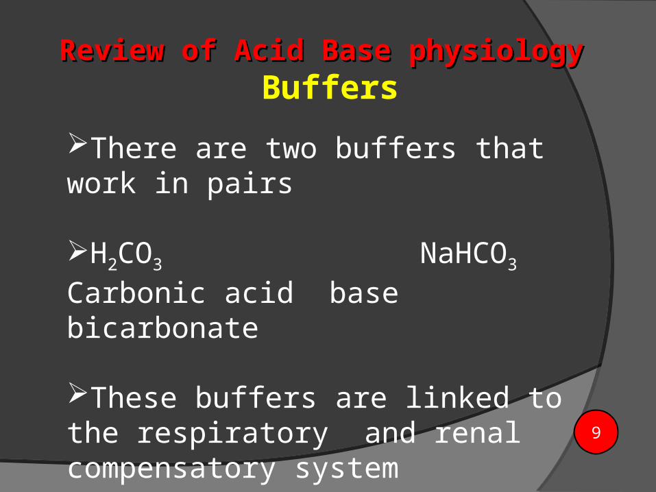
There are two buffers that work in pairs
H2CO3 NaHCO3
Carbonic acid base bicarbonate These buffers are linked to the respiratory and renal compensatory system
Review of Acid Base physiology Review of Acid Base physiology Buffers
9

Sample SourceSample Source
HOW TO TAKE AN ARTERIAL SAMPLE Heparinised syringe Blood with drawn from radial, brachial,
femoral artery Analyse as soon as possible Air bubble should be eliminated Sample should be capped and placed in an ice
bag Excessive heparin in syringe – lower pH and
decrease PCO2, variable effect on PO2 Temp. and F1O2 be mentioned in request
form10

Components of arterial blood Components of arterial blood gasesgases
pH [H+]
PCO2 Partial pressure CO2
PO2 Partial pressure O2
HCO3 Bicarbonate
BE Base excess
SaO2 Oxygen Saturation
contd.
11

Components of the Arterial Blood Components of the Arterial Blood GasGas
pH
Measurement of acidity or alkalinity, based on the hydrogen (H+)
ions present.
The normal range is 7.35 to 7.45
PaO2
The partial pressure of oxygen that is dissolved in arterial blood.
The normal range is 80 to 100 mm Hg.
SaO2
The arterial oxygen saturation.
The normal range is 95% to 100%.
contd.
12

PaCO2
The amount of carbon dioxide dissolved in arterial blood.
The normal range is 35 to 45 mm Hg.
HCO3
The calculated value of the amount of bicarbonate in the
bloodstream.
The normal range is 22 to 26 mEq/liter
B.E.
The base excess indicates the amount of excess or insufficient level
of bicarbonate in the system.
The normal range is –2 to +2 mEq/liter.
(A negative base excess indicates a base deficit in the blood.)
Components of the Arterial Blood Gas contd.Components of the Arterial Blood Gas contd.
13

Normal ABG valuesNormal ABG values
pH 7.35 – 7.45
PCO2 35 – 45 mmHg
PO2 80 – 100 mmHg
HCO3 22 – 26 mmol/L
BE -2 - +2
SaO2 >95% 14

Practical approach to Practical approach to diagnosing diagnosing
acid base disorders from the acid base disorders from the ABGABG
Step 1: Is PH normal?
Step 2: Is the CO2 normal?
Step 3: Is CO2 the HCO3 normal?
Step 4: Match the CO2 or the HCO3 with the PH?
Step 5: Does the CO2 or the HCO3 go the opposite direction of the PH?
Step 6: Are the pO2 and the O2 saturation normal?
15

Respiratory acidosis Metabolic
acidosis
Respiratory alkalosis Metabolic alkalosis
Acids based disordersAcids based disorders
16

Blood Gas ReportBlood Gas Report
Acid-Base Information•pH•PCO2
•HCO3 [calculated vs measured]
Oxygenation Information•PO2 [oxygen tension]•SO2 [oxygen saturation]
17
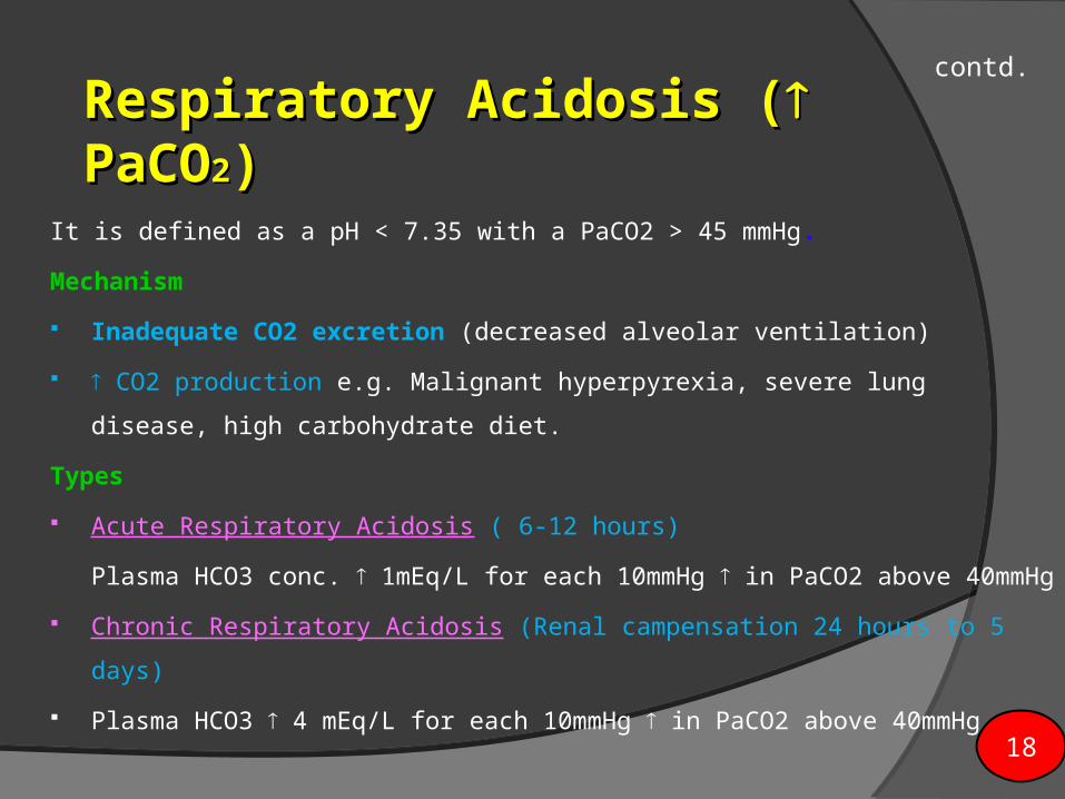
Respiratory AcidosisRespiratory Acidosis ( ( PaCOPaCO22))
It is defined as a pH < 7.35 with a PaCO2 > 45 mmHg.
Mechanism
Inadequate CO2 excretion (decreased alveolar ventilation)
CO2 production e.g. Malignant hyperpyrexia, severe lung disease, high
carbohydrate diet.
Types
Acute Respiratory Acidosis ( 6-12 hours)
Plasma HCO3 conc. 1mEq/L for each 10mmHg in PaCO2 above
40mmHg
Chronic Respiratory Acidosis (Renal campensation 24 hours to 5 days)
Plasma HCO3 4 mEq/L for each 10mmHg in PaCO2 above 40mmHg
contd.
18

Causes of respiratory Causes of respiratory acidosis acidosis
• Central nervous system depression related to head injury or medications such as narcotics, sedatives, or anesthesia
Impaired respiratory muscle function related to spinal cord injury, neuromuscular diseases,or neuromuscular blocking drugs
• Pulmonary disorders such as atelectasis, pneumonia, pneumothorax, pulmonary edema, or bronchial obstruction
• Massive pulmonary embolism
• Hypoventilation due to pain, chest wall injury/deformity, or abdominal distension
contd.
19

Respiratory AcidosisRespiratory AcidosisTreatment
Increase alveolar ventilation (bronchodilatation,
reversal of narcosis, doxapram, diuresis)
Reduce CO2 production by dantrolene, muscle
paralysis, antithyroid medication or reduced
carbohydrate intake
Mechanical ventilation, 20

Respiratory AlkalosisRespiratory AlkalosisRespiratory alkalosis is defined as a pH > 7.45 with a PaCO2 <35 mm Hg.
CAUSES:
Any condition that causes hyperventilation can result in respiratory alkalosis
Psychological responses, such as anxiety or fear
Pain
Increased metabolic demands, such as fever, sepsis, pregnancy, or
thyrotoxicosis
Medications, such as respiratory stimulants.
Central nervous system lesions
MANAGEMENT:
Treat the underlying cause 21

Metabolic Acidosis Metabolic acidosis is defined as a bicarbonate level
of <22 mEq/L with a pH of < 7.35
Types
High anion gap (nonvoltile acids)
Normal anion gap (hyperchloremia)
Anion gap
Plasma cations – plasma anion
[Na- (Cl+ Hco3)]
Anion gap:
140 – (104 + 24) = 12 mEq/L ( normal 12 – 18 mEq/L)
contd.
22

The Causes High Anion Gap
AcidosisM - MethanolU - UremiaD - DKAP - ParaldehydeI - INHL - Lactic AcidosisE - Ehylene GlycolS - Salicylate
Normal Anion Gap AcidosisHyperalimentationAcetazolamideRTA (Calculate
urine anion gap)DiarrheaPancreatic Fistula
contd.
23

Metabolic AcidosisMetabolic Acidosis
Treatment
NaHCO3 1 mEq/Kg
Base deficit X 30% X b.w
Give 50% calculated dose & repeat ABG
24

Metabolic AlkalosisMetabolic AlkalosisMetabolic alkalosis is defined as a bicarbonate level > 26
mEq/liter with a pH >7.45.
CAUSES
Either an excess of base or a loss of acid within the body can
cause metabolic alkalosis.
Excess base occurs from ingestion of antacids, excess use of
bicarbonate, or use of lactate in dialysis.
Loss of acids can occur secondary to protracted vomiting,
gastric suction, hypochloremia, excess administration of
diuretics, or high levels of aldosterone.
contd.
25

Management of Management of metabolic alkalosismetabolic alkalosis
Metabolic alkalosis is one of the most difficult acid-base
imbalances to treat.
Bicarbonate excretion through the kidneys can be
stimulated with drugs such as acetazolamide
(Diamox™),
In severe cases, IV administration of acids may be used.
It is significant to note that metabolic alkalosis in
hospitalized patients is usually iatrogenic in nature. 26

The relationships between The relationships between pH, PaCO2 and HCO3pH, PaCO2 and HCO3.
pH PaCo2 HCO3
Respiratory Acidosis ↓ ↑ Normal
Respiratory alkalosis ↑ ↓ Normal
Metabolic Acidosis ↓ Normal ↓
Metabolic Alkalosis ↑ Normal ↑
Remember Acronym ROME – Respiratory opposite, Metabolic equivalent
27

Normal Compensatory Normal Compensatory ResponsesResponses
Disturbance Response Expected Change
Respiratory Acidosis
Acute Co2 HCO3 conc. 1 mEq/L/10mmHg
increase in PaCO2Chronic Co2 HCO3 conc. 4 mEq/L/10mmHg
increase in PaCO2
Respiratory AlkalosisAcute Co2 HCO3 conc 2 mEq/L/10mmHg
decrease in PaCO2
Chronic Co2 HCO3 conc 4 mEq/L/10mmHg decrease in
PaCO2
Contd:
28

Normal Compensatory Responses (ContdNormal Compensatory Responses (Contd))
Disturbance Response Expected Change
Metabolic Acidosis HCO3 PaCO2 1.2 X the decrease in HCO3 conc.
Metabolic Alkalosis HCO3 PaCO2 0.7 X the increase in HCO3 conc.
29

pH PaCO2 HCO3
Respiratory Acidosis Normal, but < 7.40 ↑ ↑
Respiratory Alkalosis Normal, but < 7.40 ↓ ↓
Metabolic Acidosis Normal, but < 7.40 ↓ ↓
Metabolic Alkalosis Normal, but < 7.40 ↑ ↑
pH PaCO2 HCO3
Respiratory Acidosis ↓ ↑ ↑
Respiratory Alkalosis ↑ ↓ ↓
Metabolic Acidosis ↓ ↓ ↓
Metabolic Alkalosis ↑ ↑ ↑
Fully Compensated States
Partially Compensated States
30

Take Home Message: Valuable information can be gained from an ABG as to the patients physiologic condition
Remember that ABG analysis if only part of the patient assessment. Be systematic with your analysis, start with ABC’s as always and look for hypoxia (which you can usually treat quickly), then follow the four steps. A quick assessment of patient oxygenation can be achieved with a pulse oximeter which measures SaO2.
31

Example 1
A 45-year-old female presents to ER with a severe asthma
attack.
She has been experiencing increasing shortness of breath
since admission three hours ago.
Stat ABGs
pH 7.22
PaCO2 55
HCO3- 25
contd.
32

Follow the steps: 1. Assess the pH. It is low (normal 7.35-7.45);
2. Assess the PaCO2. It is high (normal 35-45) and in the opposite
direction of the pH.
3. Assess the HCO3. It has remained within the normal range (22-
26).
Management Improve the ventilation status
Oxygen therapy,
Administering bronchodilators
Mechanical ventilation
pH PCO2 HCO3
Respiratory Acidosis ↓ ↑ Normal
33

Example 2
55-year-old male presented to ER with a recurring
bowel obstruction. He has been experiencing
intractable vomiting for the last several hours despite
the use of antiemetics.
Stat ABGs
pH 7.50
PaCO2 42
HCO3- 33
contd.
34

Follow the three steps again:1. Assess the pH. It is high (normal 7.35-7.45),
therefore, indicating alkalosis.2. Assess the PaCO2. It is within the normal range
(normal 35-45).3. Assess the HCO3. It is high (normal 22-26) and
moving in the same direction as the pH.
Management:Administration of I.V.fluids and measures to
reduce the excess base.
pH PCO2 HCO3
Metabolic Alkalosis ↑ Normal ↓
35
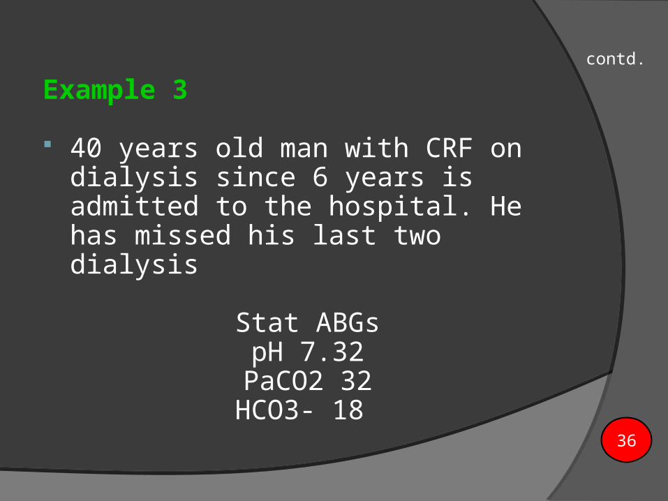
Example 3
40 years old man with CRF on dialysis since 6 years is admitted to the hospital. He has missed his last two dialysis
Stat ABGspH 7.32
PaCO2 32HCO3- 18
contd.
36

Follow the three steps:
1. Assess the pH. It is low (normal 7.35-7.45); therefore we have acidosis.
2. Assess the PaCO2. It is low. Normally we would expect the pH and PaCO2 to move in opposite directions, but this is not the case. Because the pH and PaCO2 are moving in the same direction, it indicates that the acid-base disorder is primarily metabolic. In this case, the lungs, acting as the primary acid-base buffer, are now attempting to compensate by “blowing off excessive C02”, and therefore increasing the pH.
3. Assess the HCO3. It is low (normal 22-26). We would expect the pH and the HCO3 - to move in the same direction, confirming that the primary problem is metabolic.
4. What is your interpretation? Partially compensated metabolic acidosis.
pH PaCO2 HCO3
Metabolic Acidosis ↓ ↓ ↓ 37

Example 4
70 year old man RR 50/min B.P 80/50 pH 7.1 PCO2 22mmHg SBE - 21
Metabolic Acidosis + Resp. Compensation
contd.
38

Example 5
A 6 week old child with projectile vomiting. pH 7.5 PCO2 48mmHg BE + 11
Metabolic Alkalosis + Resp. Compensation
39

Example 6
pH 7.08 PCO2 80mmHg PaO2 37 Na 138, HCO3 26, Cl 100
Respiratory Acidosis
40

Example 7
60 years old man with history of COPD since 20 years is scheduled for laprascopic cholecystectomy
stat ABGs pH 7.35
PaCO2 48HCO3- 28
contd.
41

Follow the three steps:
1. Assess the pH. It is within the normal range, but on the low
side of neutral (<7.40).
2. Assess the PaCO2. It is high (normal 35-45).
3. Assess the HCO3. It is also high (22-26).
Interpretation of this ABG a fully compensated respiratory
acidosis.
pH PaCO2 HCO3
Respiratory Acidosis Normal, but < 7.40 ↑ ↑
42

Example 8
16 years old boy after an RTA presents in ER with mental status.
Stat ABGspH 7.33
PaC02 62HC03 35
contd.
43

Follow the three steps:1. Assess the pH. It is low (normal 7.35-7.45). This indicates that an
acidosis exists.2. Assess the PaC02. It is high (normal 35-45). The pH and PaC02 are
moving in opposite directions, as we would expect if the problem were primarily respiratory in nature.
3. Assess the HC03. It is high (normal 22-26). Normally, the pH and HC03 should move in the same direction. Because they are moving in opposite directions, it also confirms that the primary acid-base disorder is respiratory in nature. In this case, the kidneys are attempting to compensate by retaining HCO3 in the blood in an order to return the pH back towards its normal range. Because there is evidence of compensation occurring (pH and HC03 moving in opposite directions), and seeing that the pH has not yet been restored to its normal range,
ABG interpretation Partially compensated respiratory
acidosis.44

Example 9
54-year-old female admitted for an ileus. She had been experiencing nausea and vomiting. An NG tube has been in place for the last 24 hours.
Here are the last ABG resultspH 7.43
PaC02 48HC03 36:
contd.
45

Follow the three steps:
1. Assess the pH. It is normal, but on the high side of neutral (>7.40).
2. Assess the PaC02. It is high (normal 35-45). Normally, we would expect the pH and PaC02 to move in opposite directions. In this case, they are moving in the same direction indicating that the primary acid-base disorder is metabolic in nature. In this case, the lungs, acting as the primary acid-base buffer system, are retaining C02 (hypoventilation) in order to help lower the pH back towards its normal range.
3. Assess the HC03. It is high (normal 22-26). Because it is moving in the same direction, as we would expect, it confirms the primary acid-base disorder is metabolic alkalosis
Interpretation
fully compensated metabolic alkalosis.
pH PaCO2 HCO3
Metabolic Alkalosis Normal, but >7.40 ↑ ↑
46

Example 10
A 42 year female diabetic, Temp 38.8oC WBC 14000 disoriented.
pH 7.23 PCO2 25mmHg,PO2118 Na 135, K 4.8, HCO3
12, Cl 99
Metabolic Acidosis + Resp. Compensation
47

Example 11A Female 23 years
pH 7.54 PCO2 22mmHg PO2 115 room air HCO3 22
Acute Respiratory Alkalosis
48

TThhaannkk yyoouu

Practice ABG’s
1. PaO2 90 SaO2 95 pH 7.48 PaCO2 32 HCO3 242. PaO2 60 SaO2 90 pH 7.32 PaCO2 48 HCO3 253. PaO2 95 SaO2 100 pH 7.30 PaCO2 40 HCO3 184. PaO2 87 SaO2 94 pH 7.38 PaCO2 48 HCO3 285. PaO2 94 SaO2 99 pH 7.49 PaCO2 40 HCO3 306. PaO2 62 SaO2 91 pH 7.35 PaCO2 48 HCO3 277. PaO2 93 SaO2 97 pH 7.45 PaCO2 47 HCO3 298. PaO2 95 SaO2 99 pH 7.31 PaCO2 38 HCO3 159. PaO2 65 SaO2 89 pH 7.30 PaCO2 50 HCO3 2410. PaO2 110 SaO2 100 pH 7.48 PaCO2 40 HCO3 30

Answers to Practice ABG’s
1. Respiratory alkalosis2. Respiratory acidosis3. Metabolic acidosis4. Compensated Respiratory acidosis5. Metabolic alkalosis6. Compensated Respiratory acidosis7. Compensated Metabolic alkalosis8. Metabolic acidosis9. Respiratory acidosis10.Metabolic alkalosis
Recommended