Copyright © 2017 Abcam. All rights reserved
Version 1 Last updated 26 June 2017
ab222944Fatty Acid Oxidation Complete Assay Kit
For the convenient measurement of Fatty Acid Oxidation (FAO) in live cells when used in combination with Extracellular Oxygen Consumption Assay (ab197243).
This product is for research use only and is not intended for diagnostic use.
Copyright © 2017 Abcam. All rights reserved
1. Overview
Fatty Acid Oxidation Complete Assay Kit (ab222944) offers a complete solution to measure Fatty Acid Oxidation (FAO) in live cells using conventional fluorescence plate readers. This product combines Fatty Acid Oxidation Assay (ab217602) and Extracellular Oxygen Consumption Assay (ab197243) in one practical kit for single purchase.
The assay uses the 18C unsaturated fatty acid Oleate as substrate, and includes two FAO modulators, etomoxir and FCCP. Etomoxir, an inhibitor of the carnitine transporter CPT1, prevents Oleate import and thereby limits the supply of reducing equivalents to the ETC, reducing oxygen consumption in turn. The remaining ETC (electron transport chain) activity is driven by non-long chain FAO. FCCP treatment induces maximal ETC activity by dissipating the mitochondrial membrane potential, while the increased demand for reducing equivalents causes a concomitant increase in the FAO activity. If exogenous long-chain fatty acid is unavailable or import is inhibited, FAO activity will be limited.
Fatty acid oxidation (FAO) is the primary metabolic pathway for degradation of fatty acids. Figure 1 gives an overview of long-chain fatty acid activation, import and oxidation. FAO is an important process in many tissues during periods of glucose deprivation. In organs, such as liver and skeletal muscle, FAO can provide over 75% of cellular ATP while in cardiac tissue it can be responsible for up to 90% of cellular energy requirements. FAO is also now acknowledged as a key factor in cancer metabolism and is also implicated in drug-induced microsteatosis.
Copyright © 2017 Abcam. All rights reserved
Version 3 Last updated 26 June 2017
ab217602 Fatty Acid Oxidation Assay
For the convenient measurement of Fatty Acid Oxidation (FAO) in live cells when used in combination with Extracellular Oxygen Consumption Assay (ab197243).
This product is for research use only and is not intended for diagnostic use.
Copyright © 2017 Abcam. All rights reserved
Table of Contents
1. Overview 1
2. Protocol Summary 3
3. Precautions 4
4. Storage and Stability 4
5. Limitations 5
6. Materials Supplied 5
7. Materials Required, Not Supplied 6
8. Technical Hints 7
9. Reagent Preparation 8
10. Sample Preparation 9
11. Assay Controls set up 12
12. Assay Procedure 14
13. Typical Assays/Data 17
14. Notes 20
ab217602 Fatty Acid Oxidation Assay Kit 1
2. Overview
Fatty Acid Oxidation Assay (ab217602) allows the detection of Fatty Acid Oxidation (FAO) in live cells. This product is designed to be used in combination with our Extracellular Oxygen Consumption Assay (ab197243).The assay uses the 18C unsaturated fatty acid Oleate as substrate, and includes two FAO modulators, etomoxir and FCCP. Etomoxir, an inhibitor of the carnitine transporter CPT1, prevents Oleate import and thereby limits the supply of reducing equivalents to the ETC, reducing oxygen consumption in turn. The remaining ETC (electron transport chain) activity is driven by non-long chain FAO. FCCP treatment induces maximal ETC activity by dissipating the mitochondrial membrane potential, while the increased demand for reducing equivalents causes a concomitant increase in the FAO activity. If exogenous long-chain fatty acid is unavailable or import is inhibited, FAO activity will be limited.
Fatty acid oxidation (FAO) is the primary metabolic pathway for degradation of fatty acids. Figure 1 gives an overview of long-chain fatty acid activation, import and oxidation. FAO is an important process in many tissues during periods of glucose deprivation. In organs, such as liver and skeletal muscle, FAO can provide over 75% of cellular ATP while in cardiac tissue it can be responsible for up to 90% of cellular energy requirements. FAO is also now acknowledged as a key factor in cancer metabolism and is also implicated in drug-induced microsteatosis.

ab217602 Fatty Acid Oxidation Assay Kit 2
Figure 1. Overview of long-chain fatty acid activation, import and oxidation.
ab217602 Fatty Acid Oxidation Assay Kit 3
3. Protocol Summary
Day 1Plate cells and return to culture overnight
Prepare Base Measurement Media
Incubate Over NightDay 2 (OPTIONAL)
Prepare Glucose-Deprivation Media
Replace culture media with Glucose-Deprivation Media and return cells to culture
Incubate Over NightDay 3
Prepare FA-Free & FA Measurement MediaPrepare FAO kit controls (Etoxomir, FCCP and BSA)
Prepare O2 Consumption reagent (ab197243)
Wash cells twice with FA-Free Measurement Media
Add FA/FA-Free Measurement Media and O2 consumption reagent
Add controls (Etoxomir, FCCP, BSA)
Overlay plate with High Sensitivity mineral oil (ab197243)
Measure on fluorescence plate reader
Analyze kinetic data output to determine FAO-driven ETC activity
ab217602 Fatty Acid Oxidation Assay Kit 4
4. Precautions
Please read these instructions carefully prior to beginning the assay.
All assay kit components have been formulated and quality control tested to function successfully as a kit.
We understand that, occasionally, experimental protocols might need to be modified to meet unique experimental circumstances. However, we cannot guarantee the performance of the product outside the conditions detailed in this protocol booklet.
Reagents should be treated as possible mutagens and should be handle with care and disposed of properly. Please review the Safety Datasheet (SDS) provided with the product for information on the specific components.
Observe good laboratory practices. Gloves, lab coat, and protective eyewear should always be worn. Never pipette by mouth. Do not eat, drink or smoke in the laboratory areas.
All biological materials should be treated as potentially hazardous and handled as such. They should be disposed of in accordance with established safety procedures.
5. Storage and Stability
Kit has a storage time of 1 year from receipt. Please observe storage conditions of each individual component described in the Materials Supplied section for correct storage upon receipt.
Note: Reconstituted reagents are stable for 3 months.
ab217602 Fatty Acid Oxidation Assay Kit 5
6. Limitations
Kit intended for research use only. Not for use in diagnostic procedures.
Do not mix or substitute reagents or materials from other kit lots or vendors. Kits are QC tested as a set of components and performance cannot be guaranteed if utilized separately or substituted.
7. Materials Supplied
Item Quantity
Storage temperature (before
prep)
Storage temperatur
e (after prep)
FAO Conjugate (3 mM) 1 mL 4ºC 4ºC
FAO Control (1.5 mM) 500 μL 4ºC 4ºC
FAO Tablet (base media) 1 tablet RT 4ºC
L-Carnitine 1 vial 4ºC -20ºC
FCCP 1 vial -20ºC -20ºC
Etomoxir 1 vial -20ºC -20ºC
ab217602 Fatty Acid Oxidation Assay Kit 6
8. Materials Required, Not Supplied
These materials are not included in the kit, but will be required to successfully perform this assay: Extracellular Oxygen Consumption Assay (ab197243) Microplate reader capable of measuring fluorescence, with
suitable filter and plate temperature control – see Instrument and Measurement Settings section on the Extracellular Oxygen Consumption Assay (ab197243) protocol for suitable plate readers
MilliQ water or other type of double distilled water (ddH2O) DMSO Pipettes and pipette tips, including multi-channel pipette Assorted glassware for the preparation of reagents and buffer
solutions Tubes for the preparation of reagents and buffer solutions Sterile 96-well plate (black wall with clear flat bottom), or standard
clear plates for cell culture Cell culture media Base glucose deprivation media: glucose-free DMEM media, 1 mM
glucose, 1 mM L-glutamine, 1% FBS, Penicillin/streptomycin solution (100 U/mL /0.1 mg/mL)
Glucose: to prepare FA-Free Measurement Media (Optional) Plate block heater for plate preparation.To prepare Base measurement media: HCl and NaOH: to bring media to pH7.4 Appropriate 0.2 µm filter to sterilize media
ab217602 Fatty Acid Oxidation Assay Kit 7
9. Technical Hints
This kit is sold based on number of tests. Number of tests based on described procedure, stock dilutions, concentrations and wash steps. Review the protocol completely to confirm this kit meets your requirements. Please contact our Technical Support staff with any questions.
Selected components in this kit are supplied in surplus amount to account for additional dilutions, evaporation, or instrumentation settings where higher volumes are required. They should be disposed of in accordance with established safety procedures.
Avoid foaming or bubbles when mixing or reconstituting components.
Avoid cross contamination of samples or reagents by changing tips between sample and reagent additions.
Ensure plates are properly sealed or covered during incubation steps.
Ensure all reagents and solutions are at the appropriate temperature before starting the assay.
Make sure all necessary equipment is switched on and set at the appropriate temperature.
Refer to Instrument and Measurement Settings Table (Table 1) in the user manual for Extracellular O2 Consumption Assay (ab197243) for recommended settings for your plate reader.
ab217602 Fatty Acid Oxidation Assay Kit 8
10.Reagent Preparation
Briefly centrifuge small vials at low speed prior to opening.
10.1 FAO Conjugate (Oleate-BSA conjugate, 3 mM) (1mL):Ready to use as supplied. Equilibrate to room temperature prior to use. Store at 4ºC.
10.2 FAO Control (BSA, 1.5 mM) (500 µL):Ready to use as supplied. Equilibrate to room temperature prior to use. Store at 4ºC.
10.3 FAO Tablet (1 Base media tablet):To prepare Base Measurement Media, dissolve tablet provided in 100 mL ddH2O and warm solution to 37°C to ensure table is completely dissolved. Adjust pH to 7.4 using HCl and NaOH. Filter sterilize base media. Filtered Base Measurement Media can be stored at 4°C for 3 weeks.
10.4 L-Carnitine (L-carnitine hydrochloride, 4 mg):Prepare a 50 mM L-Carnitine stock solution (100X) by dissolving vial contents in 400 µL ddH2O. Aliquot L-Carnitine stock solution so that you have enough volume to perform the desired number of assays (recommendation: 100 µL to cover 100 tests). Store at -20°C. Once reconstituted, use component within 3 months.
10.5 FCCP (0.004 mg):Prepare a 250 µM FCCP stock solution (100X) by dissolving vial contents in 60 µL of DMSO. Aliquot FCCP stock solution so that you have enough volume to perform the desired number of assays (recommendation: 20 µL to cover 20 tests). Store at - 20°C. Once reconstituted, use component within 3 months.
10.6 Etomoxir (0.074 mg):Prepare a 400 µM Etomoxir stock solution (10X) by dissolving vial contents in 550 µL ddH2O. Aliquot Etomoxir stock solution so that you have enough volume to perform the desired number of assays (recommendation: 50 µL or 100 µL to cover 10 or 20 tests respectively). Store at -20°C. Once reconstituted, use component within 3 months.
ab217602 Fatty Acid Oxidation Assay Kit 9
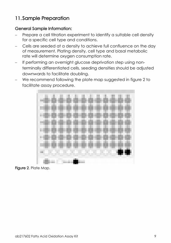
11.Sample Preparation
General Sample Information: Prepare a cell titration experiment to identify a suitable cell density
for a specific cell type and conditions. Cells are seeded at a density to achieve full confluence on the day
of measurement. Plating density, cell type and basal metabolic rate will determine oxygen consumption rate.
If performing an overnight glucose deprivation step using non-terminally differentiated cells, seeding densities should be adjusted downwards to facilitate doubling.
We recommend following the plate map suggested in figure 2 to facilitate assay procedure.
Figure 2. Plate Map.

ab217602 Fatty Acid Oxidation Assay Kit 10
If using extended culture periods (> 2 days), we recommend following plate map (figure 3), adding 200 µL of culture medium to all outer wells. This minimizes plate effects related to inconsistent cell growth across the microplate.
Figure 3. Recommended plate map for extended culture times (> 2 days)
Inconsistent growth in some cell types can be additionally reduced by allowing the plate to stand at RT for 30 minutes after plating before returning plate to cell culture.
ab217602 Fatty Acid Oxidation Assay Kit 11
11.1 Adherent cells (2D cell culture):11.1.1 Seed cells in a 96-well plate a density of 6 x 10⁴ cells/well in
200 µL culture medium.11.1.2 Incubate overnight in a CO2 incubator at 37°C (typical
incubation time > 14 hours).11.1.3 OPTIONAL: Glucose deprivation step. Note: Including a glucose deprivation step before performing the assay increases cellular dependence on FAO. For maximum FAO dependence, concentrations of L-Carnitine, glucose and FAO Conjugate should be optimized for each cell type.11.1.3.1 Prepare Base glucose deprivation media as described in
Section 7 (media can be stored for 2 weeks at 4°C).11.1.3.2 Prepare Glucose deprivation media by adding 0.5 mM L-
Carnitine (1:100 of stock) to Base glucose deprivation media.11.1.3.3 Wash cells twice with Base glucose deprivation media.11.1.3.4 Add 200 µL of Glucose deprivation media.11.1.3.5 Incubate overnight in a CO2 incubator at 37°C (typical
incubation time > 14 hours).11.2 3D cell culture:11.2.1 Plate cells for 3D cultures at a higher density than optimized for
2D cultures. When plating 3D cultures, prepare the plate or matrix solution in advance as per manufacturer’s instructions.
Note: test compounds can take longer to penetrate and affect 3D cultures and longer treatment times may be necessary.11.2.2 OPTIONAL: Glucose deprivation step. Note: Including a glucose deprivation step before performing the assay increases cellular dependence on FAO. For maximum FAO dependence, concentrations of L-Carnitine, glucose and FAO Conjugate should be optimized for each cell type.11.2.2.1 Prepare Base glucose deprivation media as described in
Section 7 (media can be stored for 2 weeks at 4°C).11.2.2.2 Prepare Glucose deprivation media by adding 0.5 mM L-
Carnitine (1:100 of stock) to Base glucose deprivation media.11.2.2.3 Wash cells twice with Base glucose deprivation media.11.2.2.4 Add 200 µL of Glucose deprivation media.11.2.2.5 Incubate overnight in a CO2 incubator at 37°C (typical
incubation time > 14 hours).
ab217602 Fatty Acid Oxidation Assay Kit 12
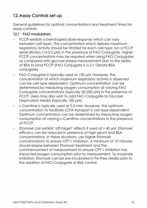
12.Assay Controls set up
General guidelines for optimal concentrations and treatment times for assay controls:12.1 FAO modulators: FCCP exhibits a bell-shaped dose-response which can vary
between cell types. The concentration which delivers maximum respiratory activity should be titrated for each cell type: run a FCCP serial dilution (15-0.5 µM) in the presence of FAO Conjugate. Higher FCCP concentrations may be required when using FAO Conjugates as compared with glucose-based measurement due to the ability of BSA to bind FCCP (FAO Conjugate is a 2:1 Oleate-BSA conjugate).
FAO Conjugate is typically used at 150 μM. However, the concentration at which maximum respiratory activity is observed can be cell type dependent. Optimum concentration can be determined by measuring oxygen consumption at varying FAO Conjugate concentrations (typically 50-200 μM) in the presence of FCCP. Users may also wish to add FAO Conjugate to Glucose Deprivation Media (typically 100 μM).
L-Carnitine is typically used at 0.5 mM. However, the optimum concentration to facilitate LCFA transport is cell type-dependent. Optimum concentration can be determined by measuring oxygen consumption at varying L-Carnitine concentrations in the presence of FCCP.
Etomoxir can exhibit ‘off-target’ effects if used at > 40 µM. Etomoxir efficacy can be reduced in presence of high serum and BSA concentrations. In these situations, use higher Etomoxir concentrations to ensure CPT-1 inhibition. A minimum of 10 minutes should elapse between Etomoxir treatment and the commencement of measurement to ensure CPT-1 inhibition has impacted oxygen consumption prior to measurement. To maximize inhibition, Etomoxir can be pre-incubated in FA-Free Media prior to the addition of FAO Conjugate or BSA control.
ab217602 Fatty Acid Oxidation Assay Kit 13
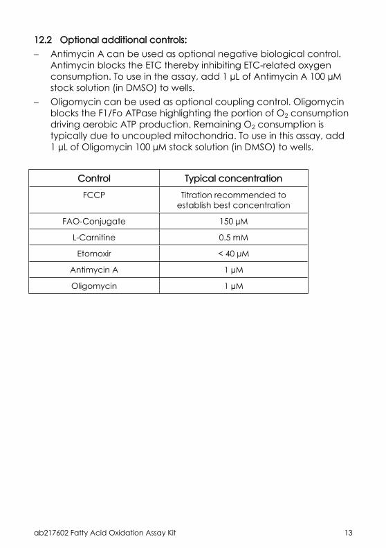
12.2 Optional additional controls: Antimycin A can be used as optional negative biological control.
Antimycin blocks the ETC thereby inhibiting ETC-related oxygen consumption. To use in the assay, add 1 µL of Antimycin A 100 µM stock solution (in DMSO) to wells.
Oligomycin can be used as optional coupling control. Oligomycin blocks the F1/Fo ATPase highlighting the portion of O2 consumption driving aerobic ATP production. Remaining O2 consumption is typically due to uncoupled mitochondria. To use in this assay, add 1 µL of Oligomycin 100 µM stock solution (in DMSO) to wells.
Control Typical concentration
FCCP Titration recommended to establish best concentration
FAO-Conjugate 150 µM
L-Carnitine 0.5 mM
Etomoxir < 40 µM
Antimycin A 1 µM
Oligomycin 1 µM
ab217602 Fatty Acid Oxidation Assay Kit 14
13.Assay Procedure
This assay is designed to be used as companion kit together in combination with Extracellular Oxygen Consumption Assay (ab197243) The Extracellular Oxygen Consumption Assay User Manual describes instrument set-up, assay optimization, data analysis and troubleshooting. The described instrument set-up and signal optimization steps should be performed prior running an FAO assay.
We recommend that you assay all controls and samples in triplicate.
Use a plate block heater for plate preparation and pre-warm plate reader to measurement temperature (typically 37°C).
Compounds are typically added immediately pre-treatment to determine their effect on FAO and related mitochondrial functional. For some 3D models, a pre-incubation step can be incorporated to ensure compounds access cells within the 3D construct. Longer treatment times can be used as required: in these instances, compound should be present in both culture media (during incubation) and measurement media.
Long-term measurement with CO2 control: additional media buffering capacity is required when conducting long term measurements (> 2 h) outside 5% CO2. This is achieved by supplementing Measurement Media with 5 mM HEPES. Supplementation is not required if plate reader has environmental gas control where 5% CO2 can be maintained within the measurement chamber.
13.1 Prepare additional reagents:13.1.1 FA-Free Measurement Media: To Base Measurement Media
(Step 9.3), add 0.5 mM L-Carnitine (1:100 final dilution) and 2.5 mM glucose.
Note: Optimal L-Carnitine and glucose concentration may be cell-type specific and maybe require additional optimization. Note: If long term measurements are being performed outside 5% CO2 a HEPES supplement is recommended.13.1.2 FA Measurement Media: FA-free Measurement Media + 150 µM
FAO Conjugate (1:20 dilution from 3 mM stock – Step 9.1).
ab217602 Fatty Acid Oxidation Assay Kit 15
13.1.3 Extracellular O2 Consumption Reagent: prepare reagent as described in Extracellular O2 Consumption Assay (ab197243) user manual.
13.2 Wash cells:13.2.1 Place the plate on a plate block heater set to assay
temperature (typically 37°C) and remove spent culture media with an aspirator (be careful not to dislodge cells from the base of the wells).
13.2.2 With a multichannel or repeater pipette, add 100 μL of the pre-warmed FA-Free Media to each well.
13.2.3 Repeat wash step one more time.13.3 Add assay media to wells: Signal control wells (wells with no cells; row H) = 90 µL of pre-
warmed FA Measurement Media. Blank control wells (H11 and H12) = 90 µL of pre-warmed FA
Measurement Media. Sample wells = 90 μL of pre-warmed FA Measurement Media. FA-Free control wells =85 μL of FA-Free Measurement Media + 5 μL
of BSA control. Note: FA-Free Measurement Media is used as a control to measure O2 consumption without FAO Conjugate. BSA control is added to ensure that the free concentrations of test compounds are consistent between FA and FA-Free conditions. BSA concentration in FA-free control wells should be consistent with the BSA concentration in samples containing FAO Conjugate.13.3.1 Add 10 μL of Extracellular O2 Consumption Reagent (Step 12.1.3)
to each sample, FA-Free control and signal control wells. Do not add to blank control wells.
13.3.2 Add 10 µL of FA Measurement Media to Blank Control wells.Note: If measuring a full 96-well plate, we recommend diluting reconstituted ab197243 stock 1 in 10 in the relevant measurement media and, using a multichannel pipette, to add 100 μL to each well. Add 100 μL of FA Measurement Media (no ab197243 reagent) to the Blank Control wells.13.4 Treat cells:Add relevant treatment or compounds to cells (see Section 13 for more detailed information on how to perform assays). The procedure below describes how to perform drug screening:
ab217602 Fatty Acid Oxidation Assay Kit 16
13.4.1 Add test compound or vehicle (typically 1-5 μL) to test wells (Step 12.3): we recommend using 6-8 compound dilutions.
Note: We recommend keeping volume of added compound as low as possible to minimize any potential vehicle effects. Note: Additional BSA control stock is added to wells without Oleate (Step 12.3.1-12.3.2). Cells should be co-treated with FCCP if impact on maximal FAO is being determined. Etomoxir is used as a control.13.5 Measurement:13.5.1 Seal each well with 100 µL of pre-warmed High Sensitivity Mineral
Oil (component from ab197243), taking care to avoid bubbles.13.5.2 Read plate immediately in a fluorescence microplate reader as
described in the protocol booklet for Extracellular Oxygen Consumption Assay (ab197243).
13.6 Calculations:13.6.1 Process data as described in Section 15 of the protocol booklet
for Extracellular Oxygen Consumption Assay (ab197243).
ab217602 Fatty Acid Oxidation Assay Kit 17
14.Typical Assays/Data
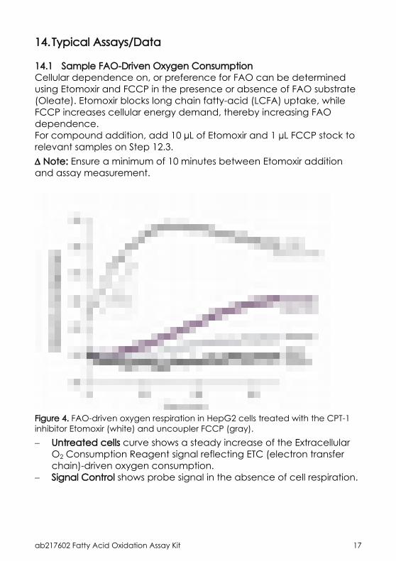
14.1 Sample FAO-Driven Oxygen ConsumptionCellular dependence on, or preference for FAO can be determined using Etomoxir and FCCP in the presence or absence of FAO substrate (Oleate). Etomoxir blocks long chain fatty-acid (LCFA) uptake, while FCCP increases cellular energy demand, thereby increasing FAO dependence.For compound addition, add 10 µL of Etomoxir and 1 µL FCCP stock to relevant samples on Step 12.3. Note: Ensure a minimum of 10 minutes between Etomoxir addition and assay measurement.
Figure 4. FAO-driven oxygen respiration in HepG2 cells treated with the CPT-1 inhibitor Etomoxir (white) and uncoupler FCCP (gray). Untreated cells curve shows a steady increase of the Extracellular
O2 Consumption Reagent signal reflecting ETC (electron transfer chain)-driven oxygen consumption.
Signal Control shows probe signal in the absence of cell respiration.
ab217602 Fatty Acid Oxidation Assay Kit 18
Etomoxir treatment prevents oleate import, resulting in reduced availability of reducing equivalents and a resultant decrease in ETC activity. The remaining ETC activity (difference between Etoxomir treatment and Signal Control) is driven by metabolic activity other than long chain FAO.
FCCP treatment induces maximal ETC activity by dissipating the mitochondrial membrane potential. Increased demand for reducing equivalents causes a concomitant increase in FAO as indicated by the rapid increase in Extracellular O2 Consumption Reagent signal. This strong increase in ETC activity is not observed where exogenous LCFA is unavailable or where import is inhibited.
ab217602 Fatty Acid Oxidation Assay Kit 19
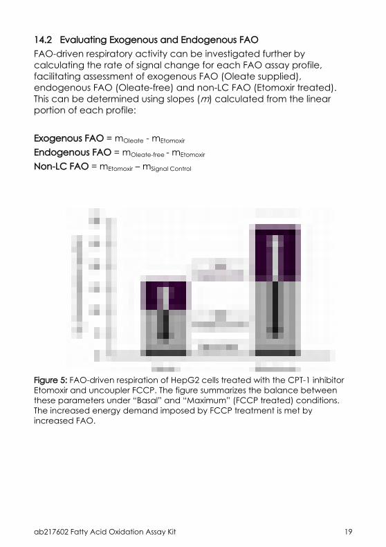
14.2 Evaluating Exogenous and Endogenous FAOFAO-driven respiratory activity can be investigated further by calculating the rate of signal change for each FAO assay profile, facilitating assessment of exogenous FAO (Oleate supplied), endogenous FAO (Oleate-free) and non-LC FAO (Etomoxir treated). This can be determined using slopes (m) calculated from the linear portion of each profile:
Exogenous FAO = mOleate - mEtomoxir
Endogenous FAO = mOleate-free - mEtomoxir
Non-LC FAO = mEtomoxir – mSignal Control
Figure 5: FAO-driven respiration of HepG2 cells treated with the CPT-1 inhibitor Etomoxir and uncoupler FCCP. The figure summarizes the balance between these parameters under “Basal” and “Maximum” (FCCP treated) conditions. The increased energy demand imposed by FCCP treatment is met by increased FAO.
ab217602 Fatty Acid Oxidation Assay Kit 20
15.Notes
Copyright © 2017 Abcam. All rights reserved
Version 11 Last updated 26 June 2017
ab197243Extracellular O2 Consumption Assay
For the measurement of extracellular oxygen consumption in isolated mitochondria, cell populations, 3D culture models, tissues and enzymes.
This product is for research use only and is not intended for diagnostic use.

Copyright © 2016 Abcam. All rights reserved
Table of Contents
1. Overview 23
2. Protocol Summary 24
3. Precautions 25
4. Storage and Stability 25
5. Limitations 26
6. Materials Supplied 26
7. Materials Required, Not Supplied 27
8. Technical Hints 28
9. Reagent Preparation 29
10. Plate Reader Set-Up 30
11. Signal Optimization 36
12. Sample Preparation 37
13. Assay Procedure 39
14. Assay Procedure for 384 well plate 41
15. Calculations 42
16. Typical Data 43
17. Assay Throughput and Performance 44
18. Additional Assays/Data 46
19. Notes 51
ab197243 Extracellular O2 Consumption Assay 23
1. Overview
Extracellular O2 Consumption Assay (ab197243) measures extracellular oxygen consumption rate (OCR) in a variety of samples.Mitochondrial dysfunction is implicated in numerous disease states and is also a major mechanism of drug-induced toxicity. Oxygen consumption is one of the most informative and direct measures of mitochondrial function. Traditional methods of measuring oxygen consumption are hampered by the limitations of low throughput and high complexity. The Extracellular O2 Consumption Assay (ab197243) solves these limitations by providing a direct, real-time measurement of extracellular oxygen consumption rate (OCR) to analyze cellular respiration and mitochondrial function.
The assay is based on the ability of oxygen to quench the excited state of Extracellular O2 Consumption reagent present in the kit. As the test material respires, oxygen is depleted in the surrounding environment, which is seen as an increase in phosphorescence signal. The addition of a high-sensitivity mineral oil is used to limit back diffusion of ambient oxygen. Measured on standard fluorescence plate readers (96- or 384- well), with standard cell culture microplates, the Extracellular O2 Consumption Assay (ab197243) is suitable for use with whole cell populations (both adherent and suspension cells), isolated mitochondria, a wide range of 3D culture models, tissues, small organisms, as well as isolated enzymes, bacteria, yeasts and molds.
The flexible plate reader format, allows multiparametric or multiplex combination with other similar products. For example, in combination with Glycolysis Assay (ab197244), the Extracellular O2 Consumption Assay (ab197243) allows simultaneous real-time measurement of mitochondrial respiration and glycolysis and the analysis of the metabolic phenotype of cells and the shift (flux) between the two pathways under pathological states.
ab197243 Extracellular O2 Consumption Assay 24
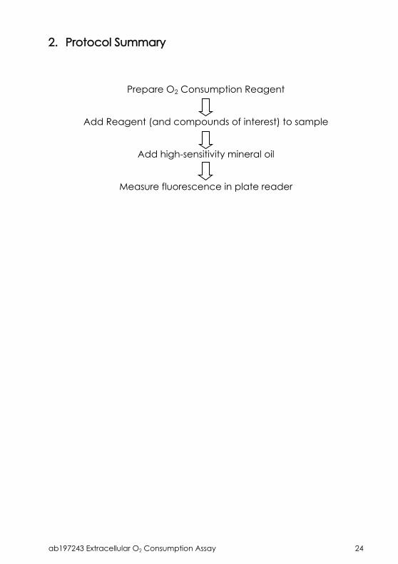
2. Protocol Summary
Prepare O2 Consumption Reagent
Add Reagent (and compounds of interest) to sample
Add high-sensitivity mineral oil
Measure fluorescence in plate reader
ab197243 Extracellular O2 Consumption Assay 25
3. Precautions
Please read these instructions carefully prior to beginning the assay.
All kit components have been formulated and quality control tested to function successfully as a kit.
We understand that, occasionally, experimental protocols might need to be modified to meet unique experimental circumstances. However, we cannot guarantee the performance of the product outside the conditions detailed in this protocol booklet.
Reagents should be treated as possible mutagens and should be handle with care and disposed of properly. Please review the Safety Datasheet (SDS) provided with the product for information on the specific components.
Observe good laboratory practices. Gloves, lab coat, and protective eyewear should always be worn. Never pipet by mouth. Do not eat, drink or smoke in the laboratory areas.
All biological materials should be treated as potentially hazardous and handled as such. They should be disposed of in accordance with established safety procedures.
4. Storage and Stability
Store kit at 4°C in the dark immediately upon receipt. Kit has a storage time of 1 year from receipt, providing components have not been reconstituted.Refer to list of materials supplied for storage conditions of individual components. Observe the storage conditions for individual prepared components in the Materials Supplied section.Aliquot components in working volumes before storing at the recommended temperature.
Note: Reconstituted reagent is stable for 1 month.
ab197243 Extracellular O2 Consumption Assay 26
5. Limitations
Assay kit intended for research use only. Not for use in diagnostic procedures.
Do not mix or substitute reagents or materials from other kit lots or vendors. Kits are QC tested as a set of components and performance cannot be guaranteed if utilized separately or substituted.
6. Materials Supplied
Quantity
Item96 tests 4 x 96
tests
Storage condition (before prep)
Storage condition
(after prep)
Extracellular O2 Consumption Reagent 1 vial 4 x 1 vial 4°C -20°C
High Sensitivity Mineral Oil 1 dropper bottle
4 x 1 dropper bottle
4°C RT
ab197243 Extracellular O2 Consumption Assay 27
7. Materials Required, Not Supplied
These materials are not included in the kit, but will be required to successfully perform this assay: Microplate reader capable of measuring fluorescence, with
suitable filter and plate temperature control – see Instrument and Measurement Settings section for suitable plate readers
Double distilled water (ddH2O) Pipettes and pipette tips, including multi-channel pipette Assorted glassware for the preparation of reagents and buffer
solutions Tubes for the preparation of reagents and buffer solutions Sterile 96 well plate (black wall with clear flat bottom), or
standard clear plates for cell cultureFor cells:
Cell culture mediumFor isolated mitochondria:
Measurement buffer (250 mM sucrose, 15 mM KCl, 1 mM EGTA, 5 mM MgCl2, 30 mM, K2HPO4, pH 7.4)
Mitochondrial substrate (succinate, glutamate or malate) ADP
ab197243 Extracellular O2 Consumption Assay 28
8. Technical Hints
This kit is sold based on number of tests. A “test” simply refers to a single assay well. The number of wells that contain sample, control or standard will vary by product. Review the protocol completely to confirm this kit meets your requirements. Please contact our Technical Support staff with any questions.
Selected components in this kit are supplied in surplus amount to account for additional dilutions, evaporation, or instrumentation settings where higher volumes are required. They should be disposed of in accordance with established safety procedures.
Avoid foaming or bubbles when mixing or reconstituting components.
Avoid cross contamination of samples or reagents by changing tips between sample, standard and reagent additions.
Ensure plates are properly sealed or covered during incubation steps.
Ensure all reagents and solutions are at the appropriate temperature before starting the assay.
Make sure all necessary equipment is switched on and set at the appropriate temperature.
Refer to Instrument and Measurement Settings table (Table 1) for recommended settings for your plate reader.
While compatible with all plate types, black border clear bottom plats give optimal signal-to-noise ratios.
For first time users, we recommend performing a Signal Optimization Step (see Section 11).
ab197243 Extracellular O2 Consumption Assay 29
9. Reagent Preparation
Briefly centrifuge small vials at low speed prior to opening.
9.1 Extracellular O2 Consumption Reagent:Prepare a stock solution of the Extracellular O2 Consumption Reagent by adding 1 mL of ddH2O, PBS, culture media or buffer to the vial. Mix by gently aspirating 3 – 4 times.Recommended working dilution = 1/15 (10 µL per 150 µL of sample for 1x 96-wp).Avoid freeze/thaw. Reconstituted reagent is stable for one month.
9.2 High Sensitivity Mineral Oil:Ready to use as supplied. Pre-warm to 37°C prior to use.Although mineral oil is provided in a dropper bottle for convenience, we recommend using a repeater pipette for routine use. To apply oil using a repeater pipette, trim 3 – 4 mm off the tip at a 45° angle. Remove internal nozzle cap from the dropper bottle and slowly pick up the pre-warmed mineral oil.Store mineral oil at room temperature in the dark.
ab197243 Extracellular O2 Consumption Assay 30
10.Plate Reader Set-Up
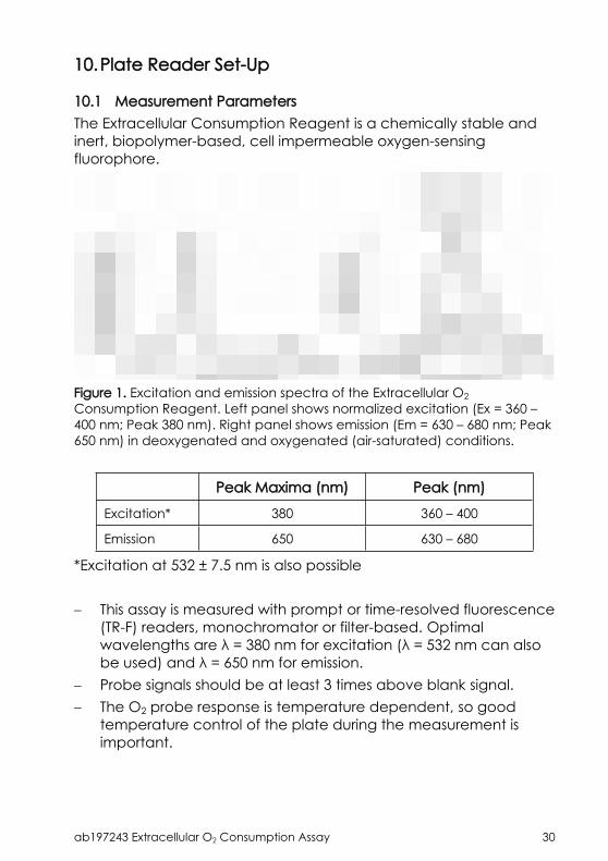
10.1 Measurement ParametersThe Extracellular Consumption Reagent is a chemically stable and inert, biopolymer-based, cell impermeable oxygen-sensing fluorophore.
Figure 1. Excitation and emission spectra of the Extracellular O2 Consumption Reagent. Left panel shows normalized excitation (Ex = 360 – 400 nm; Peak 380 nm). Right panel shows emission (Em = 630 – 680 nm; Peak 650 nm) in deoxygenated and oxygenated (air-saturated) conditions.
Peak Maxima (nm) Peak (nm)
Excitation* 380 360 – 400
Emission 650 630 – 680
*Excitation at 532 ± 7.5 nm is also possible
This assay is measured with prompt or time-resolved fluorescence (TR-F) readers, monochromator or filter-based. Optimal wavelengths are λ = 380 nm for excitation (λ = 532 nm can also be used) and λ = 650 nm for emission.
Probe signals should be at least 3 times above blank signal. The O2 probe response is temperature dependent, so good
temperature control of the plate during the measurement is important.
ab197243 Extracellular O2 Consumption Assay 31
10.2 Fluorescence measurementsOutlined below are three fluorescence modalities that can be used with this assay, depending on the plate reader type and instrument setup.10.2.1Basic: Intensity MeasurementMeasurement of Signal Intensity (sometimes referred to as Prompt) provides flexibility to use wide range of commonly available fluorescence-, monochromator or filter-based plate readers. Optimal wavelengths are λ = 380 nm for excitation and λ = 650 nm for emission, with detection gain parameters (PMT) typically set at medium or high. Note: Extracellular consumption reagent should return Signal to Blank ratio (S:B) ≥ 3.
10.2.2Standard: TR-F MeasurementUsing time-resolved fluorescence (TR-F) will increase performance levels. TR-F measurement reduces non-specific background and increases sensitivity.Optimal delay time is 30 µs and gate (integration) time is 100 µs. Note: Extracellular consumption reagent should return Signal to Blank ratio (S:B) ≥ 3. S:B ~ 10 are typical.
10.2.3Advanced: Dual-Read TR-F (Lifetime calculation)Optimal performance can be achieved using dual-read TR-F in combination with subsequent ratiometric Lifetime calculation, to maximize dynamic range. Note: Extracellular consumption reagent should return Signal to Blank ratio (S:B) ≥ 3. S:B up to 60 are possible.
Dual-read TR-F and subsequent Lifetime calculation allows measurement of the rate of fluorescence decay of the Extracellular consumption reagent, and can provide measurements of oxygen consumption that are more stable and with a wider dynamic range than measuring signal intensity.
ab197243 Extracellular O2 Consumption Assay 32
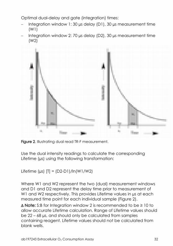
Optimal dual-delay and gate (integration) times: Integration window 1: 30 µs delay (D1), 30 µs measurement time
(W1) Integration window 2: 70 µs delay (D2), 30 µs measurement time
(W2)
Figure 2. Illustrating dual read TR-F measurement.
Use the dual intensity readings to calculate the corresponding Lifetime (µs) using the following transformation:
Lifetime (µs) [T] = (D2-D1)/ln(W1/W2)
Where W1 and W2 represent the two (dual) measurement windows and D1 and D2 represent the delay time prior to measurement of W1 and W2 respectively. This provides Lifetime values in µs at each measured time point for each individual sample (Figure 2). Note: S:B for Integration window 2 is recommended to be ≥ 10 to allow accurate Lifetime calculation. Range of Lifetime values should be 22 – 68 µs, and should only be calculated from samples containing reagent. Lifetime values should not be calculated from blank wells.
ab197243 Extracellular O2 Consumption Assay 33
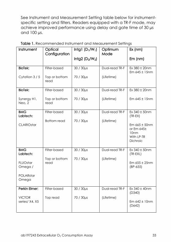
See Instrument and Measurement Setting table below for instrument-specific setting and filters. Readers equipped with a TR-F mode, may achieve improved performance using delay and gate time of 30 µs and 100 µs.
Table 1. Recommended Instrument and Measurement SettingsInstrument Optical
Configuration Intg1 (D1/W1)
Intg2 (D2/W2)
Optimum Mode
Ex (nm)
Em (nm)
BioTek:
Cytation 3 / 5
Filter-based
Top or bottom read
30 / 30μs
70 / 30μs
Dual-read TR-F
(Lifetime)
Ex 380 ± 20nmEm 645 ± 15nm
BioTek:
Synergy H1, Neo, 2
Filter-based
Top or bottom read
30 / 30μs
70 / 30μs
Dual-read TR-F
(Lifetime)
Ex 380 ± 20nm
Em 645 ± 15nm
BMG Labtech:
CLARIOstar
Filter-based
Bottom read
30 / 30μs
70 / 30μs
Dual-read TR-F
(Lifetime)
Ex 340 ± 50nm (TR-EX)
Em 665 ± 50nm or Em 645± 10nmWith LP-TR Dichroic
BMG Labtech:
FLUOstar Omega /
POLARstar Omega
Filter-based
Top or bottom read
30 / 30μs
70 / 30μs
Dual-read TR-F
(Lifetime)
Ex 340 ± 50nm (TR-EXL)
Em 655 ± 25nm (BP-655)
Perkin Elmer:
VICTOR series/ X4, X5
Filter-based
Top read
30 / 30μs
70 / 30μs
Dual-read TR-F
(Lifetime)
Ex 340 ± 40nm (D340)
Em 642 ± 10nm (D642)
ab197243 Extracellular O2 Consumption Assay 34
Instrument Optical Configuration
Intg1 (D1/W1)
Intg2 (D2/W2)
Optimum Mode
Ex (nm)
Em (nm)
Tecan:
Infinite M1000Pro /
F200Pro
Monochromator /
Filter-based
Top or bottom read
30 / 30μs
70 / 30μs
Dual-read TR-F
(Lifetime)
Ex 380 ± 20nm
Em 650 ±20nm or Em 670±40nm
BioTek:
Synergy HTx / Mx
Monochromator /
Filter-based
Top or bottom read
30 / 100μs
n/a
TR-F Ex 380±20nm
Em 650±15nm
BMG Labtech:
PHERAstar FS
Filter-based
Top or bottom read
40 / 100μs
n/a
TR-F Ex 337 nm (HTRF Module)
Em 665 nm (HTRF Module)
BMG Labtech:
FLUOstar Optima /
POLARstar Optima
Filter-based
Top or bottom read
30 / 100μs
n/a
TR-F Ex 340 ± 50nm (TR-EXL)
Em 655 ± 50nm (BP-655)
ab197243 Extracellular O2 Consumption Assay 35
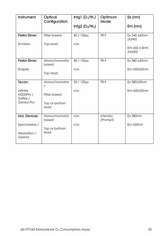
Instrument Optical Configuration
Intg1 (D1/W1)
Intg2 (D2/W2)
Optimum Mode
Ex (nm)
Em (nm)
Perkin Elmer:
EnVision
Filter-based
Top read
40 / 100μs
n/a
TR-F Ex 340 ±60nm (X340)
Em 650 ± 8nm (M650)
Perkin Elmer:
EnSpire
Monochromator based
Top read
40 / 100μs
n/a
TR-F Ex 380 ±20nm
Em 650±20nm
Tecan:
Infinite M200Pro / Saffire / Genios Pro
Monochromator/
Filter-based
Top or bottom read
30 / 100μs
n/a
TR-F Ex 380±20nm
Em 650±20nm
Mol. Devices:
SpectraMax /
Flexstation / Gemini
Monochromator based
Top or bottom read
n/a
n/a
Intensity (Prompt)
Ex 380nm
Em 650nm
ab197243 Extracellular O2 Consumption Assay 36
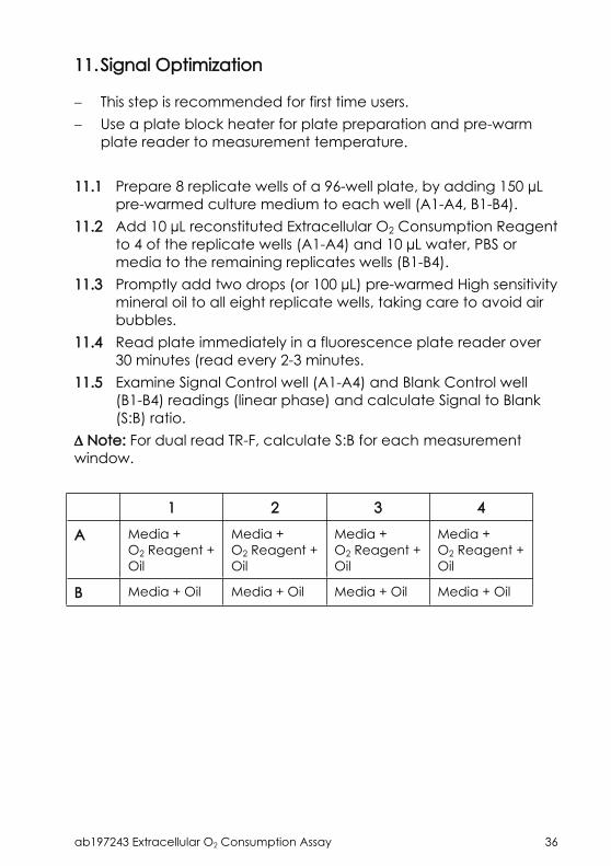
11.Signal Optimization
This step is recommended for first time users. Use a plate block heater for plate preparation and pre-warm
plate reader to measurement temperature.
11.1 Prepare 8 replicate wells of a 96-well plate, by adding 150 µL pre-warmed culture medium to each well (A1-A4, B1-B4).
11.2 Add 10 µL reconstituted Extracellular O2 Consumption Reagent to 4 of the replicate wells (A1-A4) and 10 µL water, PBS or media to the remaining replicates wells (B1-B4).
11.3 Promptly add two drops (or 100 µL) pre-warmed High sensitivity mineral oil to all eight replicate wells, taking care to avoid air bubbles.
11.4 Read plate immediately in a fluorescence plate reader over 30 minutes (read every 2-3 minutes.
11.5 Examine Signal Control well (A1-A4) and Blank Control well (B1-B4) readings (linear phase) and calculate Signal to Blank (S:B) ratio.
Note: For dual read TR-F, calculate S:B for each measurement window.
1 2 3 4
A Media +O2 Reagent + Oil
Media +O2 Reagent + Oil
Media +O2 Reagent + Oil
Media +O2 Reagent + Oil
B Media + Oil Media + Oil Media + Oil Media + Oil
ab197243 Extracellular O2 Consumption Assay 37
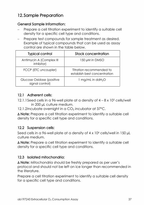
12.Sample Preparation
General Sample Information: Prepare a cell titration experiment to identify a suitable cell
density for a specific cell type and conditions. Prepare test compounds for sample treatment as desired.
Example of typical compounds that can be used as assay control are shown in the table below.
Typical control Stock concentration
Antimycin A (Complex III inhibitor)
150 µM in DMSO
FCCP (ETC uncoupler) Titration recommended to establish best concentration
Glucose Oxidase (positive signal control)
1 mg/mL in ddH2O
12.1 Adherent cells:12.1.1Seed cells in a 96-well plate at a density of 4 – 8 x 10⁵ cells/well
in 200 µL culture medium.12.1.2Incubate overnight in a CO2 incubator at 37°C. Note: Prepare a cell titration experiment to identify a suitable cell density for a specific cell type and conditions.
12.2 Suspension cells:Seed cells in a 96-well plate at a density of 4 x 10⁶ cells/well in 150 µL culture medium. Note: Prepare a cell titration experiment to identify a suitable cell density for a specific cell type and conditions.
12.3 Isolated mitochondria: Note: Mitochondria should be freshly prepared as per user’s protocol and should not be left on ice longer than recommended in the literature.Prepare a cell titration experiment to identify a suitable cell density for a specific cell type and conditions.

ab197243 Extracellular O2 Consumption Assay 38
Initial isolated mitochondria assay optimization: prepare a six-point dilution series of mitochondrial preparation in respiration buffer in 1.5 mL total volume for each concentration.
12.3.1Prepare measurement buffer as follows: 250 mM sucrose, 15 mM KCl, 1 mM EGTA, 5 mM MgCl2, 30 mM, K2HPO4; adjust to pH 7.4.
12.3.2Dilute isolated mitochondria to the desired concentration (typical range = 0.125 – 1.5 mg/mL final concentration) in measurement buffer, depending on the substrate(s) used and which respiration state is being measured.
ab197243 Extracellular O2 Consumption Assay 39
13.Assay Procedure
We recommend that you assay all controls and samples in duplicate.
Prepare all controls and samples as directed in the previous sections.
Use a plate block heater for plate preparation and pre-warm plate reader to measurement temperature (typically 37°C; 30°C for mitochondria).
Sufficient cell numbers are required to produce measurable signal changes. Oxygen consumption rate is cell-type dependent – highly glycolytic cells may need to be trypsinized and concentrated prior to measurement.
PROTOCOL FOR CELLS:13.1 Plate loading:13.1.1Adherent cells: remove culture media from all assay wells and
replace with 150 µL of fresh culture media.Suspension cells: ready to use as prepared in Step 12.2.
13.1.2Blank controls (we suggest using wells H11 and H12): add 150 µL fresh culture media.
13.2 Assay set up:13.2.1Add 10 µL reconstituted Extracellular O2 Consumption Reagent
to each sample well.13.2.2Add 10 µL of fresh culture media to blank control wells.13.2.3Add 1 – 10 µL test compound (vehicle control and/or stock) to
the wells. Note: we recommend keeping the volume of added compound low to minimize any potential effects of solvent vehicle.13.2.4Promptly seal each well by adding 100 µL (or 2 drops) of pre-
warmed High Sensitivity mineral oil, taking care to avoid air bubbles.
Note: plate preparation time should be kept to a minimum.

ab197243 Extracellular O2 Consumption Assay 40
13.3 Measurement:13.3.1Insert the prepared plate into a fluorescence plate reader
pre-set to the measurement temperature (typically 37°C).13.3.2Measure Extracellular O2 Consumption signal at 1.5 min
intervals for 90 – 120 minutes (longer for more glycolytic cells) at Ex/Em = 380/650 nm.
PROTOCOL FOR ISOLATED MITOCHONDRIA:13.4 Dilute reconstituted Extracellular O2 Consumption Reagent
(Step 9.1)1:10 in measurement buffer.13.5 Add 100 µL reconstituted probe to each sample well. 13.6 Add 1 µL test compound in appropriate solvent to the wells.13.7 Add 50 µL of diluted isolated mitochondria (Step 12.3) to each
test well. For blank control wells (wells H11 and H12), add 200 µL fresh culture media.
13.8 Dissolve substrate in measurement buffer and add 50 µL of solution to test wells (see table below for suggested concentrations). Do not add substrate to blank control wells.
Substrate Mitochondria Concentration (mg/mL)
Typical final substrate concentration (mM)
Basal state [State 2]
Glutamate/Malate 1.5 12.5 / 12.5
Succinate 1.0 25
ADP-stimulated respiration rate [State 3]
Glutamate/Malate/ADP 1.0 12.5 / 12.5 / 1.65
Succinate/ADP 0.5 25 / 1.65
13.9 Promptly seal each well by adding 100 µL (or 2 drops) of High Sensitivity mineral oil, pre-warmed at 30°C, taking care to avoid bubbles.
13.10 Insert the prepared plate into a fluorescence plate reader pre-set to 30°C.
13.11 Measure Extracellular O2 Consumption signal at 1.5 min intervals for 10 – 30 minutes at Ex/Em = 380/650 nm.
ab197243 Extracellular O2 Consumption Assay 41
14.Assay Procedure for 384 well plate
This kit provides enough reagent to perform 200 tests in 384-wp format (half plate).Follow the same recommendations outlined in Section 13. Adherent cells: seed cells in a 384-wp at a density of 2 – 4 x 10⁵
cells/well in 100 µL culture medium overnight.14.1 Cell preparation: Adherent cells: seed cells in a 384-wp at a density of 2 – 4 x 10⁵
cells/well in 100 µL culture medium overnight. Suspension cells: prepare a cell concentration stock of 4 x 10⁶
cells/mL. Add 75 µL cells per well. Note: Prepare a cell titration experiment to identify a suitable cell density for a specific cell type and conditions.14.2 Plate loading:14.2.1Adherent cells: remove culture media from all assay wells and
replace with 75 µL of fresh culture media.Suspension cells: ready to use as prepared in Step 14.1.
14.2.2Blank controls (we suggest using wells H11 and H12): add 75 µL fresh culture media (for cell-based assay).
14.3 Assay set up:14.3.1Add 5 µL reconstituted Extracellular O2 Consumption Reagent
to each sample well.14.3.2Add 5 µL of fresh culture media to blank control wells.14.3.3Add 1 – 5 µL test compound (vehicle control and/or stock) to
the wells. Note: we recommend keeping the volume of added compound low to minimize any potential effects of solvent vehicle.14.3.4Promptly seal each well by adding 50 µL (or 1 drops) of pre-
warmed High Sensitivity mineral oil, taking care to avoid air bubbles.
Note: High Sensitivity mineral oil is very viscous and it might be difficult to plate into smaller wells. Note: plate preparation time should be kept to a minimum.14.4 Measurement:14.4.1Follow instructions described in page 18.

ab197243 Extracellular O2 Consumption Assay 42
15.Calculations
15.1 Plot the Blank control well-corrected Extracellular O2 consumption assay Intensity or Lifetime values versus Time (min).
15.2 Select the linear proportion of the signal profile (avoiding any initial lag of subsequent plateau) and apply linear regression to determine the slope (OCR) and correlation coefficient for each well.
Note: this approach is preferable to calculating a slope from averaged profiles.15.3 Tabulate the slope values for each test sample, calculating
appropriate average and standard deviation values across replicate wells. If optional Signal Control wells are included, the slope obtained for the Signal Control (sample without cells) should be subtracted from all test values.
ab197243 Extracellular O2 Consumption Assay 43
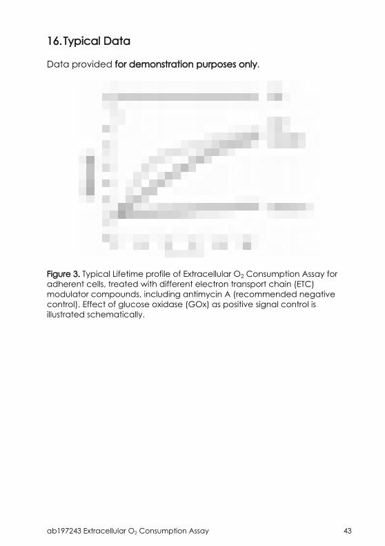
16.Typical Data
Data provided for demonstration purposes only.
Figure 3. Typical Lifetime profile of Extracellular O2 Consumption Assay for adherent cells, treated with different electron transport chain (ETC) modulator compounds, including antimycin A (recommended negative control). Effect of glucose oxidase (GOx) as positive signal control is illustrated schematically.
ab197243 Extracellular O2 Consumption Assay 44
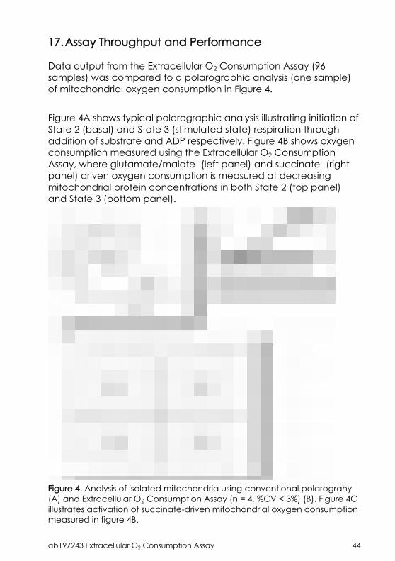
17.Assay Throughput and Performance
Data output from the Extracellular O2 Consumption Assay (96 samples) was compared to a polarographic analysis (one sample) of mitochondrial oxygen consumption in Figure 4.
Figure 4A shows typical polarographic analysis illustrating initiation of State 2 (basal) and State 3 (stimulated state) respiration through addition of substrate and ADP respectively. Figure 4B shows oxygen consumption measured using the Extracellular O2 Consumption Assay, where glutamate/malate- (left panel) and succinate- (right panel) driven oxygen consumption is measured at decreasing mitochondrial protein concentrations in both State 2 (top panel) and State 3 (bottom panel).
Figure 4. Analysis of isolated mitochondria using conventional polarograhy (A) and Extracellular O2 Consumption Assay (n = 4, %CV < 3%) (B). Figure 4C illustrates activation of succinate-driven mitochondrial oxygen consumption measured in figure 4B.
ab197243 Extracellular O2 Consumption Assay 45
The compatibility of this assay with the microplate format allows analysis under 96 or 384 discrete conditions. The effectiveness of this level of throughput in analyzing isolated mitochondria is highlighted in Figure 4B, which examines increasing mitochondrial protein concentrations on glutamate/malate- and succinate-driven respiration in both basal (State 2) and ADP activated (State 3) states, all tested in quadruplicates.The performance of the assay is highlighted in Figure 4C, with a coefficient of variance (%CV) below 3%.

ab197243 Extracellular O2 Consumption Assay 46
18.Additional Assays/Data
18.1 Monitoring cell respirationThe ability of this product to assess cellular respiration is illustrated in Figure 4. Dilutions curves for HepG2 cells (panel A) and primary rat hepatocytes (panel B) are presented.
Figure 5. Cell dilutions measured on 96-well plates using Extracellular O2 Assay. HepG2 cells (A) were assayed after an overnight (open squares) or 2-day culture period (dark squares); primary rat hepatocytes (B) were assayed after an overnight culture period. Rates of probe signal change (slope of fluorescence signal) were normalized against initial intensity.
ab197243 Extracellular O2 Consumption Assay 47
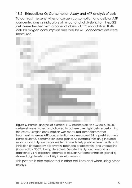
18.2 Extracellular O2 Consumption Assay and ATP analysis of cellsTo contrast the sensitivities of oxygen consumption and cellular ATP concentrations as indicators of mitochondrial dysfunction, HepG2 cells were treated with a panel of classical ETC modulators. Both cellular oxygen consumption and cellular ATP concentrations were measured.
Figure 6. Parallel analysis of classical ETC inhibitors on HepG2 cells. 80,000 cells/well were plated and allowed to adhere overnight before performing the assay. Oxygen consumption was measured immediately after treatment, whereas ATP concentration was measured 24 hr post-treatment. Extracellular O2 consumption data (panel A) illustrates that drug induced mitochondrial dysfunction is evident immediately post-treatment with both inhibition (induced by oligomycin, rotenone or antimycin) and uncoupling (induced by FCCP) being detected. Despite this dysfunction and an additional 24 hr exposure, analysis of cellular ATP concentration (panel B) showed high levels of viability in most scenarios.This pattern is also replicated in other cell lines and when using other assays.
ab197243 Extracellular O2 Consumption Assay 48
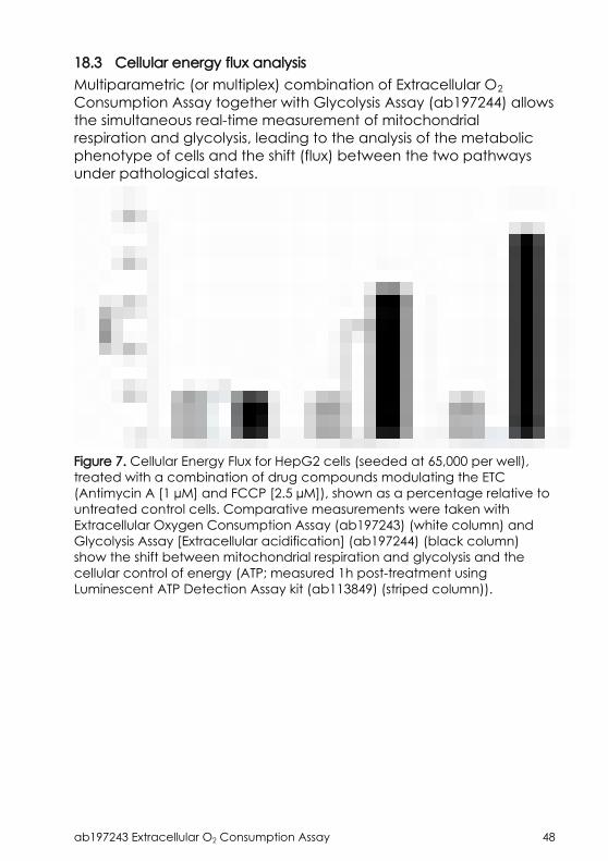
18.3 Cellular energy flux analysisMultiparametric (or multiplex) combination of Extracellular O2 Consumption Assay together with Glycolysis Assay (ab197244) allows the simultaneous real-time measurement of mitochondrial respiration and glycolysis, leading to the analysis of the metabolic phenotype of cells and the shift (flux) between the two pathways under pathological states.
Figure 7. Cellular Energy Flux for HepG2 cells (seeded at 65,000 per well), treated with a combination of drug compounds modulating the ETC (Antimycin A [1 µM] and FCCP [2.5 µM]), shown as a percentage relative to untreated control cells. Comparative measurements were taken with Extracellular Oxygen Consumption Assay (ab197243) (white column) and Glycolysis Assay [Extracellular acidification] (ab197244) (black column) show the shift between mitochondrial respiration and glycolysis and the cellular control of energy (ATP; measured 1h post-treatment using Luminescent ATP Detection Assay kit (ab113849) (striped column)).
ab197243 Extracellular O2 Consumption Assay 49
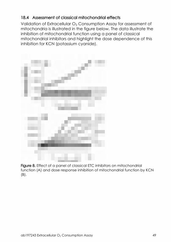
18.4 Assessment of classical mitochondrial effectsValidation of Extracellular O2 Consumption Assay for assessment of mitochondria is illustrated in the figure below. The data illustrate the inhibition of mitochondrial function using a panel of classical mitochondrial inhibitors and highlight the dose dependence of this inhibition for KCN (potassium cyanide).
Figure 8. Effect of a panel of classical ETC inhibitors on mitochondrial function (A) and dose response inhibition of mitochondrial function by KCN (B).
ab197243 Extracellular O2 Consumption Assay 50
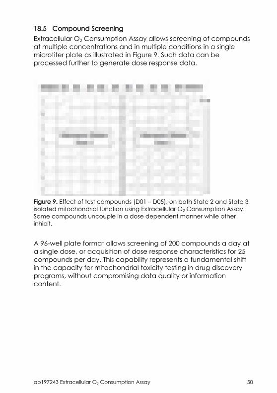
18.5 Compound ScreeningExtracellular O2 Consumption Assay allows screening of compounds at multiple concentrations and in multiple conditions in a single microtiter plate as illustrated in Figure 9. Such data can be processed further to generate dose response data.
Figure 9. Effect of test compounds (D01 – D05), on both State 2 and State 3 isolated mitochondrial function using Extracellular O2 Consumption Assay. Some compounds uncouple in a dose dependent manner while other inhibit.
A 96-well plate format allows screening of 200 compounds a day at a single dose, or acquisition of dose response characteristics for 25 compounds per day. This capability represents a fundamental shift in the capacity for mitochondrial toxicity testing in drug discovery programs, without compromising data quality or information content.

ab197243 Extracellular O2 Consumption Assay 51
19.Notes

Copyright © 2017 Abcam. All rights reserved
Technical Support
Copyright © 2017 Abcam, All Rights Reserved. The Abcam logo is a registered trademark. All information / detail is correct at time of going to print.
[email protected] | [email protected] | [email protected] | [email protected] | 91-114-65-60
[email protected] Deutsch: 043-501-64-24 | Français: 061-500-05-30UK, EU and [email protected] | +44(0)1223-696000
[email protected] | 877-749-8807US and Latin [email protected] | 888-772-2226
Asia Pacific [email protected] | (852) [email protected] | +86-21-5110-5938 | [email protected] | +81-(0)3-6231-0940Singapore [email protected] | 800 188-5244
[email protected] | +61-(0)3-8652-1450New Zealand [email protected] | +64-(0)9-909-7829
Recommended

![Cytometry Kit ab118183 Human Flow Fatty Acid Oxidation Fatt… · [MIM:609016] and maternal acute fatty liver of pregnancy (AFLP) [MIM:609016]. ab118183 Fatty Acid Oxidation Human](https://img.pdfslide.us/doc/110x75/5e19b466bf456616480a7f6d/cytometry-kit-ab118183-human-flow-fatty-acid-oxidation-fatt-mim609016-and-maternal.jpg)