The Faculty of Natural Resources and Agricultural Sciences
A small growth study of Aspergillus section Flavi, and potentially aflatoxigenic fungi and aflatoxin occurrence in Brazil nuts from local markets in Manaus, Brazil
Ellinor Andersson
Department of Microbiology
Independent project • 15 hec • First cycle, G2E
Bachelor of Food Science • Examensarbete/Sveriges lantbruksuniversitet,
Institutionen för mikrobiologi: 2012:13 • ISSN 1101-8151
Uppsala 2012


A small growth study of Aspergillus section Flavi, and potentially aflatoxigenic fungi and aflatoxin occurrence in Brazil nuts from local markets in Manaus, Brazil
Ellinor Andersson
Supervisor: Su-lin Leong, Swedish University of Agricultural Sciences,
Department of Microbiology
Assistant Supervisor: Marta Taniwaki, Instituto de Technologia de Alimentos (ITAL),
Campinas, Brazil
Examiner: Volkmar Passoth, Swedish University of Agricultural Sciences,
Department of Microbiology
Credits: 15 hec
Level: First cycle, G2E
Course title: Independent project in Food Science - bachelor project
Course code: EX0669
Programme/education: Bachelor of Food Science
Place of publication: Uppsala
Year of publication: 2012
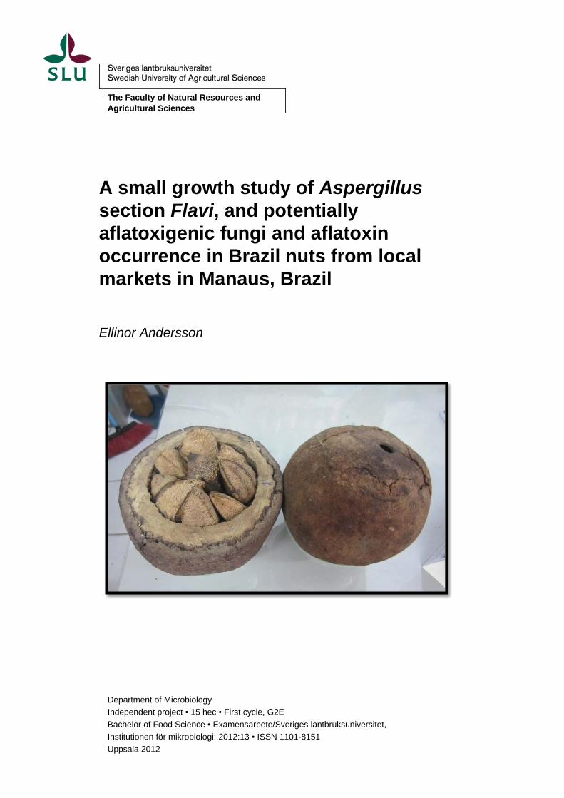
Picture Cover: Brazil nut pods. Photographer: Ellinor Andersson
Title of series: Examensarbete/Sveriges lantbruksuniversitet, Institutionen för mikrobiologi
no: 2012:13
ISSN: 1101-8151
Online publication: http://stud.epsilon.slu.se
Key Words: Aspergillus section Flavi, Aflatoxin, Brazil nut, mould growth, Brazil,
morphological identification
Sveriges lantbruksuniversitet
Swedish University of Agricultural Sciences
The Faculty of Natural Resources and Agricultural Sciences
Uppsala BioCenter
Department of Microbiology

Abstract
This study aimed to gain more knowledge about morphological features of eight different
species of Aspergillus section Flavi, which may aid in identification of these species. The
presence of potentially toxigenic Aspergillus section Flavi, and occurrence of aflatoxins, in
ready-to-eat Brazil nuts (Bertholletia excelsa) were also investigated.
Seventeen different strains, representing eight species from section Flavi, were grown at
different water activities and different temperatures. After 7 days, as well as at 14 and 28
days for the lower water activities, colonies were visually inspected, and diameter and
macroscopic characteristics were noted. Ten samples of Brazil nuts were collected from
different market stalls in the city of Manaus, Brazil. Aspergillus section Flavi were isolated
and identified by morphology and toxin profile. Toxin production was evaluated by the
agar plug technique and quantification of aflatoxins in the Brazil nuts was performed by
high-performance liquid chromatography.
It was found that macromorphology can be used to differentiate between some of the
species if the different methods were combined, but for more certain identification, other
available methods are better. One hundred and thirty five strains of suspected Aspergillus
section Flavi were isolated and aflatoxin production was observed in 100 of them (74%).
The most common species was Aspergillus nomius, followed by A. flavus and A. tamarii.
Total aflatoxin content detected in the nuts did not exceed the European legislative limit of
10 µg/kg for ready-to-eat Brazil nuts, which would be considered safe to be consumed.
Key words: Aspergillus section Flavi, Aflatoxin, Brazil nut, mould growth, Brazil, mor-
phological identification

Table of contents
1 Introduction 7
2 Methods 10 2.1 Growth study of 17 isolates available at Instituto de Technologia de Alimentos,
(ITAL) Campinas, Brazil. ............................................................................................. 10 2.1.1 Media preparation and isolates ........................................................................ 10 2.1.2 Inoculation ....................................................................................................... 11 2.1.3 Water activity and inspection ........................................................................... 11
2.2 Isolation and identification of potentially toxigenic fungi from Brazil nuts collected from market stalls. ...................................................................................................... 11 2.2.1 Sampling.......................................................................................................... 11 2.2.2 Water activity ................................................................................................... 11 2.2.3 Fungal isolation and identification of potential aflatoxigenic species ............... 12 2.2.4 Aflatoxin analysis ............................................................................................. 12
3 Results and discussion 14 3.1 Growth study ............................................................................................................... 14
3.1.1 Colony appearance ......................................................................................... 14 3.1.2 Colony size at different temperatures and different water activities ................. 15 3.1.3 Summary: Growth ............................................................................................ 18
3.2 Isolation and identification of toxigenic fungi from collected Brazil nuts. ..................... 18 3.2.1 Infection ........................................................................................................... 18 3.2.2 Aspergillus section Flavi from Brazil nuts ........................................................ 19 3.2.3 Aflatoxin in Brazil nuts ..................................................................................... 21 3.2.4 Summary: Fungal infection and aflatoxin production in Brazil nuts .................. 21
3.3 Conclusions ................................................................................................................ 21
4 Acknowledgements 23
5 References 24
6 Appendices 27 6.1 Water activity of CYA media ....................................................................................... 27 6.2 Water activity of Brazil nut kernels .............................................................................. 27 6.3 Infection with total fungi, Aspergillus section Flavi, section Nigri and Penicillium
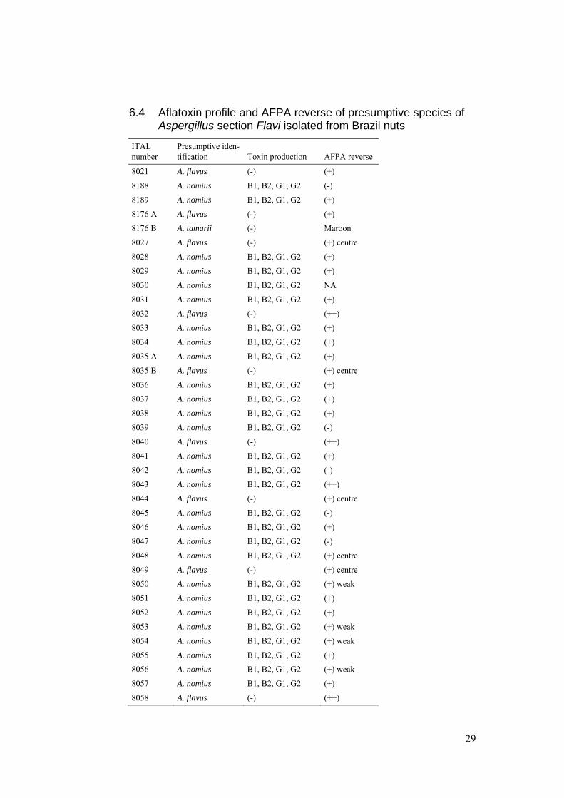
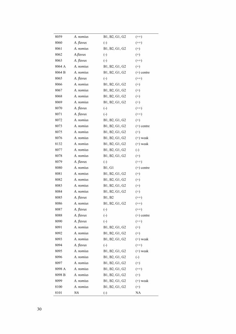
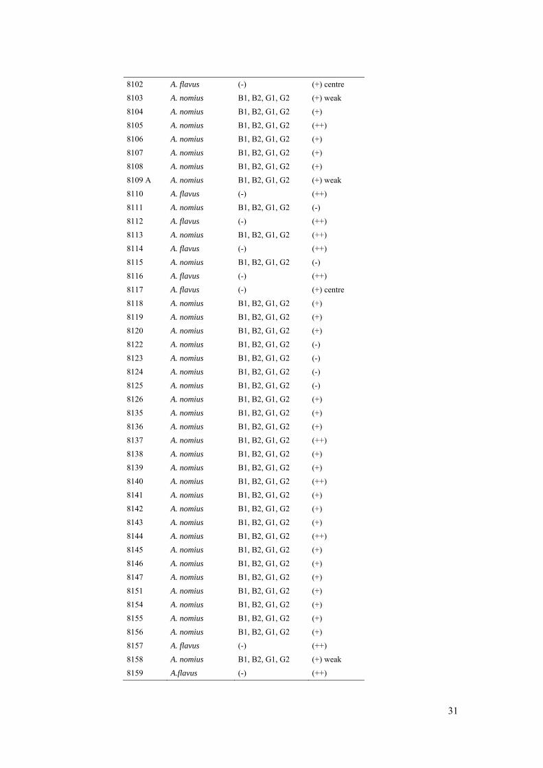
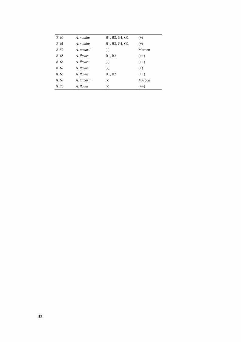
species on Brazil nuts ................................................................................................. 28 6.4 Aflatoxin profile and AFPA reverse of presumptive species of Aspergillus section
Flavi isolated from Brazil nuts ..................................................................................... 29

7
1 Introduction
The Brazil nut (Bertholletia excelsa) comes from a tall tree in the family
Lecythidaceae and is native to the Amazon rain forest in South America. The nut
is mainly found in Brazil and Bolivia but also in Peru, Guiana, Venezuela, Suri-
name and Colombia (FAO, 1993) and the trees are scattered along the Amazonian
river systems in dense groups. The Brazil nut tree is one of the tallest in Amazonas
and can reach a height of almost 60 m. It bears hard, large, spherical fruit pods
weighing up to 3 kg, with a capsule that contains 10–25 edible seeds. The seeds
are 3.5–5 cm long and have the distinct triangular shape characteristic of Brazil
nuts. Upon ripening in the rainy season (December to March), the fruit falls to the
ground with the capsule still closed, and once on the ground it can be collected and
cut open. The principal use of the Brazil nut is raw consumption, as well as an in-
gredient in food products due to its high nutritional value (Shanley, 2002). It is
rich in fat (approximately 66 %) and protein (approximately 14 %) and has higher
energy content than peanuts (NFA, 2012). Brazil nut protein contains all the essen-
tial amino acids and is especially rich in methionine. Brazil nuts are also high in
vitamins and minerals. These qualities make the nut very important in the socio-
economic setting, providing income and work for the local Amazon communities
(Newing & Harrop, 2000). Approximately 20,000 tons of Brazil nuts are produced
annually (Collison et al., 2002) with the majority being exported to Europe. The
complex ecological chain of the pollination by specific bees has made plantations
unsuccessful, and the nuts are mainly supplied from the wild (Newing & Harrop,
2000). Being produced organically and with a low technical production chain, the
Brazil nut is considered an environmentally friendly product.
One of the main problems identified in the production of Brazil nut is contami-
nation by moulds, which make the nut visually unattractive, inedible and increase
the risk of toxic metabolites such as aflatoxins and ochratoxins. This problem has
been a strong obstacle to marketing the product, mainly in foreign markets, given
the strict regulations of European countries and the United States regarding levels
of toxins in food. A common contaminant is Aspergillus, a mould that grows well
in a warm and humid environment. Aspergillus is known to spoil foods such as
peanuts, corn, figs, spices, rice, cocoa and coffee beans as well as Brazil nuts.
8
Some Aspergillus species, mainly in the Flavi-group, produce secondary metabo-
lites called aflatoxins. Aflatoxins can occur in the above-mentioned foods as a re-
sult of fungal contamination before and after harvest (EFSA, 2012). The Interna-
tional Agency of Research on Cancer (IARC, n.d.) has stated that aflatoxins are
among the most potent natural carcinogens known affecting animals and humans.
Chronic low-level exposure may increase the risk of developing hepatic cancer,
especially in individuals with chronic Hepatitis B viral infection (Tillet, 2010).
Aflatoxins also have acute toxic effects. Although not as common, acute high-
level exposures occur sporadically and have been known since the 60’s when
100 000 domesticated birds in England died after consuming peanuts contaminat-
ed with Aspergillus (FAO, 1991). On a few rare occasions, aflatoxins have also
caused the death of humans (Pitt & Hocking, 2009). In these cases, a very high
dose was consumed and the lethal dose is approximately 10 mg.
There are four different naturally occurring aflatoxins: B1, B2, G1 and G2 where
B1 is considered to be the most potent carcinogen. The letters refer to the colour
they fluoresce under UV-light; blue and green, respectively.
A recent overview of Aspergillus section Flavi by Varga et al. (2011) concluded
that the section includes 22 species. Despite the ability of aflatoxin production by
nine of these species, Aspergillus flavus has been considered the most important
for food products in general (Pitt & Hocking, 2009) and A. nomius an important
producer in Brazil nuts (Olsen et al., 2008). Taxonomic concepts within section
Flavi have evolved in recent years, with some species producing aflatoxins and
others not. Thus, development of reliable and economical methods to differentiate
between the various species is an important research field. Molecular techniques
and extrolite studies are the most precise for differentiating species, but for a
smaller laboratory without the possibility of these techniques, morphological
methods are more practical.
The occurrence of high levels of aflatoxins in Brazil nuts has been known for
several years. In 1998, the European Union (Commission Regulation, 1998) set
regulations regarding maximum residue levels for aflatoxin in food. To guarantee
a safe consumption, the limits for Brazil nuts were set to 4 µg/kg for total
aflatoxins and 2 µg/kg for aflatoxin B1. However, the industry faced a hard time
meeting the new quality requirements and formal complaints were made to the
World Trade Organization (WTO) from producing countries. The strict regulation
led to a decline in sales, mainly to the European market, of almost 90 % between
the years 2000–2004 (STDF, 2010). Recently new total aflatoxin limits have been
set by Codex alimentarius (CAC, 2010) with 10 µg/kg for shelled, ready-to-eat
nuts and 20 µg/kg for in-shell nuts. The European Commission (2010) also relaxed
their levels to meet the new Codex alimentarius limits, and in 2011, Brazil adapted
similar regulations in order to meet food safety standards. Still, many nuts sold in
9
local markets have not gone through any sorting or processing, and high levels of
toxins might occur.
There were two main objectives of this study. The first one focused on growing
17 different strains of Aspergillus section Flavi, from previously collected samples
of Brazil nuts (and 1 peanut) on agar media of different water activities and at dif-
ferent temperatures, in order to gather more knowledge about morphological fea-
tures which may aid in identification of these species. The second objective was to
isolate and identify toxigenic fungi in samples of Brazil nuts, both from the kernel
and the shell, that have not gone through any processing, and to quantify aflatoxins
in these samples.
10
2 Methods
2.1 Growth study of 17 isolates available at Instituto de Technologia de Alimentos (ITAL), Campinas, Brazil.
2.1.1 Media preparation and isolates
Two media were prepared, Czapek yeast extract (CYA) and Aspergillus Flavus
and Parasiticus (AFPA) agar. All media used in the experiment were prepared ac-
cording to Pitt & Hocking (2009) and autoclaved at 121 °C for 15 minutes. Before
incubation, all Petri-dishes were stacked in piles of approximately 16 plates and
wrapped with common household cling film. ITAL codes and scientific names of
isolates can be found in Table 1.
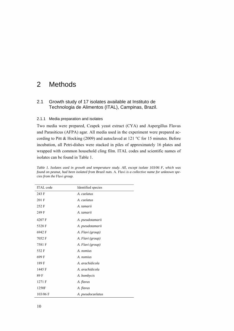
Table 1. Isolates used in growth and temperature study. All, except isolate 103/06 F, which was found on peanut, had been isolated from Brazil nuts. A. Flavi is a collective name for unknown spe-cies from the Flavi group.
ITAL code Identified species
243 F A. caelatus
201 F A. caelatus
252 F A. tamarii
249 F A. tamarii
4207 F A. pseudotamarii
5328 F A. pseudotamarii
6942 F A. Flavi (group)
7052 F A. Flavi (group)
7581 F A. Flavi (group)
532 F A. nomius
699 F A. nomius
189 F A. arachidicola
1445 F A. arachidicola
89 F A. bombycis
1271 F A. flavus
1250F A. flavus
103/06 F A. pseudocaelatus
11
2.1.2 Inoculation
A total of 17 strains of Aspergillus section Flavi, 16 previously obtained from
Brazil nuts and 1 from peanut and preserved on silica gel, were inoculated on
CYA by pouring the silica balls straight onto the media; plates were incubated at
25 °C for 5 days. Due to contamination, the strains were sub-cultured a second
time to obtain pure cultures: all Aspergillus section Flavi were isolated on CYA
again by carefully collecting spores from each plate with a needle and inoculating
at three points on the new plates. The plates were incubated at 25 °C for 4 days.
The pure isolates were three-point inoculated in one replicate on CYA and incu-
bated at 25 °C, 37 °C and 42 °C for 7 days. In order to screen for potentially toxi-
genic species, three-point inoculation was also made on Aspergillus Flavus and
Parasiticus (AFPA) agar and incubated at 25 °C. Potentially aflatoxigenic species
will show an orange reverse on this media due to production of ferric chelates of
aspergillic acids (Samson et al, 2010). All isolates were also inoculated in dupli-
cates on CYA media with different water activities (see 2.1.3) and incubated at 25
°C for 7 days.
2.1.3 Water activity and inspection
The water activity of CYA was modified by adding different amounts of glycerol
to the media. The water activities of the plates were measured in triplicate in a
Decagon Aqualab Series 3TE instrument at 25 °C, and came to aw 0.79, 0.87, 0.91,
0.94 and 0.97 (Appendix 6.1). After 7 days, three out of the total six colonies,
were randomly chosen for macroscopic studies. Macroscopic characteristics such
as colony diameter, colour, texture and AFPA reverse colour were noted. At the
lower water activities (aw 0.87 and 0.79, respectively), colonies were also inspect-
ed after 14 and 28 days.
2.2 Isolation and identification of potentially toxigenic fungi from Brazil nuts collected from market stalls.
2.2.1 Sampling
Samples of Brazil nuts were collected from 10 different market stalls in the city of
Manaus, Brazil. The nuts arrive at market early in the morning and are not subject
to any sorting or processing. In each market stall, approximately 2–4 kilos of nuts
were purchased. All nuts were ready to eat, three of the samples with the husks
still on, and seven without, depending on availability.
2.2.2 Water activity
Before further experiments, the water activity of the nuts was determined by ana-
lyzing the samples in triplicate in a Decagon Aqualab Series 3TE instrument at 25
°C (Appendix 6.2).
12
2.2.3 Fungal isolation and identification of potential aflatoxigenic species
From each sample, approximately 100 g of nuts or shells were sub-sampled. The
nuts and shells were surface disinfected separately with freshly prepared hypo-
chlorite solution (equivalent to 0.4 % chlorine) for 2 minutes and 50 pieces were
aseptically direct plated onto Dichloran 18% Glycerol (DG18) agar, five particles
per plate (Pitt & Hocking, 2009). The plates were stacked and wrapped with
household cling film and incubated at 25 °C. After 5–7 days the plates were in-
spected for fungal growth, and total infection percentage of the Brazil nuts was
calculated. Infection percentage with Aspergillus section Flavi, section Nigri and
with Penicillium spp. was also noted. All possible Aspergillus spp. were isolated
onto Czapek Yeast Extract (CYA) agar to obtain pure cultures (Pitt & Hocking,
2009). After successful isolation, the fungi were grown on AFPA media for 7 days
at 25ºC and reverse colour was noted. Fungi identified as potential producers of
aflatoxins were inoculated onto Yeast Extract Sucrose (YES) agar for 7 days at
25ºC and the capability of isolates to produce aflatoxins was evaluated by the agar
plug technique (Samson et al., 2010). Small pieces of the fungus and agar were cut
out with a scalpel and extracted with chloroform: methanol (1:1). The plugs were
placed on thin layer chromatography (TLC) plates, developed in a toluene: ethyl
acetate:90% formic acid: chloroform (7:5:2:5) mobile phase, and visualized under
UV light at 365 nm. A positive control with a mixture of aflatoxins B1, B2, G1 and
G2(Sigma, St. Louis, USA) was used for comparison. With aid from supervisors
Beatriz T. Iamanaka and Marta H. Taniwaki, the colonies were identified based on
both macroscopic and microscopic characteristics on CYA, AFPA reverse colour
and aflatoxin profile.
2.2.4 Aflatoxin analysis
Brazil nut kernels (25 g) from each sample were finely ground using a hand-held
mixer. Extraction was performed by adding 2.5 g NaCl and 100 ml of metha-
nol:water solution (8:2, v/v) and blending for 3 min at high speed (10,000 rpm)
using an Ultra-Turrax homogenizer (Polytron, Switzerland). The homogenized
solution was filtered twice, first through Nalgon 3551 filter paper and then Vicam
microfiber filters. An aliquot of 10 ml filtrate was diluted in 60 ml of previously
prepared phosphate buffered saline (PBS), after which it was passed through an
Aflatest WB immunoafffinity column (Vicam, USA) at a flow rate of 2–3 ml/min,
followed by washing with 30 ml distilled water. The columns were conditioned
with PBS before application of the sample. The aflatoxins were eluted by first add-
ing 500 µl HPLC-grade methanol and after 1 minute adding another 750 µl. The
eluate was diluted with 1750 µl Milli Q water. A positive control was prepared by
spiking a clean sample of nuts with no known growth of Aspergillus section Flavi
with 100 μl of a mixture of aflatoxins B1, B2, G1 and G2(Sigma, St. Louis, USA).
The control was treated in the same way as the samples.
13
For the quantification of aflatoxins in the samples, a chromatographic method
was used. A Shimadzu LC-10VP HPLC system (Shimadzu, Japan) was used with
a fluorescence detector set at 362 nm excitation and 455 nm emission for
aflatoxins G1 and G2, and 425 nm emission for aflatoxins B1 and B2. A Shimadzu
CLC G-ODS (4×10 mm) guard column and Shimadzu Shimpack (4.6×250 mm)
column were employed. The mobile phase consisted of water: acetonitrile: metha-
nol (6:2:3) containing 119 mg/lKBr and 4 M, 350 μl/l nitric acid. The flow rate
was 1 ml/min. A post-column derivatization of aflatoxins B1 and G1 was per-
formed with bromine using a KobraCell (R-Biopharm Rhône Ltd, Scotland). The
injection volume was 100 μL.
14
3 Results and discussion
3.1 Growth study
In this part of the study, 17 isolates from Aspergillus section Flavi were grown on
CYA at different temperatures, on CYA with different water activity and on
AFPA. The isolates represented eight different species: A. bombycis, A.
arachidicola, A. caelatus, A. flavus, A. nomius, A. pseudocaelatus, A.
pseudotamarii and A. tamarii, as well as three unidentified species from section
Flavi.
3.1.1 Colony appearance
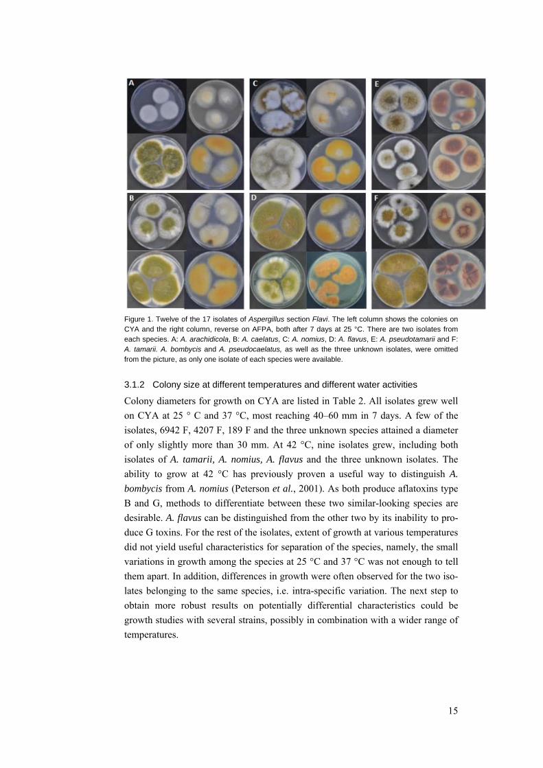
In general, the colonies were in shades of yellowish to brownish green (Figure 1)
with the exception of some more cream-coloured colonies due to low sporulation.
Some differences between the species could be seen, but were not sufficient to
consistently differentiate among the species. The reverse colour on AFPA varied
between bright orange to weaker orange and maroon. In some of the isolates, con-
tamination had occurred, making distinguishing between them even harder. The
importance of working with pure, uncontaminated colonies was highlighted.
15
Figure 1. Twelve of the 17 isolates of Aspergillus section Flavi. The left column shows the colonies on CYA and the right column, reverse on AFPA, both after 7 days at 25 °C. There are two isolates from each species. A: A. arachidicola, B: A. caelatus, C: A. nomius, D: A. flavus, E: A. pseudotamarii and F: A. tamarii. A. bombycis and A. pseudocaelatus, as well as the three unknown isolates, were omitted from the picture, as only one isolate of each species were available.
3.1.2 Colony size at different temperatures and different water activities
Colony diameters for growth on CYA are listed in Table 2. All isolates grew well
on CYA at 25 ° C and 37 °C, most reaching 40–60 mm in 7 days. A few of the
isolates, 6942 F, 4207 F, 189 F and the three unknown species attained a diameter
of only slightly more than 30 mm. At 42 °C, nine isolates grew, including both
isolates of A. tamarii, A. nomius, A. flavus and the three unknown isolates. The
ability to grow at 42 °C has previously proven a useful way to distinguish A.
bombycis from A. nomius (Peterson et al., 2001). As both produce aflatoxins type
B and G, methods to differentiate between these two similar-looking species are
desirable. A. flavus can be distinguished from the other two by its inability to pro-
duce G toxins. For the rest of the isolates, extent of growth at various temperatures
did not yield useful characteristics for separation of the species, namely, the small
variations in growth among the species at 25 °C and 37 °C was not enough to tell
them apart. In addition, differences in growth were often observed for the two iso-
lates belonging to the same species, i.e. intra-specific variation. The next step to
obtain more robust results on potentially differential characteristics could be
growth studies with several strains, possibly in combination with a wider range of
temperatures.
16
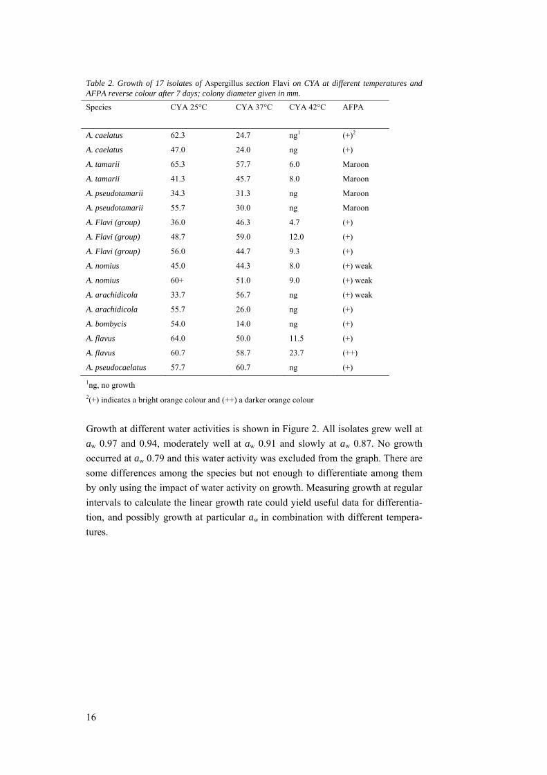
Table 2. Growth of 17 isolates of Aspergillus section Flavi on CYA at different temperatures and AFPA reverse colour after 7 days; colony diameter given in mm.
Species CYA 25°C CYA 37°C CYA 42°C AFPA
A. caelatus 62.3 24.7 ng1 (+)2
A. caelatus 47.0 24.0 ng (+)
A. tamarii 65.3 57.7 6.0 Maroon
A. tamarii 41.3 45.7 8.0 Maroon
A. pseudotamarii 34.3 31.3 ng Maroon
A. pseudotamarii 55.7 30.0 ng Maroon
A. Flavi (group) 36.0 46.3 4.7 (+)
A. Flavi (group) 48.7 59.0 12.0 (+)
A. Flavi (group) 56.0 44.7 9.3 (+)
A. nomius 45.0 44.3 8.0 (+) weak
A. nomius 60+ 51.0 9.0 (+) weak
A. arachidicola 33.7 56.7 ng (+) weak
A. arachidicola 55.7 26.0 ng (+)
A. bombycis 54.0 14.0 ng (+)
A. flavus 64.0 50.0 11.5 (+)
A. flavus 60.7 58.7 23.7 (++)
A. pseudocaelatus 57.7 60.7 ng (+)
1ng, no growth 2(+) indicates a bright orange colour and (++) a darker orange colour
Growth at different water activities is shown in Figure 2. All isolates grew well at
aw 0.97 and 0.94, moderately well at aw 0.91 and slowly at aw 0.87. No growth
occurred at aw 0.79 and this water activity was excluded from the graph. There are
some differences among the species but not enough to differentiate among them
by only using the impact of water activity on growth. Measuring growth at regular
intervals to calculate the linear growth rate could yield useful data for differentia-
tion, and possibly growth at particular aw in combination with different tempera-
tures.
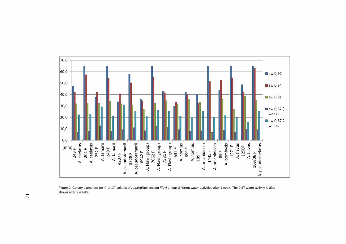
Figure 2. Colony diameters (mm) of 17 isolates of Aspergillus section Flavi at four different water activities after 1week. The 0.87 water activity is also shown after 2 weeks.
0,0
10,0
20,0
30,0
40,0
50,0
60,0
70,0
243 F
A. caelatus
201 F
A. caelatus
252 F
A. tam
arii
249 F
A. tam
arii
4207 F
A. pseudotamarii
5328 F
A. pseudotamarii
6942 F
A. Flavi (group)
7052 F
A. Flavi (group)
7581 F
A. Flavi (group)
532 F
A. nomius
699 F
A. nomius
189 F
A. arachidicola
1445 F
A. arachidicola
89 F
A. bombycis
1271 F
A. flavus
1250F
A. flavus
103/06 F
A. pseudocaelatus
aw 0,97
aw 0,94
aw 0,91
aw 0,87 (1 week)
aw 0,87 2 weeks
(mm)
17
18
3.1.3 Summary: Growth
Individual characteristics, colony size, colony colour, growth at various water ac-
tivities, and temperature tolerance, were not sufficient to distinguish the species
within section Flavi. Macromorphology could be used to differentiate some of the
species from others, but it was not enough to separate very similar looking species.
Identification based on looking at the fungi takes practice, and requires some
background knowledge of how the fungi should look. Temperature tolerance can
be a good way of separating some species, but only at certain temperatures. Water
activity seems to be the least reliable option, as the eight different species in this
study gave similar results. In order to partly identify the species in section Flavi,
one has to combine these three common identification features with each other.
Further studies should also include micromorphology and probably also molecular
methods. However, since many of the species are relatively new, appropriate keys
are not yet available. It is important that a sufficient amount of replicates are used
and preferably grown on the same batch of media to avoid differences within the
same species that might complicate the identification. Other methods used for full
identification include the production of aflatoxins and other extrolites (secondary
metabolites), and molecular techniques.
3.2 Isolation and identification of toxigenic fungi from collected Brazil nuts.
The second part of this study aimed at isolating and identifying potentially toxi-
genic fungi in samples of Brazil nuts, and to evaluate the presence of aflatoxins in
these samples.
3.2.1 Infection
In total, 10 samples of Brazil nut kernels were examined for total fungal and indi-
vidual percentage endogenous infection by Aspergillus section Flavi, Aspergillus
section Nigri and Penicillium spp. From three of these samples, the shell was also
examined. Percentage infection is shown in Table 3 (raw data in Appendix 6.3).
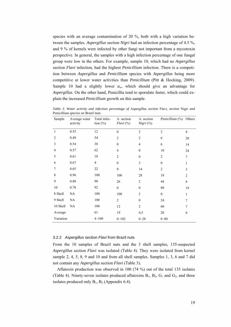
The average percentage fungal infection of the combined samples (nuts and shells)
was 61 %, with a variation of 4–100 % infection between the samples. The highest
infection rates occurred in the three shell samples (8, 9 and 10), and also in sample
8 kernel. The high presence of fungi in these samples correlated with the water
activity, where sample 8 with the highest infection rate also had the highest water
activity (Table 3). The samples with the highest overall infection rates also had the
highest infection with Aspergillus section Flavi, with sample 8 again being the
highest. The water activity of the shells was not tested. The lowest infection rate
occurred in sample 6. The most common contaminants were Aspergillus section
Flavi, with an average contamination of 21 %, closely followed by Penicillium
19
species with an average contamination of 20 %, both with a high variation be-
tween the samples. Aspergillus section Nigri had an infection percentage of 4.5 %,
and 9 % of kernels were infected by other fungi not important from a mycotoxin
perspective. In general, the samples with a high infection percentage of one fungal
group were low in the others. For example, sample 10, which had no Aspergillus
section Flavi infection, had the highest Penicillium infection. There is a competi-
tion between Aspergillus and Penicillium species with Aspergillus being more
competitive at lower water activities than Penicillium (Pitt & Hocking, 2009).
Sample 10 had a slightly lower aw, which should give an advantage for
Aspergillus. On the other hand, Penicillia tend to sporulate faster, which could ex-
plain the increased Penicillium growth on this sample.
Table 3. Water activity and infection percentage of Aspergillus section Flavi, section Nigri and Penicillium species on Brazil nuts.
Sample Average water activity
Total infec-tion (%)
A. section Flavi (%)
A. section Nigri (%)
Penicillium (%) Others
1 0.55 12 0 2 2 4
2 0.49 54 2 2 6 20
3 0.54 38 0 4 6 14
4 0.57 62 4 0 10 24
5 0.61 18 2 0 2 7
6 0.67 4 0 2 0 1
7 0.65 22 0 14 2 3
8 0.96 100 100 28 18 2
9 0.88 90 26 2 44 9
10 0.78 92 0 0 80 19
8 Shell NA 100 100 2 0 1
9 Shell NA 100 2 0 24 7
10 Shell NA 100 12 2 60 7
Average 61 19 4,5 20 9
Variation 4–100 0–102 0–28 0–80
3.2.2 Aspergillus section Flavi from Brazil nuts
From the 10 samples of Brazil nuts and the 3 shell samples, 135-suspected
Aspergillus section Flavi was isolated (Table 4). They were isolated from kernel
sample 2, 4, 5, 8, 9 and 10 and from all shell samples. Samples 1, 3, 6 and 7 did
not contain any Aspergillus section Flavi (Table 3).
Aflatoxin production was observed in 100 (74 %) out of the total 135 isolates
(Table 4). Ninety-seven isolates produced aflatoxins B1, B2, G1 and G2, and three
isolates produced only B1, B2 (Appendix 6.4).
20
Based on macromorphological differences, AFPA reverse colour (Appendix
6.4), and the ability to produce aflatoxins, the isolates were divided into different
groups. In one group, the colony colour was white to cream with no sporulation.
The reverse had a yellow colour on CYA and they were AFPA positive with an
orange reverse. Aflatoxins B1, B2, G1 and G2 were produced. Isolates in the second
group were also white with no sporulation, yellow reverse on CYA, and produced
aflatoxins B1, B2, G1 and G2. However, isolates in this group were AFPA negative.
AFPA results might not be fully reliable, as the medium was originally developed
for A. flavus and A. parasiticus. For other species, such as A. nomius, results may
vary. The third group of isolates had a green colour with abundant sporulation and
produced aflatoxins B1, B2, G1 and G2. They were AFPA positive. In the first in-
oculation on CYA, two different batches of Czapek solution were used in the me-
dia, giving a consistent difference in colony appearance between the two batches.
A second inoculation, using a freshly made Czapek solution showed that these
isolates did indeed sporulate. Based on the new morphological results, production
of aflatoxins B1, B2, G1, G2 and microscopic studies following appropriate keys,
these first three groups were presumptively identified as A. nomius (Table 4). A.
nomius, which produces all four aflatoxins, has previously been found to be of
importance in Brazil nuts because it is frequently isolated from this substrate
(Goncalves et al., 2011).
The next two groups were combined and listed as possible A. flavus. One group
had isolates with only little sporulation, were all aflatoxin negative and AFPA pos-
itive, whereas the other group had more sporulation, were AFPA positive and pro-
duced B1 and B2 toxins. Not all isolates of A. flavus are toxigenic and when they
are, they solely produce the B-type aflatoxins (Johnsson et al., 2008). These iso-
lates grew on CYA at 37 and 42 °C, and combining this information with colony
appearance and microscopic studies, they were presumptively identified as A.
flavus (Table 4).
The last group, consisting of possible A. tamarii (Table 4), had brown colonies
on CYA with abundant sporulation, a maroon reverse on AFPA and was aflatoxin
negative. Representatives from each group were selected and inoculated onto CYA
at 37 and 42 C°. The representatives all grew at these temperatures. One isolate
was never identified due to problems with repeated contamination and the time
limitations of this study; this isolate was non-toxigenic.
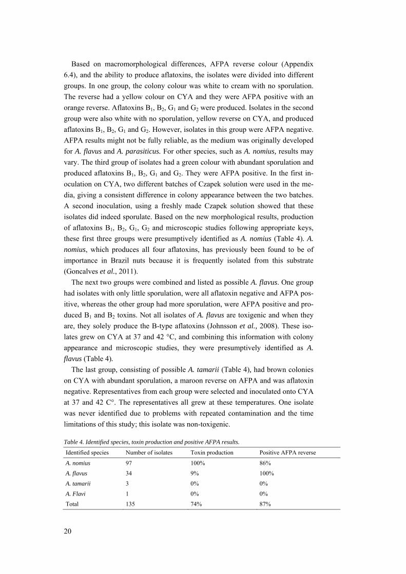
Table 4. Identified species, toxin production and positive AFPA results.
Identified species Number of isolates Toxin production Positive AFPA reverse
A. nomius 97 100% 86%
A. flavus 34 9% 100%
A. tamarii 3 0% 0%
A. Flavi 1 0% 0%
Total 135 74% 87%
21
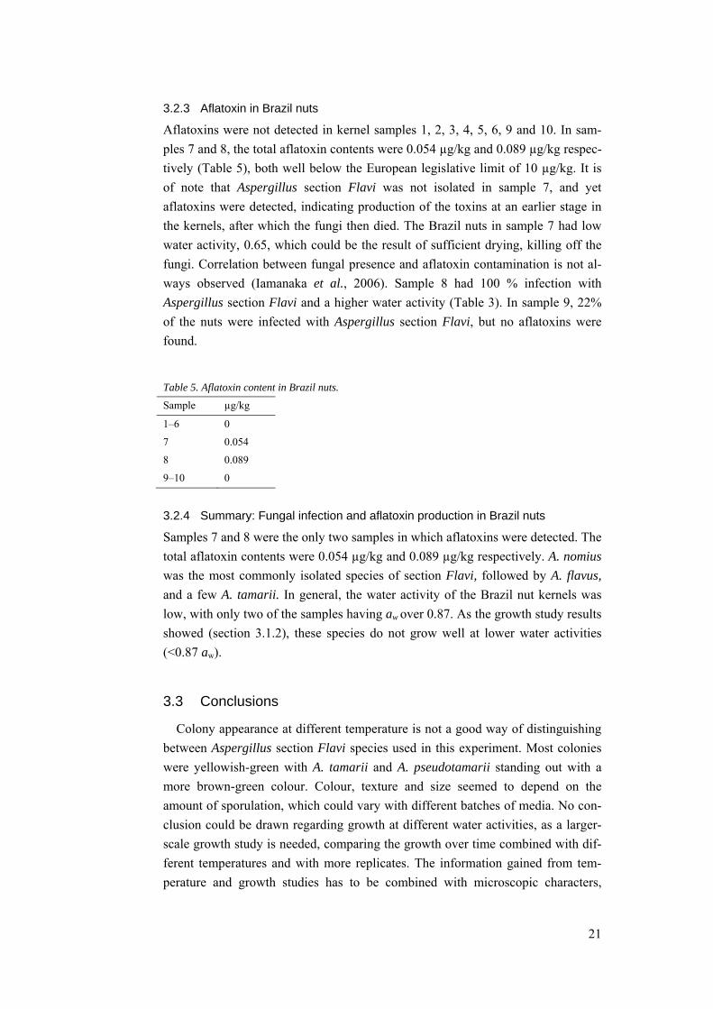
3.2.3 Aflatoxin in Brazil nuts
Aflatoxins were not detected in kernel samples 1, 2, 3, 4, 5, 6, 9 and 10. In sam-
ples 7 and 8, the total aflatoxin contents were 0.054 µg/kg and 0.089 µg/kg respec-
tively (Table 5), both well below the European legislative limit of 10 µg/kg. It is
of note that Aspergillus section Flavi was not isolated in sample 7, and yet
aflatoxins were detected, indicating production of the toxins at an earlier stage in
the kernels, after which the fungi then died. The Brazil nuts in sample 7 had low
water activity, 0.65, which could be the result of sufficient drying, killing off the
fungi. Correlation between fungal presence and aflatoxin contamination is not al-
ways observed (Iamanaka et al., 2006). Sample 8 had 100 % infection with
Aspergillus section Flavi and a higher water activity (Table 3). In sample 9, 22%
of the nuts were infected with Aspergillus section Flavi, but no aflatoxins were
found.
Table 5. Aflatoxin content in Brazil nuts.
Sample µg/kg
1–6 0
7 0.054
8 0.089
9–10 0
3.2.4 Summary: Fungal infection and aflatoxin production in Brazil nuts
Samples 7 and 8 were the only two samples in which aflatoxins were detected. The
total aflatoxin contents were 0.054 µg/kg and 0.089 µg/kg respectively. A. nomius
was the most commonly isolated species of section Flavi, followed by A. flavus,
and a few A. tamarii. In general, the water activity of the Brazil nut kernels was
low, with only two of the samples having aw over 0.87. As the growth study results
showed (section 3.1.2), these species do not grow well at lower water activities
(<0.87 aw).
3.3 Conclusions
Colony appearance at different temperature is not a good way of distinguishing
between Aspergillus section Flavi species used in this experiment. Most colonies
were yellowish-green with A. tamarii and A. pseudotamarii standing out with a
more brown-green colour. Colour, texture and size seemed to depend on the
amount of sporulation, which could vary with different batches of media. No con-
clusion could be drawn regarding growth at different water activities, as a larger-
scale growth study is needed, comparing the growth over time combined with dif-
ferent temperatures and with more replicates. The information gained from tem-
perature and growth studies has to be combined with microscopic characters,
22
extrolite production such as toxin- and exudate production, and possibly molecular
identification in order to fully identify the fungi. However, after working with the
fungi over a longer period, one might become more skilled in using the above in-
formation, as it takes a lot of practice to see small differences between colonies by
eye.
All species in this experiment grew well at the temperatures 25 and 30°C, and at
the water activities 0.91, 0.94 and 0.97, meaning that the fungus is very capable of
growing in the warm and humid environment of the Amazonas rainforest. As not
only one species contributes to aflatoxin content, it may not be necessary to al-
ways identify the Aspergillus strains down to species level in order to assess the
risk of aflatoxin contamination, even though some species are non-toxigenic. For
producers, merchandisers and re-sellers of Brazil nuts, focus should be on drying
and storing the nuts in such a way that the risk of Aspergillus section Flavi con-
tamination in general is kept to a minimum.
Aflatoxin was detected in 2 out of 10 samples. In both samples, the toxin-level
was well below the new European legislative limit of 10 µg/kg. Water activity
played a role for the amount of aflatoxigenic fungi found on the Brazil nuts, and
nuts purchased with the shell still on had the highest water activity. Buying and
consuming pre-shelled nuts with adequate drying probably reduces the risk of
aflatoxigenic fungi in general, but does not completely reduce the risk of
aflatoxins. As one sample result showed, toxins may be present in the nuts even
though the water activity is low and no fungi are found. However, all the nuts from
the 10 different market stalls in this study would be considered safe to consume.
23
4 Acknowledgements
First I would like to thank SIDA (Swedish International Development Cooperation
Agency) for the scholarship which supported this Minor Field Study at ITAL
(Instituto de Technologia de Alimentos) in Campinas, Brazil. I also want to say
thank you to everyone at ITAL, with an extra thought to my supervisors Marta
Taniwaki and Beatriz Iamanaka for all their help and support, and for making me
feel welcome at the lab and in their homes; to Camila Possari for assistance in the
lab; and to Larissa Ferranti for an interesting adventure in Manaus. A special thank
you goes to Su-lin Leong who made it possible for me to go to Brazil in the first
place, and who helped me with the Minor Field study application and supported
me throughout the project. I am also grateful for the much-needed Skype session
with Eva Edlund Tjernberg during my stay in Brazil and for Maja Sidstedt being a
good friend.
24
5 References
CAC (2010) Codex Alimentarius Commission. Report of the Fourth Session of
the Codex Committee on Contaminants in Foods. CL 2010/13-CF. Izmir, Turkey.
Joint FAO/WHO Food Standards Program, Rome, Italy.
Collinson, C., Burnett, D., Agreda, V. (2000). Peru. Natural Resources and Eth-
ical Trade Program, Natural Resources Institute, University of Greenwich.
Commission Regulation (1998) No 1525-98. Amending Commission Regulation
(EEC) No 752/93 laying down provisions for the implementation of Council Regu-
lation (EEC) No 3911/92 on the export of cultural goods. Official Journal of the
European Communities, L 201/47
Commission Regulation (2010) No 165/2010.Amending Regulation (EC) No
1881/2006 Setting maximum levels for certain contaminants in foodstuffs as re-
gards aflatoxins. Official Journal of the European Union, L 50/8 EFSA (2012) European Food Safety Authority - Aflatoxins in food. Available
at: http://www.efsa.europa.eu/en/topics/topic/aflatoxins.htm?wtrl=01. Accessed
2012-08-22
FAO (1991) Food and Agriculture Organization. Mycotoxins and food supply.
Food, Nutrition and Agriculture. Available at: http. Accessed 2012-08-22
FAO (1993) Food and Agriculture Organization. Selected species and strategies
to enhance income generation from Amazonian forests. Working Paper. Available
at: http://www.fao.org/docrep/V0784E/V0784E00.htm. Accessed 2012-08-22
Goncalves J.S., Ferracin, L.M., Carneiro Vieira, M.L., Iamanaka, B.T.,
Taniwaki, M.H., Pelegrinelli Fungaro, M.H. (2012) Molecular analysis of
Aspergillus section Flavi isolated from Brazil nuts. World Journal of Microbiolo-
gy and Biotechnology 28:1817-1825
Iamanaka, B.T., Castle de Menezes, H., Vicente, E., Leite, R.S.F., Taniwaki,
M.H. (2007) Aflatoxigenic fungi and aflatoxins occurrence in sultanas and dried
figs commercialized in Brazil. Food Control 18: 454–457
IARC (n.d.). International Agency for Research on Cancer (IARC). Monograph
100F – Aflatoxins. Available at:
http://monographs.iarc.fr/ENG/Monographs/vol100F/mono100F-23.pdf. Accessed
2012-08-22
25
Johnsson, P., Lindblad, M., Thim, A.M., Jonsson, N., Vargas, E.A., Medeiros,
N.L., Brabet, C., Quaresma de Araújo, M., Olsen, M. (2008). Growth of
aflatoxigenic moulds and aflatoxin formation in Brazil nuts. World Mycotoxin
Journal, 1: 127-137
Newing, H., Harrop, S. (2000). European Health Regulations and Brazil nuts:
Implications for Biodiversity Conservation and Sustainable Rural Livelihoods in
the Amazon. Journal of International Wildlife Law and Policy 3: 109-124.
Olsen, M., Johnsson, P., Möller, T., Paladino, R., Lindblad, M. (2008).
Aspergillus nomius, an important aflatoxin producer in Brazil nuts? World
Mycotoxin Journal 1: 123–126.
Pitt, J.I., & Hocking, A.D. (2009). Fungi and food spoilage. Springer, New
York, USA.Third edition.
Samson, R.A., Houbraken, J., Frisvad, J.C., Thrane, U., Andersen, B. (2010).
Food and Indoor fungi.CBS Laboratory Manual series 2. Utrecht : CBS-KNAW
Fungal Biodiversity Centre.
Shanley, P., Pierce, A.R., Laird, S.A., Guillen, A. (2002). Tapping the Green
Market: Certification and Management of Non-Timber Forest Products. Earthscan
Publications Ltd, United Kingdom. First edition.
NFA (2012) Swedish National Food Agency. Livsmedelsverkets livsmedelsda-
tabas version 2012-01-26 Available at:
http://www7.slv.se/Naringssok/Naringsamnen.aspx. Accessed: 2012-08-22
STDF (2008) Standards and Trade Development Facility, Project 114 - SafeNut.
Available at: http://www.stdf-safenutproject.com. Accessed 2012-08-22
Tillet, T. (2010). Carcinogenic Crops: Analyzing the Effect of Aflatoxin on
Global Liver Cancer Rates. Environmental Health Perspectives, 118:A 258.

26
6 Appendices
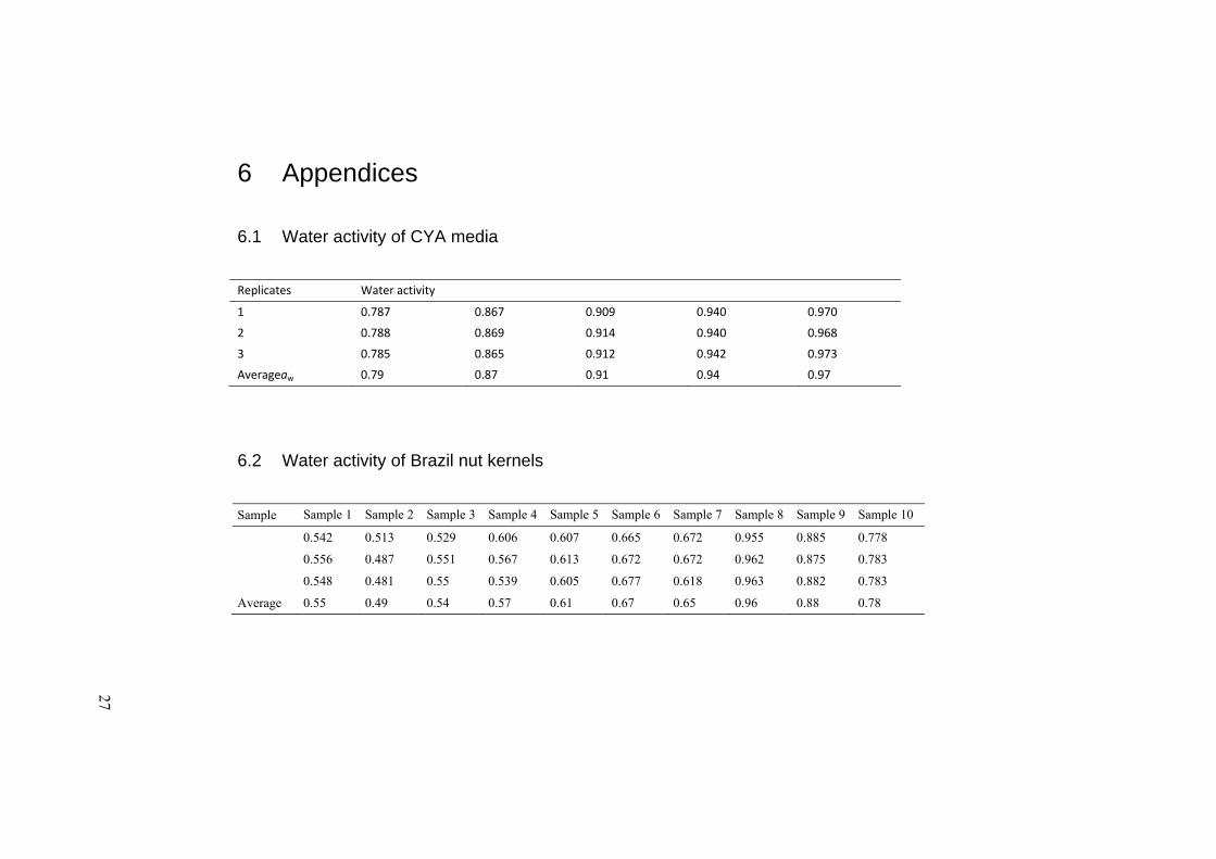
6.1 Water activity of CYA media
Replicates Water activity
1 0.787 0.867 0.909 0.940 0.970
2 0.788 0.869 0.914 0.940 0.968
3 0.785 0.865 0.912 0.942 0.973
Averageaw 0.79 0.87 0.91 0.94 0.97
6.2 Water activity of Brazil nut kernels
Sample Sample 1 Sample 2 Sample 3 Sample 4 Sample 5 Sample 6 Sample 7 Sample 8 Sample 9 Sample 10
0.542 0.513 0.529 0.606 0.607 0.665 0.672 0.955 0.885 0.778
0.556 0.487 0.551 0.567 0.613 0.672 0.672 0.962 0.875 0.783
0.548 0.481 0.55 0.539 0.605 0.677 0.618 0.963 0.882 0.783
Average 0.55 0.49 0.54 0.57 0.61 0.67 0.65 0.96 0.88 0.78
27
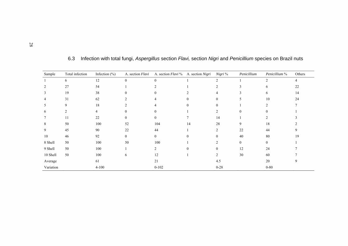
6.3 Infection with total fungi, Aspergillus section Flavi, section Nigri and Penicillium species on Brazil nuts
Sample Total infection Infection (%) A. section Flavi A. section Flavi % A. section Nigri Nigri % Penicillium Penicillium % Others
1 6 12 0 0 1 2 1 2 4
2 27 54 1 2 1 2 3 6 22
3 19 38 0 0 2 4 3 6 14
4 31 62 2 4 0 0 5 10 24
5 9 18 2 4 0 0 1 2 7
6 2 4 0 0 1 2 0 0 1
7 11 22 0 0 7 14 1 2 3
8 50 100 52 104 14 28 9 18 2
9 45 90 22 44 1 2 22 44 9
10 46 92 0 0 0 0 40 80 19
8 Shell 50 100 50 100 1 2 0 0 1
9 Shell 50 100 1 2 0 0 12 24 7
10 Shell 50 100 6 12 1 2 30 60 7
Average 61 21 4.5 20 9
Variation 4-100 0-102 0-28 0-80
28
29
6.4 Aflatoxin profile and AFPA reverse of presumptive species of Aspergillus section Flavi isolated from Brazil nuts
ITAL number
Presumptive iden-tification Toxin production AFPA reverse
8021 A. flavus (-) (+)
8188 A. nomius B1, B2, G1, G2 (-)
8189 A. nomius B1, B2, G1, G2 (+)
8176 A A. flavus (-) (+)
8176 B A. tamarii (-) Maroon
8027 A. flavus (-) (+) centre
8028 A. nomius B1, B2, G1, G2 (+)
8029 A. nomius B1, B2, G1, G2 (+)
8030 A. nomius B1, B2, G1, G2 NA
8031 A. nomius B1, B2, G1, G2 (+)
8032 A. flavus (-) (++)
8033 A. nomius B1, B2, G1, G2 (+)
8034 A. nomius B1, B2, G1, G2 (+)
8035 A A. nomius B1, B2, G1, G2 (+)
8035 B A. flavus (-) (+) centre
8036 A. nomius B1, B2, G1, G2 (+)
8037 A. nomius B1, B2, G1, G2 (+)
8038 A. nomius B1, B2, G1, G2 (+)
8039 A. nomius B1, B2, G1, G2 (-)
8040 A. flavus (-) (++)
8041 A. nomius B1, B2, G1, G2 (+)
8042 A. nomius B1, B2, G1, G2 (-)
8043 A. nomius B1, B2, G1, G2 (++)
8044 A. flavus (-) (+) centre
8045 A. nomius B1, B2, G1, G2 (-)
8046 A. nomius B1, B2, G1, G2 (+)
8047 A. nomius B1, B2, G1, G2 (-)
8048 A. nomius B1, B2, G1, G2 (+) centre
8049 A. flavus (-) (+) centre
8050 A. nomius B1, B2, G1, G2 (+) weak
8051 A. nomius B1, B2, G1, G2 (+)
8052 A. nomius B1, B2, G1, G2 (+)
8053 A. nomius B1, B2, G1, G2 (+) weak
8054 A. nomius B1, B2, G1, G2 (+) weak
8055 A. nomius B1, B2, G1, G2 (+)
8056 A. nomius B1, B2, G1, G2 (+) weak
8057 A. nomius B1, B2, G1, G2 (+)
8058 A. flavus (-) (++)
30
8059 A. nomius B1, B2, G1, G2 (++)
8060 A. flavus (-) (++)
8061 A. nomius B1, B2, G1, G2 (+)
8062 A.flavus (-) (+)
8063 A. flavus (-) (++)
8064 A A. nomius B1, B2, G1, G2 (+)
8064 B A. nomius B1, B2, G1, G2 (+) centre
8065 A. flavus (-) (++)
8066 A. nomius B1, B2, G1, G2 (+)
8067 A. nomius B1, B2, G1, G2 (+)
8068 A. nomius B1, B2, G1, G2 (+)
8069 A. nomius B1, B2, G1, G2 (+)
8070 A. flavus (-) (++)
8071 A. flavus (-) (++)
8072 A. nomius B1, B2, G1, G2 (+)
8073 A. nomius B1, B2, G1, G2 (+) centre
8075 A. nomius B1, B2, G1, G2 (+)
8076 A. nomius B1, B2, G1, G2 (+) weak
8132 A. nomius B1, B2, G1, G2 (+) weak
8077 A. nomius B1, B2, G1, G2 (-)
8078 A. nomius B1, B2, G1, G2 (+)
8079 A. flavus (-) (++)
8080 A. nomius B1, G1 (+) centre
8081 A. nomius B1, B2, G1, G2 (+)
8082 A. nomius B1, B2, G1, G2 (+)
8083 A. nomius B1, B2, G1, G2 (+)
8084 A. nomius B1, B2, G1, G2 (+)
8085 A. flavus B1, B2 (++)
8086 A. nomius B1, B2, G1, G2 (++)
8087 A. flavus (-) (++)
8088 A. flavus (-) (+) centre
8090 A. flavus (-) (++)
8091 A. nomius B1, B2, G1, G2 (+)
8092 A. nomius B1, B2, G1, G2 (+)
8093 A. nomius B1, B2, G1, G2 (+) weak
8094 A. flavus (-) (++)
8095 A. nomius B1, B2, G1, G2 (+) weak
8096 A. nomius B1, B2, G1, G2 (-)
8097 A. nomius B1, B2, G1, G2 (+)
8098 A A. nomius B1, B2, G1, G2 (++)
8098 B A. nomius B1, B2, G1, G2 (+)
8099 A. nomius B1, B2, G1, G2 (+) weak
8100 A. nomius B1, B2, G1, G2 (+)
8101 NA (-) NA
31
8102 A. flavus (-) (+) centre
8103 A. nomius B1, B2, G1, G2 (+) weak
8104 A. nomius B1, B2, G1, G2 (+)
8105 A. nomius B1, B2, G1, G2 (++)
8106 A. nomius B1, B2, G1, G2 (+)
8107 A. nomius B1, B2, G1, G2 (+)
8108 A. nomius B1, B2, G1, G2 (+)
8109 A A. nomius B1, B2, G1, G2 (+) weak
8110 A. flavus (-) (++)
8111 A. nomius B1, B2, G1, G2 (-)
8112 A. flavus (-) (++)
8113 A. nomius B1, B2, G1, G2 (++)
8114 A. flavus (-) (++)
8115 A. nomius B1, B2, G1, G2 (-)
8116 A. flavus (-) (++)
8117 A. flavus (-) (+) centre
8118 A. nomius B1, B2, G1, G2 (+)
8119 A. nomius B1, B2, G1, G2 (+)
8120 A. nomius B1, B2, G1, G2 (+)
8122 A. nomius B1, B2, G1, G2 (-)
8123 A. nomius B1, B2, G1, G2 (-)
8124 A. nomius B1, B2, G1, G2 (-)
8125 A. nomius B1, B2, G1, G2 (-)
8126 A. nomius B1, B2, G1, G2 (+)
8135 A. nomius B1, B2, G1, G2 (+)
8136 A. nomius B1, B2, G1, G2 (+)
8137 A. nomius B1, B2, G1, G2 (++)
8138 A. nomius B1, B2, G1, G2 (+)
8139 A. nomius B1, B2, G1, G2 (+)
8140 A. nomius B1, B2, G1, G2 (++)
8141 A. nomius B1, B2, G1, G2 (+)
8142 A. nomius B1, B2, G1, G2 (+)
8143 A. nomius B1, B2, G1, G2 (+)
8144 A. nomius B1, B2, G1, G2 (++)
8145 A. nomius B1, B2, G1, G2 (+)
8146 A. nomius B1, B2, G1, G2 (+)
8147 A. nomius B1, B2, G1, G2 (+)
8151 A. nomius B1, B2, G1, G2 (+)
8154 A. nomius B1, B2, G1, G2 (+)
8155 A. nomius B1, B2, G1, G2 (+)
8156 A. nomius B1, B2, G1, G2 (+)
8157 A. flavus (-) (++)
8158 A. nomius B1, B2, G1, G2 (+) weak
8159 A.flavus (-) (++)
32
8160 A. nomius B1, B2, G1, G2 (+)
8161 A. nomius B1, B2, G1, G2 (+)
8150 A. tamarii (-) Maroon
8165 A. flavus B1, B2 (++)
8166 A. flavus (-) (++)
8167 A. flavus (-) (+)
8168 A. flavus B1, B2 (++)
8169 A. tamarii (-) Maroon
8170 A. flavus (-) (++)
Recommended