ORIGINAL ARTICLE BREAST SURGERY
A New Tool for Breast Anthropometric Measurements:Presentation and Validation for Women and Men
Maksym Mikołajczyk1• Anna Kasielska-Trojan1
• Bogusław Antoszewski1
Received: 17 April 2019 / Accepted: 20 July 2019 / Published online: 1 August 2019
� The Author(s) 2019
Abstract
Introduction Anthropometric measurements of breasts are
crucial for planning surgical procedures; however, there are
no practical solutions for their quick, digital performance.
The aim of the study was to present and validate a self-
designed web application BreastIdea (BI) designed for
indirect breast anthropometry.
Methods Ten male and 10 female volunteers had their
chests measured directly according to the routine clinical
practice. Then their chests were photographed in non-s-
tandardised conditions. Corresponding measurements were
performed using BI. Accuracy and both relative and
absolute reliability of BI measurements were investigated.
Results Breast assessments using BI yielded highly accu-
rate results and presented near-perfect precision when
compared to direct anthropometric measurements of the
breast. Indirect anthropometry eliminates the necessity to
trace the body’s curves, which usually introduces a bias to
linear measurements.
Conclusion BI web application is a reliable tool for indi-
rect breast measurements in a clinical setting, providing
accurate results regardless of chest pathology and pho-
tograph standardisation.
Level of Evidence IV This journal requires that authors
assign a level of evidence to each article. For a full
description of these Evidence-Based Medicine ratings,
please refer to the Table of Contents or the online
Instructions to Authors www.springer.com/00266.
Keywords Anthropometry � Breast � Linear measurement �Web application
Introduction
Planning a surgical procedure on the breast, be it recon-
structive, reductive, oncological or aesthetic, breast mea-
surements prove indispensable for state-of-the-art surgery
[1]. Proportions, asymmetries and levels play a major role
in planning and optimising treatment results. Preoperative
chest evaluation is also crucial to accurate breast implant
choices for aesthetic and reconstructive reasons [2].
Even though the consistency of outcome of manual
measurements is highly dependent on the measurer’s skills
and experience, as well as available precision of the tools
used, those crucial measurements are mostly done by hand
(direct anthropometry). The female breast is one of the
most difficult organs to assess objectively [2]. Not only
size, but also contour, volume and asymmetry need to be
considered [3]. Some breast surgeons use 3D scanning,
which would yield accurate results. The technology is not
yet as accessible in clinical practice as a simple tape
measure. What is more, it is time-consuming and requires
expensive devices and a trained specialist to acquire and
analyse the scan [4]. There have been attempts to perform
breast measurements on photographs using graphics soft-
ware packages [5]. However, the studies were based on
photographs captured in standardised conditions, which are
not possible to replicate in clinical settings. What is more,
the software used was not clinically oriented.
Maksym Mikołajczyk and Anna Kasielska-Trojan equally contributed
to this work.
& Maksym Mikołajczyk
1 Plastic, Reconstructive and Aesthetic Surgery Clinic, Medical
University of Łodz, Kopcinskiego 22, 90-153 Lodz, Poland
123
Aesth Plast Surg (2019) 43:1160–1170
https://doi.org/10.1007/s00266-019-01467-6
Due to the lack of freely accessible and easy to use tools
for indirect breast anthropometry, we decided to design
such software. The tool was not created as an alternative to
direct measurements, which remain a gold standard in
breast surgeons’ clinical practice. Photograph-based breast
analysis may, however, be helpful as an additional source
of clinical data. It may help to extract metric data from the
image, e.g. while consulting the patient with other surgeons
and assessing asymmetry. Moreover, it may serve as a
valuable tool for scientific research, like metric evaluation
of breast asymmetry performed by independent observers
without the need for personal contact with the patient.
Our aim is to present the self-designed web applica-
tion—BreastIdea (BI)—a tool for linear breast measure-
ments, allowing to substitute a physical pen and tape
measure with their digital counterparts, thus increasing the
objectivity of breast assessment. In the article, we show the
process of its validation, as well as some pitfalls to be
avoided by potential users.
Methods
The protocol of the study was approved by the Bioethics
Committee of the Medical University (RNN/100/18/KE).
Written informed consent was obtained from all subjects.
The study consisted of three phases. The first was web
application design and development, the second—direct
breast measurements and the third—digital measurements
using BI (indirect breast measurements). The measure-
ments performed with BI were compared with direct
measurements performed by a plastic surgeon.
The study groups consisted of 10 males, aged 18–37
(mean age 23.7 years, SD 5.04 years) and 10 females, aged
18–45 (mean age 29.6 years, SD 9.38). The volunteers
were randomly selected from students of the Medical
University and from patients at the Plastic, Reconstructive
and Aesthetic Surgery Clinic, University Hospital,
regardless of age (over 18 years old), breast size, breast
ptosis and deformation (e.g. Poland syndrome). In the
female group, 6 women (volunteers: students, patients) had
natural breasts and 4 were clinic patients after breast sur-
gery [breast reduction and lift (n = 2), breast enlargement
(n = 1), breast symmetrisation due to Poland syndrome
(n = 1)]. None of the male participants had chest surgery;
however, two had mild lipomastia. The exclusion criterion
for the female and male groups was obesity—body mass
index above 30. The study process was identical for all
patients.
Web Application Design and Development
Our initial assumption was to design an application that
was easy to use and access (through a web browser). The
user interface was written in Hypertext Markup Language
(HTML) with Cascading Style Sheets (CSS), while the
logic was supplied by JavaScript (JS)—a standard in web
development [6]. Seven plastic surgeons specialising in
breast surgery were asked to identify clinically relevant
breast measurements. The following linear breast parame-
ters were chosen: the difference in the levels of nipples,
medial line–nipple distance (right and left), sternal notch–
nipple distance (right and left), the difference in the levels
of inframammary folds apices, medial line–inframammary
fold apex distance (right and left), inframammary fold
apex–nipple distance (right and left), the difference in the
levels of both upper pole apices, and upper pole apex–
nipple distance (right and left) (Fig. 1). The parameters
were not gender specific, applying to both males and
females.
For aesthetic assessment, we added proportions of the
upper to the lower pole as well as medial to lateral part,
derived from the measurements listed above. Following
Mallucci et al., for vertical parameters we used the ratio of
distance from upper pole apex to nipple and distance from
inframammary fold apex to nipple (with the most appealing
proportion of 45:55) [7]. For horizontal parameters, we
used the ratio of distance from the breast’s lateral border to
the nipple and distance from the breast’s medial border to
the nipple (with most appealing proportion of 40:60), as
suggested by Lewin et al. [8].
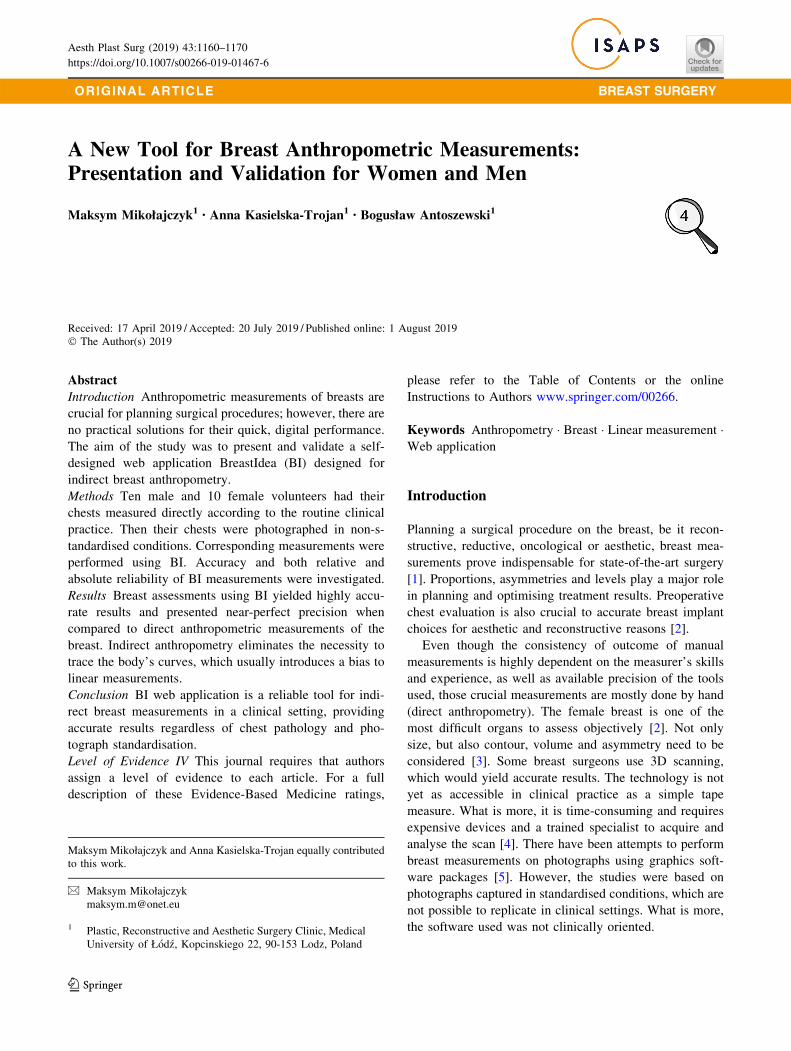
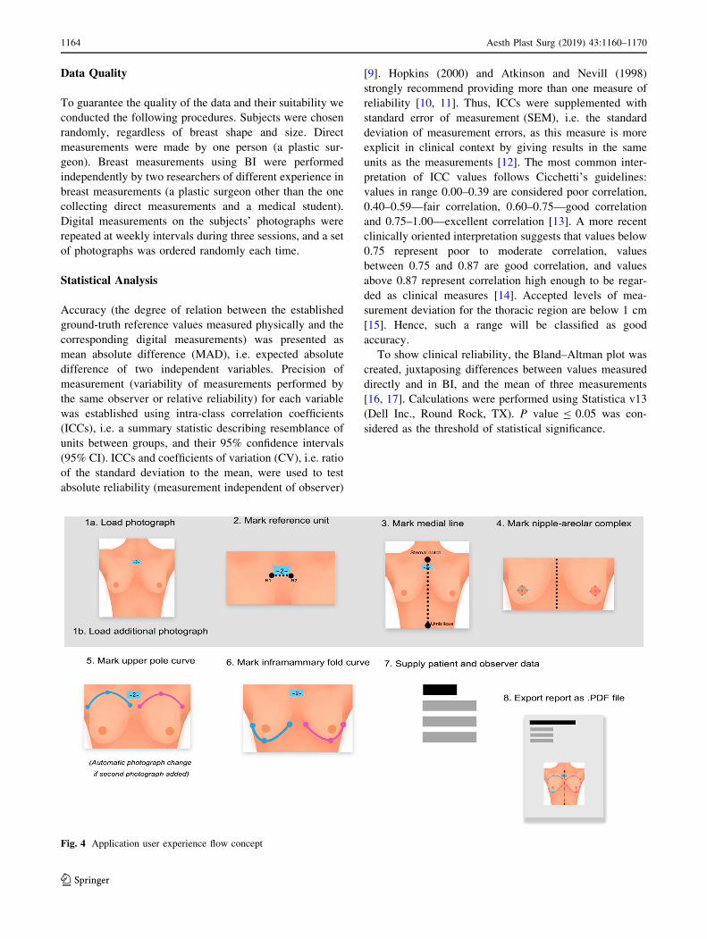
BI guides the user through the process of digital breast
measurements. Each measurement consists of placing a
number of digital markers on the photograph by clicking in
locations suggested by dynamic instructions. If needed, the
Fig. 1 Illustration of all the chosen landmarks and measurements. ’
Left, SN sternal notch, UPA upper pole apex, N nipple, IMFA
inframammary fold apex, ML medial line, UMB umbilicus
Aesth Plast Surg (2019) 43:1160–1170 1161
123
markers are automatically connected by lines. Most of the
measurements are based upon straightforward vector length
computation. Level difference measurements need an
additional step of horizontal projection of the digital
markers onto the midline and then calculating the distance
between the resulting coordinates. To allow precise point
marking, BI functions are enhanced with photograph
transformation capabilities—translation, scaling and rota-
tion. The user can also adjust its brightness and contrast
(Fig. 2).
In many patients, the upper breast border is not evident,
especially on photographical data. To determine the breast
footprint, the breast has to be lifted. Taking this into
account, an option to use a second photograph was added.
This option was designed especially for patients with breast
ptosis—the patient is photographed while lifting their
breasts to visualise inframammary folds and upper breast
borders. This function makes it possible to correctly assess
parameters referring to the inframammary fold and better
depict the upper pole of the breasts. The two photographs
are spatially bound together by marking the patient’s
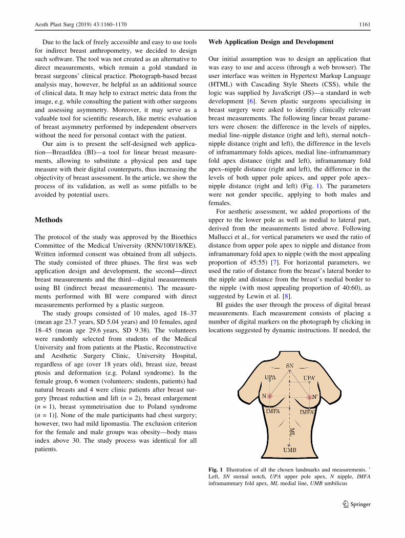
medial line on both of them. After all the measurements are
made, BI generates a report with all the markers and lines
present on the photograph, as well as essential patient
information (Fig. 3). The report can then be printed or
saved as a PDF file, providing an easy way to enclose the
results of the breast assessment process in both physical
and electronic medical documentation.
The application uses the web browser only as an envi-
ronment to run JS logic. The photographs used in BI are not
uploaded to the web. No data were sent out or fetched from
a database—data were only handled locally in the browser.
BI is available free of charge for everyone at www.
breastidea.com/app.
Direct Breast Measurements
Direct breast measurements took place at different times of
day and in random lighting conditions. The surgeon placed
a reference unit (2 cm long) on the chest, either below a
clavicle or on the sternum, below the sternal angle. The
location of the reference unit did not affect measurement
accuracy, so optimal reference unit placement was depen-
dent on the patient’s body shape, as long as the surface was
flat and perpendicular to the camera. The unit was used to
translate virtual length to real-world length. A digital
photograph of the subject’s chest was taken, making sure
the camera (We consider that most potential users will take
pictures with their smartphones. However, because of
security restrictions imposed by the Ethics Committee, we
were not allowed to take photographs with a mobile phone.
We made sure the digital camera parameters were as close
to a standard smartphone’s camera as possible.) was per-
pendicular to the ground so as not to skew the perspective.
Based on breast size and ptosis, the need for an additional
photograph was evaluated. In those situations, the subject
was asked to elevate their breasts for the second
photograph.
Next, to acquire the reference data, all previously listed
measurements were performed directly (gold standard
method). All essential lines were drawn on the subject’s
chest using a marker. Then, using a tape measure, the linear
parameters were measured. To measure level differences,
the right and left inframammary folds apices, upper pole
apices and nipples were projected as points to the midline
and the differences between these points were measured.
The duration of measurements was noted. All measure-
ments were done by one person (a plastic surgeon).
Digital Indirect Breast Measurements
After transferring the photographs onto a computer, all
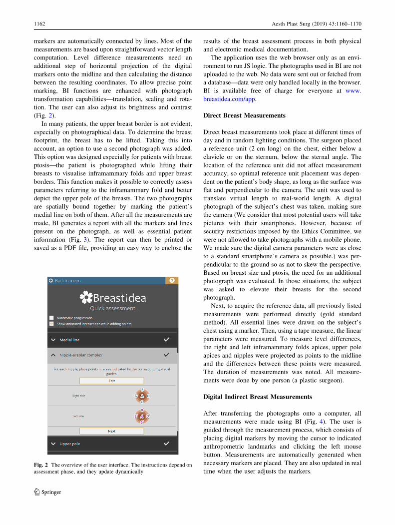
measurements were made using BI (Fig. 4). The user is
guided through the measurement process, which consists of
placing digital markers by moving the cursor to indicated
anthropometric landmarks and clicking the left mouse
button. Measurements are automatically generated when
necessary markers are placed. They are also updated in real
time when the user adjusts the markers.Fig. 2 The overview of the user interface. The instructions depend on
assessment phase, and they update dynamically
1162 Aesth Plast Surg (2019) 43:1160–1170
123
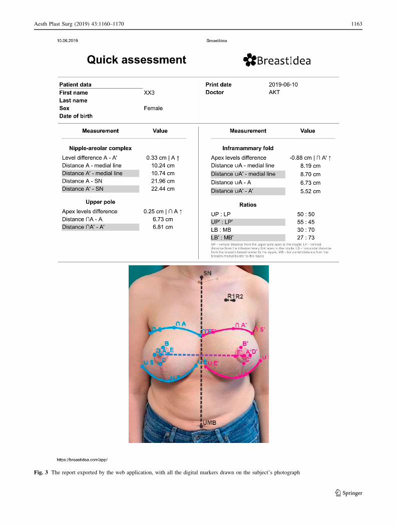
Fig. 3 The report exported by the web application, with all the digital markers drawn on the subject’s photograph
Aesth Plast Surg (2019) 43:1160–1170 1163
123
Data Quality
To guarantee the quality of the data and their suitability we
conducted the following procedures. Subjects were chosen
randomly, regardless of breast shape and size. Direct
measurements were made by one person (a plastic sur-
geon). Breast measurements using BI were performed
independently by two researchers of different experience in
breast measurements (a plastic surgeon other than the one
collecting direct measurements and a medical student).
Digital measurements on the subjects’ photographs were
repeated at weekly intervals during three sessions, and a set
of photographs was ordered randomly each time.
Statistical Analysis
Accuracy (the degree of relation between the established
ground-truth reference values measured physically and the
corresponding digital measurements) was presented as
mean absolute difference (MAD), i.e. expected absolute
difference of two independent variables. Precision of
measurement (variability of measurements performed by
the same observer or relative reliability) for each variable
was established using intra-class correlation coefficients
(ICCs), i.e. a summary statistic describing resemblance of
units between groups, and their 95% confidence intervals
(95% CI). ICCs and coefficients of variation (CV), i.e. ratio
of the standard deviation to the mean, were used to test
absolute reliability (measurement independent of observer)
[9]. Hopkins (2000) and Atkinson and Nevill (1998)
strongly recommend providing more than one measure of
reliability [10, 11]. Thus, ICCs were supplemented with
standard error of measurement (SEM), i.e. the standard
deviation of measurement errors, as this measure is more
explicit in clinical context by giving results in the same
units as the measurements [12]. The most common inter-
pretation of ICC values follows Cicchetti’s guidelines:
values in range 0.00–0.39 are considered poor correlation,
0.40–0.59—fair correlation, 0.60–0.75—good correlation
and 0.75–1.00—excellent correlation [13]. A more recent
clinically oriented interpretation suggests that values below
0.75 represent poor to moderate correlation, values
between 0.75 and 0.87 are good correlation, and values
above 0.87 represent correlation high enough to be regar-
ded as clinical measures [14]. Accepted levels of mea-
surement deviation for the thoracic region are below 1 cm
[15]. Hence, such a range will be classified as good
accuracy.
To show clinical reliability, the Bland–Altman plot was
created, juxtaposing differences between values measured
directly and in BI, and the mean of three measurements
[16, 17]. Calculations were performed using Statistica v13
(Dell Inc., Round Rock, TX). P value B 0.05 was con-
sidered as the threshold of statistical significance.
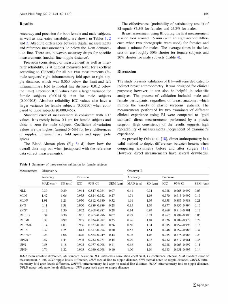
Fig. 4 Application user experience flow concept
1164 Aesth Plast Surg (2019) 43:1160–1170
123
Results
Accuracy and precision for both female and male subjects,
as well as inter-rater variability, are shown in Tables 1, 2
and 3. Absolute differences between digital measurements
and reference measurements lie below the 1-cm demarca-
tion line. There are, however, accuracy drops for specific
measurements (medial line–nipple distance).
Precision (consistency of measurement) as well as inter-
rater reliability, is at clinical measures level (or excellent
according to Cichetti) for all but two measurements (fe-
male subjects’ right inframammary fold apex to right nip-
ple distance, which was 0.060 below the limit and left
inframammary fold to medial line distance, 0.012 below
the limit). Precision ICC values have a larger variance for
female subjects (0.001415) than for male subjects
(0.000705). Absolute reliability ICC values also have a
larger variance for female subjects (0.00290) when com-
pared to male subjects (0.0003465).
Standard error of measurement is consistent with ICC
values. It is mostly below 0.1 cm for female subjects and
close to zero for male subjects. Coefficient-of-variation
values are the highest (around 5–6%) for level differences
of nipples, inframammary fold apices and upper pole
apices.
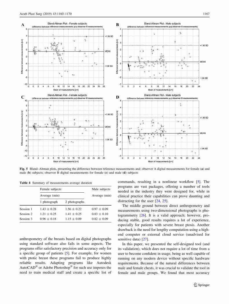
The Bland–Altman plots (Fig. 5a–d) show how the
overall data map out when juxtaposed with the reference
data (direct measurements).
The effectiveness (probability of satisfactory result) of
BI equals 87.5% for females and 95.8% for males.
Breast assessment using BI during the first measurement
session took around 1.5 min (with an eight-second differ-
ence when two photographs were used) for females and
about a minute for males. The average times in the last
session are roughly 30% shorter for female subjects and
20% shorter for male subjects (Table 4).
Discussion
The study presents validation of BI—software dedicated to
indirect breast anthropometry. It was designed for clinical
purposes; however, it can also be helpful in scientific
analyses. The process of validation included male and
female participants, regardless of breast anatomy, which
mimics the variety of plastic surgeons’ patients. The
measurements performed by two examiners of different
clinical experience using BI were compared to ‘gold
standard’ direct measurements performed by a plastic
surgeon. High consistency of the results suggests high
repeatability of measurements independent of examiner’s
experience.
As proved by Odo et al. [18], direct anthropometry is a
valid method to depict differences between breasts when
comparing asymmetry before and after surgery [18].
However, direct measurements have several drawbacks.
Table 1 Summary of three-session validation for female subjects
Measurement Observer A Observer B
Accuracy Precision Accuracy Precision
MAD (cm) SD (cm) ICC 95% CI SEM (cm) MAD (cm) SD (cm) ICC 95% CI SEM (cm)
NLD 0.30 0.29 0.944 0.847–0.984 0.07 0.41 0.31 0.988 0.965–0.997 0.03
MLN 1.42 1.06 0.935 0.824–0.982 0.27 1.71 1.08 0.970 0.915–0.992 0.19
MLN* 1.91 1.21 0.930 0.812–0.980 0.32 1.61 1.03 0.958 0.883–0.988 0.21
SNN 0.11 1.38 0.960 0.889–0.989 0.28 0.15 1.07 0.977 0.935–0.994 0.16
SNN* 0.12 1.30 0.952 0.868–0.987 0.28 0.14 0.94 0.969 0.913–0.991 0.17
IMFLD 0.34 0.30 0.951 0.865–0.986 0.07 0.29 0.24 0.962 0.894–0.990 0.05
IMFML 0.39 0.99 0.935 0.824–0.982 0.25 0.26 1.04 0.926 0.802–0.979 0.28
IMF*ML 0.14 1.03 0.936 0.827–0.982 0.26 0.50 1.31 0.985 0.957–0.996 0.16
IMFN 0.32 1.25 0.843 0.617–0.954 0.50 0.53 1.51 0.948 0.857–0.986 0.34
IMF*N* 0.26 1.06 0.826 0.584–0.949 0.44 0.05 1.08 0.955 0.875–0.988 0.23
UPLD 0.57 1.44 0.905 0.752–0.973 0.45 0.70 1.33 0.932 0.817–0.981 0.35
UPN 0.58 1.18 0.992 0.977–0.998 0.11 0.68 1.00 0.988 0.965–0.997 0.11
UPN* 0.70 1.22 0.993 0.980–0.998 0.10 1.00 1.04 0.983 0.951–0.995 0.14
MAD mean absolute difference, SD standard deviation, ICC intra-class correlation coefficient, CI confidence interval, SEM standard error of
measurement, * left, NLD nipple levels difference, MLN medial line to nipple distance, SNN sternal notch to nipple distance, IMFLD infra-
mammary fold apex levels difference, IMFML inframammary fold apex to medial line distance, IMFN inframammary fold to nipple distance,
UPLD upper pole apex levels difference, UPN upper pole apex to nipple distance
Aesth Plast Surg (2019) 43:1160–1170 1165
123
While some body regions can be assessed in a mostly linear
manner, the chest has prominent curvatures, making mea-
surements less accurate [2, 19, 20]. Also, the region is not
only affected by whole-body movements, but also ribcage
expansion while breathing. Indirect anthropometry on the
other hand presents a wide array of advantages. According
to numerous studies, not only does it allow higher relia-
bility, but it is also much less stressful for the patient
[19, 21–23]. Data are also usually relatively easy to record,
already being in digital format. Nonetheless, indirect
Table 2 Summary of three-session validation for male subjects
Measurement Observer A Observer B
Accuracy Precision Accuracy Precision
MAD (cm) SD (cm) ICC 95% CI SEM (cm) MAD (cm) SD (cm) ICC 95% CI SEM (cm)
NLD 0.36 0.20 0.887 0.711–0.968 0.07 0.31 0.18 0.953 0.870–0.987 0.04
MLN 0.47 0.46 0.986 0.960–0.996 0.05 0.62 0.47 0.996 0.988–0.999 0.03
MLN* 0.21 0.40 0.984 0.954–0.996 0.05 0.33 0.39 0.994 0.983–0.998 0.03
SNN 0.89 0.84 0.973 0.924–0.993 0.14 1.12 0.75 0.994 0.983–0.998 0.06
SNN* 0.74 0.77 0.973 0.924–0.993 0.13 0.90 0.68 0.991 0.974–0.998 0.06
IMFLD 0.27 0.22 0.909 0.761–0.974 0.07 0.26 0.22 0.950 0.862–0.986 0.05
IMFML 0.32 0.50 0.987 0.963–0.996 0.06 0.27 0.43 0.993 0.980–0.998 0.04
IMF*ML 0.09 0.59 0.972 0.921–0.992 0.10 0.07 0.54 0.976 0.932–0.993 0.08
IMFN 0.15 0.34 0.988 0.965–0.997 0.04 0.23 0.34 0.990 0.971–0.997 0.03
IMF*N* 0.37 0.38 0.979 0.940–0.994 0.06 0.41 0.41 0.997 0.991–0.999 0.02
UPLD 0.16 0.35 0.963 0.897–0.990 0.07 0.27 0.29 0.959 0.886–0.989 0.06
UPN 0.11 0.34 0.991 0.974–0.998 0.03 0.20 0.28 0.998 0.994–0.999 0.01
UPN* 0.14 0.32 0.996 0.988–0.999 0.02 0.29 0.31 0.999 0.997–1.000 0.01
MAD mean absolute difference, SD standard deviation, ICC intra-class correlation coefficient, CI confidence interval, SEM standard error of
measurement, * left, NLD nipple levels difference, MLN medial line to nipple distance, SNN sternal notch to nipple distance, IMFLD infra-
mammary fold apex levels difference, IMFML inframammary fold apex to medial line distance, IMFN inframammary fold to nipple distance,
UPLD upper pole apex levels difference, UPN upper pole apex to nipple distance
Table 3 Summary of intra-
observer reliability between
Observer A and Observer B,
based on average values from
three sessions
Measurement Female subjects Male subjects
CV (%) ICC 95% CI SEM (cm) CV (%) ICC 95% CI SEM (cm)
NLD 5.7 0.968 0.910–0.991 0.01 6.3 0.943 0.844–0.984 0.01
MLN 1.2 0.968 0.910–0.991 0.02 0.7 0.996 0.988–0.999 0.00
MLN* 0.1 0.924 0.797–0.979 0.00 0.5 0.998 0.994–0.999 0.00
SNN 0.1 0.989 0.968–0.997 0.00 0.6 0.996 0.988–0.999 0.01
SNN* 0.0 0.986 0.960–0.996 0.00 0.4 0.997 0.991–0.999 0.00
IMFLD 5.8 0.909 0.761–0.974 0.01 0.4 0.957 0.881–0.988 0.00
IMFML 2.2 0.953 0.870–0.987 0.04 0.2 0.992 0.977–0.998 0.00
IMF*ML 3.0 0.858 0.648–0.959 0.11 0.1 0.995 0.985–0.999 0.00
IMFN 4.5 0.810 0.553–0.943 0.15 0.9 0.998 0.994–0.999 0.00
IMF*N* 1.2 0.898 0.736–0.971 0.04 0.9 0.995 0.985–0.999 0.01
UPLD 4.9 0.944 0.847–0.984 0.09 0.5 0.967 0.907–0.991 0.00
UPN 2.3 0.987 0.963–0.996 0.02 0.6 0.998 0.994–0.999 0.00
UPN* 3.8 0.952 0.868–0.987 0.08 1.0 0.999 0.997–1.000 0.00
CV coefficient of variation, ICC intra-class correlation coefficient, CI confidence interval, SEM standard
error of measurement, * left, NLD nipple levels difference, MLN medial line to nipple distance, SNN sternal
notch to nipple distance, IMFLD inframammary fold apex levels difference, IMFML inframammary fold
apex to medial line distance, IMFN inframammary fold to nipple distance, UPLD upper pole apex levels
difference, UPN upper pole apex to nipple distance
1166 Aesth Plast Surg (2019) 43:1160–1170
123
anthropometry of the breasts based on digital photographs
using standard software also fails in some aspects. The
programs offer satisfactory precision and accuracy only for
a specific group of patients [5]. For example, for women
with ptotic breast these programs fail to produce highly
reliable results. Adapting programs like Autodesk
AutoCAD� or Adobe Photoshop� for such use imposes the
need to train medical staff and create a specific list of
commands, resulting in a nonlinear workflow [5]. The
programs are vast packages, offering a number of tools
needed in the industry they were designed for, while in
clinical practice their capabilities can prove daunting and
distracting for the user [24, 25].
The middle ground between direct anthropometry and
measurements using two-dimensional photographs is pho-
togrammetry [26]. It is a valid approach; however, pro-
ducing stable, good results requires a lot of experience,
especially for patients with severe breast ptosis. Another
drawback is the need for lengthy computation using a high-
end computer or external cloud service (unadvised for
sensitive data) [27].
In this paper, we presented the self-designed tool (and
its validation), which does not require a lot of time from a
user to become confident in usage, being as well capable of
running on any modern device without specific hardware
requirements. Because of the natural differences between
male and female chests, it was crucial to validate the tool in
female and male groups. We found that most accuracy
Fig. 5 Bland–Altman plots, presenting the difference between reference measurements and: observer A digital measurements for female (a) and
male (b) subjects; observer B digital measurements for female (c) and male (d) subjects
Table 4 Summary of measurements average duration
Female subjects Male subjects
Average (min) Average (min)
1 photograph 2 photographs
Session 1 1.43 ± 0.28 1.56 ± 0.22 0.97 ± 0.09
Session 2 1.21 ± 0.25 1.41 ± 0.25 0.83 ± 0.10
Session 3 0.96 ± 0.18 1.15 ± 0.09 0.82 ± 0.09
Aesth Plast Surg (2019) 43:1160–1170 1167
123
values for female subjects vary by ± 1 cm, consistently
between observers. The exceptions are nipple level dif-
ferences and inframammary fold apex level differences.
Their standard deviation was around 0.30 cm. Standard
deviation of mean absolute difference above 1 cm was not
detected in measurements in males. BI measurements,
which were juxtaposed with measurements mostly affected
by curves in direct anthropometry are least accurate, with a
mean absolute difference between 1.42 and 1.91 cm (me-
dial line–nipple distance). This may result from the fact
that for distances like the one traced from the nipple to the
body’s medial line, one should demarcate a line segment
tangent to the natural breast curve, which is not a routine
approach in clinical breast measurements [2]. It appeared
that BI gives valid results when compared to a textbook
procedure (measuring straight lines instead of following
natural curves) [2]. The discussed problem is also depicted
in the Bland–Altman graphs, where the majority of points
fall between the two limits, proving suitability for clinical
use, with a cluster of outlying data present for female
subjects for both observers, corresponding to the mea-
surements listed above.
To properly evaluate BI’s accuracy, we need to consider
measurements which were truly linear both in the direct
and indirect anthropometry using the web application.
These measurements (sternal notch to nipple distance,
medial line to inframammary fold apex distance, upper
pole apex to nipple distance) mostly present satisfactory
accuracy with excellent correspondence between obser-
vers. MAD of the distance from the inframammary fold
apex to the medial line was initially above 2 cm, but
decreased to about 0.30 cm within the next two sessions.
What is more, the average accuracy of both observers grew
with each session, accompanied by a significant decrease in
procedure duration and time variation between different
subjects. In the case of the distance from the inframam-
mary fold apex to the medial line, we suppose the progress
can be attributed to better visual extrapolation of anthro-
pometric landmarks—applying experience from assessing
subjects, whose specific points were more distinct, to
female subjects with breasts that are more difficult to
assess. This would also explain why there was no accuracy
increase for male subjects—their chests were much easier
to measure correctly.
For both male and female subjects, nearly all the ICC
values fall above 0.87 and can be considered clinical
measures—most reach at least 0.93. Reliability of direct
anthropometry lies between 0.03 and 0.25 cm [28]. The
SEM values both for inter and intra-rater reliability prove
BI’s reliability is higher. Manual measurements usually
have a coefficient of variation of 5% or more [28].
Although this percentage is still acceptable for the thoracic
region, measurements with BI, on average, scored less than
2.7% for female subjects and 1.0% for male subjects.
When compared to graphics software adapted for breast
measurements, BI’s accuracy is similar. However, mean
absolute differences for female breasts obtained using BI
generally deviate more when referred to standard deviation
values presented by Quieregatto et al., what may be
attributed to the fact, that their direct anthropometry mea-
surements were made using callipers (truly linear mea-
surement), while we used a tape measure and introduced
curvature bias [5]. Analysing BI in comparison to 3D
scanners, it should be highlighted that low- to medium-tier
3D scanners provide a reliability comparable to manual
methods [29–31]. When comparing BI to the 3D scanner
used by Conkle et al., the accuracy results for linear
measurements are similar, while reliability outcomes are
somewhat better [24]. On the other hand, BI’s accuracy is
hindered by pixel resolution and is ten times lower than
accuracy of an Artec Eva high-precision laser 3D scanner
(Artec, Luxembourg) used by Seminati et al., while
achieving comparable reliability [32].
What is more, even though we designed the application
with ease of use as our top priority, we did not manage to
avoid a learning curve for the user. While precision did not
show a major change during the three sessions, accuracy
and measurement time improved. The progress was
noticeable for the assessment of female breasts.
Last but not least, striving for aesthetic breast assess-
ment, we have used ratios proposed by Mallucci et al. and
Lewin et al. [7, 8]. When performing direct measurements,
the clinical practitioner had to calculate those proportions
themselves. In BI the ratios are automatically calculated,
saving the observer some effort.
One of the main limitations of the study was the number
of participants. We decided ten males and ten females were
sufficient to validate the application, especially when the
stated data quality procedures were taken into account. The
problem was tackled by random choice of subjects,
resulting in a wide array of different breast shapes and
sizes. However, this may also have resulted in data bias, so
further study with a larger sample of patients should be
performed. It is possible that separate validations should
have been conducted for different breast deformations.
Further limitation is related to the specificity of using
indirect anthropometry—the technique that cannot address
skin extensibility. On the other hand, skin extensibility
makes direct measurement dependent on the technique of
taking the measurements by certain surgeons, which is
overcome by standardised indirect technique.
1168 Aesth Plast Surg (2019) 43:1160–1170
123
Conclusion
To conclude, we found that BI may be a useful tool in
breast surgeon’s clinical practice. It provides an objective
method of breast measurement, offers high accuracy and
precision without the need to process 3D data and omits the
need for drawing dots and lines on the patient’s body. The
user can accurately assess asymmetry, not being limited by
chest deformations, severe ptosis or the necessity to take
photographs in specific conditions. Finally, by using a
digital medium, breast measurements no longer have to be
written on physical paper or typed in a spreadsheet, while
the report generated by BI may help analyse the treatment
plan and consult it with another specialist regardless of the
patient’s presence.
Funding This research did not receive any specific grant from
funding agencies in the public, commercial or not-for-profit sectors.
Compliance with Ethical Standards
Conflict of interest All authors declare that there are no conflicts of
interest in this work.
Ethical Approval The protocol of the study was approved by the
Bioethics Committee of the Medical University (RNN/100/18/KE).
Informed Consent Written informed consent was obtained from all
subjects.
Open Access This article is distributed under the terms of the
Creative Commons Attribution 4.0 International License (http://
creativecommons.org/licenses/by/4.0/), which permits unrestricted
use, distribution, and reproduction in any medium, provided you give
appropriate credit to the original author(s) and the source, provide a
link to the Creative Commons license, and indicate if changes were
made.
References
1. Choppin SB, Wheat JS, Gee M et al (2016) The accuracy of
breast volume measurement methods: a systematic review. Breast
28:121–129
2. Westreich M (1997) Anthropomorphic breast measurement:
protocol and results in 50 women with aesthetically perfect
breasts and clinical application. Plast Reconstr Surg
100(2):468–479
3. Watmough DJ (1982) Diaphanography: mechanism responsible
for the images. Acta Radiol Oncol 21(1):11–15
4. Daneshmand M, Helmi A, Avots E et al (2018) 3D scanning: a
comprehensive survey. arXiv:801.08863v1 [cs.CV]. January 24,
2018. Available from: Cornell University, Ithaca, NY. Accessed
22 Aug 2018
5. Quieregatto PR, Hochman B, Furtado F et al (2014) Image
analysis software versus direct anthropometry for breast mea-
surements. Acta Cir Bras 29(10):688–695
6. Flanagan D (2011) JavaScript: the definitive guide, 6th edn.
O’Reilly Media, Sebastopol
7. Mallucci P, Branford OA (2014) Population analysis of the per-
fect breast: a morphometric analysis. Plast Reconstr Surg
134(3):436–447
8. Lewin R, Amoroso M, Plate N et al (2016) The aesthetically ideal
position of the nipple–areola complex on the breast. Aesth Plast
Surg 40:724–732
9. Bruton A, Conway JH, Holgate ST (2000) Reliability: what is it,
and how is it measured? Physiotherapy 86(2):94–99
10. Hopkins WG (2000) Measures of reliability in sports medicine
and science. Sports Med 30(1):1–15
11. Atkinson G, Nevill AM (1998) Statistical methods for assessing
measurement error (reliability) in variables relevant to sports
medicine. Sports Med 26(4):217–238
12. Serbetar I (2015) Establishing some measures of absolute and
relative reliability of a motor tests. Croat J Educ 17(1):37–48
13. Cicchetti DV (1994) Guidelines, criteria, and rules of thumb for
evaluating normed and standardized assessment instruments in
psychology. Psychol Assess 6(4):284–290
14. Portney LG, Watkins MP (2000) Foundations of clinical research:
applications to practice. Prentice Hall Inc., New Jersey
15. Mony PK, Swaminathan S, Gajendran JK et al (2016) Quality
assurance for accuracy of anthropometric measurements in clin-
ical and epidemiological studies: [Errare humanum est = to err is
human]. Indian J Community Med 41(2):98–102
16. Altman DG, Bland JM (1983) Measurement in medicine: the
analysis of method comparison studies. J R Stat Soc D
32:307–317
17. Giavarina D (2015) Understanding bland altman analysis. Bio-
chem Med 25(2):141–151
18. Odo LM, Guimaraes PA, Silva AL et al (2009) Assessing the
outcome of surgical treatment of breast asymmetry by means of
linear measures. Arq Catarin Med 38(Suppl. 1):43–45
19. Nechala P, Mahoney J, Farkas LG (1999) Digital two-dimen-
sional photogrammetry: a comparison of three techniques of
obtaining digital photographs. Plast Reconstr Surg
103(7):1819–1825
20. Pietruski P, Majak M, Debski T et al (2017) A novel computer
system for the evaluation of nasolabial morphology, symmetry
and aesthetics after cleft lip and palate treatment. Part 1: general
concept and validation. J Craniomaxillofac Surg 45(4):491–504
21. Zancanaro C, Milanese C, Lovato C et al (2015) Reliability of
three-dimensional photonic scanner anthropometry performed by
skilled and naıve operators. IJEG 5(1):1–11
22. Medina-Inojosa J, Somers VK, Ngwa T et al (2016) Reliability of
a 3D body scanner for anthropometric measurements of central
obesity. Obes Open Access 2(3):1–4
23. Conkle J, Suchdev PS, Alexander E et al (2018) Accuracy and
reliability of a low-cost, handheld 3D imaging system for child
anthropometry. PLoS One 13(10):e0205320
24. Weisberg DE (2006) Chapter 8: Autodesk and AutoCAD,
Autodesk and AutoCAD—the early days. http://cadhistory.net/
08%20Autodesk%20and%20AutoCAD.pdf. Accessed 06 Aug
2018
25. Tool galleries (2017) Photoshop user guide. https://helpx.adobe.
com/photoshop/using/tools.html. Accessed 06 Sep 2018
26. Schenk T (2005) Introduction to photogrammetry, 1st edn. The
Ohio State University, Columbus
27. Hashizume K, Rosado DG, Fernandez-Medina E et al (2013) An
analysis of security issues for cloud computing. J Internet Serv
Appl 4(5):1–1326
28. Gordon CC, Blackwell CL, Bradtmiller B et al (2012) Anthro-
pometric survey of U.S. Army Personnel: methods and summary
statistics. U.S. Army Natick Soldier Research, Development and
Engineering Center, Natick
29. Koepke N, Zwahlen M, Wells JC et al (2017) Comparison of 3D
laser-based photonic scans and manual anthropometric
Aesth Plast Surg (2019) 43:1160–1170 1169
123

measurements of body size and shape in a validation study of 123
young Swiss men. Peer J 5:e2980
30. Parker C, Gill S, Hayes S (2017) 3D body scanning has suit-
able reliability: an anthropometric investigation for garment
construction. In: Proceedings of 3DBODY.TECH, pp 298–305
31. Bougourd JP, Dekker L, Grant Ross P et al (2000) A comparison
of women’s sizing by 3D electronic scanning and traditional
anthropometry. J Tex I 91(2):163–173
32. Seminati E, Canepa Talamas D, Young M et al (2017) Validity
and reliability of a novel 3D scanner for assessment of the shape
and volume of amputees’ residual limb models. PLoS ONE
12(9):e0184498
Publisher’s Note Springer Nature remains neutral with regard to
jurisdictional claims in published maps and institutional affiliations.
1170 Aesth Plast Surg (2019) 43:1160–1170
123
Recommended