J Korean Surg Soc 2010;78:133-139□ 원 저 □
DOI: 10.4174/jkss.2010.78.3.133
133
Correspondence to: Yong Suk Cho, Department of General Surgery,Hangang Sacred Heart Hospital, Hallym University Medical Center,94-200, Yeongdeungpo-dong 7-ga, Yeongdeungpo-gu, Seoul 150- 719, Korea. Tel: 02-2639-5442, Fax: 02-2678-4386, E-mail: [email protected]
Received October 5, 2009, Accepted December 18, 2009
A Clinical Study of Stevens-Johnson Syndrome and Toxic Epidermal Necrolysis: Efficacy of Treatment in Burn Intensive Care Unit
Departments of Surgery and 1Plastic Surgery, Burn Center, Hangang Sacred Heart Hospital, College of Medicine, Hallym University, 2Department of Dermatology and Cutaneous Biology Research Institute,
Yonsei University College of Medicine, Seoul, Korea
Haejun Yim, M.D., Jin Mo Park, M.D.2, Yong Suk Cho, M.D., Dohern Kim, M.D.,
Jun Hur, M.D., Wook Chun, M.D., Jong Hyun Kim, M.D., Dong Kook Seo, M.D.1
Purpose: Stevens-Johnson syndrome (SJS) and toxic epidermal necrolysis (TEN), potentially life-threatening skin diseases with organ failures caused by drugs, require specialized intensive care. However, SJS and TEN have usually been managed in general wards and intensive care units by most doctors. This study describes the efficacy of treatment in the burn intensive care unit (BICU) compared to previous general treatments.Methods: To investigate the clinical features, outcomes and benefits of 11 patients with SJS and TEN treated in our burn intensive care unit. Data on 11 patients who were treated between January 2004 and December 2008 were collected via a retrospective chart review. Also, the data were reviewed with previous literatures on SJS and TEN treatments.Results: Patients were classified with overlap SJS/TEN (n=4, 36.36%) or TEN (n=7, 63.64%). Nonsteroidal anti-inflammatory drugs (NSAIDs) were the most common causative agents. Hepatitis was the most common organ involvement in both overlap SJS/TEN (n=1, 9.1%) and TEN (n=4, 36.36%). Renal dysfunction (n=4, 36.36%) and respiratory disorders (n=3, 27.27%) were seen in some cases. Mean time of total reepithelization was 9 days and mean hospital day was 14.66 days. Two patients with TEN died from sepsis with multi-organ failure, and the mortality rate was 18.18%.Conclusion: Adequate treatment of SJS and TEN in the BICU supports efficacy with a low mortality rate, short healing time, short hospitalization and fewer complications. (J Korean Surg Soc 2010;78:133-139)
Key Words: Burn intensive care unit, Stevens-Johnson syndrome, Toxic epidermal necrolysis
INTRODUCTION
Stevens-Johnson syndrome (SJS) and toxic epidermal ne-
crolysis (TEN) are characterized by widespread epidermal
necrosis and mucosal involvement secondary to keratino-
cyte apoptosis mostly by drugs with high mortality. Though
the pathophysiology has not yet been fully elucidated, both
disorders are considered to be within the same spectrum,
except the involved body areas.(1-4) Several treatments with
advanced dressing material and drug therapy were intro-
duced. Some authors not by dermatologist but by surgeons
suggested some advantages of burn intensive care unit
(BICU) treatment in SJS and TEN. There were some
reports of clinical studies of SJS and TEN in Korean
dermatologic literature, however, only limited number of
reports included treatment in the burn intensive care
unit.(5-7) However, the BICU supports the patients with
proper thermoregulations, intensive fluid replacement with
electrolyte balance, enteral nutrition, infection control and
wound management with specialized nursing. Those spe-
cialized treatments of BICU provide the efficacy with a low
mortality rate, short healing time, short hospitalization and
134 J Korean Surg Soc. Vol. 78, No. 3
fewer complications. Therefore, the aim of the present
study is to present the efficacy of the burn intensive care
unit treatment with necessity in SJS and TEN.
Herein, we report our interesting retrospective study in
treating SJS and TEN in the burn intensive care unit with
literature reviews.
METHODS
1) Patients
A retrospective review was performed on all 11 patients
who visited our hospital burn center for SJS/TEN from
January 2004 to December 2008. All of them were ad-
mitted to the burn intensive care unit.
2) Diagnostic criteria
Diagnoses were made by dermatologists with histopa-
thological confirmation. The patients were divided into
three groups according to the following criteria of Bas-
tuji-Garin et al.(8,9). Bullous erythema multiforme (EM):
epidermal detachment involving <10% of the body sur-
face, coupled with localized typical targets or raised atypical
targets. SJS: epidermal detachment of <10% of the body
surface in association with widespread erythematous or
purpuric macules or flat atypical targets. SJS/TEN overlap:
epidermal detachment of 10% to 30% of the body surface
plus widespread purpuric macules or flat atypical targets.
TEN with spots: epidermal detachment of >30% of the
body surface coupled with wide spread purpuric macules
or flat atypical targets. TEN without spots: large sheets of
epidermal detachment involving >10% of the body surface
without purpuric macules or target lesions.
3) Evaluations
Data regarding demographics, causative agents, pattern
of involvement, underlying diseases, complications, mortali-
ty, and morbidity were obtained. As it is difficult to con-
firm which drugs are responsible for SJS/TEN, we checked
all drugs used within 3 weeks of onset.(5) Severity of illness
score for toxic epidermal necrolysis (SCORTEN) was eva-
luated during the first 24 hours of admission. From the
SCORTEN score, expected mortality and expected death
case were calculated. SCORTEN includes seven clinical
variables: 1) age above 40 years, 2) presence of malignancy,
3) tachycardia above 120/min, 4) involvement of >10%
of body surface area, 5) serum urea >28 mg/dl, 6) serum
glucose >252 mg/dl and 7) bicarbonate <20 mEq/L.(10)
4) Treatment
All patients received proper fluid and electrolyte resusci-
tation, pain management, nutritional support, wound care,
surgical debridement of dead tissue by intensive care unit
specialist. For the wound management, moisture retentive
dressings such as MedifoamⓇ (Hydrophilic polyurethane
foam dressing; Il Dong & Biopol, Korea), AQUACELⓇ
(ConvaTec, UK) or ActicoatTM (Smith & nephew, Canada)
were applied. Sulfonamide-containing topical agents were
avoided. Antibiotics were applied only to treat systemic
infections depending on the wound, urine, and blood
cultures, which were checked twice a week. Steroids were
prohibited and any steroid agents used prior to admission
were discontinued. Ten patients in the burn intensive care
unit were treated with intravenous immunoglobulin (IVIG)
at a dose of 1 g/kg/day for 3 to 7 days (mean 4.3 days).
RESULTS
1) Demographics
A total of 11 patients (9 males and 2 females, mean age
31.81 years, range 5∼83 years) were included in this study.
According to the criteria of Bastuji-Garin et al.,(8,9) four
patients were diagnosed with SJS/TEN (n=4, 36.36%) and
seven with TEN (n=7, 63.64%). Six (54.55%) of the pa-
tients had underlying diseases, including hypertension, con-
gestive heart failure, gout, nephritic syndrome, epilepsy,
and glaucoma (Table 1).
2) Medication history
The most common causative drugs were NSAIDs (6 of
11 patients, 54.55%) for upper respiratory infections. Two
patients (18.18%) took allopurinol for gout. Two others
had taken prednisolone for nephrotic syndrome. One (9.1%)
Haejun Yim, et al:A Clinical Study of Stevens-Johnson Syndrome and Toxic Epidermal Necrolysis: Efficacy of Treatment in Burn Intensive Care Unit 135
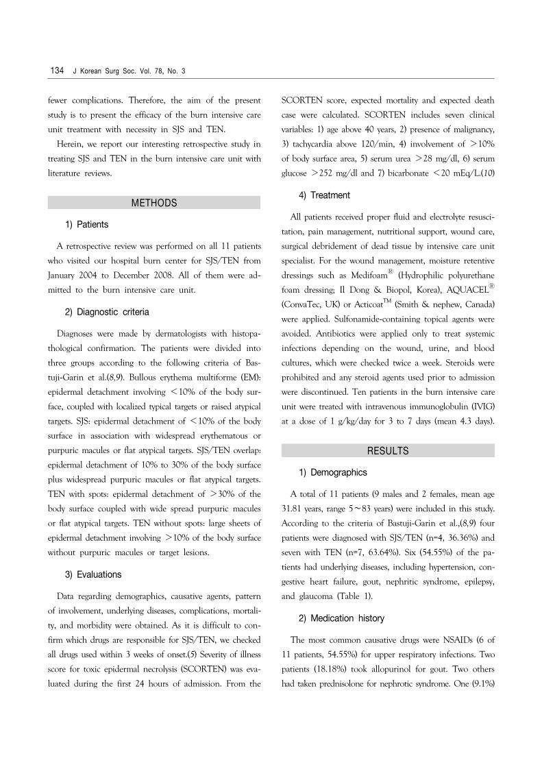
Table 1. The clinical profiles related with medication of 11 patients with SJS/TEN overlap and TEN
Patient No. Criteria Age/SexOnset*(days)
TBSA†
(%)Underlying preexisting disease
Previousdrug allergy
Offending drugs
1 SJS/TEN 41/M 5 18 Upper respiratory infection − NSAIDs‡
2 SJS/TEN 6/F 5 27 Acute pharyngeal tonsillitis, mycoplasma pneumonia − NSAIDs 3 SJS/TEN 83/F 3 27 Upper respiratory infection − NSAIDs 4 SJS/TEN 5/M 3 27 Acute pharyngeal tonsillitis − NSAIDs 5 TEN 48/M 21 36 Hypertension, congestive heart failure, gout − Allopurinol 6 TEN 79/M 5 40 Hypertension, congestive heart failure, gout − Allopurinol 7 TEN 7/M 5 60 Upper respiratory infection − NSAIDs 8 TEN 8/M 9 60 Epilepsy − Carbamazepine 9 TEN 15/M 3 90 Nephrotic syndrome − Prednisolone10 TEN 45/M 5 95 Upper respiratory infection, glaucoma − NSAIDs11 TEN 13/M 35 100 Nephrotic syndrome − Prednisolone
*Time of onset clinical disease following the institution of a new drug regimen; †TBSA = total body surface area; ‡NSAIDs = nonsteroidal anti-inflammatory drugs.
patient had taken the antiepileptic drug carbamazepine for
epilepsy. The average period between taking the relevant
drug to the appearance of symptoms was 9 days. Both
overlap SJS/TEN (100%) and TEN (71.4%) showed symp-
toms within 2 weeks. The mean percentage total body
surface area (TBSA) of skin involvement was 24.75% (18∼
30%) in overlap SJS/TEN and 68.7% (31∼100%) in TEN
(Table 1).
3) Clinical courses
The time from appearance of the first skin lesions to
the initiation of therapy varied from 1 to 10 days (mean
4.27 days). The mean period of hospital care to complete
skin healing time was 9 days (8∼12 days). The mean
period of hospitalization was 14.66 days (7∼22 days). All
patients showed involvement of the mucous membranes,
including the buccal, conjunctival, and genital mucosae.
4) Complications
During admission, coagulase negative staphylococcus, P.
aeruginosa, A. baumannii, and methicillin-resistant staphylo-
coccus aureus were cultured. On laboratory examination,
neutropenia was found in three cases and normocytic
anemia in six cases. The most common complication was
hepatitis, which was seen in one case in the overlap
SJS/TEN group and four cases in the TEN group. Acute
renal failure occurred in four cases in the TEN group.
Continuous renal replacement therapy (CRRT) was applied
in two patients. Two patients developed sepsis and three
patients had disseminated intravascular coagulation (DIC).
Conjunctivitis developed in six patients.
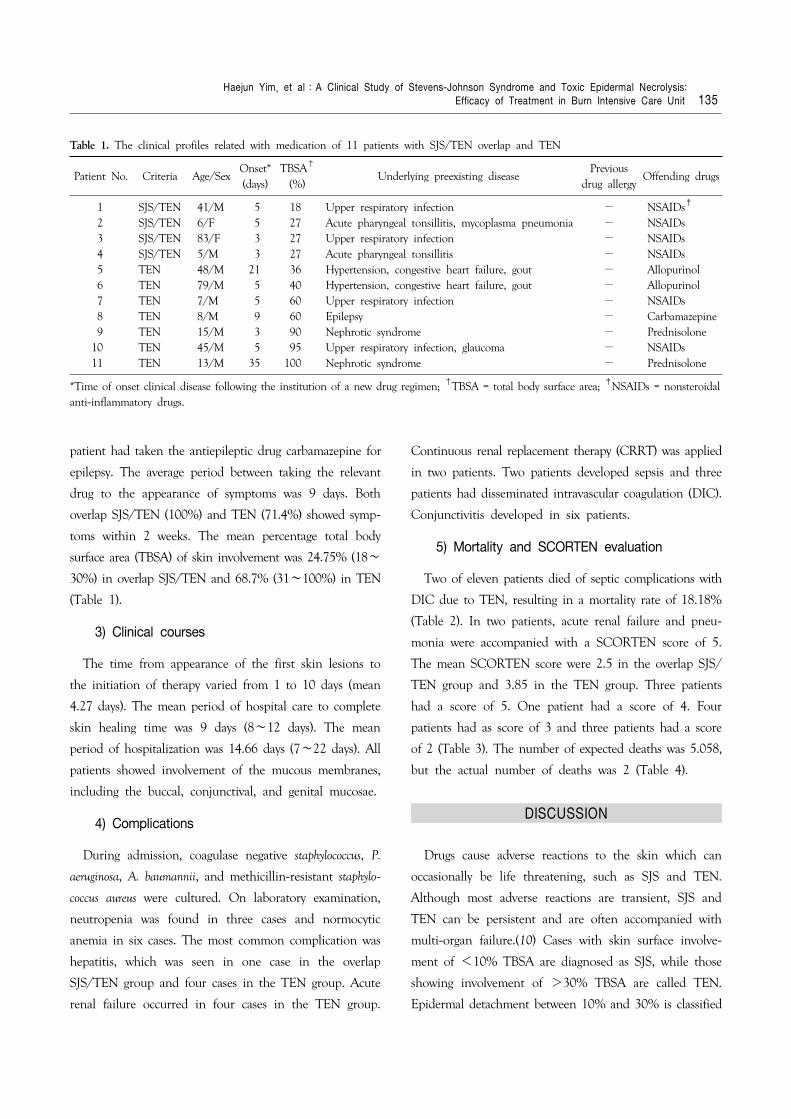
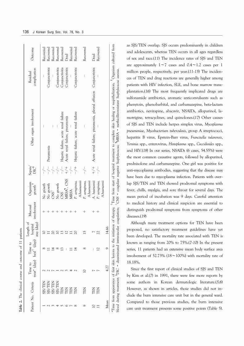
5) Mortality and SCORTEN evaluation
Two of eleven patients died of septic complications with
DIC due to TEN, resulting in a mortality rate of 18.18%
(Table 2). In two patients, acute renal failure and pneu-
monia were accompanied with a SCORTEN score of 5.
The mean SCORTEN score were 2.5 in the overlap SJS/
TEN group and 3.85 in the TEN group. Three patients
had a score of 5. One patient had a score of 4. Four
patients had as score of 3 and three patients had a score
of 2 (Table 3). The number of expected deaths was 5.058,
but the actual number of deaths was 2 (Table 4).
DISCUSSION
Drugs cause adverse reactions to the skin which can
occasionally be life threatening, such as SJS and TEN.
Although most adverse reactions are transient, SJS and
TEN can be persistent and are often accompanied with
multi-organ failure.(10) Cases with skin surface involve-
ment of <10% TBSA are diagnosed as SJS, while those
showing involvement of >30% TBSA are called TEN.
Epidermal detachment between 10% and 30% is classified
136 J Korean Surg Soc. Vol. 78, No. 3
as SJS/TEN overlap. SJS occurs predominantly in children
and adolescents, whereas TEN occurs in all ages regardless
of sex and race.(11) The incidence rates of SJS and TEN
are approximately 1∼7 cases and 0.4∼1.2 cases per 1
million people, respectively, per year.(11-15) The inciden-
ces of TEN and drug reactions are generally higher among
patients with HIV infection, SLE, and bone marrow trans-
plantation.(16) The most frequently implicated drugs are
sulfonamide antibiotics, aromatic anticonvulsants such as
phenytoin, phenobarbital, and carbamazepine, beta-lactam
antibiotics, nevirapine, abacavir, NSAIDs, allopurinol, la-
motrigine, tetracyclines, and quinolones.(17) Other causes
of SJS and TEN include herpes simplex virus, Mycoplasma
pneumoniae, Mycobacterium tuberculosis, group A streptococci,
hepatitis B virus, Epstein-Barr virus, Francisella tularensis,
Yersinia spp., enterovirus, Histoplasma spp., Coccidioides spp.,
and HIV.(18) In our series, NSAIDs (6 cases, 54.55%) were
the most common causative agents, followed by allopurinol,
prednisolone and carbamazepine. One girl was positive for
anti-mycoplasma antibodies, suggesting that the disease may
have been due to mycoplasma infection. Patients with over-
lap SJS/TEN and TEN showed prodromal symptoms with
fever, chills, myalgia, and sore throat for several days. The
mean period of incubation was 9 days. Careful attention
to medical history and clinical suspicion are essential to
distinguish prodromal symptoms from symptoms of other
diseases.(19)
Although many treatment options for TEN have been
proposed, no satisfactory treatment guidelines have yet
been developed. The mortality rate associated with TEN is
known as ranging from 20% to 75%.(7-10) In the present
series, 11 patients had an extensive mean body surface area
involvement of 52.73% (18∼100%) with mortality rate of
18.18%.
Since the first report of clinical studies of SJS and TEN
by Kim et al.(7) in 1991, there were few more reports by
some authors in Korean dermatologic literature.(5,6)
However, as shown in articles, those studies did not in-
clude the burn intensive care unit but in the general ward.
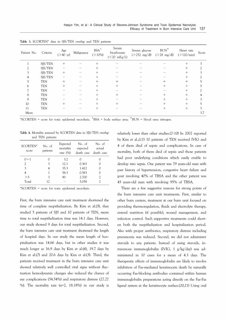
Compared to those previous studies, the burn intensive
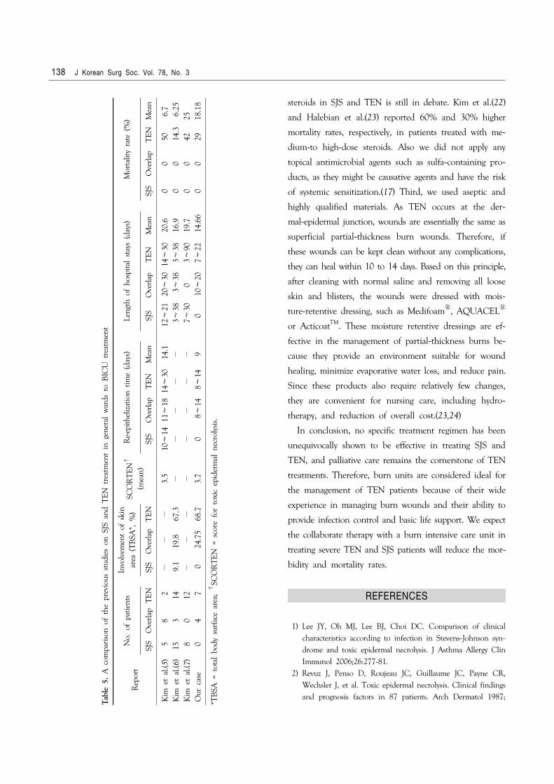
care unit treatment presents some positive points (Table 5).
Tabl
e 2.
The
clin
ical
cou
rse
and
outc
ome
of 1
1 pa
tient
s
Patie
nt N
o.C
rite
ria
Tim
e to
trea
t* (
days
)T
ime
tohe
al†
(da
ys)
Leng
th o
fho
spita
lst
ay (
days
)
Muc
osal
invo
lvem
ent
Org
anism
grow
th‡
Seps
is/
DIC
§O
ther
org
an i
nvol
vem
ent
Res
idua
lco
mpl
icat
ion
Out
com
e
1
2
3
4
5
6
7
8
9
10
11
M
ean
SJS/
TEN
SJS/
TEN
SJS/
TEN
SJS/
TEN
TEN
TEN
TEN
TEN
TEN
TEN
TEN
2 2 7 1 2 3 4 2 10 7 7 4.2
7
8 11 14 8 13 − 11 14 8 − 12 9
10 11 20 10 13 12 11 20 15 7 22 14.6
6
+ + + + + + + + + + +
No
grow
thN
o gr
owth
CN
S∥
No
grow
thC
NS
MR
SA¶ ,
CN
SM
RSA
P. a
erug
inos
a, A
.bau
man
nii
P. a
erug
inos
a, A
.bau
man
nii
A.
baum
anni
iP.
aer
ugin
osa,
A.b
aum
anni
i
−/−
−/−
−/−
−/−
−/−
+/+ −/−
−/+
−/−
+/+ −/−
−Pn
eum
onia
− −H
epat
ic f
ailu
re,
acut
e re
nal
failu
reA
cute
ren
al f
ailu
re,
pneu
mon
ia−
Hep
atic
fai
lure
, ac
ute
rena
l fa
ilure
−A
cute
ren
al f
ailu
re,
pneu
mon
ia,
pleu
ral
effu
sion
−
−C
onju
nctiv
itis
−C
onju
nctiv
itis
Con
junc
tiviti
sC
onju
nctiv
itis
−C
onju
nctiv
itis
−C
onju
nctiv
itis
−
Rec
over
edR
ecov
ered
Rec
over
edR
ecov
ered
Rec
over
edD
ead
Rec
over
edR
ecov
ered
Rec
over
ed
Dea
dR
ecov
ered
*Tim
e fr
om a
ppea
ranc
e of
fir
st s
kin
lesi
ons
to t
he i
nitia
tion
of t
hera
py;
†T
ime
from
sta
rt o
f ho
spita
l tr
eatm
ent
to c
ompl
ete
skin
hea
ling
or r
eepi
thel
izatio
n; ‡
Org
anism
cul
ture
d fr
ombl
ood
duri
ng t
reat
men
t; § D
IC =
diss
emin
ated
int
rava
scul
ar c
oagu
latio
n; ∥
CN
S =
coag
ulas
e ne
gativ
e St
aphy
loco
ccus
; ¶ M
RSA
= m
ethi
cilli
n-re
sista
nt S
taph
yloco
ccus
aur
esus
.
Haejun Yim, et al:A Clinical Study of Stevens-Johnson Syndrome and Toxic Epidermal Necrolysis: Efficacy of Treatment in Burn Intensive Care Unit 137
Table 4. Mortality assessed by SCORTEN data in SJS/TEN overlapand TEN patients
SCORTEN*score
No. ofpatients
Expectedmortalityrate (%)
No. ofexpected
death case
No. of actual
death case
0∼1 0 3.2 0 02 3 12.1 0.363 03 4 35.3 1.412 04 1 58.3 0.583 0>5 3 90 2.700 2Total 12 − 5.058 2
*SCORTEN = score for toxic epidermal necrolysis.
Table 3. SCORTEN* data in SJS/TEN overlap and TEN patients
Patient No. CriteriaAge
(>40 yr)Malignancy
BSA†
(>10%)
Serumbicarbonate
(<20 mEq/L)
Serum glucose(>252 mg/dl)
BUN‡
(>28 mg/dl)Heart rate
(>120/min)Score
1 SJS/TEN + − + − − − + 3 2 SJS/TEN − − + − − − + 2 3 SJS/TEN + − + − − − + 3 4 SJS/TEN − − + − − − + 2 5 TEN + − + + − + + 5 6 TEN + − + + − + + 5 7 TEN − − + − − − + 2 8 TEN − − + + − + + 4 9 TEN − − + − − + + 310 TEN + − + + − + + 511 TEN − − + − − + + 3Mean 3.7
*SCORTEN = score for toxic epidermal necrolysis; †BSA = body surface area; ‡BUN = blood urea nitrogen.
First, the burn intensive care unit treatment shortened the
time of complete reepithelization. By Kim et al.(5), they
studied 5 patients of SJS and 10 patients of TEN, mean
time to total reepithelization time was 14.1 days. However,
our study showed 9 days for total reepithelization. Second,
the burn intensive care unit treatment shortened the length
of hospital days. In our study the mean length of hos-
pitalization was 14.66 days, but in other studies it was
much longer as 16.9 days by Kim et al.(6), 19.7 days by
Kim et al.(7) and 20.6 days by Kim et al.(5). Third, the
patients received treatment in the burn intensive care unit
showed relatively well controlled vital signs without fluc-
tuation hemodynamic changes also reduced the chance of
eye complications (54.54%) and respiratory distress (27.27
%). The mortality rate (n=2, 18.18%) in our study is
relatively lower than other studies.(7-10) In 2002 reported
by Kim et al.,(11) 10 patients of TEN received IVIG and
4 of them died of sepsis and complications. In case of
mortality, both of them died of sepsis and those patients
had poor underlying conditions which easily enable to
develop into sepsis. One patient was 79 years-old man with
past history of hypertension, congestive heart failure and
gout involving 40% of TBSA and the other patient was
45 years-old man with involving 95% of TBSA.
There are a few suggestive reasons for strong points of
the burn intensive care unit treatments. First, similar to
other burn centers, treatment at our burn unit focused on
providing thermoregulation, fluids and electrolyte therapy,
enteral nutrition (if possible), wound management, and
infection control. Such supportive treatments could short-
en both the reepithelization and hospitalization period.
Also with proper antibiotics, respiratory distress including
pneumonia was reduced. Second, we did not administer
steroids to any patients. Instead of using steroids, in-
travenous immunoglobulin (IVIG, 1 g/kg/day) was ad-
ministered in 10 cases for a mean of 4.3 days. The
therapeutic effects of immunoglobulin are likely to involve
inhibition of Fas-mediated keratinocyte death by naturally
occurring Fas-blocking antibodies contained within human
immunoglobulin preparations acting directly on the Fas-Fas
ligand system at the keratinocyte surface.(20,21) Using oral
138 J Korean Surg Soc. Vol. 78, No. 3
steroids in SJS and TEN is still in debate. Kim et al.(22)
and Halebian et al.(23) reported 60% and 30% higher
mortality rates, respectively, in patients treated with me-
dium-to high-dose steroids. Also we did not apply any
topical antimicrobial agents such as sulfa-containing pro-
ducts, as they might be causative agents and have the risk
of systemic sensitization.(17) Third, we used aseptic and
highly qualified materials. As TEN occurs at the der-
mal-epidermal junction, wounds are essentially the same as
superficial partial-thickness burn wounds. Therefore, if
these wounds can be kept clean without any complications,
they can heal within 10 to 14 days. Based on this principle,
after cleaning with normal saline and removing all loose
skin and blisters, the wounds were dressed with mois-
ture-retentive dressing, such as MedifoamⓇ, AQUACELⓇ
or ActicoatTM. These moisture retentive dressings are ef-
fective in the management of partial-thickness burns be-
cause they provide an environment suitable for wound
healing, minimize evaporative water loss, and reduce pain.
Since these products also require relatively few changes,
they are convenient for nursing care, including hydro-
therapy, and reduction of overall cost.(23,24)
In conclusion, no specific treatment regimen has been
unequivocally shown to be effective in treating SJS and
TEN, and palliative care remains the cornerstone of TEN
treatments. Therefore, burn units are considered ideal for
the management of TEN patients because of their wide
experience in managing burn wounds and their ability to
provide infection control and basic life support. We expect
the collaborate therapy with a burn intensive care unit in
treating severe TEN and SJS patients will reduce the mor-
bidity and mortality rates.
REFERENCES
1) Lee JY, Oh MJ, Lee BJ, Choi DC. Comparison of clinical characteristics according to infection in Stevens-Johnson syn-
drome and toxic epidermal necrolysis. J Asthma Allergy Clin Immunol 2006;26:277-81.
2) Revuz J, Penso D, Roujeau JC, Guillaume JC, Payne CR, Wechsler J, et al. Toxic epidermal necrolysis. Clinical findings and prognosis factors in 87 patients. Arch Dermatol 1987;
Tabl
e 5.
A c
ompa
riso
n of
the
pre
viou
s st
udie
s on
SJS
and
TEN
tre
atm
ent
in g
ener
al w
ards
to
BIC
U t
reat
men
t
Rep
ort
No.
of
patie
nts
In
volv
emen
t of
ski
nar
ea (
TB
SA*,
%)
SCO
RT
EN†
(mea
n)
Re-
epith
eliz
atio
n tim
e (d
ays)
Le
ngth
of
hosp
ital
stay
s (d
ays)
Mor
talit
y ra
te (
%)
SJS
Ove
rlap
TEN
SJ
SO
verla
pT
ENSJ
SO
verl
apT
ENM
ean
SJ
SO
verla
pT
ENM
ean
SJS
Ove
rlap
TEN
Mea
n
Kim
et
al.(5
) 5
8 2
−
−−
3.5
10∼
1411
∼18
14∼
30 1
4.1
12
∼21
20∼
3014
∼30
20.6
00
50
6.
7K
im e
t al
.(6)
153
149.
119
.867
.3−
−−
−−
3∼
38
3∼38
3
∼38
16
.90
0 1
4.3
6.
25K
im e
t al
.(7)
80
12−
−−
−−
−−
−
7∼30
0 3
∼90
19
.70
0 4
225
Our
cas
e 0
4 7
0
24.
7568
.73.
70
8∼
14 8
∼14
9
010
∼20
7∼
22 1
4.66
00
29
18.1
8
*TB
SA =
tot
al b
ody
surf
ace
area
; †
SCO
RT
EN =
sco
re f
or t
oxic
epi
derm
al n
ecro
lysis
.
Haejun Yim, et al:A Clinical Study of Stevens-Johnson Syndrome and Toxic Epidermal Necrolysis: Efficacy of Treatment in Burn Intensive Care Unit 139
123:1160-5.3) McGee T, Munster A. Toxic epidermal necrolysis syndrome:
mortality rate reduced with early referral to regional burn center. Plast Reconstr Surg 1998;102:1018-22.
4) Ruiz-Maldonado R. Acute disseminated epidermal necrosis types 1, 2, and 3: study of sixty cases. J Am Acad Dermatol 1985;13:623-35.
5) Kim JW, Kim ST, Song DH. A clinical observation of the patients with Stevens-Johnson syndrome and toxic epidermal necrolysis in Jeju island. Korean J Dermatol 2004;42:579-91.
6) Kim EJ, Lee JB, Kwon YH, Yun SJ, Kim SJ, Lee SC, et al. A clinical study of Stevens-Johnson syndrome and toxic epidermal necrolysis over the last five-year period in the Gwangju-Chon-
nam area. Korean J Dermatol 2006;44:574-8.7) Kim YG, Cho KH, Chung JH. A comparative clinical study of
toxic epidermal necrolysis and Stevens-Johnson syndrome. Kor-ean J Dermatol 1991;29:602-9.
8) Halebian P, Corder V, Herndon D, Shires GT. Clinical ma-
nagement: a burn center experience with toxic epidermal ne-
crolysis. J Burn Care Rehabil 1983;4:176-83.9) Bastuji-Garin S, Rzany B, Stern RS, Shear NH, Naldi L,
Roujeau JC. Clinical classification of cases of toxic epidermal necrolysis, Stevens-Johnson syndrome, and erythema multi-forme. Arch Dermatol 1993;129:92-6.
10) Pereira FA, Mudgil AV, Rosmarin DM. Toxic epidermal ne-
crolysis. J Am Acad Dermatol 2007;56:181-200.11) Kim KJ, Jee MS, Han MH, Choi JH, Sung KJ, Moon KC, et
al. The effect of high-dose intravenous immunoglobulin for the treatment of toxic epidermal necrolysis. Korean J Dermatol 2002;40:766-71.
12) Wolkenstein P, Latarjet J, Roujeau JC, Duguet JC, Boudeau S, Vaillant L, et al. Randomised comparison of thalidomide versus placebo in toxic epidermal necrolysis. Lancet 1998;352:1586-9.
13) Lohmeier K, Megahed M, Schulte KW, Stannigel H, Mayatepek E, Schroten H. Toxic epidermal necrolysis in a premature infant of 27 weeks’ gestational age. Br J Dermatol 2005;152:150-1.
14) Chan HL, Stern RS, Arndt KA, Langlois J, Jick SS, Jick H, et al. The incidence of erythema multiforme, Stevens-Johnson syndrome, and toxic epidermal necrolysis. A population-based study with particular reference to reactions caused by drugs
among outpatients. Arch Dermatol 1990;126:43-7.15) Roujeau JC, Guillaume JC, Fabre JP, Penso D, Flechet ML,
Girre JP. Toxic epidermal necrolysis (Lyell syndrome). Incidence and drug etiology in France, 1981-1985. Arch Dermatol 1990; 126:37-42.
16) Strom BL, Carson JL, Halpern AC, Schinnar R, Snyder ES, Shaw M, et al. A population-based study of Stevens-Johnson syndrome. Incidence and antecedent drug exposures. Arch Dermatol 1991;127:831-8.
17) McKenna JK, Leiferman KM. Dermatologic drug reactions. Immunol Allergy Clin North Am 2004;24:399-423, vi.
18) Roujeau JC, Kelly JP, Naldi L, Rzany B, Stern RS, Anderson T, et al. Medication use and the risk of Stevens-Johnson syndrome or toxic epidermal necrolysis. N Engl J Med 1995; 333:1600-7.
19) Fritsch PO, Ruiz-Maldonado R. Erythema multiforme, Stevens- Johnson syndrome and toxic epidermal necrolysis. In: Freedberg IM, Eisen AZ, Wolff K, Austen KF, Goldsmith LA, Katz SI, editors. Fitzpatrick’s Dermatology in General Medicine. 6th ed. New York: McGraw-Hill; 2003:543-57.
20) Schopf E, Stuhmer A, Rzany B, Victor N, Zentgraf R, Kapp JF. Toxic epidermal necrolysis and Stevens-Johnson syndrome. An epidemiologic study from West Germany. Arch Dermatol 1991; 127:839-42.
21) Viard I, Wehrli P, Bullani R, Schneider P, Holler N, Salomon D, et al. Inhibition of toxic epidermal necrolysis by blockade of CD95 with human intravenous immunoglobulin. Science 1998;282:490-3.
22) Kim PS, Goldfarb IW, Gaisford JC, Slater H. Burn rounds: Stevens-Johnson syndrome and toxic epidermal necrolysis: a pathophysiologic review with recommendations for a treatment protocol. J Burn Care Rehabil 1983;4:91-100.
23) Halebian PH, Corder VJ, Madden MR, Finklestein JL, Shires GT. Improved burn center survival of patients with toxic epi-dermal necrolysis managed without corticosteroids. Ann Surg 1986;204:503-12.
24) Choi SW, Suh MS, Park SJ, Lim YK. Burn care of toxic epidermal necrolysis using allevynⓇ: a report of two cases. J Korean Burn Soc 2006;9:74-8.
Recommended