Lesson Overview7.1 Life is Cellular

Lesson Overview Life Is Cellular
The Discovery of the Cell
What is the cell theory?
The cell theory states:
• All living things are made up of cells.
• Cells are the basic units of structure and function in
living things.
• New cells are produced from existing cells.

Lesson Overview Life Is Cellular
Early Microscopes
It was not until the mid-1600s that scientists began to use microscopes to
observe living things.
In 1665, Englishman Robert Hooke used an early compound microscope to
look at a nonliving thin slice of cork, a plant material.
Under the microscope, cork seemed to be made of thousands of tiny, empty
chambers that Hooke called “cells”. The term cell is used in biology to this
day.
Today we know that living cells are not empty chambers, but contain a huge
array of working parts, each with its own function.
Lesson Overview Life Is Cellular
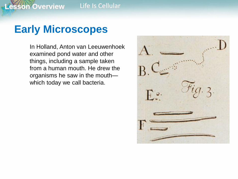
Early Microscopes
In Holland, Anton van Leeuwenhoek
examined pond water and other
things, including a sample taken
from a human mouth. He drew the
organisms he saw in the mouth—
which today we call bacteria.
Lesson Overview Life Is Cellular
The Cell Theory
Soon after Leeuwenhoek, observations made by other scientists made it clear that cells were the basic units of life.
In 1838, German botanist Matthias Schleiden concluded that all plants are made of cells.
The next year, German biologist Theodor Schwann stated that all animals were made of cells.
In 1855, German physician Rudolf Virchow concluded that new cells could be produced only from the division of existing cells, confirming a suggestion made by German Lorenz Oken 50 years earlier.
Lesson Overview Life Is Cellular
The Cell Theory
These discoveries are summarized in the cell theory,
a fundamental concept of biology.
The cell theory states:
-All living things are made up of cells.
-Cells are the basic units of structure and function in living things.
-New cells are produced from existing cells.

Lesson Overview Life Is Cellular
Exploring the Cell
How do microscopes work?
Most microscopes use lenses to magnify the image of
an object by focusing light or electrons.

Lesson Overview Life Is Cellular
Light Microscopes and Cell Stains
A typical light microscope allows light to pass through a specimen and uses two lenses to form an image.
The first set of lenses, located just above the specimen, produces an enlarged image of the specimen.
The second set of lenses magnifies this image still further.
Because light waves are diffracted, or scattered, as they pass through matter, light microscopes can produce clear images of objects only to a magnification of about 1000 times.
Lesson Overview Life Is Cellular
Light Microscopes and Cell Stains
Another problem with light microscopy is that
most living cells are nearly transparent, making it
difficult to see the structures within them.
Using chemical stains or dyes can usually solve
this problem. Some of these stains are so
specific that they reveal only compounds or
structures within the cell.
Lesson Overview Life Is Cellular
Light Microscopes and Cell Stains
Some dyes give off light of a particular color when viewed under specific
wavelengths of light, a property called fluorescence.
Fluorescent dyes can be attached to specific molecules and can then be
made visible using a special fluorescence microscope.
Fluorescence microscopy makes it possible to see and identify the locations
of these molecules, and even to watch them move about in a living cell.
Lesson Overview Life Is Cellular
Electron Microscopes
Light microscopes can be used to see cells and cell structures as small as 1 millionth of a meter. To study something smaller than that, scientists need to use electron microscopes.
Electron microscopes use beams of electrons, not light, that are focused by magnetic fields.
Electron microscopes offer much higher resolution than light microscopes.
There are two major types of electron microscopes: transmission and scanning.
Lesson Overview Life Is Cellular
Electron Microscopes
Transmission electron microscopes make it possible
to explore cell structures and large protein
molecules.
Because beams of electrons can only pass through
thin samples, cells and tissues must be cut first into
ultra thin slices before they can be examined under
a transmission electron microscope.
Transmission electron microscopes produce flat,
two-dimensional images.

Lesson Overview Life Is Cellular
Electron Microscopes
In scanning electron microscopes, a pencil-like
beam of electrons is scanned over the surface of a
specimen.
Because the image is of the surface, specimens
viewed under a scanning electron microscope do
not have to be cut into thin slices to be seen.
Scanning electron microscopes produce three-
dimensional images of the specimen’s surface.

Lesson Overview Life Is Cellular
Electron Microscopes
Because electrons are easily scattered by molecules
in the air, samples examined in both types of
electron microscopes must be placed in a vacuum in
order to be studied.
Researchers chemically preserve their samples first
and then carefully remove all of the water before
placing them in the microscope.
This means that electron microscopy can be used to
examine only nonliving cells and tissues.
Lesson Overview Life Is Cellular
Prokaryotes and Eukaryotes
How are prokaryotic and eukaryotic cells different?
Prokaryotic cells do not separate their genetic
material within a nucleus.
In eukaryotic cells, the nucleus separates the genetic
material from the rest of the cell.
Lesson Overview Life Is Cellular
Prokaryotes and Eukaryotes
Although typical cells range from 5 to 50 micrometers in diameter, the
smallest Mycoplasma bacteria are only 0.2 micrometers across, so small
that they are difficult to see under even the best light microscopes.
In contrast, the giant amoeba Chaos chaos may be 1000 micrometers in
diameter, large enough to be seen with the unaided eye as a tiny speck in
pond water.
Despite their differences, all cells contain the
molecule that carries biological information—DNA.
In addition, all cells are surrounded by a thin, flexible
barrier called a cell membrane.
Lesson Overview Life Is Cellular
Prokaryotes and Eukaryotes
Cells fall into two broad categories, depending on whether
they contain a nucleus.
The nucleus is a large membrane-enclosed structure
that contains the cell’s genetic material in the form
of DNA. The nucleus controls many of the cell’s
activities.
Lesson Overview Life Is Cellular
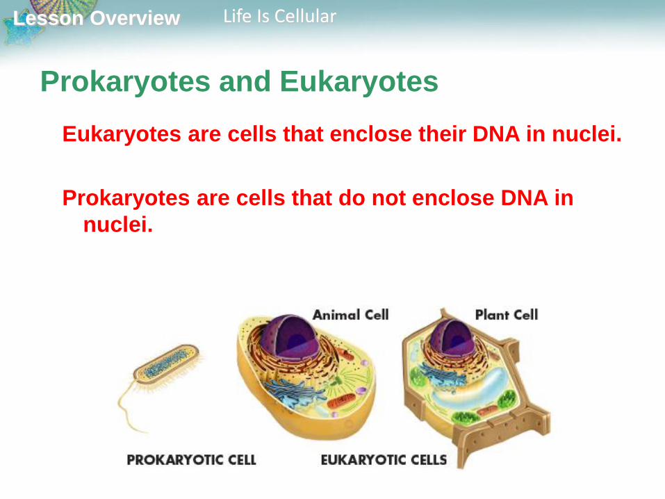
Prokaryotes and Eukaryotes
Eukaryotes are cells that enclose their DNA in nuclei.
Prokaryotes are cells that do not enclose DNA in
nuclei.

Lesson Overview Life Is Cellular
Prokaryotic cells are generally smaller and simpler
than eukaryotic cells.
Despite their simplicity, prokaryotes grow, reproduce, and respond to the
environment, and some can even move by gliding along surfaces or
swimming through liquids.
The organisms we call bacteria are prokaryotes.
Prokaryotes
Lesson Overview Life Is Cellular
Eukaryotes
Eukaryotic cells are generally larger and more
complex than prokaryotic cells.
Most eukaryotic cells contain dozens of structures
and internal membranes. Many eukaryotes are
highly specialized.
There are many types of eukaryotes: plants, animals,
fungi, and organisms commonly called “protists.”

Lesson Overview Cell Structure
The Fluid Mosaic Model Although many substances can cross biological
membranes, some are too large or too strongly charged
to cross the lipid bilayer.
If a substance is able to cross a membrane, the
membrane is said to be permeable to it.
A membrane is impermeable to substances that cannot
pass across it.
Most biological membranes are selectively permeable,
meaning that some substances can pass across them
and others cannot. Selectively permeable membranes
are also called semipermeable membranes.

Lesson Overview7.2 Cell Structure
Lesson Overview Cell Structure
Cell Organization
What is the role of the cell nucleus?
The nucleus contains nearly all the cell’s DNA and, with it, the coded
instructions for making proteins and other important molecules.
Lesson Overview Cell Structure
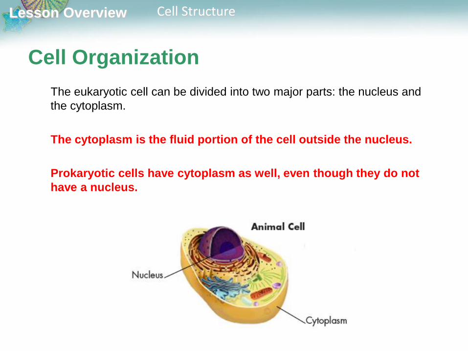
Cell Organization
The eukaryotic cell can be divided into two major parts: the nucleus and
the cytoplasm.
The cytoplasm is the fluid portion of the cell outside the nucleus.
Prokaryotic cells have cytoplasm as well, even though they do not
have a nucleus.
Lesson Overview Cell Structure
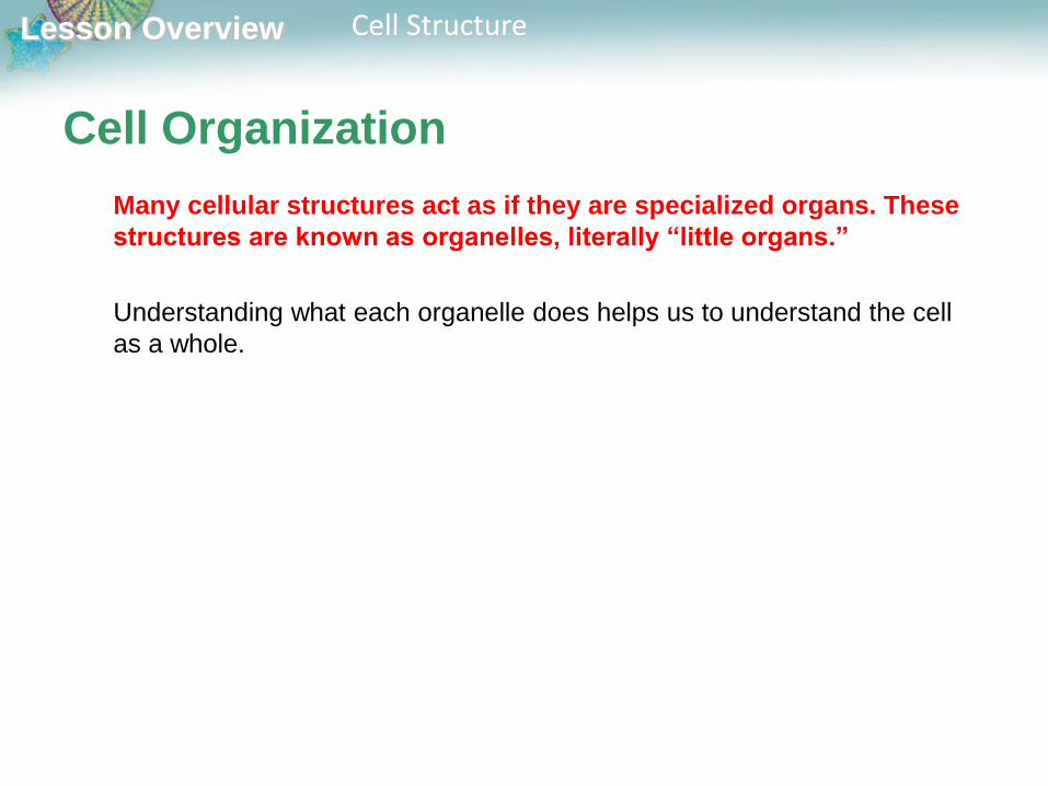
Cell Organization
Many cellular structures act as if they are specialized organs. These
structures are known as organelles, literally “little organs.”
Understanding what each organelle does helps us to understand the cell
as a whole.
Lesson Overview Cell Structure
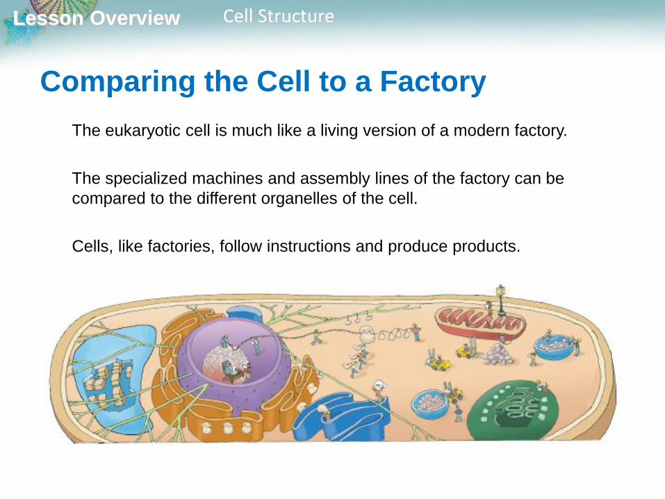
Comparing the Cell to a Factory
The eukaryotic cell is much like a living version of a modern factory.
The specialized machines and assembly lines of the factory can be
compared to the different organelles of the cell.
Cells, like factories, follow instructions and produce products.
Lesson Overview Cell Structure
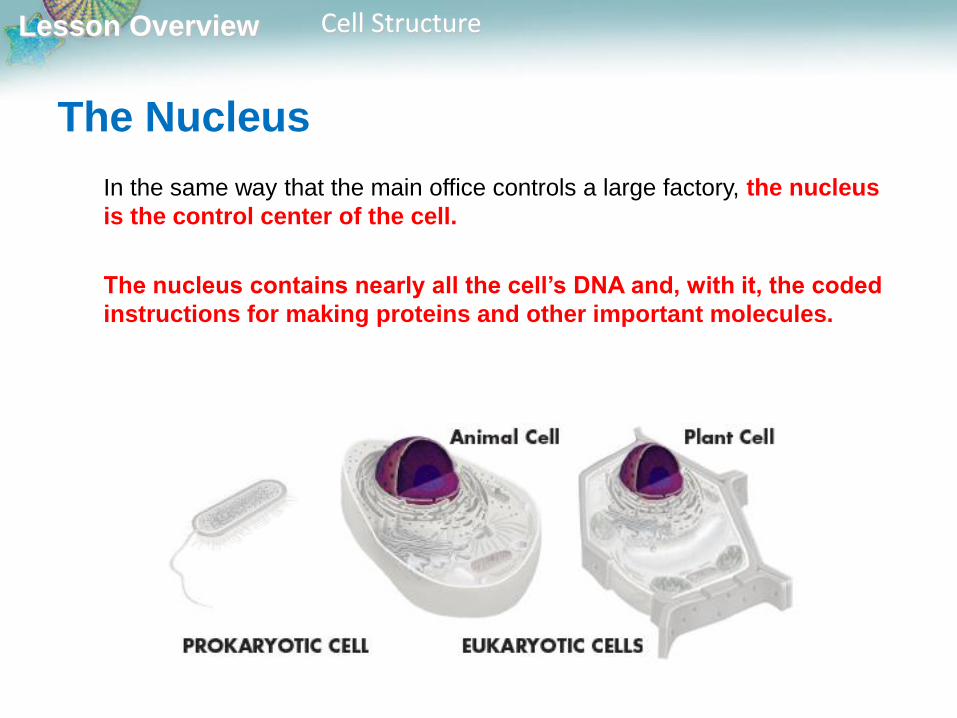
The Nucleus
In the same way that the main office controls a large factory, the nucleus
is the control center of the cell.
The nucleus contains nearly all the cell’s DNA and, with it, the coded
instructions for making proteins and other important molecules.

Lesson Overview Cell Structure
The Nucleus
The nucleus is surrounded by a nuclear envelope composed of two
membranes.
Lesson Overview Cell Structure
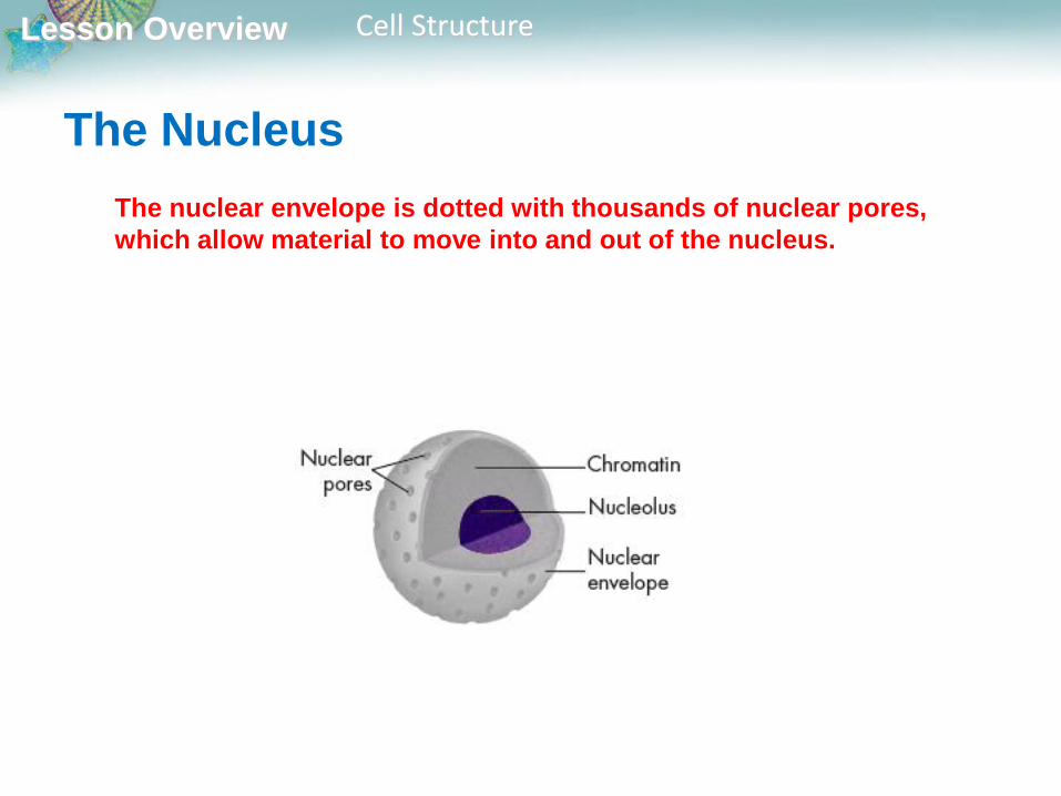
The nuclear envelope is dotted with thousands of nuclear pores,
which allow material to move into and out of the nucleus.
The Nucleus
Lesson Overview Cell Structure
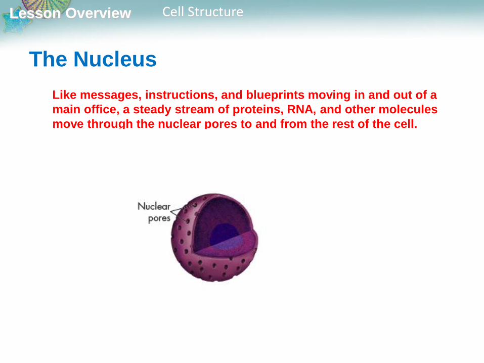
Like messages, instructions, and blueprints moving in and out of a
main office, a steady stream of proteins, RNA, and other molecules
move through the nuclear pores to and from the rest of the cell.
The Nucleus
Lesson Overview Cell Structure
The Nucleus
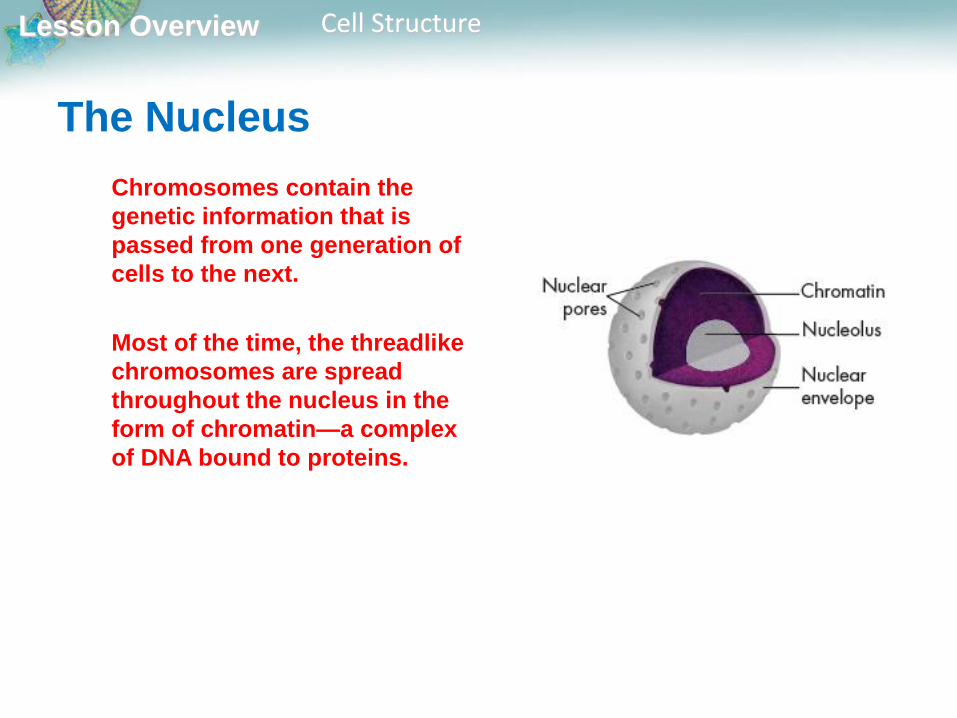
Chromosomes contain the
genetic information that is
passed from one generation of
cells to the next.
Most of the time, the threadlike
chromosomes are spread
throughout the nucleus in the
form of chromatin—a complex
of DNA bound to proteins.

Lesson Overview Cell Structure
The Nucleus
When a cell divides, its
chromosomes condense and can
be seen under a microscope.
Lesson Overview Cell Structure
The Nucleus
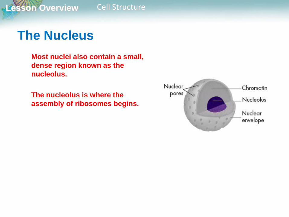
Most nuclei also contain a small,
dense region known as the
nucleolus.
The nucleolus is where the
assembly of ribosomes begins.
Lesson Overview Cell Structure
Organelles That Store, Clean Up, and
Support
What are the functions of vacuoles, lysosomes, and the cytoskeleton?
Vacuoles store materials like water, salts, proteins, and
carbohydrates.
Lysosomes break down lipids, carbohydrates, and proteins into small
molecules that can be used by the rest of the cell. They are also
involved in breaking down organelles that have outlived their
usefulness.
The cytoskeleton helps the cell maintain its shape and is also
involved in movement.

Lesson Overview Cell Structure
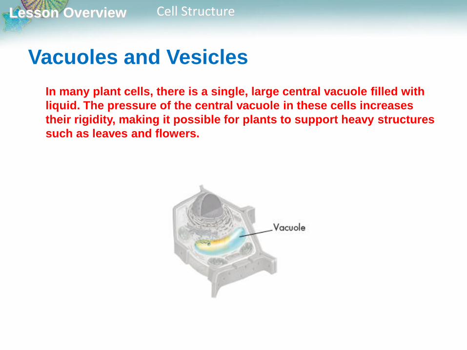
Vacuoles and Vesicles
Many cells contain large, saclike, membrane-enclosed structures called
vacuoles that store materials such as water, salts, proteins, and
carbohydrates.
Lesson Overview Cell Structure
Vacuoles and Vesicles
In many plant cells, there is a single, large central vacuole filled with
liquid. The pressure of the central vacuole in these cells increases
their rigidity, making it possible for plants to support heavy structures
such as leaves and flowers.

Lesson Overview Cell Structure
Vacuoles are also found in some unicellular organisms and in some
animals.
The paramecium contains an organelle called a contractile vacuole. By
contracting rhythmically, this specialized vacuole pumps excess water
out of the cell.
Vacuoles and Vesicles
Lesson Overview Cell Structure
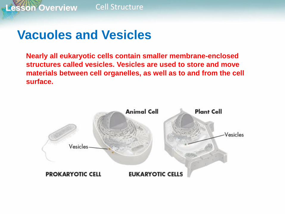
Vacuoles and Vesicles
Nearly all eukaryotic cells contain smaller membrane-enclosed
structures called vesicles. Vesicles are used to store and move
materials between cell organelles, as well as to and from the cell
surface.
Lesson Overview Cell Structure
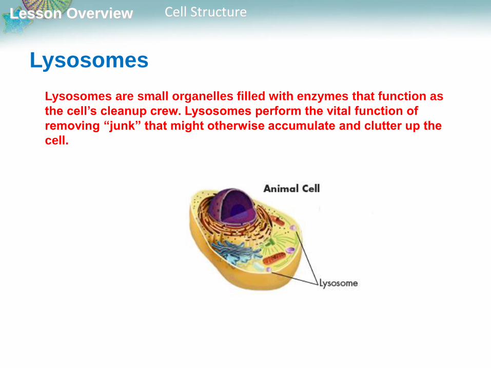
Lysosomes
Lysosomes are small organelles filled with enzymes that function as
the cell’s cleanup crew. Lysosomes perform the vital function of
removing “junk” that might otherwise accumulate and clutter up the
cell.

Lesson Overview Cell Structure
Lysosomes
One function of lysosomes is the breakdown of lipids, carbohydrates,
and proteins into small molecules that can be used by the rest of the
cell.

Lesson Overview Cell Structure
Lysosomes
Lysosomes are also involved in breaking down organelles that have
outlived their usefulness.
Biologists once thought that lysosomes were only found in animal cells, but it
is now clear that lysosomes are also found in a few specialized types of
plant cells as well.
Lesson Overview Cell Structure
The Cytoskeleton
Eukaryotic cells are given their shape and internal organization by a
network of protein filaments known as the cytoskeleton.
Certain parts of the cytoskeleton also help to transport materials
between different parts of the cell, much like conveyer belts that carry
materials from one part of a factory to another.
Microfilaments and microtubules are two of the principal protein
filaments that make up the cytoskeleton.
Lesson Overview Cell Structure
Microfilaments
Microfilaments are threadlike structures made up of a protein called
actin.
They form extensive networks in some cells and produce a tough,
flexible framework that supports the cell.
Microfilaments also help cells move.
Microfilament assembly and disassembly is responsible for the
cytoplasmic movements that allow cells, such as amoebas, to crawl
along surfaces.
Lesson Overview Cell Structure
Microtubules
Microtubules are hollow structures made up of proteins known as
tubulins.
They play critical roles in maintaining cell shape.
Microtubules are also important in cell division, where they form a
structure known as the mitotic spindle, which helps to separate
chromosomes.
Lesson Overview Cell Structure
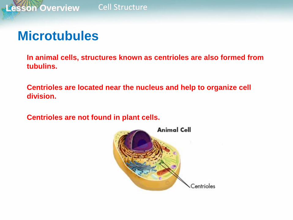
Microtubules
In animal cells, structures known as centrioles are also formed from
tubulins.
Centrioles are located near the nucleus and help to organize cell
division.
Centrioles are not found in plant cells.

Lesson Overview Cell Structure
Microtubules
Microtubules help to build projections from the cell surface,
which are known as cilia and flagella, that enable cells to swim
rapidly through liquids.
Microtubules are arranged in a “9 + 2” pattern.
Small cross-bridges between the microtubules in these
organelles use chemical energy to pull on, or slide along, the
microtubules, allowing cells to produce controlled movements.
Lesson Overview Cell Structure
Organelles That Build Proteins
What organelles help make and transport proteins?
Proteins are assembled on ribosomes.
Proteins made on the rough endoplasmic reticulum include those
that will be released, or secreted, from the cell as well as many
membrane proteins and proteins destined for lysosomes and other
specialized locations within the cell.
The Golgi apparatus modifies, sorts, and packages proteins and
other materials from the endoplasmic reticulum for storage in the
cell or release outside the cell.

Lesson Overview Cell Structure
Organelles That Build Proteins
Cells need to build new molecules all the time, especially proteins, which
catalyze chemical reactions and make up important structures in the cell.
Because proteins carry out so many of the essential functions of living
things, a big part of the cell is devoted to their production and distribution.
Proteins are synthesized on ribosomes, sometimes in association
with the rough endoplasmic reticulum in eukaryotes.
Lesson Overview Cell Structure
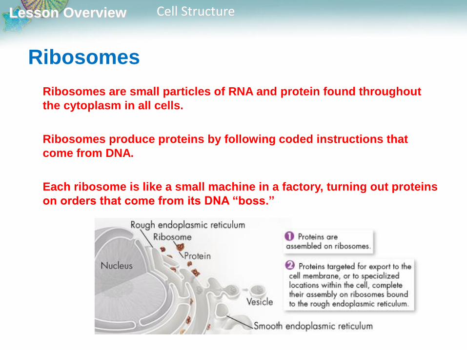
Ribosomes
Ribosomes are small particles of RNA and protein found throughout
the cytoplasm in all cells.
Ribosomes produce proteins by following coded instructions that
come from DNA.
Each ribosome is like a small machine in a factory, turning out proteins
on orders that come from its DNA “boss.”
Lesson Overview Cell Structure
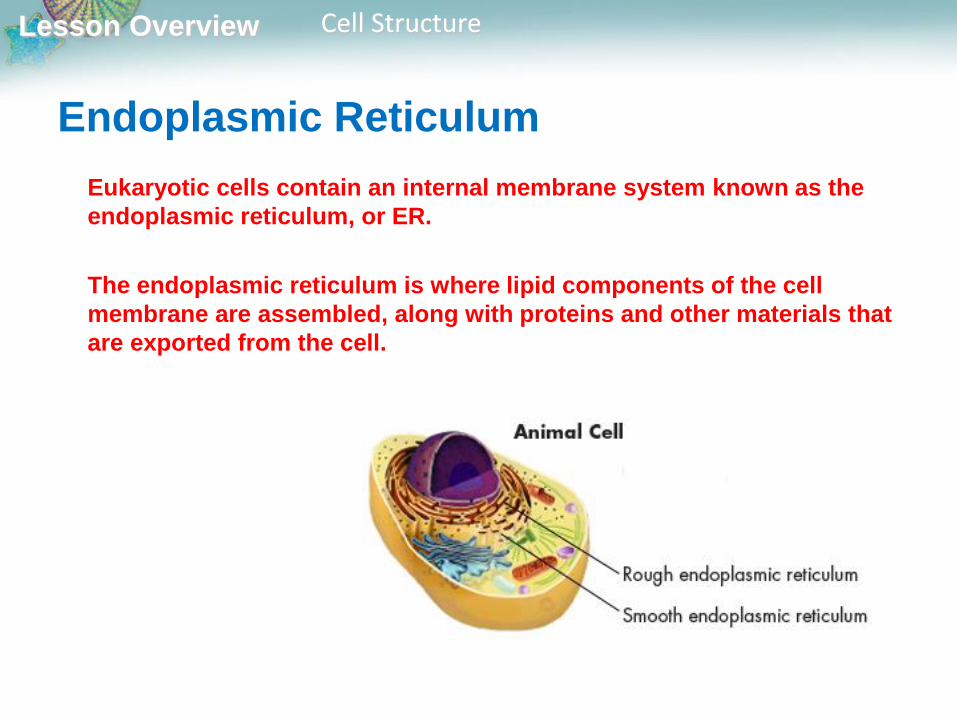
Endoplasmic Reticulum
Eukaryotic cells contain an internal membrane system known as the
endoplasmic reticulum, or ER.
The endoplasmic reticulum is where lipid components of the cell
membrane are assembled, along with proteins and other materials that
are exported from the cell.
Lesson Overview Cell Structure
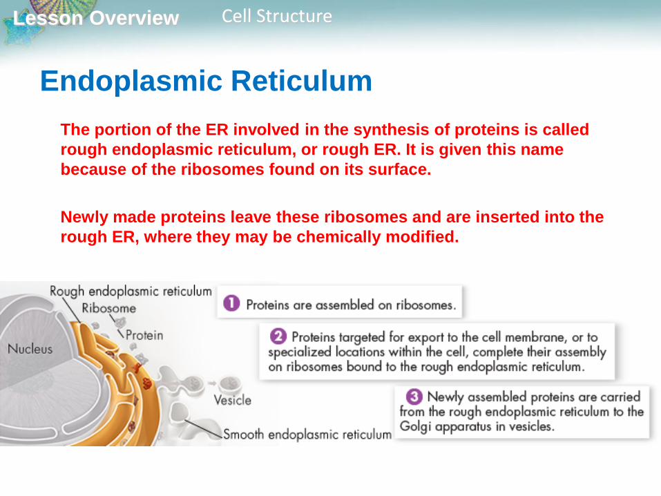
Endoplasmic Reticulum
The portion of the ER involved in the synthesis of proteins is called
rough endoplasmic reticulum, or rough ER. It is given this name
because of the ribosomes found on its surface.
Newly made proteins leave these ribosomes and are inserted into the
rough ER, where they may be chemically modified.
Lesson Overview Cell Structure
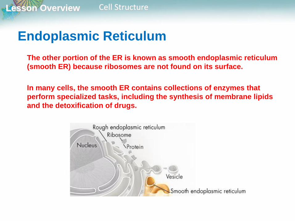
Endoplasmic Reticulum
The other portion of the ER is known as smooth endoplasmic reticulum
(smooth ER) because ribosomes are not found on its surface.
In many cells, the smooth ER contains collections of enzymes that
perform specialized tasks, including the synthesis of membrane lipids
and the detoxification of drugs.

Lesson Overview Cell Structure
Golgi Apparatus
Proteins produced in the rough ER move next into the Golgi apparatus,
which appears as a stack of flattened membranes.
The proteins are bundled into tiny vesicles that bud from the ER and
carry them to the Golgi apparatus.

Lesson Overview Cell Structure
The Golgi apparatus modifies, sorts, and packages proteins and other
materials from the ER for storage in the cell or release outside the cell.
It is somewhat like a customization shop, where the finishing touches
are put on proteins before they are ready to leave the “factory.”
Golgi Apparatus

Lesson Overview Cell Structure
From the Golgi apparatus, proteins are “shipped” to their final
destination inside or outside the cell.
Golgi Apparatus
Lesson Overview Cell Structure
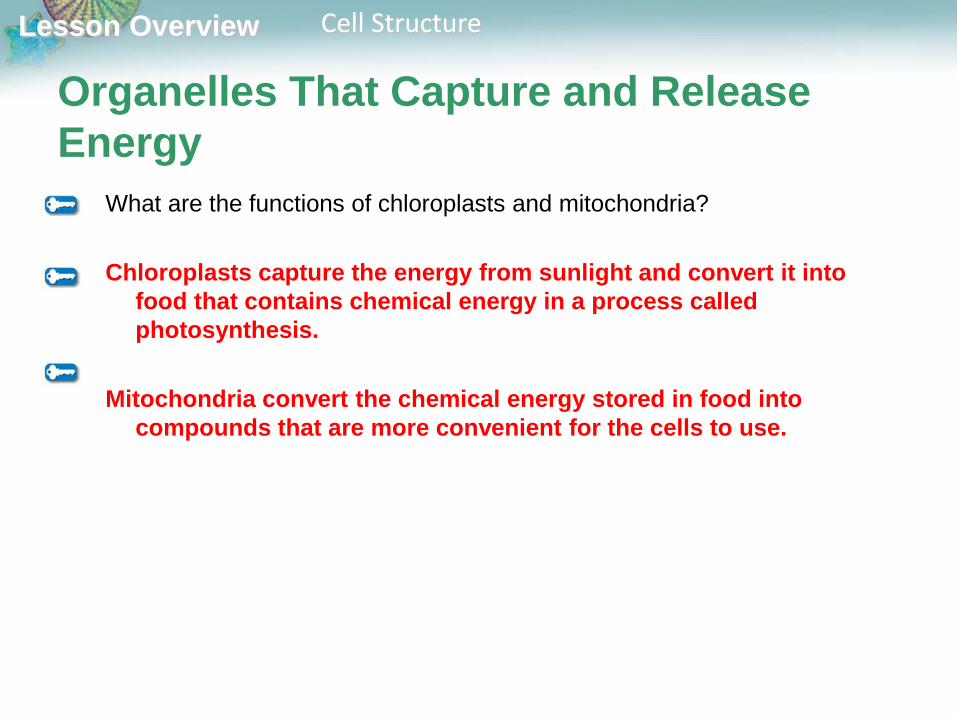
Organelles That Capture and Release
Energy
What are the functions of chloroplasts and mitochondria?
Chloroplasts capture the energy from sunlight and convert it into
food that contains chemical energy in a process called
photosynthesis.
Mitochondria convert the chemical energy stored in food into
compounds that are more convenient for the cells to use.

Lesson Overview Cell Structure
Organelles That Capture and Release
EnergyAll living things require a source of energy. Most cells are powered by
food molecules that are built using energy from the sun.
Chloroplasts and mitochondria are both involved in energy
conversion processes within the cell.
Lesson Overview Cell Structure
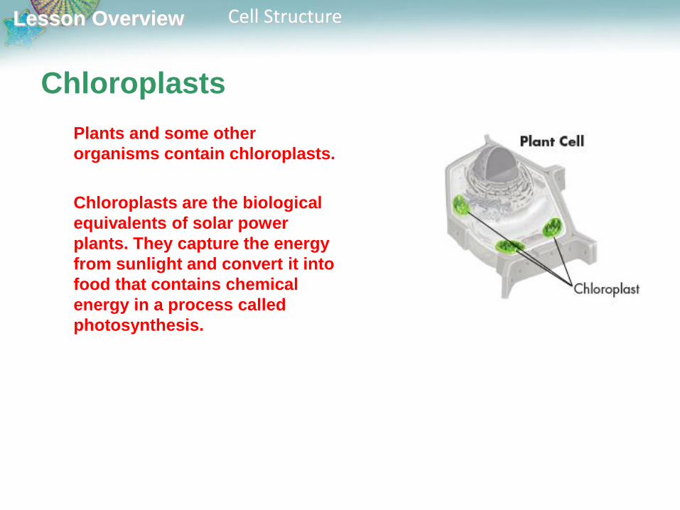
Chloroplasts
Plants and some other
organisms contain chloroplasts.
Chloroplasts are the biological
equivalents of solar power
plants. They capture the energy
from sunlight and convert it into
food that contains chemical
energy in a process called
photosynthesis.
Lesson Overview Cell Structure
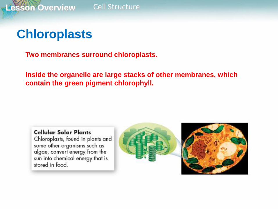
Chloroplasts
Two membranes surround chloroplasts.
Inside the organelle are large stacks of other membranes, which
contain the green pigment chlorophyll.

Lesson Overview Cell Structure
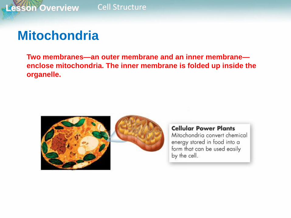
Mitochondria
Nearly all eukaryotic cells, including plants, contain mitochondria.
Mitochondria are the power plants of the cell. They convert the
chemical energy stored in food into compounds that are more
convenient for the cell to use.
Lesson Overview Cell Structure
Mitochondria
Two membranes—an outer membrane and an inner membrane—
enclose mitochondria. The inner membrane is folded up inside the
organelle.
Lesson Overview Cell Structure
Mitochondria
One of the most interesting aspects of mitochondria is the way in which they
are inherited.
In humans, all or nearly all of our mitochondria come from the
cytoplasm of the ovum, or egg cell. You get your mitochondria from
Mom!
Lesson Overview Cell Structure
Mitochondria
Chloroplasts and mitochondria contain their own genetic information
in the form of small DNA molecules.
The endosymbiotic theory suggests that chloroplasts and
mitochondria may have descended from independent microorganisms.

Lesson Overview Cell Structure
Cellular Boundaries
What is the function of the cell membrane?
The cell membrane regulates what enters and leaves the cell and
also protects and supports the cell.
Lesson Overview Cell Structure
Cellular Boundaries
A working factory has walls and a roof to protect it from the environment
outside, and also to serve as a barrier that keeps its products safe and
secure until they are ready to be shipped out.

Lesson Overview Cell Structure
Cellular Boundaries
Similarly, cells are surrounded by a barrier known as the cell
membrane.
Many cells, including most prokaryotes, also produce a strong
supporting layer around the membrane known as a cell wall.

Lesson Overview Cell Structure
Cell Walls
The main function of the cell wall is to provide support and protection
for the cell.
Prokaryotes, plants, algae, fungi, and many prokaryotes have cell
walls. Animal cells do not have cell walls.
Cell walls lie outside the cell membrane and most are porous enough
to allow water, oxygen, carbon dioxide, and certain other substances
to pass through easily.

Lesson Overview Cell Structure
Cell Membranes
All cells contain a cell membrane that regulates what enters and leaves
the cell and also protects and supports the cell.

Lesson Overview Cell Structure
Cell Membranes
The composition of nearly all cell membranes is a double-layered sheet
called a lipid bilayer, which gives cell membranes a flexible structure
and forms a strong barrier between the cell and its surroundings.

Lesson Overview Cell Structure
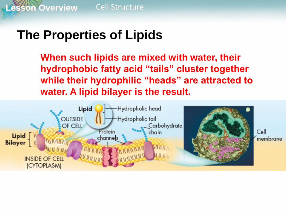
The Properties of Lipids
Many lipids have oily fatty acid chains attached to chemical groups
that interact strongly with water.
The fatty acid portions of such a lipid are
hydrophobic, or “water-hating,” while the
opposite end of the molecule is hydrophilic, or
“water-loving.”
Lesson Overview Cell Structure
The Properties of Lipids
When such lipids are mixed with water, their
hydrophobic fatty acid “tails” cluster together
while their hydrophilic “heads” are attracted to
water. A lipid bilayer is the result.
Lesson Overview Cell Structure
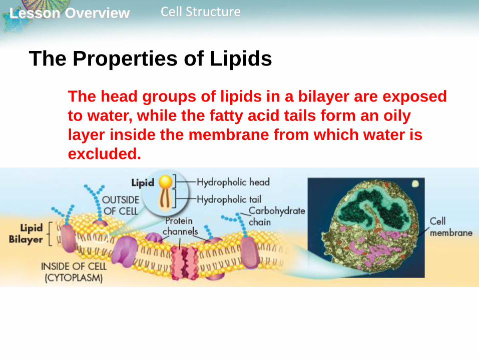
The Properties of Lipids
The head groups of lipids in a bilayer are exposed
to water, while the fatty acid tails form an oily
layer inside the membrane from which water is
excluded.

Lesson Overview Cell Structure
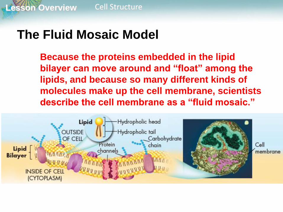
The Fluid Mosaic Model
Most cell membranes contain protein molecules
that are embedded in the lipid bilayer.
Carbohydrate molecules are attached to many of
these proteins.
Lesson Overview Cell Structure
The Fluid Mosaic Model
Because the proteins embedded in the lipid
bilayer can move around and “float” among the
lipids, and because so many different kinds of
molecules make up the cell membrane, scientists
describe the cell membrane as a “fluid mosaic.”

Lesson Overview Cell Structure
The Fluid Mosaic Model
Some of the proteins form channels and pumps
that help to move material across the cell
membrane.
Many of the carbohydrate molecules act like
chemical identification cards, allowing individual
cells to identify one another.
Lesson Overview Cell Structure
The Fluid Mosaic Model Although many substances can cross biological
membranes, some are too large or too strongly charged
to cross the lipid bilayer.
If a substance is able to cross a membrane, the
membrane is said to be permeable to it.
A membrane is impermeable to substances that cannot
pass across it.
Most biological membranes are selectively permeable,
meaning that some substances can pass across them
and others cannot. Selectively permeable membranes
are also called semipermeable membranes.

Lesson Overview7.3 Cell Transport
Lesson Overview Cell Transport
Passive Transport
What is passive transport?
The movement of materials across the cell
membrane without using cellular energy is called
passive transport.

Lesson Overview Cell Transport
Passive Transport
Every living cell exists in a liquid environment.
One of the most important functions of the cell
membrane is to keep the cell’s internal conditions
relatively constant. It does this by regulating the
movement of molecules from one side of the
membrane to the other side.
Lesson Overview Cell Transport
Diffusion
The cytoplasm of a cell is a solution of many different
substances dissolved in water.
In any solution, solute particles tend to move from an area
where they are more concentrated to an area where they are
less concentrated.
The process by which particles move from an area of high
concentration to an area of lower concentration is known as
diffusion.
Diffusion is the driving force behind the movement of many
substances across the cell membrane.

Lesson Overview Cell Transport
Diffusion
Suppose a substance is present in unequal
concentrations on either side of a cell membrane.

Lesson Overview Cell Transport
Diffusion
If the substance can cross the cell membrane, its
particles will tend to move toward the area where it is
less concentrated until it is evenly distributed.
Lesson Overview Cell Transport
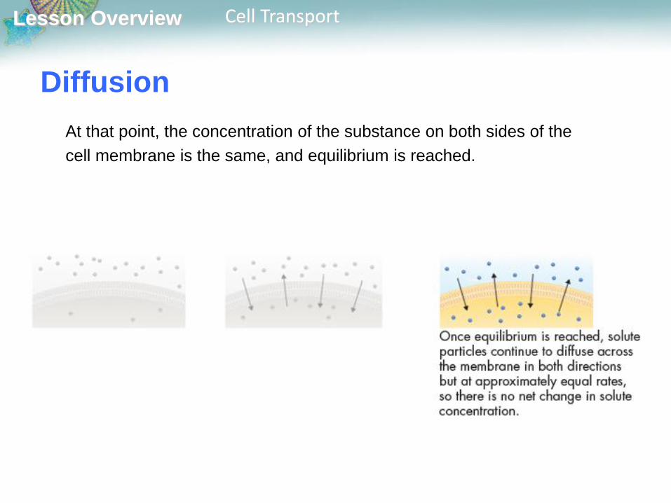
Diffusion
At that point, the concentration of the substance on both sides of the
cell membrane is the same, and equilibrium is reached.

Lesson Overview Cell Transport
Diffusion
Even when equilibrium is reached, particles of a solution will
continue to move across the membrane in both directions.
Because almost equal numbers of particles move in each
direction, there is no net change in the concentration on either
side.

Lesson Overview Cell Transport
Diffusion
Diffusion depends upon random particle movements.
Substances diffuse across membranes without requiring the
cell to use additional energy.
The movement of materials across the cell membrane without
using cellular energy is called passive transport.

Lesson Overview Cell Transport
Facilitated Diffusion
Cell membranes have proteins that act as carriers, or channels,
making it easy for certain molecules to cross.
Molecules that cannot directly diffuse across the membrane
pass through special protein channels in a process known as
facilitated diffusion.
Hundreds of different proteins have been found that allow particular
substances to cross cell membranes.
The movement of molecules by facilitated diffusion does not
require any additional use of the cell’s energy.
Lesson Overview Cell Transport
Osmosis: An Example of Facilitated
Diffusion
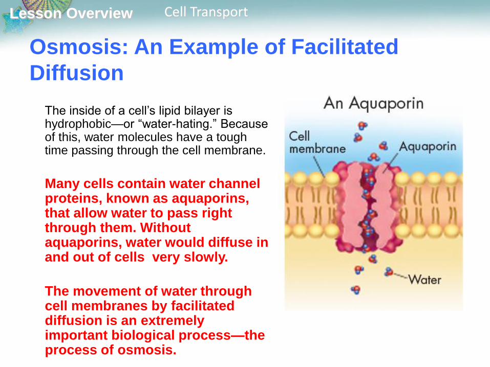
The inside of a cell’s lipid bilayer is hydrophobic—or “water-hating.” Because of this, water molecules have a tough time passing through the cell membrane.
Many cells contain water channel proteins, known as aquaporins, that allow water to pass right through them. Without aquaporins, water would diffuse in and out of cells very slowly.
The movement of water through cell membranes by facilitated diffusion is an extremely important biological process—the process of osmosis.
Lesson Overview Cell Transport
Osmosis: An Example of Facilitated
Diffusion
Osmosis is the diffusion of water through a selectively
permeable membrane.
Osmosis involves the movement of water molecules from an
area of higher concentration to an area of lower concentration.
Lesson Overview Cell Transport
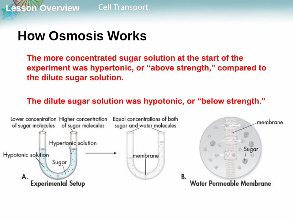
How Osmosis Works
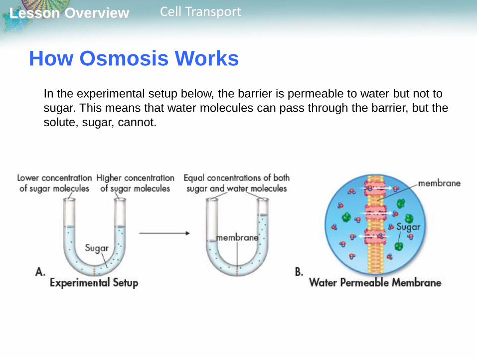
In the experimental setup below, the barrier is permeable to water but not to
sugar. This means that water molecules can pass through the barrier, but the
solute, sugar, cannot.

Lesson Overview Cell Transport
There are more sugar molecules on the right side of the barrier than on the
left side. Therefore, the concentration of water is lower on the right, where
more of the solution is made of sugar.
How Osmosis Works
Lesson Overview Cell Transport
How Osmosis Works
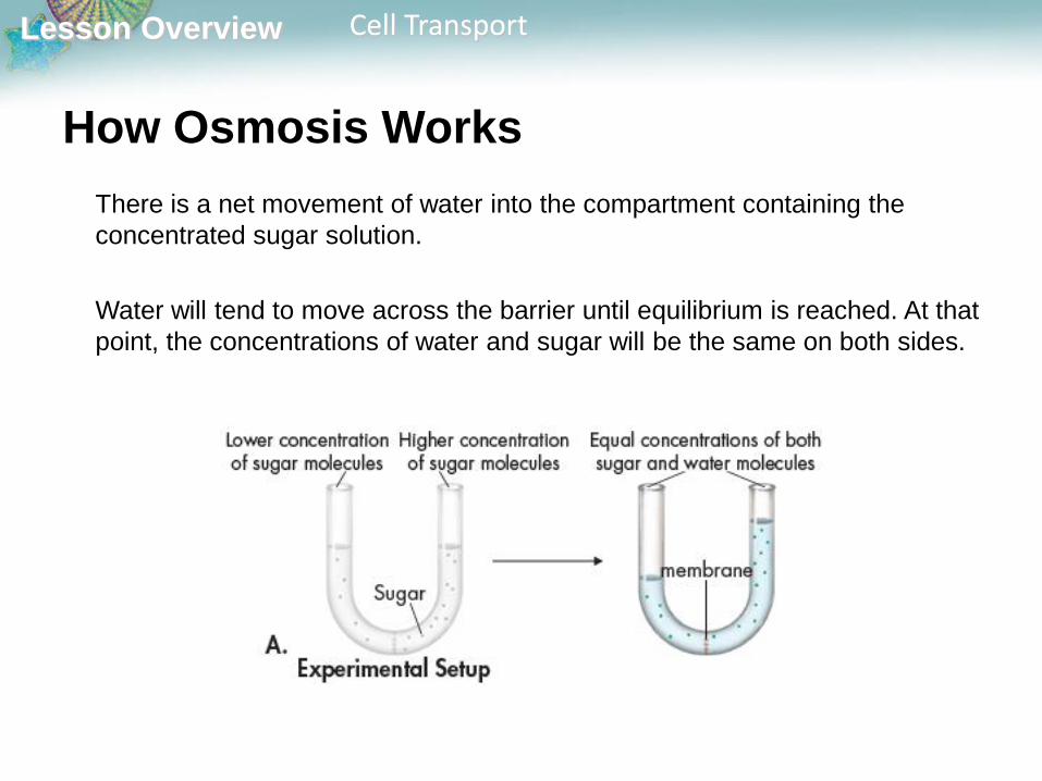
There is a net movement of water into the compartment containing the
concentrated sugar solution.
Water will tend to move across the barrier until equilibrium is reached. At that
point, the concentrations of water and sugar will be the same on both sides.
Lesson Overview Cell Transport
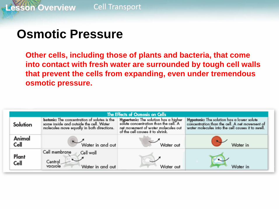
How Osmosis Works
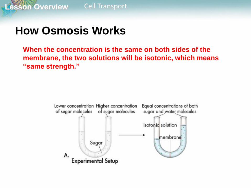
When the concentration is the same on both sides of the
membrane, the two solutions will be isotonic, which means
“same strength.”
Lesson Overview Cell Transport
How Osmosis Works
The more concentrated sugar solution at the start of the
experiment was hypertonic, or “above strength,” compared to
the dilute sugar solution.
The dilute sugar solution was hypotonic, or “below strength.”
Lesson Overview Cell Transport
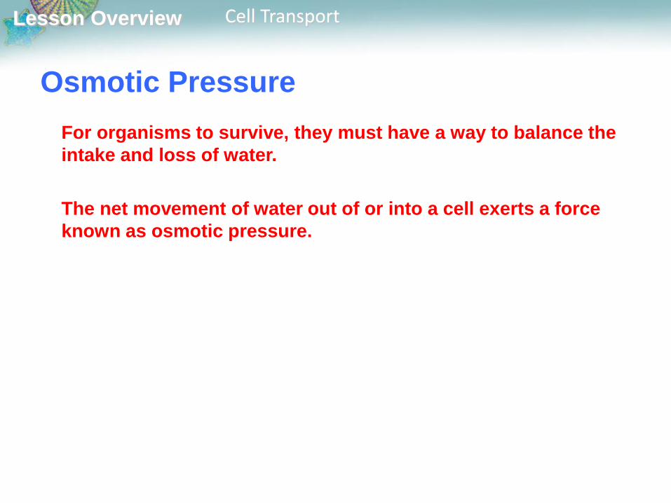
Osmotic Pressure
For organisms to survive, they must have a way to balance the
intake and loss of water.
The net movement of water out of or into a cell exerts a force
known as osmotic pressure.
Lesson Overview Cell Transport
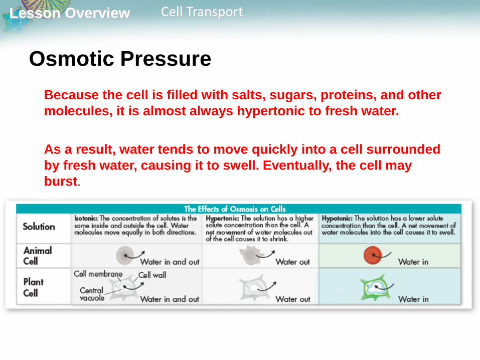
Osmotic Pressure
Because the cell is filled with salts, sugars, proteins, and other
molecules, it is almost always hypertonic to fresh water.
As a result, water tends to move quickly into a cell surrounded
by fresh water, causing it to swell. Eventually, the cell may
burst.
Lesson Overview Cell Transport
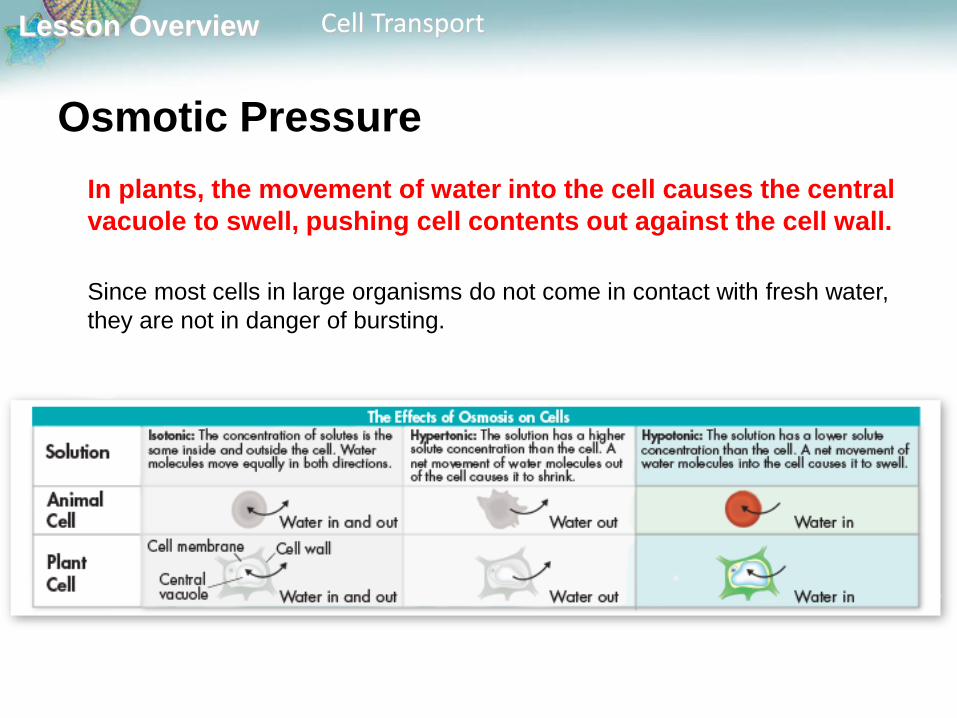
Osmotic Pressure
In plants, the movement of water into the cell causes the central
vacuole to swell, pushing cell contents out against the cell wall.
Since most cells in large organisms do not come in contact with fresh water,
they are not in danger of bursting.

Lesson Overview Cell Transport
Osmotic Pressure
Instead, the cells are bathed in fluids, such as blood, that are isotonic and
have concentrations of dissolved materials roughly equal to those in the
cells.
Cells placed in an isotonic solution neither gain nor lose water.

Lesson Overview Cell Transport
In a hypertonic solution, water rushes out of the cell, causing
animal cells to shrink and plant cell vacuoles to collapse.
Osmotic Pressure
Lesson Overview Cell Transport
Osmotic Pressure
Some cells, such as the eggs laid by fish and frogs, must come into contact
with fresh water. These types of cells tend to lack water channels.
As a result, water moves into them so slowly that osmotic pressure does not
become a problem.
Lesson Overview Cell Transport
Osmotic Pressure
Other cells, including those of plants and bacteria, that come
into contact with fresh water are surrounded by tough cell walls
that prevent the cells from expanding, even under tremendous
osmotic pressure.

Lesson Overview Cell Transport
Osmotic Pressure
Notice how the plant cell holds its shape in hypotonic solution, while the
animal red blood cell does not.
However, the increased osmotic pressure makes such cells extremely
vulnerable to injuries to their cell walls.
Lesson Overview Cell Transport
Active Transport
What is active transport?
The movement of materials against a concentration difference
is known as active transport. Active transport requires
energy.
Lesson Overview Cell Transport
Active Transport
Cells sometimes must move materials against a concentration
difference.
The movement of material against a concentration difference is
known as active transport. Active transport requires energy.

Lesson Overview Cell Transport
Active Transport
The active transport of
small molecules or ions
across a cell membrane is
generally carried out by
transport proteins, or
protein “pumps,” that are
found in the membrane
itself.
Lesson Overview Cell Transport
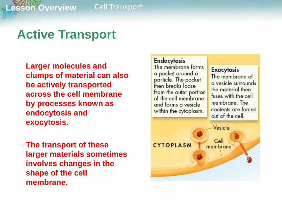
Active Transport
Larger molecules and
clumps of material can also
be actively transported
across the cell membrane
by processes known as
endocytosis and
exocytosis.
The transport of these
larger materials sometimes
involves changes in the
shape of the cell
membrane.

Lesson Overview Cell Transport
Molecular Transport
Small molecules and ions are carried across membranes by proteins in the membrane that act like pumps.
Many cells use such proteins to move calcium, potassium, and sodium ions across cell membranes.
Changes in protein shape seem to play an important role in the pumping process.
Lesson Overview Cell Transport
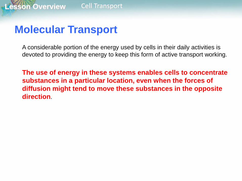
Molecular Transport
A considerable portion of the energy used by cells in their daily activities is
devoted to providing the energy to keep this form of active transport working.
The use of energy in these systems enables cells to concentrate
substances in a particular location, even when the forces of
diffusion might tend to move these substances in the opposite
direction.
Lesson Overview Cell Transport
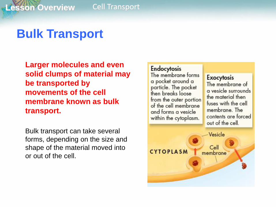
Bulk Transport
Larger molecules and even
solid clumps of material may
be transported by
movements of the cell
membrane known as bulk
transport.
Bulk transport can take several
forms, depending on the size and
shape of the material moved into
or out of the cell.
Lesson Overview Cell Transport
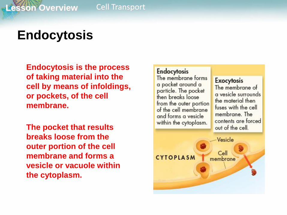
Endocytosis
Endocytosis is the process
of taking material into the
cell by means of infoldings,
or pockets, of the cell
membrane.
The pocket that results
breaks loose from the
outer portion of the cell
membrane and forms a
vesicle or vacuole within
the cytoplasm.

Lesson Overview Cell Transport
Endocytosis
Large molecules, clumps of
food, and even whole cells
can be taken up by
endocytosis.
Two examples of
endocytosis are
phagocytosis and
pinocytosis.
Lesson Overview Cell Transport
Endocytosis
In phagocytosis, extensions of cytoplasm surround a particle
and package it within a food vacuole. The cell then engulfs it.
Amoebas use this method for taking in food.
Engulfing material in this way requires a considerable amount
of energy and, therefore, is a form of active transport.

Lesson Overview Cell Transport
Endocytosis
In pinocytosis, cells take up liquid from the surrounding
environment by forming tiny pockets along the cell membrane.
The pockets fill with liquid and pinch off to form vacuoles within
the cell.

Lesson Overview Cell Transport
Exocytosis
Many cells also release
large amounts of material
from the cell, a process
known as exocytosis.
During exocytosis, the
membrane of the vacuole
surrounding the material
fuses with the cell
membrane, forcing the
contents out of the cell.
Lesson Overview Homeostasis and Cells
Lesson Overview7.4 Homeostasis and Cells
Lesson Overview Homeostasis and Cells
The Cell as an Organism
How do individual cells maintain homeostasis?
To maintain homeostasis, unicellular organisms grow, respond
to the environment, transform energy, and reproduce.

Lesson Overview Homeostasis and Cells
The Cell as an Organism
A single-celled, or unicellular, organism does everything you would expect
a living thing to do.
Just like other living things, unicellular organisms must
achieve homeostasis, relatively constant internal physical and
chemical conditions.
To maintain homeostasis, unicellular organisms grow, respond
to the environment, transform energy, and reproduce.
Lesson Overview Homeostasis and Cells
The Cell as an Organism
In terms of their numbers, unicellular organisms dominate life on Earth.
Unicellular organisms include both prokaryotes and
eukaryotes.
Prokaryotes, especially bacteria, are remarkably adaptable and live almost
everywhere—in the soil, on leaves, in the ocean, in the air, and even within
the human body.
Lesson Overview Homeostasis and Cells
The Cell as an Organism
Many eukaryotes also spend their lives as single cells.
Some types of algae, which contain chloroplasts and are found in oceans,
lakes, and streams around the world, are single celled.
Yeasts, or unicellular fungi, are also widespread. Yeasts play an important
role in breaking down complex nutrients, which makes them available for
other organisms.
Lesson Overview Homeostasis and Cells
The Cell as an Organism
Whether a prokaryote or a eukaryote, homeostasis is an issue for each
unicellular organism.
Every unicellular organism needs to find sources of energy or food, to keep
concentrations of water and minerals within certain levels, and to respond
quickly to changes in its environment.
Lesson Overview Homeostasis and Cells
Multicellular Life
How do the cells of multicellular organisms work together to maintain
homeostasis?
The cells of multicellular organisms become specialized for
particular tasks and communicate with one another to
maintain homeostasis.
Lesson Overview Homeostasis and Cells
Multicellular Life
The cells of multicellular organisms are interdependent, and like the
members of a successful baseball team, they work together.
In baseball, players take on a particular role, such as pitcher, catcher,
infielder, or outfielder. Messages and signals are sent and understood by
teammates and coaches to play the game effectively.
Cells in a multicellular organism work the same way. The cells of
multicellular organisms become specialized for particular
tasks and communicate with one another in order to maintain
homeostasis.
Lesson Overview Homeostasis and Cells
Cell Specialization
The cells of multicellular organisms are specialized, with different cell
types playing different roles.
Some cells are specialized to move, others to react to the environment,
and still others to produce substances that the organism needs.
No matter what the role, each specialized cell contributes to the overall
homeostasis of the organism.
Lesson Overview Homeostasis and Cells
Specialized Animal Cells
Particles of dust, smoke, and bacteria are part of even the cleanest air.
Specialized animal cells act like street sweepers to keep the particles
out of the lungs.
These cells are full of mitochondria, which provide a steady supply of
the ATP that powers the cilia on their upper surfaces.
Lesson Overview Homeostasis and Cells
Specialized Plant Cells
Pollen grains are highly specialized cells that are tiny and light, with
thick cell walls to protect the cell’s contents.
Pine pollen grains have two tiny wings that enable the slightest breeze
to carry them great distances.
Lesson Overview Homeostasis and Cells
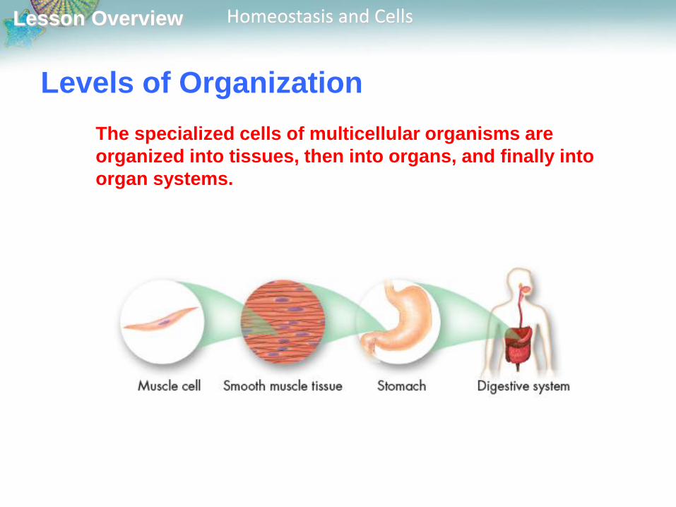
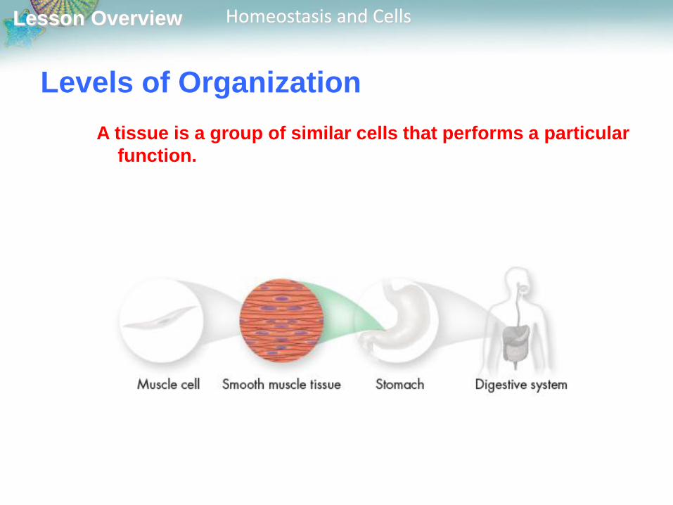
Levels of Organization
The specialized cells of multicellular organisms are
organized into tissues, then into organs, and finally into
organ systems.
Lesson Overview Homeostasis and Cells
Levels of Organization
A tissue is a group of similar cells that performs a particular
function.
Lesson Overview Homeostasis and Cells
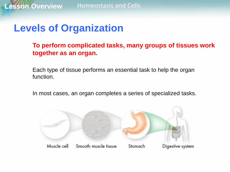
Levels of Organization
To perform complicated tasks, many groups of tissues work
together as an organ.
Each type of tissue performs an essential task to help the organ
function.
In most cases, an organ completes a series of specialized tasks.
Lesson Overview Homeostasis and Cells
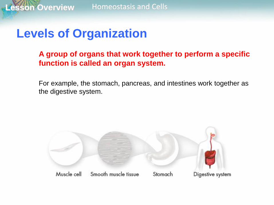
A group of organs that work together to perform a specific
function is called an organ system.
For example, the stomach, pancreas, and intestines work together as
the digestive system.
Levels of Organization
Lesson Overview Homeostasis and Cells
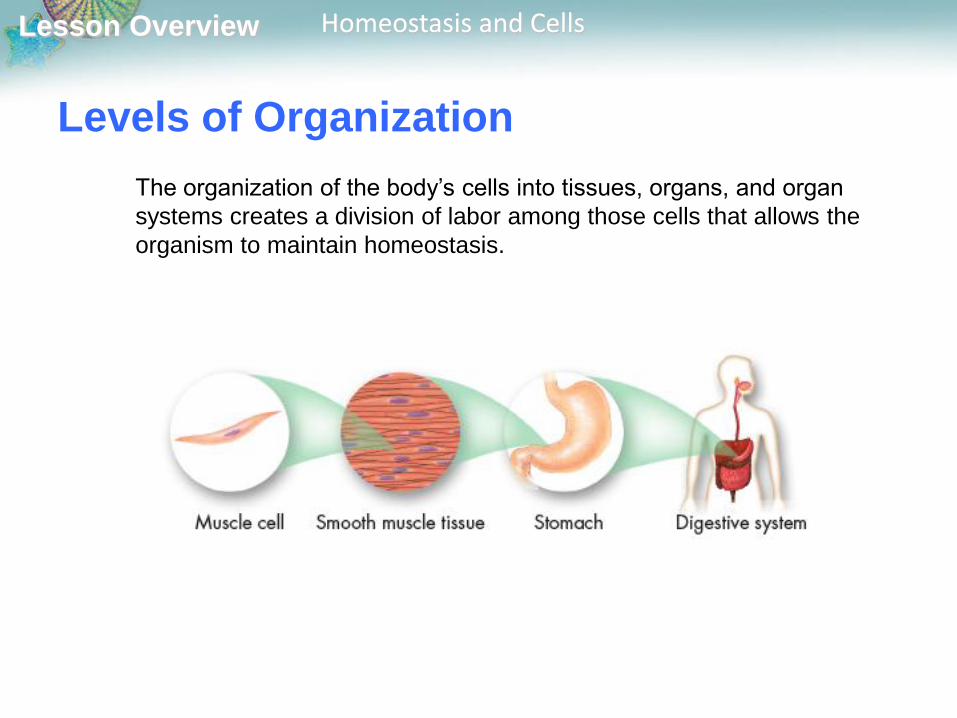
Levels of Organization
The organization of the body’s cells into tissues, organs, and organ
systems creates a division of labor among those cells that allows the
organism to maintain homeostasis.

Lesson Overview Homeostasis and Cells
Cellular Communication
Cells in a large organism communicate by means of
chemical signals that are passed from one cell to another.
These cellular signals can speed up or slow down the activities of the
cells that receive them, and can cause a cell to change what it is doing.
Lesson Overview Homeostasis and Cells
Cellular Communication
Some cells form connections, or cellular junctions, to
neighboring cells.
Some junctions hold cells firmly together.

Lesson Overview Homeostasis and Cells
Cellular Communication
Other junctions allow small molecules carrying chemical
messages to pass directly from one cell to the next.
To respond to one of these chemical signals, a cell must
have a receptor to which the signaling molecule can bind.
Sometimes these receptors are on the cell membrane,
although the receptors for certain types of signals are inside
the cytoplasm.
The chemical signals sent by various types of cells can cause important
changes in cellular activity. For example, such junctions enable the cells
of the heart muscle to contract in a coordinated fashion.
Recommended


![[5] v. PAPA JOHN'S INTERNATIONAL, INC.; PAPA JOHN'S …wps.prenhall.com/.../ch_7/Pizza_Hut_Inc_v_Papa_Johns_Inter.pdf · introduction in 1995 until May 1997. However, the slogan "became](https://img.pdfslide.us/doc/110x75/5abc82cf7f8b9a76038df8db/5-v-papa-johns-international-inc-papa-johns-wps-in-1995-until-may-1997.jpg)