3.5.3 Skeletal muscles are stimulated to contract by nerves and act as effectors.
The sliding filament theory of muscle contraction
Gross and microscopic structure of skeletal muscle. The ultrastructure of a myofibril.
The roles of actin, myosin, calcium ions and ATP in myofibril contraction.
The roles of calcium ions and tropomyosin in the cycle of actinomyosin bridge formation.
Muscles as effectorsThe role of ATP and phosphocreatine in providing the energy supply during muscle contraction.
The structure, location and general properties of slow and fast skeletal muscle fibres.

There are 3 types of muscle in our body…
We have conscious control over skeletal muscle only.

Muscles are made of many fibres, for strength (think of a rope!)
Muscle cells have become fused together to form MUSCLE FIBRES
These share nuclei and SARCOPLASM (cytoplasm in muscle cells)
There are many MITOCHONDRIA and ENDOPLASMIC RETICULUM

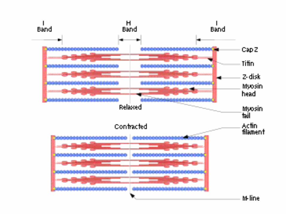
The ultrastructure of a myofibril.

The arrangement of fibres

Close up of The Filaments…

The Bands…

Arrangement of Actin and Myosin

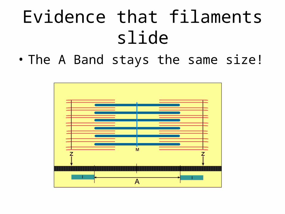
Evidence that filaments slide
• The A Band stays the same size!

Myosin

Actin
Actin and Tropomyosin

The roles of actin, myosin, calcium ions and ATP in myofibril contraction
The roles of calcium ions and tropomyosin in the cycle of
actinomyosin bridge formation.

The Neuromuscular junction


Clip on Actinomyosin bridges


The role of ATP in muscle contraction:
1. To provide energy for the movement of the myosin heads.
2. To provide energy for the active reabsorption of Calcium ions, into the Sarcoplasmic Reticulum, when the nervous stimulation has ceased.

The role of Phosphocreatine• In very active muscles, aerobic respiration may
not occur quickly enough to provide ATP
• Anaerobic Respiration may occur to compensate since so much ATP is needed.
• Phosphocreatine is stored in muscle cells and acts as a reserve supply of Phosphate so that ATP can be regenerated quickly.

There are 2 types of Muscle Fibre…
• Slow Muscle Fibres (Slow-twitch)
• Fast Muscle Fibres (Fast-twitch)

Slow-Twitch Fibres…
• Function… Contractions over a long period of time. E.g. running a marathon, keeping our body upright.
• Location… Calf Muscles (or any place where support to keep us upright is needed)
• Structure… Lots of MYOGLOBIN (stores lots of oxygen so these muscles are very red), Numerous mitochondria, a store of glycogen, huge blood supply

Fast-Twitch Fibres
• Function… Rapid, Powerful contractions. E.g. Weight-Lifting
• Location… Biceps (or any area where muscles carry out short bursts of intense acticity)
• Structure… Thicker, more numerous Myosin filaments. A store of phosphocreatine. More enzymes for Anaerobic Respiration.
Recommended