CENTER FOR DRUG EVALUATION AND RESEARCH
APPLICATION NUMBER:
208065Orig1s000
PHARMACOLOGY REVIEW(S)
MEMORANDUM
Tagrisso (osimertinib)
Date: November 4, 2015To: File for NDA 208065From: John K. Leighton, PhD, DABT
Director, Division of Hematology Oncology ToxicologyOffice of Hematology and Oncology Products
I have examined pharmacology/toxicology supporting and labeling reviews for Tagrisso conducted by Dr. Weis, and secondary memorandum and labeling provided by Dr. Helms. I concur with Dr. Helms’ conclusion that Tagrisso may be approved for the proposed indication.
Reference ID: 3842505
---------------------------------------------------------------------------------------------------------This is a representation of an electronic record that was signedelectronically and this page is the manifestation of the electronicsignature.---------------------------------------------------------------------------------------------------------/s/----------------------------------------------------
JOHN K LEIGHTON11/04/2015
Reference ID: 3842505
MEMORANDUM
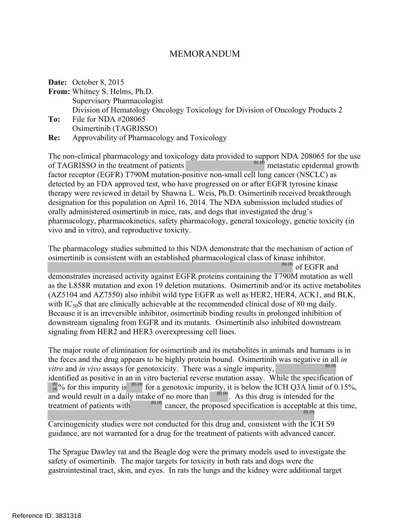
Date: October 8, 2015From: Whitney S. Helms, Ph.D.
Supervisory PharmacologistDivision of Hematology Oncology Toxicology for Division of Oncology Products 2
To: File for NDA #208065Osimertinib (TAGRISSO)
Re: Approvability of Pharmacology and Toxicology
The non-clinical pharmacology and toxicology data provided to support NDA 208065 for the use of TAGRISSO in the treatment of patients metastatic epidermal growth factor receptor (EGFR) T790M mutation-positive non-small cell lung cancer (NSCLC) as detected by an FDA approved test, who have progressed on or after EGFR tyrosine kinase therapy were reviewed in detail by Shawna L. Weis, Ph.D. Osimertinib received breakthrough designation for this population on April 16, 2014. The NDA submission included studies of orally administered osimertinib in mice, rats, and dogs that investigated the drug’s pharmacology, pharmacokinetics, safety pharmacology, general toxicology, genetic toxicity (in vivo and in vitro), and reproductive toxicity.
The pharmacology studies submitted to this NDA demonstrate that the mechanism of action of osimertinib is consistent with an established pharmacological class of kinase inhibitor.
of EGFR and demonstrates increased activity against EGFR proteins containing the T790M mutation as well as the L858R mutation and exon 19 deletion mutations. Osimertinib and/or its active metabolites (AZ5104 and AZ7550) also inhibit wild type EGFR as well as HER2, HER4, ACK1, and BLK, with IC50S that are clinically achievable at the recommended clinical dose of 80 mg daily. Because it is an irreversible inhibitor, osimertinib binding results in prolonged inhibition of downstream signaling from EGFR and its mutants. Osimertinib also inhibited downstream signaling from HER2 and HER3 overexpressing cell lines.
The major route of elimination for osimertinib and its metabolites in animals and humans is in the feces and the drug appears to be highly protein bound. Osimertinib was negative in all in vitro and in vivo assays for genotoxicity. There was a single impurity, identified as positive in an in vitro bacterial reverse mutation assay. While the specification of
% for this impurity is for a genotoxic impurity, it is below the ICH Q3A limit of 0.15%, and would result in a daily intake of no more than . As this drug is intended for the treatment of patients with cancer, the proposed specification is acceptable at this time,
Carcinogenicity studies were not conducted for this drug and, consistent with the ICH S9 guidance, are not warranted for a drug for the treatment of patients with advanced cancer.
The Sprague Dawley rat and the Beagle dog were the primary models used to investigate the safety of osimertinib. The major targets for toxicity in both rats and dogs were the gastrointestinal tract, skin, and eyes. In rats the lungs and the kidney were additional target
Reference ID: 3831318
(b) (4)
(b) (4)
(b) (4)
(b) (4)
(b) (4)
(b) (4)
(b) (4)
(b) (4)
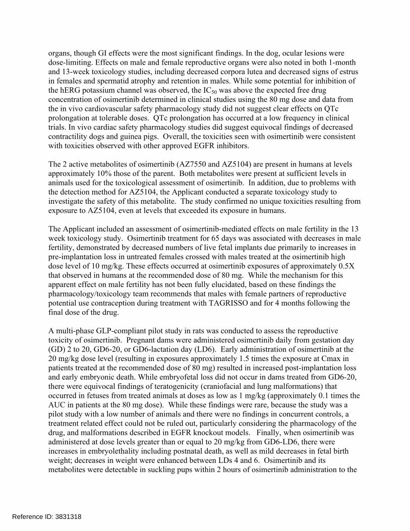
organs, though GI effects were the most significant findings. In the dog, ocular lesions were dose-limiting. Effects on male and female reproductive organs were also noted in both 1-month and 13-week toxicology studies, including decreased corpora lutea and decreased signs of estrus in females and spermatid atrophy and retention in males. While some potential for inhibition of the hERG potassium channel was observed, the IC50 was above the expected free drug concentration of osimertinib determined in clinical studies using the 80 mg dose and data from the in vivo cardiovascular safety pharmacology study did not suggest clear effects on QTc prolongation at tolerable doses. QTc prolongation has occurred at a low frequency in clinical trials. In vivo cardiac safety pharmacology studies did suggest equivocal findings of decreased contractility dogs and guinea pigs. Overall, the toxicities seen with osimertinib were consistent with toxicities observed with other approved EGFR inhibitors.
The 2 active metabolites of osimertinib (AZ7550 and AZ5104) are present in humans at levels approximately 10% those of the parent. Both metabolites were present at sufficient levels in animals used for the toxicological assessment of osimertinib. In addition, due to problems with the detection method for AZ5104, the Applicant conducted a separate toxicology study to investigate the safety of this metabolite. The study confirmed no unique toxicities resulting from exposure to AZ5104, even at levels that exceeded its exposure in humans.
The Applicant included an assessment of osimertinib-mediated effects on male fertility in the 13 week toxicology study. Osimertinib treatment for 65 days was associated with decreases in male fertility, demonstrated by decreased numbers of live fetal implants due primarily to increases in pre-implantation loss in untreated females crossed with males treated at the osimertinib high dose level of 10 mg/kg. These effects occurred at osimertinib exposures of approximately 0.5X that observed in humans at the recommended dose of 80 mg. While the mechanism for this apparent effect on male fertility has not been fully elucidated, based on these findings the pharmacology/toxicology team recommends that males with female partners of reproductive potential use contraception during treatment with TAGRISSO and for 4 months following the final dose of the drug.
A multi-phase GLP-compliant pilot study in rats was conducted to assess the reproductive toxicity of osimertinib. Pregnant dams were administered osimertinib daily from gestation day (GD) 2 to 20, GD6-20, or GD6-lactation day (LD6). Early administration of osimertinib at the 20 mg/kg dose level (resulting in exposures approximately 1.5 times the exposure at Cmax in patients treated at the recommended dose of 80 mg) resulted in increased post-implantation loss and early embryonic death. While embryofetal loss did not occur in dams treated from GD6-20, there were equivocal findings of teratogenicity (craniofacial and lung malformations) that occurred in fetuses from treated animals at doses as low as 1 mg/kg (approximately 0.1 times the AUC in patients at the 80 mg dose). While these findings were rare, because the study was a pilot study with a low number of animals and there were no findings in concurrent controls, a treatment related effect could not be ruled out, particularly considering the pharmacology of the drug, and malformations described in EGFR knockout models. Finally, when osimertinib was administered at dose levels greater than or equal to 20 mg/kg from GD6-LD6, there were increases in embryolethality including postnatal death, as well as mild decreases in fetal birth weight; decreases in weight were enhanced between LDs 4 and 6. Osimertinib and its metabolites were detectable in suckling pups within 2 hours of osimertinib administration to the
Reference ID: 3831318
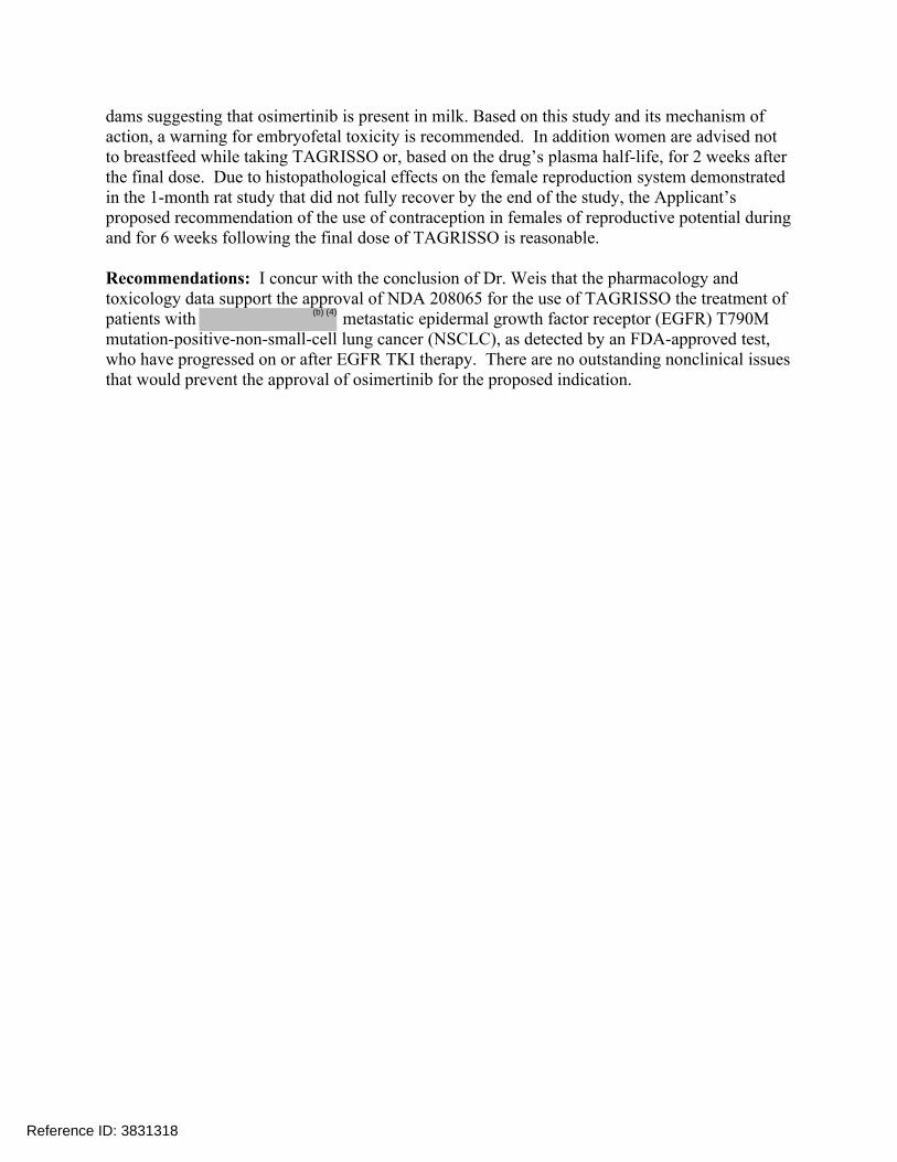
dams suggesting that osimertinib is present in milk. Based on this study and its mechanism of action, a warning for embryofetal toxicity is recommended. In addition women are advised not to breastfeed while taking TAGRISSO or, based on the drug’s plasma half-life, for 2 weeks after the final dose. Due to histopathological effects on the female reproduction system demonstrated in the 1-month rat study that did not fully recover by the end of the study, the Applicant’s proposed recommendation of the use of contraception in females of reproductive potential during and for 6 weeks following the final dose of TAGRISSO is reasonable.
Recommendations: I concur with the conclusion of Dr. Weis that the pharmacology and toxicology data support the approval of NDA 208065 for the use of TAGRISSO the treatment of patients with metastatic epidermal growth factor receptor (EGFR) T790M mutation-positive-non-small-cell lung cancer (NSCLC), as detected by an FDA-approved test, who have progressed on or after EGFR TKI therapy. There are no outstanding nonclinical issues that would prevent the approval of osimertinib for the proposed indication.
Reference ID: 3831318
(b) (4)
---------------------------------------------------------------------------------------------------------This is a representation of an electronic record that was signedelectronically and this page is the manifestation of the electronicsignature.---------------------------------------------------------------------------------------------------------/s/----------------------------------------------------
WHITNEY S HELMS10/08/2015
Reference ID: 3831318
1
DEPARTMENT OF HEALTH AND HUMAN SERVICES PUBLIC HEALTH SERVICE
FOOD AND DRUG ADMINISTRATION CENTER FOR DRUG EVALUATION AND RESEARCH
PHARMACOLOGY/TOXICOLOGY NDA/BLA REVIEW AND EVALUATION
Application number: NDA 208,065
Supporting document/s: 001
Applicant’s letter date: 05 June 2015
CDER stamp date: 05 June 2015
Product: TAGRISSO (osimertinib)
Indication: Non-Small Cell Lung Cancer (NSCLC)
Applicant: AstraZeneca Pharmaceuticals LP
1800 Concord Pike
Wilmington, DE 19803
UNITED STATES
Review Division: DHOT/DOP2
Reviewer: Shawna L. Weis, PhD
Supervisor/Team Leader: Whitney S. Helms, PhD
Division Director: John K. Leighton, PhD, DABT / Patricia Keegan,
MD
Project Manager: Ingrid Y. Fan
Disclaimer Except as specifically identified, all data and information discussed below and necessary for approval of NDA 208,065 are owned by AstraZeneca Pharmaceuticals LP or are data for which AstraZeneca Pharmaceuticals LP has obtained a written right of reference. Any information or data necessary for approval of NDA 208,065 that AstraZeneca Pharmaceuticals LP does not own or have a written right to reference constitutes one of the following: (1) published literature, or (2) a prior FDA finding of safety or effectiveness for a listed drug, as reflected in the drug’s approved labeling. Any data or information described or referenced below from reviews or publicly available summaries of a previously approved application is for descriptive purposes only and is not relied upon for approval of NDA 208,065.
Reference ID: 3831295
NDA # 208,065 Reviewer: Shawna L. Weis, PhD
2
TABLE OF CONTENTS
1 EXECUTIVE SUMMARY ....................................................................................... 11
1.1 INTRODUCTION .................................................................................................. 11 1.2 BRIEF DISCUSSION OF NONCLINICAL FINDINGS .................................................... 11
1.3 RECOMMENDATIONS .......................................................................................... 13
2 DRUG INFORMATION .......................................................................................... 13
2.1 DRUG ............................................................................................................... 13
KINASE INHIBITOR ..................................................................................................... 14
2.2 RELEVANT INDS, NDAS, BLAS AND DMFS ......................................................... 14
2.3 DRUG FORMULATION ......................................................................................... 14
2.4 COMMENTS ON NOVEL EXCIPIENTS ..................................................................... 15
2.5 COMMENTS ON IMPURITIES/DEGRADANTS OF CONCERN ....................................... 15 2.6 PROPOSED CLINICAL POPULATION AND DOSING REGIMEN .................................... 15
2.7 REGULATORY BACKGROUND .............................................................................. 15
3 STUDIES SUBMITTED .......................................................................................... 16
3.1 STUDIES REVIEWED ........................................................................................... 16 3.2 STUDIES NOT REVIEWED ................................................................................... 18 3.3 PREVIOUS REVIEWS REFERENCED...................................................................... 19
4 PHARMACOLOGY ................................................................................................ 19
4.1 PRIMARY PHARMACOLOGY ................................................................................. 19
4.2 SECONDARY PHARMACOLOGY ............................................................................ 43 4.3 SAFETY PHARMACOLOGY ................................................................................... 46
5 PHARMACOKINETICS/ADME/TOXICOKINETICS .............................................. 61
5.1 PK/ADME ........................................................................................................ 61
6 GENERAL TOXICOLOGY ..................................................................................... 72
6.2 REPEAT-DOSE TOXICITY .................................................................................... 72
7 GENETIC TOXICOLOGY ...................................................................................... 89
7.1 IN VITRO REVERSE MUTATION ASSAY IN BACTERIAL CELLS (AMES) ....................... 89 7.2 IN VITRO ASSAYS IN MAMMALIAN CELLS .............................................................. 93
7.3 IN VIVO CLASTOGENICITY ASSAY IN RODENT (MICRONUCLEUS ASSAY) .................. 96
8 CARCINOGENICITY ............................................................................................. 97
9 REPRODUCTIVE AND DEVELOPMENTAL TOXICOLOGY ................................ 98
9.2 EMBRYONIC FETAL DEVELOPMENT ..................................................................... 98
10 SPECIAL TOXICOLOGY STUDIES ................................................................. 109
11 INTEGRATED SUMMARY AND SAFETY EVALUATION ............................... 122
Reference ID: 3831295
NDA # 208,065 Reviewer: Shawna L. Weis, PhD
3
12 APPENDIX/ATTACHMENTS ........................................................................... 127
Reference ID: 3831295
NDA # 208,065 Reviewer: Shawna L. Weis, PhD
4
Table of Tables
Table 1: Composition of AZD9291 film-coated tablets .................................................. 14 Table 2: Inhibition of cellular phosphorylation (pEGFR) by AZD9291 and its metabolites (IC50, µM) ..................................................................................................................... 20 Table 3: pEGFR suppression by AZD9291 and its metabolites in the HTRF assay (IC50, µM) ................................................................................................................................ 20 Table 4: Mean IC50s (nM) for AZD9291 and its metabolites, AZ13575104 and AZ1397550 in cultures of NSCLC cells bearing WT or mutant EGFR ........................... 21 Table 5: Percent kinase inhibition at 1µM and IC50 values for kinases inhibited by AZD9291 or its metabolites ........................................................................................... 25 Table 6: IC50 for inhibition of pIGF1R by AZD9291, AZ5104 or A7550 (mean±95%CI; nM) ................................................................................................................................ 25
Table 7: Apparent IC50 inhibition of pHER2 for AD9291 and AD5104 (mean±95%CI; nM) ................................................................................................................................ 26 Table 8: Summary of study designs .............................................................................. 28 Table 9: Summary of mutation analyses from studies of EX19del PC9 cells selected for resistance with gefitinib, afatinib, or AZD9291 ................................................ 41 Table 10: Comparative inhibitory activity of different compounds for pErbB2 and pErbB3 production in HRG1-stimulated MCF-7 cells or HER2-amplified BT474 cells ............... 42 Table 11: Summary of AZD9291 off-target pharmacological activity (mean IC50s) ...... 43 Table 12: Summary of in vitro binding and enzyme inhibition assays with AZ13575104 ...................................................................................................................................... 44 Table 13: Effect of AZ13597550 in in vitro radioligand binding, enzyme and electrophysiology assays .............................................................................................. 45 Table 14: Effect of AZ13597550 in in vitro electrophysiology assays ............................ 45
Table 15: Analytical chemistry results from the in vitro hERG assay ............................ 47 Table 16: Effect of AZ13597550 in electrophysiology assays using CHO or HEK cells expressing human cardiac voltage-gated ion channels ................................................. 47 Table 17: Effect of AZ13575104 in electrophysiology assays using CHO or HEK cells expressing human cardiac voltage-gated ion channels ................................................. 48 Table 18: Effect of in electrophysiology assays using CHO or HEK cells expressing human cardiac voltage-gated ion channels ................................................. 49 Table 19: Mean Plasma Concentrations Measured in the Conscious Telemetered Beagle Dog at 4-Hours Post-Dose ................................................................................ 52
Table 20: Groups and Dose Levels ............................................................................... 53 Table 21: Pupil Size Following Single Dose AZD9291 ................................................. 54 Table 22: Visual Acuity Following Administration of a Single Oral Dose of AZD9291 to male Han Wistar rats ..................................................................................................... 55 Table 23: Spatial Repositioning [mean (±SEM)] data Obtained Following Administration of a Single Oral Dose of AZD9291 to male Han Wistar rats .......................................... 55 Table 24: Effect of AZD9291 on expiratory time ............................................................ 57 Table 25: Weight of stomach contents (grams) ............................................................. 57 Table 26: Intestinal transport distance (%) .................................................................... 58 Table 27: Summary of mean plasma concentrations for AZD9291 in male Han Wistar rats following oral administration of AZD9291 ............................................................... 58
Reference ID: 3831295
(b) (4)
(b) (4)
NDA # 208,065 Reviewer: Shawna L. Weis, PhD
5
Table 28: Summary of mean plasma concentrations for AZ13597550 in male Han Wistar rats following oral administration of AZD9291 .................................................... 59 Table 29: Covalent binding and fraction of covalent binding of [3H]AZD9291 and [3H]zomepirac in cultured hepatocyte incubations ......................................................... 65 Table 30: Design of the rat distribution study ................................................................ 66 Table 31: Mean recovery of radioactivity (0-168 hours) after single I or PO dose of [14C]AZD9291 in male and female rats (% of administered dose) ................................. 67 Table 32: Design of the rat mass balance study ........................................................... 71 Table 33: Summary of mean (±SD) total recovery of radiation after a single IV or oral dose of [14C]AZD9291 to male and female rats (% administered dose) ........................ 71 Table 34: Summary of mean plasma and blood concentrations of total radioactivity following oral administration of 10 mg/kg [14C]AZD9291 to male and female rats ......... 71 Table 35: Changes in mean hematology parameters in the rat (%Δ vs. concurrent control) .......................................................................................................................... 74 Table 36: Changes in mean chemistry parameters in the rat (%Δ vs. concurrent control) ...................................................................................................................................... 75 Table 37: Changes in mean urine chemistry parameters in the rat (%Δ vs. concurrent control) .......................................................................................................................... 76 Table 38: Summary of treatment-related organ weight changes ................................... 77 Table 39: Summary of histopathological findings in the rat ........................................... 78 Table 40: Summary of individual pregnancy data ......................................................... 80 Table 41: Day 91 Summary of mean toxicokinetic parameters for AZD9291 and its metabolite, AZ1397550 ................................................................................................. 81 Table 42: Dose holidays in high dose animals .............................................................. 85 Table 43: Summary of mean clinical chemistry changes (% change vs. concurrent controls) in male and female dogs ................................................................................ 85
Table 44: 13-Week Dog TK .......................................................................................... 89 Table 45: Historical solvent control data ........................................................................ 90 Table 46: Plate Incorporation Method - Without Metabolic Activation ..... 91 Table 47: Plate Incorporation Method - With Metabolic Activation .......... 92 Table 48: Mouse lymphoma LY5178Y cell historical control data from August 2006-December 2011 ............................................................................................................. 94 Table 49: AZ19291: In Vitro Mouse Lymphoma TK Assay Mutation Test: 3 Hour Exposure in the absence of S9 ..................................................................................... 94
Table 50: AZD9291: In Vitro Mouse Lymphoma TK Assay Mutation Test: 3 Hours Exposure in the presence of S9 .................................................................................... 95 Table 51: AZ19291: In Vitro Mouse Lymphoma TK Assay Mutation Test: 24 Hour exposure in the absence of S9 ...................................................................................... 95
Table 52: Summary of control group mean and range of micronucleus counts in 2000 IE in male Han Wistar rats ................................................................................................. 97 Table 53: Mean incidence of micronucleated immature erythrocytes (MIEs) in rat bone marrow and percentages of immature erythrocytes (IE) ................................................ 97 Table 54: Summary of mean AZD9291 TK parameters in dams (Phase 2) ................ 102 Table 55: Summary of mean metabolite (AZ13597550) TK parameters in dams (Phase 2) ................................................................................................................................. 103
Reference ID: 3831295
(b) (4)
(b) (4)
NDA # 208,065 Reviewer: Shawna L. Weis, PhD
6
Table 56: Summary of mean plasma concentrations of AZD9291 and metabolite AZ13597550 in dams (Phase 2) and fetuses (Phase 1) .............................................. 103 Table 57: Summary of mean AZD9291 and metabolite AZ13597550 plasma concentrations in rat pups and dams (Phase 3) .......................................................... 103 Table 58: Summary of Pregnancy Data (Phase 1) ...................................................... 104 Table 59: Summary of Pregnancy Data (Phase 2) ...................................................... 104 Table 60: Survival Indices – Phase 3 Batch 1 ............................................................. 105 Table 61: Survival Indices – Phase 3 Batch 2 ............................................................. 105 Table 62: Litter performance – Phase 3, batch 1 ........................................................ 106 Table 63: Litter performance – Phase 3, batch 2 ........................................................ 106 Table 64: Litter and pup weight - Phase 3(Batch 1) .................................................... 107 Table 65: Table 40: Litter and pup weight - Phase 3(Batch 2) .................................... 107 Table 66: Summary of fetal malformations - Phase 2* ................................................ 108
Table 67: Summary of minor fetal abnormalities and variations: Phase 2 ................... 108 Table 68: Summary of minor pup abnormalities and variations - Phase 3 batch 1 ...... 109 Table 69: Summary of minor pup abnormalities and variations - Phase 3 batch 2 ...... 109 Table 70: Design of the rat lung ultrastructure study ................................................... 110 Table 71: Study Design of the repeat-dose toxicology study of AZ13575104 and
in the rat ................................................................................................. 112 Table 72: Summary of histopathological findings in the 4-week rat metabolite qualification study ........................................................................................................ 113 Table 73: Summary of mean AZ13575104 toxicokinetic parameters in the rat metabolite qualification study ........................................................................................................ 114 Table 74: Design of the 1-month oral impurity qualification toxicology study in the rat 114 Table 75: Summary of impurities contained in Batch 1 and Batch 2 test articles ........ 114 Table 76: Summary of AZD9291 Day 28 group mean TK parameters ........................ 115
Table 77: Individual plate counts from the dose-ranging mutagenicity study of (-S9) ....................................................................................................... 116
Table 78: Individual plate counts from the dose-ranging mutagenicity study of (+S9) ...................................................................................................... 117
Table 79: Plate counts from the confirmatory mutagenicity study with (-S9) .................................................................................................................................... 117 Table 80: Plate counts from the confirmatory mutagenicity study with (-S9) .................................................................................................................................... 118 Table 81: Individual plate counts from the dose-ranging mutagenicity study of
(-S9) ....................................................................................................... 119 Table 82: Individual plate counts from the dose-ranging mutagenicity study of
(+S9) ...................................................................................................... 120
Table 83: Individual plate counts from the dose-ranging mutagenicity study of (-S9) ....................................................................................................... 121
Table 84: Individual plate counts from the dose-ranging mutagenicity study of (+S9) ...................................................................................................... 122
Table 85: Treatment-Related Hematology Changes (%) in Treated Groups vs. Concurrent Controls .................................................................................................... 130 Table 86: Treatment-Related Clinical Chemistry Changes (%) in Treated Groups vs. Concurrent Controls .................................................................................................... 130
Reference ID: 3831295
(b) (4)
(b) (4)
(b) (4)
(b) (4)
(b) (4)
(b) (4)
(b) (4)
(b) (4)
(b) (4)
NDA # 208,065 Reviewer: Shawna L. Weis, PhD
7
Table 87: Summary of Changes (Expressed as % Change of Control) in Rat Urine Chemistry Following Treatment with AZ9291 .............................................................. 131 Table 88: Summary of CYP Metabolic Enzyme Levels Measured in Rat Livers Following Treatment with AZD9291 ............................................................................................ 133 Table 89: Summary of AZD9291 Toxicokinetic Parameters in the Rat on Study Days 1 and 28 ......................................................................................................................... 134 Table 90: Summary of AZ13597550 Exposures in the Rat Following Oral Administration of AZD9291 on Study Days 1 and 28 .......................................................................... 134
Table 91: Incidence of Ophthalmology Findings Associated with AZD9291 Administration in Dogs ................................................................................................ 138 Table 92: Summary AZD9291 TK Parameters in the Dog Following Administration of AZD9291 ..................................................................................................................... 143 Table 93: Summary AZ13597550 TK Parameters in the Dog Following Administration of AZD9291 ..................................................................................................................... 143
Reference ID: 3831295
NDA # 208,065 Reviewer: Shawna L. Weis, PhD
8
Table of Figures
Figure 1: Time-course for IC50 determination (pEGFR formation) in NCI H1975 cells (L858R/T790M) ............................................................................................................. 22 Figure 2: Time-course for IC50 determination (pEGFR formation) in PC9 cells (Ex19del) ...................................................................................................................................... 22 Figure 3: Time-course for IC50 determination (pEGFR formation) in LOVO cells (WT) 23 Figure 4: Timecourse of pEGFR suppression in NCI H1975 (T790M/L858R) following drug-washout (%DMSO control vs. time) ...................................................................... 23 Figure 5: Timecourse of pEGFR suppression in PC9 (Ex19Del) cells following drug-washout (%DMSO control vs. time) .............................................................................. 24 Figure 6: Tumor growth inhibition of AZD9291 in female SCID mice harboring PC9 (EGFR Ex19del) xenografts .......................................................................................... 27
Figure 7: Tumor growth inhibition of AZD9291 in female athymic (nu/nu) mice harboring H1975 (EGFR T790M/L858R) xenografts ..................................................................... 27 Figure 8: Tumor growth inhibition of AZD9291 in female athymic (nu/nu) mice harboring A431 (EGFR WT) xenografts ........................................................................................ 28 Figure 9: Effect of AZ13575104 on tumor growth in PC9 (EGFRm+;Ex19del) SCID xenografts ..................................................................................................................... 29 Figure 10: Effect of AZ13575104 on tumor growth in H1975 (EGFRm+; L585R/T790M) nu/nu xenografts ........................................................................................................... 29 Figure 11: Effect of AZ13575104 on tumor growth in A431 (EGFRwt) nu/nu xenografts ...................................................................................................................................... 30 Figure 12: Dose-response and timecourse of pEGFR and pAKT expression following oral administration of AD9291 in H1975 xenografts by ELISA ...................................... 31 Figure 13: Dose-response and timecourse of pEGFR and pAKT expression following oral administration of AD9291 in PC9 xenografts by ELISA .......................................... 31 Figure 14: Dose-response and timecourse of pEGFR and pERK expression following oral administration of AD9291 in A431 (EGFR WT) xenografts .................................... 32 Figure 15: Immunohistochemical timecourse of pEGFR and pERK suppression in H1975 (EGFRm) tumors treated in vivo with AZ9291 ................................................... 32 Figure 16: Effect of AZD9291 on bi-transgenic EGFRm+/T790M tumor growth in comparison with Iressa .................................................................................. 33 Figure 17: IHC for pEGFR (Y1173) in skin epidermis and hair follicles in the bi-transgenic mouse study ................................................................................................ 34
Figure 18: Tumor growth inhibition in NCI-H1975 nude mouse xenografts during and after treatment with AZD9291 ....................................................................................... 35 Figure 19: Effect of AD9291 treatment on body weight in NCI-H1975 (L858R/T790M) NSCLC xenograft-bearing female nude mice ................................................................ 36 Figure 20: Tumor growth inhibition following administration of AZD9291 or gefitinib in the PC9 NSLC xenograft model .................................................................................... 37 Figure 21: Tumor growth inhibition in NCI-H3255 NSLC xenografts following treatment with gefitinib, afatinib or AZD9291 ................................................................................. 38 Figure 22: Summary of tumor growth inhibition by AZD9291 in a PC9 brain metastasis model in athymic nu/nu mice ......................................................................................... 39
Reference ID: 3831295
(b) (4)
NDA # 208,065 Reviewer: Shawna L. Weis, PhD
9
Figure 23: Effect of AZD9291 on survival in the murine NSCLC brain metastasis model in athymic nu/nu mice .................................................................................................... 39 Figure 24: Concentration-time curve for AZD9291 in the blood, brain and CSF in athymic nu/nu mice ....................................................................................................... 39 Figure 25: Proportion of resistant cells harboring T790M mutations following treatment with gefitinib, afatinib, or AZD9291 ................................................................. 40 Figure 26: Kinetics of resistance acquisition in cultured PC9 cells treated with AZD9291, gefitinib, afatinib or .......................................................................................... 42 Figure 27: AZD9291 hERG Concentration-Effect Curve ............................................... 46 Figure 28: Effect of AZD9291 on Heart Rate in the Conscious Telemetered Beagle Dog ...................................................................................................................................... 51 Figure 29: Effect of AZD9291 on dP/dt+ in the Conscious Telemetered Beagle Dog ... 51 Figure 30: Effect of AZD9291 on Heart Rate-Corrected QT (QTcR) Intervals in the Conscious Telemetered Beagle Dog ............................................................................. 52 Figure 31: Effect of AZD9291 on overnight body weight ............................................... 54 Figure 32: Effect of AD9291 on Respiratory Rate in the Rat ......................................... 56 Figure 33: Effect of AZD9291 on Minute Volume in the Rat .......................................... 56 Figure 34: Effect of AZD9291 on the Diastolic Blood Pressure of Rats ......................... 60 Figure 35: Effect of AZD9291 on the Systolic Blood Pressure of Rats .......................... 60 Figure 36: Activity Response following a 100 mg/kg oral dose of AZD9291 in the Rat . 61 Figure 37: Position of the 14C label on AZD9291 for the dog disposition study ............. 61 Figure 38: Mean cumulative recovery of radioactivity in excreta following administration of a 1 mg/kg IV dose of [14C]AD9291 to male beagle dogs ........................................... 62 Figure 39: Mean cumulative recovery of radioactivity in excreta following administration of a 2 mg/kg oral dose of [14C]AD9291 in male beagle dogs ......................................... 63 Figure 40: Mean concentration of radioactivity in blood and plasma and concentrations of AD9291 and AZ13597550 following administration of a single IV dose of 1 mg/kg [14C]AD9291 in male beagle dogs ................................................................................. 63 Figure 41: Mean concentration of radioactivity in blood and plasma and concentrations of AD9291 and AZ13597550 following administration of a single oral dose of 2 mg/kg [14C]AD9291 in male beagle dogs ................................................................................. 64 Figure 42: Position of the 14C label in the rat mass balance study ................................ 66 Figure 43: Mean cumulative recovery of radioactivity in excreta following administration of a single oral dose of [14C]AZD9291 (10 mg/kg) to male and female rats ................. 67
Figure 44: Mean cumulative recovery of radioactivity in excreta following administration of a single oral dose of [14C]AZD9291 (2 mg/kg) to male and female rats ................... 68 Figure 45: Concentrations of [14C]AZD9291 and/or its Metabolites in Selected Tissues at Different Times After Oral Administration of 4 mg/kg to Male Partially Pigment Rats ... 69
Figure 46: Concentrations of [14C]AZD9291 and/or its Metabolites in Selected Tissues at Different Times after Oral Administration of 4 mg/kg to Male Albino Rats ..................... 70 Figure 47: Concentrations of [14C]AZD9291 and/or its Metabolites in Selected Tissues at Different Times after Oral Administration of 4 mg/kg to Female Albino Rats ................. 70 Figure 48: Mean Male Body Weights ............................................................................ 73 Figure 49: Mean Female Body Weights ........................................................................ 74
Figure 50: Mean concentration-time profiles for AZD9291 in male and female rats following oral administration of AZD9291 on Day 91 ..................................................... 81
Reference ID: 3831295
(b) (4)
(b) (4)
NDA # 208,065 Reviewer: Shawna L. Weis, PhD
10
Figure 51: Mean concentration-time profiles for AZ13597550 in male and female rats following oral administration of AZD9291 on Day 91 ..................................................... 82 Figure 52: Mean male body weights ............................................................................. 84
Figure 53: Mean female body weights........................................................................... 84 Figure 54: Mean maternal body weights (Phase 1) ..................................................... 100 Figure 55: Mean Maternal Body Weights (Phase 2) .................................................... 101 Figure 56: Mean Maternal Body Weights (Phase 3 Batch 1) ....................................... 101 Figure 57: Mean Maternal Body Weights (Phase 3 Batch 2) ....................................... 101
Figure 58: High magnification of alveolar macrophage in a control male that contained multiple secondary lysosomes .................................................................................... 110 Figure 59: High magnification of alveolar macrophages with lysosomes containing clusters of electron-lucent circular structures in a high dose male .............................. 111 Figure 60: High magnification of alveolar macrophages with lysosomes containing whorls of membranes and electron-lucent circular structures in a high dose female .. 111 Figure 61: Structural model of Osimertinib binding to the EGFR T790M mutant ......... 123
Figure 62: Osimertinib proposed metabolic scheme in humans .................................. 124
Figure 63: Body Weights of Main and Recovery Cohort Males ................................... 129 Figure 64: Body Weights of Main and Recovery Cohort Females ............................... 129 Figure 65: Male Body Weights .................................................................................... 136
Figure 66: Female Body Weights ................................................................................ 136 Figure 67: Male Feed Consumption ............................................................................ 137
Figure 68: Female Feed Consumption ........................................................................ 137 Figure 69: Effects of AZD9291 on Insulin Levels on Study Day 1 in the Male Dog ..... 140 Figure 70: Effects of AZD9291 on Insulin Levels on Study Day 1 in the Female Dog . 140
Figure 71: Effects of AZD9291 on Insulin Levels on Study Day 28 in the Male Dog ... 141 Figure 72: Effects of AZD9291 on Insulin Levels on Study Day 28 in the Female Dog141
Figure 73: Effect of AZD9291 on Plasma Glucose in Male Dogs (Day 1) ................... 141 Figure 74: Effect of AZD9291 on Plasma Glucose in Female Dogs (Day 1) ............... 142
Figure 75: Effect of AZD9291 on Plasma Glucose in Male Dogs (Day 28) ................. 142 Figure 76: Effect of AZD9291 on Plasma Glucose in Female Dogs (Day 28) ............. 142
Reference ID: 3831295
NDA # 208,065 Reviewer: Shawna L. Weis, PhD
11
1 Executive Summary
1.1 Introduction
AstraZeneca (the Applicant) has submitted NDA 208065 to support the approval of TAGRISSO (osimertinib) for the treatment patients with metastatic epidermal growth factor receptor (EGFR) T790M mutation-positive-non-small-cell lung cancer (NSCLC), as detected by an FDA-approved test, who have progressed on or after EGFR TKI therapy. Osimertinib is an irreversible inhibitor of EGFR that shows greater inhibitory activity against mutant EGFR isoforms, including the T790M and L858R point-mutants, or exon 19 deletion mutants, than against wild-type EGFR. The product received breakthrough designation for treatment of the proposed patient population on 16 April 2014.
1.2 Brief Discussion of Nonclinical Findings
The Applicant conducted a series of pharmacology studies to assess the relative inhibitory activity of osimertinib against mutant forms of EGFR (EGFRm) relative to wild-type (wt) EGFR. Osimertinib exhibited greater anti-tumor activity in murine tumor models that are predominantly driven by mutant EGFR isoforms, including T790M, L858R, or exon 19 deletion mutants, than in those that express wild-type EGFR, a finding that correlated with the increased biochemical activity of osimertinib against EGFR mutants relative to wild type EGFR. In vitro, osimertinib also exhibited the potential to inhibit other members of the EGFR family (HER2, HER3, and HER4) as well as ACK1 and BLK at clinically relevant concentrations. While some potential for inhibition of cardiac ion channels, including hERG, and the L-type calcium channels, was observed, data from the in vivo cardiovascular safety pharmacology study did not suggest notable electrocardiology effects at tolerable doses, though equivocal findings of decreased contractility occurred in dogs and guinea pigs. AZD9291 was evaluated in 13-week repeat-dose toxicology studies in the rat and the dog. Consistent with its pharmacologic mechanism of action as an inhibitor of EGFR, administration, administration of AZD9291 to dogs and rats was associated with adverse gastrointestinal (GI) clinical symptoms (loose feces, and/or inappetence), skin lesions (ulceration), and ocular lesions (corneal atrophy). Other histological target organs included the lungs (rats and dogs; macrophage infiltration) and kidney (rat). In the dog, ocular lesions were dose-limiting, whereas in the rat, GI effects were dose-limiting. The highest doses of osimertinib administered to rats in the 13-week toxicology studies produced plasma exposures that were similar to the clinical Cmax of 501 nM and AUC of 11258 nM*h at the recommended dose of 80 mg daily. Plasma AUCs achieved in the dog study at the highest-tolerated dose of 6 mg/kg/day were approximately 0.48X those observed in patients who received the 80 mg daily oral dose.
Reference ID: 3831295
(b) (4)
NDA # 208,065 Reviewer: Shawna L. Weis, PhD
12
Two major pharmacologically-active metabolites were identified in humans and animals: AZ13575104 and AZ13597550. The Applicant demonstrated that both metabolites exhibit comparable pharmacodynamic and target-inhibitory activities as osimertinib. In humans, plasma exposure to each metabolite is approximately 10% of those of osimertinib on the basis of both AUC and Cmax. While exposure to both metabolites was demonstrated in the rat and the dog, the Applicant only calculated TK parameters for AZ1397550 due to problems with bioanalytical reproducibility for AZ13575104. In the rat, exposure to AZ13597550 was approximately 0.8X those observed in humans on an AUC basis. In the dog, exposure to AZ13597550 (AZ7550) was approximately 0.51X that of humans on an AUC basis. Due to difficulties with the detection assay for AZ13575104 (AZ5104) in animals, the Applicant performed an additional 1-month metabolite-characterization study of AZ5104 in the rat. This study revealed no new toxicities at doses that exceeded the clinical exposure of this metabolite in patients treated with osimertinib. Thus, the Applicant has adequately characterized the toxicity of osimertinib and its two major metabolites in the overall toxicological assessment for osimertinib. Osimertinib was non-mutagenic in bacterial and mammalian cell assays when tested in the presence and absence of metabolic activation by Aroclor-induced rat S9 liver fractions. Osimertinib was also and negative for induction of structural chromosome aberrations in primary human peripheral blood mononuclear cells both in the presence and absence of metabolic activation by Aroclor-induced rat S9 liver fractions and the in vivo rat micronucleus assay. The Applicant assessed the potential for reproductive toxicity in a variety of studies. In an assessment of male fertility that was conducted in the context of the 13 week rat study, males were treated with osimertinib for 65 days prior to mating with untreated females. Osimertinib treatment was associated with decreases in male fertility, as demonstrated by decreased numbers of live fetal implants. These reductions were primarily due to increases in pre-implantation loss in naïve females crossed with males treated at the high dose level of 20 mg/kg and occurred at osimertinib exposures of approximately 0.5X those observed in humans at the recommended dose of 80 mg. While the mechanism for this apparent effect on male fertility has not been fully elucidated, a recommendation for male contraception for at least 4 months during treatment with TAGRISSO is warranted.
The Applicant conducted a GLP-compliant dose range-finding study of AZD9291 in pregnant dams. When administered to dams between gestation days (GDs) 2-20 (i.e. prior to implantation through the end of organogenesis), osimertinib exposure led to increased post-implantation loss and early embryonic death. The adverse effects on reproduction occurred at maternal exposures of approximately 1.5-times the clinical Cmax observed in patients who receive the 80 mg oral dose. No clear adverse effects on pregnancy maintenance were noted when osimertinib was administered between GD6 and GD20 at doses up to 30 mg/kg; however, an equivocal increase in the rate of fetal malformations (anencephaly and missing lung lobe) and variations was observed in treated litters relative to those of concurrent controls at doses greater than or equal to 1 mg/kg (approximately 0.1 times the AUC in patients at the 80 mg dose). Given the
Reference ID: 3831295
NDA # 208,065 Reviewer: Shawna L. Weis, PhD
13
small number of dams included in the study and the mechanism of action of osimertinib, the relationship of these findings to osimertinib treatment cannot be excluded. When osimertinib was administered to pregnant dams during organogenesis through lactation day 6 (GDs 6-LD6), an increase in total litter loss including in postnatal death occurred at 30 mg/kg/Day. At 20 mg/kg, there was an increase in postnatal death as well as a slight reduction in mean pup weight at birth that increased in magnitude between lactation Days 4 and 6. Toxicokinetic exposures were also measured in fetuses and/or nursing pups. Fetal exposure to osimertinib at the end of gestation (GD20) was approximately 36% of that observed in dams on GD16. Fetal exposure to AZ13597550 was also demonstrated; however, fetal metabolite levels were relatively low - less than 7% of maternal levels. A low level of exposure to osimertinib and its metabolite was demonstrated in nursing pups, suggesting that osimertinib and/or AZ13597550 may be excreted in milk. At the maternal Cmax (2 hours post-dose), neonatal exposure to osimertinib was approximately 2% of the maternal exposure levels. Peak neonatal exposure to AZ13597550, however, was approximately 12% of maternal levels at 2 hours post-maternal-dose.
1.3 Recommendations
1.3.1 Approvability
From a nonclinical perspective, TAGRISSO is approvable for the treatment of patients with metastatic epidermal growth factor receptor (EGFR) T790M mutation-positive-non-small-cell lung cancer (NSCLC), as detected by an FDA-approved test, who have progressed on or after EGFR TKI therapy.
1.3.3 Labeling
A separate labeling review will be performed if warranted.
2 Drug Information
2.1 Drug
CAS Registry Number (Optional) 1421373-65-0 Generic Name Osimertinib Code Name AZD9291,
Reference ID: 3831295
(b) (4)
(b) (4)
NDA # 208,065 Reviewer: Shawna L. Weis, PhD
14
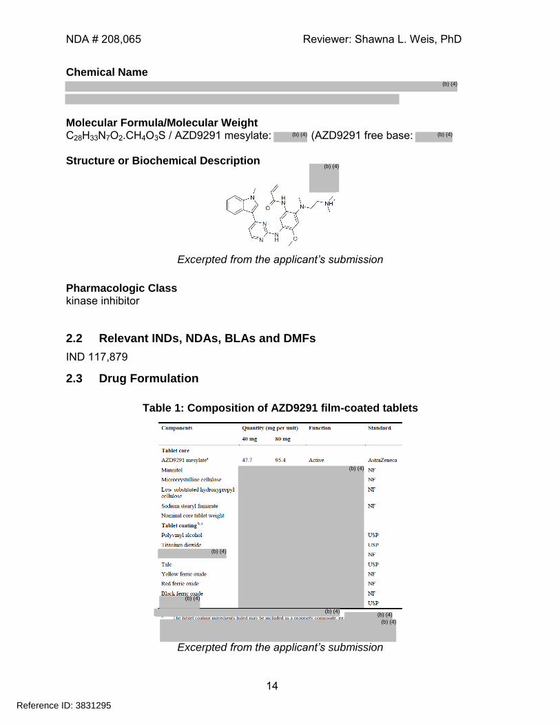
Chemical Name
Molecular Formula/Molecular Weight C28H33N7O2.CH4O3S / AZD9291 mesylate: (AZD9291 free base: Structure or Biochemical Description
Excerpted from the applicant’s submission
Pharmacologic Class kinase inhibitor
2.2 Relevant INDs, NDAs, BLAs and DMFs
IND 117,879
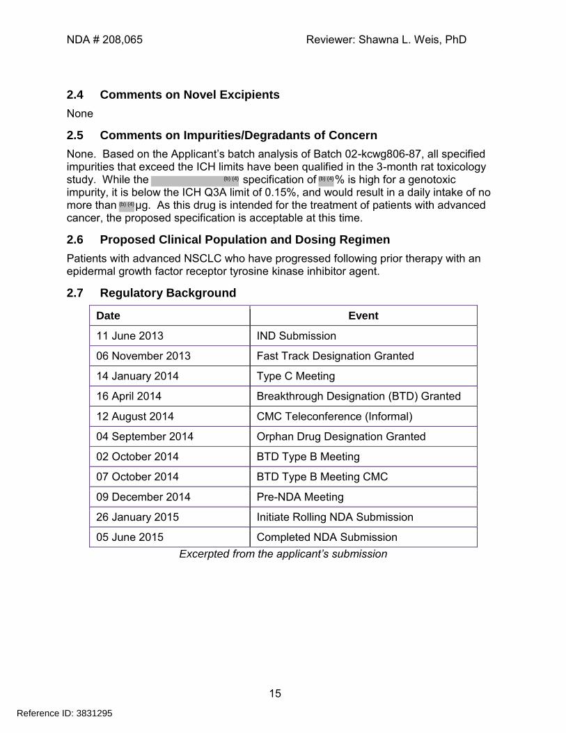
2.3 Drug Formulation
Table 1: Composition of AZD9291 film-coated tablets
Excerpted from the applicant’s submission
Reference ID: 3831295
(b) (4)
(b) (4)(b) (4)
(b) (4)
(b) (4)
(b) (4)
(b) (4)
(b) (4)(b) (4)
(b) (4)
NDA # 208,065 Reviewer: Shawna L. Weis, PhD
15
2.4 Comments on Novel Excipients
None
2.5 Comments on Impurities/Degradants of Concern
None. Based on the Applicant’s batch analysis of Batch 02-kcwg806-87, all specified impurities that exceed the ICH limits have been qualified in the 3-month rat toxicology study. While the specification of % is high for a genotoxic impurity, it is below the ICH Q3A limit of 0.15%, and would result in a daily intake of no more than μg. As this drug is intended for the treatment of patients with advanced cancer, the proposed specification is acceptable at this time.
2.6 Proposed Clinical Population and Dosing Regimen
Patients with advanced NSCLC who have progressed following prior therapy with an epidermal growth factor receptor tyrosine kinase inhibitor agent.
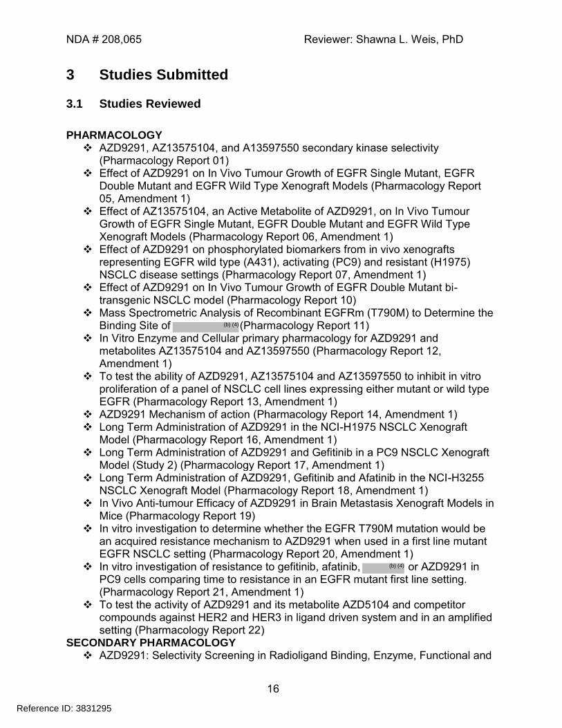
2.7 Regulatory Background
Date Event
11 June 2013 IND Submission
06 November 2013 Fast Track Designation Granted
14 January 2014 Type C Meeting
16 April 2014 Breakthrough Designation (BTD) Granted
12 August 2014 CMC Teleconference (Informal)
04 September 2014 Orphan Drug Designation Granted
02 October 2014 BTD Type B Meeting
07 October 2014 BTD Type B Meeting CMC
09 December 2014 Pre-NDA Meeting
26 January 2015 Initiate Rolling NDA Submission
05 June 2015 Completed NDA Submission
Excerpted from the applicant’s submission
Reference ID: 3831295
(b) (4) (b) (4)
(b) (4)
NDA # 208,065 Reviewer: Shawna L. Weis, PhD
16
3 Studies Submitted
3.1 Studies Reviewed
PHARMACOLOGY
AZD9291, AZ13575104, and A13597550 secondary kinase selectivity (Pharmacology Report 01)
Effect of AZD9291 on In Vivo Tumour Growth of EGFR Single Mutant, EGFR Double Mutant and EGFR Wild Type Xenograft Models (Pharmacology Report 05, Amendment 1)
Effect of AZ13575104, an Active Metabolite of AZD9291, on In Vivo Tumour Growth of EGFR Single Mutant, EGFR Double Mutant and EGFR Wild Type Xenograft Models (Pharmacology Report 06, Amendment 1)
Effect of AZD9291 on phosphorylated biomarkers from in vivo xenografts representing EGFR wild type (A431), activating (PC9) and resistant (H1975) NSCLC disease settings (Pharmacology Report 07, Amendment 1)
Effect of AZD9291 on In Vivo Tumour Growth of EGFR Double Mutant bi-transgenic NSCLC model (Pharmacology Report 10)
Mass Spectrometric Analysis of Recombinant EGFRm (T790M) to Determine the Binding Site of (Pharmacology Report 11)
In Vitro Enzyme and Cellular primary pharmacology for AZD9291 and metabolites AZ13575104 and AZ13597550 (Pharmacology Report 12, Amendment 1)
To test the ability of AZD9291, AZ13575104 and AZ13597550 to inhibit in vitro proliferation of a panel of NSCLC cell lines expressing either mutant or wild type EGFR (Pharmacology Report 13, Amendment 1)
AZD9291 Mechanism of action (Pharmacology Report 14, Amendment 1) Long Term Administration of AZD9291 in the NCI-H1975 NSCLC Xenograft
Model (Pharmacology Report 16, Amendment 1) Long Term Administration of AZD9291 and Gefitinib in a PC9 NSCLC Xenograft
Model (Study 2) (Pharmacology Report 17, Amendment 1) Long Term Administration of AZD9291, Gefitinib and Afatinib in the NCI-H3255
NSCLC Xenograft Model (Pharmacology Report 18, Amendment 1) In Vivo Anti-tumour Efficacy of AZD9291 in Brain Metastasis Xenograft Models in
Mice (Pharmacology Report 19) In vitro investigation to determine whether the EGFR T790M mutation would be
an acquired resistance mechanism to AZD9291 when used in a first line mutant EGFR NSCLC setting (Pharmacology Report 20, Amendment 1)
In vitro investigation of resistance to gefitinib, afatinib, or AZD9291 in PC9 cells comparing time to resistance in an EGFR mutant first line setting. (Pharmacology Report 21, Amendment 1)
To test the activity of AZD9291 and its metabolite AZD5104 and competitor compounds against HER2 and HER3 in ligand driven system and in an amplified setting (Pharmacology Report 22)
SECONDARY PHARMACOLOGY AZD9291: Selectivity Screening in Radioligand Binding, Enzyme, Functional and
Reference ID: 3831295
(b) (4)
(b) (4)
NDA # 208,065 Reviewer: Shawna L. Weis, PhD
17
Electrophysiological Assays in vitro (Report 1112SY) AZ13575104: Selectivity Screening in Radioligand Binding, Enzyme, Functional
and Electrophysiological Assays in vitro (Report 1120SY) AZ13597550: Selectivity Screening in Radioligand Binding, Enzyme and
Electrophysiological Assays in vitro (Report 1121SY) AZ13597550 : Selectivity Screening in Cardiac Ion Channel Electrophysiological
Assays in vitro (Report 3472SV) AZ13575104 : Selectivity Screening in Cardiac Ion Channel Electrophysiological
Assays in vitro (Report 3473SV) : Selectivity Screening in Cardiac Ion Channel Electrophysiological
Assays in vitro (Report 3535SV) SAFETY PHARMACOLOGY
AZ13540484 and Cardiovascular Effects in Anaesthetised Guinea-Pigs following Intravenous Infusion (0264SG)
AZD9291: Cardiovascular Effects in Conscious, Telemetered Beagle Dogs following Single Oral Administration - Report Amendment (1352ZD)
AZD9291: Nervous System, Visual, Respiratory and Gastrointestinal Transit Effects in the Han Wistar Rat following Single Oral Administration (3464SR)
Cardiovascular Effects of in vivo: Rat Telemetry (ONC.000-574-813)
AZD9291: Effects on Human Ether-a-go-go-related Gene (hERG) Encoded Potassium Channel in vitro (VKS0795)
ADME Validation of a High Performance Liquid Chromatography-Tandem
Mass Spectrometry (HPLC-MS/MS) Method for the Determination of AstraZeneca AZD9291, AZ13597550 and AZ13575104 Concentrations in Rat and Dog Plasma (D9291 KPV005)
AZD9291: In Vitro Covalent Binding of [3H]-Labelled AZD9291 to Human Hepatic Proteins in Human Hepatocyte Incubations (111123_CVB_KXZZ856)
AZD9291: The Tissue Distribution of Total Radioactivity in the Rat Following Oral Administration of [14C]AZD9291 (Quantitative Whole Body Autoradiography) (Report #8265542)
AZD9291: Mass Balance and Radioactive Pharmacokinetics Following Oral and Intravenous Administration of [14C]AZD9291 to the Rat (Report #196187)
AZD9291: The disposition of [14C]AZD9291 following intravenous and oral administration in the dog (Report 197725)
TOXICOLOGY AZD9291: Three Month Oral (Gavage) Toxicity Study in the Rat – Report
Amendment (Report 52648) AZD9291: Three Month Oral (Gavage) Toxicity Study in the Dog (Report 526253 AZD9291: Genetic Toxicity Evaluation Using the Bacterial Reverse Mutation Test
in Salmonella typhimurium TA 1535, TA 100, TA 1537 and TA 98 and Escherichia coli WP2uvrA (pKM101) (Report #793061)
AZD9291: Genetic Toxicity Evaluation using the Mouse Lymphoma Cell Thymidine Kinase Locus Assay (Report #793056)
AZD9291: Genetic Toxicity Evaluation Using the Rat Micronucleus Test After
Reference ID: 3831295
(b) (4)
(b) (4)
(b) (4)
NDA # 208,065 Reviewer: Shawna L. Weis, PhD
18
Two Oral Doses (Report #793538) AZD9291: Oral (Gavage) Investigative Dose Range Finding Embryofetal
Development and Pre and Post Natal Study in the Rat (Report #496800) AZD9291: Evaluation of in vitro phototoxicity on Balb/c 3T3 fibroblasts using the
Neutral Red Uptake assay (8273754) IMPURITY AND METABOLITE QUALIFICATION
AZD9291: One Month Oral (Gavage) Toxicity Study in the Rat (Report # 528622) AZ13575104 and : One Month Investigatory Toxicity Study in the
Rat (3500KR) Genetic Toxicity Evaluation Using the Bacterial Reverse Mutation
Test in Salmonella typhimurium TA1535, TA100, TA1537 and TA98 and Escherichia coli WP2 uvrA (pKM101) (Report # 8303534)
: Genetic Toxicity Evaluation Using the Bacterial Reverse Mutation Test in Salmonella typhimurium TA1535, TA100, TA1537 and TA98 and Escherichia coli WP2 uvrA (pKM101) (Report # 8303535)
: Genetic Toxicity Evaluation Using the Bacterial Reverse Mutation Test in Salmonella typhimurium TA1535, TA100, TA1537 and TA98 and Escherichia coli WP2 uvrA (pKM101) (Report # 8303536)
: Genetic Toxicity Evaluation Using the Bacterial Reverse Mutation Test in Salmonella typhimurium TA1535, TA100, TA1537 and TA98 and Escherichia coli WP2 uvrA pKM101(Report # 8306010)
3.2 Studies Not Reviewed
ADME Exposure in male beagle dogs following oral administration f a 20 mg dose of
AZD9291 mesylate salt as a solution, or solid in a capsule and tablets (Report CPK-13-0099)
Pharmacokinetics of AZD9291 following intravenous and oral administration in the rat (Report 8308627)
AZD9291: In vivo pharmacokinetics in plasma, brain and H1975 tumour of AZD9291 in tumour bearing SCID mice following a single oral dose at 5 and 25 mg/kg (Report #BE000343-60)
AZD9291: Validation of an Analytical Method for the Determination of AZD9291 and Metabolites (AZ13597550 and AZ13575104) in Rat Plasma Using Protein Precipitation Followed by LC-MS/MS (Report #317706)
AZD9291: Partial Validation of a Method for the Determination of AZD9291 and Metabolites (AZ13597550 and AZ13575104) in Dog Plasma Using Protein Precipitation Followed by LC-MS/MS (Report #318322)
Investigation of ISR Failure and Further Development of an Analytical Method for the Determination of AZ13575104 in Rat and Dog Plasma by LC-MS/MS (Report # 319719)
Qualification of a Method for the Determination of AZD9291, AZ13575104 and AZ13597550 in Rat Plasma using Protein Precipitation followed by Liquid Chromatography with Tandem Mass Spectrometric Detection (LC-MS/MS) Study (Report #8309952)
Reference ID: 3831295
(b) (4)
(b) (4)
(b) (4)
(b) (4)
(b) (4)
NDA # 208,065 Reviewer: Shawna L. Weis, PhD
19
Administration of AZD9291 in the Rat Following Oral Administration to Produce Plasma to Investigate a Rat Bioanalytical Method (Report #8309953)
Validation of a High Performance Liquid Chromatography-Tandem Mass Spectrometry (HPLC-MS/MS) Method for the Determination of AstraZeneca AZD9291, AZ13597550 and AZ13575104 Concentrations in Rat and Dog Plasma (Report #D9291 KPV005)
TOXICOLOGY Maximum Tolerated Dose and Repeated Dose Oral Toxicity Study
in the Dog (1324DD)
AZD9291: One Month Oral Toxicity Study with Assessment of Recovery in the Dog - Report Amendment (1351AD)
7 Day Oral Gavage MTD Toxicity Study in the Rat (3278DR)
: 14 Day Oral Toxicity Study in the Rat (3310DR) AZD9291: One Month Oral Toxicity Study with Assessment of Recovery in the
Rat (3416AR) Maximum Tolerated Dose and Repeated Dose Oral Toxicity Study
in the Dog (Report # 1324DD)
3.3 Previous Reviews Referenced
Review of IND 117,879 by Shawna L. Weis, PhD
4 Pharmacology
4.1 Primary Pharmacology
4.1.7 In Vitro Enzyme and Cellular primary pharmacology for AZD9291 and metabolites AZ13575104 and AZ13597550 (Pharmacology Report 12, Amendment 1)
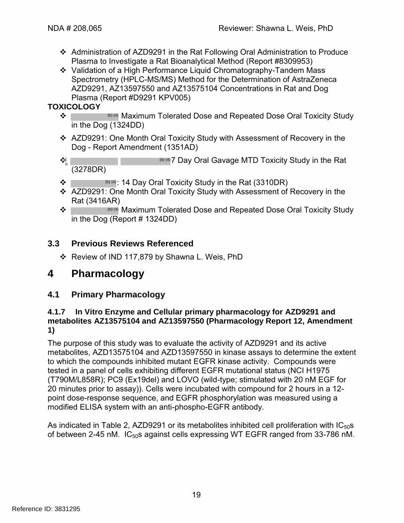
The purpose of this study was to evaluate the activity of AZD9291 and its active metabolites, AZD13575104 and AZD13597550 in kinase assays to determine the extent to which the compounds inhibited mutant EGFR kinase activity. Compounds were tested in a panel of cells exhibiting different EGFR mutational status (NCI H1975 (T790M/L858R); PC9 (Ex19del) and LOVO (wild-type; stimulated with 20 nM EGF for 20 minutes prior to assay)). Cells were incubated with compound for 2 hours in a 12-point dose-response sequence, and EGFR phosphorylation was measured using a modified ELISA system with an anti-phospho-EGFR antibody. As indicated in Table 2, AZD9291 or its metabolites inhibited cell proliferation with IC50s of between 2-45 nM. IC50s against cells expressing WT EGFR ranged from 33-786 nM.
Reference ID: 3831295
(b) (4)
(b
(b) (4)
(b) (4)
(b) (4)
NDA # 208,065 Reviewer: Shawna L. Weis, PhD
20
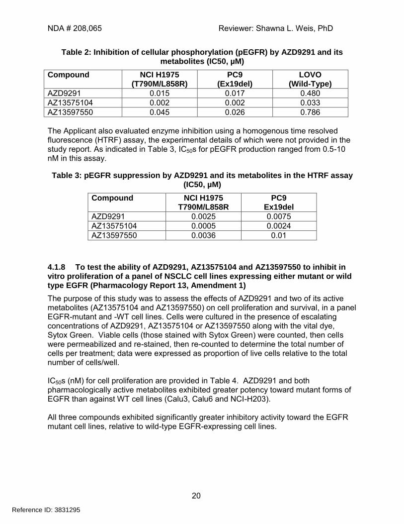
Table 2: Inhibition of cellular phosphorylation (pEGFR) by AZD9291 and its metabolites (IC50, µM)
Compound NCI H1975 (T790M/L858R)
PC9 (Ex19del)
LOVO (Wild-Type)
AZD9291 0.015 0.017 0.480
AZ13575104 0.002 0.002 0.033
AZ13597550 0.045 0.026 0.786
The Applicant also evaluated enzyme inhibition using a homogenous time resolved fluorescence (HTRF) assay, the experimental details of which were not provided in the study report. As indicated in Table 3, IC50s for pEGFR production ranged from 0.5-10 nM in this assay.
Table 3: pEGFR suppression by AZD9291 and its metabolites in the HTRF assay (IC50, µM)
Compound NCI H1975 T790M/L858R
PC9 Ex19del
AZD9291 0.0025 0.0075
AZ13575104 0.0005 0.0024
AZ13597550 0.0036 0.01
4.1.8 To test the ability of AZD9291, AZ13575104 and AZ13597550 to inhibit in vitro proliferation of a panel of NSCLC cell lines expressing either mutant or wild type EGFR (Pharmacology Report 13, Amendment 1)
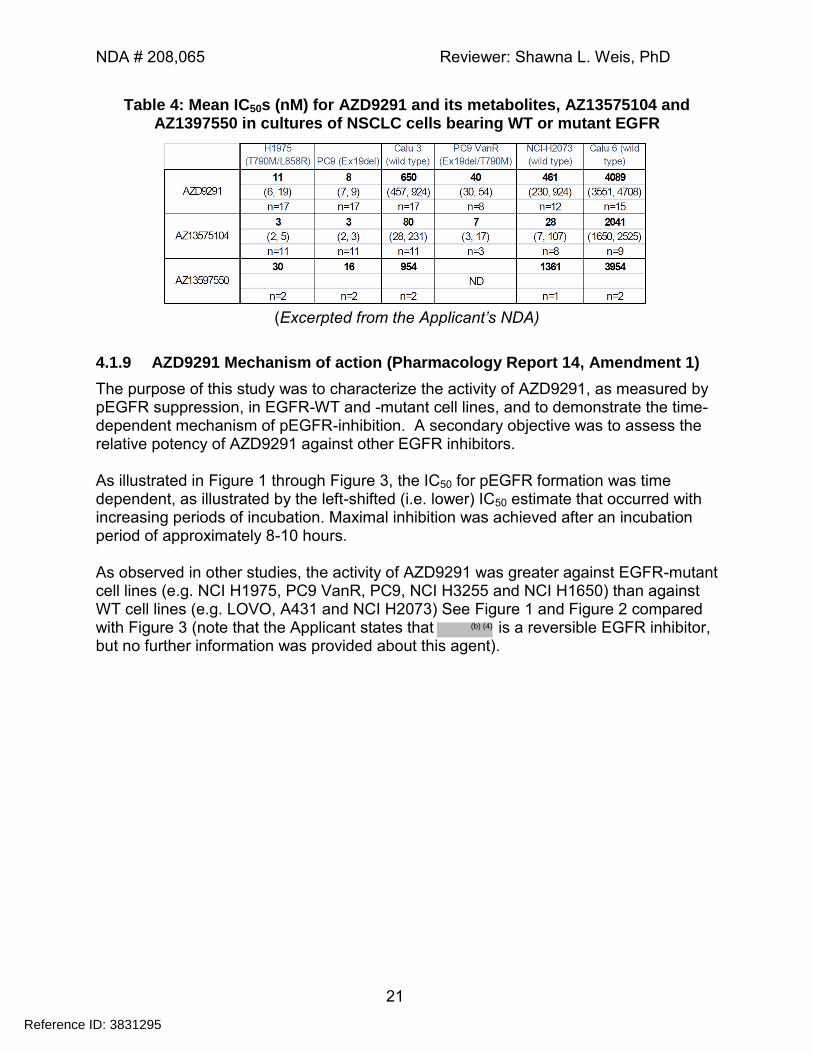
The purpose of this study was to assess the effects of AZD9291 and two of its active metabolites (AZ13575104 and AZ13597550) on cell proliferation and survival, in a panel EGFR-mutant and -WT cell lines. Cells were cultured in the presence of escalating concentrations of AZD9291, AZ13575104 or AZ13597550 along with the vital dye, Sytox Green. Viable cells (those stained with Sytox Green) were counted, then cells were permeabilized and re-stained, then re-counted to determine the total number of cells per treatment; data were expressed as proportion of live cells relative to the total number of cells/well. IC50s (nM) for cell proliferation are provided in Table 4. AZD9291 and both pharmacologically active metabolites exhibited greater potency toward mutant forms of EGFR than against WT cell lines (Calu3, Calu6 and NCI-H203). All three compounds exhibited significantly greater inhibitory activity toward the EGFR mutant cell lines, relative to wild-type EGFR-expressing cell lines.
Reference ID: 3831295
NDA # 208,065 Reviewer: Shawna L. Weis, PhD
21
Table 4: Mean IC50s (nM) for AZD9291 and its metabolites, AZ13575104 and AZ1397550 in cultures of NSCLC cells bearing WT or mutant EGFR
(Excerpted from the Applicant’s NDA)
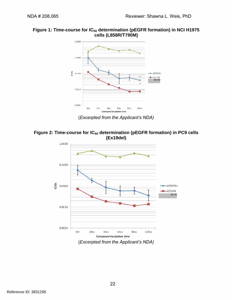
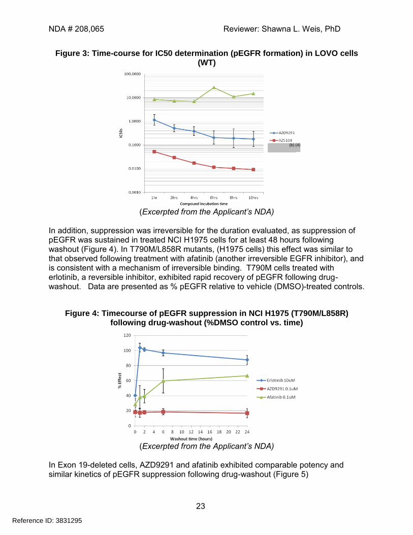
4.1.9 AZD9291 Mechanism of action (Pharmacology Report 14, Amendment 1)
The purpose of this study was to characterize the activity of AZD9291, as measured by pEGFR suppression, in EGFR-WT and -mutant cell lines, and to demonstrate the time-dependent mechanism of pEGFR-inhibition. A secondary objective was to assess the relative potency of AZD9291 against other EGFR inhibitors. As illustrated in Figure 1 through Figure 3, the IC50 for pEGFR formation was time dependent, as illustrated by the left-shifted (i.e. lower) IC50 estimate that occurred with increasing periods of incubation. Maximal inhibition was achieved after an incubation period of approximately 8-10 hours. As observed in other studies, the activity of AZD9291 was greater against EGFR-mutant cell lines (e.g. NCI H1975, PC9 VanR, PC9, NCI H3255 and NCI H1650) than against WT cell lines (e.g. LOVO, A431 and NCI H2073) See Figure 1 and Figure 2 compared with Figure 3 (note that the Applicant states that is a reversible EGFR inhibitor, but no further information was provided about this agent).
Reference ID: 3831295
(b) (4)
NDA # 208,065 Reviewer: Shawna L. Weis, PhD
22
Figure 1: Time-course for IC50 determination (pEGFR formation) in NCI H1975 cells (L858R/T790M)
(Excerpted from the Applicant’s NDA)
Figure 2: Time-course for IC50 determination (pEGFR formation) in PC9 cells (Ex19del)
(Excerpted from the Applicant’s NDA)
Reference ID: 3831295
(b) (4)
(b) (4)
NDA # 208,065 Reviewer: Shawna L. Weis, PhD
23
Figure 3: Time-course for IC50 determination (pEGFR formation) in LOVO cells (WT)
(Excerpted from the Applicant’s NDA)
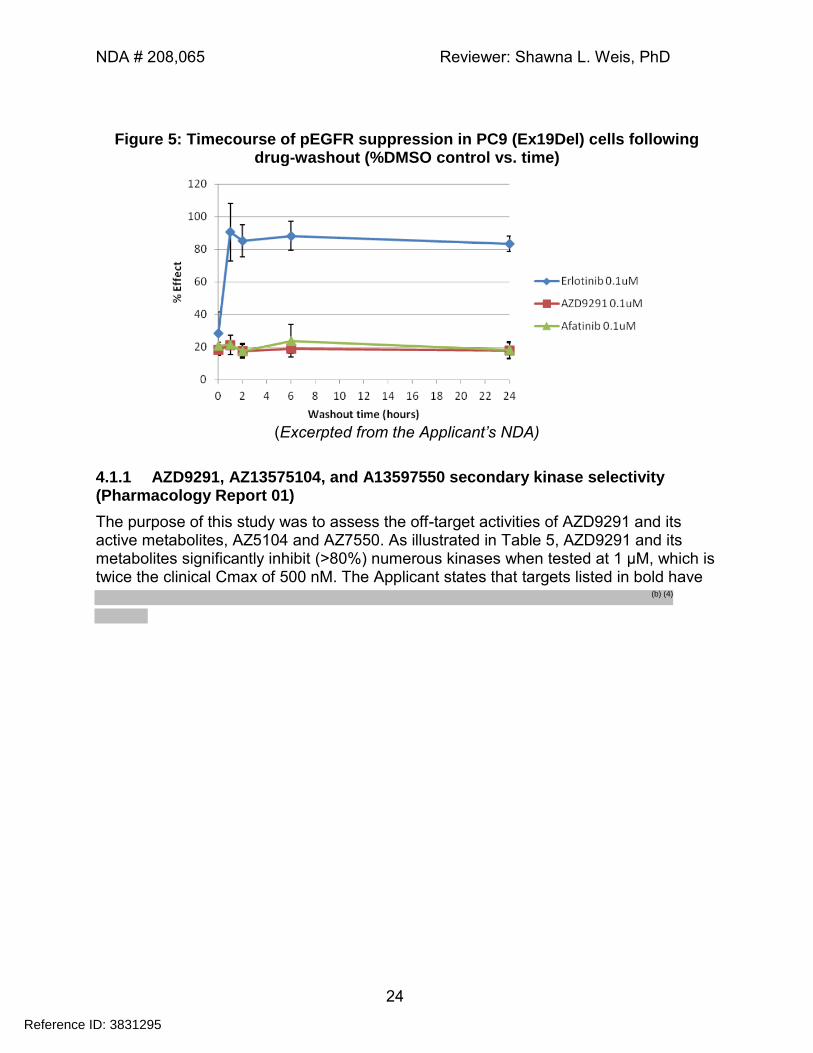
In addition, suppression was irreversible for the duration evaluated, as suppression of pEGFR was sustained in treated NCI H1975 cells for at least 48 hours following washout (Figure 4). In T790M/L858R mutants, (H1975 cells) this effect was similar to that observed following treatment with afatinib (another irreversible EGFR inhibitor), and is consistent with a mechanism of irreversible binding. T790M cells treated with erlotinib, a reversible inhibitor, exhibited rapid recovery of pEGFR following drug-washout. Data are presented as % pEGFR relative to vehicle (DMSO)-treated controls.
Figure 4: Timecourse of pEGFR suppression in NCI H1975 (T790M/L858R) following drug-washout (%DMSO control vs. time)
(Excerpted from the Applicant’s NDA)
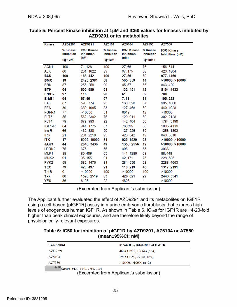
In Exon 19-deleted cells, AZD9291 and afatinib exhibited comparable potency and similar kinetics of pEGFR suppression following drug-washout (Figure 5)
Reference ID: 3831295
(b) (4)
NDA # 208,065 Reviewer: Shawna L. Weis, PhD
24
Figure 5: Timecourse of pEGFR suppression in PC9 (Ex19Del) cells following drug-washout (%DMSO control vs. time)
(Excerpted from the Applicant’s NDA)
4.1.1 AZD9291, AZ13575104, and A13597550 secondary kinase selectivity (Pharmacology Report 01)
The purpose of this study was to assess the off-target activities of AZD9291 and its active metabolites, AZ5104 and AZ7550. As illustrated in Table 5, AZD9291 and its metabolites significantly inhibit (>80%) numerous kinases when tested at 1 µM, which is twice the clinical Cmax of 500 nM. The Applicant states that targets listed in bold have
Reference ID: 3831295
(b) (4)
NDA # 208,065 Reviewer: Shawna L. Weis, PhD
25
Table 5: Percent kinase inhibition at 1µM and IC50 values for kinases inhibited by AZD9291 or its metabolites
(Excerpted from Applicant’s submission)
The Applicant further evaluated the effect of AZD9291 and its metabolites on IGF1R using a cell-based (pIGF1R) assay in murine embryonic fibroblasts that express high levels of exogenous human IGF1R. As shown in Table 6, IC50s for IGF1R are ~4-20-fold higher than peak clinical exposures, and are therefore likely beyond the range of physiologically-relevant exposures.
Table 6: IC50 for inhibition of pIGF1R by AZD9291, AZ5104 or A7550 (mean±95%CI; nM)
(Excerpted from Applicant’s submission)
Reference ID: 3831295
(b) (4)
NDA # 208,065 Reviewer: Shawna L. Weis, PhD
26
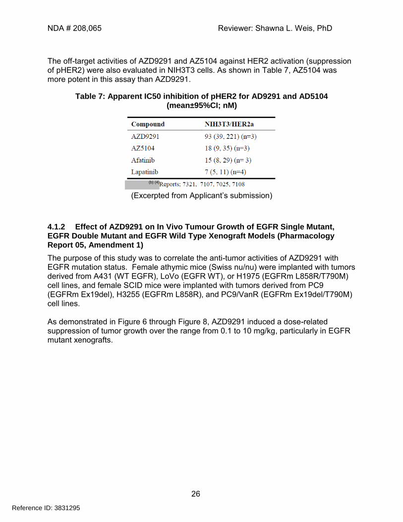
The off-target activities of AZD9291 and AZ5104 against HER2 activation (suppression of pHER2) were also evaluated in NIH3T3 cells. As shown in Table 7, AZ5104 was more potent in this assay than AZD9291.
Table 7: Apparent IC50 inhibition of pHER2 for AD9291 and AD5104 (mean±95%CI; nM)
(Excerpted from Applicant’s submission)
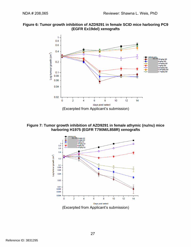
4.1.2 Effect of AZD9291 on In Vivo Tumour Growth of EGFR Single Mutant, EGFR Double Mutant and EGFR Wild Type Xenograft Models (Pharmacology Report 05, Amendment 1)
The purpose of this study was to correlate the anti-tumor activities of AZD9291 with EGFR mutation status. Female athymic mice (Swiss nu/nu) were implanted with tumors derived from A431 (WT EGFR), LoVo (EGFR WT), or H1975 (EGFRm L858R/T790M) cell lines, and female SCID mice were implanted with tumors derived from PC9 (EGFRm Ex19del), H3255 (EGFRm L858R), and PC9/VanR (EGFRm Ex19del/T790M) cell lines. As demonstrated in Figure 6 through Figure 8, AZD9291 induced a dose-related suppression of tumor growth over the range from 0.1 to 10 mg/kg, particularly in EGFR mutant xenografts.
Reference ID: 3831295
(b) (4)
NDA # 208,065 Reviewer: Shawna L. Weis, PhD
27
Figure 6: Tumor growth inhibition of AZD9291 in female SCID mice harboring PC9 (EGFR Ex19del) xenografts
(Excerpted from Applicant’s submission)
Figure 7: Tumor growth inhibition of AZD9291 in female athymic (nu/nu) mice harboring H1975 (EGFR T790M/L858R) xenografts
(Excerpted from Applicant’s submission)
Reference ID: 3831295
(b) (4)
(b) (4)
NDA # 208,065 Reviewer: Shawna L. Weis, PhD
28
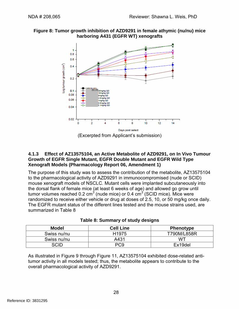
Figure 8: Tumor growth inhibition of AZD9291 in female athymic (nu/nu) mice harboring A431 (EGFR WT) xenografts
(Excerpted from Applicant’s submission)
4.1.3 Effect of AZ13575104, an Active Metabolite of AZD9291, on In Vivo Tumour Growth of EGFR Single Mutant, EGFR Double Mutant and EGFR Wild Type Xenograft Models (Pharmacology Report 06, Amendment 1)
The purpose of this study was to assess the contribution of the metabolite, AZ13575104 to the pharmacological activity of AZD9291 in immunocompromised (nude or SCID) mouse xenograft models of NSCLC. Mutant cells were implanted subcutaneously into the dorsal flank of female mice (at least 6 weeks of age) and allowed go grow until tumor volumes reached 0.2 cm3 (nude mice) or 0.4 cm3 (SCID mice). Mice were randomized to receive either vehicle or drug at doses of 2.5, 10, or 50 mg/kg once daily. The EGFR mutant status of the different lines tested and the mouse strains used, are summarized in Table 8
Table 8: Summary of study designs
Model Cell Line Phenotype
Swiss nu/nu H1975 T790M/L858R
Swiss nu/nu A431 WT
SCID PC9 Ex19del
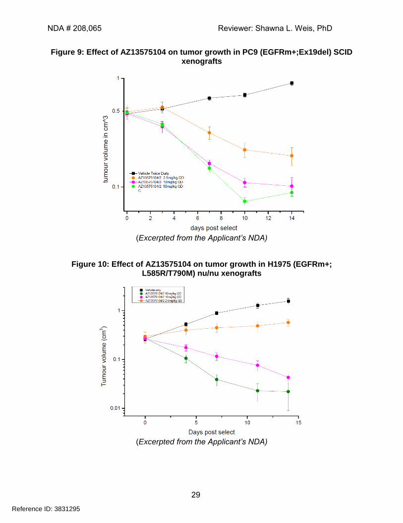
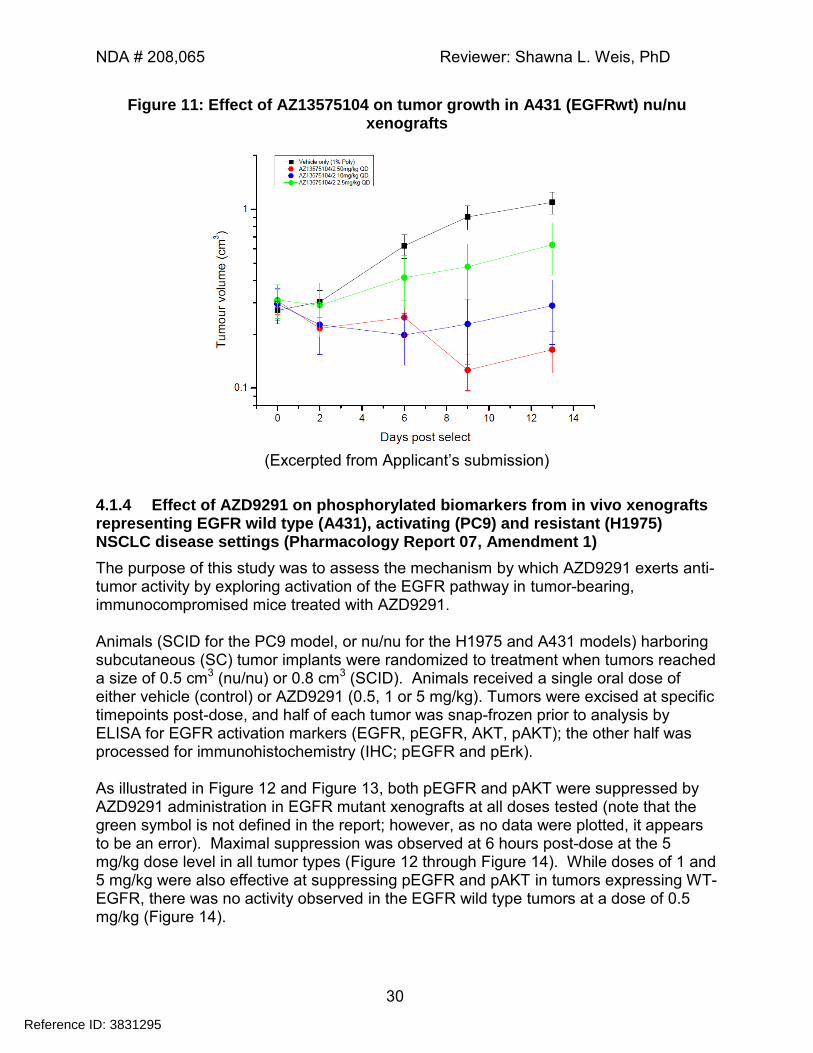
As illustrated in Figure 9 through Figure 11, AZ13575104 exhibited dose-related anti-tumor activity in all models tested; thus, the metabolite appears to contribute to the overall pharmacological activity of AZD9291.
Reference ID: 3831295
(b) (4)
NDA # 208,065 Reviewer: Shawna L. Weis, PhD
29
Figure 9: Effect of AZ13575104 on tumor growth in PC9 (EGFRm+;Ex19del) SCID xenografts
(Excerpted from the Applicant’s NDA)
Figure 10: Effect of AZ13575104 on tumor growth in H1975 (EGFRm+; L585R/T790M) nu/nu xenografts
(Excerpted from the Applicant’s NDA)
Reference ID: 3831295
NDA # 208,065 Reviewer: Shawna L. Weis, PhD
30
Figure 11: Effect of AZ13575104 on tumor growth in A431 (EGFRwt) nu/nu xenografts
(Excerpted from Applicant’s submission)
4.1.4 Effect of AZD9291 on phosphorylated biomarkers from in vivo xenografts representing EGFR wild type (A431), activating (PC9) and resistant (H1975) NSCLC disease settings (Pharmacology Report 07, Amendment 1)
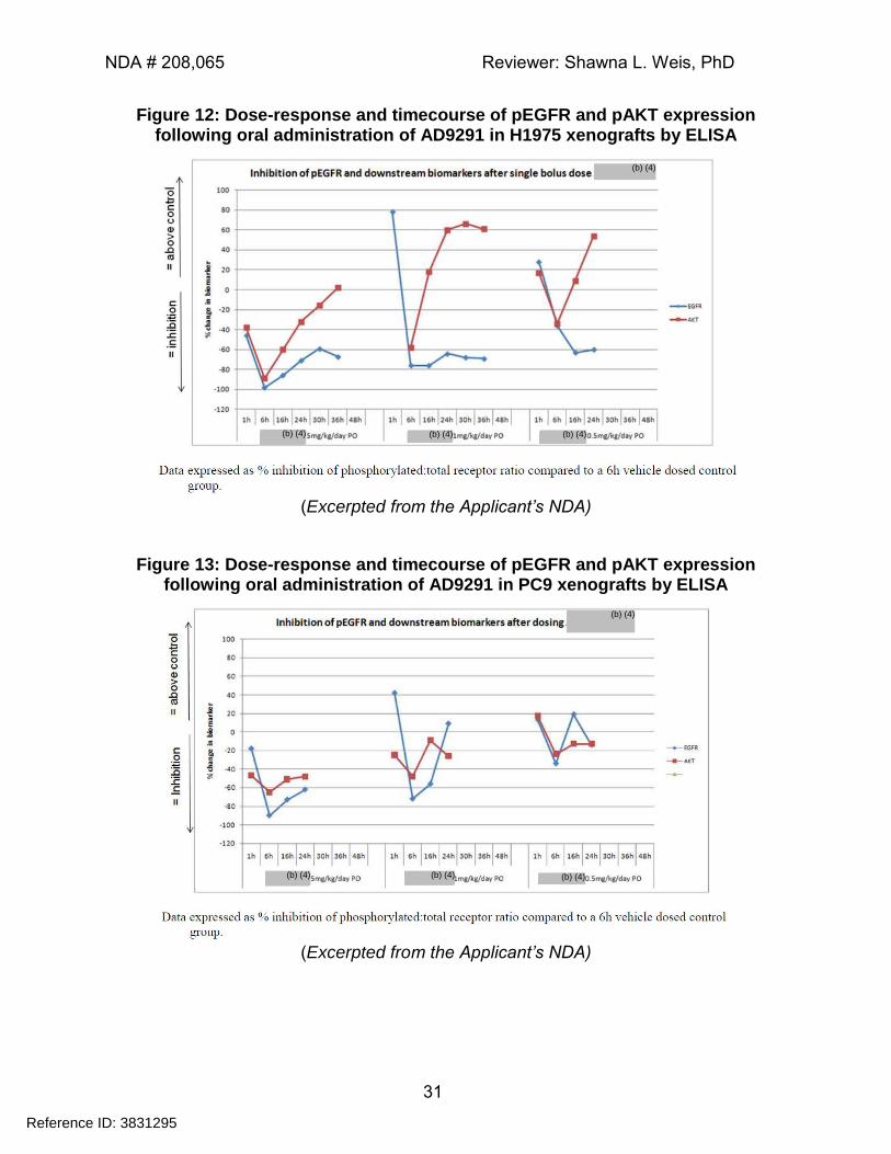
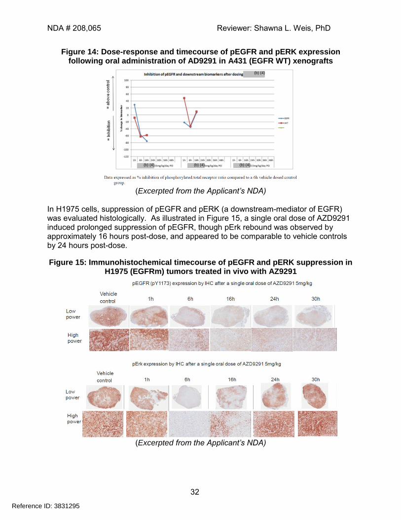
The purpose of this study was to assess the mechanism by which AZD9291 exerts anti-tumor activity by exploring activation of the EGFR pathway in tumor-bearing, immunocompromised mice treated with AZD9291. Animals (SCID for the PC9 model, or nu/nu for the H1975 and A431 models) harboring subcutaneous (SC) tumor implants were randomized to treatment when tumors reached a size of 0.5 cm3 (nu/nu) or 0.8 cm3 (SCID). Animals received a single oral dose of either vehicle (control) or AZD9291 (0.5, 1 or 5 mg/kg). Tumors were excised at specific timepoints post-dose, and half of each tumor was snap-frozen prior to analysis by ELISA for EGFR activation markers (EGFR, pEGFR, AKT, pAKT); the other half was processed for immunohistochemistry (IHC; pEGFR and pErk). As illustrated in Figure 12 and Figure 13, both pEGFR and pAKT were suppressed by AZD9291 administration in EGFR mutant xenografts at all doses tested (note that the green symbol is not defined in the report; however, as no data were plotted, it appears to be an error). Maximal suppression was observed at 6 hours post-dose at the 5 mg/kg dose level in all tumor types (Figure 12 through Figure 14). While doses of 1 and 5 mg/kg were also effective at suppressing pEGFR and pAKT in tumors expressing WT-EGFR, there was no activity observed in the EGFR wild type tumors at a dose of 0.5 mg/kg (Figure 14).
Reference ID: 3831295
NDA # 208,065 Reviewer: Shawna L. Weis, PhD
31
Figure 12: Dose-response and timecourse of pEGFR and pAKT expression following oral administration of AD9291 in H1975 xenografts by ELISA
(Excerpted from the Applicant’s NDA)
Figure 13: Dose-response and timecourse of pEGFR and pAKT expression following oral administration of AD9291 in PC9 xenografts by ELISA
(Excerpted from the Applicant’s NDA)
Reference ID: 3831295
(b) (4) (b) (4) (b) (4)
(b) (4)
(b) (4)
(b) (4) (b) (4) (b) (4)
NDA # 208,065 Reviewer: Shawna L. Weis, PhD
32
Figure 14: Dose-response and timecourse of pEGFR and pERK expression following oral administration of AD9291 in A431 (EGFR WT) xenografts
(Excerpted from the Applicant’s NDA)
In H1975 cells, suppression of pEGFR and pERK (a downstream-mediator of EGFR) was evaluated histologically. As illustrated in Figure 15, a single oral dose of AZD9291 induced prolonged suppression of pEGFR, though pErk rebound was observed by approximately 16 hours post-dose, and appeared to be comparable to vehicle controls by 24 hours post-dose.
Figure 15: Immunohistochemical timecourse of pEGFR and pERK suppression in H1975 (EGFRm) tumors treated in vivo with AZ9291
(Excerpted from the Applicant’s NDA)
Reference ID: 3831295
(b) (4) (b) (4) (b) (4)
(b) (4)
NDA # 208,065 Reviewer: Shawna L. Weis, PhD
33
4.1.5 Effect of AZD9291 on In Vivo Tumour Growth of EGFR Double Mutant bi-transgenic NSCLC model (Pharmacology Report 10)
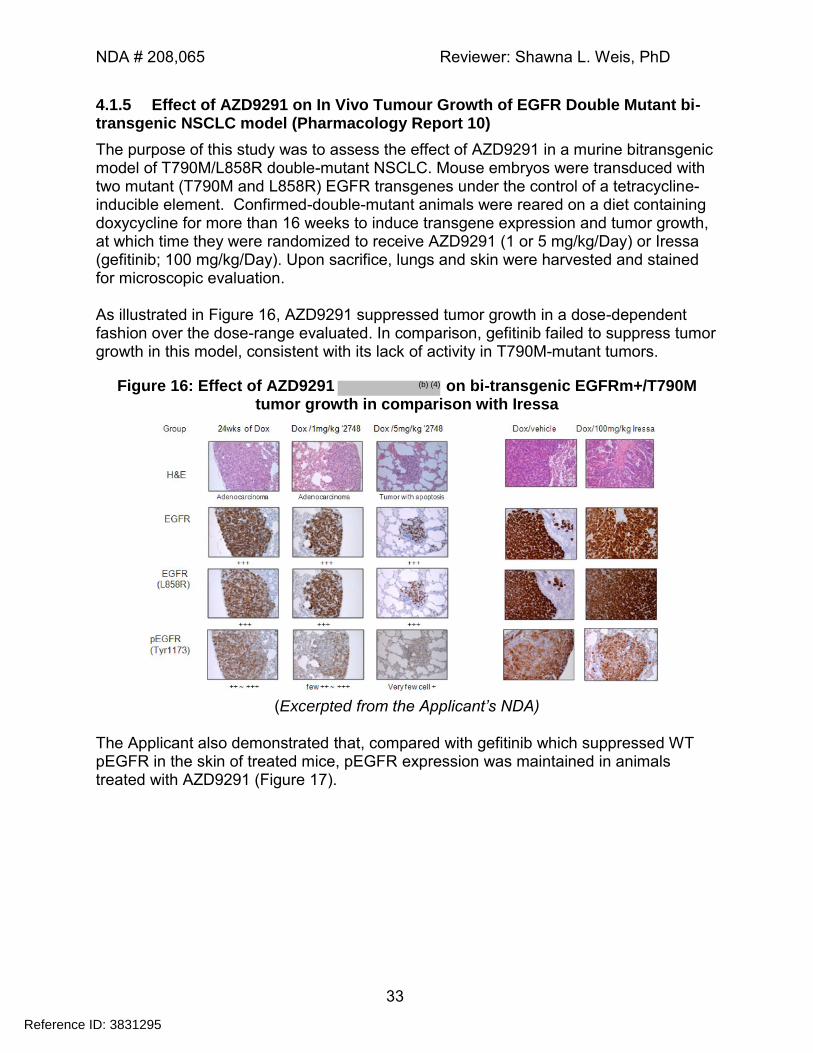
The purpose of this study was to assess the effect of AZD9291 in a murine bitransgenic model of T790M/L858R double-mutant NSCLC. Mouse embryos were transduced with two mutant (T790M and L858R) EGFR transgenes under the control of a tetracycline-inducible element. Confirmed-double-mutant animals were reared on a diet containing doxycycline for more than 16 weeks to induce transgene expression and tumor growth, at which time they were randomized to receive AZD9291 (1 or 5 mg/kg/Day) or Iressa (gefitinib; 100 mg/kg/Day). Upon sacrifice, lungs and skin were harvested and stained for microscopic evaluation. As illustrated in Figure 16, AZD9291 suppressed tumor growth in a dose-dependent fashion over the dose-range evaluated. In comparison, gefitinib failed to suppress tumor growth in this model, consistent with its lack of activity in T790M-mutant tumors.
Figure 16: Effect of AZD9291 on bi-transgenic EGFRm+/T790M tumor growth in comparison with Iressa
(Excerpted from the Applicant’s NDA)
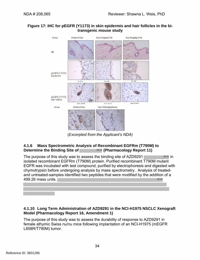
The Applicant also demonstrated that, compared with gefitinib which suppressed WT pEGFR in the skin of treated mice, pEGFR expression was maintained in animals treated with AZD9291 (Figure 17).
Reference ID: 3831295
(b) (4)
NDA # 208,065 Reviewer: Shawna L. Weis, PhD
34
Figure 17: IHC for pEGFR (Y1173) in skin epidermis and hair follicles in the bi-transgenic mouse study
(Excerpted from the Applicant’s NDA)
4.1.6 Mass Spectrometric Analysis of Recombinant EGFRm (T790M) to Determine the Binding Site of (Pharmacology Report 11)
The purpose of this study was to assess the binding site of AZD9291 in isolated recombinant EGFRm (T790M) protein. Purified recombinant T790M mutant EGFR was incubated with test compound, purified by electrophoresis and digested with chymotrypsin before undergoing analysis by mass spectrometry. Analysis of treated- and untreated-samples identified two peptides that were modified by the addition of a 499.26 mass units.
4.1.10 Long Term Administration of AZD9291 in the NCI-H1975 NSCLC Xenograft Model (Pharmacology Report 16, Amendment 1)
The purpose of this study was to assess the durability of response to AZD9291 in female athymic Swiss nu/nu mice following implantation of an NCI-H1975 (mEGFR L858R/T790M) tumor.
Reference ID: 3831295
(b) (4)
(b) (4)
(b) (4)
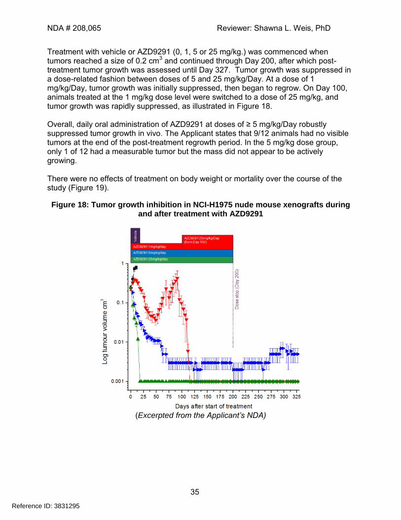
NDA # 208,065 Reviewer: Shawna L. Weis, PhD
35
Treatment with vehicle or AZD9291 (0, 1, 5 or 25 mg/kg.) was commenced when tumors reached a size of 0.2 cm3 and continued through Day 200, after which post-treatment tumor growth was assessed until Day 327. Tumor growth was suppressed in a dose-related fashion between doses of 5 and 25 mg/kg/Day. At a dose of 1 mg/kg/Day, tumor growth was initially suppressed, then began to regrow. On Day 100, animals treated at the 1 mg/kg dose level were switched to a dose of 25 mg/kg, and tumor growth was rapidly suppressed, as illustrated in Figure 18. Overall, daily oral administration of AZD9291 at doses of ≥ 5 mg/kg/Day robustly suppressed tumor growth in vivo. The Applicant states that 9/12 animals had no visible tumors at the end of the post-treatment regrowth period. In the 5 mg/kg dose group, only 1 of 12 had a measurable tumor but the mass did not appear to be actively growing. There were no effects of treatment on body weight or mortality over the course of the study (Figure 19).
Figure 18: Tumor growth inhibition in NCI-H1975 nude mouse xenografts during and after treatment with AZD9291
(Excerpted from the Applicant’s NDA)
Reference ID: 3831295
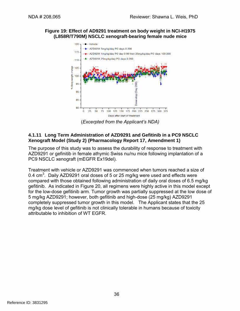
NDA # 208,065 Reviewer: Shawna L. Weis, PhD
36
Figure 19: Effect of AD9291 treatment on body weight in NCI-H1975 (L858R/T790M) NSCLC xenograft-bearing female nude mice
(Excerpted from the Applicant’s NDA)
4.1.11 Long Term Administration of AZD9291 and Gefitinib in a PC9 NSCLC Xenograft Model (Study 2) (Pharmacology Report 17, Amendment 1)
The purpose of this study was to assess the durability of response to treatment with AZD9291 or gefinitib in female athymic Swiss nu/nu mice following implantation of a PC9 NSCLC xenograft (mEGFR Ex19del). Treatment with vehicle or AZD9291 was commenced when tumors reached a size of 0.4 cm3. Daily AZD9291 oral doses of 5 or 25 mg/kg were used and effects were compared with those obtained following administration of daily oral doses of 6.5 mg/kg gefitinib. As indicated in Figure 20, all regimens were highly active in this model except for the low-dose gefitinib arm. Tumor growth was partially suppressed at the low dose of 5 mg/kg AZD9291; however, both gefitinib and high-dose (25 mg/kg) AZD9291 completely suppressed tumor growth in this model. The Applicant states that the 25 mg/kg dose level of gefitinib is not clinically tolerable in humans because of toxicity attributable to inhibition of WT EGFR.
Reference ID: 3831295
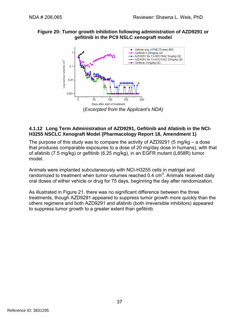
NDA # 208,065 Reviewer: Shawna L. Weis, PhD
37
Figure 20: Tumor growth inhibition following administration of AZD9291 or gefitinib in the PC9 NSLC xenograft model
(Excerpted from the Applicant’s NDA)
4.1.12 Long Term Administration of AZD9291, Gefitinib and Afatinib in the NCI-H3255 NSCLC Xenograft Model (Pharmacology Report 18, Amendment 1)
The purpose of this study was to compare the activity of AZD9291 (5 mg/kg – a dose that produces comparable exposures to a dose of 20 mg/day dose in humans), with that of afatinib (7.5 mg/kg) or gefitinib (6.25 mg/kg), in an EGFR mutant (L858R) tumor model. Animals were implanted subcutaneously with NCI-H3255 cells in matrigel and randomized to treatment when tumor volumes reached 0.4 cm3. Animals received daily oral doses of either vehicle or drug for 75 days, beginning the day after randomization. As illustrated in Figure 21, there was no significant difference between the three treatments, though AZD9291 appeared to suppress tumor growth more quickly than the others regimens and both AZD9291 and afatinib (both irreversible inhibitors) appeared to suppress tumor growth to a greater extent than gefitinib.
Reference ID: 3831295
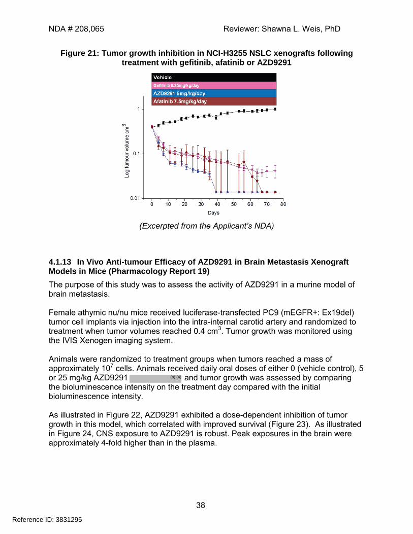
NDA # 208,065 Reviewer: Shawna L. Weis, PhD
38
Figure 21: Tumor growth inhibition in NCI-H3255 NSLC xenografts following treatment with gefitinib, afatinib or AZD9291
(Excerpted from the Applicant’s NDA)
4.1.13 In Vivo Anti-tumour Efficacy of AZD9291 in Brain Metastasis Xenograft Models in Mice (Pharmacology Report 19)
The purpose of this study was to assess the activity of AZD9291 in a murine model of brain metastasis. Female athymic nu/nu mice received luciferase-transfected PC9 (mEGFR+: Ex19del) tumor cell implants via injection into the intra-internal carotid artery and randomized to treatment when tumor volumes reached 0.4 cm3. Tumor growth was monitored using the IVIS Xenogen imaging system. Animals were randomized to treatment groups when tumors reached a mass of approximately 107 cells. Animals received daily oral doses of either 0 (vehicle control), 5 or 25 mg/kg AZD9291 and tumor growth was assessed by comparing the bioluminescence intensity on the treatment day compared with the initial bioluminescence intensity. As illustrated in Figure 22, AZD9291 exhibited a dose-dependent inhibition of tumor growth in this model, which correlated with improved survival (Figure 23). As illustrated in Figure 24, CNS exposure to AZD9291 is robust. Peak exposures in the brain were approximately 4-fold higher than in the plasma.
Reference ID: 3831295
(b) (4)
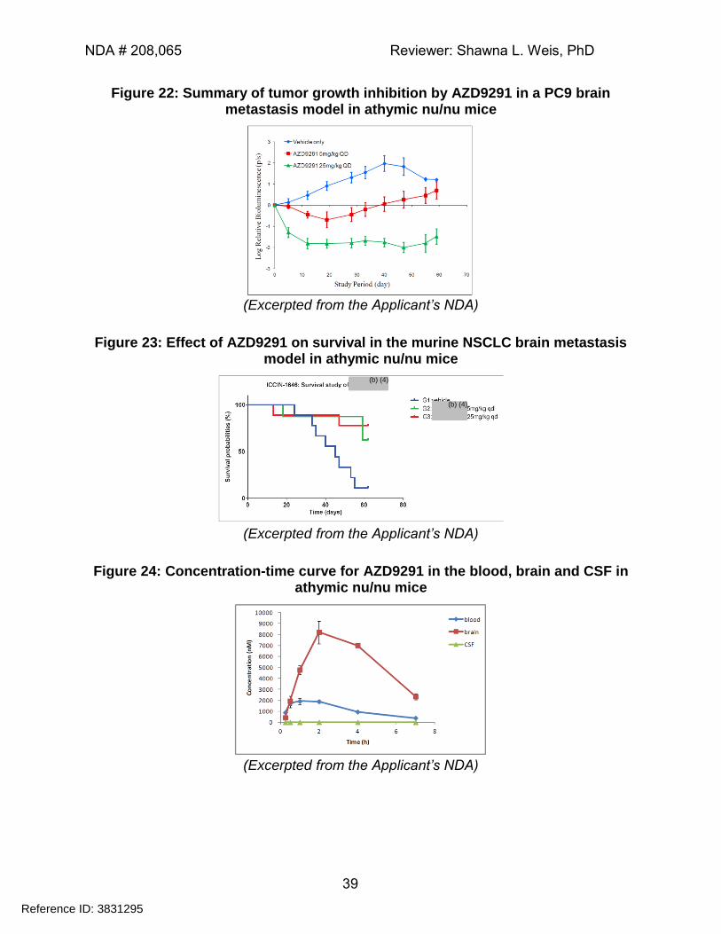
NDA # 208,065 Reviewer: Shawna L. Weis, PhD
39
Figure 22: Summary of tumor growth inhibition by AZD9291 in a PC9 brain metastasis model in athymic nu/nu mice
(Excerpted from the Applicant’s NDA)
Figure 23: Effect of AZD9291 on survival in the murine NSCLC brain metastasis model in athymic nu/nu mice
(Excerpted from the Applicant’s NDA)
Figure 24: Concentration-time curve for AZD9291 in the blood, brain and CSF in athymic nu/nu mice
(Excerpted from the Applicant’s NDA)
Reference ID: 3831295
(b) (4)
(b) (4)
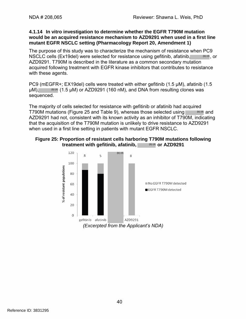
NDA # 208,065 Reviewer: Shawna L. Weis, PhD
40
4.1.14 In vitro investigation to determine whether the EGFR T790M mutation would be an acquired resistance mechanism to AZD9291 when used in a first line mutant EGFR NSCLC setting (Pharmacology Report 20, Amendment 1)
The purpose of this study was to characterize the mechanism of resistance when PC9 NSCLC cells (Ex19del) were selected for resistance using gefitinib, afatinib, , or AZD9291. T790M is described in the literature as a common secondary mutation acquired following treatment with EGFR kinase inhibitors that contributes to resistance with these agents. PC9 (mEGFR+; EX19del) cells were treated with either gefitinib (1.5 µM), afatinib (1.5 µM), (1.5 µM) or AZD9291 (160 nM), and DNA from resulting clones was sequenced. The majority of cells selected for resistance with gefitinib or afatinib had acquired T790M mutations (Figure 25 and Table 9), whereas those selected using and AZD9291 had not, consistent with its known activity as an inhibitor of T790M, indicating that the acquisition of the T790M mutation is unlikely to drive resistance to AZD9291 when used in a first line setting in patients with mutant EGFR NSCLC.
Figure 25: Proportion of resistant cells harboring T790M mutations following treatment with gefitinib, afatinib, or AZD9291
(Excerpted from the Applicant’s NDA)
Reference ID: 3831295
(b) (4)
(b) (4)
(b) (4)
(b) (4)
(b) (4)
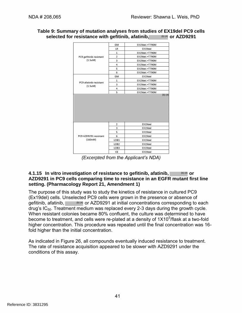
NDA # 208,065 Reviewer: Shawna L. Weis, PhD
41
Table 9: Summary of mutation analyses from studies of EX19del PC9 cells selected for resistance with gefitinib, afatinib, or AZD9291
(Excerpted from the Applicant’s NDA)
4.1.15 In vitro investigation of resistance to gefitinib, afatinib, or AZD9291 in PC9 cells comparing time to resistance in an EGFR mutant first line setting. (Pharmacology Report 21, Amendment 1)
The purpose of this study was to study the kinetics of resistance in cultured PC9 (Ex19del) cells. Unselected PC9 cells were grown in the presence or absence of gefitinib, afatinib, or AZD9291 at initial concentrations corresponding to each drug’s IC50. Treatment medium was replaced every 2-3 days during the growth cycle. When resistant colonies became 80% confluent, the culture was determined to have become to treatment, and cells were re-plated at a density of 1X10⁵/flask at a two-fold higher concentration. This procedure was repeated until the final concentration was 16-fold higher than the initial concentration. As indicated in Figure 26, all compounds eventually induced resistance to treatment. The rate of resistance acquisition appeared to be slower with AZD9291 under the conditions of this assay.
Reference ID: 3831295
(b) (4)
(b) (4)
(b) (4)
(b) (4)
NDA # 208,065 Reviewer: Shawna L. Weis, PhD
42
Figure 26: Kinetics of resistance acquisition in cultured PC9 cells treated with AZD9291, gefitinib, afatinib
(Excerpted from the Applicant’s NDA)
4.1.16 To test the activity of AZD9291 and its metabolite AZD5104 and competitor compounds against HER2 and HER3 in ligand driven system and in an amplified setting (Pharmacology Report 22)
The purpose of this study was to assess the ability of AZD9291 and its metabolite, AZ5104 to inhibit HRG1-stimulated HER2 and HER3 phosphorylation in stimulated MCF7 cells, and to inhibit pHER2 production in HER2 amplified BT474 cells. As illustrated in Table 10, AZD9291 and its metabolite, AZ5104 suppressed pErbB2 production in both HRG1-stimulated and HER2 amplified cells. In addition, AZD9291 and AZ5104 both suppressed pErbB3 production in HRG1-stimulated cells. The potency of AZD9291 was comparable to the other irreversible inhibitors (e.g. AZ5104, and afatinib). Gefitinib and erlotinib (both reversible inhibitors) were less potent in the context of HER2 amplification. The potency of AZD9291 in the context of HER2 amplification was comparable (within ~1.5X) of that observed in the context of HRG1-stimulation.
Table 10: Comparative inhibitory activity of different compounds for pErbB2 and pErbB3 production in HRG1-stimulated MCF-7 cells or HER2-amplified BT474
cells
Compound Cell Line pHER2 IC50 (µM) pHER3 IC50 (µM)
AZD9291 MCF7 0.095 0.128
BT474 0.1227 --
Reference ID: 3831295
(b) (4)
(b) (4)
(b) (4)
(b) (4)
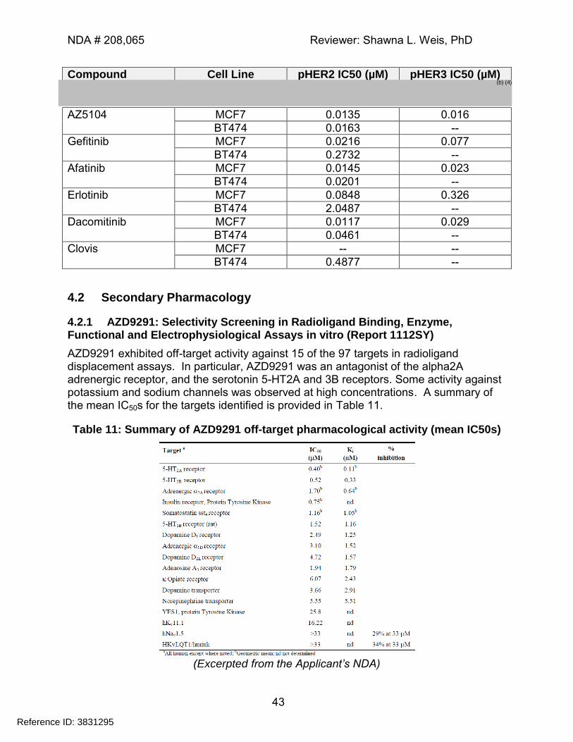
NDA # 208,065 Reviewer: Shawna L. Weis, PhD
43
Compound Cell Line pHER2 IC50 (µM) pHER3 IC50 (µM)
AZ5104 MCF7 0.0135 0.016
BT474 0.0163 --
Gefitinib MCF7 0.0216 0.077
BT474 0.2732 --
Afatinib MCF7 0.0145 0.023
BT474 0.0201 --
Erlotinib MCF7 0.0848 0.326
BT474 2.0487 --
Dacomitinib MCF7 0.0117 0.029
BT474 0.0461 --
Clovis MCF7 -- --
BT474 0.4877 --
4.2 Secondary Pharmacology
4.2.1 AZD9291: Selectivity Screening in Radioligand Binding, Enzyme, Functional and Electrophysiological Assays in vitro (Report 1112SY)
AZD9291 exhibited off-target activity against 15 of the 97 targets in radioligand displacement assays. In particular, AZD9291 was an antagonist of the alpha2A adrenergic receptor, and the serotonin 5-HT2A and 3B receptors. Some activity against potassium and sodium channels was observed at high concentrations. A summary of the mean IC50s for the targets identified is provided in Table 11.
Table 11: Summary of AZD9291 off-target pharmacological activity (mean IC50s)
(Excerpted from the Applicant’s NDA)
Reference ID: 3831295
(b) (4)
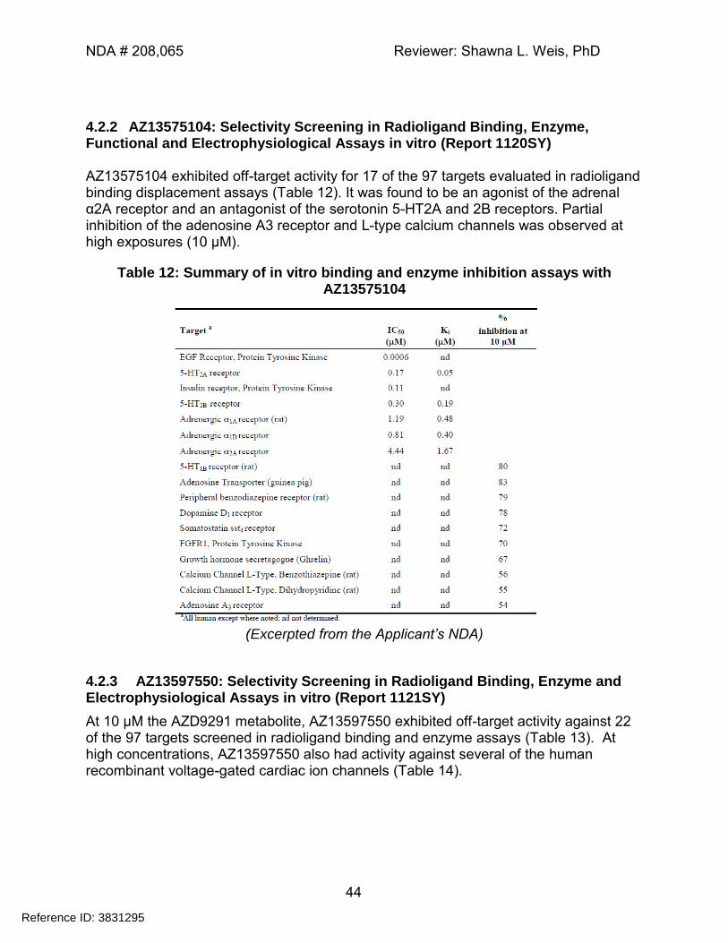
NDA # 208,065 Reviewer: Shawna L. Weis, PhD
44
4.2.2 AZ13575104: Selectivity Screening in Radioligand Binding, Enzyme, Functional and Electrophysiological Assays in vitro (Report 1120SY) AZ13575104 exhibited off-target activity for 17 of the 97 targets evaluated in radioligand binding displacement assays (Table 12). It was found to be an agonist of the adrenal α2A receptor and an antagonist of the serotonin 5-HT2A and 2B receptors. Partial inhibition of the adenosine A3 receptor and L-type calcium channels was observed at high exposures (10 µM).
Table 12: Summary of in vitro binding and enzyme inhibition assays with AZ13575104
(Excerpted from the Applicant’s NDA)
4.2.3 AZ13597550: Selectivity Screening in Radioligand Binding, Enzyme and Electrophysiological Assays in vitro (Report 1121SY)
At 10 µM the AZD9291 metabolite, AZ13597550 exhibited off-target activity against 22 of the 97 targets screened in radioligand binding and enzyme assays (Table 13). At high concentrations, AZ13597550 also had activity against several of the human recombinant voltage-gated cardiac ion channels (Table 14).
Reference ID: 3831295
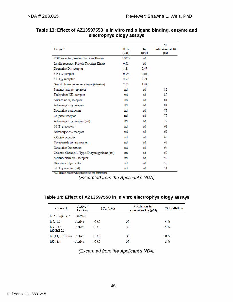
NDA # 208,065 Reviewer: Shawna L. Weis, PhD
45
Table 13: Effect of AZ13597550 in in vitro radioligand binding, enzyme and electrophysiology assays
(Excerpted from the Applicant’s NDA)
Table 14: Effect of AZ13597550 in in vitro electrophysiology assays
(Excerpted from the Applicant’s NDA)
Reference ID: 3831295
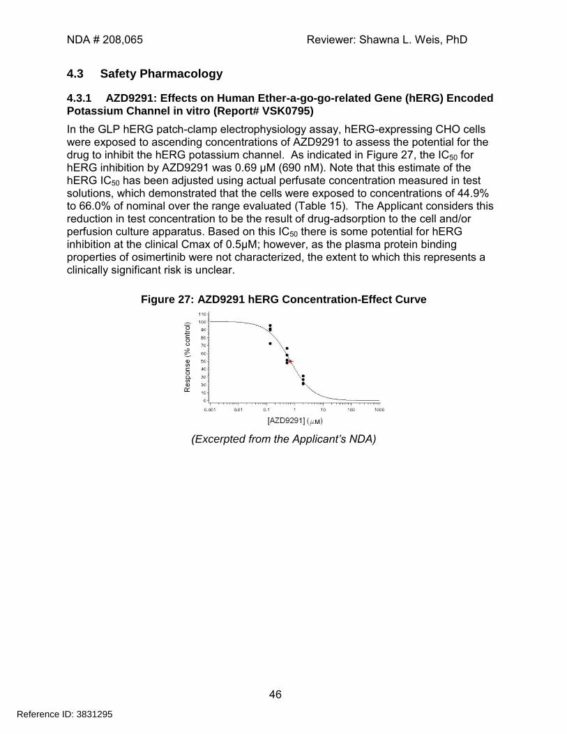
NDA # 208,065 Reviewer: Shawna L. Weis, PhD
46
4.3 Safety Pharmacology
4.3.1 AZD9291: Effects on Human Ether-a-go-go-related Gene (hERG) Encoded Potassium Channel in vitro (Report# VSK0795)
In the GLP hERG patch-clamp electrophysiology assay, hERG-expressing CHO cells were exposed to ascending concentrations of AZD9291 to assess the potential for the drug to inhibit the hERG potassium channel. As indicated in Figure 27, the IC50 for hERG inhibition by AZD9291 was 0.69 μM (690 nM). Note that this estimate of the hERG IC50 has been adjusted using actual perfusate concentration measured in test solutions, which demonstrated that the cells were exposed to concentrations of 44.9% to 66.0% of nominal over the range evaluated (Table 15). The Applicant considers this reduction in test concentration to be the result of drug-adsorption to the cell and/or perfusion culture apparatus. Based on this IC50 there is some potential for hERG inhibition at the clinical Cmax of 0.5µM; however, as the plasma protein binding properties of osimertinib were not characterized, the extent to which this represents a clinically significant risk is unclear.
Figure 27: AZD9291 hERG Concentration-Effect Curve
(Excerpted from the Applicant’s NDA)
Reference ID: 3831295
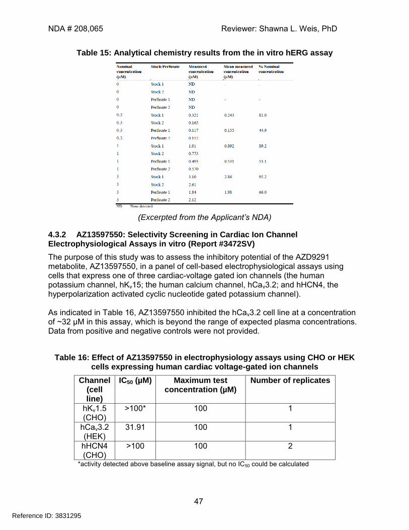
NDA # 208,065 Reviewer: Shawna L. Weis, PhD
47
Table 15: Analytical chemistry results from the in vitro hERG assay
(Excerpted from the Applicant’s NDA)
4.3.2 AZ13597550: Selectivity Screening in Cardiac Ion Channel Electrophysiological Assays in vitro (Report #3472SV)
The purpose of this study was to assess the inhibitory potential of the AZD9291 metabolite, AZ13597550, in a panel of cell-based electrophysiological assays using cells that express one of three cardiac-voltage gated ion channels (the human potassium channel, hKv15; the human calcium channel, hCav3.2; and hHCN4, the hyperpolarization activated cyclic nucleotide gated potassium channel). As indicated in Table 16, AZ13597550 inhibited the hCav3.2 cell line at a concentration of ~32 µM in this assay, which is beyond the range of expected plasma concentrations. Data from positive and negative controls were not provided.
Table 16: Effect of AZ13597550 in electrophysiology assays using CHO or HEK cells expressing human cardiac voltage-gated ion channels
Channel (cell line)
IC50 (µM) Maximum test concentration (µM)
Number of replicates
hKv1.5 (CHO)
>100* 100 1
hCav3.2 (HEK)
31.91 100 1
hHCN4 (CHO)
>100 100 2
*activity detected above baseline assay signal, but no IC50 could be calculated
Reference ID: 3831295
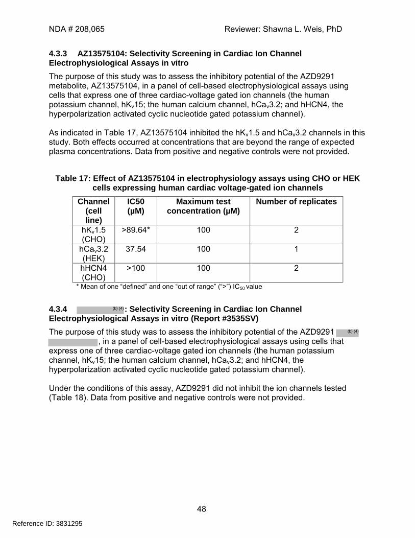
NDA # 208,065 Reviewer: Shawna L. Weis, PhD
48
4.3.3 AZ13575104: Selectivity Screening in Cardiac Ion Channel Electrophysiological Assays in vitro
The purpose of this study was to assess the inhibitory potential of the AZD9291 metabolite, AZ13575104, in a panel of cell-based electrophysiological assays using cells that express one of three cardiac-voltage gated ion channels (the human potassium channel, hKv15; the human calcium channel, hCav3.2; and hHCN4, the hyperpolarization activated cyclic nucleotide gated potassium channel). As indicated in Table 17, AZ13575104 inhibited the hKv1.5 and hCav3.2 channels in this study. Both effects occurred at concentrations that are beyond the range of expected plasma concentrations. Data from positive and negative controls were not provided.
Table 17: Effect of AZ13575104 in electrophysiology assays using CHO or HEK cells expressing human cardiac voltage-gated ion channels
Channel (cell line)
IC50 (µM)
Maximum test concentration (µM)
Number of replicates
hKv1.5 (CHO)
>89.64* 100 2
hCav3.2 (HEK)
37.54 100 1
hHCN4 (CHO)
>100 100 2
* Mean of one “defined” and one “out of range” (“>”) IC50 value
4.3.4 : Selectivity Screening in Cardiac Ion Channel Electrophysiological Assays in vitro (Report #3535SV)
The purpose of this study was to assess the inhibitory potential of the AZD9291 , in a panel of cell-based electrophysiological assays using cells that
express one of three cardiac-voltage gated ion channels (the human potassium channel, hKv15; the human calcium channel, hCav3.2; and hHCN4, the hyperpolarization activated cyclic nucleotide gated potassium channel). Under the conditions of this assay, AZD9291 did not inhibit the ion channels tested (Table 18). Data from positive and negative controls were not provided.
Reference ID: 3831295
(b) (4)
(b) (4)
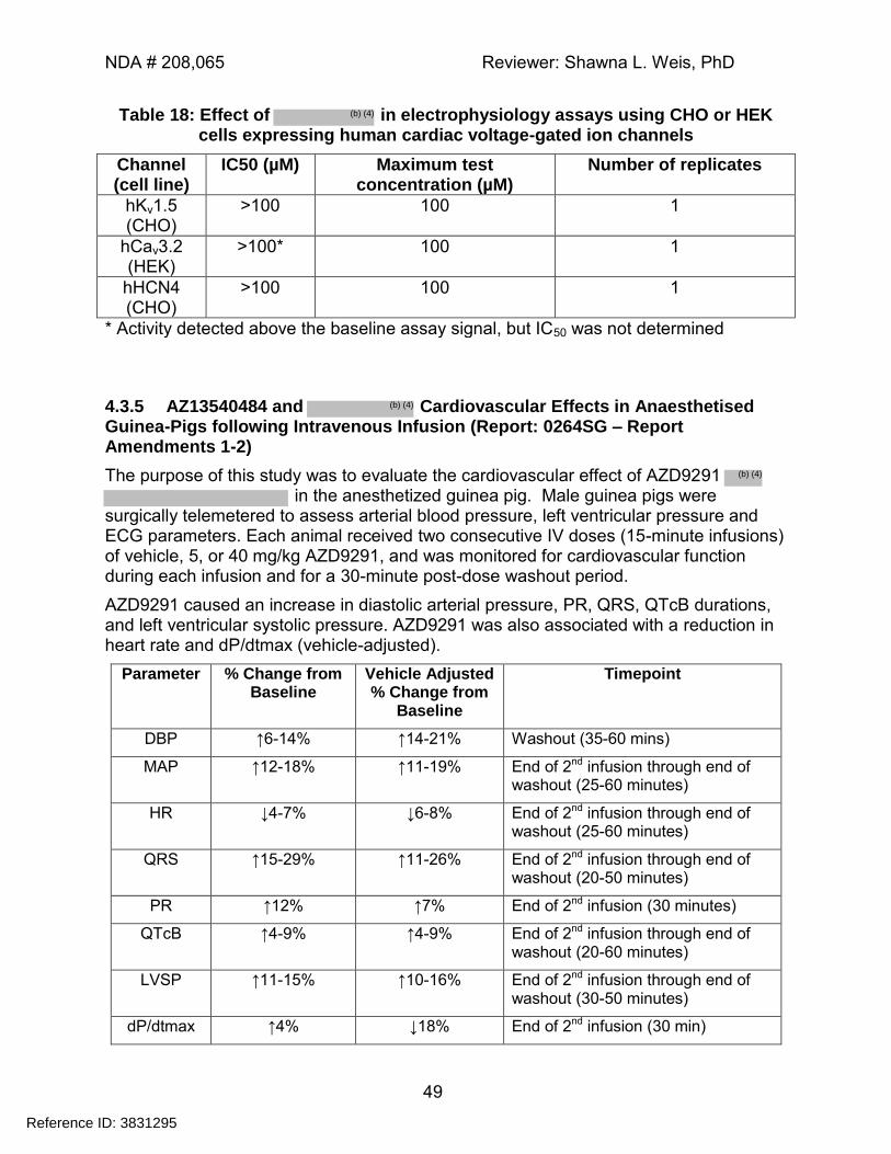
NDA # 208,065 Reviewer: Shawna L. Weis, PhD
49
Table 18: Effect of in electrophysiology assays using CHO or HEK cells expressing human cardiac voltage-gated ion channels
Channel (cell line)
IC50 (µM) Maximum test concentration (µM)
Number of replicates
hKv1.5 (CHO)
>100 100 1
hCav3.2 (HEK)
>100* 100 1
hHCN4 (CHO)
>100 100 1
* Activity detected above the baseline assay signal, but IC50 was not determined
4.3.5 AZ13540484 and Cardiovascular Effects in Anaesthetised Guinea-Pigs following Intravenous Infusion (Report: 0264SG – Report Amendments 1-2)
The purpose of this study was to evaluate the cardiovascular effect of AZD9291 in the anesthetized guinea pig. Male guinea pigs were
surgically telemetered to assess arterial blood pressure, left ventricular pressure and ECG parameters. Each animal received two consecutive IV doses (15-minute infusions) of vehicle, 5, or 40 mg/kg AZD9291, and was monitored for cardiovascular function during each infusion and for a 30-minute post-dose washout period.
AZD9291 caused an increase in diastolic arterial pressure, PR, QRS, QTcB durations, and left ventricular systolic pressure. AZD9291 was also associated with a reduction in heart rate and dP/dtmax (vehicle-adjusted).
Parameter % Change from Baseline
Vehicle Adjusted % Change from
Baseline
Timepoint
DBP ↑6-14% ↑14-21% Washout (35-60 mins)
MAP ↑12-18% ↑11-19% End of 2nd infusion through end of washout (25-60 minutes)
HR ↓4-7% ↓6-8% End of 2nd infusion through end of washout (25-60 minutes)
QRS ↑15-29% ↑11-26% End of 2nd infusion through end of washout (20-50 minutes)
PR ↑12% ↑7% End of 2nd infusion (30 minutes)
QTcB ↑4-9% ↑4-9% End of 2nd infusion through end of washout (20-60 minutes)
LVSP ↑11-15% ↑10-16% End of 2nd infusion through end of washout (30-50 minutes)
dP/dtmax ↑4% ↓18% End of 2nd infusion (30 min)
Reference ID: 3831295
(b) (4)
(b) (4)
(b) (4)
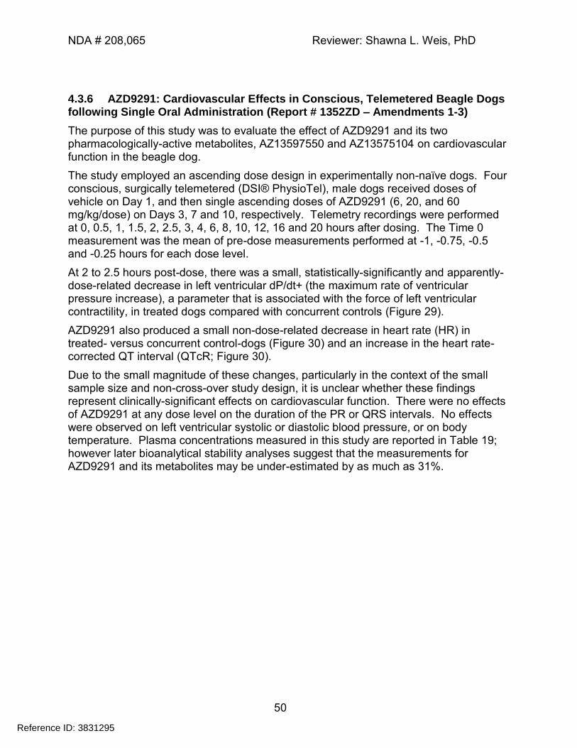
NDA # 208,065 Reviewer: Shawna L. Weis, PhD
50
4.3.6 AZD9291: Cardiovascular Effects in Conscious, Telemetered Beagle Dogs following Single Oral Administration (Report # 1352ZD – Amendments 1-3)
The purpose of this study was to evaluate the effect of AZD9291 and its two pharmacologically-active metabolites, AZ13597550 and AZ13575104 on cardiovascular function in the beagle dog.
The study employed an ascending dose design in experimentally non-naïve dogs. Four conscious, surgically telemetered (DSI® PhysioTel), male dogs received doses of vehicle on Day 1, and then single ascending doses of AZD9291 (6, 20, and 60 mg/kg/dose) on Days 3, 7 and 10, respectively. Telemetry recordings were performed at 0, 0.5, 1, 1.5, 2, 2.5, 3, 4, 6, 8, 10, 12, 16 and 20 hours after dosing. The Time 0 measurement was the mean of pre-dose measurements performed at -1, -0.75, -0.5 and -0.25 hours for each dose level.
At 2 to 2.5 hours post-dose, there was a small, statistically-significantly and apparently-dose-related decrease in left ventricular dP/dt+ (the maximum rate of ventricular pressure increase), a parameter that is associated with the force of left ventricular contractility, in treated dogs compared with concurrent controls (Figure 29).
AZD9291 also produced a small non-dose-related decrease in heart rate (HR) in treated- versus concurrent control-dogs (Figure 30) and an increase in the heart rate-corrected QT interval (QTcR; Figure 30).
Due to the small magnitude of these changes, particularly in the context of the small sample size and non-cross-over study design, it is unclear whether these findings represent clinically-significant effects on cardiovascular function. There were no effects of AZD9291 at any dose level on the duration of the PR or QRS intervals. No effects were observed on left ventricular systolic or diastolic blood pressure, or on body temperature. Plasma concentrations measured in this study are reported in Table 19; however later bioanalytical stability analyses suggest that the measurements for AZD9291 and its metabolites may be under-estimated by as much as 31%.
Reference ID: 3831295
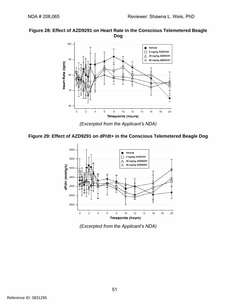
NDA # 208,065 Reviewer: Shawna L. Weis, PhD
51
Figure 28: Effect of AZD9291 on Heart Rate in the Conscious Telemetered Beagle Dog
(Excerpted from the Applicant’s NDA)
Figure 29: Effect of AZD9291 on dP/dt+ in the Conscious Telemetered Beagle Dog
(Excerpted from the Applicant’s NDA)
Reference ID: 3831295
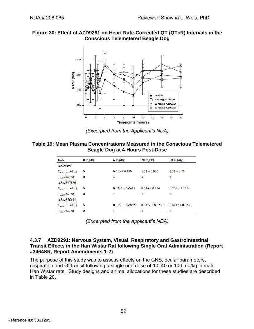
NDA # 208,065 Reviewer: Shawna L. Weis, PhD
52
Figure 30: Effect of AZD9291 on Heart Rate-Corrected QT (QTcR) Intervals in the Conscious Telemetered Beagle Dog
(Excerpted from the Applicant’s NDA)
Table 19: Mean Plasma Concentrations Measured in the Conscious Telemetered Beagle Dog at 4-Hours Post-Dose
(Excerpted from the Applicant’s NDA)
4.3.7 AZD9291: Nervous System, Visual, Respiratory and Gastrointestinal Transit Effects in the Han Wistar Rat following Single Oral Administration (Report #3464SR, Report Amendments 1-2)
The purpose of this study was to assess effects on the CNS, ocular parameters, respiration and GI transit following a single oral dose of 10, 40 or 100 mg/kg in male Han Wistar rats. Study designs and animal allocations for these studies are described in Table 20.
Reference ID: 3831295
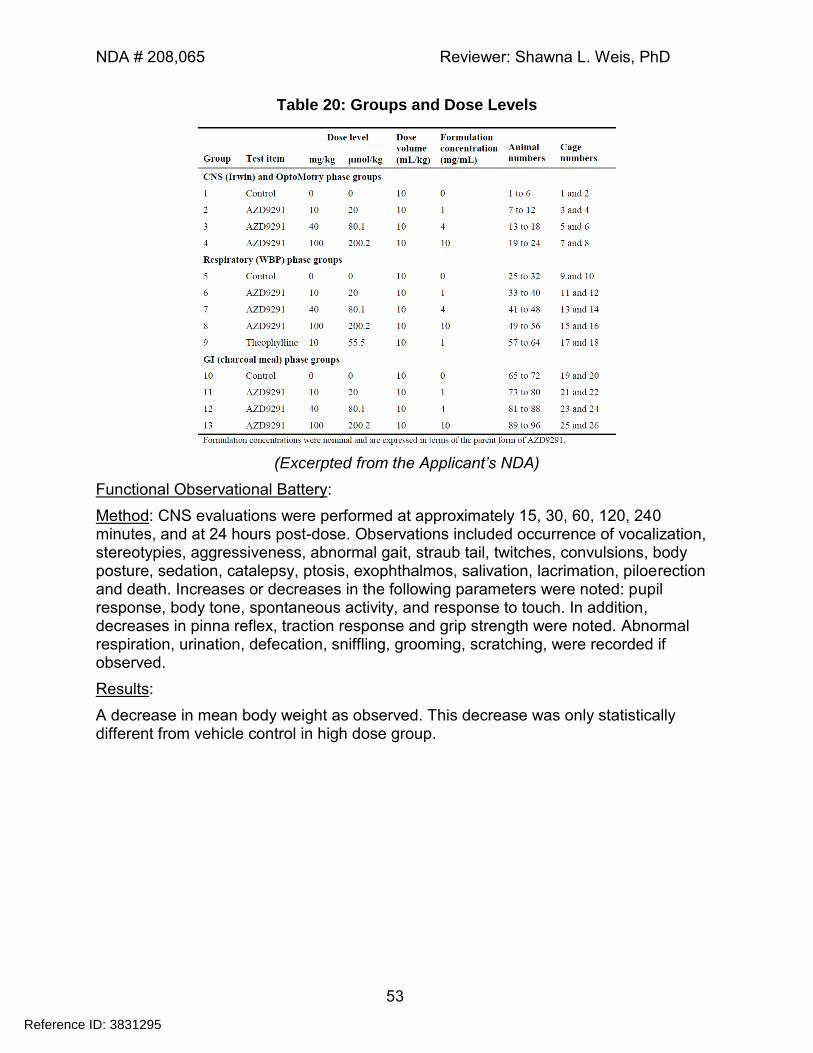
NDA # 208,065 Reviewer: Shawna L. Weis, PhD
53
Table 20: Groups and Dose Levels
(Excerpted from the Applicant’s NDA)
Functional Observational Battery:
Method: CNS evaluations were performed at approximately 15, 30, 60, 120, 240 minutes, and at 24 hours post-dose. Observations included occurrence of vocalization, stereotypies, aggressiveness, abnormal gait, straub tail, twitches, convulsions, body posture, sedation, catalepsy, ptosis, exophthalmos, salivation, lacrimation, piloerection and death. Increases or decreases in the following parameters were noted: pupil response, body tone, spontaneous activity, and response to touch. In addition, decreases in pinna reflex, traction response and grip strength were noted. Abnormal respiration, urination, defecation, sniffling, grooming, scratching, were recorded if observed.
Results:
A decrease in mean body weight as observed. This decrease was only statistically different from vehicle control in high dose group.
Reference ID: 3831295
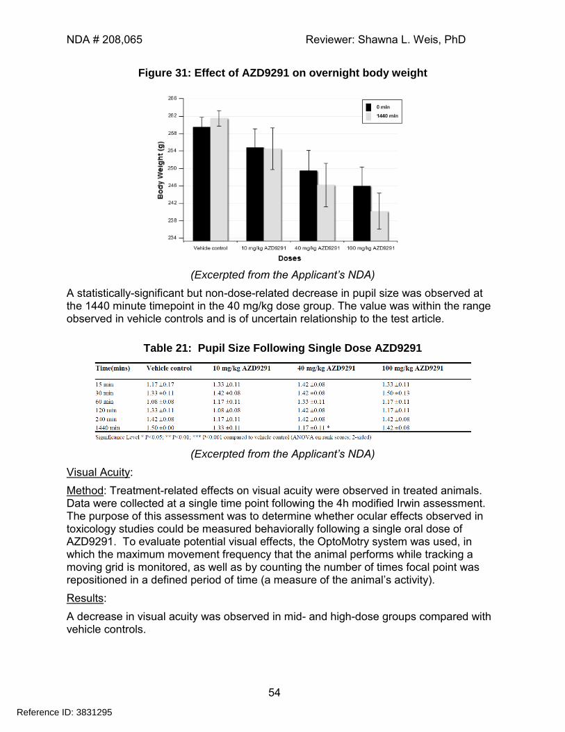
NDA # 208,065 Reviewer: Shawna L. Weis, PhD
54
Figure 31: Effect of AZD9291 on overnight body weight
(Excerpted from the Applicant’s NDA)
A statistically-significant but non-dose-related decrease in pupil size was observed at the 1440 minute timepoint in the 40 mg/kg dose group. The value was within the range observed in vehicle controls and is of uncertain relationship to the test article.
Table 21: Pupil Size Following Single Dose AZD9291
(Excerpted from the Applicant’s NDA)
Visual Acuity:
Method: Treatment-related effects on visual acuity were observed in treated animals. Data were collected at a single time point following the 4h modified Irwin assessment. The purpose of this assessment was to determine whether ocular effects observed in toxicology studies could be measured behaviorally following a single oral dose of AZD9291. To evaluate potential visual effects, the OptoMotry system was used, in which the maximum movement frequency that the animal performs while tracking a moving grid is monitored, as well as by counting the number of times focal point was repositioned in a defined period of time (a measure of the animal’s activity).
Results:
A decrease in visual acuity was observed in mid- and high-dose groups compared with vehicle controls.
Reference ID: 3831295
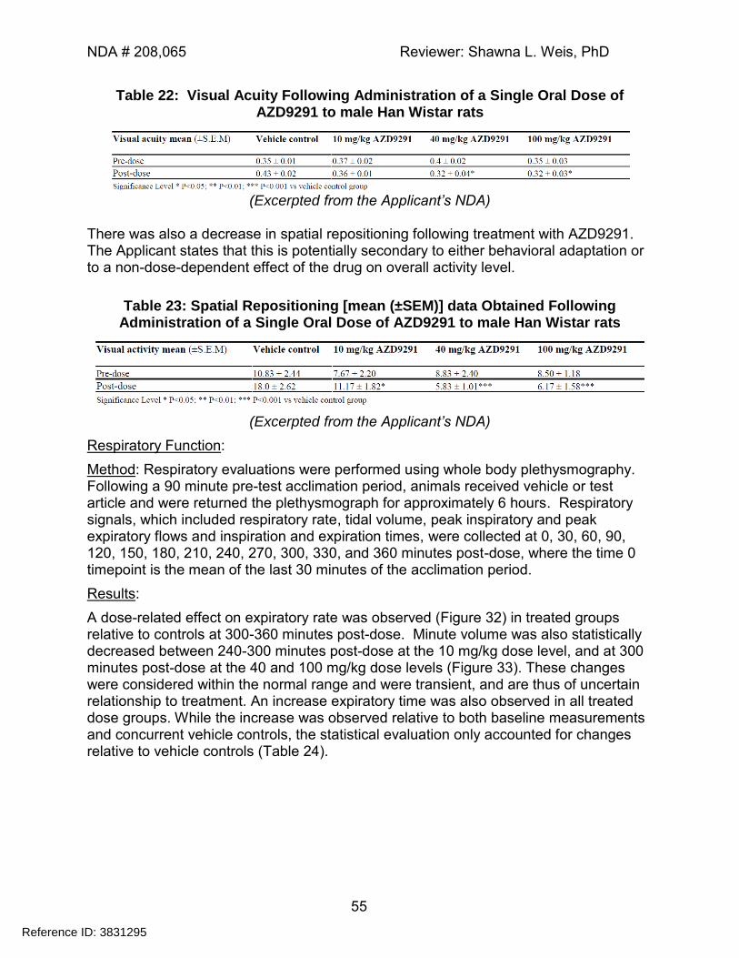
NDA # 208,065 Reviewer: Shawna L. Weis, PhD
55
Table 22: Visual Acuity Following Administration of a Single Oral Dose of AZD9291 to male Han Wistar rats
(Excerpted from the Applicant’s NDA)
There was also a decrease in spatial repositioning following treatment with AZD9291. The Applicant states that this is potentially secondary to either behavioral adaptation or to a non-dose-dependent effect of the drug on overall activity level.
Table 23: Spatial Repositioning [mean (±SEM)] data Obtained Following Administration of a Single Oral Dose of AZD9291 to male Han Wistar rats
(Excerpted from the Applicant’s NDA)
Respiratory Function:
Method: Respiratory evaluations were performed using whole body plethysmography. Following a 90 minute pre-test acclimation period, animals received vehicle or test article and were returned the plethysmograph for approximately 6 hours. Respiratory signals, which included respiratory rate, tidal volume, peak inspiratory and peak expiratory flows and inspiration and expiration times, were collected at 0, 30, 60, 90, 120, 150, 180, 210, 240, 270, 300, 330, and 360 minutes post-dose, where the time 0 timepoint is the mean of the last 30 minutes of the acclimation period.
Results:
A dose-related effect on expiratory rate was observed (Figure 32) in treated groups relative to controls at 300-360 minutes post-dose. Minute volume was also statistically decreased between 240-300 minutes post-dose at the 10 mg/kg dose level, and at 300 minutes post-dose at the 40 and 100 mg/kg dose levels (Figure 33). These changes were considered within the normal range and were transient, and are thus of uncertain relationship to treatment. An increase expiratory time was also observed in all treated dose groups. While the increase was observed relative to both baseline measurements and concurrent vehicle controls, the statistical evaluation only accounted for changes relative to vehicle controls (Table 24).
Reference ID: 3831295
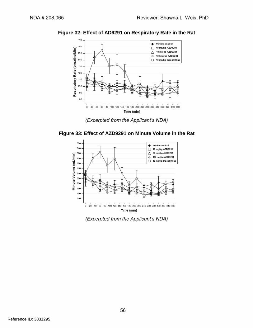
NDA # 208,065 Reviewer: Shawna L. Weis, PhD
56
Figure 32: Effect of AD9291 on Respiratory Rate in the Rat
(Excerpted from the Applicant’s NDA)
Figure 33: Effect of AZD9291 on Minute Volume in the Rat
(Excerpted from the Applicant’s NDA)
Reference ID: 3831295
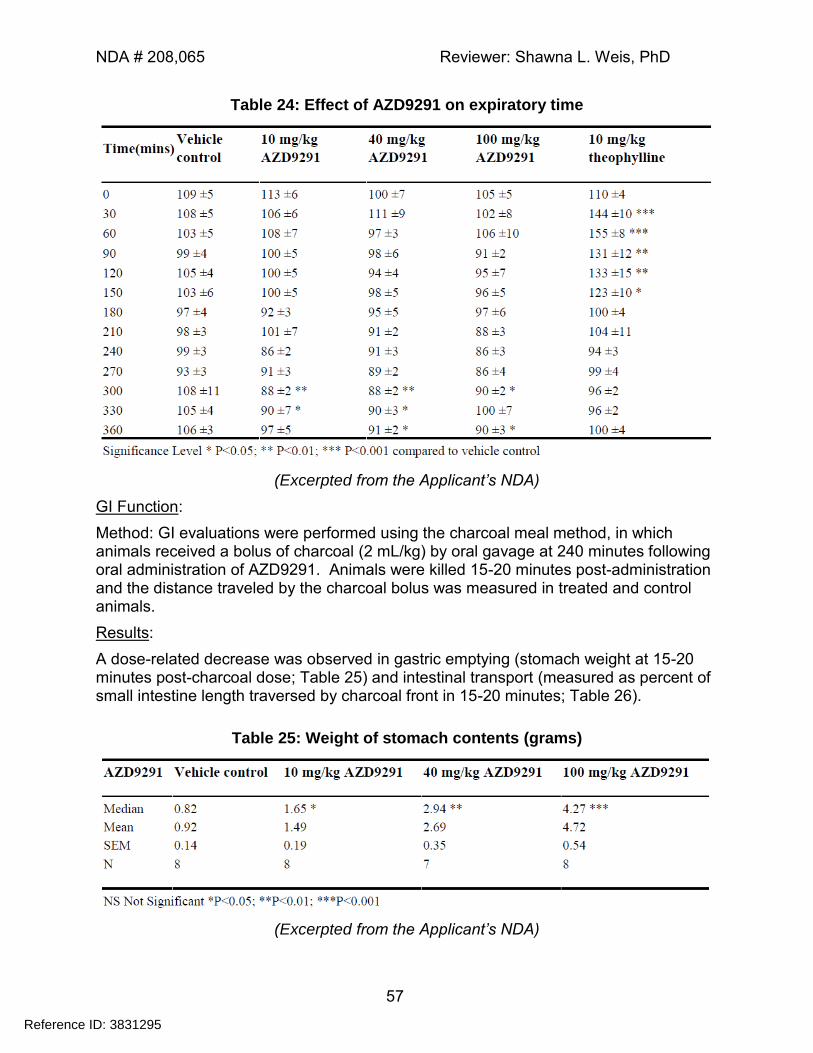
NDA # 208,065 Reviewer: Shawna L. Weis, PhD
57
Table 24: Effect of AZD9291 on expiratory time
(Excerpted from the Applicant’s NDA)
GI Function:
Method: GI evaluations were performed using the charcoal meal method, in which animals received a bolus of charcoal (2 mL/kg) by oral gavage at 240 minutes following oral administration of AZD9291. Animals were killed 15-20 minutes post-administration and the distance traveled by the charcoal bolus was measured in treated and control animals.
Results:
A dose-related decrease was observed in gastric emptying (stomach weight at 15-20 minutes post-charcoal dose; Table 25) and intestinal transport (measured as percent of small intestine length traversed by charcoal front in 15-20 minutes; Table 26).
Table 25: Weight of stomach contents (grams)
(Excerpted from the Applicant’s NDA)
Reference ID: 3831295
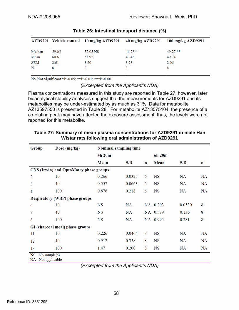
NDA # 208,065 Reviewer: Shawna L. Weis, PhD
58
Table 26: Intestinal transport distance (%)
(Excerpted from the Applicant’s NDA)
Plasma concentrations measured in this study are reported in Table 27; however, later bioanalytical stability analyses suggest that the measurements for AZD9291 and its metabolites may be under-estimated by as much as 31%. Data for metabolite AZ13597550 is presented in Table 28. For metabolite AZ13575104, the presence of a co-eluting peak may have affected the exposure assessment; thus, the levels were not reported for this metabolite.
Table 27: Summary of mean plasma concentrations for AZD9291 in male Han Wistar rats following oral administration of AZD9291
(Excerpted from the Applicant’s NDA)
Reference ID: 3831295
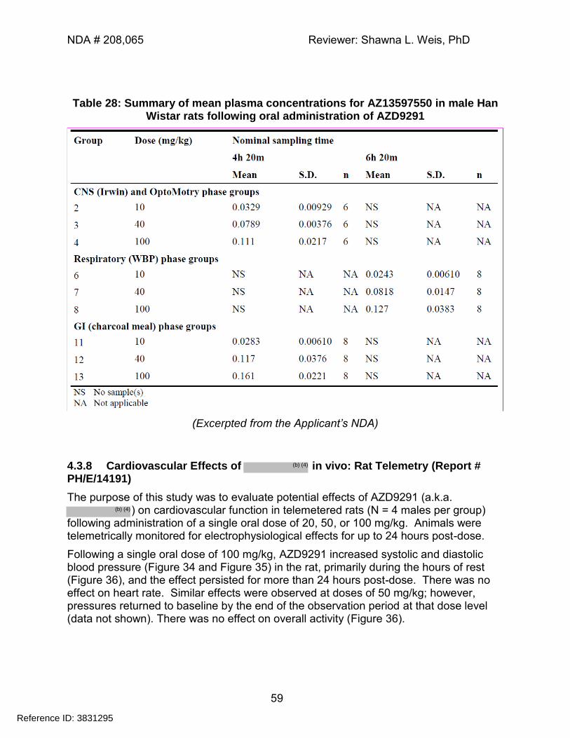
NDA # 208,065 Reviewer: Shawna L. Weis, PhD
59
Table 28: Summary of mean plasma concentrations for AZ13597550 in male Han Wistar rats following oral administration of AZD9291
(Excerpted from the Applicant’s NDA)
4.3.8 Cardiovascular Effects of in vivo: Rat Telemetry (Report # PH/E/14191)
The purpose of this study was to evaluate potential effects of AZD9291 (a.k.a. ) on cardiovascular function in telemetered rats (N = 4 males per group)
following administration of a single oral dose of 20, 50, or 100 mg/kg. Animals were telemetrically monitored for electrophysiological effects for up to 24 hours post-dose.
Following a single oral dose of 100 mg/kg, AZD9291 increased systolic and diastolic blood pressure (Figure 34 and Figure 35) in the rat, primarily during the hours of rest (Figure 36), and the effect persisted for more than 24 hours post-dose. There was no effect on heart rate. Similar effects were observed at doses of 50 mg/kg; however, pressures returned to baseline by the end of the observation period at that dose level (data not shown). There was no effect on overall activity (Figure 36).
Reference ID: 3831295
(b) (4)
(b) (4)
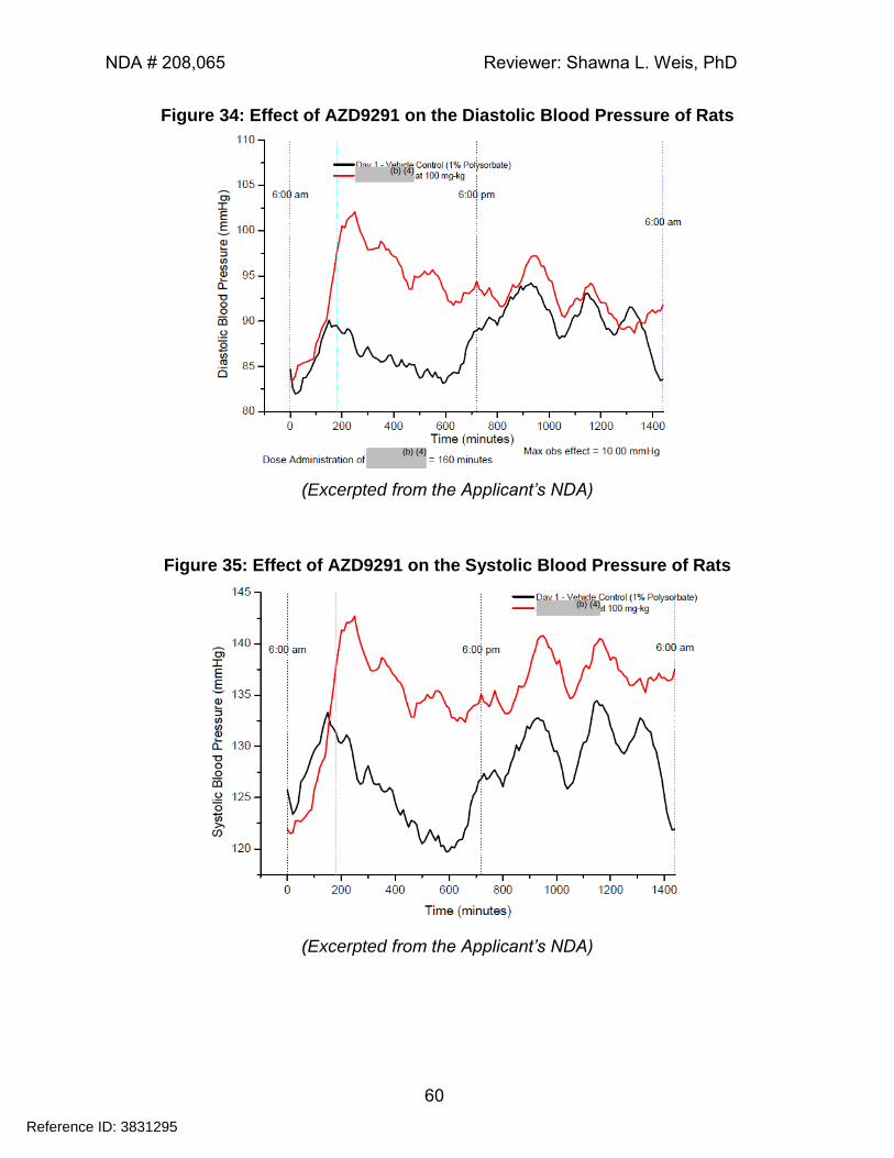
NDA # 208,065 Reviewer: Shawna L. Weis, PhD
60
Figure 34: Effect of AZD9291 on the Diastolic Blood Pressure of Rats
(Excerpted from the Applicant’s NDA)
Figure 35: Effect of AZD9291 on the Systolic Blood Pressure of Rats
(Excerpted from the Applicant’s NDA)
Reference ID: 3831295
(b) (4)
(b) (4)
(b) (4)
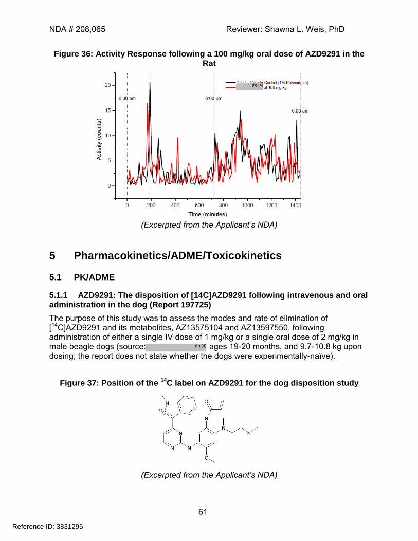
NDA # 208,065 Reviewer: Shawna L. Weis, PhD
61
Figure 36: Activity Response following a 100 mg/kg oral dose of AZD9291 in the Rat
(Excerpted from the Applicant’s NDA)
5 Pharmacokinetics/ADME/Toxicokinetics
5.1 PK/ADME
5.1.1 AZD9291: The disposition of [14C]AZD9291 following intravenous and oral administration in the dog (Report 197725)
The purpose of this study was to assess the modes and rate of elimination of [14C]AZD9291 and its metabolites, AZ13575104 and AZ13597550, following administration of either a single IV dose of 1 mg/kg or a single oral dose of 2 mg/kg in male beagle dogs (source: ages 19-20 months, and 9.7-10.8 kg upon dosing; the report does not state whether the dogs were experimentally-naïve).
Figure 37: Position of the 14C label on AZD9291 for the dog disposition study
(Excerpted from the Applicant’s NDA)
Reference ID: 3831295
(b) (4)
(b) (4)
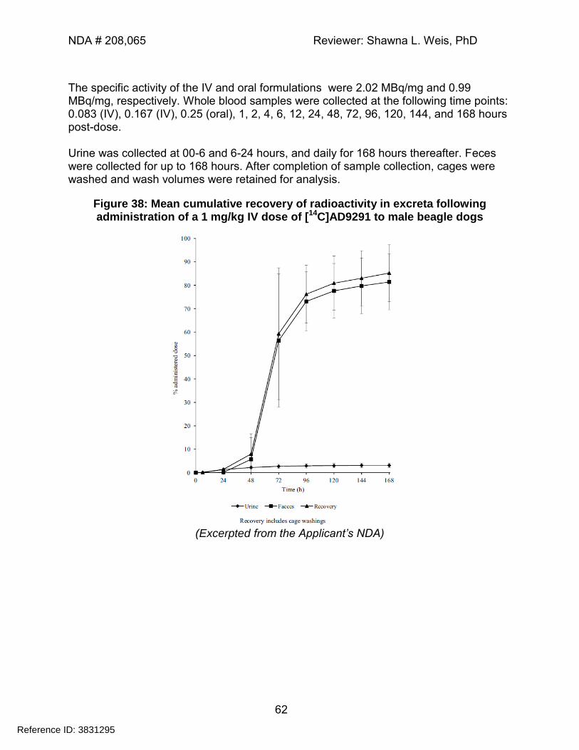
NDA # 208,065 Reviewer: Shawna L. Weis, PhD
62
The specific activity of the IV and oral formulations were 2.02 MBq/mg and 0.99 MBq/mg, respectively. Whole blood samples were collected at the following time points: 0.083 (IV), 0.167 (IV), 0.25 (oral), 1, 2, 4, 6, 12, 24, 48, 72, 96, 120, 144, and 168 hours post-dose. Urine was collected at 00-6 and 6-24 hours, and daily for 168 hours thereafter. Feces were collected for up to 168 hours. After completion of sample collection, cages were washed and wash volumes were retained for analysis.
Figure 38: Mean cumulative recovery of radioactivity in excreta following administration of a 1 mg/kg IV dose of [14C]AD9291 to male beagle dogs
(Excerpted from the Applicant’s NDA)
Reference ID: 3831295
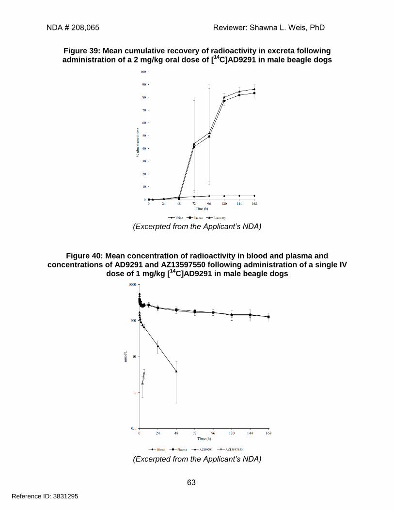
NDA # 208,065 Reviewer: Shawna L. Weis, PhD
63
Figure 39: Mean cumulative recovery of radioactivity in excreta following administration of a 2 mg/kg oral dose of [14C]AD9291 in male beagle dogs
(Excerpted from the Applicant’s NDA)
Figure 40: Mean concentration of radioactivity in blood and plasma and concentrations of AD9291 and AZ13597550 following administration of a single IV
dose of 1 mg/kg [14C]AD9291 in male beagle dogs
(Excerpted from the Applicant’s NDA)
Reference ID: 3831295
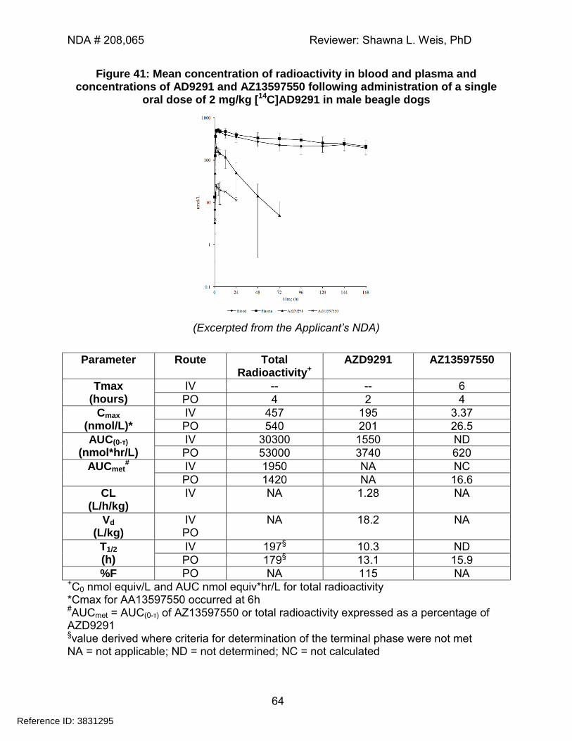
NDA # 208,065 Reviewer: Shawna L. Weis, PhD
64
Figure 41: Mean concentration of radioactivity in blood and plasma and concentrations of AD9291 and AZ13597550 following administration of a single
oral dose of 2 mg/kg [14C]AD9291 in male beagle dogs
(Excerpted from the Applicant’s NDA)
Parameter Route Total Radioactivity+
AZD9291 AZ13597550
Tmax (hours)
IV -- -- 6
PO 4 2 4
Cmax (nmol/L)*
IV 457 195 3.37
PO 540 201 26.5
AUC(0-τ)
(nmol*hr/L) IV 30300 1550 ND
PO 53000 3740 620
AUCmet#
IV 1950 NA NC
PO 1420 NA 16.6
CL (L/h/kg)
IV NA 1.28 NA
Vd (L/kg)
IV PO
NA 18.2 NA
T1/2 (h)
IV 197§ 10.3 ND
PO 179§ 13.1 15.9
%F PO NA 115 NA +C0 nmol equiv/L and AUC nmol equiv*hr/L for total radioactivity *Cmax for AA13597550 occurred at 6h #AUCmet = AUC(0-τ) of AZ13597550 or total radioactivity expressed as a percentage of AZD9291 §value derived where criteria for determination of the terminal phase were not met NA = not applicable; ND = not determined; NC = not calculated
Reference ID: 3831295
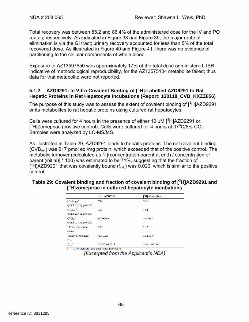
NDA # 208,065 Reviewer: Shawna L. Weis, PhD
65
Total recovery was between 85.2 and 86.4% of the administered dose for the IV and PO routes, respectively. As indicated in Figure 38 and Figure 39, the major route of elimination is via the GI tract; urinary recovery accounted for less than 5% of the total recovered dose. As illustrated in Figure 40 and Figure 41, there was no evidence of partitioning to the cellular components of whole blood. Exposure to AZ13597550 was approximately 17% of the total dose administered. ISR, indicative of methodological reproducibility, for the AZ13575104 metabolite failed; thus data for that metabolite were not reported.
5.1.2 AZD9291: In Vitro Covalent Binding of [3H]-Labelled AZD9291 to Rat Hepatic Proteins in Rat Hepatocyte Incubations (Report: 120118_CVB_KXZZ856)
The purpose of this study was to assess the extent of covalent binding of [3H]AZD9291 or its metabolites to rat hepatic proteins using cultured rat hepatocytes. Cells were cultured for 4 hours in the presence of either 10 µM [3H]AZD9291 or [3H]Zomepriac (positive control). Cells were cultured for 4 hours at 37oC/5% CO2.
Samples were analyzed by LC-MS/MS. As illustrated in Table 29, AZD9291 binds to hepatic proteins. The net covalent binding (CVBnet) was 217 pmol eq./mg protein, which exceeded that of the positive control. The metabolic turnover (calculated as 1-[(concentration parent at end) / concentration of parent (initial)] * 100) was estimated to be 71%, suggesting that the fraction of [3H]AZD9291 that was covalently bound (fcvb) was 0.020, which is similar to the positive control.
Table 29: Covalent binding and fraction of covalent binding of [3H]AZD9291 and [3H]zomepirac in cultured hepatocyte incubations
(Excerpted from the Applicant’s NDA)
Reference ID: 3831295
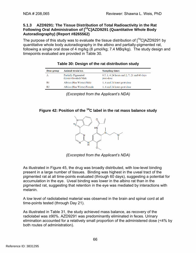
NDA # 208,065 Reviewer: Shawna L. Weis, PhD
66
5.1.3 AZD9291: The Tissue Distribution of Total Radioactivity in the Rat Following Oral Administration of [14C]AZD9291 (Quantitative Whole Body Autoradiography) (Report #8265562)
The purpose of this study was to evaluate the tissue distribution of [14C]AZD9291 by quantitative whole body autoradiography in the albino and partially-pigmented rat, following a single oral dose of 4 mg/kg (8 µmol/kg; 7.4 MBq/kg). The study design and timepoints evaluated are provided in Table 30.
Table 30: Design of the rat distribution study
(Excerpted from the Applicant’s NDA)
Figure 42: Position of the 14C label in the rat mass balance study
(Excerpted from the Applicant’s NDA)
As illustrated in Figure 45, the drug was broadly distributed, with low-level binding present in a large number of tissues. Binding was highest in the uveal tract of the pigmented rat at all time-points evaluated (through 60 days), suggesting a potential for accumulation in the eye. Uveal binding was lower in the albino rat than in the pigmented rat, suggesting that retention in the eye was mediated by interactions with melanin. A low level of radiolabeled material was observed in the brain and spinal cord at all time-points tested (through Day 21). As illustrated in Table 31, the study achieved mass balance, as recovery of the radiolabel was ≥90%. AZD9291 was predominantly eliminated in feces. Urinary elimination accounted for a relatively small proportion of the administered dose (<4% by both routes of administration).
Reference ID: 3831295
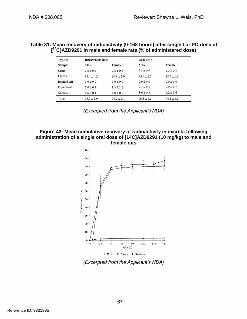
NDA # 208,065 Reviewer: Shawna L. Weis, PhD
67
Table 31: Mean recovery of radioactivity (0-168 hours) after single I or PO dose of [14C]AZD9291 in male and female rats (% of administered dose)
(Excerpted from the Applicant’s NDA)
Figure 43: Mean cumulative recovery of radioactivity in excreta following administration of a single oral dose of [14C]AZD9291 (10 mg/kg) to male and
female rats
(Excerpted from the Applicant’s NDA)
Reference ID: 3831295
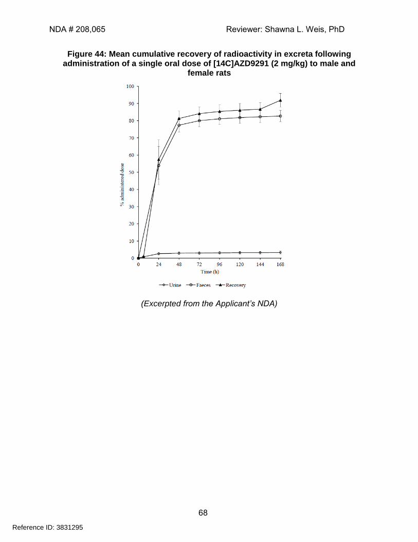
NDA # 208,065 Reviewer: Shawna L. Weis, PhD
68
Figure 44: Mean cumulative recovery of radioactivity in excreta following administration of a single oral dose of [14C]AZD9291 (2 mg/kg) to male and
female rats
(Excerpted from the Applicant’s NDA)
Reference ID: 3831295
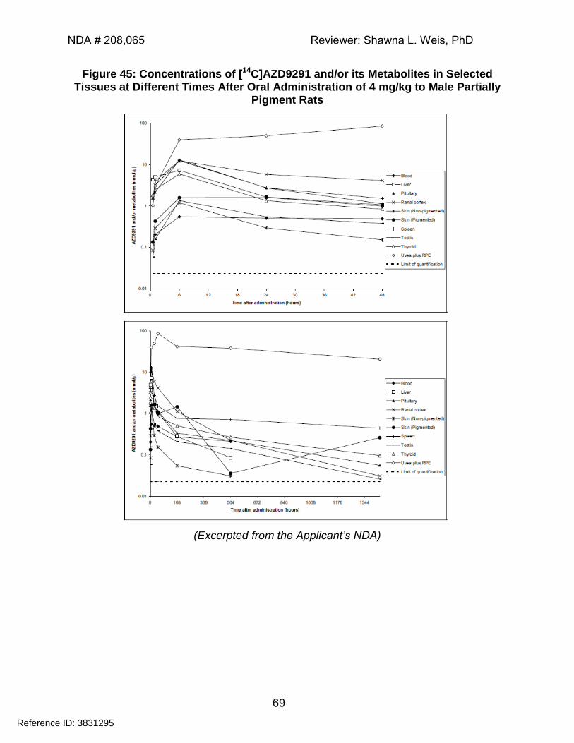
NDA # 208,065 Reviewer: Shawna L. Weis, PhD
69
Figure 45: Concentrations of [14C]AZD9291 and/or its Metabolites in Selected Tissues at Different Times After Oral Administration of 4 mg/kg to Male Partially
Pigment Rats
(Excerpted from the Applicant’s NDA)
Reference ID: 3831295
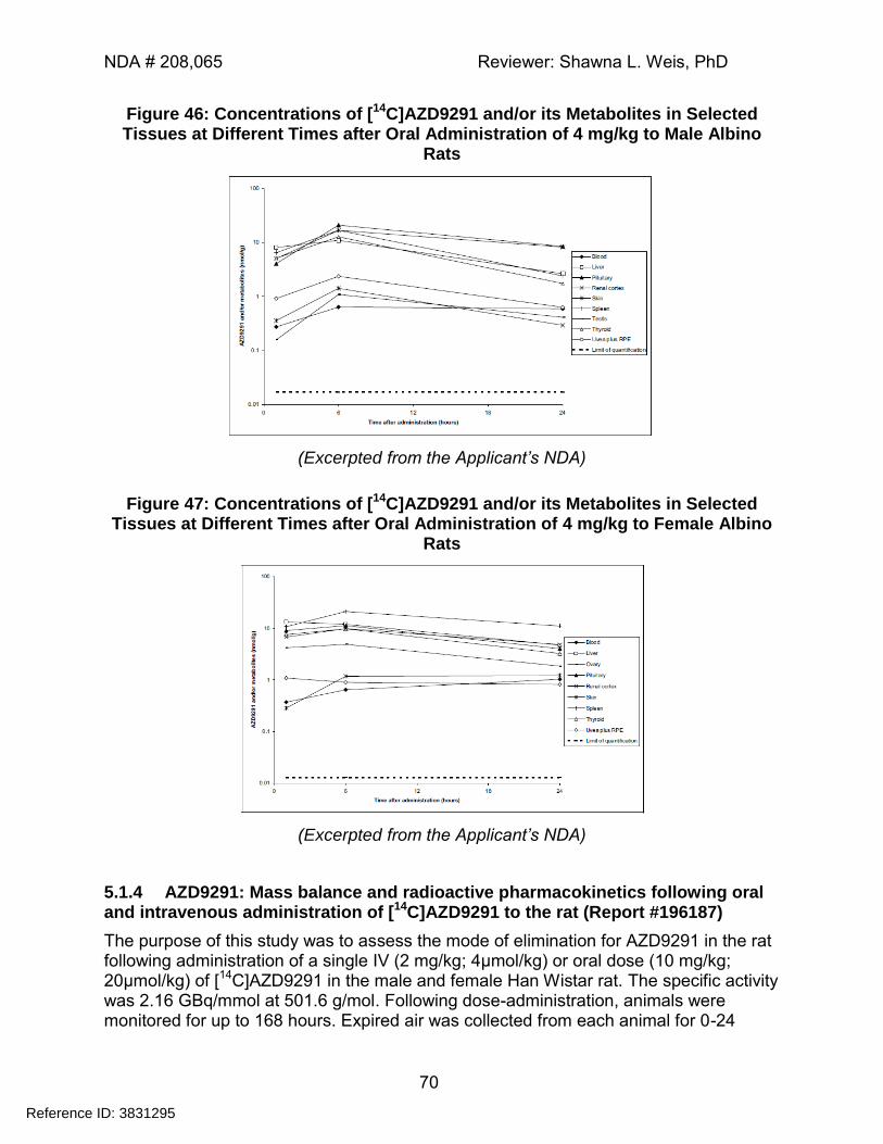
NDA # 208,065 Reviewer: Shawna L. Weis, PhD
70
Figure 46: Concentrations of [14C]AZD9291 and/or its Metabolites in Selected Tissues at Different Times after Oral Administration of 4 mg/kg to Male Albino
Rats
(Excerpted from the Applicant’s NDA)
Figure 47: Concentrations of [14C]AZD9291 and/or its Metabolites in Selected Tissues at Different Times after Oral Administration of 4 mg/kg to Female Albino
Rats
(Excerpted from the Applicant’s NDA)
5.1.4 AZD9291: Mass balance and radioactive pharmacokinetics following oral and intravenous administration of [14C]AZD9291 to the rat (Report #196187)
The purpose of this study was to assess the mode of elimination for AZD9291 in the rat following administration of a single IV (2 mg/kg; 4µmol/kg) or oral dose (10 mg/kg; 20µmol/kg) of [14C]AZD9291 in the male and female Han Wistar rat. The specific activity was 2.16 GBq/mmol at 501.6 g/mol. Following dose-administration, animals were monitored for up to 168 hours. Expired air was collected from each animal for 0-24
Reference ID: 3831295
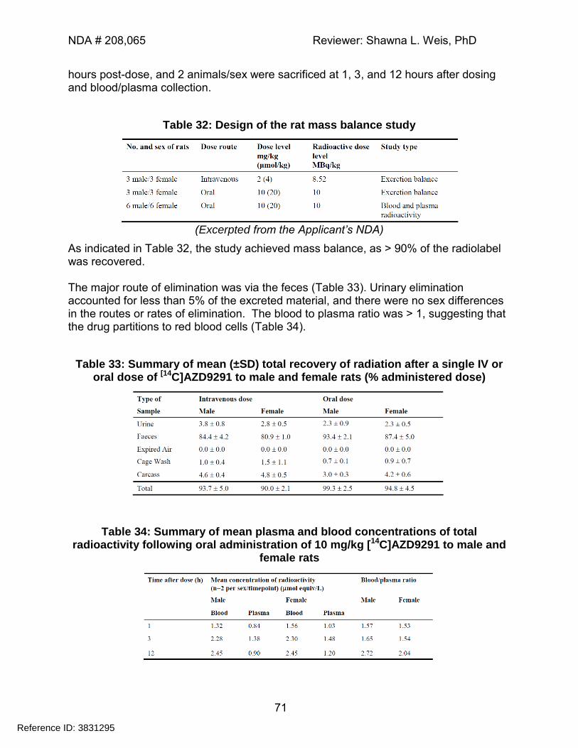
NDA # 208,065 Reviewer: Shawna L. Weis, PhD
71
hours post-dose, and 2 animals/sex were sacrificed at 1, 3, and 12 hours after dosing and blood/plasma collection.
Table 32: Design of the rat mass balance study
(Excerpted from the Applicant’s NDA)
As indicated in Table 32, the study achieved mass balance, as > 90% of the radiolabel was recovered. The major route of elimination was via the feces (Table 33). Urinary elimination accounted for less than 5% of the excreted material, and there were no sex differences in the routes or rates of elimination. The blood to plasma ratio was > 1, suggesting that the drug partitions to red blood cells (Table 34).
Table 33: Summary of mean (±SD) total recovery of radiation after a single IV or oral dose of [14C]AZD9291 to male and female rats (% administered dose)
Table 34: Summary of mean plasma and blood concentrations of total radioactivity following oral administration of 10 mg/kg [14C]AZD9291 to male and
female rats
Reference ID: 3831295
NDA # 208,065 Reviewer: Shawna L. Weis, PhD
72
6 General Toxicology
6.2 Repeat-Dose Toxicity
6.2.1 AZD9291: Three Month Oral (Gavage) Toxicity Study in the Rat - Report
Amendment
Study no.: 526248 Study report location: 4.2.3.2
Conducting laboratory and location:
Date of study initiation: 06 November 2013 (1st dose) GLP compliance: Yes
QA statement: Yes Drug, lot #, and % purity: Drug: AZD9291 mesylate salt
Lot: 02-kcwg806-87 Purity: 95.6% (HPLC % area)
Key Study Findings:
Target organs included the eyes (atrophy of the corneal epithelium and retinal dysplasia), skin (inflammatory cell infiltration), lung (inflammatory cell infiltration), kidneys (tubular basophilia and inflammatory cell infiltration) and GI tract.
Hematology changes included decreased red blood cell indices, which correlated with increased incidences of erythrophagocytosis in high dose animals; increased WBC indices; increased fibrinogen and decreased APTT.
Clinical chemistry changes included increased ALT and AST; decreased triglycerides, albumin, and total protein. Decreased calcium was also observed; however, this is likely secondary to the observed decreases in albumin.
Methods
Doses: 0, 1, 10, or 40/20 mg/kg/Day (males/females)** Frequency of dosing: Daily
Route of administration: Oral gavage Dose volume: 10 mL/kg
Formulation/Vehicle: MilliQ water pH adjusted with methane sulfonic acid to a final pH of 3.0-3.5
Species/Strain: Rattus norvegicus (Han Wistar) Number/Sex/Group: 10/sex/group
Age: Approximately 8-10 weeks at initiation of dosing Weight: Males: 253-337g (Day 1 dosing)
Females: 163-211 g (Day 1 dosing) Satellite groups: Toxicokinetics (3/sex)
Unique study design: **Dose holiday between Days 56-62 and
Reference ID: 3831295
(b) (4)
(b) (4)
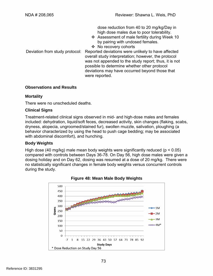
NDA # 208,065 Reviewer: Shawna L. Weis, PhD
73
dose reduction from 40 to 20 mg/kg/Day in high dose males due to poor tolerability.
Assessment of male fertility during Week 10 by pairing with undosed females.
No recovery cohorts Deviation from study protocol: Reported deviations were unlikely to have affected
overall study interpretation; however, the protocol was not appended to the study report; thus, it is not possible to determine whether other protocol deviations may have occurred beyond those that were reported.
Observations and Results
Mortality
There were no unscheduled deaths.
Clinical Signs
Treatment-related clinical signs observed in mid- and high-dose males and females included: dehydration, liquid/soft feces, decreased activity, skin changes (flaking, scabs, dryness, alopecia, ungroomed/stained fur), swollen muzzle, salivation, ploughing (a behavior characterized by using the head to push cage bedding; may be associated with abdominal discomfort), and hunching.
Body Weights
High dose (40 mg/kg) male mean body weights were significantly reduced (p < 0.05) compared with controls between Days 36-78. On Day 56, high dose males were given a dosing holiday and on Day 62, dosing was resumed at a dose of 20 mg/kg. There were no statistically significant changes in female body weights versus concurrent controls during the study.
Figure 48: Mean Male Body Weights
Reference ID: 3831295
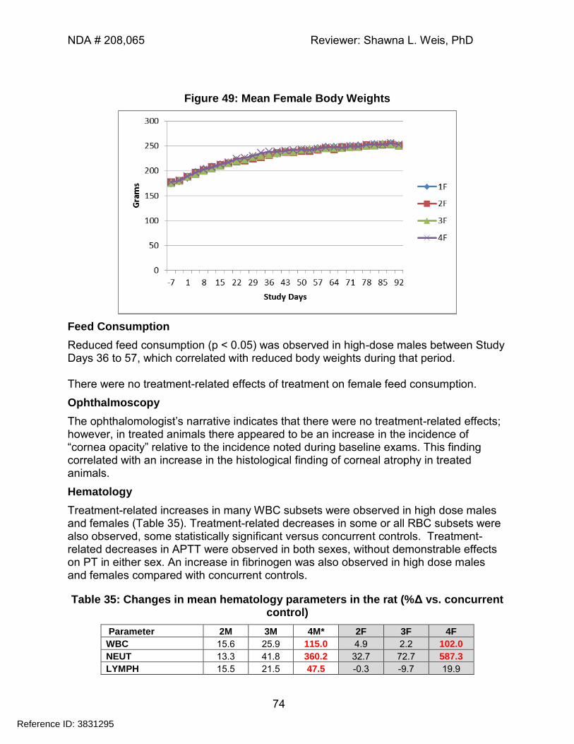
NDA # 208,065 Reviewer: Shawna L. Weis, PhD
74
Figure 49: Mean Female Body Weights
Feed Consumption
Reduced feed consumption (p < 0.05) was observed in high-dose males between Study Days 36 to 57, which correlated with reduced body weights during that period. There were no treatment-related effects of treatment on female feed consumption.
Ophthalmoscopy
The ophthalomologist’s narrative indicates that there were no treatment-related effects; however, in treated animals there appeared to be an increase in the incidence of “cornea opacity” relative to the incidence noted during baseline exams. This finding correlated with an increase in the histological finding of corneal atrophy in treated animals.
Hematology
Treatment-related increases in many WBC subsets were observed in high dose males and females (Table 35). Treatment-related decreases in some or all RBC subsets were also observed, some statistically significant versus concurrent controls. Treatment-related decreases in APTT were observed in both sexes, without demonstrable effects on PT in either sex. An increase in fibrinogen was also observed in high dose males and females compared with concurrent controls.
Table 35: Changes in mean hematology parameters in the rat (%Δ vs. concurrent control)
Parameter 2M 3M 4M* 2F 3F 4F
WBC 15.6 25.9 115.0 4.9 2.2 102.0
NEUT 13.3 41.8 360.2 32.7 72.7 587.3
LYMPH 15.5 21.5 47.5 -0.3 -9.7 19.9
Reference ID: 3831295
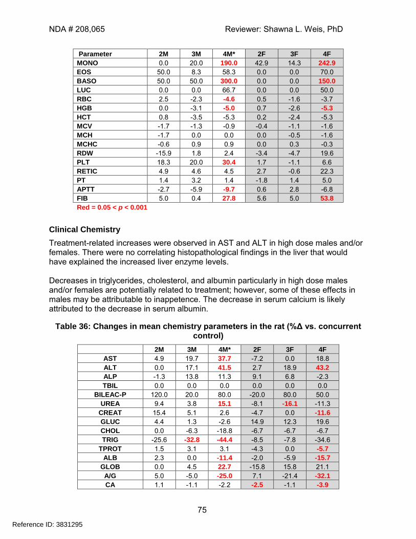
NDA # 208,065 Reviewer: Shawna L. Weis, PhD
75
Parameter 2M 3M 4M* 2F 3F 4F
MONO 0.0 20.0 190.0 42.9 14.3 242.9
EOS 50.0 8.3 58.3 0.0 0.0 70.0
BASO 50.0 50.0 300.0 0.0 0.0 150.0
LUC 0.0 0.0 66.7 0.0 0.0 50.0
RBC 2.5 -2.3 -4.6 0.5 -1.6 -3.7
HGB 0.0 -3.1 -5.0 0.7 -2.6 -5.3
HCT 0.8 -3.5 -5.3 0.2 -2.4 -5.3
MCV -1.7 -1.3 -0.9 -0.4 -1.1 -1.6
MCH -1.7 0.0 0.0 0.0 -0.5 -1.6
MCHC -0.6 0.9 0.9 0.0 0.3 -0.3
RDW -15.9 1.8 2.4 -3.4 -4.7 19.6
PLT 18.3 20.0 30.4 1.7 -1.1 6.6
RETIC 4.9 4.6 4.5 2.7 -0.6 22.3
PT 1.4 3.2 1.4 -1.8 1.4 5.0
APTT -2.7 -5.9 -9.7 0.6 2.8 -6.8
FIB 5.0 0.4 27.8 5.6 5.0 53.8
Red = 0.05 < p < 0.001
Clinical Chemistry
Treatment-related increases were observed in AST and ALT in high dose males and/or females. There were no correlating histopathological findings in the liver that would have explained the increased liver enzyme levels. Decreases in triglycerides, cholesterol, and albumin particularly in high dose males and/or females are potentially related to treatment; however, some of these effects in males may be attributable to inappetence. The decrease in serum calcium is likely attributed to the decrease in serum albumin.
Table 36: Changes in mean chemistry parameters in the rat (%Δ vs. concurrent control)
2M 3M 4M* 2F 3F 4F
AST 4.9 19.7 37.7 -7.2 0.0 18.8
ALT 0.0 17.1 41.5 2.7 18.9 43.2
ALP -1.3 13.8 11.3 9.1 6.8 -2.3
TBIL 0.0 0.0 0.0 0.0 0.0 0.0
BILEAC-P 120.0 20.0 80.0 -20.0 80.0 50.0
UREA 9.4 3.8 15.1 -8.1 -16.1 -11.3
CREAT 15.4 5.1 2.6 -4.7 0.0 -11.6
GLUC 4.4 1.3 -2.6 14.9 12.3 19.6
CHOL 0.0 -6.3 -18.8 -6.7 -6.7 -6.7
TRIG -25.6 -32.8 -44.4 -8.5 -7.8 -34.6
TPROT 1.5 3.1 3.1 -4.3 0.0 -5.7
ALB 2.3 0.0 -11.4 -2.0 -5.9 -15.7
GLOB 0.0 4.5 22.7 -15.8 15.8 21.1
A/G 5.0 -5.0 -25.0 7.1 -21.4 -32.1
CA 1.1 -1.1 -2.2 -2.5 -1.1 -3.9
Reference ID: 3831295
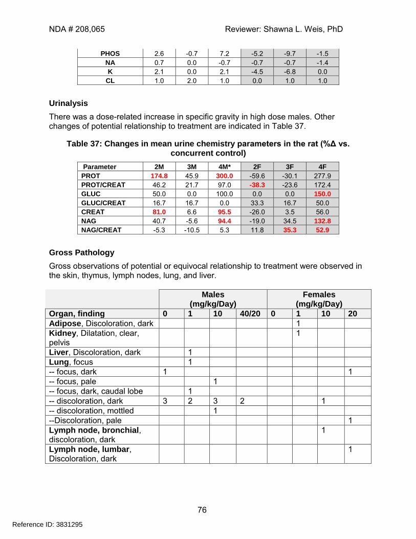
NDA # 208,065 Reviewer: Shawna L. Weis, PhD
76
PHOS 2.6 -0.7 7.2 -5.2 -9.7 -1.5
NA 0.7 0.0 -0.7 -0.7 -0.7 -1.4
K 2.1 0.0 2.1 -4.5 -6.8 0.0
CL 1.0 2.0 1.0 0.0 1.0 1.0
Urinalysis
There was a dose-related increase in specific gravity in high dose males. Other changes of potential relationship to treatment are indicated in Table 37.
Table 37: Changes in mean urine chemistry parameters in the rat (%Δ vs. concurrent control)
Parameter 2M 3M 4M* 2F 3F 4F
PROT 174.8 45.9 300.0 -59.6 -30.1 277.9
PROT/CREAT 46.2 21.7 97.0 -38.3 -23.6 172.4
GLUC 50.0 0.0 100.0 0.0 0.0 150.0
GLUC/CREAT 16.7 16.7 0.0 33.3 16.7 50.0
CREAT 81.0 6.6 95.5 -26.0 3.5 56.0
NAG 40.7 -5.6 94.4 -19.0 34.5 132.8
NAG/CREAT -5.3 -10.5 5.3 11.8 35.3 52.9
Gross Pathology
Gross observations of potential or equivocal relationship to treatment were observed in the skin, thymus, lymph nodes, lung, and liver.
Males (mg/kg/Day)
Females (mg/kg/Day)
Organ, finding 0 1 10 40/20 0 1 10 20
Adipose, Discoloration, dark 1
Kidney, Dilatation, clear, pelvis
1
Liver, Discoloration, dark 1
Lung, focus 1
-- focus, dark 1 1
-- focus, pale 1
-- focus, dark, caudal lobe 1
-- discoloration, dark 3 2 3 2 1
-- discoloration, mottled 1
--Discoloration, pale 1
Lymph node, bronchial, discoloration, dark
1
Lymph node, lumbar, Discoloration, dark
1
Reference ID: 3831295
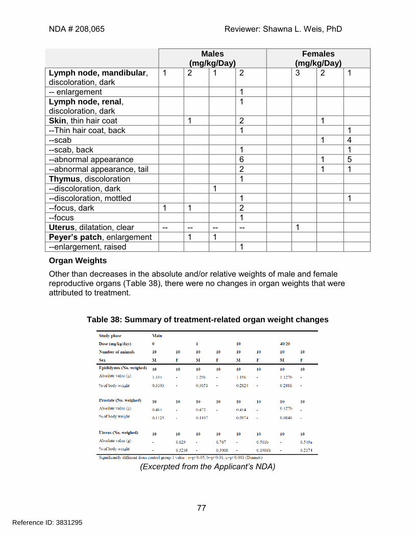
NDA # 208,065 Reviewer: Shawna L. Weis, PhD
77
Males (mg/kg/Day)
Females (mg/kg/Day)
Lymph node, mandibular, discoloration, dark
1 2 1 2 3 2 1
-- enlargement 1
Lymph node, renal, discoloration, dark
1
Skin, thin hair coat 1 2 1
--Thin hair coat, back 1 1
--scab 1 4
--scab, back 1 1
--abnormal appearance 6 1 5
--abnormal appearance, tail 2 1 1
Thymus, discoloration 1
--discoloration, dark 1
--discoloration, mottled 1 1
--focus, dark 1 1 2
--focus 1
Uterus, dilatation, clear -- -- -- -- 1
Peyer’s patch, enlargement 1 1
--enlargement, raised 1
Organ Weights
Other than decreases in the absolute and/or relative weights of male and female reproductive organs (Table 38), there were no changes in organ weights that were attributed to treatment.
Table 38: Summary of treatment-related organ weight changes
(Excerpted from the Applicant’s NDA)
Reference ID: 3831295
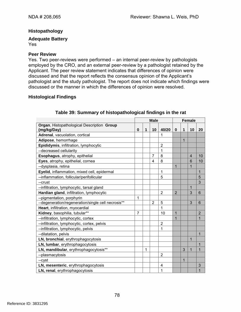
NDA # 208,065 Reviewer: Shawna L. Weis, PhD
78
Histopathology
Adequate Battery Yes
Peer Review Yes. Two peer-reviews were performed – an internal peer-review by pathologists employed by the CRO, and an external peer-review by a pathologist retained by the Applicant. The peer review statement indicates that differences of opinion were discussed and that the report reflects the consensus opinion of the Applicant’s pathologist and the study pathologist. The report does not indicate which findings were discussed or the manner in which the differences of opinion were resolved.
Histological Findings
Table 39: Summary of histopathological findings in the rat
Male Female
Organ, Histopathological Description Group (mg/kg/Day) 0 1 10 40/20 0 1 10 20
Adrenal, vacuolation, cortical 1
Adipose, hemorrhage 1
Epididymis, infiltration, lymphocytic 2
--decreased cellularity 1
Esophagus, atrophy, epithelial 7 8 4 10
Eyes, atrophy, epithelial, cornea 4 8 6 10
--dysplasia, retina 1 1
Eyelid, inflammation, mixed cell, epidermal 1 1
--inflammation, follicular/perifollicular 5 5
--crust 3
--infiltration, lymphocytic, tarsal gland 1
Hardian gland, infiltration, lymphocytic 2 2 3 6
--pigmentation, porphyrin 1
--degeneration/regeneration/single cell necrosis** 2 5 3 6
Heart, infiltration, myocardial 1
Kidney, basophilia, tubular** 7 10 1 2
--infiltration, lymphocytic, cortex 1 1
--infiltration, lymphocytic, cortex, pelvis 2
--infiltration, lymphocytic, pelvis 1
--dilatation, pelvis 1
LN, bronchial, erythrophagocytosis 1
LN, lumbar, erythrophagocytosis 1
LN, mandibular, erythrophagocytosis** 1 3 1 1
--plasmacytosis 2
--cyst 1
LN, mesenteric, erythrophagocytosis 4 3
LN, renal, erythrophagocytosis 1 1
Reference ID: 3831295
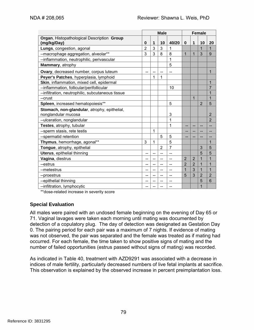
NDA # 208,065 Reviewer: Shawna L. Weis, PhD
79
Male Female
Organ, Histopathological Description Group (mg/kg/Day) 0 1 10 40/20 0 1 10 20
Lungs, congestion, agonal 2 3 3 1 1 1
--macrophage aggregation, alveolar** 3 3 8 8 1 1 3 9
--inflammation, neutrophilic, perivascular 1
Mammary, atrophy 5
Ovary, decreased number, corpus luteum -- -- -- -- 1
Peyer's Patches, hyperplasia, lymphoid 1 1
Skin, inflammation, mixed cell, epidermal 1
--inflammation, follicular/perifollicular 10 7
--infiltration, neutrophilic, subcutaneous tissue 1
--crust 1 1
Spleen, increased hematopoiesis** 5 2 5
Stomach, non-glandular, atrophy, epithelial, nonglandular mucosa 3 2
--ulceration, nonglandular 1 2
Testes, atrophy, tubular 1 -- -- -- --
--sperm stasis, rete testis 1 -- -- -- --
--spermatid retention 5 5 -- -- -- --
Thymus, hemorrhage, agonal** 3 1 5 1
Tongue, atrophy, epithelial 2 7 3 5
Uterus, epithelial thinning -- -- -- -- 5 5
Vagina, diestrus -- -- -- -- 2 2 1 1
--estrus -- -- -- -- 2 2 1 1
--metestrus -- -- -- -- 1 3 1 1
--proestrus -- -- -- -- 5 3 2 2
--epithelial thinning -- -- -- -- 5 6
--infiltration, lymphocytic -- -- -- -- 1
**dose-related increase in severity score
Special Evaluation
All males were paired with an undosed female beginning on the evening of Day 65 or 71. Vaginal lavages were taken each morning until mating was documented by detection of a copulatory plug. The day of detection was designated as Gestation Day 0. The pairing period for each pair was a maximum of 7 nights. If evidence of mating was not observed, the pair was separated and the female was treated as if mating had occurred. For each female, the time taken to show positive signs of mating and the number of failed opportunities (estrus passed without signs of mating) was recorded. As indicated in Table 40, treatment with AZD9291 was associated with a decrease in indices of male fertility, particularly decreased numbers of live fetal implants at sacrifice. This observation is explained by the observed increase in percent preimplantation loss.
Reference ID: 3831295
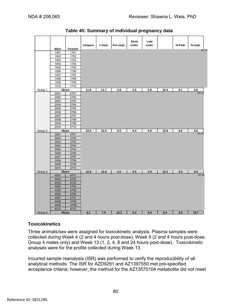
NDA # 208,065 Reviewer: Shawna L. Weis, PhD
80
Table 40: Summary of individual pregnancy data
1001 1701
1002 1702
1003 1703
1004 1704
1005 1705
1006 1706
1007 1707
1008 1708
1009 1709
1010 1710
11.8 11.7 0.8 0.6 0.0 12.4 4.1 4.8
2001 2701
2002 2702
2003 2703
2004 2704
2005 2705
2006 2706
2007 2707
2008 2708
2009 2709
2010 2710
12.0 12.0 0.0 0.4 0.0 12.9 3.6 3.6
3001 3701
3002 3702
3003 3703
3004 3704
3005 3705
3006 3706
3007 3707
3008 3708
3009 3709
3010 3710
12.8 12.8 0.0 0.5 0.0 12.3 3.9 3.9
4001 4701
4002 4702
4003 4703
4004 4704
4005 4705
4006 4706
4007 4707
4008 4708
4009 -4709
4010 4710
9.1 7.9 16.2 0.3 0.0 8.4 3.9 19.7
Mean
Mean
Mean
Group 2
Group 3
Group 4 Mean
Pre-impl.
Early
embr.
Late
embr. % Post- % impl. Corpora # Impl.
Male Female
Group 1
Toxicokinetics
Three animals/sex were assigned for toxicokinetic analysis. Plasma samples were collected during Week 4 (2 and 4 hours post-dose), Week 9 (2 and 4 hours post-dose, Group 4 males only) and Week 13 (1, 2, 4, 8 and 24 hours post-dose). Toxicokinetic analyses were for the profile collected during Week 13. Incurred sample reanalysis (ISR) was performed to verify the reproducibility of all analytical methods. The ISR for AZD9291 and AZ1397550 met pre-specified acceptance criteria; however, the method for the AZ13575104 metabolite did not meet
Reference ID: 3831295
(b) (4)
(b) (4)
(b) (4)
(b) (4)
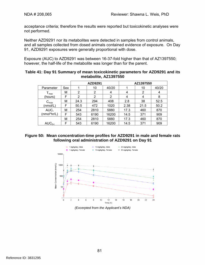
NDA # 208,065 Reviewer: Shawna L. Weis, PhD
81
acceptance criteria; therefore the results were reported but toxicokinetic analyses were not performed. Neither AZD9291 nor its metabolites were detected in samples from control animals, and all samples collected from dosed animals contained evidence of exposure. On Day 91, AZD9291 exposures were generally proportional with dose. Exposure (AUC) to AZD9291 was between 16-37-fold higher than that of AZ1397550; however, the half-life of the metabolite was longer than for the parent.
Table 41: Day 91 Summary of mean toxicokinetic parameters for AZD9291 and its metabolite, AZ1397550
AZD9291 AZ1397550
Parameter Sex 1 10 40/20 1 10 40/20
Tmax (hours)
M 2 2 4 4 2 4
F 2 2 2 4 4 8
Cmax (nmol/L)
M 24.3 294 408 2.8 38 52.5
F 50.5 472 1020 2.38 21.5 50.2
AUCτ (nmol*hr/L)
M 254 2810 5880 17.3 460 870
F 543 6190 16200 14.5 371 909
AUC0-t
M 254 2810 5880 17.3 460 870
F 543 6190 16200 14.5 371 909
Figure 50: Mean concentration-time profiles for AZD9291 in male and female rats following oral administration of AZD9291 on Day 91
(Excerpted from the Applicant’s NDA)
Reference ID: 3831295
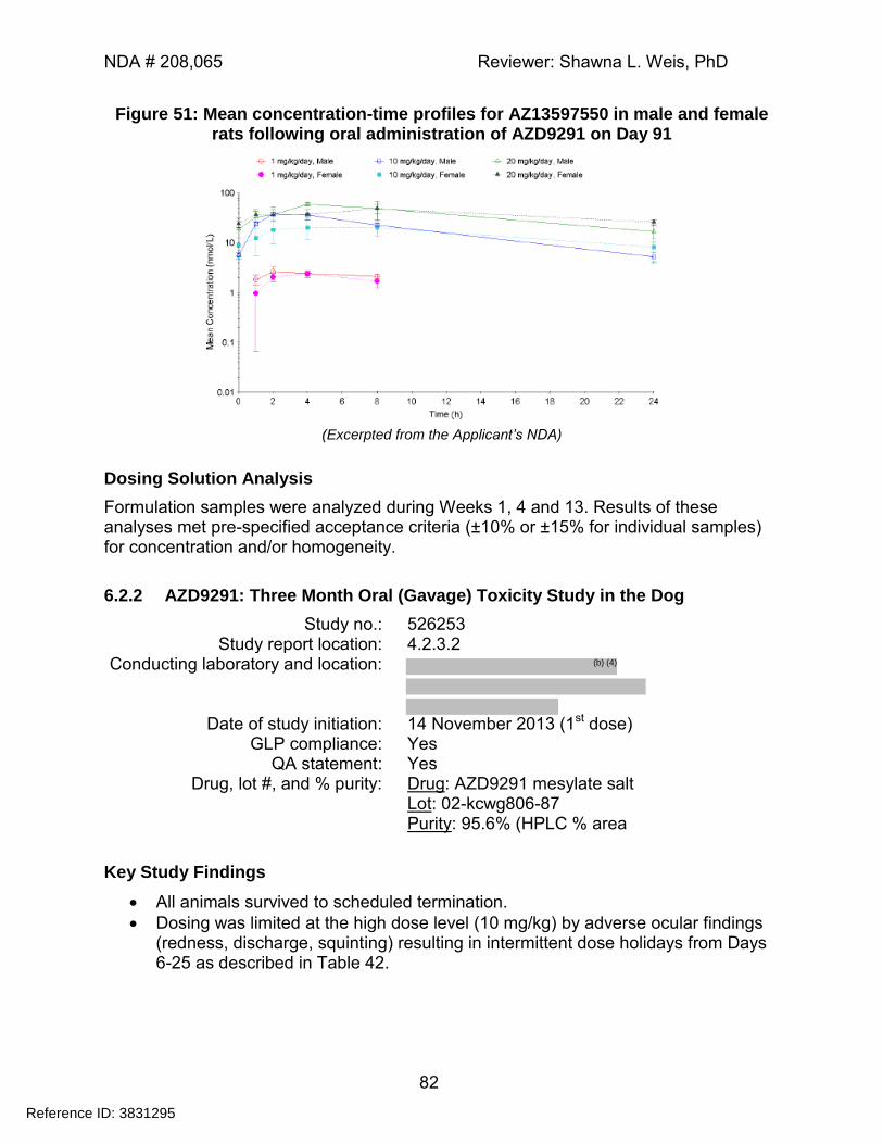
NDA # 208,065 Reviewer: Shawna L. Weis, PhD
82
Figure 51: Mean concentration-time profiles for AZ13597550 in male and female rats following oral administration of AZD9291 on Day 91
(Excerpted from the Applicant’s NDA)
Dosing Solution Analysis
Formulation samples were analyzed during Weeks 1, 4 and 13. Results of these analyses met pre-specified acceptance criteria (±10% or ±15% for individual samples) for concentration and/or homogeneity.
6.2.2 AZD9291: Three Month Oral (Gavage) Toxicity Study in the Dog
Study no.: 526253 Study report location: 4.2.3.2
Conducting laboratory and location:
Date of study initiation: 14 November 2013 (1st dose)
GLP compliance: Yes QA statement: Yes
Drug, lot #, and % purity: Drug: AZD9291 mesylate salt Lot: 02-kcwg806-87 Purity: 95.6% (HPLC % area
Key Study Findings
All animals survived to scheduled termination.
Dosing was limited at the high dose level (10 mg/kg) by adverse ocular findings (redness, discharge, squinting) resulting in intermittent dose holidays from Days 6-25 as described in Table 42.
Reference ID: 3831295
(b) (4)
NDA # 208,065 Reviewer: Shawna L. Weis, PhD
83
Methods Doses: 0, 1, 3, 10/6 mg/kg
Frequency of dosing: Daily Route of administration: Oral gavage
Dose volume: 5 mL/kg Formulation/Vehicle: Milli-Q water pH adjusted with methane sulfonic
acid (final pH 3 to pH 3.5) Species/Strain: Canis familiaris (Beagle dog; animals were
supplied to the CRO by the Applicant) Number/Sex/Group: 4/sex/group
Age: Males: 24 to 28 months Females: 25 to 28 months
Weight: Males: 11.2 – 13.8 kg Females: 8.3 – 13.4 kg
Satellite groups: None Unique study design: Dose reduction in high dose animals on
Day 23. Deviation from study protocol: In general, the reported deviations were
considered unlikely to have affected the outcome of the study; however, there were numerous intermittent dose holidays granted for poor tolerability to high dose animals (due to ocular lesions and GI effects) during Days 9-25 of the study, after which animals were dose-reduced. Given the number of animals involved and the number of reported dose holidays, these events are likely to have underrepresented the toxicity of AZD9291 at the 10/6 mg/kg dose level when administered over a 3 month duration.
Observations and Results
Mortality
There were no preterm deaths.
Clinical Signs
Treatment-related clinical signs in mid- and high-dose animals included: eye discharge; eye closed or partially closed; eye red; emesis; soft/yellow/mucoid feces; and skin lesions.
Body Weights
A decrease in mean body weight was observed in high dose males and females during the first week of the dosing period, which showed signs of recovery after the dose reduction on Day 23.
Reference ID: 3831295
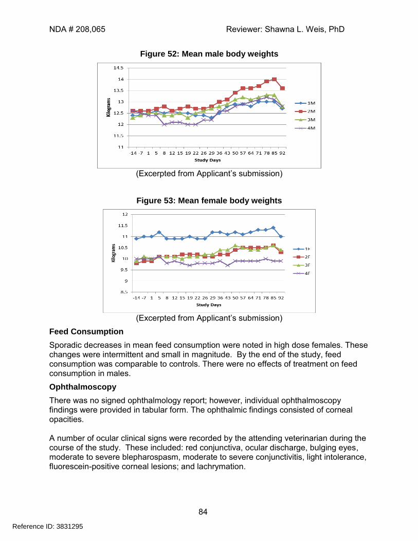
NDA # 208,065 Reviewer: Shawna L. Weis, PhD
84
Figure 52: Mean male body weights
(Excerpted from Applicant’s submission)
Figure 53: Mean female body weights
(Excerpted from Applicant’s submission)
Feed Consumption
Sporadic decreases in mean feed consumption were noted in high dose females. These changes were intermittent and small in magnitude. By the end of the study, feed consumption was comparable to controls. There were no effects of treatment on feed consumption in males.
Ophthalmoscopy
There was no signed ophthalmology report; however, individual ophthalmoscopy findings were provided in tabular form. The ophthalmic findings consisted of corneal opacities. A number of ocular clinical signs were recorded by the attending veterinarian during the course of the study. These included: red conjunctiva, ocular discharge, bulging eyes, moderate to severe blepharospasm, moderate to severe conjunctivitis, light intolerance, fluorescein-positive corneal lesions; and lachrymation.
Reference ID: 3831295
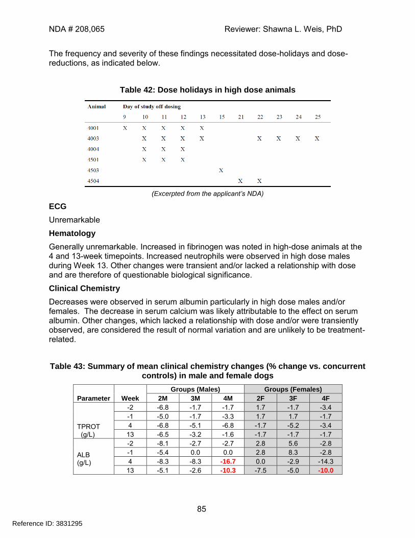
NDA # 208,065 Reviewer: Shawna L. Weis, PhD
85
The frequency and severity of these findings necessitated dose-holidays and dose-reductions, as indicated below.
Table 42: Dose holidays in high dose animals
(Excerpted from the applicant’s NDA)
ECG
Unremarkable
Hematology
Generally unremarkable. Increased in fibrinogen was noted in high-dose animals at the 4 and 13-week timepoints. Increased neutrophils were observed in high dose males during Week 13. Other changes were transient and/or lacked a relationship with dose and are therefore of questionable biological significance.
Clinical Chemistry
Decreases were observed in serum albumin particularly in high dose males and/or females. The decrease in serum calcium was likely attributable to the effect on serum albumin. Other changes, which lacked a relationship with dose and/or were transiently observed, are considered the result of normal variation and are unlikely to be treatment-related.
Table 43: Summary of mean clinical chemistry changes (% change vs. concurrent controls) in male and female dogs
Parameter Week
Groups (Males) Groups (Females)
2M 3M 4M 2F 3F 4F
TPROT (g/L)
-2 -6.8 -1.7 -1.7 1.7 -1.7 -3.4
-1 -5.0 -1.7 -3.3 1.7 1.7 -1.7
4 -6.8 -5.1 -6.8 -1.7 -5.2 -3.4
13 -6.5 -3.2 -1.6 -1.7 -1.7 -1.7
ALB (g/L)
-2 -8.1 -2.7 -2.7 2.8 5.6 -2.8
-1 -5.4 0.0 0.0 2.8 8.3 -2.8
4 -8.3 -8.3 -16.7 0.0 -2.9 -14.3
13 -5.1 -2.6 -10.3 -7.5 -5.0 -10.0
Reference ID: 3831295
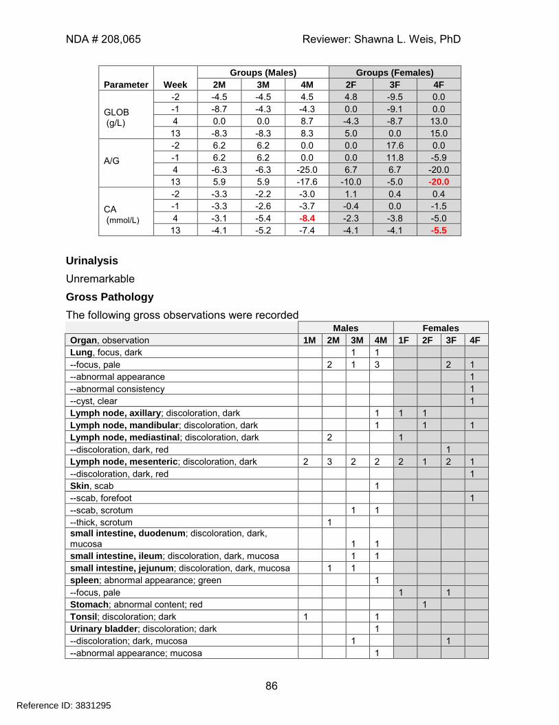
NDA # 208,065 Reviewer: Shawna L. Weis, PhD
86
Parameter Week
Groups (Males) Groups (Females)
2M 3M 4M 2F 3F 4F
GLOB (g/L)
-2 -4.5 -4.5 4.5 4.8 -9.5 0.0
-1 -8.7 -4.3 -4.3 0.0 -9.1 0.0
4 0.0 0.0 8.7 -4.3 -8.7 13.0
13 -8.3 -8.3 8.3 5.0 0.0 15.0
A/G
-2 6.2 6.2 0.0 0.0 17.6 0.0
-1 6.2 6.2 0.0 0.0 11.8 -5.9
4 -6.3 -6.3 -25.0 6.7 6.7 -20.0
13 5.9 5.9 -17.6 -10.0 -5.0 -20.0
CA (mmol/L)
-2 -3.3 -2.2 -3.0 1.1 0.4 0.4
-1 -3.3 -2.6 -3.7 -0.4 0.0 -1.5
4 -3.1 -5.4 -8.4 -2.3 -3.8 -5.0
13 -4.1 -5.2 -7.4 -4.1 -4.1 -5.5
Urinalysis
Unremarkable
Gross Pathology
The following gross observations were recorded Males Females
Organ, observation 1M 2M 3M 4M 1F 2F 3F 4F
Lung, focus, dark 1 1
--focus, pale 2 1 3 2 1
--abnormal appearance 1
--abnormal consistency 1
--cyst, clear 1
Lymph node, axillary; discoloration, dark 1 1 1
Lymph node, mandibular; discoloration, dark 1 1 1
Lymph node, mediastinal; discoloration, dark 2 1
--discoloration, dark, red 1
Lymph node, mesenteric; discoloration, dark 2 3 2 2 2 1 2 1
--discoloration, dark, red 1
Skin, scab 1
--scab, forefoot 1
--scab, scrotum 1 1
--thick, scrotum 1
small intestine, duodenum; discoloration, dark, mucosa 1 1
small intestine, ileum; discoloration, dark, mucosa 1 1
small intestine, jejunum; discoloration, dark, mucosa 1 1
spleen; abnormal appearance; green 1
--focus, pale 1 1
Stomach; abnormal content; red 1
Tonsil; discoloration; dark 1 1
Urinary bladder; discoloration; dark 1
--discoloration; dark, mucosa 1 1
--abnormal appearance; mucosa 1
Reference ID: 3831295
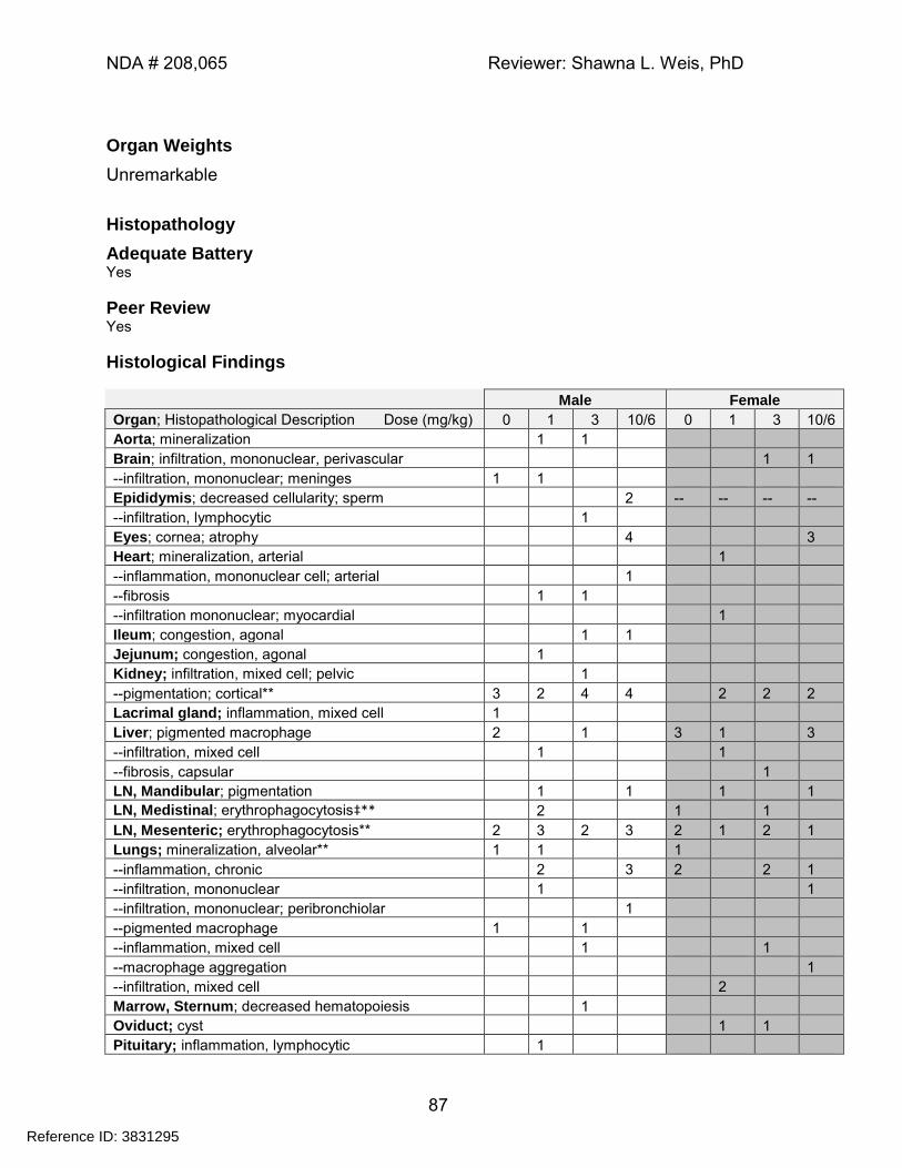
NDA # 208,065 Reviewer: Shawna L. Weis, PhD
87
Organ Weights
Unremarkable
Histopathology
Adequate Battery Yes
Peer Review Yes
Histological Findings
Male Female
Organ; Histopathological Description Dose (mg/kg) 0 1 3 10/6 0 1 3 10/6
Aorta; mineralization 1 1
Brain; infiltration, mononuclear, perivascular 1 1
--infiltration, mononuclear; meninges 1 1
Epididymis; decreased cellularity; sperm 2 -- -- -- --
--infiltration, lymphocytic 1
Eyes; cornea; atrophy 4 3
Heart; mineralization, arterial 1
--inflammation, mononuclear cell; arterial 1
--fibrosis 1 1
--infiltration mononuclear; myocardial 1
Ileum; congestion, agonal 1 1
Jejunum; congestion, agonal 1
Kidney; infiltration, mixed cell; pelvic 1
--pigmentation; cortical** 3 2 4 4 2 2 2
Lacrimal gland; inflammation, mixed cell 1
Liver; pigmented macrophage 2 1 3 1 3
--infiltration, mixed cell 1 1
--fibrosis, capsular 1
LN, Mandibular; pigmentation 1 1 1 1
LN, Medistinal; erythrophagocytosis‡** 2 1 1
LN, Mesenteric; erythrophagocytosis** 2 3 2 3 2 1 2 1
Lungs; mineralization, alveolar** 1 1 1
--inflammation, chronic 2 3 2 2 1
--infiltration, mononuclear
1 1
--infiltration, mononuclear; peribronchiolar 1
--pigmented macrophage 1 1
--inflammation, mixed cell 1 1
--macrophage aggregation 1
--infiltration, mixed cell 2
Marrow, Sternum; decreased hematopoiesis 1
Oviduct; cyst 1 1
Pituitary; inflammation, lymphocytic 1
Reference ID: 3831295
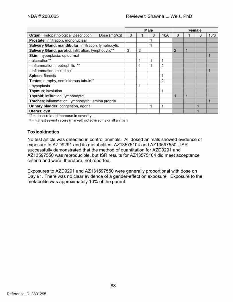
NDA # 208,065 Reviewer: Shawna L. Weis, PhD
88
Male Female
Organ; Histopathological Description Dose (mg/kg) 0 1 3 10/6 0 1 3 10/6
Prostate; infiltration, mononuclear 1
Salivary Gland, mandibular; infiltration, lymphocytic 1
Salivary Gland, parotid; infiltration, lymphocytic** 3 2 2 1
Skin; hyperplasia, epidermal 1
--ulceration** 1 1 1
--inflammation, neutrophilic‡** 1 1 2
--inflammation, mixed cell 1
Spleen; fibrosis 1
Testes; atrophy, seminiferous tubule** 2
--hypoplasia 1
Thymus; involution 1
Thyroid; infiltration, lymphocytic 1 1
Trachea; inflammation, lymphocytic; lamina propria 1
Urinary bladder; congestion, agonal 1 1 1
Uterus; cyst 1
** = dose-related increase in severity
‡ = highest severity score (marked) noted in some or all animals
Toxicokinetics
No test article was detected in control animals. All dosed animals showed evidence of exposure to AZD9291 and its metabolites, AZ13575104 and AZ13597550. ISR successfully demonstrated that the method of quantitation for AZD9291 and AZ13597550 was reproducible, but ISR results for AZ13575104 did meet acceptance criteria and were, therefore, not reported. Exposures to AZD9291 and AZ131597550 were generally proportional with dose on Day 91. There was no clear evidence of a gender-effect on exposure. Exposure to the metabolite was approximately 10% of the parent.
Reference ID: 3831295
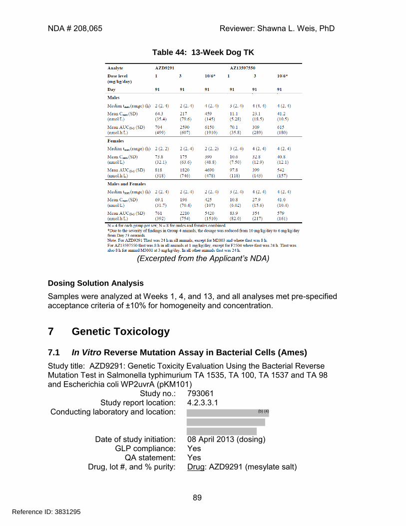
NDA # 208,065 Reviewer: Shawna L. Weis, PhD
89
Table 44: 13-Week Dog TK
(Excerpted from the Applicant’s NDA)
Dosing Solution Analysis
Samples were analyzed at Weeks 1, 4, and 13, and all analyses met pre-specified acceptance criteria of ±10% for homogeneity and concentration.
7 Genetic Toxicology
7.1 In Vitro Reverse Mutation Assay in Bacterial Cells (Ames)
Study title: AZD9291: Genetic Toxicity Evaluation Using the Bacterial Reverse Mutation Test in Salmonella typhimurium TA 1535, TA 100, TA 1537 and TA 98 and Escherichia coli WP2uvrA (pKM101)
Study no.: 793061 Study report location: 4.2.3.3.1
Conducting laboratory and location:
Date of study initiation: 08 April 2013 (dosing)
GLP compliance: Yes QA statement: Yes
Drug, lot #, and % purity: Drug: AZD9291 (mesylate salt)
Reference ID: 3831295
(b) (4)
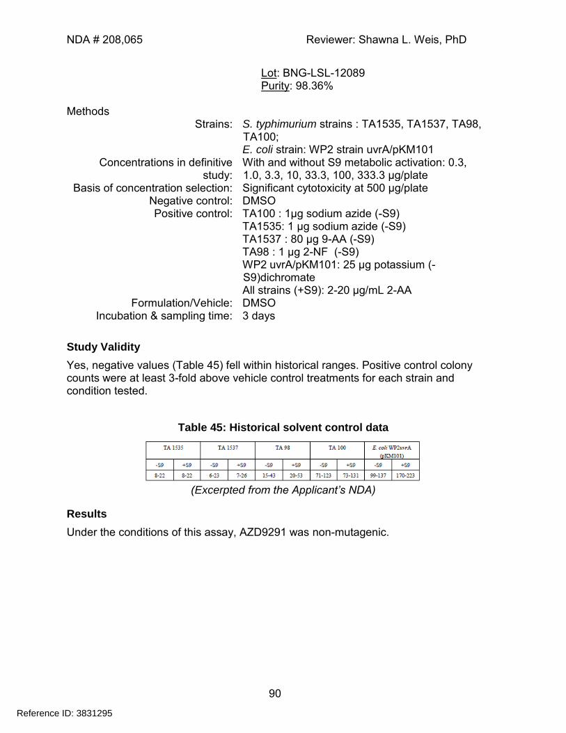
NDA # 208,065 Reviewer: Shawna L. Weis, PhD
90
Lot: BNG-LSL-12089 Purity: 98.36%
Methods
Strains: S. typhimurium strains : TA1535, TA1537, TA98, TA100; E. coli strain: WP2 strain uvrA/pKM101
Concentrations in definitive study:
With and without S9 metabolic activation: 0.3, 1.0, 3.3, 10, 33.3, 100, 333.3 µg/plate
Basis of concentration selection: Significant cytotoxicity at 500 µg/plate Negative control: DMSO Positive control: TA100 : 1µg sodium azide (-S9)
TA1535: 1 μg sodium azide (-S9) TA1537 : 80 μg 9-AA (-S9) TA98 : 1 μg 2-NF (-S9) WP2 uvrA/pKM101: 25 μg potassium (-S9)dichromate All strains (+S9): 2-20 µg/mL 2-AA
Formulation/Vehicle: DMSO Incubation & sampling time: 3 days
Study Validity
Yes, negative values (Table 45) fell within historical ranges. Positive control colony counts were at least 3-fold above vehicle control treatments for each strain and condition tested.
Table 45: Historical solvent control data
(Excerpted from the Applicant’s NDA)
Results
Under the conditions of this assay, AZD9291 was non-mutagenic.
Reference ID: 3831295
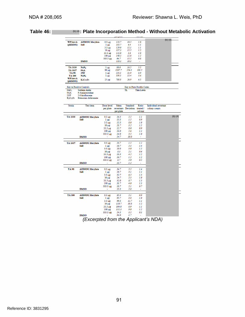
NDA # 208,065 Reviewer: Shawna L. Weis, PhD
91
Table 46: : Plate Incorporation Method - Without Metabolic Activation
(Excerpted from the Applicant’s NDA)
Reference ID: 3831295
(b) (4)
(b) (4)
(b) (4)
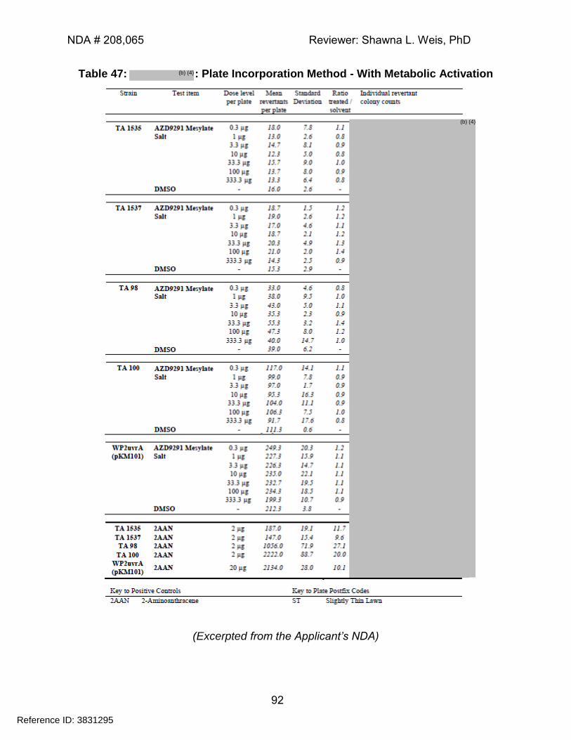
NDA # 208,065 Reviewer: Shawna L. Weis, PhD
92
Table 47: : Plate Incorporation Method - With Metabolic Activation
(Excerpted from the Applicant’s NDA)
Reference ID: 3831295
(b) (4)
(b) (4)
NDA # 208,065 Reviewer: Shawna L. Weis, PhD
93
7.2 In Vitro Assays in Mammalian Cells
Study title: AZD9291: Genetic Toxicity Evaluation using the Mouse Lymphoma Cell Thymidine Kinase Locus Assay
Study no.: 793056 Study report location: 4.2.3.3.1
Conducting laboratory and location:
Date of study initiation: 15 January 2013 (experimental start)
GLP compliance: Yes QA statement: Yes
Drug, lot #, and % purity: Drug: AZD9291 (mesylate salt) Batch: BNG-LSL-12089 Purity: 98.6%
Methods
Cell line: L5175Y TK+/- Concentrations in definitive study: Minus S9: 1, 2, 3, 4, 5, 6, 7, 8 µM
Plus S9: 2.5, 4, 5.5, 7, 8.5, 10, 11.5, 13 µM Basis of concentration selection: Cytotoxicity
Negative control: Solvent (DMSO) Positive control: Minus S9: MMS
Plus S9: 3-methylcholanthrene Formulation/Vehicle: DMSO
Incubation & sampling time: 3 hours±S9; 24 hours + S9; cloning efficiency assessed on Day 2
Study Validity
Yes, with the following exception, all positive and negative controls were within historical range provided:
The mutant frequency for 3-MC-treated cultures was 2491 X 10-6 in the culture treated at a concentration of 10µM. While this value exceeded previous control range provided, the result was considered sufficient to demonstrate the sensitivity of the assay as it demonstrated a statistically and biologically significant increase in revertants compared with the concurrent control. There is no impact of this exception on the interpretation of the data.
While a few small increases in mutant frequency were observed at some concentrations in the 3-hour incubation performed in the presence of S9 activation, the increases were not considered biologically significant.
Reference ID: 3831295
(b) (4)
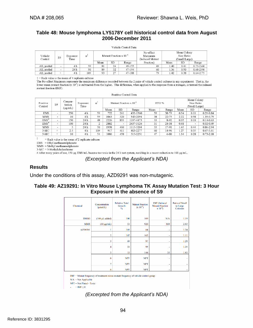
NDA # 208,065 Reviewer: Shawna L. Weis, PhD
94
Table 48: Mouse lymphoma LY5178Y cell historical control data from August 2006-December 2011
(Excerpted from the Applicant’s NDA)
Results
Under the conditions of this assay, AZD9291 was non-mutagenic.
Table 49: AZ19291: In Vitro Mouse Lymphoma TK Assay Mutation Test: 3 Hour Exposure in the absence of S9
(Excerpted from the Applicant’s NDA)
Reference ID: 3831295
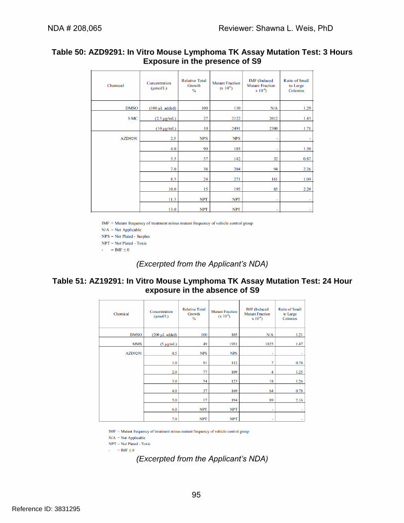
NDA # 208,065 Reviewer: Shawna L. Weis, PhD
95
Table 50: AZD9291: In Vitro Mouse Lymphoma TK Assay Mutation Test: 3 Hours Exposure in the presence of S9
(Excerpted from the Applicant’s NDA)
Table 51: AZ19291: In Vitro Mouse Lymphoma TK Assay Mutation Test: 24 Hour exposure in the absence of S9
(Excerpted from the Applicant’s NDA)
Reference ID: 3831295
NDA # 208,065 Reviewer: Shawna L. Weis, PhD
96
7.3 In Vivo Clastogenicity Assay in Rodent (Micronucleus Assay)
Study title: AZD9291: Genetic Toxicity Evaluation Using the Rat Micronucleus Test After Two Oral Doses
Study no: 793538 Study report location: 4.2.3.3.2
Conducting laboratory and location:
Date of study initiation: 07 May 2013 (1st dose)
GLP compliance: Yes QA statement: Yes
Drug, lot #, and % purity: Drug: AZD9291 (mesylate salt) Batch: BNG-LSL-12089 Purity: 98.6%
Key Study Findings
Under the conditions of this assay, AZD9291 was considered negative for induction of micronucleated immature erythrocytes in the Han Wistar rat. Methods
Doses in definitive study: 0, 30, 150, 300 mg/kg Frequency of dosing: Daily (2 days)
Route of administration: Oral gavage Dose volume: 10 mL/kg
Formulation/Vehicle: 0.5% HPMC (3500-5600 cps) Species/Strain: Rattus norvegicus / Han Wistar (CRL:WI(Han))
Number/Sex/Group: 7M/Group Satellite groups: None
Basis of dose selection: MTD Negative control: Vehicle Positive control: Cyclophosphamide (20 mg/kg)
Study Validity
In Test 1, the formulation analysis results indicated that the test solutions were sub-potent. Because the results failed to meet pre-specified acceptance criteria of ±10%; the formulations were considered out of speciation, and the data from Test 1 were not analyzed. The formulations used in Test 2 were considered acceptable. Because the positive control demonstrated the sensitivity of the system using a known inducer of micronucleus formation, and the negative control data fell within historical ranges, the study met the criteria for a valid test.
Reference ID: 3831295
(b) (4)
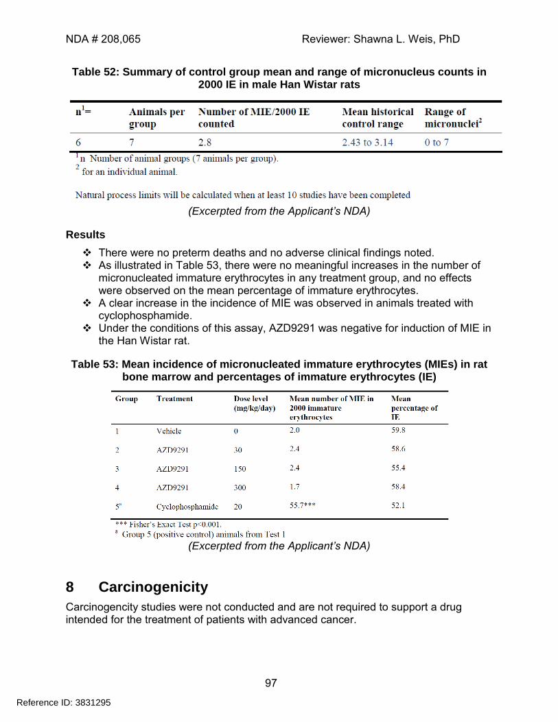
NDA # 208,065 Reviewer: Shawna L. Weis, PhD
97
Table 52: Summary of control group mean and range of micronucleus counts in 2000 IE in male Han Wistar rats
(Excerpted from the Applicant’s NDA)
Results
There were no preterm deaths and no adverse clinical findings noted. As illustrated in Table 53, there were no meaningful increases in the number of
micronucleated immature erythrocytes in any treatment group, and no effects were observed on the mean percentage of immature erythrocytes.
A clear increase in the incidence of MIE was observed in animals treated with cyclophosphamide.
Under the conditions of this assay, AZD9291 was negative for induction of MIE in the Han Wistar rat.
Table 53: Mean incidence of micronucleated immature erythrocytes (MIEs) in rat bone marrow and percentages of immature erythrocytes (IE)
(Excerpted from the Applicant’s NDA)
8 Carcinogenicity
Carcinogencity studies were not conducted and are not required to support a drug intended for the treatment of patients with advanced cancer.
Reference ID: 3831295
NDA # 208,065 Reviewer: Shawna L. Weis, PhD
98
9 Reproductive and Developmental Toxicology
9.2 Embryonic Fetal Development
Study title: AZD9291: Oral (Gavage) Investigative Dose Range Finding Embryofetal Development and Pre and Post Natal Study in the Rat
Study no.: 496800 Study report location: 4.2.3.5.2
Conducting laboratory and location:
Date of study initiation: 28 March 2014 (1st dose) GLP compliance: Yes
QA statement: Yes Drug, lot #, and % purity: Drug: AZD9291 mesylate salt
Lot: 02-kcwg806-87 Purity: 95.6% (HPLC % area)
Key Study Findings
Embryofetal and neonatal death: o Administration of AZD9291 from GD 2-20 led to increased post-
implantation loss and early embryonic death. o When administered during organogenesis through Day 6 of lactation
(GD6-LD6), AZD9291 exposure led to an increase in postnatal death and an increase in total litter loss, as well as a reduction in mean pup weight.
Effects on fetal weight:
o When administered during organogenesis (GD 6-16), AZD9291 exposure led to reduced fetal weight at maternal doses of ≥20 mg/kg/Day.
Malformations:
o Visceral abnormalities and malformations were observed in some pups of treated dams; however, the incidences were of low frequency and did not exhibit a clear relationship to dose. Due to the small number of litters used and the absence of historical control data, the relationship of these findings to maternal AZD9291 exposure cannot be excluded.
Placental and lactational transfer: o Toxicokinetic exposures were evaluated in dams and fetuses and/or
nursing pups. Fetal exposure at the end of gestation (GD20) was approximately 36% of those observed in dams on GD16. Fetal exposure to the AZ13597550 metabolite was also demonstrated; however, fetal metabolite levels were relatively low - less than 7% of maternal levels.
o A low level of exposure to AZD9291 and its metabolite was demonstrated in nursing pups. At the maternal Cmax (2 hours post-dose), neonatal exposure to AZD9291 was approximately 2% of the maternal exposure
Reference ID: 3831295
(b) (4)
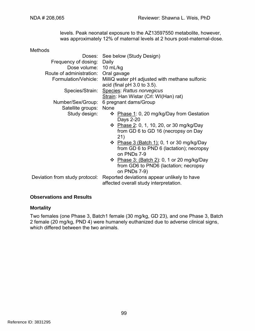
NDA # 208,065 Reviewer: Shawna L. Weis, PhD
99
levels. Peak neonatal exposure to the AZ13597550 metabolite, however, was approximately 12% of maternal levels at 2 hours post-maternal-dose.
Methods
Doses: See below (Study Design) Frequency of dosing: Daily
Dose volume: 10 mL/kg Route of administration: Oral gavage
Formulation/Vehicle: MilliQ water pH adjusted with methane sulfonic acid (final pH 3.0 to 3.5).
Species/Strain: Species: Rattus norvegicus Strain: Han Wistar (Crl: WI(Han) rat)
Number/Sex/Group: 6 pregnant dams/Group Satellite groups: None
Study design: Phase 1: 0, 20 mg/kg/Day from Gestation Days 2-20
Phase 2: 0, 1, 10, 20, or 30 mg/kg/Day from GD 6 to GD 16 (necropsy on Day 21)
Phase 3 (Batch 1): 0, 1 or 30 mg/kg/Day from GD 6 to PND 6 (lactation); necropsy on PNDs 7-9
Phase 3: (Batch 2): 0, 1 or 20 mg/kg/Day from GD6 to PND6 (lactation; necropsy on PNDs 7-9)
Deviation from study protocol: Reported deviations appear unlikely to have affected overall study interpretation.
Observations and Results
Mortality
Two females (one Phase 3, Batch1 female (30 mg/kg, GD 23), and one Phase 3, Batch 2 female (20 mg/kg, PND 4) were humanely euthanized due to adverse clinical signs, which differed between the two animals.
Reference ID: 3831295
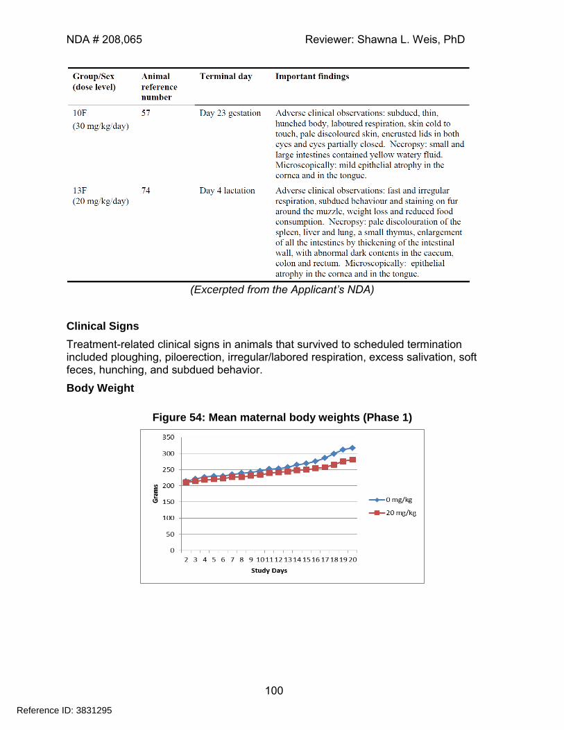
NDA # 208,065 Reviewer: Shawna L. Weis, PhD
100
(Excerpted from the Applicant’s NDA)
Clinical Signs
Treatment-related clinical signs in animals that survived to scheduled termination included ploughing, piloerection, irregular/labored respiration, excess salivation, soft feces, hunching, and subdued behavior.
Body Weight
Figure 54: Mean maternal body weights (Phase 1)
Reference ID: 3831295
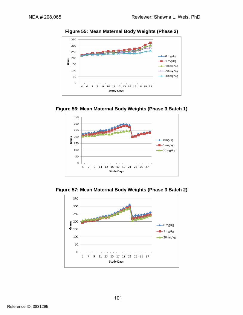
NDA # 208,065 Reviewer: Shawna L. Weis, PhD
101
Figure 55: Mean Maternal Body Weights (Phase 2)
Figure 56: Mean Maternal Body Weights (Phase 3 Batch 1)
Figure 57: Mean Maternal Body Weights (Phase 3 Batch 2)
Reference ID: 3831295
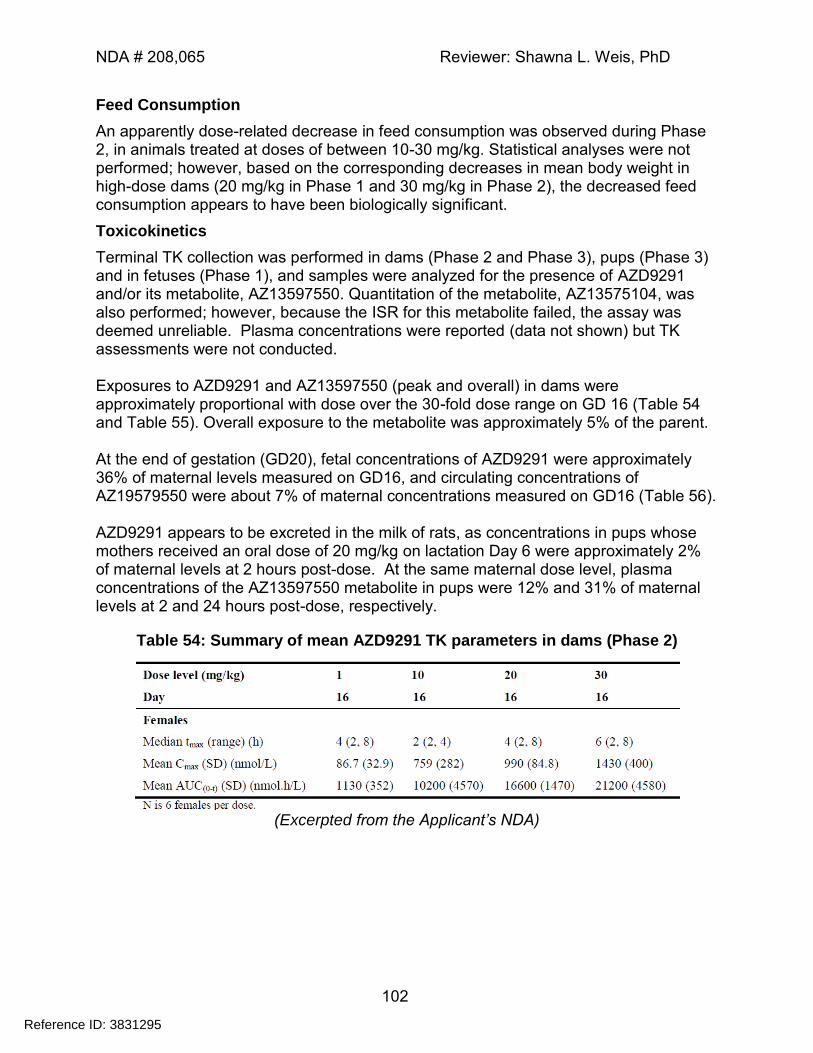
NDA # 208,065 Reviewer: Shawna L. Weis, PhD
102
Feed Consumption
An apparently dose-related decrease in feed consumption was observed during Phase 2, in animals treated at doses of between 10-30 mg/kg. Statistical analyses were not performed; however, based on the corresponding decreases in mean body weight in high-dose dams (20 mg/kg in Phase 1 and 30 mg/kg in Phase 2), the decreased feed consumption appears to have been biologically significant.
Toxicokinetics
Terminal TK collection was performed in dams (Phase 2 and Phase 3), pups (Phase 3) and in fetuses (Phase 1), and samples were analyzed for the presence of AZD9291 and/or its metabolite, AZ13597550. Quantitation of the metabolite, AZ13575104, was also performed; however, because the ISR for this metabolite failed, the assay was deemed unreliable. Plasma concentrations were reported (data not shown) but TK assessments were not conducted. Exposures to AZD9291 and AZ13597550 (peak and overall) in dams were approximately proportional with dose over the 30-fold dose range on GD 16 (Table 54 and Table 55). Overall exposure to the metabolite was approximately 5% of the parent. At the end of gestation (GD20), fetal concentrations of AZD9291 were approximately 36% of maternal levels measured on GD16, and circulating concentrations of AZ19579550 were about 7% of maternal concentrations measured on GD16 (Table 56). AZD9291 appears to be excreted in the milk of rats, as concentrations in pups whose mothers received an oral dose of 20 mg/kg on lactation Day 6 were approximately 2% of maternal levels at 2 hours post-dose. At the same maternal dose level, plasma concentrations of the AZ13597550 metabolite in pups were 12% and 31% of maternal levels at 2 and 24 hours post-dose, respectively.
Table 54: Summary of mean AZD9291 TK parameters in dams (Phase 2)
(Excerpted from the Applicant’s NDA)
Reference ID: 3831295
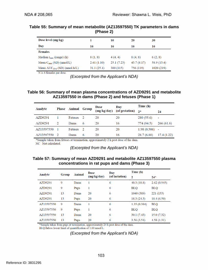
NDA # 208,065 Reviewer: Shawna L. Weis, PhD
103
Table 55: Summary of mean metabolite (AZ13597550) TK parameters in dams (Phase 2)
(Excerpted from the Applicant’s NDA)
Table 56: Summary of mean plasma concentrations of AZD9291 and metabolite AZ13597550 in dams (Phase 2) and fetuses (Phase 1)
(Excerpted from the Applicant’s NDA)
Table 57: Summary of mean AZD9291 and metabolite AZ13597550 plasma concentrations in rat pups and dams (Phase 3)
(Excerpted from the Applicant’s NDA)
Reference ID: 3831295
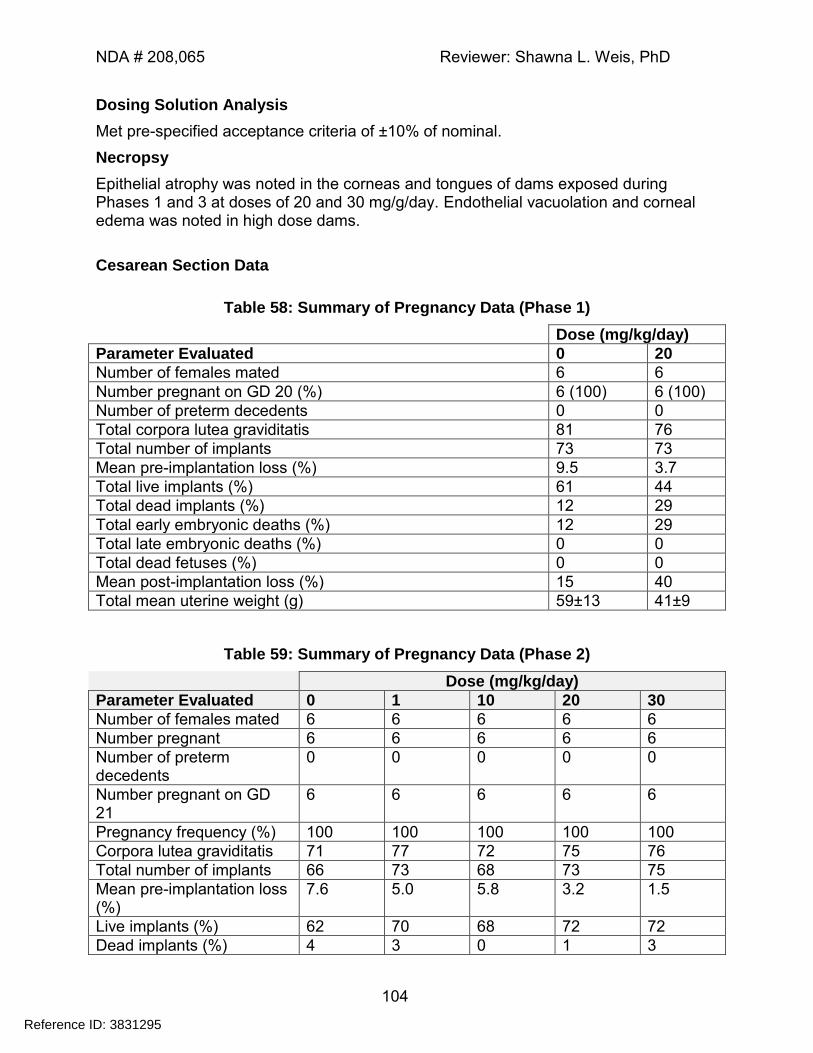
NDA # 208,065 Reviewer: Shawna L. Weis, PhD
104
Dosing Solution Analysis
Met pre-specified acceptance criteria of ±10% of nominal.
Necropsy
Epithelial atrophy was noted in the corneas and tongues of dams exposed during Phases 1 and 3 at doses of 20 and 30 mg/g/day. Endothelial vacuolation and corneal edema was noted in high dose dams.
Cesarean Section Data
Table 58: Summary of Pregnancy Data (Phase 1)
Dose (mg/kg/day)
Parameter Evaluated 0 20
Number of females mated 6 6
Number pregnant on GD 20 (%) 6 (100) 6 (100)
Number of preterm decedents 0 0
Total corpora lutea graviditatis 81 76
Total number of implants 73 73
Mean pre-implantation loss (%) 9.5 3.7
Total live implants (%) 61 44
Total dead implants (%) 12 29
Total early embryonic deaths (%) 12 29
Total late embryonic deaths (%) 0 0
Total dead fetuses (%) 0 0
Mean post-implantation loss (%) 15 40
Total mean uterine weight (g) 59±13 41±9
Table 59: Summary of Pregnancy Data (Phase 2)
Dose (mg/kg/day)
Parameter Evaluated 0 1 10 20 30
Number of females mated 6 6 6 6 6
Number pregnant 6 6 6 6 6
Number of preterm decedents
0 0 0 0 0
Number pregnant on GD 21
6 6 6 6 6
Pregnancy frequency (%) 100 100 100 100 100
Corpora lutea graviditatis 71 77 72 75 76
Total number of implants 66 73 68 73 75
Mean pre-implantation loss (%)
7.6 5.0 5.8 3.2 1.5
Live implants (%) 62 70 68 72 72
Dead implants (%) 4 3 0 1 3
Reference ID: 3831295
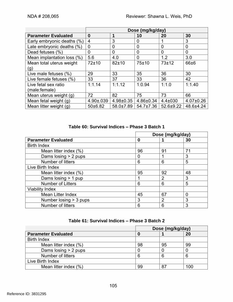
NDA # 208,065 Reviewer: Shawna L. Weis, PhD
105
Dose (mg/kg/day)
Parameter Evaluated 0 1 10 20 30
Early embryonic deaths (%) 4 3 0 1 3
Late embryonic deaths (%) 0 0 0 0 0
Dead fetuses (%) 0 0 0 0 0
Mean implantation loss (%) 5.6 4.0 0 1.2 3.0
Mean total uterus weight (g)
72±10 82±10 75±10 73±12 66±6
Live male fetuses (%) 29 33 35 36 30
Live female fetuses (%) 33 37 33 36 42
Live fetal sex ratio (male:female)
1:1.14 1:1.12 1:0.94 1:1.0 1:1.40
Mean uterus weight (g) 72 82 75 73 66
Mean fetal weight (g) 4.90±.039 4.98±0.35 4.86±0.34 4.4±030 4.07±0.26
Mean litter weight (g) 50±6.82 58.0±7.89 54.7±7.36 52.6±9.22 48.6±4.24
Table 60: Survival Indices – Phase 3 Batch 1
Dose (mg/kg/day)
Parameter Evaluated 0 1 30
Birth Index
Mean litter index (%) 96 91 71
Dams losing > 2 pups 0 1 3
Number of litters 6 6 5
Live Birth Index
Mean litter index (%) 95 92 48
Dams losing > 1 pup 1 2 3
Number of Litters 6 6 5
Viability Index
Mean Litter Index 45 67 0
Number losing > 3 pups 3 2 3
Number of litters 6 6 3
Table 61: Survival Indices – Phase 3 Batch 2
Dose (mg/kg/day)
Parameter Evaluated 0 1 20
Birth Index
Mean litter index (%) 98 95 99
Dams losing > 2 pups 0 0 0
Number of litters 6 6 6
Live Birth Index
Mean litter index (%) 99 87 100
Reference ID: 3831295
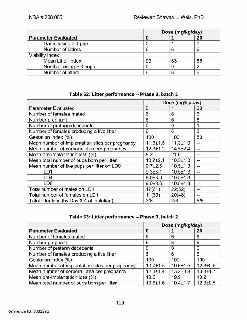
NDA # 208,065 Reviewer: Shawna L. Weis, PhD
106
Dose (mg/kg/day)
Parameter Evaluated 0 1 20
Dams losing > 1 pup 0 1 0
Number of Litters 6 6 6
Viability Index
Mean Litter Index 99 83 65
Number losing > 3 pups 0 0 2
Number of litters 6 6 6
Table 62: Litter performance – Phase 3, batch 1
Dose (mg/kg/day)
Parameter Evaluated 0 1 30
Number of females mated 6 6 6
Number pregnant 6 6 6
Number of preterm decedents 0 0 1
Number of females producing a live litter 6 6 3
Gestation Index (%) 100 100 50
Mean number of implantation sites per pregnancy 11.3±1.5 11.3±1.0 --
Mean number of corpora lutea per pregnancy 12.3±1.2 14.5±2.4 --
Mean pre-implantation loss (%) 8.2 21.0 --
Mean total number of pups born per litter 10.7±2.1 10.5±1.3 --
Mean number of live pups per litter on LD0 9.7±2.5 10.5±1.3 --
LD1 9.3±3.1 10.5±1.3 --
LD4 9.0±3.6 10.5±1.3 --
LD6 9.0±3.6 10.5±1.3 --
Total number of males on LD1 17(61) 22(52) --
Total number of females on LD1 11(39) 20(48) --
Total litter loss (by Day 3-4 of lactation) 3/6 2/6 5/5
Table 63: Litter performance – Phase 3, batch 2
Dose (mg/kg/day)
Parameter Evaluated 0 1 20
Number of females mated 6 6 6
Number pregnant 6 6 6
Number of preterm decedents 0 0 0
Number of females producing a live litter 6 6 6
Gestation Index (%) 100 100 100
Mean number of implantation sites per pregnancy 10.7±1.5 10.6±1.5 12.3±0.5
Mean number of corpora lutea per pregnancy 12.3±1.4 13.2±0.8 13.8±1.7
Mean pre-implantation loss (%) 13.5 19.9 10.2
Mean total number of pups born per litter 10.5±1.6 10.4±1.7 12.3±0.5
Reference ID: 3831295
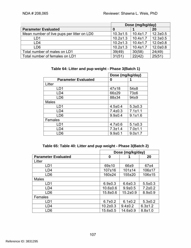
NDA # 208,065 Reviewer: Shawna L. Weis, PhD
107
Dose (mg/kg/day)
Parameter Evaluated 0 1 20
Mean number of live pups per litter on LD0 10.3±1.5 10.4±1.7 12.3±0.5
LD1 10.2±1.3 10.4±1.7 12.3±0.5
LD4 10.2±1.3 10.4±1.7 12.0±0.8
LD6 10.2±1.3 10.4±1.7 12.0±0.8
Total number of males on LD1 39(49) 30(58) 24(49)
Total number of females on LD1 31(51) 22(42) 25(51)
Table 64: Litter and pup weight - Phase 3(Batch 1)
Dose (mg/kg/day)
Parameter Evaluated 0 1
Litter
LD1 47±18 54±8
LD4 66±29 73±6
LD6 88±34 94±9
Males
LD1 4.5±0.4 5.3±0.3
LD4 7.4±0.3 7.1±1.1
LD6 9.9±0.4 9.1±1.6
Females
LD1 4.7±0.6 5.1±0.3
LD4 7.3±1.4 7.0±1.1
LD6 9.9±0.1 9.0±1.7
Table 65: Table 40: Litter and pup weight - Phase 3(Batch 2)
Dose (mg/kg/day)
Parameter Evaluated 0 1 20
Litter
LD1 69±10 66±9 67±4
LD4 107±16 101±14 106±17
LD6 160±24 155±20 106±15
Males
LD1 6.9±0.3 6.6±0.3 5.5±0.3
LD4 10.6±0.6 9.9±0.5 7.2±0.2
LD6 15.8±0.6 15.2±0.9 8.9±0.9
Females
LD1 6.7±0.2 6.1±0.2 5.3±0.2
LD4 10.2±0.3 9.4±0.2 6.3±1.2
LD6 15.6±0.5 14.6±0.9 8.8±1.0
Reference ID: 3831295
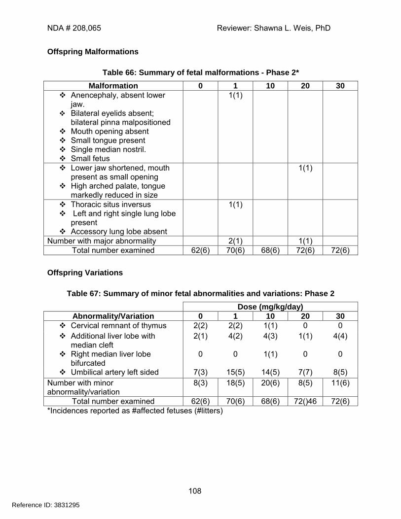
NDA # 208,065 Reviewer: Shawna L. Weis, PhD
108
Offspring Malformations
Table 66: Summary of fetal malformations - Phase 2*
Malformation 0 1 10 20 30
Anencephaly, absent lower jaw.
Bilateral eyelids absent; bilateral pinna malpositioned
Mouth opening absent Small tongue present Single median nostril. Small fetus
1(1)
Lower jaw shortened, mouth present as small opening
High arched palate, tongue markedly reduced in size
1(1)
Thoracic situs inversus Left and right single lung lobe
present Accessory lung lobe absent
1(1)
Number with major abnormality 2(1) 1(1)
Total number examined 62(6) 70(6) 68(6) 72(6) 72(6)
Offspring Variations
Table 67: Summary of minor fetal abnormalities and variations: Phase 2
Dose (mg/kg/day)
Abnormality/Variation 0 1 10 20 30
Cervical remnant of thymus 2(2) 2(2) 1(1) 0 0
Additional liver lobe with median cleft
2(1) 4(2) 4(3) 1(1) 4(4)
Right median liver lobe bifurcated
0 0 1(1) 0 0
Umbilical artery left sided 7(3) 15(5) 14(5) 7(7) 8(5)
Number with minor abnormality/variation
8(3) 18(5) 20(6) 8(5) 11(6)
Total number examined 62(6) 70(6) 68(6) 72()46 72(6)
*Incidences reported as #affected fetuses (#litters)
Reference ID: 3831295
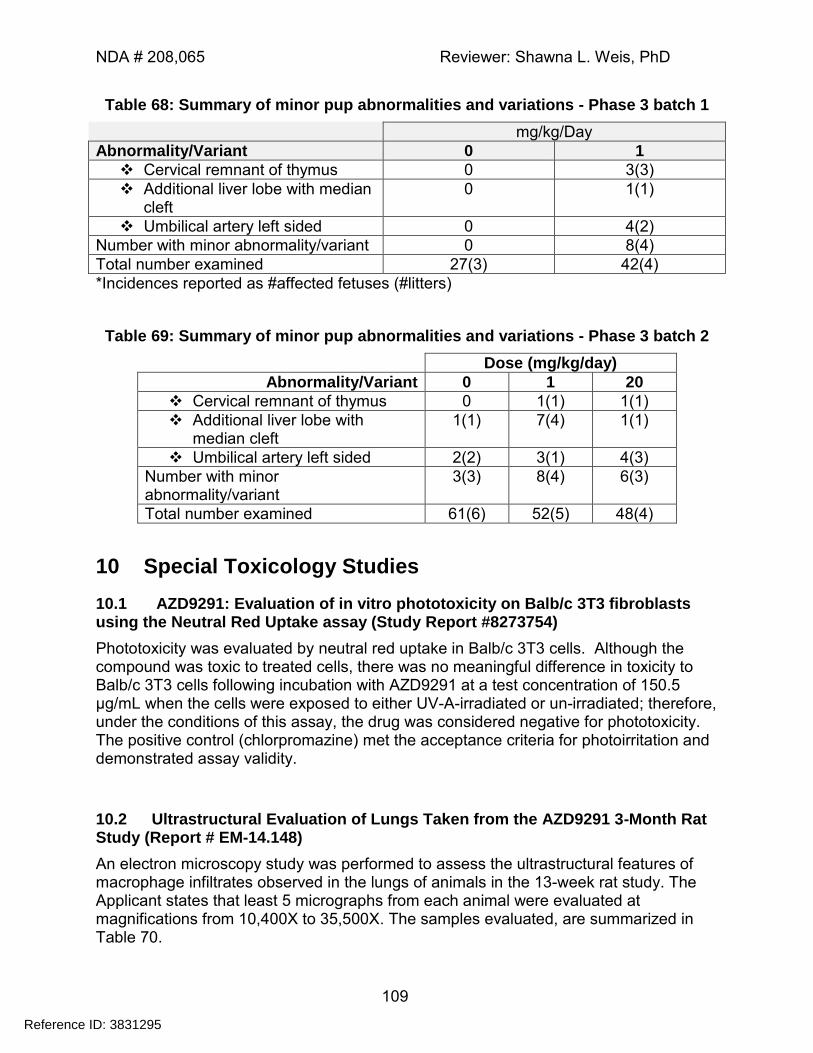
NDA # 208,065 Reviewer: Shawna L. Weis, PhD
109
Table 68: Summary of minor pup abnormalities and variations - Phase 3 batch 1
mg/kg/Day
Abnormality/Variant 0 1
Cervical remnant of thymus 0 3(3)
Additional liver lobe with median cleft
0 1(1)
Umbilical artery left sided 0 4(2)
Number with minor abnormality/variant 0 8(4)
Total number examined 27(3) 42(4)
*Incidences reported as #affected fetuses (#litters)
Table 69: Summary of minor pup abnormalities and variations - Phase 3 batch 2
Dose (mg/kg/day)
Abnormality/Variant 0 1 20
Cervical remnant of thymus 0 1(1) 1(1)
Additional liver lobe with median cleft
1(1) 7(4) 1(1)
Umbilical artery left sided 2(2) 3(1) 4(3)
Number with minor abnormality/variant
3(3) 8(4) 6(3)
Total number examined 61(6) 52(5) 48(4)
10 Special Toxicology Studies
10.1 AZD9291: Evaluation of in vitro phototoxicity on Balb/c 3T3 fibroblasts using the Neutral Red Uptake assay (Study Report #8273754)
Phototoxicity was evaluated by neutral red uptake in Balb/c 3T3 cells. Although the compound was toxic to treated cells, there was no meaningful difference in toxicity to Balb/c 3T3 cells following incubation with AZD9291 at a test concentration of 150.5 μg/mL when the cells were exposed to either UV-A-irradiated or un-irradiated; therefore, under the conditions of this assay, the drug was considered negative for phototoxicity. The positive control (chlorpromazine) met the acceptance criteria for photoirritation and demonstrated assay validity.
10.2 Ultrastructural Evaluation of Lungs Taken from the AZD9291 3-Month Rat Study (Report # EM-14.148)
An electron microscopy study was performed to assess the ultrastructural features of macrophage infiltrates observed in the lungs of animals in the 13-week rat study. The Applicant states that least 5 micrographs from each animal were evaluated at magnifications from 10,400X to 35,500X. The samples evaluated, are summarized in Table 70.
Reference ID: 3831295
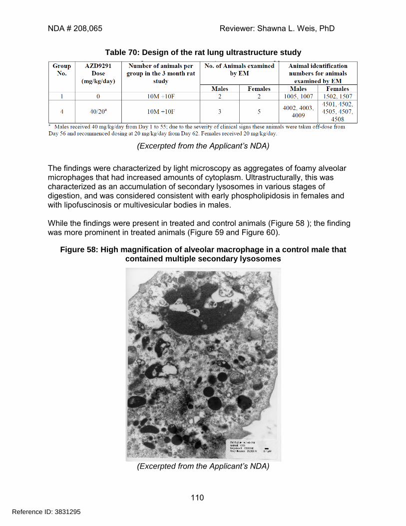
NDA # 208,065 Reviewer: Shawna L. Weis, PhD
110
Table 70: Design of the rat lung ultrastructure study
(Excerpted from the Applicant’s NDA)
The findings were characterized by light microscopy as aggregates of foamy alveolar microphages that had increased amounts of cytoplasm. Ultrastructurally, this was characterized as an accumulation of secondary lysosomes in various stages of digestion, and was considered consistent with early phospholipidosis in females and with lipofuscinosis or multivesicular bodies in males. While the findings were present in treated and control animals (Figure 58 ); the finding was more prominent in treated animals (Figure 59 and Figure 60).
Figure 58: High magnification of alveolar macrophage in a control male that contained multiple secondary lysosomes
(Excerpted from the Applicant’s NDA)
Reference ID: 3831295
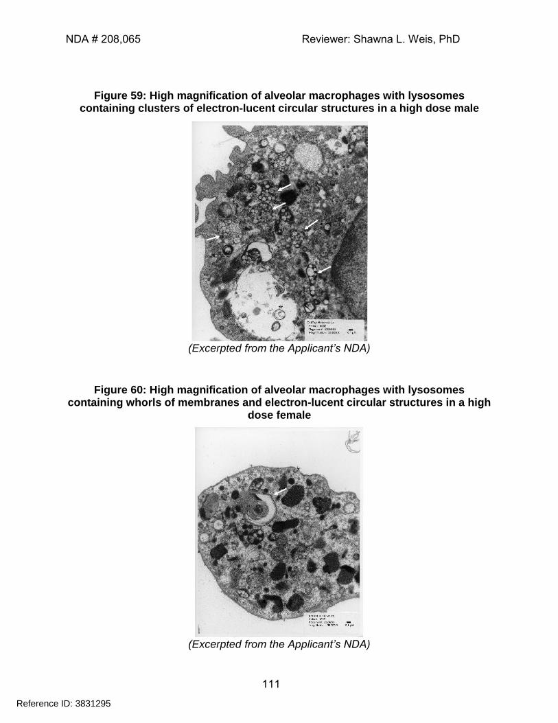
NDA # 208,065 Reviewer: Shawna L. Weis, PhD
111
Figure 59: High magnification of alveolar macrophages with lysosomes containing clusters of electron-lucent circular structures in a high dose male
(Excerpted from the Applicant’s NDA)
Figure 60: High magnification of alveolar macrophages with lysosomes containing whorls of membranes and electron-lucent circular structures in a high
dose female
(Excerpted from the Applicant’s NDA)
Reference ID: 3831295
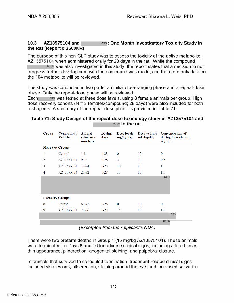
NDA # 208,065 Reviewer: Shawna L. Weis, PhD
112
10.3 AZ13575104 and : One Month Investigatory Toxicity Study in the Rat (Report # 3500KR)
The purpose of this non-GLP study was to assess the toxicity of the active metabolite, AZ13575104 when administered orally for 28 days in the rat. While the compound
was also investigated in this study, the report states that a decision to not progress further development with the compound was made, and therefore only data on the 104 metabolite will be reviewed. The study was conducted in two parts: an initial dose-ranging phase and a repeat-dose phase. Only the repeat-dose phase will be reviewed. Each was tested at three dose levels, using 8 female animals per group. High dose recovery cohorts (N = 3 females/compound; 28 days) were also included for both test agents. A summary of the repeat-dose phase is provided in Table 71.
Table 71: Study Design of the repeat-dose toxicology study of AZ13575104 and in the rat
(Excerpted from the Applicant’s NDA)
There were two preterm deaths in Group 4 (15 mg/kg AZ13575104). These animals were terminated on Days 8 and 16 for adverse clinical signs, including altered feces, thin appearance, piloerection, anogenital staining, and palpebral closure. In animals that survived to scheduled termination, treatment-related clinical signs included skin lesions, piloerection, staining around the eye, and increased salivation.
Reference ID: 3831295
(b) (4)
(b) (4)
(b) (4)
(b) (4)
(b) (4)
(b) (4)
(b) (4)
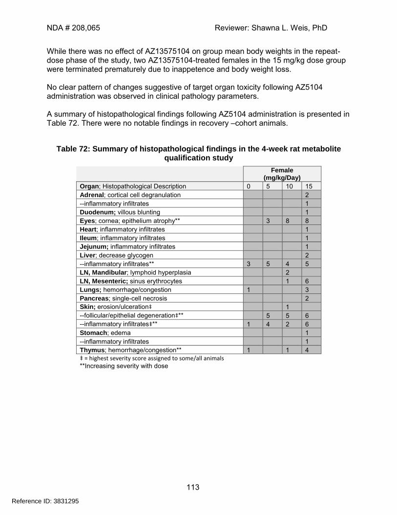
NDA # 208,065 Reviewer: Shawna L. Weis, PhD
113
While there was no effect of AZ13575104 on group mean body weights in the repeat-dose phase of the study, two AZ13575104-treated females in the 15 mg/kg dose group were terminated prematurely due to inappetence and body weight loss. No clear pattern of changes suggestive of target organ toxicity following AZ5104 administration was observed in clinical pathology parameters. A summary of histopathological findings following AZ5104 administration is presented in Table 72. There were no notable findings in recovery –cohort animals.
Table 72: Summary of histopathological findings in the 4-week rat metabolite qualification study
Female (mg/kg/Day)
Organ; Histopathological Description 0 5 10 15
Adrenal; cortical cell degranulation 2
--inflammatory infiltrates 1
Duodenum; villous blunting 1
Eyes; cornea; epithelium atrophy** 3 8 8
Heart; inflammatory infiltrates 1
Ileum; inflammatory infiltrates 1
Jejunum; inflammatory infiltrates 1
Liver; decrease glycogen 2
--inflammatory infiltrates** 3 5 4 5
LN, Mandibular; lymphoid hyperplasia 2
LN, Mesenteric; sinus erythrocytes 1 6
Lungs; hemorrhage/congestion 1 3
Pancreas; single-cell necrosis 2
Skin; erosion/ulceration‡ 1
--follicular/epithelial degeneration‡** 5 5 6
--inflammatory infiltrates‡** 1 4 2 6
Stomach; edema 1
--inflammatory infiltrates 1
Thymus; hemorrhage/congestion** 1 1 4
‡ = highest severity score assigned to some/all animals **Increasing severity with dose
Reference ID: 3831295
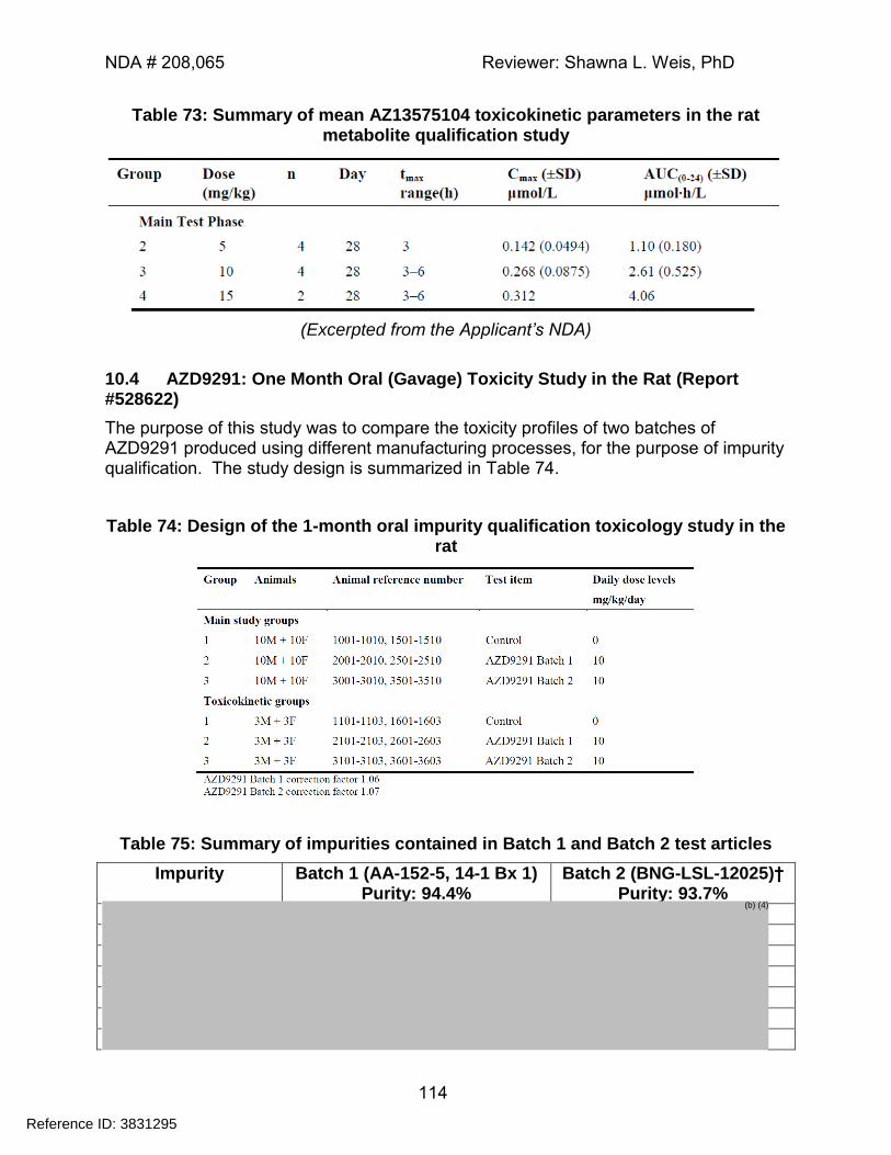
NDA # 208,065 Reviewer: Shawna L. Weis, PhD
114
Table 73: Summary of mean AZ13575104 toxicokinetic parameters in the rat metabolite qualification study
(Excerpted from the Applicant’s NDA)
10.4 AZD9291: One Month Oral (Gavage) Toxicity Study in the Rat (Report #528622)
The purpose of this study was to compare the toxicity profiles of two batches of AZD9291 produced using different manufacturing processes, for the purpose of impurity qualification. The study design is summarized in Table 74.
Table 74: Design of the 1-month oral impurity qualification toxicology study in the rat
Table 75: Summary of impurities contained in Batch 1 and Batch 2 test articles
Impurity Batch 1 (AA-152-5, 14-1 Bx 1) Purity: 94.4%
Batch 2 (BNG-LSL-12025)† Purity: 93.7%
Reference ID: 3831295
(b) (4)
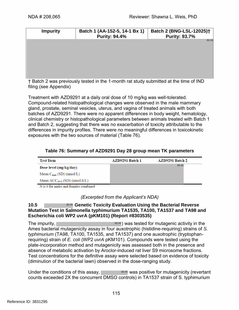
NDA # 208,065 Reviewer: Shawna L. Weis, PhD
115
Impurity Batch 1 (AA-152-5, 14-1 Bx 1) Purity: 94.4%
Batch 2 (BNG-LSL-12025)† Purity: 93.7%
† Batch 2 was previously tested in the 1-month rat study submitted at the time of IND filing (see Appendix) Treatment with AZD9291 at a daily oral dose of 10 mg/kg was well-tolerated. Compound-related histopathological changes were observed in the male mammary gland, prostate, seminal vesicles, uterus, and vagina of treated animals with both batches of AZD9291. There were no apparent differences in body weight, hematology, clinical chemistry or histopathological parameters between animals treated with Batch 1 and Batch 2, suggesting that there was no exacerbation of toxicity attributable to the differences in impurity profiles. There were no meaningful differences in toxicokinetic exposures with the two sources of material (Table 76).
Table 76: Summary of AZD9291 Day 28 group mean TK parameters
(Excerpted from the Applicant’s NDA)
10.5 Genetic Toxicity Evaluation Using the Bacterial Reverse Mutation Test in Salmonella typhimurium TA1535, TA100, TA1537 and TA98 and Escherichia coli WP2 uvrA (pKM101) (Report #8303535)
The impurity, ) was tested for mutagenic activity in the Ames bacterial mutagenicity assay in four auxotrophic (histidine-requiring) strains of S. typhimurium (TA98, TA100, TA1535, and TA1537) and one auxotrophic (tryptophan-requiring) strain of E. coli (WP2 uvrA pKM101). Compounds were tested using the plate-incorporation method and mutagenicity was assessed both in the presence and absence of metabolic activation by Aroclor-induced rat liver S9 microsome fractions. Test concentrations for the definitive assay were selected based on evidence of toxicity (diminution of the bacterial lawn) observed in the dose-ranging study. Under the conditions of this assay, was positive for mutagenicity (revertant counts exceeded 2X the concurrent DMSO controls) in TA1537 strain of S. typhimurium
Reference ID: 3831295
(b) (4)
(b) (4)
(b) (4)
(b) (4)
(b) (4)
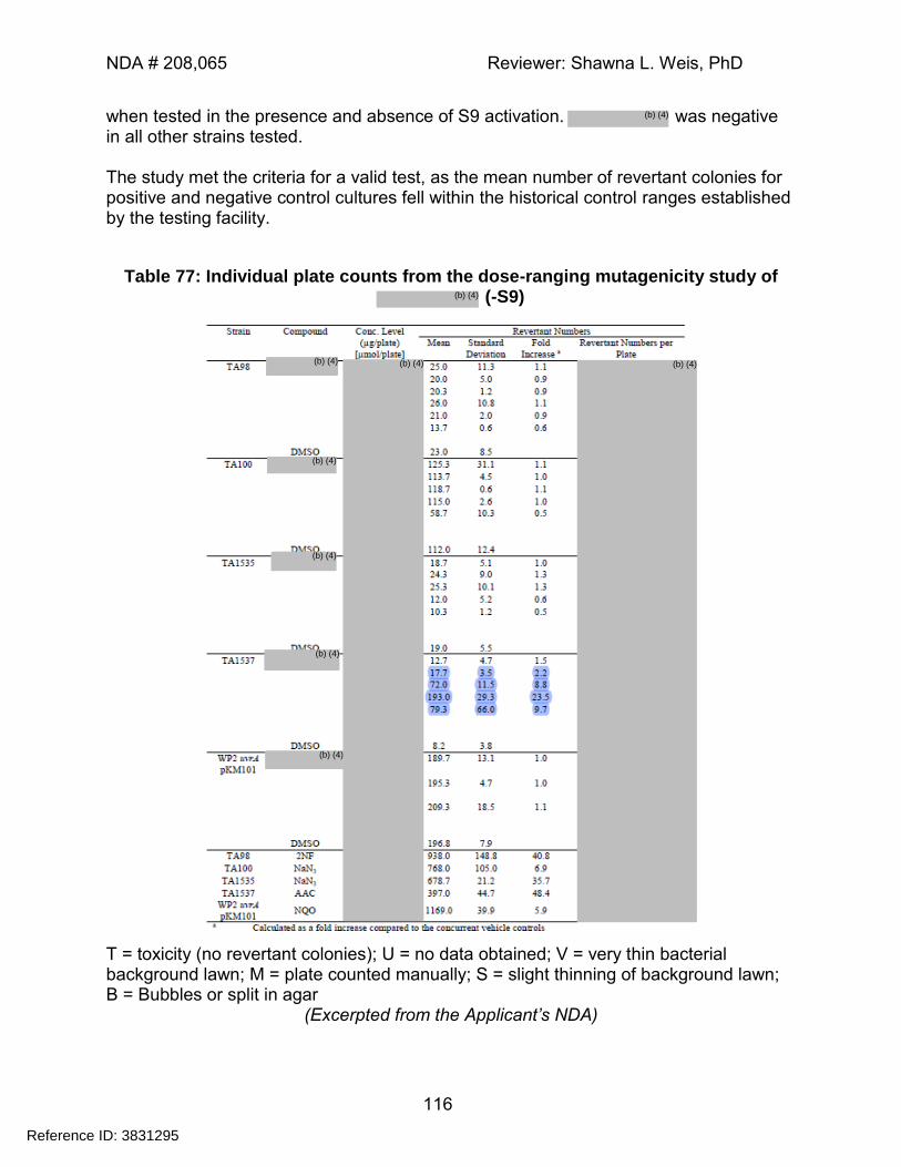
NDA # 208,065 Reviewer: Shawna L. Weis, PhD
116
when tested in the presence and absence of S9 activation. was negative in all other strains tested. The study met the criteria for a valid test, as the mean number of revertant colonies for positive and negative control cultures fell within the historical control ranges established by the testing facility.
Table 77: Individual plate counts from the dose-ranging mutagenicity study of (-S9)
T = toxicity (no revertant colonies); U = no data obtained; V = very thin bacterial background lawn; M = plate counted manually; S = slight thinning of background lawn; B = Bubbles or split in agar
(Excerpted from the Applicant’s NDA)
Reference ID: 3831295
(b) (4)
(b) (4)
(b) (4)
(b) (4)
(b) (4)
(b) (4)
(b) (4)
(b) (4)(b) (4)
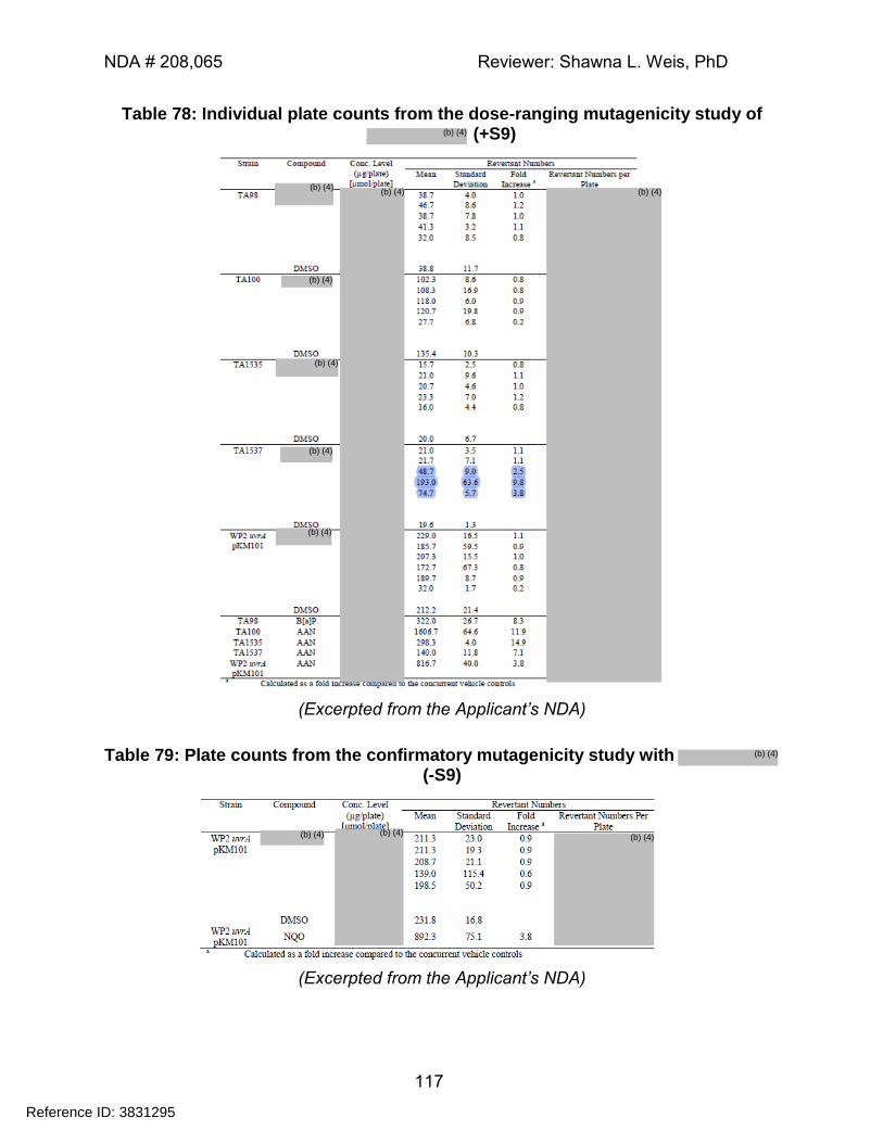
NDA # 208,065 Reviewer: Shawna L. Weis, PhD
117
Table 78: Individual plate counts from the dose-ranging mutagenicity study of (+S9)
(Excerpted from the Applicant’s NDA)
Table 79: Plate counts from the confirmatory mutagenicity study with (-S9)
(Excerpted from the Applicant’s NDA)
Reference ID: 3831295
(b) (4)
(b) (4)
(b) (4)
(b) (4)
(b) (4)
(b) (4)
(b) (4)
(b) (4) (b) (4)
(b) (4) (b) (4) (b) (4)
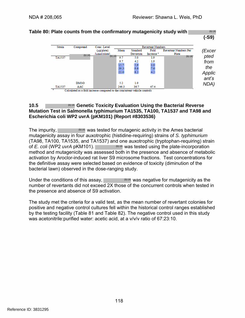
NDA # 208,065 Reviewer: Shawna L. Weis, PhD
118
Table 80: Plate counts from the confirmatory mutagenicity study with (-S9)
(Excerpted from the
Applicant’s NDA)
10.5 Genetic Toxicity Evaluation Using the Bacterial Reverse Mutation Test in Salmonella typhimurium TA1535, TA100, TA1537 and TA98 and Escherichia coli WP2 uvrA (pKM101) (Report #8303536)
The impurity, was tested for mutagenic activity in the Ames bacterial mutagenicity assay in four auxotrophic (histidine-requiring) strains of S. typhimurium (TA98, TA100, TA1535, and TA1537) and one auxotrophic (tryptophan-requiring) strain of E. coli (WP2 uvrA pKM101). was tested using the plate-incorporation method and mutagenicity was assessed both in the presence and absence of metabolic activation by Aroclor-induced rat liver S9 microsome fractions. Test concentrations for the definitive assay were selected based on evidence of toxicity (diminution of the bacterial lawn) observed in the dose-ranging study. Under the conditions of this assay, was negative for mutagenicity as the number of revertants did not exceed 2X those of the concurrent controls when tested in the presence and absence of S9 activation. The study met the criteria for a valid test, as the mean number of revertant colonies for positive and negative control cultures fell within the historical control ranges established by the testing facility (Table 81 and Table 82). The negative control used in this study was acetonitrile:purified water: acetic acid, at a v/v/v ratio of 67:23:10.
Reference ID: 3831295
(b) (4)
(b) (4)
(b) (4)
(b) (4)
(b) (4)
(b) (4) (b) (4) (b) (4)
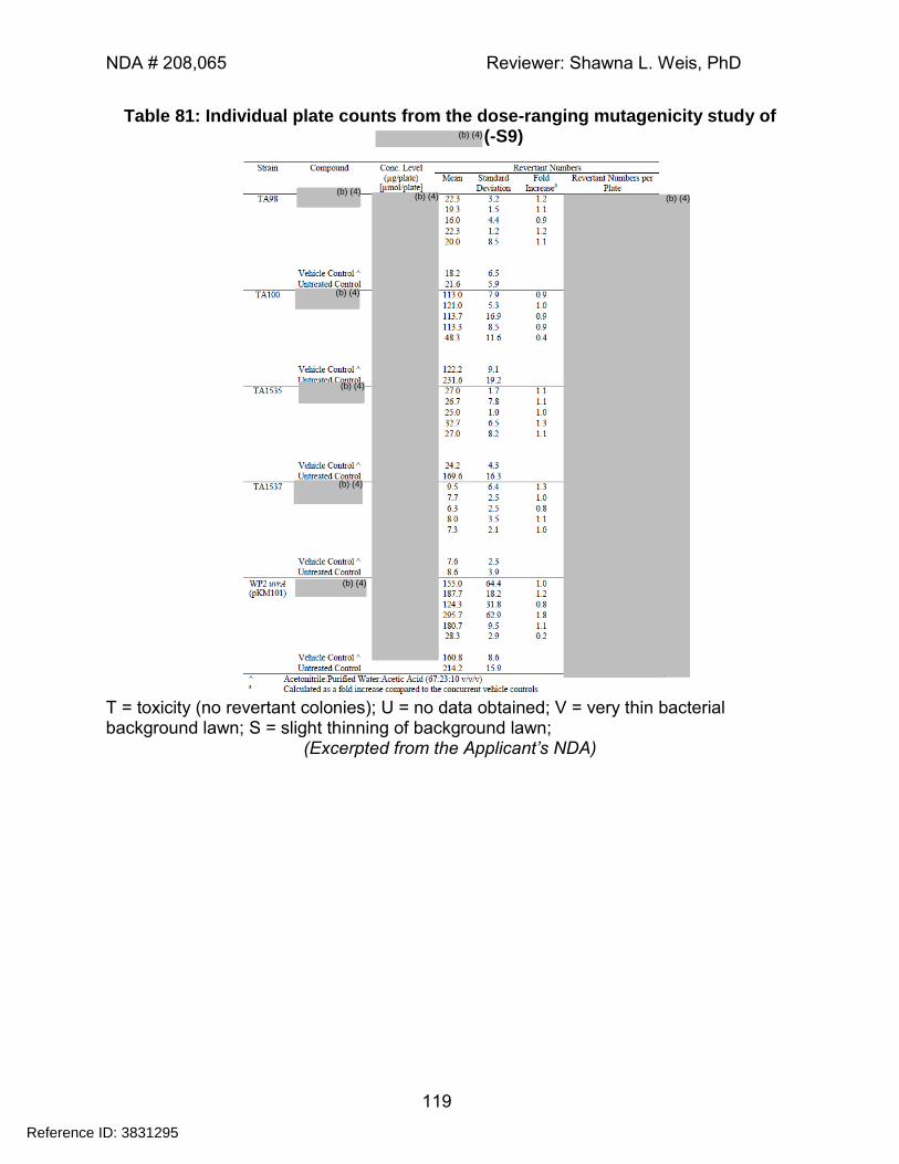
NDA # 208,065 Reviewer: Shawna L. Weis, PhD
119
Table 81: Individual plate counts from the dose-ranging mutagenicity study of (-S9)
T = toxicity (no revertant colonies); U = no data obtained; V = very thin bacterial background lawn; S = slight thinning of background lawn;
(Excerpted from the Applicant’s NDA)
Reference ID: 3831295
(b) (4)
(b) (4)
(b) (4)
(b) (4)
(b) (4)
(b) (4)
(b) (4) (b) (4)
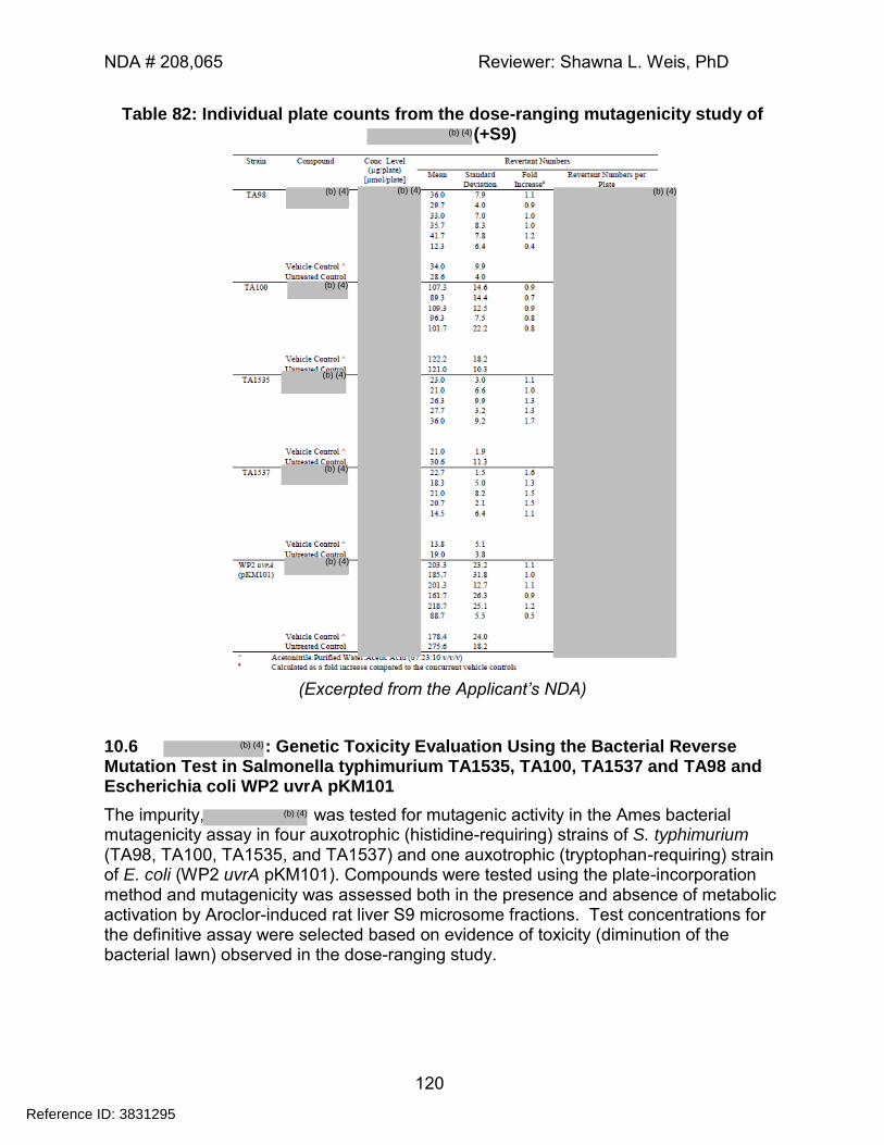
NDA # 208,065 Reviewer: Shawna L. Weis, PhD
120
Table 82: Individual plate counts from the dose-ranging mutagenicity study of (+S9)
(Excerpted from the Applicant’s NDA)
10.6 : Genetic Toxicity Evaluation Using the Bacterial Reverse Mutation Test in Salmonella typhimurium TA1535, TA100, TA1537 and TA98 and Escherichia coli WP2 uvrA pKM101
The impurity, was tested for mutagenic activity in the Ames bacterial mutagenicity assay in four auxotrophic (histidine-requiring) strains of S. typhimurium (TA98, TA100, TA1535, and TA1537) and one auxotrophic (tryptophan-requiring) strain of E. coli (WP2 uvrA pKM101). Compounds were tested using the plate-incorporation method and mutagenicity was assessed both in the presence and absence of metabolic activation by Aroclor-induced rat liver S9 microsome fractions. Test concentrations for the definitive assay were selected based on evidence of toxicity (diminution of the bacterial lawn) observed in the dose-ranging study.
Reference ID: 3831295
(b) (4)
(b) (4)
(b) (4)
(b) (4)
(b) (4)
(b) (4)
(b) (4)
(b) (4)
(b) (4) (b) (4)
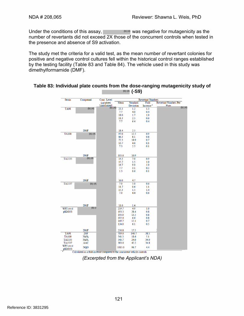
NDA # 208,065 Reviewer: Shawna L. Weis, PhD
121
Under the conditions of this assay, was negative for mutagenicity as the number of revertants did not exceed 2X those of the concurrent controls when tested in the presence and absence of S9 activation. The study met the criteria for a valid test, as the mean number of revertant colonies for positive and negative control cultures fell within the historical control ranges established by the testing facility (Table 83 and Table 84). The vehicle used in this study was dimethylformamide (DMF).
Table 83: Individual plate counts from the dose-ranging mutagenicity study of (-S9)
(Excerpted from the Applicant’s NDA)
Reference ID: 3831295
(b) (4)
(b) (4)
(b) (4)
(b) (4)
(b) (4)
(b) (4)
(b) (4
(b) (4) (b) (4)
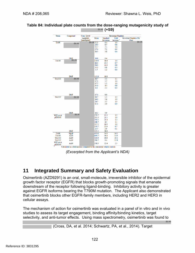
NDA # 208,065 Reviewer: Shawna L. Weis, PhD
122
Table 84: Individual plate counts from the dose-ranging mutagenicity study of (+S9)
(Excerpted from the Applicant’s NDA)
11 Integrated Summary and Safety Evaluation
Osimertinib (AZD9291) is an oral, small-molecule, irreversible inhibitor of the epidermal growth factor receptor (EGFR) that blocks growth-promoting signals that emanate downstream of the receptor following ligand-binding. Inhibitory activity is greater against EGFR isoforms bearing the T790M mutation. The Applicant also demonstrated that osimertinib blocks other EGFR-family members, including HER2 and HER3 in cellular assays. The mechanism of action for osimertinib was evaluated in a panel of in vitro and in vivo studies to assess its target engagement, binding affinity/binding kinetics, target selectivity, and anti-tumor effects. Using mass spectrometry, osimertinib was found to
(Cross, DA, et al. 2014; Schwartz, PA, et al., 2014). Target
Reference ID: 3831295
(b) (4)
(b) (4)
(b) (4)
(b) (4)
(b) (4)
(b) (4
(b) (4) (b) (4)
(b) (4)
NDA # 208,065 Reviewer: Shawna L. Weis, PhD
123
engagement was found to be a time-dependent process, and to persist for at least 48 hours after washout in cultured cell models.
Figure 61: Structural model of Osimertinib binding to the EGFR T790M mutant
The Applicant also evaluated the cross-species metabolic profile of osimertinib in an in vitro panel and demonstrated that osimertinib is extensively metabolized, but that there are no unique human metabolites. The proposed human metabolic scheme for osimertinib is given in Figure 62. Two of its metabolites (AZ13575104 (denoted as M6) and AZ13597550 (denoted as M3)) exhibit a similar degree of pharmacological activity and selectivity as the parent, and are present at approximately 10% of the parent AUC; thus, the pharmacology and toxicity of these compounds were evaluated in tandem throughout the development program. The structures of M3, and M6, are provided below.
Reference ID: 3831295
(b) (4)
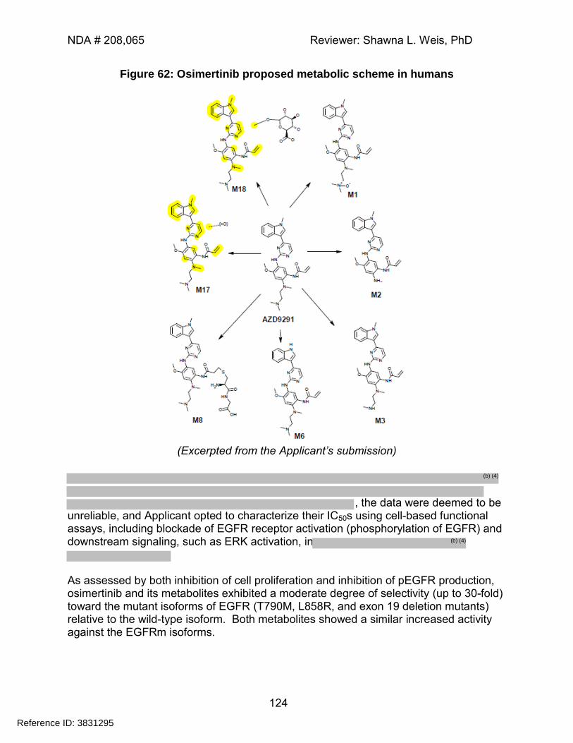
NDA # 208,065 Reviewer: Shawna L. Weis, PhD
124
Figure 62: Osimertinib proposed metabolic scheme in humans
(Excerpted from the Applicant’s submission)
, the data were deemed to be
unreliable, and Applicant opted to characterize their IC50s using cell-based functional assays, including blockade of EGFR receptor activation (phosphorylation of EGFR) and downstream signaling, such as ERK activation, in
As assessed by both inhibition of cell proliferation and inhibition of pEGFR production, osimertinib and its metabolites exhibited a moderate degree of selectivity (up to 30-fold) toward the mutant isoforms of EGFR (T790M, L858R, and exon 19 deletion mutants) relative to the wild-type isoform. Both metabolites showed a similar increased activity against the EGFRm isoforms.
Reference ID: 3831295
(b) (4)
(b) (4)

NDA # 208,065 Reviewer: Shawna L. Weis, PhD
125
The Applicant provided data from several murine tumor models demonstrating the anti-tumor activity of osimertinib and its metabolite, AZ13575104. Consistent with its in vitro properties, both osimertinib and AZ13575104 exhibited greater suppression of Ex19del and T790M/L858R xenografts than were observed in WT tumor models. The Applicant evaluated off-target binding-selectivity and demonstrated that osimertinib and/or its metabolites significantly inhibited the following kinases at potentially physiologically-relevant concentrations (i.e. IC50s≤ 100nM): HER2, HER4, ACK-1, and BLK. Osimertinib demonstrated some potential for inhibition of IGF1R based on IC50 concentrations approaching the clinical Cmax. While inhibition of this receptor seemed unlikely at IC50s that did not factor in free versus bound protein, the Applicant submitted follow-up studies to demonstrate this point. Effects on IGF1R were evaluated in cultured cells that overexpress exogenous human IGF1R; based on these studies, effects of osimertinib and AZ13575104 on IGF1R signaling are likely outside of the physiological range. In receptor binding and enzyme inhibition studies, osimertinib and its metabolites, AZ13575104 and AZ13597550, also inhibited a number of neurotransmitter pathways and/or cardiac ion channels at low multiples (<3-5X) of the observed clinical Cmax. These compounds were found to be less potent than predicted on the basis of receptor binding and enzyme inhibition assays, when they were tested in in electrophysiology assays in whole cells. IC50s in these assays ranged upwards of 30µM, suggesting that osimertinib and its metabolites are unlikely to mediate cardiac toxicity via the hKv1.5, hCav3.2, or hHCN4 channels. The hERG IC50 for osimertinib was 690 nM; therefore, some inhibition is expected at the clinical Cmax of 501 nM, though the free levels of osimertinib are unlikely to reach this level. Osimertinib and/or its metabolites were evaluated in a complete panel of safety pharmacology studies to evaluate their effects on vital organ function, including cardiac, respiratory, CNS, visual, and GI parameters. In the anesthetized Guinea pig, osimertinib administration increased diastolic, mean arterial, and left ventricular systolic pressures; PR, QRS, and QTcB durations, and was associated with a reduction in heart rate and dP/dtmax. When tested in the conscious telemetered beagle dog, osimertinib was also associated with an increase in heart-rate-corrected QTcR, and decreases in dP/dt+ and heart rate. Effects were also observed in the rat telemetry study, particularly during the hours of rest. Administration of osimertinib in rats led to increased blood pressure without a change in heart rate at oral doses of 50 and 100 mg/kg. In the combined visual, respiratory and GI transit study, there were no effects on CNS activity as measured by the functional observational battery. A decrease in pupil size and visual acuity was observed at the mid- or mid- and low-dose levels, respectively. In the respiratory assessment, a decrease in minute volume and expiratory rate were also observed by whole body plethysmography, and in the GI transit study, a decrease in gastric emptying and GI transit was observed.
Reference ID: 3831295
NDA # 208,065 Reviewer: Shawna L. Weis, PhD
126
The general toxicology studies for osimertinib consisted of dose-ranging and repeat-dose GLP toxicology studies in the rat and dog. The selection of toxicology species was made on the basis of pharmacodynamic and metabolic similarity to humans, and dose-selection for pivotal toxicology studies (3-month rat and dog studies) on the basis of MTDs determined in studies of shorter durations. In the 13-week toxicity study in the rat, there were no preterm deaths, but target organs included the eyes (atrophy of the corneal epithelium and retinal dysplasia), skin (inflammatory cell infiltration), lung (inflammatory cell infiltration), kidneys (tubular basophilia and inflammatory cell infiltration) and GI tract (soft feces/hunching/salivation). Hematology changes included decreased red blood cell indices, which correlated with increased incidences of erythrophagocytosis in high dose animals; increased WBC indices; increased fibrinogen and decreased APTT. Clinical chemistry changes included mildly increased ALT and AST; decreased triglycerides, albumin, and total protein. Decreased calcium was also observed; however, this was likely secondary to the observed decreases in albumin. Mating treated males with untreated dams on Days 65-71 indicated a treatment-related decrease in male fertility, demonstrated by decreased numbers of live fetal implants and increased preimplantation loss at the high dose level. Exposures in this study were approximately 0.5-1X the observed clinical AUC in males and females, respectively. While levels of AZ13575104 were measured, they were not reported, due to technical problems with the bioanalytical assay, involving the presence of a co-eluting peak that interfered with the quantitation of this metabolite. In the 3-month rat study, relative exposure to AZ1397550 in high dose animals was approximately 0.77-0.81X those observed in humans at the 80 mg/day dose level. In a separate metabolite qualification study performed with AZ13575104 no unique toxicities were identified compared with those observed with osimertinib. The AUCs achieved at the AZ13575104 maximum tolerated dose of 10 mg/kg were 2.3-fold higher than those expected to occur in humans; thus, the toxicity of both metabolites is considered adequately characterized for use in patients with advanced cancer. In the 13-week dog study, ocular toxicity (corneal atrophy) was dose-limiting, and necessitated a dose-reduction from 10 mg/kg/Day to 6 mg/kg/Day after Day 25. Other histological target organs included the lung (mononuclear infiltration/aggregation), skin (hyperplasia, inflammatory cell infiltration), bone marrow (decreased hematopoiesis) and testes (atrophy). Achieved exposures at the highest dose level in this study were 0.42X of the human AUCs for osimertinib, and 0.51X the human AUC for AZ13597550. Osimertinib was negative in the bacterial and mammalian cell mutagenicity assays, both in the presence and absence of S9 metabolic activation, and negative for induction of structural chromosome aberrations in primary peripheral blood mononuclear cells and in the rat in vivo micronucleus assay. One impurity, was identified as mutagenic in the in vitro reverse mutation assay in bacteria (Ames). The specification for this impurity is no more than %, which, though high for a genotoxic impurity, is below the impurity qualification threshold of 0.15% described in ICH Q3A. At the clinical dose of osimertinib of 80 mg daily, this specification would result in clinical exposures of this impurity of no more than μg/day. Given that this drug is intended for the
Reference ID: 3831295
(b) (4)
(b) (4)
(b) (4)
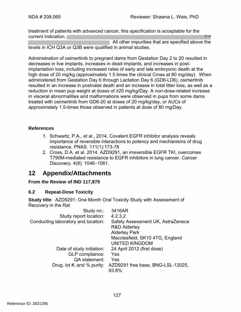
NDA # 208,065 Reviewer: Shawna L. Weis, PhD
127
treatment of patients with advanced cancer, this specification is acceptable for the current indication,
All other impurities that are specified above the levels in ICH Q3A or Q3B were qualified in animal studies. Administration of osimertinib to pregnant dams from Gestation Day 2 to 20 resulted in decreases in live implants, increases in dead implants, and increases in post-implantation loss, including increased rates of early and late embryonic death at the high dose of 20 mg/kg (approximately 1.5 times the clinical Cmax at 80 mg/day). When administered from Gestation Day 6 through Lactation Day 6 (GD6-LD6), osimertinib resulted in an increase in postnatal death and an increase in total litter loss, as well as a reduction in mean pup weight at doses of ≥20 mg/kg/Day. A non-dose-related increase in visceral abnormalities and malformations were observed in pups from some dams treated with osimertinib from GD6-20 at doses of 20 mg/kg/day, or AUCs of approximately 1.5-times those observed in patients at dose of 80 mg/Day.
References
1. Schwartz, P.A., et al., 2014. Covalent EGFR inhibitor analysis reveals importance of reversible interactions to potency and mechanisms of drug resistance. PNAS. 111(1):173-78
2. Cross, D.A. et al. 2014. AZD9291, an irreversible EGFR TKI, overcomes T790M-mediated resistance to EGFR inhibitors in lung cancer. Cancer Discovery. 4(9): 1046–1061.
12 Appendix/Attachments
From the Review of IND 117,879
6.2 Repeat-Dose Toxicity
Study title: AZD9291: One Month Oral Toxicity Study with Assessment of Recovery in the Rat
Study no.: 3416AR Study report location: 4.2.3.2
Conducting laboratory and location: Safety Assessment UK, AstraZeneca R&D Alderley Alderley Park Macclesfield, SK10 4TG, England UNITED KINGDOM
Date of study initiation: 24 April 2012 (first dose) GLP compliance: Yes
QA statement: Yes Drug, lot #, and % purity: AZD9291 free base, BNG-LSL-12025,
93.8%
Reference ID: 3831295
(b) (4)
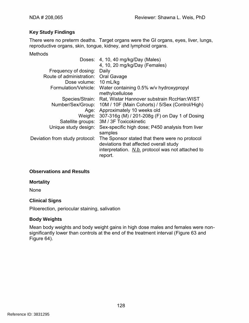
NDA # 208,065 Reviewer: Shawna L. Weis, PhD
128
Key Study Findings
There were no preterm deaths. Target organs were the GI organs, eyes, liver, lungs, reproductive organs, skin, tongue, kidney, and lymphoid organs.
Methods Doses: 4, 10, 40 mg/kg/Day (Males)
4, 10, 20 mg/kg/Day (Females) Frequency of dosing: Daily
Route of administration: Oral Gavage Dose volume: 10 mL/kg
Formulation/Vehicle: Water containing 0.5% w/v hydroxypropyl methylcellulose
Species/Strain: Rat, Wistar Hannover substrain RccHan:WIST Number/Sex/Group: 10M / 10F (Main Cohorts) / 5/Sex (Control/High)
Age: Approximately 10 weeks old Weight: 307-316g (M) / 201-208g (F) on Day 1 of Dosing
Satellite groups: 3M / 3F Toxicokinetic Unique study design: Sex-specific high dose; P450 analysis from liver
samples Deviation from study protocol: The Sponsor stated that there were no protocol
deviations that affected overall study interpretation. N.b. protocol was not attached to report.
Observations and Results
Mortality
None
Clinical Signs
Piloerection, periocular staining, salivation
Body Weights
Mean body weights and body weight gains in high dose males and females were non-significantly lower than controls at the end of the treatment interval (Figure 63 and Figure 64).
Reference ID: 3831295
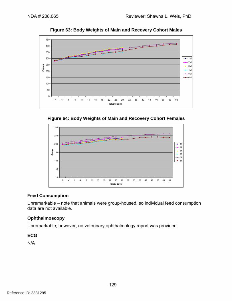
NDA # 208,065 Reviewer: Shawna L. Weis, PhD
129
Figure 63: Body Weights of Main and Recovery Cohort Males
0
50
100
150
200
250
300
350
400
450
-7 -4 1 4 8 11 15 18 22 25 29 32 36 39 43 46 50 53 56
Study Days
Gra
ms
1M
2M
3M
4M
5M
6M
Figure 64: Body Weights of Main and Recovery Cohort Females
0
50
100
150
200
250
300
-7 -4 1 4 8 11 15 18 22 25 29 32 36 39 43 46 50 53 56
Study Days
Gra
ms
1F
2F
3F
4F
5F
6F
Feed Consumption
Unremarkable – note that animals were group-housed, so individual feed consumption data are not available.
Ophthalmoscopy
Unremarkable; however, no veterinary ophthalmology report was provided.
ECG
N/A
Reference ID: 3831295
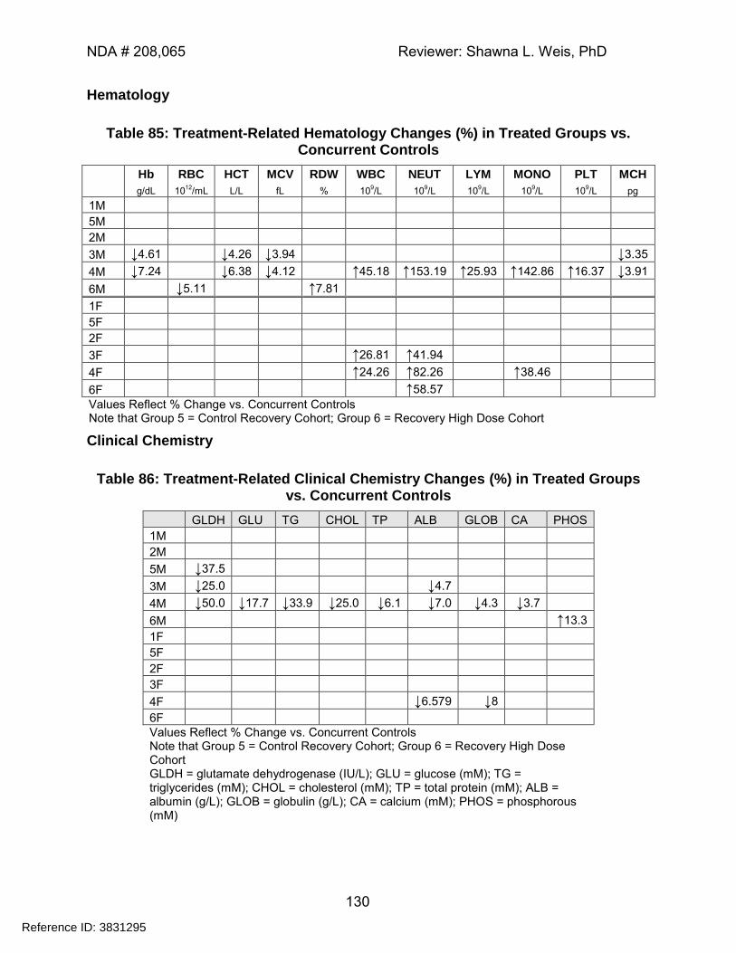
NDA # 208,065 Reviewer: Shawna L. Weis, PhD
130
Hematology
Table 85: Treatment-Related Hematology Changes (%) in Treated Groups vs. Concurrent Controls
Hb RBC HCT MCV RDW WBC NEUT LYM MONO PLT MCH
g/dL 1012
/mL L/L fL % 109/L 10
9/L 10
9/L 10
9/L 10
9/L pg
1M
5M
2M
3M ↓4.61 ↓4.26 ↓3.94 ↓3.35
4M ↓7.24 ↓6.38 ↓4.12 ↑45.18 ↑153.19 ↑25.93 ↑142.86 ↑16.37 ↓3.91
6M ↓5.11 ↑7.81
1F
5F
2F
3F ↑26.81 ↑41.94
4F ↑24.26 ↑82.26 ↑38.46
6F ↑58.57
Values Reflect % Change vs. Concurrent Controls Note that Group 5 = Control Recovery Cohort; Group 6 = Recovery High Dose Cohort
Clinical Chemistry
Table 86: Treatment-Related Clinical Chemistry Changes (%) in Treated Groups vs. Concurrent Controls
GLDH GLU TG CHOL TP ALB GLOB CA PHOS
1M
2M
5M ↓37.5
3M ↓25.0 ↓4.7
4M ↓50.0 ↓17.7 ↓33.9 ↓25.0 ↓6.1 ↓7.0 ↓4.3 ↓3.7
6M ↑13.3
1F
5F
2F
3F
4F ↓6.579 ↓8
6F
Values Reflect % Change vs. Concurrent Controls Note that Group 5 = Control Recovery Cohort; Group 6 = Recovery High Dose Cohort GLDH = glutamate dehydrogenase (IU/L); GLU = glucose (mM); TG = triglycerides (mM); CHOL = cholesterol (mM); TP = total protein (mM); ALB = albumin (g/L); GLOB = globulin (g/L); CA = calcium (mM); PHOS = phosphorous (mM)
Reference ID: 3831295
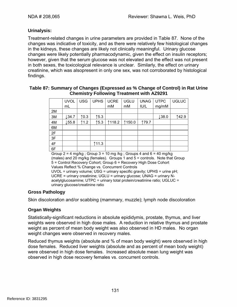
NDA # 208,065 Reviewer: Shawna L. Weis, PhD
131
Urinalysis:
Treatment-related changes in urine parameters are provided in Table 87. None of the changes was indicative of toxicity, and as there were relatively few histological changes in the kidneys, these changes are likely not clinically meaningful. Urinary glucose changes were likely potentially pharmacodynamic, given the effect on insulin receptors; however, given that the serum glucose was not elevated and the effect was not present in both sexes, the toxicological relevance is unclear. Similarly, the effect on urinary creatinine, which was alsopresent in only one sex, was not corroborated by histological findings.
Table 87: Summary of Changes (Expressed as % Change of Control) in Rat Urine Chemistry Following Treatment with AZ9291
UVOL USG UPHS UCRE UGLU UNAG UTPC UGLUC
mL mM mM IU/L mg/mM
2M
3M ↓34.7 ↑0.3 ↑5.3 ↓38.0 ↑42.9
4M ↓55.8 ↑1.2 ↑5.3 ↑118.2 ↑150.0 ↑79.7
6M
2F
3F
4F ↑11.3
6F
Group 2 = 4 mg/kg ; Group 3 = 10 mg /kg ; Groups 4 and 6 = 40 mg/kg (males) and 20 mg/kg (females). Groups 1 and 5 = controls. Note that Group 5 = Control Recovery Cohort; Group 6 = Recovery High Dose Cohort Values Reflect % Change vs. Concurrent Controls UVOL = urinary volume; USG = urinary specific gravity; UPHS = urine pH; UCRE = urinary creatinine; UGLU = urinary glucose; UNAG = urinary N-acetylglucosamine; UTPC = urinary total protein/creatinine ratio; UGLUC = urinary glucose/creatinine ratio
Gross Pathology
Skin discoloration and/or scabbing (mammary, muzzle); lymph node discoloration
Organ Weights
Statistically-significant reductions in absolute epididymis, prostate, thymus, and liver weights were observed in high dose males. A reduction in relative thymus and prostate weight as percent of mean body weight was also observed in HD males. No organ weight changes were observed in recovery males.
Reduced thymus weights (absolute and % of mean body weight) were observed in high dose females. Reduced liver weights (absolute and as percent of mean body weight) were observed in high dose females. Increased absolute mean lung weight was observed in high dose recovery females vs. concurrent controls.
Reference ID: 3831295
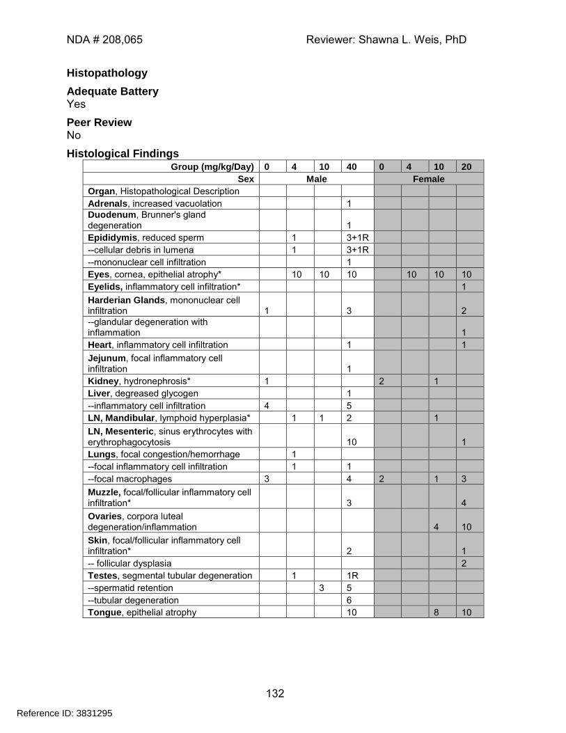
NDA # 208,065 Reviewer: Shawna L. Weis, PhD
132
Histopathology
Adequate Battery Yes
Peer Review No
Histological Findings Group (mg/kg/Day) 0 4 10 40 0 4 10 20
Sex Male Female
Organ, Histopathological Description
Adrenals, increased vacuolation 1
Duodenum, Brunner's gland degeneration 1
Epididymis, reduced sperm 1 3+1R
--cellular debris in lumena 1 3+1R
--mononuclear cell infiltration 1
Eyes, cornea, epithelial atrophy* 10 10 10 10 10 10
Eyelids, inflammatory cell infiltration* 1
Harderian Glands, mononuclear cell infiltration 1 3 2
--glandular degeneration with inflammation 1
Heart, inflammatory cell infiltration 1 1
Jejunum, focal inflammatory cell infiltration 1
Kidney, hydronephrosis* 1 2 1
Liver, degreased glycogen 1
--inflammatory cell infiltration 4 5
LN, Mandibular, lymphoid hyperplasia* 1 1 2 1
LN, Mesenteric, sinus erythrocytes with erythrophagocytosis 10 1
Lungs, focal congestion/hemorrhage 1
--focal inflammatory cell infiltration 1 1
--focal macrophages 3 4 2 1 3
Muzzle, focal/follicular inflammatory cell infiltration* 3 4
Ovaries, corpora luteal degeneration/inflammation 4 10
Skin, focal/follicular inflammatory cell infiltration* 2 1
-- follicular dysplasia 2
Testes, segmental tubular degeneration 1 1R
--spermatid retention 3 5
--tubular degeneration 6
Tongue, epithelial atrophy 10 8 10
Reference ID: 3831295
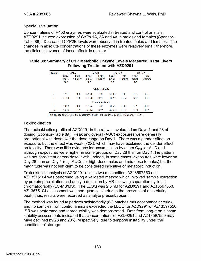
NDA # 208,065 Reviewer: Shawna L. Weis, PhD
133
Special Evaluation
Concentrations of P450 enzymes were evaluated in treated and control animals. AZD9291 induced expression of CYPs 1A, 3A and 4A in males and females (Sponsor-Table 88). Decreased CYP2B levels were observed in treated males and females. The changes in absolute concentrations of these enzymes were relatively small; therefore, the clinical relevance of these effects is unclear.
Table 88: Summary of CYP Metabolic Enzyme Levels Measured in Rat Livers Following Treatment with AZD9291
Toxicokinetics
The toxicokinetics profile of AZD9291 in the rat was evaluated on Days 1 and 28 of dosing (Sponsor-Table 89). Peak and overall (AUC) exposures were generally proportional with dose over the dose range on Day 1. There was a gender effect on exposure, but the effect was weak (<2X), which may have explained the gender effect on toxicity. There was little evidence for accumulation by either Cmax or AUC and although exposures were higher in some groups on Day 28 than on Day 1, the pattern was not consistent across dose levels; indeed, in some cases, exposures were lower on Day 28 than on Day 1 (e.g. AUCs for high-dose males and mid-dose females) but the magnitude was not sufficient to be considered indicative of metabolic induction.
Toxicokinetic analysis of AZD9291 and its two metabolites, AZ13597550 and AZ13575104 was performed using a validated method which involved sample extraction by protein precipitation and analyte detection by MS following separation by liquid chromatography (LC-MS/MS). The LLOQ was 2.5 nM for AZD9291 and AZ13597550. AZ13575104 assessment was non-quantitative due to the presence of a co-eluting peak; thus, results were recorded as analyte present/absent.
The method was found to perform satisfactorily (8/8 batches met acceptance criteria), and no samples from control animals exceeded the LLOQ for AZD9291 or AZ13597550. ISR was performed and reproducibility was demonstrated. Data from long-term plasma stability assessments indicated that concentrations of AZD9291 and AZ13597550 may have declined by 23 and 20%, respectively, due to temporal instability under the conditions of storage.
Reference ID: 3831295
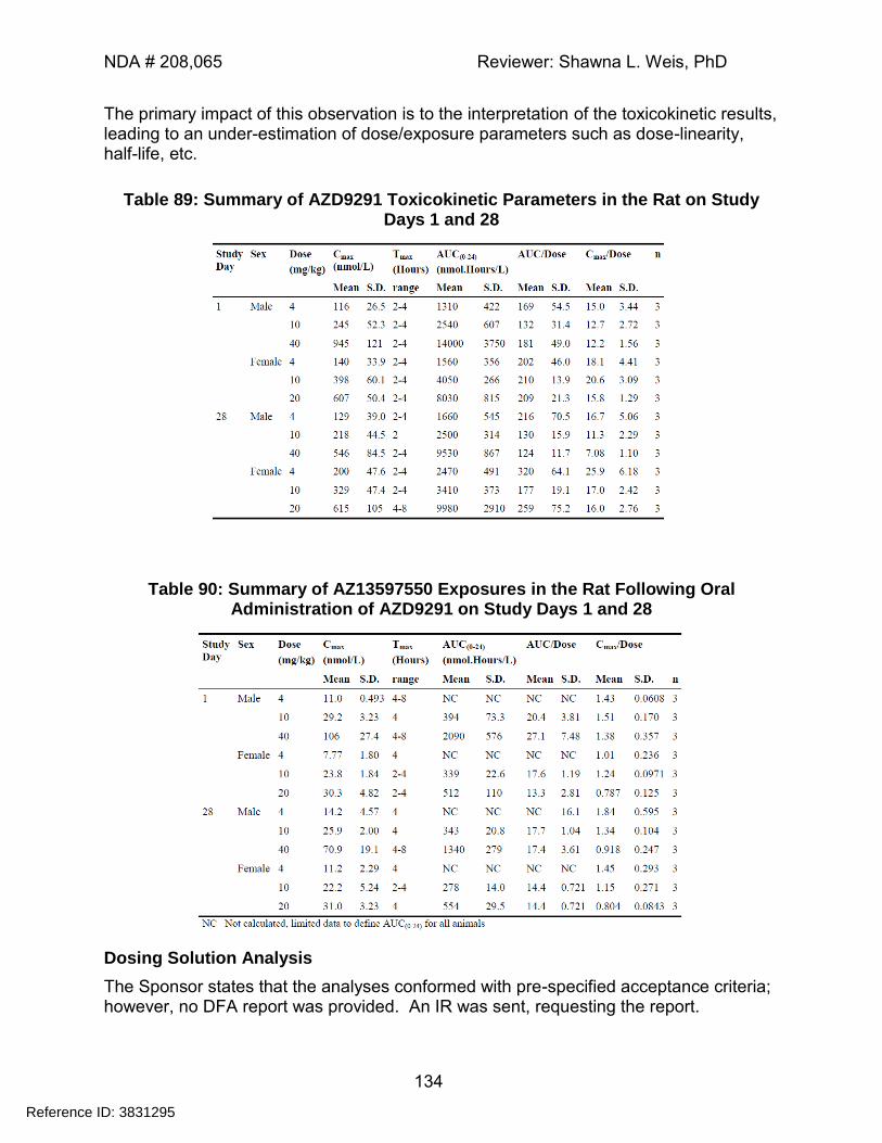
NDA # 208,065 Reviewer: Shawna L. Weis, PhD
134
The primary impact of this observation is to the interpretation of the toxicokinetic results, leading to an under-estimation of dose/exposure parameters such as dose-linearity, half-life, etc.
Table 89: Summary of AZD9291 Toxicokinetic Parameters in the Rat on Study Days 1 and 28
Table 90: Summary of AZ13597550 Exposures in the Rat Following Oral Administration of AZD9291 on Study Days 1 and 28
Dosing Solution Analysis
The Sponsor states that the analyses conformed with pre-specified acceptance criteria; however, no DFA report was provided. An IR was sent, requesting the report.
Reference ID: 3831295
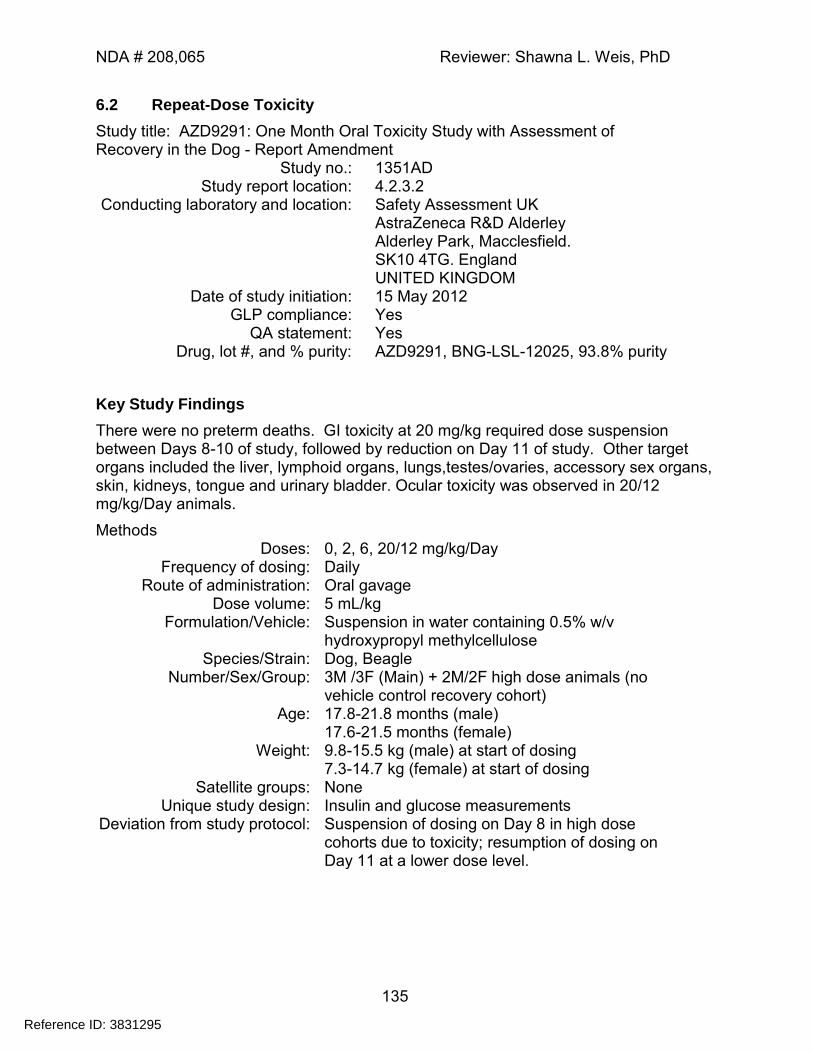
NDA # 208,065 Reviewer: Shawna L. Weis, PhD
135
6.2 Repeat-Dose Toxicity
Study title: AZD9291: One Month Oral Toxicity Study with Assessment of Recovery in the Dog - Report Amendment
Study no.: 1351AD Study report location: 4.2.3.2
Conducting laboratory and location: Safety Assessment UK AstraZeneca R&D Alderley Alderley Park, Macclesfield. SK10 4TG. England UNITED KINGDOM
Date of study initiation: 15 May 2012 GLP compliance: Yes
QA statement: Yes Drug, lot #, and % purity: AZD9291, BNG-LSL-12025, 93.8% purity
Key Study Findings
There were no preterm deaths. GI toxicity at 20 mg/kg required dose suspension between Days 8-10 of study, followed by reduction on Day 11 of study. Other target organs included the liver, lymphoid organs, lungs,testes/ovaries, accessory sex organs, skin, kidneys, tongue and urinary bladder. Ocular toxicity was observed in 20/12 mg/kg/Day animals.
Methods Doses: 0, 2, 6, 20/12 mg/kg/Day
Frequency of dosing: Daily Route of administration: Oral gavage
Dose volume: 5 mL/kg Formulation/Vehicle: Suspension in water containing 0.5% w/v
hydroxypropyl methylcellulose Species/Strain: Dog, Beagle
Number/Sex/Group: 3M /3F (Main) + 2M/2F high dose animals (no vehicle control recovery cohort)
Age: 17.8-21.8 months (male) 17.6-21.5 months (female)
Weight: 9.8-15.5 kg (male) at start of dosing 7.3-14.7 kg (female) at start of dosing
Satellite groups: None Unique study design: Insulin and glucose measurements
Deviation from study protocol: Suspension of dosing on Day 8 in high dose cohorts due to toxicity; resumption of dosing on Day 11 at a lower dose level.
Reference ID: 3831295
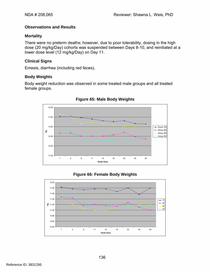
NDA # 208,065 Reviewer: Shawna L. Weis, PhD
136
Observations and Results
Mortality
There were no preterm deaths; however, due to poor tolerability, dosing in the high dose (20 mg/kg/Day) cohorts was suspended between Days 8-10, and reinitiated at a lower dose level (12 mg/kg/Day) on Day 11.
Clinical Signs
Emesis, diarrhea (including red feces),
Body Weights
Body weight reduction was observed in some treated male groups and all treated female groups.
Figure 65: Male Body Weights
11.50
12.00
12.50
13.00
13.50
14.00
1 4 8 11 15 18 22 25 29
Study Days
Kg
Group 1M
Group 2M
Group 3M
Group 4M
Figure 66: Female Body Weights
10.40
10.60
10.80
11.00
11.20
11.40
11.60
11.80
12.00
1 4 8 11 15 18 22 25 29
Study Days
Kg
1F
2F
3F
4F
Reference ID: 3831295
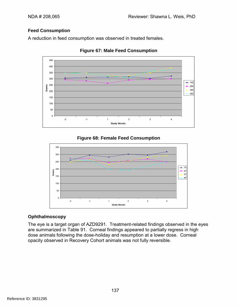
NDA # 208,065 Reviewer: Shawna L. Weis, PhD
137
Feed Consumption
A reduction in feed consumption was observed in treated females.
Figure 67: Male Feed Consumption
0
50
100
150
200
250
300
350
400
450
-2 -1 1 2 3 4
Study Weeks
Gra
ms
1M
2M
3M
4M
Figure 68: Female Feed Consumption
0
50
100
150
200
250
300
350
-2 -1 1 2 3 4
Study Weeks
Gra
ms
1F
2F
3F
4F
Ophthalmoscopy
The eye is a target organ of AZD9291. Treatment-related findings observed in the eyes are summarized in Table 91. Corneal findings appeared to partially regress in high dose animals following the dose-holiday and resumption at a lower dose. Corneal opacity observed in Recovery Cohort animals was not fully reversible.
Reference ID: 3831295
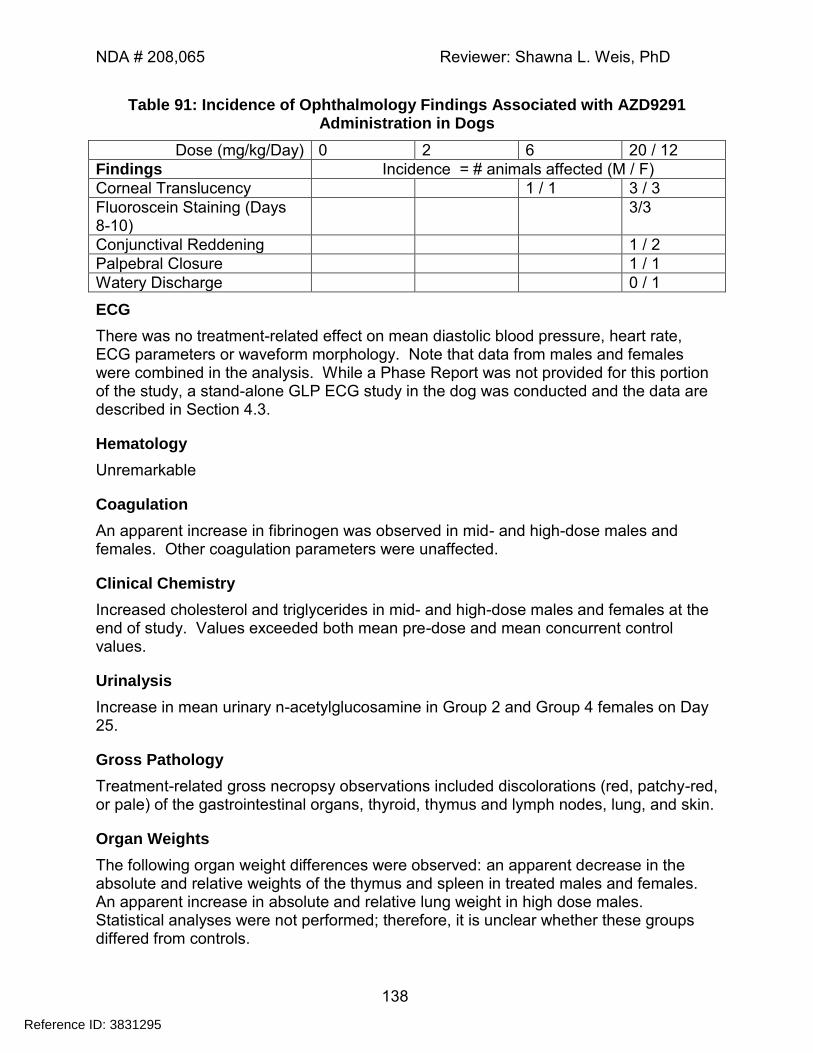
NDA # 208,065 Reviewer: Shawna L. Weis, PhD
138
Table 91: Incidence of Ophthalmology Findings Associated with AZD9291 Administration in Dogs
Dose (mg/kg/Day) 0 2 6 20 / 12
Findings Incidence = # animals affected (M / F)
Corneal Translucency 1 / 1 3 / 3
Fluoroscein Staining (Days 8-10)
3/3
Conjunctival Reddening 1 / 2
Palpebral Closure 1 / 1
Watery Discharge 0 / 1
ECG
There was no treatment-related effect on mean diastolic blood pressure, heart rate, ECG parameters or waveform morphology. Note that data from males and females were combined in the analysis. While a Phase Report was not provided for this portion of the study, a stand-alone GLP ECG study in the dog was conducted and the data are described in Section 4.3.
Hematology
Unremarkable
Coagulation
An apparent increase in fibrinogen was observed in mid- and high-dose males and females. Other coagulation parameters were unaffected.
Clinical Chemistry
Increased cholesterol and triglycerides in mid- and high-dose males and females at the end of study. Values exceeded both mean pre-dose and mean concurrent control values.
Urinalysis
Increase in mean urinary n-acetylglucosamine in Group 2 and Group 4 females on Day 25.
Gross Pathology
Treatment-related gross necropsy observations included discolorations (red, patchy-red, or pale) of the gastrointestinal organs, thyroid, thymus and lymph nodes, lung, and skin.
Organ Weights
The following organ weight differences were observed: an apparent decrease in the absolute and relative weights of the thymus and spleen in treated males and females. An apparent increase in absolute and relative lung weight in high dose males. Statistical analyses were not performed; therefore, it is unclear whether these groups differed from controls.
Reference ID: 3831295
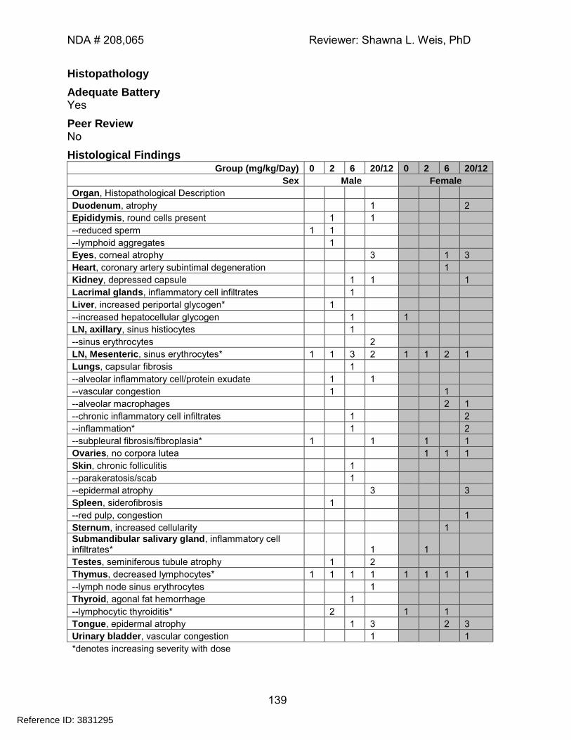
NDA # 208,065 Reviewer: Shawna L. Weis, PhD
139
Histopathology
Adequate Battery Yes
Peer Review No
Histological Findings Group (mg/kg/Day) 0 2 6 20/12 0 2 6 20/12
Sex Male Female
Organ, Histopathological Description
Duodenum, atrophy 1 2
Epididymis, round cells present 1 1
--reduced sperm 1 1
--lymphoid aggregates 1
Eyes, corneal atrophy 3 1 3
Heart, coronary artery subintimal degeneration 1
Kidney, depressed capsule 1 1 1
Lacrimal glands, inflammatory cell infiltrates 1
Liver, increased periportal glycogen* 1
--increased hepatocellular glycogen 1 1
LN, axillary, sinus histiocytes 1
--sinus erythrocytes 2
LN, Mesenteric, sinus erythrocytes* 1 1 3 2 1 1 2 1
Lungs, capsular fibrosis 1
--alveolar inflammatory cell/protein exudate 1 1
--vascular congestion 1 1
--alveolar macrophages 2 1
--chronic inflammatory cell infiltrates 1 2
--inflammation* 1 2
--subpleural fibrosis/fibroplasia* 1 1 1 1
Ovaries, no corpora lutea 1 1 1
Skin, chronic folliculitis 1
--parakeratosis/scab 1
--epidermal atrophy 3 3
Spleen, siderofibrosis 1
--red pulp, congestion 1
Sternum, increased cellularity 1
Submandibular salivary gland, inflammatory cell infiltrates* 1 1
Testes, seminiferous tubule atrophy 1 2
Thymus, decreased lymphocytes* 1 1 1 1 1 1 1 1
--lymph node sinus erythrocytes 1
Thyroid, agonal fat hemorrhage 1
--lymphocytic thyroiditis* 2 1 1
Tongue, epidermal atrophy 1 3 2 3
Urinary bladder, vascular congestion 1 1
*denotes increasing severity with dose
Reference ID: 3831295
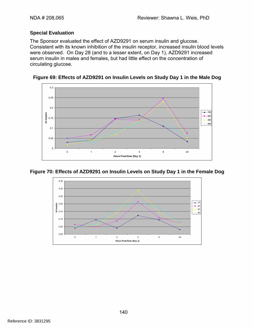
NDA # 208,065 Reviewer: Shawna L. Weis, PhD
140
Special Evaluation
The Sponsor evaluated the effect of AZD9291 on serum insulin and glucose. Consistent with its known inhibition of the insulin receptor, increased insulin blood levels were observed. On Day 28 (and to a lesser extent, on Day 1), AZD9291 increased serum insulin in males and females, but had little effect on the concentration of circulating glucose.
Figure 69: Effects of AZD9291 on Insulin Levels on Study Day 1 in the Male Dog
0
0.05
0.1
0.15
0.2
0.25
0.3
0 1 2 4 8 24
Hours Post-Dose (Day 1)
IU I
nsu
lin 1M
2M
3M
4M
Figure 70: Effects of AZD9291 on Insulin Levels on Study Day 1 in the Female Dog
0.00
0.05
0.10
0.15
0.20
0.25
0.30
0.35
0 1 2 4 8 24
Hours Post-Dose (Day 1)
IU I
nsu
lin 1F
2F
3F
4F
Reference ID: 3831295
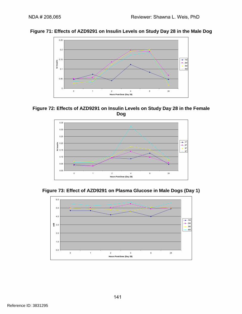
NDA # 208,065 Reviewer: Shawna L. Weis, PhD
141
Figure 71: Effects of AZD9291 on Insulin Levels on Study Day 28 in the Male Dog
0
0.05
0.1
0.15
0.2
0.25
0 1 2 4 8 24
Hours Post-Dose (Day 28)
IU I
nsu
lin 1M
2M
3M
4M
Figure 72: Effects of AZD9291 on Insulin Levels on Study Day 28 in the Female Dog
0.00
0.05
0.10
0.15
0.20
0.25
0.30
0.35
0 1 2 4 8 24
Hours Post-Dose (Day 28)
IU I
nsu
lin 1F
2F
3F
4F
Figure 73: Effect of AZD9291 on Plasma Glucose in Male Dogs (Day 1)
0.0
1.0
2.0
3.0
4.0
5.0
6.0
0 1 2 4 8 24
Hours Post-Dose (Day 28)
mM
1M
2M
3M
4M
Reference ID: 3831295
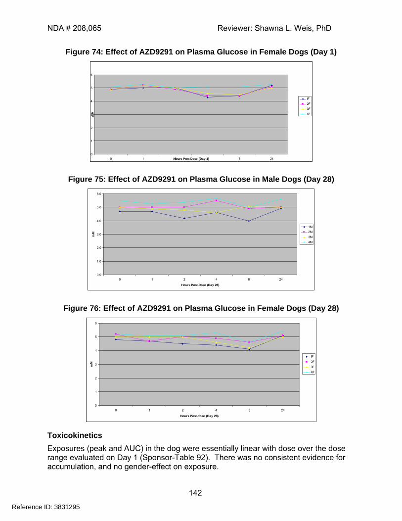
NDA # 208,065 Reviewer: Shawna L. Weis, PhD
142
Figure 74: Effect of AZD9291 on Plasma Glucose in Female Dogs (Day 1)
0
1
2
3
4
5
6
0 1 2 4 8 24Hours Post-Dose (Day 1)
mM
IF
2F
3F
4F
Figure 75: Effect of AZD9291 on Plasma Glucose in Male Dogs (Day 28)
0.0
1.0
2.0
3.0
4.0
5.0
6.0
0 1 2 4 8 24
Hours Post-Dose (Day 28)
mM
1M
2M
3M
4M
Figure 76: Effect of AZD9291 on Plasma Glucose in Female Dogs (Day 28)
0
1
2
3
4
5
6
0 1 2 4 8 24
Hours Post-dose (Day 28)
mM
IF
2F
3F
4F
Toxicokinetics
Exposures (peak and AUC) in the dog were essentially linear with dose over the dose range evaluated on Day 1 (Sponsor-Table 92). There was no consistent evidence for accumulation, and no gender-effect on exposure.
Reference ID: 3831295
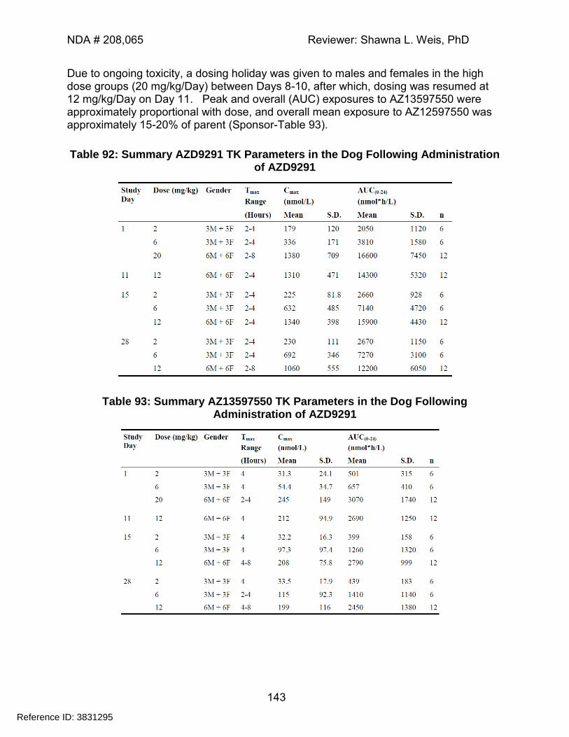
NDA # 208,065 Reviewer: Shawna L. Weis, PhD
143
Due to ongoing toxicity, a dosing holiday was given to males and females in the high dose groups (20 mg/kg/Day) between Days 8-10, after which, dosing was resumed at 12 mg/kg/Day on Day 11. Peak and overall (AUC) exposures to AZ13597550 were approximately proportional with dose, and overall mean exposure to AZ12597550 was approximately 15-20% of parent (Sponsor-Table 93).
Table 92: Summary AZD9291 TK Parameters in the Dog Following Administration of AZD9291
Table 93: Summary AZ13597550 TK Parameters in the Dog Following Administration of AZD9291
Reference ID: 3831295
---------------------------------------------------------------------------------------------------------This is a representation of an electronic record that was signedelectronically and this page is the manifestation of the electronicsignature.---------------------------------------------------------------------------------------------------------/s/----------------------------------------------------
SHAWNA L WEIS10/08/2015
WHITNEY S HELMS10/08/2015
Reference ID: 3831295
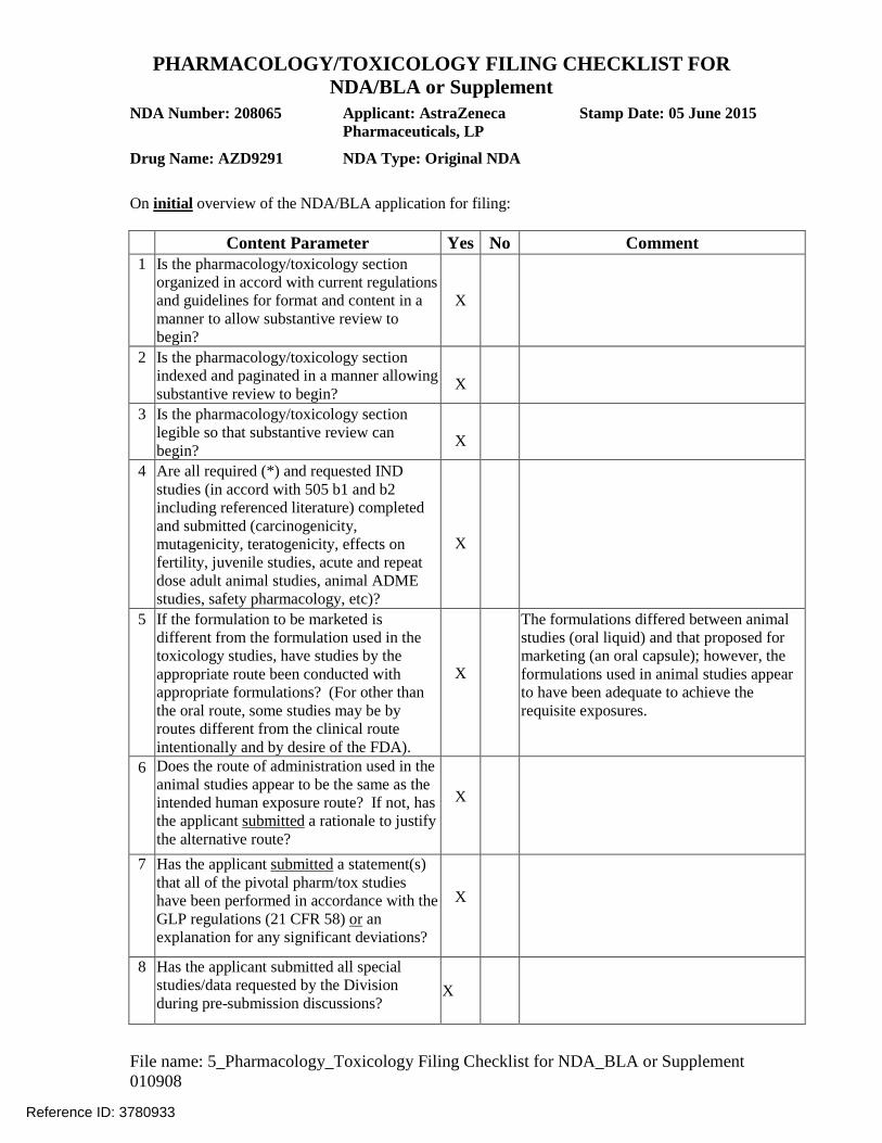
PHARMACOLOGY/TOXICOLOGY FILING CHECKLIST FOR NDA/BLA or Supplement
File name: 5_Pharmacology_Toxicology Filing Checklist for NDA_BLA or Supplement 010908
NDA Number: 208065 Applicant: AstraZeneca Pharmaceuticals, LP
Stamp Date: 05 June 2015
Drug Name: AZD9291 NDA Type: Original NDA
On initial overview of the NDA/BLA application for filing:
Content Parameter
Yes
No
Comment 1 Is the pharmacology/toxicology section
organized in accord with current regulations and guidelines for format and content in a manner to allow substantive review to begin?
X
2
Is the pharmacology/toxicology section indexed and paginated in a manner allowing substantive review to begin?
X
3
Is the pharmacology/toxicology section legible so that substantive review can begin?
X
4
Are all required (*) and requested IND studies (in accord with 505 b1 and b2 including referenced literature) completed and submitted (carcinogenicity, mutagenicity, teratogenicity, effects on fertility, juvenile studies, acute and repeat dose adult animal studies, animal ADME studies, safety pharmacology, etc)?
X
5
If the formulation to be marketed is different from the formulation used in the toxicology studies, have studies by the appropriate route been conducted with appropriate formulations? (For other than the oral route, some studies may be by routes different from the clinical route intentionally and by desire of the FDA).
X
The formulations differed between animal studies (oral liquid) and that proposed for marketing (an oral capsule); however, the formulations used in animal studies appear to have been adequate to achieve the requisite exposures.
6
Does the route of administration used in the animal studies appear to be the same as the intended human exposure route? If not, has the applicant submitted a rationale to justify the alternative route?
X
7 Has the applicant submitted a statement(s) that all of the pivotal pharm/tox studies have been performed in accordance with the GLP regulations (21 CFR 58) or an explanation for any significant deviations?
X
8 Has the applicant submitted all special studies/data requested by the Division during pre-submission discussions?
X
Reference ID: 3780933
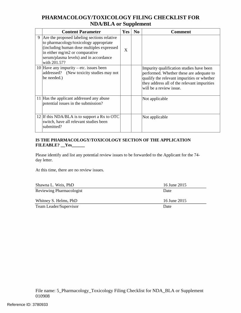
PHARMACOLOGY/TOXICOLOGY FILING CHECKLIST FOR NDA/BLA or Supplement
File name: 5_Pharmacology_Toxicology Filing Checklist for NDA_BLA or Supplement 010908
Content Parameter
Yes
No
Comment
9 Are the proposed labeling sections relative to pharmacology/toxicology appropriate (including human dose multiples expressed in either mg/m2 or comparative serum/plasma levels) and in accordance with 201.57?
X
10 Have any impurity – etc. issues been addressed? (New toxicity studies may not be needed.)
Impurity qualification studies have been performed. Whether these are adequate to qualify the relevant impurities or whether they address all of the relevant impurities will be a review issue.
11 Has the applicant addressed any abuse potential issues in the submission?
Not applicable
12 If this NDA/BLA is to support a Rx to OTC switch, have all relevant studies been submitted?
Not applicable
IS THE PHARMACOLOGY/TOXICOLOGY SECTION OF THE APPLICATION FILEABLE? __Yes______ Please identify and list any potential review issues to be forwarded to the Applicant for the 74-day letter. At this time, there are no review issues. Shawna L. Weis, PhD 16 June 2015 Reviewing Pharmacologist Date Whitney S. Helms, PhD 16 June 2015 Team Leader/Supervisor Date
Reference ID: 3780933

---------------------------------------------------------------------------------------------------------This is a representation of an electronic record that was signedelectronically and this page is the manifestation of the electronicsignature.---------------------------------------------------------------------------------------------------------/s/----------------------------------------------------
SHAWNA L WEIS06/18/2015
WHITNEY S HELMS06/19/2015
Reference ID: 3780933
Recommended