2017 RECOMMENDATIONS FOR THE
DIAGNOSIS, TREATMENT AND
MANAGEMENT OF TUBERCULOSIS
(Mycobacterium tuberculosis) IN ELEPHANTS
IN HUMAN CARE
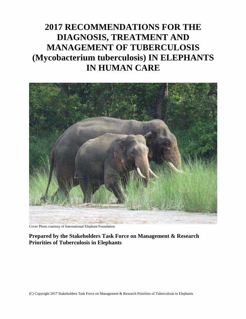
Cover Photo courtesy of International Elephant Foundation
Prepared by the Stakeholders Task Force on Management & Research
Priorities of Tuberculosis in Elephants
(C) Copyright 2017 Stakeholders Task Force on Management & Research Priorities of Tuberculosis in Elephants
Page 1 of 58
2017 RECOMMENDATIONS FOR THE DIAGNOSIS, TREATMENT
AND MANAGEMENT OF TUBERCULOSIS (Mycobacterium tuberculosis)
IN ELEPHANTS IN HUMAN CARE
Prepared by the Stakeholders Task Force on
Management & Research Priorities of Tuberculosis in Elephants
Executive Summary
African and Asian elephants are both susceptible to infection by Mycobacterium
tuberculosis (Mtb). The Asian elephant has lived in close association with humans in Asian
range countries for thousands of years and this close partnership is likely responsible for the
exposure of the Asian elephant to a human disease. African elephants, in contrast, with whom
historically have had fewer contacts with humans the disease has been less common. Incidental
reports of Mtb-like disease in the Asian elephant go back thousands of years, (Chalke 1962). The
confirmation of the existence of Mtb infection in elephants has only occurred recently after a
testing program was initiated in 1996 in elephant-holding facilities in the United States.
Mycobacterium tuberculosis (Mtb) is now recognized as a disease primarily of captive Asian
elephants (Elephas maximus) though some cases have been identified in wild Asian elephants
and one case not confirmed by culture in a wild African elephant. Although two decades of
routine testing and monitoring in the United States have taught us a great deal about Mtb in
elephants, our understanding of its epidemiology and pathophysiology in elephants is still
evolving.
In general, Mtb is transmitted through close, prolonged contact with an infected person or
animal shedding the organism. Transmission of the disease from elephants to humans is therefore
an occupational health concern of elephant caretakers rather than a general public health concern.
Prevalence studies from 1997-2011 have shown an Mtb point prevalence of 5.1% in the living
captive U.S. Asian elephant population and 0% for African elephants (Feldman 2013).
Asian elephants infected with Mtb may have variable disease manifestations but most
infected elephants do not show clinical signs unless the disease becomes advanced. Multiple
diagnostic and screening tools are available to assist in diagnosis but confirmation remains
challenging. Currently the only test available for identifying truly infected elephants is through
culture of the organism, the gold standard test for diagnosis. Samples for culture are typically
obtained from living elephants using trunk wash samples, considered the equivalent of human
sputum samples.
As of October 2015, the United States Department of Agriculture, Division of Animal
and Plant Health Inspection Services (USDA APHIS) no longer regulates the surveillance,
diagnosis and treatment of the disease in elephants within the United States. Currently, USDA
APHIS places this responsibility in the hands of the attending veterinarian. Thus, veterinarians
working with elephants need a thorough understanding of the disease in these species and a
willingness to work closely with their state veterinarian and public health officials to manage the
disease appropriately if diagnosed.
This document is a continuing multi-year effort of the Elephant Care Stakeholders
(hereon “the stakeholders”), a group of veterinarians, elephant managers, public health
specialists, epidemiologists, pharmacologists, physicians and other professionals with many
Page 2 of 58
years of experience working with elephants in zoos, circuses, and private facilities. Their efforts
were initiated following a recommendation from USDA APHIS to bring more transparency and
stakeholder involvement to the development of useful, consistent and easy to follow guidelines
for dealing with elephant tuberculosis.
The stakeholders offer these 2017 Recommendations for the Diagnosis, Management,
and Treatment of Tuberculosis in Elephants in Human Care’ as a guide for veterinarians,
elephant caretakers, government regulators and public health officials dealing with elephants as
well as an accurate source of information for the general public. This document is a multi-year
collaborative effort that is updated regularly as the science and knowledge of Mtb in the elephant
advances. The stakeholders advocate good management, medical surveillance, scientific
cooperation, and the use of evidence based medicine.
The findings and conclusions in this report are those of the authors and do not necessarily
represent the views of the Centers for Disease Control and Prevention, National Institute for
Occupational Safety and Health.
_________________
Acknowledgments: The Elephant Care Stakeholders Task Force would like to thank the
following organizations and persons for their support:
• American Association of Zoo Veterinarians (AAZV)
• International Elephant Foundation (IEF)
• Feld Entertainment, Inc.
• Elephant Managers Association (EMA)
• The many professionals who volunteered their time and expertise in the creation of this
document.
Editors: Dr. Kay Backues, DVM, DACZM and Dr. Ellen Wiedner, VMD, DACVIM, DACZM
Attendees to 2016 Stakeholders Meeting Atlanta, Georgia and Contributors to 2017
Version in Alphabetical Order: Noha Abou-Madi, DVM, MSc, DACZM, Tom Albert, Nelly
Amador-Jehn, DVM, Kay Backues, DVM, DACZM, Sharon Bloom, MD, Tarrie Crnic, DVM,
Ethan Fechter-Leggett, DVM, MPVM, Joan Galvin, JD, Frank Goeritz, DVM, MRCVS, Kelly
Helmick, DVM, DACZM, Thomas Hildebrandt, Dr med vet, MRCVS, Darryl Hoffman, Lauren
Howard, DVM, DACZM, Robert Hunter MS, PhD*, Ramiro Isaza DVM, MPH, DACZM, Kari
Johnson, Jaime Landolfi, DVM, PhD, Dan Loper RPh, DVM, Rita McManamon, DVM, Dave
Miller, DVM, PhD, DACZM, DACAW, Julia Murphy, DVM, MS, DACVPM Deborah Olson,
Jeff Nelson, DVM*, Elisabeth Patton, DVM, Fred Quinn MS, PhD, Laura Reynolds, MPH,
BSN, RN, Sam Rivera, DVM, DACZM, Suelee Robbe-Austerman, DVM, PhD*, Victor Rutten,
DVM, Carlos Sanchez, DVM, Dennis Schmitt, DVM, PhD, DACT, William Schaffner, MD,
Willem Schaftnaar, DVM, Hanspeter Steinmetz, DVM, DACZM, Tim Storms, DVM, Susanne
Tomasi DVM, MPH, DACVPM, Mark Wilson, DVM, Lydia Young DVM.
* Attended via Webinar.
Printing of this document was funded by the International Elephant Foundation
Page 3 of 58
Quick Reference and Contents Guide:
Executive Summary page 1
Acknowledgments page 2
Quick Reference and Contents Guide pages 3-5
Background Information on Elephants page 6
Frequently Asked Questions pages 7-9
Frequently Asked Questions for State and Regulatory Veterinarians page 10
Diagnostic Approach pages 11-13
Risk Assessments for Elephants page 11
Testing for Mtb in Elephants page 12
Risk Categories for Elephants page 17
The Trunk Wash Positive Elephant: First Steps pages 19-21
Mandatory notifications page 19
Drug sourcing, purchasing, and testing pages 20-21
Treatment of Culture Positive Elephants pages 22-23
Goals of treatment page 22
Overview of regimens page 22
Characteristics of Drugs Used page 22
Treatment Protocols pages 26-27
Overview of Treatment for Mtb Culture Positive Elephants page 26
Frequency of Dosing page 26
Two-Phase Protocol: Initiation Phase & Continuation Phase page 27
Single-Phase Protocol (aka Combination Therapy Protocol) page 27
Page 4 of 58
Monitoring during Treatment pages 29-33
Frequency of Trunk Washes During Treatment page 29
Health Assessment of Elephant Receiving Treatment page 29
Monitoring Herdmates page 29
Overview of Measuring Drug Levels in Blood pages 29-30
Specifics of Measuring Drug Levels page 30
Adverse Effects in Elephants of Antitubercular Drugs pages 31-32
Specific Signs of Drug Intolerance/Adverse Effects page 32
Managing Adverse Effects of drugs in elephants page 32
Treatment Failure page 33
Recurrence vs Reinfection page 33
Monitoring after Treatment is Completed page 33
Human Health Considerations pages 34-35
Human-to-Human Transmission of Mtb page 34
Elephant-to-Human Transmission of Mtb page 34
Human-to-Elephant Transmission of Mtb page 35
Occupational Health Recommendations page 35
Tables
Table 1: Diagnostic Tests for Mtb in Elephants pages 14-16
Table 2: Risk Categories for Elephants with Regard to Mtb page 18
Table 3: Antitubercular Drug Characteristics and Usage page 24
Table 4: Routes of Drug Administration in Elephants page 25
Table 5: Example of a 2-Phase Mtb Treatment Protocol page 28
Table 6: Time to Tmax for Drugs Used Against Mtb in Elephants page 31
Table 7: Starting Drug Doses for Elephants with Mtb page 31
Page 5 of 58
Appendices
Appendix 1: References pages 36-39
Appendix 2: Trunk Wash Technique pages 40-42
Appendix 3: CVI Form page 43
Appendix 4: Laboratories for Mtb culture and qPCR samples pages 44-45
Appendix 5: Laboratory information for Therapeutic Drug Level
Monitoring page 46
Appendix 6: Epidemiological Definitions pages 47-48
Appendix 7: Resources for Occupational Health and Safety Information page 49
Appendix 8: A Novel Method of Making Orally Administered
Medicated Capsules pages 50-55
Appendix 9: Participants of the Elephant Care Stakeholders
Meetings 2016 pages 56-57
Page 6 of 58
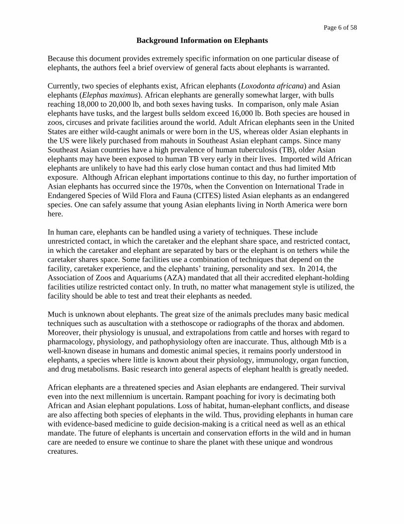
Background Information on Elephants
Because this document provides extremely specific information on one particular disease of
elephants, the authors feel a brief overview of general facts about elephants is warranted.
Currently, two species of elephants exist, African elephants (Loxodonta africana) and Asian
elephants (Elephas maximus). African elephants are generally somewhat larger, with bulls
reaching 18,000 to 20,000 lb, and both sexes having tusks. In comparison, only male Asian
elephants have tusks, and the largest bulls seldom exceed 16,000 lb. Both species are housed in
zoos, circuses and private facilities around the world. Adult African elephants seen in the United
States are either wild-caught animals or were born in the US, whereas older Asian elephants in
the US were likely purchased from mahouts in Southeast Asian elephant camps. Since many
Southeast Asian countries have a high prevalence of human tuberculosis (TB), older Asian
elephants may have been exposed to human TB very early in their lives. Imported wild African
elephants are unlikely to have had this early close human contact and thus had limited Mtb
exposure. Although African elephant importations continue to this day, no further importation of
Asian elephants has occurred since the 1970s, when the Convention on International Trade in
Endangered Species of Wild Flora and Fauna (CITES) listed Asian elephants as an endangered
species. One can safely assume that young Asian elephants living in North America were born
here.
In human care, elephants can be handled using a variety of techniques. These include
unrestricted contact, in which the caretaker and the elephant share space, and restricted contact,
in which the caretaker and elephant are separated by bars or the elephant is on tethers while the
caretaker shares space. Some facilities use a combination of techniques that depend on the
facility, caretaker experience, and the elephants’ training, personality and sex. In 2014, the
Association of Zoos and Aquariums (AZA) mandated that all their accredited elephant-holding
facilities utilize restricted contact only. In truth, no matter what management style is utilized, the
facility should be able to test and treat their elephants as needed.
Much is unknown about elephants. The great size of the animals precludes many basic medical
techniques such as auscultation with a stethoscope or radiographs of the thorax and abdomen.
Moreover, their physiology is unusual, and extrapolations from cattle and horses with regard to
pharmacology, physiology, and pathophysiology often are inaccurate. Thus, although Mtb is a
well-known disease in humans and domestic animal species, it remains poorly understood in
elephants, a species where little is known about their physiology, immunology, organ function,
and drug metabolisms. Basic research into general aspects of elephant health is greatly needed.
African elephants are a threatened species and Asian elephants are endangered. Their survival
even into the next millennium is uncertain. Rampant poaching for ivory is decimating both
African and Asian elephant populations. Loss of habitat, human-elephant conflicts, and disease
are also affecting both species of elephants in the wild. Thus, providing elephants in human care
with evidence-based medicine to guide decision-making is a critical need as well as an ethical
mandate. The future of elephants is uncertain and conservation efforts in the wild and in human
care are needed to ensure we continue to share the planet with these unique and wondrous
creatures.
Page 7 of 58
Frequently Asked Questions (FAQ)
What is tuberculosis (TB)?
Tuberculosis is a disease caused by Mycobacterium tuberculosis (Mtb), a bacterium.
Tuberculosis is primarily a human disease but Mtb can transfer to other species and has caused
most cases of TB in Asian elephants. Thus, Mtb is zoonotic.
Are all organisms named Mycobacterium of equal concern from a trunk wash culture in
regards to TB disease in humans and elephants?
No. Mtb belongs to a group of organisms known as “the Mtb complex”. Members of the Mtb
complex, which also includes Mycobacterium bovis (in cattle and other hoof stock) and
Mycobacterium pinnipedii (in marine mammals) are contagious, have zoonotic potential, and
require treatment. If these organisms are isolated in a culture, the laboratory is mandated to
contact the facility to discuss the findings.
Non-Mtb complex mycobacteria however, are NOT of significant concern. Known as “atypical
mycobacteria” or “saprophytic mycobacteria,” these mycobacteria live in substrate. Examples
include Mycobacterium avium, Mycobacterium intracellulare, and numerous others. Because
elephants root around in the dirt, they may get atypical mycobacteria in their trunks, and the final
results of a trunk wash culture may mention the identification of such organisms. However no
treatment is needed for these organisms if they are reported in culture results. If there is any
question about whether a mycobacterial organism is significant or not, the laboratory should be
contacted.
How is tuberculosis transmitted?
While a number of factors influence human to human transmission, such as the concentration of
tubercle bacilli an infectious person produces, the duration of exposure, proximity to an
infectious person and the size of the space associated with the exposure, transmission typically
occurs after close, prolonged contact with a person expelling many tubercle bacilli. Mtb
transmission in humans is well documented to occur via small (1-5 microns in diameter) airborne
particles, called droplet nuclei. Infectious droplet nuclei are generated when persons who have
pulmonary or laryngeal TB disease cough, sneeze, shout, or sing. Transmission occurs when a
person inhales droplet nuclei containing Mtb organisms and the droplet nuclei traverse the mouth
or nasal passages, upper respiratory tract, and bronchi to reach the alveoli of the lungs. Mtb is
not transmitted by surface contact. https://www.cdc.gov/tb/education/corecurr/pdf/chapter2.pdf
Initial infections of elephants with Mtb have been hypothesized to come from exposure to
infectious humans, but no direct, well-documented evidence exists to confirm this suspicion. To
date, no study has documented how elephant-to-elephant transmission has occurred; aerosol
droplet and prolonged exposure similar to human-to-human transmission are presumed to have
occurred, because the affected animals were typically long-term companions, shared the same
barn, and had trunk-to trunk-contact. To date, all known verified transmission of Mtb between
elephants and humans involved facility employees working with or exposed to the housing
environment of an infected animal at that facility (Murphree et al 2011, Zlot 2016).
What is the difference between an active infection and a latent infection?
An active infection is one in which the bacteria are growing in the body, and the animal or
person is shedding Mtb organisms into the environment. An active infection can be transmitted
to another individual. In a latent infection, the bacteria are typically inactive and walled-off by
Page 8 of 58
the immune system. A latent infection cannot be transmitted to another individual, and the
person or elephant with a latent infection is neither contagious nor shedding living organisms
into the environment. Five to 10% of latently infected people will ever develop an active
tuberculosis infection. This reactivation occurs when the body, for some reason such as immune
compromise from age, other diseases or stressors, “releases” the walled off bacteria back into the
system. The relationships between infection, latency and disease is unknown in elephants.
Can you get tuberculosis from riding an elephant, or visiting a circus or zoo elephant
exhibit?
Brief incidental contact as occurs with elephant rides, public feeds, touching or viewing an
elephant is extremely unlikely to result in infection. Because Initial infections of elephants with
Mtb have been hypothesized to come from exposure to infectious humans, but no direct, well-
documented evidence exists to confirm this suspicion. To date, no study has documented how
elephant-to-elephant transmission has occurred; aerosol droplet and prolonged exposure similar
to human-to-human transmission are presumed to have occurred, because the affected animals
were typically long-term companions, shared the same barn, and had trunk-to trunk-contact. To
date, all known verified transmission of Mtb between elephants and humans involved facility
employees working with or exposed to the housing environment of an infected animal at that
facility (Murphree et al 2011, Zlot 2016). Mtb is typically transmitted through close, prolonged
aerosol contact with an infected person or animal that is actively shedding the organism,
transmission of Mtb from elephants to humans is more of an occupational health concern than a
health concern to the general public.
If I am in a barn with an infected elephant, can I spread the disease if I get the bacteria on
my clothing or shoes?
To date, this type of transmission, known as “fomite transmission,” which refers to acquiring or
spreading an infection from an inanimate object such as clothing or equipment is NOT proven as
a means of transmitting TB. https://www.cdc.gov/tb/education/corecurr/pdf/chapter2.pdf
How do I know if an elephant has tuberculosis?
One cannot tell from simply looking because elephants with Mtb usually show no signs of
disease. The best method to definitively confirm that a living elephant has an Mtb infection is to
find the Mtb organisms by culture in a trunk wash samples, lung lavage, biopsy or other fluids
excreted by the elephant. Other ancillary tests, such as serological tests, may be used to help
support a diagnosis or but are not definitive. Definitive diagnosis of Mtb in a dead elephant can
be made if positive culture results are obtained from necropsy samples of the animal.
Is it possible to make a risk assessment about the likelihood of an elephant having Mtb?
Elephants are categorized as Risk Level A, B, or C depending of their history of infection or
exposure to other Mtb-infected elephants. This concept is discussed in detail in this document.
What is a trunk wash?
A trunk wash (TW) represents a sample from an elephant’s upper and lower respiratory tract and
is the equivalent of human sputum sample. Because of their anatomy, elephants do not readily
cough. Instead, they can be trained to blow hard through their trunk into a specimen container. A
complete description of a TW can be found in Appendix 2. The procedure requires no sedation
or undue stress to the animal nor any specialized or expensive equipment, but does require time
to train the animal.
Page 9 of 58
How do I test a clinically healthy elephant for tuberculosis?
For an elephant without any prior exposure or history of Mtb disease, the triple trunk wash (TW)
technique can be performed once a year. The triple TW technique is done three times over a 7
day period. Collected samples should be submitted for mycobacterial culture to a laboratory
specializing in these organisms. If available, qPCR, a real-time Polymerase Chain Reaction test
should be requested along with the culture. Veterinarians caring for elephants may also opt to use
other tests such as serological tests for herd surveillance.
Are there other tests for tuberculosis in elephants besides the trunk wash?
Tests that use serum/blood to look for exposure to TB are available but cannot be used to
confirm TB, although they may be useful for routine herd surveillance in conjunction with
culture. A qPCR test is being developed that may be a useful adjunctive test to trunk wash
cultures. Several common tests in humans are not possible in elephants. For example a chest
radiograph (x-ray) is not possible in an elephant because of their large size, and the skin test, aka
the TST using PPD, does not work in elephants. Other tests such as gamma interferon are being
developed, but for now are experimental only.
What do I do if an elephant comes up positive by culture on a trunk wash?
The first step is to notify facility staff, public health officials, and the state veterinarian. The next
step is to confirm the culture by repeating the trunk wash. Before results are back, however, the
facility should start treatment. Treatment is discussed fully later in this document.
Do I need to isolate an infected elephant?
A facility may decide on a case-by-case basis whether an infected animal should be isolated
during the entire time of treatment, isolated only temporarily, or kept with the herd while
treatment is undertaken. In all cases, starting treatment immediately to stop shedding, and
improving hygiene and sanitation in the barn are very important.
If I do isolate the elephant, what is the minimum recommended distance an Mtb-infected
elephant should be housed from other elephants?
Currently neither this distance nor the amount of time needed to transmit Mtb from an infected
elephant to an uninfected one are known. Here too, though, steps can be taken to decrease the
risk of disease transmission. First is to start treatment of the infected animal as soon as possible
in order to stop shedding of the organism into the environment. Additionally the facility should
examine its ventilation, hygiene and sanitation protocols in order to increase the dilution of
bacteria and reduce their aerosolization.
How long should an elephant be treated for tuberculosis?
No one treatment protocol has been used in treating all infected elephants. Current
recommendations using a one or two-phase protocol recommend 12 months of treatment.
Treatment length may be affected by the antibiotics that can be successfully administered to the
elephant. Using combination protocols with less frequent dosing or antibiotics of lesser known
effectiveness may extend the treatment period for 15 to 18 months. See the treatment section for
more details.
How often should an elephant be tested after it finishes treatment for Mtb?
After treatment is completed, triple TW cultures should be performed at least quarterly (4X a
year) to monitor and confirm success of treatment. The veterinarian of record may prescribe to
do triple TW cultures more frequently.
Page 10 of 58
Frequently Asked Questions for State and Regulatory Veterinarians
Because elephants may travel across state lines for facility transfers, breeding or exhibitions,
questions about Mtb in elephants have often arisen from the regulatory sector. This FAQ
addresses the different tests, possible risks to livestock or the general public and how to
appropriately evaluate an elephant or elephants coming into a region or state.
What paperwork should accompany an elephant(s) when they enter a state?
The elephant(s) must be accompanied by a Certificate of Veterinary Inspection (CVI) written by
an accredited USDA Class II veterinarian and dated within 30 days prior to arrival into that state.
The CVI can be on a form provided by the American Association of Zoo Veterinarians (AAZV),
or a generic or livestock CVI form from the state of origin (see Appendix 3). The CVI should
include the name, age and gender of each elephant in the group, the dates of any vaccinations, if
given, and the date that the triple trunk wash (TW) series was performed. The veterinarian
writing the health certificate should list to which category each elephant on the certificate
belongs (Category A, B, or C). Elephants over the age of five should travel with copies of at
least two years of TW results. TW results should document the name of the laboratory, the name
of the elephant, that the results are “final” and that each TW was negative. (Note: a description
of the trunk wash technique can be found in Appendix 2).
What are the differences between Category A, Category B and Category C elephants?
Elephants in Category A have had no known exposure to culture positive animals within a 5 year
period. These elephants are negative on TW and have no clinical signs. Elephants in Category B
may have had contact with an Mtb positive animal within 5 years but are negative on all TW. No
travel restrictions exist for either Category A or B elephants. Category C elephants are TW
positive (i.e., Mtb organisms were isolated by culture from a TW sample) and are considered
infected. Category C elephants would only travel if necessary for medical care
Are Mtb testing recommendations the same for all elephants?
No. Category A elephants need only one triple TW series done within a 12 month period.
Category B animals should receive triple TW series done quarterly from the time that the
elephant(s) was/were placed in Category B. Category C elephants receive considerably more
testing which is described elsewhere. See description of Elephant Risk Categories, page 17.
What if the elephant is TW negative but reactive on a serological test (DPP, MAPIA,
STAT-PAK or ELISA)?
Many elephants fall into this group. If the elephant has two years of negative annual triple TW
cultures, it can be considered negative, and going forward from 2017, the qPCR test performed
on TW samples), it should be considered negative. The serologic tests are not confirmatory for
Mtb and should not be used as a solo test for regulatory purposes.
Elephants are traveling to a livestock arena or venue in my state; what precautions should
be taken to prevent the transmission of Mtb to other livestock?
No special precautions are needed. Travelling elephants (both Categories A and B) are routinely
trunk washed and negative by culture. There has been no documented case of Mtb transmission
from elephants to livestock. There are no documented cases of fomite transmission of Mtb
Page 11 of 58
DIAGNOSTIC APPROACH
Testing for Mtb infection should be part of the preventative medicine program of every elephant-
holding facility. This aspect of the preventative medicine program should be reviewed and
updated annually by the attending veterinarian.
A. Risk assessments
A standard risk assessment for Mtb infection in an elephant or its herd includes the following:
1. Complete history of the elephant and its herdmates including:
Signalment: Mtb is primarily a disease of Asian elephants although several cases were
identified in African elephants in Europe and one unconfirmed by culture report in a free
ranging African elephant (Obanda 2013).
Travel and housing history of elephant and herdmates with regard to Mtb exposure
Current health problems (including age-related diseases)
Diagnostic procedures for which the elephant is trained (i.e., TW, blood draw, etc)
History of Mtb complex testing & results over at least a five-year period
Necropsy results of herdmates at both current and previous locations
Quarantine protocols
Recent herd acquisitions
Reproductive history
2. Staff screening program for Mtb (This information is protected by HIPAA laws)
Staff testing protocols should be developed with the input of local public health officials
Staff Mtb risk assessment including:
travel to countries with high rates of human Mtb
known exposure to infected (M.bovis) animals other than elephants (dairy cattle,
deer, etc)
known exposure to infected family members
known exposure to infected elephants either at current or previous facilities
3. Husbandry practices that could increase risk of transmission of Mtb
Barn ventilation
Barn cleaning procedures, i.e., use of high-pressure hoses, which can aerosolize bacteria
(Murphree 2011)
4. General elephant health
Current weight, and weight trends over three to five years including body condition score
Physical examination
Blood work: Complete blood count (CBC), serum chemistry and blood smear
Urinalysis
Acute phase proteins/protein electrophoresis
Fecal analysis for parasites and fecal culture for enteric pathogens
Banking of serum at -80°C for future diagnostics
5. Mtb diagnostic test results
Trunk wash results over 5 years
Other samples submitted for Mtb culture such as semen, milk, feces, biopsies
Serological test results
Page 12 of 58
B. Testing for Mtb in Elephants: Ante mortem and Postmortem Diagnostic Options
Diagnostic tests for Mtb can be applied to elephants ante mortem or postmortem. Ante mortem
testing can be done for surveillance, when there is concern or confirmation of disease, or for
regulatory purposes, i.e., for moving elephants across state lines.
Two general groups of tests are available: Direct tests identify the actual Mtb organism.
Indirect tests look for exposure to the organism. Culture is the only definitive means of
diagnosing infections, although negative results cannot be construed as definitively being
equivalent to absence of infection. Tests other than culture can be used when agreed to by
parties of origin and destination, but should not be used as sole test for regulatory purposes.
General comments on testing options follow.
Ante mortem direct tests:
Culture: the gold standard for diagnoses of Mtb complex infections in live elephants is culture
of trunk wash, TW samples. A positive culture result indicates an elephant is infected but a
negative result does not rule out infection. There is the potential for intermittent or low level
shedding of bacilli. (Note: a description of the trunk wash technique can be found in Appendix
2). Where available (rare) pulmonary fluid wash or biopsies collected via endoscopy are likely
superior to trunk washes due to reduced contamination and the opportunity to collect samples
likely closer to or at the site of lesions. (Hildebrandt 2016)
Additional biological specimens that are appropriate for mycobacterial culture, where warranted
for a specific instance:
mucus
vaginal fluid
semen
tissue biopsy
lung/airway lavage fluid
esophageal cardia wash
urine
feces
milk
Submission of specimens to laboratories with extensive experience with mycobacterial culture
expertise especially with trunk wash samples that commonly are heavily contaminated with
normal upper airway microflora (See Appendix 4 for contact information). Duplicate or
additional TW samples if collected can be stored in a -30C freezer. The time to obtain culture
results is approximately eight weeks.
Polymerase Chain Reaction (PCR): Currently a real time Polymerase Chain Reaction, (RT-
qPCR) test on trunk wash fluid is being validated alongside TW culture at the USDA’s National
Veterinary Services lab in Ames, IA. (Backues 2015) The organism-based test combination of
TW culture and qPCR at this lab is showing great promise to significantly improve the
sensitivity of the standard TW series as a screening tool for Mtb infection, surveillance,
diagnosis, and response to treatment. Note though, that qPCR cannot distinguish live from dead
organisms and can detect DNA fragments of organisms. qPCR results are usually returned within
a few days. (Magnuson 2017).
Page 13 of 58
Acid-fast staining: acid-fast staining of specimens is appropriate as a fast initial screen, but is
not specific for Mtb complex.
Ante mortem indirect tests
Serology: Useful when used in combination with direct testing methods as a screening tool or
for monitoring response to treatment. False positives and false negatives are possible and the
accuracy of these tests has not been rigorously established. Many elephants are reactive on
serological test(s) but are TW negative. Co-editor has treated 2 elephants that were TW positive
but serological negative.
Immune response assays (IGRAs); not yet validated for elephants or commercially available
(Landolfi et al 2014)
Tuberculin skin testing (PPD) should not be used for Mtb complex infection testing in
elephants. (Lewerin, 2005)
Post mortem direct tests
Deceased elephants should receive a full necropsy, and tissue specimens should be collected in
duplicate with one set placed in formalin and the other frozen at -80°C. If suspicious lesions
such as granulomas, evidence of pneumonia, or enlarged lymph node are evident, or if there is
epidemiological or histological evidence suggests of Mtb complex infection, multiple tissue
specimens should be submitted for culture, PCR and histological exam. Standard tissues used for
tuberculosis diagnosis include but are not confined to:
trunk
trachea
lung
lymph nodes (bronchial, abdominal, cervical, etc), especially if abscessed or enlarged.
stomach wall
salivary glands
small intestine
lower esophagus (cardia)
feces
granulomas - although such lesions should NOT be considered pathognomonic for Mtb complex
they are highly associated with TB disease. (Lacasse 2007). Do not assume all granulomas are
Mtb, there are other disease that may cause such lesions and other Mtb complex organisms can
have similar histologic appearance, verify with culture, and PCR.
A thorough review of the details and logistics of an elephant necropsy can be found in the,
Elephant TAG/SSP Research and Necropsy Protocol of the AZA, June 2017. This document
can be found at AAZV.org under SSP necropsy protocols or AZA.org
www.aazv.org/resource/resmgr/Protocols/ELEPHANT_NEC_PROT_Jun_2017.pdf
Additional details about the tests below can be found in the ante mortem test section
Culture: Culture samples should NOT be placed in formalin, can be shipped overnight to an
appropriate laboratory on a cold pack with appropriate biohazard documentation. Samples can
also be frozen until shipping can be arranged. Samples should be double bagged in zippered
plastic bags
Page 14 of 58
PCR: PCR samples can be placed in formalin. Jars should be sealed with a wax sealant tape
such as ‘Parafilm’ around the lid, then double bagged in zippered plastic bags
Immunohistochemistry: not routinely performed for diagnosis of Mtb complex in elephants.
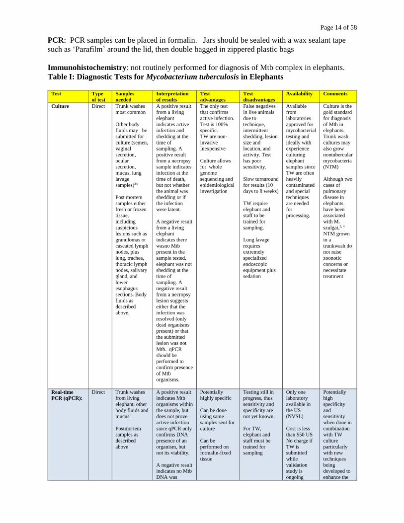
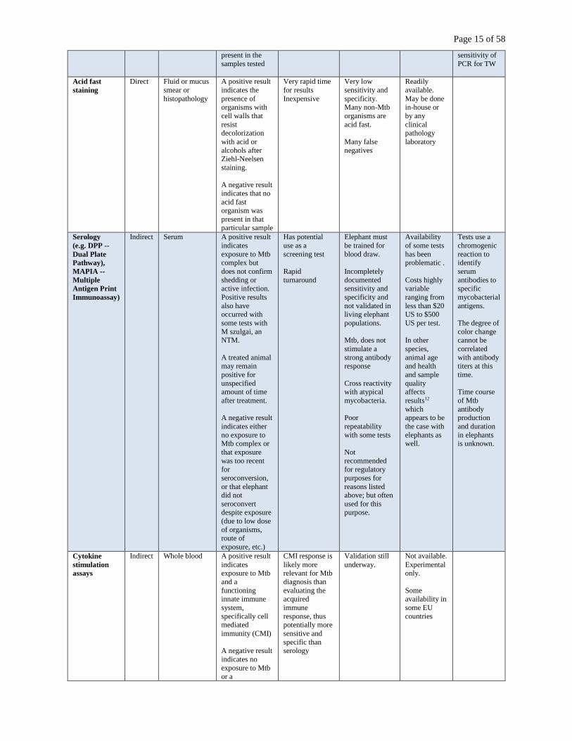
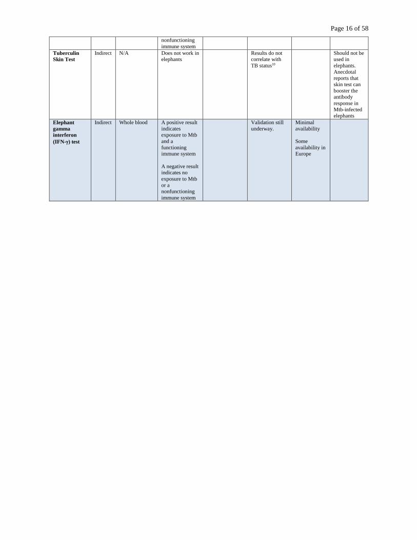
Table I: Diagnostic Tests for Mycobacterium tuberculosis in Elephants
Test Type
of test
Samples
needed
Interpretation
of results
Test
advantages
Test
disadvantages
Availability Comments
Culture
Direct Trunk washes most common
Other body fluids may be
submitted for
culture (semen, vaginal
secretion,
ocular secretion,
mucus, lung
lavage samples)34
Post mortem
samples either
fresh or frozen tissue,
including
suspicious lesions such as
granulomas or
caseated lymph nodes, plus
lung, trachea,
thoracic lymph nodes, salivary
gland, and
lower
esophagus
sections. Body
fluids as described
above.
A positive result from a living
elephant
indicates active infection and
shedding at the
time of sampling. A
positive result
from a necropsy sample indicates
infection at the
time of death, but not whether
the animal was shedding or if
the infection
were latent.
A negative result
from a living elephant
indicates there
wasno Mtb present in the
sample tested,
elephant was not shedding at the
time of
sampling. A
negative result
from a necropsy
lesion suggests either that the
infection was
resolved (only dead organisms
present) or that
the submitted lesion was not
Mtb. qPCR
should be performed to
confirm presence
of Mtb organisms.
The only test that confirms
active infection.
Test is 100% specific.
TW are non-
invasive Inexpensive
Culture allows for whole
genome
sequencing and epidemiological
investigation
False negatives in live animals
due to
technique, intermittent
shedding, lesion
size and location, and
activity. Test
has poor sensitivity.
Slow turnaround for results (10
days to 8 weeks)
TW require
elephant and staff to be
trained for
sampling.
Lung lavage
requires extremely
specialized
endoscopic equipment plus
sedation
Available from
laboratories
approved for mycobacterial
testing and
ideally with experience
culturing
elephant samples since
TW are often
heavily contaminated
and special techniques
are needed
for processing.
Culture is the gold standard
for diagnosis
of Mtb in elephants.
Trunk wash
cultures may also grow
nontubercular
mycobacteria (NTM)
Although two cases of
pulmonary disease in
elephants
have been associated
with M.
szulgai,3, 4 NTM grown
in a
trunkwash do not raise
zoonotic
concerns or necessitate
treatment
Real-time
PCR (qPCR):
Direct Trunk washes from living
elephant, other
body fluids and mucus.
Postmortem samples as
described
above
A positive result indicates Mtb
organisms within
the sample, but does not prove
active infection
since qPCR only confirms DNA
presence of an
organism, but not its viability.
A negative result indicates no Mtb
DNA was
Potentially highly specific
Can be done using same
samples sent for
culture
Can be
performed on formalin-fixed
tissue
Testing still in progress, thus
sensitivity and
specificity are not yet known.
For TW, elephant and
staff must be
trained for sampling
Only one laboratory
available in
the US (NVSL)
Cost is less than $50 US
No charge if
TW is submitted
while
validation study is
ongoing
Potentially high
specificity
and sensitivity
when done in
combination with TW
culture
particularly with new
techniques
being developed to
enhance the
Page 15 of 58
present in the
samples tested
sensitivity of
PCR for TW
Acid fast
staining
Direct Fluid or mucus
smear or
histopathology
A positive result
indicates the
presence of organisms with
cell walls that
resist decolorization
with acid or
alcohols after Ziehl-Neelsen
staining.
A negative result
indicates that no
acid fast organism was
present in that
particular sample
Very rapid time
for results
Inexpensive
Very low
sensitivity and
specificity. Many non-Mtb
organisms are
acid fast.
Many false
negatives
Readily
available.
May be done in-house or
by any
clinical pathology
laboratory
Serology
(e.g. DPP --
Dual Plate
Pathway),
MAPIA --
Multiple
Antigen Print
Immunoassay)
Indirect Serum A positive result
indicates
exposure to Mtb complex but
does not confirm
shedding or active infection.
Positive results
also have occurred with
some tests with
M szulgai, an NTM.
A treated animal may remain
positive for
unspecified amount of time
after treatment.
A negative result
indicates either
no exposure to Mtb complex or
that exposure
was too recent for
seroconversion,
or that elephant did not
seroconvert despite exposure
(due to low dose
of organisms, route of
exposure, etc.)
Has potential
use as a
screening test
Rapid
turnaround
Elephant must
be trained for
blood draw.
Incompletely
documented sensitivity and
specificity and
not validated in living elephant
populations.
Mtb, does not
stimulate a
strong antibody response
Cross reactivity with atypical
mycobacteria.
Poor
repeatability
with some tests
Not
recommended for regulatory
purposes for
reasons listed above; but often
used for this purpose.
Availability
of some tests
has been problematic .
Costs highly variable
ranging from
less than $20 US to $500
US per test.
In other
species,
animal age and health
and sample
quality affects
results12
which appears to be
the case with
elephants as well.
Tests use a
chromogenic
reaction to identify
serum
antibodies to specific
mycobacterial
antigens.
The degree of
color change cannot be
correlated
with antibody titers at this
time.
Time course
of Mtb
antibody production
and duration
in elephants is unknown.
Cytokine
stimulation
assays
Indirect Whole blood A positive result
indicates exposure to Mtb
and a
functioning innate immune
system,
specifically cell mediated
immunity (CMI)
A negative result
indicates no
exposure to Mtb or a
CMI response is
likely more relevant for Mtb
diagnosis than
evaluating the acquired
immune
response, thus potentially more
sensitive and
specific than serology
Validation still
underway.
Not available.
Experimental only.
Some availability in
some EU
countries
Page 16 of 58
nonfunctioning
immune system
Tuberculin
Skin Test
Indirect N/A Does not work in
elephants
Results do not
correlate with
TB status10
Should not be
used in
elephants.
Anecdotal reports that
skin test can
booster the antibody
response in
Mtb-infected elephants
Elephant
gamma
interferon
(IFN-) test
Indirect Whole blood A positive result
indicates exposure to Mtb
and a
functioning immune system
A negative result indicates no
exposure to Mtb
or a nonfunctioning
immune system
Validation still
underway.
Minimal
availability
Some
availability in Europe
Page 17 of 58
C. Mtb Risk Categories for Elephants
Elephants are placed into one of three groups depending on their risk of being positive for Mtb.
Testing requirements for elephants vary according to what risk group they belong. Note that
these groups are determined by two factors: herd history and a positive trunk wash culture, but
not by serological test results. The clinical veterinarian can decide to increase the amount and
type testing for any elephant based on their concerns and experience. Caveat: The ‘in validation’
qPCR on TW samples currently ongoing at NVSL should increase the sensitivity of the standard
TW. A positive qPCR test could be a false positive but should be handled as if the sample was
Mtb positive while awaiting culture confirmation of the result.
Category A elephants:
Risk of infection with Mtb complex: Low
Known exposure to an Mtb culture-positive animal: None within the past five years.
Test history: Negative for past five years on annual triple TW testing by culture.
Recommended testing: Routine. Triple TW culture technique done minimally annually.
Travel restrictions: None.
Category B elephants:
Risk of infection with Mtb complex: Moderate
Known exposure to an Mtb culture-positive animal: Exposure to an Mtb culture-
positive has occurred within the past five years, i.e., a herd mate was a confirmed positive
animal.
Test history: Consistently negative by annual triple TW testing by culture
Recommended testing: Increased. Triple TW cultures should be performed quarterly,
every three months for minimum of two years. If no positive results, triple TW cultures may be
reduced at the recommendation of the clinical veterinarian but increased surveillance beyond
annual testing is recommended for a five-year period. (EX: TW frequency at 4, 3 or 2 x a year)
If all tests remain negative after 5-years, these elephants return to Category A status, and only
need triple TW cultures once a year.
Travel restrictions: None
Category C elephants:
Risk of infection with Mtb complex: These elephants are positive on TW cultures or
culture of other body fluid.
Known exposure to an Mtb culture-positive animal: Animals with whom this elephant
has been in contact become Category B elephants. Trace back to see what other elephants and
other animals should be performed to determine what other transmission routes were possible.
Human exposure to Category C elephants should be handled with the cooperation of local and
state public health officials. See THE TRUNKWASH POSITIVE ELEPHANT: FIRST
STEPS, following Table 2.
Test history: Positive on a single or multiple TW cultures or cultures of other fluids
Recommended testing: Increased on several levels. See next section: Next steps
following identification of an Mtb culture positive elephant (Class C elephant).
Travel restrictions: Travel permitted only for specific medical reasons
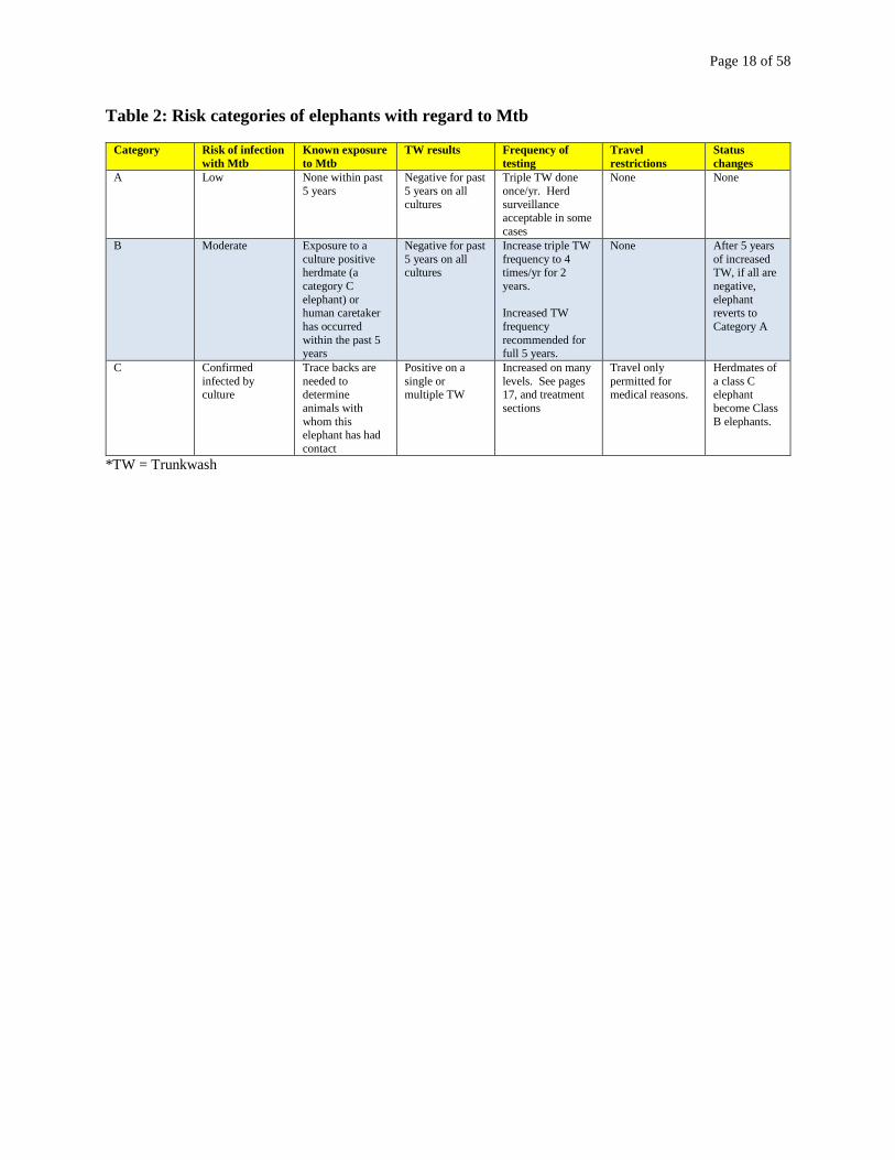
Table 2, below, summarizes the categories.
Page 18 of 58
Table 2: Risk categories of elephants with regard to Mtb
Category Risk of infection
with Mtb
Known exposure
to Mtb
TW results Frequency of
testing
Travel
restrictions
Status
changes
A Low None within past
5 years
Negative for past
5 years on all
cultures
Triple TW done
once/yr. Herd
surveillance acceptable in some
cases
None None
B Moderate Exposure to a
culture positive herdmate (a
category C
elephant) or human caretaker
has occurred
within the past 5 years
Negative for past
5 years on all cultures
Increase triple TW
frequency to 4 times/yr for 2
years.
Increased TW
frequency
recommended for full 5 years.
None After 5 years
of increased TW, if all are
negative,
elephant reverts to
Category A
C Confirmed
infected by culture
Trace backs are
needed to determine
animals with
whom this elephant has had
contact
Positive on a
single or multiple TW
Increased on many
levels. See pages 17, and treatment
sections
Travel only
permitted for medical reasons.
Herdmates of
a class C elephant
become Class
B elephants.
*TW = Trunkwash
Page 19 of 58
THE TRUNKWASH POSITIVE ELEPHANT: FIRST STEPS
After an elephant tests positive for Mtb via TW culture, the diagnostic testing laboratory will
contact the attending veterinarian. Once a positive culture is received, the elephant is considered
infected with Mtb, and the following notifications are necessary and even before confirmation of
results:
Although a positive qPCR result may represent a false positive for active disease, the
initiation of the following steps should be undertaken while waiting for confirmatory culture
results.
A. Notifications of an Mtb-culture positive elephant must include:
1. Regulatory personnel such as:
State Veterinarian
State Public Health Veterinarian
local public health officials
USDA VS Inspector veterinarian for the facility
2. Facility personnel including:
All staff working with the elephant including barn staff, veterinary staff and volunteers
Upper management & legal teams
Public relations, marketing, and communications teams
Human resources
Safety departments
3. Notification of other facilities where the Mtb culture-positive elephant has been, if
appropriate.
Discussions regarding safe elephant handling, use of personal protective equipment (PPE) such
as N-95 respirators, and barn cleaning protocols to prevent zoonotic transmission or spread to
other elephants in the herd should occur in collaboration with regulatory and facility personnel.
See Human Health Considerations starting page 33.
B. Confirmation of the positive TW culture result.
If TW samples have been banked, submit the duplicate sample to the lab for culture
Submit a new TW sample for culture.
C. Additional information needed from the diagnostic laboratories:
1. Request additional identification of the Mtb isolate using whole gene sequencing from the
diagnostic lab. There is a moderate fee for this service but older tests such as spoligotyping yield
minimal information as to the genetic relatedness of Mtb strains.
2. Test for antimicrobial susceptibility using validated CLSI references.
Note: if the laboratory is not able to perform these tests, it should ship the isolate to National
Veterinary Services Laboratory (NVSL) (contact information can be found in Appendix 4.)
Page 20 of 58
3. Identify laboratory that can measure serum drug samples, and confirm testing and handling
protocols (See Appendix 5)
D. Purchase and testing of antitubercular drugs.
1. Costs and sources for large amounts may vary; comparison shopping is recommended
2. Purchase drugs only from a licensed pharmacy and certified compounder
3. Permission from the FDA may be needed to import bulk drugs
4. Priority drugs to source are first-line drugs, Isoniazid (INH) and Rifampin (RIF).
5. New batches of bulk drugs should be tested for purity and drug activity concentrations.
Samples can be submitted to the Infectious Disease Pharmacokinetic Laboratory at the
University of Florida (Contact information is in Appendix 5).
E. Sourcing Medications:
1. The use of bulk drugs may be a viable alternative to the use of FDA approved TB products
due to elephant size, the length of treatment and the ability to properly formulate/compound for
administration to elephants.
2. Some distributers of FDA approved products in the past have restricted the dosage units that
can be purchased which means not enough could be purchased for use in elephants.
3. Bulk drugs contain an active pharmaceutical ingredient (API) typically used for human drug
compounding
4. In contrast, FDA approved TB products come in fixed dosage forms for human use.
5. The FDA recognizes that there are specific circumstances when an animal drug can be
compounded from bulk drug substances.
6. The FDA has the authority to regulate compounding for animal use and offers guidance in
purchasing active pharmaceutical ingredients (bulk drug) for treatment options.
7. The FDA has released a draft: “Guidance for Industry (GFI) #230 Compounding Animal
Drugs from Bulk Drug Substances.”
GFI #230 outlines the current FDA stance for these circumstances.
GFI#230 also documents the necessary items that must be included on a prescription for a
pharmacy to obtain bulk drug to be dispensed or compounded.
A link to that draft can be found at:
http://www.fda.gov/downloads/AnimalVeterinary/GuidanceComplianceEnforcement/Gui
danceforIndustry/UCM446862.pdf
F. Purchasing Drugs
1. Shortages resulting in delay of delivery for several weeks have occurred.
2. Always verify drug availability with a pharmacy prior to purchase and discuss duration of
treatment (total amount of drug needed) with that pharmacy.
3. Bulk drugs may be purchased by direct importation from a manufacturer or via purchase
from a licensed pharmacy.
If directly importing bulk drugs from a manufacturer:
Verify that the importing entity is registered with the FDA.
Check state rules regarding the purchase and distribution of bulk drugs.
Check if state rules require the importer to have a license equivalent to a
pharmacy wholesale license or distribution license prior to purchase or sale of bulk chemicals
(aka drugs).
Page 21 of 58
4. If purchasing directly from a licensed FDA-approved facility, a certificate of analysis (COA)
from the original manufacturer will accompany the product. The COA documents the potency of
the bulk drug.
If purchasing bulk drugs from a pharmacy: Check first with local compounding pharmacies which may have Mtb drugs
available at reasonable prices.
Larger veterinary pharmaceutical compounding pharmacies have routinely
stocked Mtb-related bulk drugs for the last several years.
When purchasing from a pharmacy, generally a COA does not come with the
prescription but can be requested from a dispensing pharmacy.
G. Concerns with bulk drug use
1. Potency of bulk drugs has been a problem.
- Loss of potency of bulk drugs, can occur for multiple reasons.
- Due to the possibility of loss of potency, samples of the bulk drug can be analyzed via a
potency assay.
- A few facilities that will perform potency analysis are listed here, but many others are
available.
Analytical Research Laboratories (ARL), 840 Research Parkway, Ste. 546, Oklahoma City,
OK 73104, 800-393-1595
Compounder’s International Analytical Laboratory (CIAL), 680 Atchison Way, Suite 100,
Castle Rock, CO 80109, 800-788-9922 Front Range Labs, 3985 S Lincoln, Loveland, Co 90537, 970-593-0171
Professional Compounding Centers of America (PCCA), 9901 South Wilcrest Drive,
Houston, TX 77099, 800-331-2498
Page 22 of 58
TREATMENT OF MTB CULTURE POSITIVE ELEPHANTS
A. Definition of Epidemiological Terms: See Appendix 6.
B. Goals of treatment:
1. Stop shedding of live Mtb organisms to protect herdmates and humans.
2. Prevent elephant from becoming ill from treatment.
3. Obtain appropriate serum/plasma concentrations of antitubercular drugs in the elephant.
4. Monitor TW cultures intensively throughout treatment. See Category C elephant requirements.
5. Treat for an adequate period of time, typically one year.
Caveats: Doses, frequency, and duration of treatment remain empirical in elephants.
C. Overview of anti-tubercular drugs regimen:
1. Mtb in humans is typically treated with a four-drug regimen for 6 to 12 months.
2. Elephant protocols have used human protocols as a starting point.
3. Drugs typically used are isoniazid (INH), rifampin (RIF), pyrazinamide (PZA) and ethambutol
(ETH).
4. INH and RIF (or a fluoroquinolone in place of RIF when compliance is an issue) are
recommended for all treatment regimens.
5. Pharmacokinetic (PK) studies in elephants have not evaluated necessary blood concentrations
needed for cure, only the amounts of drugs that need to be administered to achieve blood
concentrations.
6. The PK and pharmacodynamics (PD) of these drugs in elephants are quite different compared
to humans.
7. A fine balance exists between giving an amount of drug that does not cause adverse effects
and one that does not select for resistant organisms. Optimization of drug doses must be tailored
to each individual elephant's tolerance to medications. It is not uncommon to alter the dose or the
frequency at which a medication is given during the treatment period to accommodate the
elephant's tolerance to medications, and changes in appetite and attitude.
8. Elephants need be trained to take drugs consistently, regardless of route. Treatment will be
most effective if training is well established and routine before the elephant has a medical need
to take medications.
Routes of drug administration in elephants are described in Table 4, below.
Selected references: (Maslow et al 2005a & b: Zhu et al 2005, Peloquin et al 2006)
D. Characteristics of Drugs Used to Treat Mtb in Elephants
1. Isoniazid, INH
INH should be used in all elephant protocols unless the elephant is intolerant of the
medication or the mycobacterial isolate is found to be insensitive to it.
INH is responsible for the rapid killing of actively dividing Mtb organisms.
2. Pyrazinamide, PZA
Used in conjunction with INH to prevent the development of INH resistance.
INH and PZA should always be used initially for Mtb treatment in elephants and can be
effectively administered together rectally.
Page 23 of 58
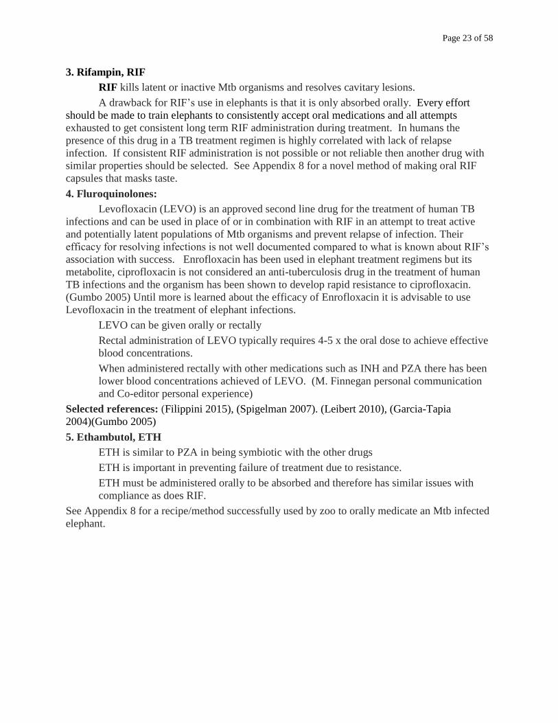
3. Rifampin, RIF
RIF kills latent or inactive Mtb organisms and resolves cavitary lesions.
A drawback for RIF’s use in elephants is that it is only absorbed orally. Every effort
should be made to train elephants to consistently accept oral medications and all attempts
exhausted to get consistent long term RIF administration during treatment. In humans the
presence of this drug in a TB treatment regimen is highly correlated with lack of relapse
infection. If consistent RIF administration is not possible or not reliable then another drug with
similar properties should be selected. See Appendix 8 for a novel method of making oral RIF
capsules that masks taste.
4. Fluroquinolones:
Levofloxacin (LEVO) is an approved second line drug for the treatment of human TB
infections and can be used in place of or in combination with RIF in an attempt to treat active
and potentially latent populations of Mtb organisms and prevent relapse of infection. Their
efficacy for resolving infections is not well documented compared to what is known about RIF’s
association with success. Enrofloxacin has been used in elephant treatment regimens but its
metabolite, ciprofloxacin is not considered an anti-tuberculosis drug in the treatment of human
TB infections and the organism has been shown to develop rapid resistance to ciprofloxacin.
(Gumbo 2005) Until more is learned about the efficacy of Enrofloxacin it is advisable to use
Levofloxacin in the treatment of elephant infections.
LEVO can be given orally or rectally
Rectal administration of LEVO typically requires 4-5 x the oral dose to achieve effective
blood concentrations.
When administered rectally with other medications such as INH and PZA there has been
lower blood concentrations achieved of LEVO. (M. Finnegan personal communication
and Co-editor personal experience)
Selected references: (Filippini 2015), (Spigelman 2007). (Leibert 2010), (Garcia-Tapia
2004)(Gumbo 2005)
5. Ethambutol, ETH
ETH is similar to PZA in being symbiotic with the other drugs
ETH is important in preventing failure of treatment due to resistance.
ETH must be administered orally to be absorbed and therefore has similar issues with
compliance as does RIF.
See Appendix 8 for a recipe/method successfully used by zoo to orally medicate an Mtb infected
elephant.
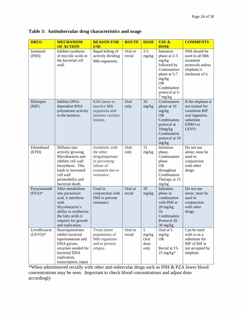
Page 24 of 58
Table 3: Antitubercular drug characteristics and usage
DRUG MECHANISM
OF ACTION
REASON FOR
USE
ROUTE DOSE USE &
DOSE
COMMENTS
Isoniazid
(INH)
Inhibits synthesis
of mycolic acids in
the bacterial cell
wall.
Rapid killing of
actively dividing
Mtb organisms.
Oral or
rectal
2-5
mg/kg
Initiation
phase at 2-3
mg/kg
followed by
Continuation
phase at 5-7
mg/kg
OR
Combination
protocol at 5-
7 mg/kg
INH should be
used in all Mtb
treatment
protocols unless
elephant is
intolerant of it
Rifampin
(RIF)
Inhibits DNA-
dependent RNA
polymerase activity
in the bacteria.
Kills latent or
inactive Mtb
organisms and
resolves cavitary
lesions.
Oral
only
10
mg/kg
Continuation
phase at 10
mg/kg
OR
Combination
protocol at
10mg/kg
Combination
protocol at 10
mg/kg
If the elephant is
not trained for
consistent RIF
oral ingestion,
substitute
ENRO or
LEVO.
Ethambutol
(ETH)
Diffuses into
actively growing
Mycobacteria and
inhibits cell wall
biosythesis. This
leads to increased
cell wall
permeability and
bacterial death.
Symbiotic with
the other
drugsImportant
in preventing
failure of
treatment due to
resistance.
Oral
only
15
mg/kg
Initiation
phase,
Continuation
phase
OR
throughout
Combination
Therapy at 15
mg/kg
Do not use
alone; must be
used in
conjunction
with other
drugs.
Pyrazinamide
(PZA)*
After metabolism
into pyrazinoic
acid, it interferes
with
Mycobacteria’s
ability to synthesize
the fatty acids it
requires for growth
and replication.
Used in
conjunction with
INH to prevent
resistance
Oral or
rectal
20
mg/kg
Initiation
phase in
combination
with INH at
20 mg/kg
Or
Combination
Protocol 20-
30 mg/kg
Do not use
alone; must be
used in
conjunction
with other
drugs.
Levofloxacin
(LEVO)*
fluoroquinolones
inhibit bacterial
topoisomerase and
DNA gyrase,
enzymes needed for
bacterial DNA
replication,
transcription, repair
Treats latent
populations of
Mtb organisms
and to prevent
relapse.
Oral or
rectal
5
mg/kg
Oral
dose
only
Oral at 5
mg/kg
OR
Rectal at 15-
25 mg/kg*
Can be used
with or as a
substitute for
RIF of RIF is
not accepted by
elephant.
*When administered rectally with other anti-tubercular drugs such as INH & PZA lower blood
concentrations may be seen. Important to check blood concentrations and adjust dose
accordingly
Page 25 of 58
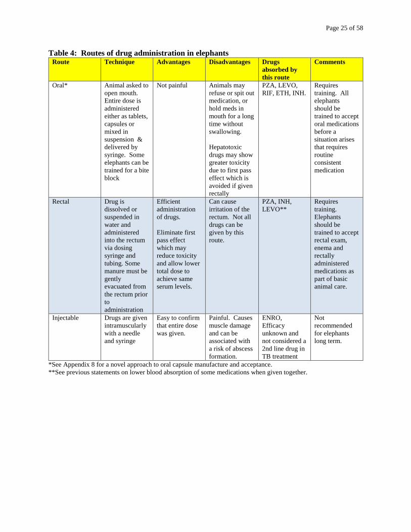
Table 4: Routes of drug administration in elephants Route Technique Advantages Disadvantages Drugs
absorbed by
this route
Comments
Oral* Animal asked to
open mouth.
Entire dose is
administered
either as tablets,
capsules or
mixed in
suspension &
delivered by
syringe. Some
elephants can be
trained for a bite
block
Not painful Animals may
refuse or spit out
medication, or
hold meds in
mouth for a long
time without
swallowing.
Hepatotoxic
drugs may show
greater toxicity
due to first pass
effect which is
avoided if given
rectally
PZA, LEVO,
RIF, ETH, INH.
Requires
training. All
elephants
should be
trained to accept
oral medications
before a
situation arises
that requires
routine
consistent
medication
Rectal Drug is
dissolved or
suspended in
water and
administered
into the rectum
via dosing
syringe and
tubing. Some
manure must be
gently
evacuated from
the rectum prior
to
administration
Efficient
administration
of drugs.
Eliminate first
pass effect
which may
reduce toxicity
and allow lower
total dose to
achieve same
serum levels.
Can cause
irritation of the
rectum. Not all
drugs can be
given by this
route.
PZA, INH,
LEVO**
Requires
training.
Elephants
should be
trained to accept
rectal exam,
enema and
rectally
administered
medications as
part of basic
animal care.
Injectable Drugs are given
intramuscularly
with a needle
and syringe
Easy to confirm
that entire dose
was given.
Painful. Causes
muscle damage
and can be
associated with
a risk of abscess
formation.
ENRO,
Efficacy
unknown and
not considered a
2nd line drug in
TB treatment
Not
recommended
for elephants
long term.
*See Appendix 8 for a novel approach to oral capsule manufacture and acceptance.
**See previous statements on lower blood absorption of some medications when given together.
Page 26 of 58
TREATMENT PROTOCOLS FOR CULTURE POSITIVE ELEPHANTS
(Note: This section was adapted from the ATS/CDC/IDS Practical Guidelines for Drug
Susceptible TB-CID 2016:63)
https://www.cdc.gov/tb/publications/guidelines/pdf/clin-infect-dis.-2016-nahid-cid_ciw376.pdf
A. Overview of Treatment
1. Treatment recommendations for elephants resemble human protocols in utilizing a two phase
protocol, but some veterinarians have used only the initiation phase treatment regimen
throughout the entire course of treatment without adverse effects:
a. The first is the Initiation Phase, characterized by frequent administration of drugs.
b. The second is a Continuation Phase in which the doses of each drug are typically
higher but are administered less frequently.
2. A Single Phase Combination Therapy protocol is possible for elephants that become very sick
during the intensive Initiation phase and consists of only a Continuation phase like schedule.
a. This protocol should last longer, ~ 18-24 months.
b. Drugs are given intermittently (3 to 4 times/week), but at higher doses.
c. In humans, this schedule has been shown to be less effective than a two-phase protocol.
d. There is no data on whether this is also true in elephants.
e. Its use should only be considered in elephants that cannot tolerate the more frequent
dosing regimens.
3. If at any point during the Single Phase Combination Therapy Protocol or during the
Continuation Phase of the Two-Phase Protocol, a positive TW by culture occurs, the sample
should receive full whole genome sequencing, WGS and repeat bacterial susceptibility testing.
See page 29 for a discussion of treatment failures.
4. To stop shedding, increased frequency of dosing during the first few months of treatment
(initiation phase) is likely applicable to elephants.
5. Some drugs have post-antibiotic effects, i.e., they continue to function even after their serum
concentrations are decreased. This enables an intermittent dosing schedule to be effective, this is
the ideal behind how the continuation phase works.
6. To try to stop bacterial shedding as quickly as possible, start rectal administration of INH with
PZA as soon as possible rather than wait for susceptibility results.
Selected references: (Backues 2015) (Simpson 2017)
Page 27 of 58
B. Two Phase Protocol: Initiation phase and Continuation Phase
1. Initiation phase - Goals:
Rapidly decrease large living Mtb bacterial populations without creating resistance
Stop detectable shedding
- Schedule: Administer 3-4 drugs concurrently during the first ~ 3 months of treatment
Give drugs either every day, or 5 days a week with 2 non-consecutive days off
Example: Drugs administered Sunday, Monday, Tuesday, Thursday & Friday
No drugs are given on Wednesday or Saturday
- Monitoring: Perform at least one TW per week throughout this phase to monitor shedding.
-Shedding should be verified to have ceased during this phase as one of the signs of effective
therapy.
-Some veterinarians have continued this type of regimen through an entire 12- month course of
treatment, but this is dependent upon elephant tolerance to medications. There is currently no
data as to whether a single intensive treatment or changing to a continuation phase are equally
effective. (M. Finnegan personal communication)
2. Continuation phase - Goals:
Continued killing of Mycobacterial organisms, including inactive or latent bacteria.
- Schedule: Administer 2-3 drugs concurrently for the remainder of the approximately 12- month
treatment regimen
Drugs are given on intermittent schedule but doses are increased.
Administration can be decreased to minimum of 3 treatments a week.
- Monitoring
Monitoring for shedding must continue throughout treatment
Perform a minimum of one triple TW series each month throughout this phase
C. Single Phase Combination Therapy Protocol - Goals: Eradication of Mtb in situations where elephants cannot tolerate the intensive Initiation
phase of the two phase drug protocol.
- Schedule: Administer 3 drugs concurrently for the entire treatment regimen which is should be
increased to a minimum of 18 months.
Give drugs on an intermittent schedule of 3 to 4 times per week.
Use doses that are 2x the doses used when an Initiation phase is being utilized.
- Monitoring
Monitoring for shedding must continue throughout treatment
Perform a minimum of one triple TW series each month throughout this phase
Page 28 of 58
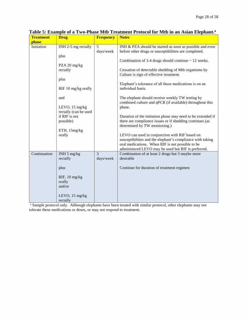
Table 5: Example of a Two-Phase Mtb Treatment Protocol for Mtb in an Asian Elephant.a
Treatment
phase
Drug Frequency Notes
Initiation INH 2-5 mg rectally
plus
PZA 20 mg/kg
rectally
plus
RIF 10 mg/kg orally
and
LEVO, 15 mg/kg
rectally (can be used
if RIF is not
possible)
ETH, 15mg/kg
orally
5
days/week
INH & PZA should be started as soon as possible and even
before other drugs or susceptibilities are completed.
Combination of 3-4 drugs should continue ~ 12 weeks.
Cessation of detectable shedding of Mtb organisms by
Culture is sign of effective treatment.
Elephant’s tolerance of all these medications is on an
individual basis.
The elephant should receive weekly TW testing by
combined culture and qPCR (if available) throughout this
phase.
Duration of the initiation phase may need to be extended if
there are compliance issues or if shedding continues (as
determined by TW monitoring.)
LEVO can used in conjunction with RIF based on
susceptibilities and the elephant’s compliance with taking
oral medications. When RIF is not possible to be
administered LEVO may be used but RIF is preferred.
Continuation INH 5 mg/kg
rectally
plus
RIF, 10 mg/kg
orally
and/or
LEVO, 15 mg/kg
rectally
3
days/week
Combination of at least 2 drugs but 3 maybe more
desirable
Continue for duration of treatment regimen
a Sample protocol only. Although elephants have been treated with similar protocol, other elephants may not
tolerate these medications or doses, or may not respond to treatment.
Page 29 of 58
MONITORING DURING TREATMENT FOR MTB
A. Increased frequency of trunk washes
1. Regardless of the treatment schedule, an increased number of TW should be performed
throughout to determine if shedding has stopped.
2. If using the two-phase protocol, submit a single trunk wash (TW) or triple TW series for
culture each week throughout the Initiation Phase. Bacterial shedding should cease during the
Initiation Phase and this phase can be increased in duration if tolerated.
3. Submit a triple TW series once a month and throughout Continuation Phase until treatment is
completed.
4. If using the single-phase Combination protocol, triple TW series weekly should continue until
detectable shedding has ceased, then reducing to monthly TW monitoring sometime after
cessation is verified.
5. If any TW are culture positive during treatment, recheck sensitivity of the organism and repeat
whole genome sequencing, WGS again to assess if a new isolate is present. The following
options can be considered:
- Increasing the amount of INH per dose
- Adding in a fifth drug
- Swapping one drug for another comparable drug, while keeping INH in the mix.
6. Some elephants shed infrequently, and true cessation may be difficult to discern.
B. Health assessments of elephant receiving treatment
1. Routine CBC/Chem/UA and blood smear should be performed monthly
2. The elephant should be assessed daily for its tolerance to the drugs.
3. If the elephant shows obvious signs of illness during treatment, additional diagnostics may be
needed. See page 31 managing adverse effects in elephants.
4. A body weight should be measured quarterly.
4. The use of serology to monitor post-treatment recovery is un-validated, but some serologically
reactive animals may eventually become non-reactive.
C. Monitoring Herdmates
All herdmates of the infected elephant become Class B elephants and should receive triple TW
testing every 3 months.
D. Measuring drug concentrations
1. Drug concentrations are used to determine if the elephant is reaching a blood concentration of
drug known to be effective against Mtb organisms in other species.
2. Therapeutic drug concentrations for humans are often not attainable in elephants so comparing
the drug concentration reached with the Mtb isolates susceptibility, MIC is often used to assess
treatment concentrations.
3. Some evidence shows that the amount of drug a particular elephant needs can have high
individual variability.
4. Drug concentrations can be measured in serum or plasma. Speak with the laboratory running
the samples to find out which is preferred.
5. The goal is to create a PK curve to determine the animal’s drug concentration is at Tmax. Table
6 shows typical Tmax times for different drugs.
Page 30 of 58
6. Tmax is the time after administration of a drug when the maximum plasma concentration is
reached.
7. Not all elephants will reach an appropriate drug level within the expected time. Some will
take longer, and some will never reach an appropriate serum level of a specific drug at the dose
being used.
8. Some drugs may inhibit the rectal absorption of other drugs (eg, rectal levo suspension and
INH) so if adding a new drug to the protocol repeat PK trials on all of the drugs.
9. If the elephant is not reaching appropriate drug levels, concerns regarding measuring or
understanding drug levels should be discussed with the laboratory. Consider purity testing and
examine sample handling techniques (PK serum/plasma samples) The drug may need increased
or effective therapy may be based on the isolates susceptibilities and cessation of shedding.
10. Plan for measuring drug concentrations once elephant is consistently taking medications
early in treatment regimen, within first 14-45 days. Compare concentrations with MIC of
organism as well as TW shedding results, and appropriate drug concentration listed for humans.
Drug concentration monitoring should be repeated ~ ½ way through treatment or anytime doses
or administration rates are changed. Consistent results with same drug and dose are not always
seen.
Selected References: (Alsultan 2014)(Egelund 2015)(Wiedner & Hunter 2013)
E. Specifics of measuring drug concentrations.
1. Blood is collected at specific timed intervals after rectal or oral administration of a particular
antibiotic, starting at Time Zero (T0; i.e., immediately before drug administration)
2. After T0, additional blood samples are collected every 15 or 30 minutes until one hour past
Tmax. Each blood sample is put into its own vacuum tube.
3. Use lithium heparin vacuum tubes for plasma collection.
4. Use red top vacuum tubes for serum collection (but not serum separation tube –red/grey tops).
5. If collecting serum, allow blood to clot in the red-top vacuum tube at room temperature, then
spin down the tube in a centrifuge for 5 minutes at 3500 RPM.
6. Plasma does not require waiting for clotting to occur before centrifugation, so the lithium
heparin tube can be spun as soon as it is filled.
7. After centrifugation, use a disposable pipette to transfer the serum or plasma into a freezer
proof plastic tube.
8. After labeling the tube with the animal’s name, whether the tube contains serum or plasma, the
date, and how long the contents were collected after antibiotic administration, place the tube in a
standard freezer if samples are going to be sent out for exam in a short period of time. Long term
storage of samples can be done in a -80C freezer.
9. Discuss shipping protocols with the laboratory prior to packaging.
10. Example: Determining drug serum concentrations at Tmax for rectally administered INH
Look at Table 6: Tmax for INH is 3 hours
You can take first blood sample at T0 before elephant is medicated.
Administer full dose of INH rectally.
Take blood every 15 minutes for first hour
Then take blood at 30 minute intervals until 3 hours past T0
Take last sample at Hour 4. There will be a total of 9 samples.
Plasma or serum should be frozen soon after collection since some of the drugs are labile
and photosensitive in the blood.
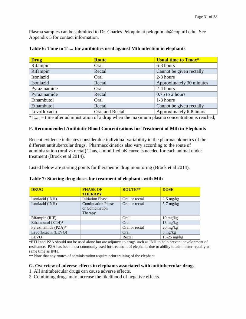
Page 31 of 58
Plasma samples can be submitted to Dr. Charles Peloquin at [email protected]. See
Appendix 5 for contact information.
Table 6: Time to Tmax for antibiotics used against Mtb infection in elephants
Drug Route Usual time to Tmax*
Rifampin Oral 6-8 hours
Rifampin Rectal Cannot be given rectally
Isoniazid Oral 2-3 hours
Isoniazid Rectal Approximately 30 minutes
Pyrazinamide Oral 2-4 hours
Pyrazinamide Rectal 0.75 to 2 hours
Ethambutol Oral 1-3 hours
Ethambutol Rectal Cannot be given rectally
Levofloxacin Oral and Rectal Approximately 6-8 hours
*Tmax = time after administration of a drug when the maximum plasma concentration is reached;
F. Recommended Antibiotic Blood Concentrations for Treatment of Mtb in Elephants
Recent evidence indicates considerable individual variability in the pharmacokinetics of the
different antitubercular drugs. Pharmacokinetics also vary according to the route of
administration (oral vs rectal) Thus, a modified pK curve is needed for each animal under
treatment (Brock et al 2014).
Listed below are starting points for therapeutic drug monitoring (Brock et al 2014).
Table 7: Starting drug doses for treatment of elephants with Mtb
DRUG PHASE OF
THERAPY
ROUTE** DOSE
Isoniazid (INH) Initiation Phase Oral or rectal 2-5 mg/kg
Isoniazid (INH)
Continuation Phase
or Combination
Therapy
Oral or rectal 5-7 mg/kg
Rifampin (RIF) Oral 10 mg/kg
Ethambutol (ETH)* Oral 15 mg/kg
Pyrazinamide (PZA)* Oral or rectal 20 mg/kg
Levofloxacin (LEVO) Oral 5 mg/kg
LEVO Rectal 15-25 mg/kg
*ETH and PZA should not be used alone but are adjuncts to drugs such as INH to help prevent development of
resistance. PZA has been most commonly used for treatment of elephants due to ability to administer rectally at
same time as INH.
** Note that any routes of administration require prior training of the elephant
G. Overview of adverse effects in elephants associated with antitubercular drugs
1. All antitubercular drugs can cause adverse effects.
2. Combining drugs may increase the likelihood of negative effects.
Page 32 of 58
3. Which drug is causing problems is not always clear, and sometimes it is the combination
together that creates adverse effects.
4. Serious adverse events are well-documented in humans, domestic animals, and elephants.
5. The adverse signs seen in elephants are more like those seen in domestic animals, rather than
those that occur in humans.
6. Some signs may be transient and resolve spontaneously; others may persist
7. Adverse effects can occur in elephants at doses of drugs recommended for humans, suggesting
that human serum concentrations can be toxic for elephants.
8. One of the most serious adverse effects with these drugs in humans is liver toxicity, leading to
liver failure.
9. In humans and elephants, increased liver enzymes may be seen on bloodwork obtained during
treatment. However, in elephants, liver enzymes vary in specificity and are poorly validated.
10. Bile acids, the gold standard for monitoring liver function in humans, do not occur in
elephants. Thus, monitoring for hepatoxicity in elephants can be very challenging.
Selected References: (Wiedner and Schmitt 2007), (Wilson 2010)(Simpson 2017).
H. Specific Signs of Drug Intolerance/Adverse effects in Elephants
1. Partial to complete anorexia
2. Depression
3. Hard, scant, black, and or fetid manure
4. Severe blepharospasm
5. Ocular tearing
6. Weakness.
7. Small amounts of frank blood in feces associated with rectal administered drugs
8. Colic/abdominal pain
9. Liver failure
10. Death
I. Managing adverse effects of drugs in elephants
1. If signs are mild such as ocular tearing and blepharospasm, providing analgesia and keeping
the elephant out of bright sunlight may be helpful.
2. If signs do not abate or are severe enough to cause significant discomfort or distress,
temporary cessation of all drug treatment is warranted.
3. Obtain routine bloodwork (CBC/Chem/blood smear) if adverse signs are noted.
4. Most signs will resolve within 7-10 days of stopping all antibiotics.
5. Supportive care for the sick elephant may include hydration using rectal fluids and analgesics.
6. Restart drugs once the elephant appears to be recovered. Repeat the CBC/Chem/blood smear.
7. If signs recur, consider using the Single Phase Combination Therapy Protocol (see page 27).
8. If signs recur, consultation with an experienced veterinary pharmacologist or zoo veterinarian
that has treated elephants with Mtb may be helpful in determining if other drugs or drug
protocols may improve the situation.
9. Adverse effects, either those described above or others, should be reported to the Food and
Drug administration (FDA) Center for Veterinary Medicine, (CVM) The stakeholders would also
appreciate being made aware of side effects and toxicity associated with treatment so that these
treatment recommendations can be amended. (see Appendix 4 for references).
Page 33 of 58
J. Treatment failures
1. Recurrence of shedding (a positive TW) indicates treatment failure.
2 Treatment failure can occur at any point during or after treatment. Humans are typically
monitored for two years after completion of antitubercular treatment.
3. Recurrence (also called relapse) rates in humans using a four-drug approach range from 0 to
27%.
4. Prevalence of treatment failure in elephants is unknown.
5. If weekly TW cultures continue to be positive eight weeks after starting treatment:
Recheck drug sensitivities
Confirm potency of medications being administered
Recheck serum drug concentrations.
6. Reported cases of INH resistant infections have occurred in elephants. If this is noted, consider
using a combination of RIF & Fluoroquinolone medications in place of INH based on reported
susceptibilities.
7. For resistant infections and/or extreme elephant sensitivity to drugs, few guidelines exist.
Thus, it is the recommendation of the authors of this document that in such cases, the
veterinarian of the facility should consult with pharmacologists and elephant veterinary experts
to determine how best to proceed. An excellent reference guide for physicians dealing with
Drug-Resistant TB in humans may also offer some guidance for veterinarians in this difficult
situation.
Curry International TB Center, Drug-Resistant TB, A Survival Guide for Clinicians.3rd Ed http://www.currytbcenter.ucsf.edu/products/cover-pages/drug-resistant-tuberculosis-survival-
guide-clinicians-3rd-edition
K. Recurrence vs Reinfection
1. Re-infection is a new infection with a different isolate and is considered different from
recurrence.
2. Distinguishing between recurrence and re-infection requires molecular techniques such as
whole genome sequencing to determine if the new isolate is the same strain as the initial one and
if it has mutated. (Bryant 2013)
L. Monitoring after treatment is completed.
1. Test by triple TW culture series every 3 months for 2 years.
2. If all TW are culture negative during this period, the elephant reverts to Category B and
should be tested as per that protocol: by triple TW technique every 3 months for an additional 2
years, and followed with increased TW monitoring for the remaining 3 years.
3. At this point (2 years), the elephant no longer has travel restrictions.
4. Herdmates continue as Category B elephants for the recommended time.
Page 34 of 58
HUMAN HEALTH CONSIDERATIONS
A. Human-to-human transmission of Mtb
1. Transmission of Mtb between humans is well studied and comparatively well defined.
Transmission can occur when Mtb are aerosolized by a person actively shedding the bacteria. In
an enclosed space, the likelihood of human-to-human transmission of Mtb is influenced by air
volume, exhaust rate, time and circulation. In large indoor settings, because of diffusion and
local circulation patterns, the degree of proximity between contacts and the index patient can
influence the likelihood of transmission
2. Indirect contact with a person who is shedding the organism is unlikely to result in
transmission. Humidity and light do not affect transmission.
3. The likelihood of infection depends on the intensity, frequency, and duration of exposure. For
example, airline passengers who are seated for >8 hours in the same or adjoining row as a person
who is contagious are much more likely to be infected than other passengers and only these
people are contacted for testing in a TB exposure investigation.
4. Other routes of Mtb exposure in humans have not been documented or are not considered
significant.
Selected references: (Heyman 2008)
https://www.cdc.gov/tb/education/corecurr/pdf/chapter2.pdf
WHO Guidelines, Tuberculosis and Air Travel: Guidelines for Prevention and Control, third
edition, 2013)
B. Elephant-to-human transmission of Mtb
1. In general, Mtb requires prolonged, close contact with a person or animal that is actively
shedding the bacteria into the environment. Transmission of Mtb from elephants to humans is,
therefore, more of an occupational health concern than a health concern for the general public.
2. Close, prolonged contact as a significant risk factor for transmission is supported by an
investigation that assessed 48 zoo patrons, 20 of whom were present at special elephant events
where a culture positive elephant actively aerosolized paint with his trunk. All zoo patrons had
<30 minutes of exposure at a distance of > 25 feet to this elephant during his presumed infectious
period and no patrons evaluated showed evidence of exposure. In contrast, that same study
documented skin test conversions in 5/31 zoo employees, all of whom were considered close
contacts to this same elephant, and one zoo volunteer, who had greater exposure (1 hour
cumulative presence in the elephant barn) than zoo patrons to the same culture positive elephant
(Zlot, et al, MMWR 2016). In the United States, facilities that maintain elephants may offer
opportunities for members of the general public to touch, feed or ride an elephant. These
opportunities are typically offered in such a way that members of the general public do not have
prolonged contact with elephants in an enclosed space. Therefore, such contacts would be
unlikely to constitute a public health risk. This is particularly true with elephants that are
routinely screened and monitored for their Mtb status via routine trunk wash (TW) cultures.
3. Close prolonged contact such as routine elephant handling, elephant training, or participating
in a necropsy on an Mtb-infected elephant can increase occupational risk. (Michalak 1998)
4. Cleaning protocols that utilized high pressure water hoses were implicated in causing humans
working close to a barn containing an Mtb-infected elephant to become PPD (skin-test) positive,
although they did not develop active infections (Murphree 2011). Thus, husbandry practices
should be examined closely.
Page 35 of 58
Selected references: (Zlot 2016), (Davis 2001), (Murphree 2011), (Oh 2002), (Montali 2001),
(Mikota and Maslow 2011), (Michalak 1998), (National Tuberculosis Controllers Association
2005), (Vogelnest 2013), (Lecu and Ball 2011).
C. Human-to-elephant transmission of Mtb
1. Initial infections of elephants with Mtb have been hypothesized to come from close contact
with infectious humans and some documented evidence exists to support this suspicion. (Payeur
2002). Several elephants in the US originally imported from South East Asia have had Mtb
isolates that originate genetically (Lineage-3 genotype) from this part of the world. (S. Robbe-
Austerman, K Backues personal communication)
2. Mtb infection has only recently been documented as a disease of wild elephants.
(Chandranaik 2017), (Perera 2014))
D. Occupational Health Recommendations
1. Managers and veterinarians of facilities that maintain elephants should consult with that
facility's occupation health staff, State Public Health Veterinarian and/or health department
tuberculosis staff to develop an occupational health program. Program development activities
should include a written infection control plan that covers measures designed to reduce direct
and indirect aerosol transmission of Mtb. Such measures may include addressing use of personal
protective equipment (PPE), elephant quarantine procedures, disinfection procedures and the
ventilation in a facility where elephants are being housed.
2. Facility protocols should assign levels of risk for Mtb exposure based on factors including
differing levels of contact staff have with elephants, species of elephant, diagnostic history of the
herd and degree of public contact.
3. Protocols should include routine annual tuberculosis screening of employees who work with
elephants and testing of new hires. Employees with acid-fast positive sputum smears or
suspicious chest films should not work directly with elephants until they have had additional
diagnostic testing. The results of this additional testing should inform decisions about an
employee’s return to work and performing elephant care duties. Human testing for Mtb should
be overseen by occupational health and/or tuberculosis control authorities.
4. Managers of elephant facilities should encourage employees who have health concerns to
discuss them with a healthcare provider, occupational health staff and/or health department
personnel. Managers should also understand that employee medical information is considered
private and discuss with health department and/or occupational health personnel how situations
associated with an employee for whom Mtb infection is a concern will be handled.
5. Routine education of staff in zoonotic disease prevention should occur, including discussion of
clinical symptoms consistent with active human tuberculosis infection.
6. Employees should be trained in the proper use of PPE.
Resources for Occupational Health and Safety Information can be found in Appendix 7.
Page 36 of 58
Appendix 1: References: Most are cited in text but others are included as an overview of
current and historical literature on Mtb in both human and veterinary medicine.
1. Alsultan A, CA Peloquin Therapeutic Drug Monitoring in the Treatment of Tuberculosis: An
Update. Drugs 2014: 74, Pp839-854.
2. American Thoracic Society, Centers for Disease Control and Prevention, Infectious Diseases
Society of America. Treatment of tuberculosis. Morbidity and mortality weekly report
2003;52:1-80.
3. Angkawanish T, Morar D, Kooten Pv, et al. The elephant interferon gamma assay: a
contribution to diagnosis of tuberculosis in elephants. Transboundary and emerging diseases
2013;60:53-59.
4. Backues KA, Robbe-Austerman S, Isaza R. Documented cessation of mycobacterial shedding
with antibiotic treatment in a Mycobacteria-tuberculosis-positive Asian elephant (Elephas
maximus) by serial culture and direct real-time polymerase chain reaction testing of trunk wash
samples Proc AAZV 2015;151-152.
5. Bontekoning I. Tuberculosis detection in the Asian elephant (Elephas maximus) population of
Thailand. 1. Development of an IFN-gamma assay 2. Evaluation of a multiple antigen iELISA
and a commercial rapid test. In. Thailand: Kasetsart University, Bangkok, Chiang Mai
University, Chiang Mai Utrecht University, Utrecht; 2009:29 pages.
6. Boyd J. Serum enzymes in the diagnosis of disease in man and animals. Journal of
comparative pathology 1988;98:381-404.
7. Blumberg H, Burman W, Chaisson R, et al. American Thoracic Society/Centers for Disease
Control and Prevention/Infectious Diseases Society of America: treatment of tuberculosis. Am J
Resp Crit Care Med 2003;167:603.
8. Brock AP, Isaza R, Egelund EF, et al. The pharmacokinetics of a single oral or rectal dose of
concurrently administered isoniazid, rifampin, pyrazinamide, and ethambutol in Asian elephants
(Elephas maximus). Journal of Veterinary Pharmacology and Therapeutics 2014.
9. Bryant JM, et al. Whole-genome Sequencing to Establish Relapse or Re-infection with
Mycobacterium tuberculosis: A Retrospective Observational Study. The Lancet 2013 V1, Pp786-
792.
10. Chalke HD. The impact of tuberculosis on history, literature and art. Medical history
1962;6:301-318.
11. Chandranaik B, Shivashankar BP, Umashankar KS, et al. Mycobacterium tuberculosis
Infection in free-roaming wild Asian elephant. Emerg Infect Dis 2017;23:555-557.
12. Davies GR, Neurmberger EL. Pharmacokinetics and pharmacodynamics in the development
of anti-tuberculosis drugs. Tuberculosis 2008;88:S65-S74.
13. Davis M. Mycobacterium tuberculosis risk for elephant handlers and veterinarians.
Parasitology international 2001;16:350-353.
14. Egelund EF, A Alsultan, CA Peloquin. Optimizing the Clinical Pharmacology of
Tuberculosis Medications. Clinical Pharmacology & Therapeutics 2015; V98(4) Pp 387-393.
15. Egelund EF, Isaza R, Alsultan A, et al. Isoniazid and rifampin pharmacokinetics in two
Asian elephants (Elephas maximus) infected with Mycobacterium tuberculosis. J Zoo Wild Med
2016;47:868-871
Page 37 of 58
16. Feldman M, Isaza R, Prins C, et al. Point prevalence and incidence of Mycobacterium
tuberculosis complex in captive elephants in the United States. Veterinary quarterly 2013;33:25-
29.
17. Filippini P, Iona E, Piccaro G, et al. Activity of drug combinations against dormant
Mycobacterium tuberculosis. Antimicrob Agents Chemother 2010;54:2712-2715.
18. Garcia-Tapia A et al. Action of Fluoroquinolones and Linezolid on Logarithmic-and
Stationary-Phase Culture of Mycobacterium tuberculosis. Chemotherapy 2004; 50: 211-213.
19. Gumbo T, et al. Pharmacodynamic Evidence that Ciprofloxacin Failure Against Tuberculosis
is not Due to Poor Microbial Kill but Rapid Emergence of Resistance. Antimicrobial Agents and
Chemotherapy. 2005 V 49(8)Pp 3178-3181.
20. Greenwald R, Lyashchenko O, Esfandiari J, et al. Highly accurate antibody assays for early
and rapid detection of tuberculosis in African and Asian elephants. Clinical and vaccine
immunology 2009;16:605-612.
21. Griffin J, Buchan G. Aetiology, pathogenesis and diagnosis of Mycobacterium bovis in deer.
Veterinary Microbiology 1994;40:193-205.
22. Heymann DL. Control of communicable diseases manual. Washington, D.C.: American
Public Health Association; 2008.
23. Isaza R, Ketz C. A trunk wash technique for the diagnosis of tuberculosis in elephants. In:
Verhandlungsbericht des Internationalen Symposiums uber die Erkrankungen der Zootiere,
Vienna 1999;121-124.
24. Isaza R, Wiedner E, Hiser S, et al. Reference intervals for acute phase protein and serum
protein electrophoresis values in captive Asian elephants (Elephas maximus). Journal of
Veterinary Diagnostic Investigation 2014;26:616-621.
25. Lacasse C, Terio K, Kinsel MJ, et al. Two cases of atypical mycobacteriosis caused by
Mycobacterium szulgai associated with mortality in captive African elephants (Loxodonta
africana). Journal of zoo and wildlife medicine 2007;38:101-107.
26. Lambert M-L, Hasker E, Van Deun A, et al. Recurrence in tuberculosis: relapse or
reinfection? The Lancet infectious diseases 2003;3:282-287.
27. Landolfi JA, Miller M, Maddox C, et al. Differences in immune cell function between
tuberculosis positive and negative Asian elephants. Tuberculosis 2014;94:374-382.
28. Landolfi, JA, et al. Pulmonary tuberculosis in Asian Elehpants (Elephas maximus):
Histologic Lesions with Correlation to Local Immune Response. Vet Path. 2015 Vol 52(3)
pp535-542.
29. Larsen RS, Salman MD, Mikota SK, et al. Evaluation of a multiple-antigen enzyme-linked
immunosorbent assay for detection of Mycobacterium tuberculosis infection in captive
elephants. Journal of zoo and wildlife medicine 2000;31:291-302.
30. Léçu A, Ball R. Mycobacterial infections in zoo animals: relevance, diagnosis and
management. International zoo yearbook 2011;45:183-202.
31. Leibert E, Rom WN. New drugs and regimens for treatment of TB. Expert Rev Anti-Infect
Ther 2010;8:801-813.
32. Lewerin, SS, Olsson S-L, Eld K, Roken B, Ghebremichael S, Koivula T, Kallenius G,
Bolske G. Outbreak of Mycobacterium tuberculosis infection among captive Asian elephants in a
Swedish zoo. Vet Rec 2005;156:171-175.
Page 38 of 58
33. Magnuson RJ, et al. Rapid Screening for Mycobacterium tuberculosis Complex in Clinical
Elehpant Trunk Wash Samples. Research in Vet Science 2017; 112 Pp52-58.
34. Maslow JN, Mikota SK, Zhu M, et al. Population pharmacokinetics of isoniazid in the
treatment of Mycobacterium tuberculosis among Asian and African elephants (Elephas maximus
and Loxodonta africana). Journal of veterinary pharmacology and therapeutics 2005;28:21-27.
35. Maslow JN, Mikota SK, Zhu M, et al. Pharmacokinetics of ethambutol (EMB) in elephants.
Journal of veterinary pharmacology and therapeutics 2005;28:321-323.
36. Michalak K, Austin C, Diesel S, et al. Mycobacterium tuberculosis infections as a zoonotic
disease: transmission between humans and elephants. Emerging infectious diseases 1998;4:283-
287.
37. Mikota SK, Maslow JN. Tuberculosis at the human-animal interface: An emerging disease
of elephants. Tuberculosis 2011;91:208-211.
38. Mikota SK, Peddie L, Peddie J, et al. Epidemiology and diagnosis of Mycobacterium
tuberculosis in captive Asian elephants (Elephas maximus). Journal of zoo and wildlife medicine
2001;32:1-16.
39. Montali RJ, Mikota SK, Cheng LI. Mycobacterium tuberculosis in zoo and wildlife species.
Review of science and technology 2001;20:291-303.
40. Murphree R, Warkentin JV, Dunn JR, et al. Elephant-to-human transmission of tuberculosis,
2009. Emerging infectious diseases 2011;17:366-371.
41. National Association of State Public Health Veterinarians. Compendium of measures to
prevent disease associated with animals in public settings, 2005. MMWR Recommendations and
reports: Morbidity and mortality weekly report Recommendations and reports/Centers for
Disease Control 2005;54:1.
42. National Tuberculosis Controllers Association. Guidelines for the investigation of contacts
of persons with infectious tuberculosis. Recommendations from the National Tuberculosis
Controllers Association and CDC. MMWR Recommendations and reports: Morbidity and
mortality weekly report, Recommendations and reports/Centers for Disease Control 2005;54:1.
43. NIOSH. Health hazard evaluation report: evaluation of potential employee exposure to
Mycobacterium tuberculosis at an elephant refuge. By Neimeier RT. MeadK, dePerio MA,
Martin SB, Burr GA Cincinnati, OH: U.S. Department of Health and Human Services, Centers
For Disease Control and Prevention, National Institute for Occupational Safety and Health; 2015.
44. Obanda V. First reported case of Fatal tuberculosis in a wild African elephant with past
human-wildlife contact. Epidemiology and Infection 2013; 141(7): 1476-1480.
45. Oh P, Granich R, Scott J, et al. Human exposure following Mycobacterium tuberculosis
infection of multiple animal species in a metropolitan zoo. Emerging infectious diseases 2002;8.
46. Papastavros T, Dolovich LR, Holbrook A, et al. Adverse events associated with
pyrazinamide and levofloxacin in the treatment of latent multidrug-resistant tuberculosis. CMAJ
2002;167:131-136.
47. Payeur J, B., Jarnagin JL, Marquardt JG, et al. Mycobacterial isolations in captive elephants
in the United States. Annals of the New York Academy of Sciences 2002;969:256-258.
48. Peloquin CA, Maslow JN, Mikota SK, et al. Dose selection and pharmacokinetics of
rifampin in elephants for the treatment of tuberculosis. J Vet Pharm Ther 2006;29:581-585.
49. Perera BVP, Salgadu MA, Gunawardena GSPdS, et al. First confirmed case of fatal
Page 39 of 58
tuberculosis in a wild Sri Lankan elephant. Gajah 2014;41:28-31.
50. Saukkonen JJ, Cohn DL, Jasmer RM, et al. An official ATS statement: hepatotoxicity of
antituberculosis therapy. American journal of respiratory and critical care medicine
2006;174:935-952.
51. Scheftel JM, Elchos BL, Cherry B, et al. Compendium of veterinary standard precautions for
zoonotic disease prevention in veterinary personnel: National Association of State Public Health
Veterinarians Veterinary Infection Control Committee 2010. Journal of the American Veterinary
Medical Association 2010;237:1403.
52. Simpson G, Zimmerman R, Shashkina E, et al. Mycobacterium tuberculosis infection among
Asian Elephants in captivity. Emerg Infect Dis 2017;23:513.
53. Spigelman MK. New tuberculosis therapeutics: a growing pipeline. J Infect Dis
2007;196:S28-S34.
54. Stephens N, Vogelnest L, Lowbridge C, et al. Transmission of Mycobacterium tuberculosis
from an Asian elephant (Elephas maximus) to a chimpanzee (Pan troglodytes) and humans in an
Australian zoo. Epidemiology and infection 2013;141:1488-1497.
55. Thompson NP, Caplin ME, Hamilton MI, et al. Anti-tuberculosis medication and the liver:
dangers and recommendations in management. European respiratory journal 1995;8:1384-1388.
56. Verma-Kumar S, Abraham D, Dendukuri N, et al. Serodiagnosis of Tuberculosis in Asian
Elephants (Elephas maximus) in Southern India: A Latent Class Analysis. PLOs one 2012;7.
57. Vogelnest L. Tuberculosis: an emerging zoonosis. New South Wales Public Health Bulletin
2013;24:32-33.
58. Vogelnest L, Hust F, Vinette HK, et al. Diagnosis and management of tuberculosis
(Mycobacterium tuberculosis) in an Asian elephant in Australia. In: Mycobacterial diseases of
wildlife 2012;242.
59. Wiedner E, RP Hunter. Antimicrobial Drug Use in Zoological Animals. In Antimicrobial
therapy in Veterinary Medicine 5th ed. editors S Giguere, JF Prescott and PM Dowling. 2013
John Wiley & Sons. Inc.
60. Wiedner E, Schmitt DL. Preliminary report of side effects associated with drugs used in the
treatment of tuberculosis in elephants. In: Proceedings of the international elephant foundation
meeting, Orlando, FL 2007;15-20.
61. Wilson E, Mikota S, Bradford JP, et al. Seropositive, culture negative tuberculosis in an
Asian elephant (Elephas maximus). In: AAZV, AAWV Joint Conference, South Padre Island,
Texas 2010;170.
62. Yee D, Valiquette C, Pelletier M, et al. Incidence of serious side effects from first-line
antituberculosis drugs among patients treated for active tuberculosis. American journal of
respiratory and critical care medicine 2003;167:1472-1477.
63. Younossian AB, Rochat T, Ketterer JP, et al. High hepatotoxicity of pyrazinamide and
ethambutol for treatment of latent tuberculosis. European respiratory journal 2005;26:462-464.
64. Zhu M, Maslow JN, Mikota SK, et al. Population pharmacokinetics of pyrazinamide in
elephants. Journal of veterinary pharmacology and therapeutics 2005;28:403-409.
65. Zlot A, Vines J, Nystrom L, et al. Diagnosis of Tuberculosis in Three Zoo Elephants and a
Human Contact—Oregon, 2013. MMWR 2016;64:1398-1402.
Page 40 of 58
Appendix 2: Trunk Wash Technique (Isaza and Ketz): The Trunk Wash Technique for
Routine Surveillance and Diagnosis of Mtb in Elephants (Isaza and Ketz 1999)
Summary
A trunk wash is a practical method of collecting a sample from an elephant’s distal respiratory
tract for Mycobacterium culture. The procedure, however, is potentially dangerous to the handlers
and requires cooperation of the elephant. Because of the limitations of using culture results as a
screening test, the trunk wash results should be interpreted with care. A positive culture result
identifies an elephant that is shedding tuberculosis organisms whereas a negative result is non-
diagnostic.
Materials and methods The trunk wash technique requires that the elephant allow the handlers to restrain and manipulate
the tip of trunk. This is difficult in an untrained elephant in that most elephants resent this
manipulation, and the trunk is many times stronger than the combined force of several handlers. It
is therefore important that the animals be trained to present the trunk, allow gentle manual restraint,
and manipulation of the trunk tip during the collection of the sample. The training period varies
with the individual elephant, the prior behavioral conditioning of the animal, and the skill of the
handlers. In our experience, most animals can be adequately trained for the procedure in 2-4 weeks.
Materials Needed:
o Sterile 0.9% saline solution
o Sterile 60 ml syringe
o 1 gallon plastic zip lock type bags (heavy duty)
o 50 ml, screw top, plastic jar or centrifuge tube
As long as attention is given to collecting a clean sample from the distal nasal passages, the
materials and techniques for the sample collection can be modified. For example, some clinicians
prefer to use a 14-gauge red rubber tube feeding tube inserted into the trunk tip instead of simply
flushing the sterile saline into the trunk tip. Another common variation is to use a sterile plastic
container to catch the trunk wash fluid instead of a plastic bag.
Procedure A routine screening of an elephant should consist of a series of three trunk wash samples collected
on separate days within a one-week period, i.e. triple trunk wash series or collection. Trunk
washings should be collected in the morning and prior to water being offered to the animal, or after
food and water has been withheld for a minimum of 2 hours. These recommendations are made in
an attempt to obtain a representative sample of the nasal flora from the previous night, and to avoid
the dilution effect caused by elephants drinking water with their trunks.
The elephant’s trunk is manually restrained by the handlers so that the tip is held up. The 60 ml
syringe filled with sterile saline is then inserted into one of the nostrils and the saline quickly
flushed into the trunk. The handler then lifts the trunk tip as high as possible to help the fluid flow
as far into the trunk as possible. The 1 gallon plastic bag is then slipped over the trunk tip and the
tip of the trunk is lowered to allow the fluid to drain. If possible, the elephant is allowed to exhale
into the bag during this collection phase of the procedure. A good sample should retrieve a
significant portion of the saline that was placed into the trunk (about 40 ml). The sample should
Page 41 of 58
contain visible mucus from the inside of the trunk and often contains dirt and food particles that
are normally found inside the trunk. The collection of moderate amounts of foreign material does
not invalidate the sample. If, however, the collector feels the contamination is excessive, a second
flush may be attempted.
Once the sample is collected in the plastic bag, it is carefully transferred into a labeled container.
Ideally, the sample is refrigerated and sent directly to a laboratory for processing and
mycobacterial culture. If the sample cannot be sent directly for culturing, it may be frozen in a
regular freezer (-20 to -10o C) until it can be sent to the laboratory. Often the recommended three
daily culture samples are collected and frozen until all samples are collected and the batch of
samples can be sent to the laboratory together.
The trunk wash as a method of collecting a culture sample from elephants has become the standard
method of screening elephants for Mtb. It is a practical way of obtaining a culture sample from a
large proportion of the elephant population. The procedure requires no sedation or undue stress to
the animal. Additionally, the procedure requires no specialized or expensive equipment.
An important consideration of this procedure is that it can potentially be very dangerous to the
handlers. This is particularly true when attempted on an uncooperative elephant, because any
attempts to manually restrain the trunk in an uncooperative elephant can lead to injury. Therefore,
the time spent training the elephant to accept this method will greatly increase the efficiency and
safety of the procedure. In some cases, with potentially dangerous or unpredictable animals, an
increased level of handler safety can be obtained by having the animal lie in sternal or lateral
recumbency prior to sample collection. This technique does not guarantee safety or successful
sample collection, as it still requires cooperation of the animal and does not replace adequate
training. In the case of elephants managed under protective contact, the animal’s trunk can be
handled though a set of bars. This method still requires that the animal is fully cooperative and,
therefore, usually requires extensive training prior to the collection.
A second safety issue is the potential for zoonotic infection. Recently there has been
documentation of a zoonotic transmission of tuberculosis between humans and elephants. During
the collection of the trunk wash sample, there is exposure to aerosolized mucus from the elephant’s
respiratory tract. The authors, therefore, suggest that the collectors and handlers wear PPE of that
includes an N-95 or greater particle mask. Minimal precautions would include a well fitted
respirator or face mask capable of filtering 0.3 micron particles, disposable gloves, and working
in a well-ventilated, area.
Mycobacterial culture as the primary method of detecting infected animals has several limitations
that are best illustrated by examination of the underlying biological assumptions. The first
assumption is that most infected elephants have respiratory infections. Although the literature
suggests that most infected elephants have respiratory infection, there have been no comprehensive
necropsy studies to confirm these observations. The second assumption is that most infected
animals shed mycobacterial organisms into the respiratory tract. There is little data that determines
if and when an infected animal will begin shedding organisms. It is unknown what proportion of
elephants can carry latent or “walled off” infections that would be missed with culturing
techniques. A third assumption is that animals that are shedding will pass mycobacteria organisms
at least once in the three-day testing period. Currently it is unknown if shedding animals pass
Page 42 of 58
organisms periodically or continuously. Finally, the samples collected from the distal trunk are
often contaminated with normal bacterial flora and foreign material. It is assumed that these
contaminants do not routinely overgrow or mask the growth of pathogenic mycobacteria, although
no studies have tested this assumption. The interpretations of the culture results should, therefore,
be limited. A positive culture is strong evidence that the animal is shedding mycobacteria and is
infected; negative culture results provide little information as to whether the elephant is infected
or not.
Culturing the distal trunks of all the animals in a population will only detect animals shedding
tuberculosis through the trunk, and not detect all animals that are infected. However, with time
and repeated cultures of all animals in the population, it may be possible to detect and treat most
of the elephants shedding infectious organisms. If these animals are then treated properly and
shedding of organisms stops, the spread of tuberculosis from elephant to elephant should decrease
in the population
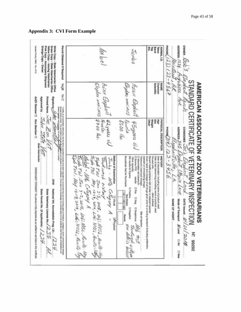
Page 43 of 58
Appendix 3: CVI Form Example
Page 44 of 58
Appendix 4: Laboratories for Mtb culture samples:
Suggested Certified Laboratories for Mycobacteria Cultures and Research status qPCR on
Trunk Washes.
1. USDA APHIS VS
National Veterinary Services Laboratories (NVSL)
1920 Dayton Avenue
Ames, IA 50010
Lab web site: http://www.aphis.usda.gov/animal_health/lab_info_services/diagnos_tests.shtml
Dr. Suelee Robbe-Austerman
Veterinarian, Mycobacteria and Brucella Section
(515) 337-7837 Fax: (515) 337-7315
Email: [email protected]
50 ml conical screw-top leak-proof centrifuge tubes are preferred and available free of
charge from NVSL.
Send trunk washes to NVSL either frozen or on icepacks by overnight express (Federal
Express handles diagnostic samples). Containers should be leak proof and double-bagged
If lesions are submitted for culture, tissues should be frozen and sent on ice packs overnight.
Lesioned tissues should be split and ½ should be sent to the histopathology lab so PCR can be
run to see if the tissue is compatible with tuberculosis. There is no charge for histopathology on
lesioned tissue.
Use the VS Form 10-4 for submission. If the formalinized tissue is sent separately from the
frozen tissue, please indicate on the submission forms that there are 2 separate packages coming
from the same animal so that the reports can be combined and accession numbers coordinated
when they reach NVSL. It is also helpful to call or email NVSL contacts when sending sample
from Mtb suspects to schedule testing and relay any relevant history of the case.
NVSL Trunk wash cost: $98 per sample for processing which includes a Gen Probe® DNA
probe on any isolate. If the sample is positive for mycobacteria and speciation is requested, the
charge is $122.00 per sample which includes biochemical analysis, 16s rDNA sequencing
analysis, spoliotyping and VNTR genotyping. DNA fingerprinting of M. tuberculosis or M.
bovis isolates is also available. Antimicrobial susceptibility testing is available for M.
tuberculosis complex organisms for $112.00 per isolate. Please contact NVSL at (515) 337-
7388 for test schedule.
To establish an account at NVSL for billing, contact Connie Osmundson (515) 337-7571 or
Email: [email protected] .
(User fees as of December 1, 2014). Call lab before shipping samples for current prices and
schedule of testing or check prices at the NVSL web site:
http://www.aphis.usda.gov/animal_health/lab_info_services/diagnos_tests.shtml
Page 45 of 58
2. Mycobacteriology Laboratory at National Jewish Medical and Research Center
National Jewish Medical and Research Center
1400 Jackson St.
Denver, CO 80206
(303) 398-1384
Manager Clinical Laboratories:
Jamie Marola, MB(ASCP)
National Jewish Health Advanced Diagnostic Laboratories
303.270.2479 Office
303.398.1339 Laboratory
720.290.2204 Mobile
303.398.1953 Fax
Clinical Laboratory Supervisor: Kimberly Sue Messina, MT-ASCP
Mycobacteriology Lab
Room K422a
Lab Phone: 303-398-1339
Office Phone: 303-398-1347
Cell Phone: 469-323-1352
For price list; sample collection and shipping instructions and requisition form:
http://www.nationaljewish.org/research/diagnostics/adx/labs/mycobacteriology/requisitions-and-
specimen-handling.aspx
3. Your State Public Health Laboratory other CDL certified Laboratory.
Page 46 of 58
Appendix 5: Lab Contact for Therapeutic Drug Level Monitoring
INFECTIOUS DISEASE PHARMACOKINETICS LABORATORY
Contact: Dr. Charles Peloquin
1600 SW Archer Rd., P4-30
Gainesville, FL 32610
Phone: 352-273-6710 Fax: 352-273-6804
E-mail: [email protected]
Website: http://idpl.pharmacy.ufl.edu
Direct link to IDPL with Submission Form
https://cop-idpl.sites.medinfo.ufl.edu/files/2017/06/IDPL-UFHealth-v05.17.pdf
Page 47 of 58
Appendix 6: Epidemiological Definitions
There are a number of epidemiologic terms that are applicable to any infectious disease, and that
must be understood to facilitate sound clinical decision-making and application of Mtb treatment
and management measures. It is imperative that the clinician consider what is known about both
the epidemiology of Mtb and the diagnostic test modalities available. Below are a few definitions
for understanding diagnostic test interpretation and TB disease epidemiology.
Mtb Direct Tests: Tests that determine the presence of Mycobacterium tuberculosis in
the sample. These tests can determine viable organisms (e.g., culture) or potentially non-
viable components of the organism, such as DNA fragments (e.g., PCR) or proteins.
Mtb Indirect Tests: Tests that measure or detect an animal’s immune response to
Mycobacterium tuberculosis.
Sensitivity: A measure of the ability of a test to identify infected animals. Sensitivity is the
frequency of a positive or abnormal test result (e.g., a test that is outside of the reference interval)
when a disease is present (i.e., the percentage of true positive results). Sensitivity = [TP ÷ (TP +
FN)] X 100 where TP = true positive; FN = false-negative). Validation of test sensitivity requires
inclusion of a full spectrum of disease states. Test sensitivity may vary among populations. No
Mycobacterium tuberculosis diagnostic test is 100% sensitive.
Specificity: A measure of the ability of a test to identify non-infected animals. Specificity is the
frequency of a negative or “normal” test result when a disease is absent (i.e., the percentage of
true-negative (TN) test results. Specificity = [TN ÷ (TN + FP)] X 100. Validation of test specificity
requires inclusion of a full spectrum of disease states. Test specificity may vary among
populations. No Mycobacterium tuberculosis diagnostic test is 100% specific.
Negative Predictive Value: A numerical value for the proportion of individuals with a negative
test result who have the target condition (i.e., the probability that a person who is a test negative is
a true negative.) This probability is relevant to determining the usefulness of a test when applied
to animals of unknown disease status, and is clinically more important than test sensitivity and
specificity. The negative predictive value of diagnostic tests can be low in populations with high
disease prevalence.
Positive Predictive Value: A numerical value for the proportion of individuals with a positive test
result who have the target condition (i.e., the probability that a person who is a test positive is a
true positive.) This probability is relevant to determining the usefulness of a test when applied to
animals of unknown disease status, and is clinically more important than test sensitivity and
specificity. The positive predictive value of diagnostic tests can be low in populations with low
disease prevalence.
Risk analysis: The phases of a risk analysis, according to World Organization for Animal Health
(OIE) Code, include hazard identification, risk assessment, risk management, and risk
communication. Data for conducting formal risk analysis for tuberculosis in elephants is limited,
although qualitatively considering the parts of formal risk assessments and risk analyses is useful
for managing the risks of tuberculosis.
Page 48 of 58
Risk assessment: The process of evaluating the likelihood of exposure, infection, or spread of a
disease. This is a part of formal risk analysis. The parts of a formal risk assessment include release,
exposure, and consequence assessments, as well as risk estimation. Data for conducting formal
risk assessments for tuberculosis in elephants is limited, although qualitatively considering the
parts of formal risk assessments and risk analyses is useful for assessing the risks of tuberculosis.
Trunk Wash (TW) Culture: A direct test designed to detect viable Mtb organisms via culture of
material obtained from a trunk wash. It is a practical method of obtaining a culture sample from a
large proportion of the elephant population. The procedure requires no sedation or undue stress to
the elephant. Additionally, the procedure requires no specialized or expensive equipment. The
recommended routine Mtb monitoring is an annual triple mycobacterial trunk wash culture. Each
testing event should consist of three independent collections on three days within a one week
period. It is recommended that food and water be withheld from elephants for 2 hours before the
TW is performed to help minimize the contamination of the TW sample.
Mtb Infected Elephant: An elephant from which one positive Mtb culture has been isolated from
a bodily discharge or lesion.
Pharmacokinetics (PK): How a drug behaves in the body; i.e. how much needs to be given to
obtain a particular drug level, where and how the drug is metabolized, how long the drug lasts in
the system.
Pharmacodynamics (PD): The action of the drug; i.e., whether it actually kills infectious
organisms, whether it causes adverse effects.
Tmax: refers to the time after administration of a drug when the maximum plasma concentration
is reached. This needs to be determined when serum drug levels are measured.
Page 49 of 58
Appendix 7: Resources for Occupational Health and Safety Information:
The Occupational Safety and Health Agency (OSHA) tuberculosis guidelines.
OSHA has regulations for recording and reporting tuberculosis infection acquired in the workplace.
These are outlined at http://www.osha.gov/SLTC/tuberculosis.
The Centers for Disease Control and Prevention (CDC) Guidelines for Preventing the Transmission
of Mycobacterium tuberculosis in Health-Care Settings in 2005. The CDC guidelines for
preventing transmission of Mtb are available at http://www.cdc.gov/mmwr/PDF/rr/rr5417.pdf.
Though not specific to elephant care settings, they contain invaluable recommendations and
guidance for workers exposed to a source of tuberculosis.
NIOSH [2015]. Health hazard evaluation report: evaluation of potential employee exposures to
Mycobacterium tuberculosis at an elephant refuge. By Niemeier RT, Mead K, de Perio MA, Martin
SB, Burr GA. Cincinnati, OH: U.S. Department of Health and Human Services, Centers for Disease
Control and Prevention, National Institute for Occupational Safety and Health, NIOSH HHE
Report No. 2010-0080-3235. Available at https://www.cdc.gov/niosh/hhe/reports/pdfs/2010-0080-
3235.pdf.
The CDC document Biosafety in Microbiological and Biomedical Laboratories published by the
U.S. Department of Health and Human Services in 2007. This document outlines best practices
for the safe conduct of work in biomedical and clinical laboratories and animal facilities in regards
to Mycobacterium tuberculosis and is available at
http://www.cdc.gov/od/ohs/biosfty/bmbl5/BMBL.
The CDC/National Institute for Occupational Safety (NIOSH) respirator guidance. The
CDC/NIOSH Health Respirator Trusted-Source Information is located at
http://www.cdc.gov/niosh/npptl/topics/respirators/disp_part/RespSource.html. This website
provides information on appropriate respirator types and usage.
Iowa State Center for Food Security and Public Health (CFSPH) disinfection guidelines.
These guidelines review disinfectants including those effective against mycobacterial species and
can be found at www.cfsph.iastate.edu/Disinfection/index.php
Protect Yourself Against Tuberculosis – A Respiratory Protection Guide for Health Care Workers
(http://www.cdc.gov/niosh/docs/96-102/pdfs/96-102.pdf) – resource booklet for employees and
outlines in Appendix II what is required in a respiratory protection program
OSHA’s Respiratory Protection informational booklet
(https://www.osha.gov/Publications/OSHA3079/osha3079.html or
https://www.osha.gov/Publications/osha3079.pdf) – about OSHA Respiratory Protection
Programs
OSHA’s Small Entity Compliance Guide for the Revised Respiratory Protection Standard
(https://osha.gov/Publications/3384small-entity-for-respiratory-protection-standard-rev.pdf) –
Explains all parts of an appropriate respiratory protection program and how to comply. Also
contains a sample respiratory protection program in Attachment 4 that can be used as a model
program.
The Washington State Department of Labor and Industries has a user-friendly, fillable template
that is helpful in developing a respiratory protection program at
http://www.lni.wa.gov/Safety/TrainingPrevention/Programs/Respiratory.asp. Compendium of Veterinary Standard Precautions for Zoonotic Disease Prevention
in Veterinary Personnel. These guidelines were developed by the National Association of State
Public Health Veterinarians in response to a growing recognition of the occupational risks inherent in veterinary practice and the need for infection control guidance for veterinarians. They are
available at http://www.nasphv.org/documentsCompendia.html
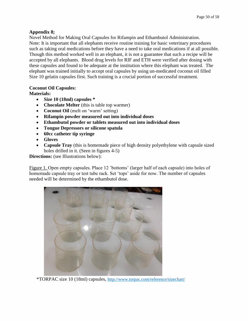
Page 50 of 58
Appendix 8;
Novel Method for Making Oral Capsules for Rifampin and Ethambutol Administration.
Note: It is important that all elephants receive routine training for basic veterinary procedures
such as taking oral medications before they have a need to take oral medications if at all possible.
Though this method worked well in an elephant, it is not a guarantee that such a recipe will be
accepted by all elephants. Blood drug levels for RIF and ETH were verified after dosing with
these capsules and found to be adequate at the institution where this elephant was treated. The
elephant was trained initially to accept oral capsules by using un-medicated coconut oil filled
Size 10 gelatin capsules first. Such training is a crucial portion of successful treatment.
Coconut Oil Capsules:
Materials:
Size 10 (18ml) capsules *
Chocolate Melter (this is table top warmer)
Coconut Oil (melt on ‘warm’ setting)
Rifampin powder measured out into individual doses
Ethambutol powder or tablets measured out into individual doses
Tongue Depressors or silicone spatula
60cc catheter tip syringe
Gloves
Capsule Tray (this is homemade piece of high density polyethylene with capsule sized
holes drilled in it. (Seen in figures 4-5)
Directions: (see Illustrations below):
Figure 1. Open empty capsules. Place 12 ’bottoms’ (larger half of each capsule) into holes of
homemade capsule tray or test tube rack. Set ‘tops’ aside for now. The number of capsules
needed will be determined by the ethambutol dose.
*TORPAC size 10 (18ml) capsules, http://www.torpac.com/reference/sizechart/
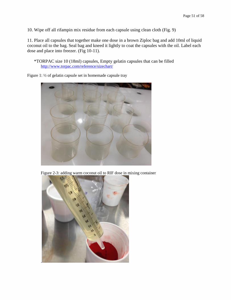
Page 51 of 58
10. Wipe off all rifampin mix residue from each capsule using clean cloth (Fig. 9)
11. Place all capsules that together make one dose in a brown Ziploc bag and add 10ml of liquid
coconut oil to the bag. Seal bag and kneed it lightly to coat the capsules with the oil. Label each
dose and place into freezer. (Fig 10-11).
*TORPAC size 10 (18ml) capsules, Empty gelatin capsules that can be filled http://www.torpac.com/reference/sizechart/
Figure 1: ½ of gelatin capsule set in homemade capsule tray
Figure 2-3: adding warm coconut oil to RIF dose in mixing container
.
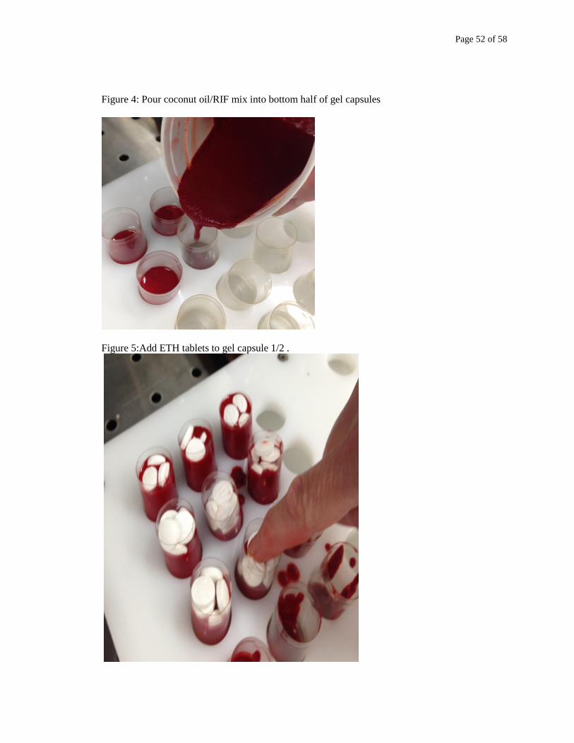
Page 52 of 58
Figure 4: Pour coconut oil/RIF mix into bottom half of gel capsules
Figure 5:Add ETH tablets to gel capsule 1/2 .
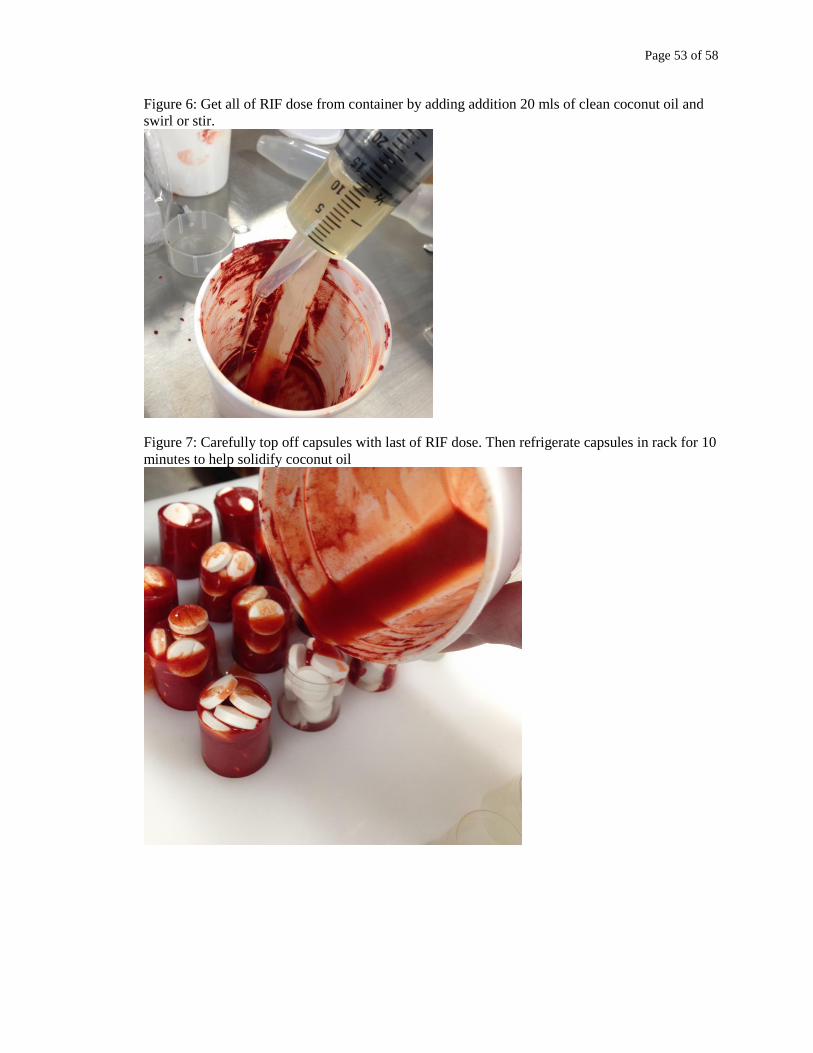
Page 53 of 58
Figure 6: Get all of RIF dose from container by adding addition 20 mls of clean coconut oil and
swirl or stir.
Figure 7: Carefully top off capsules with last of RIF dose. Then refrigerate capsules in rack for 10
minutes to help solidify coconut oil
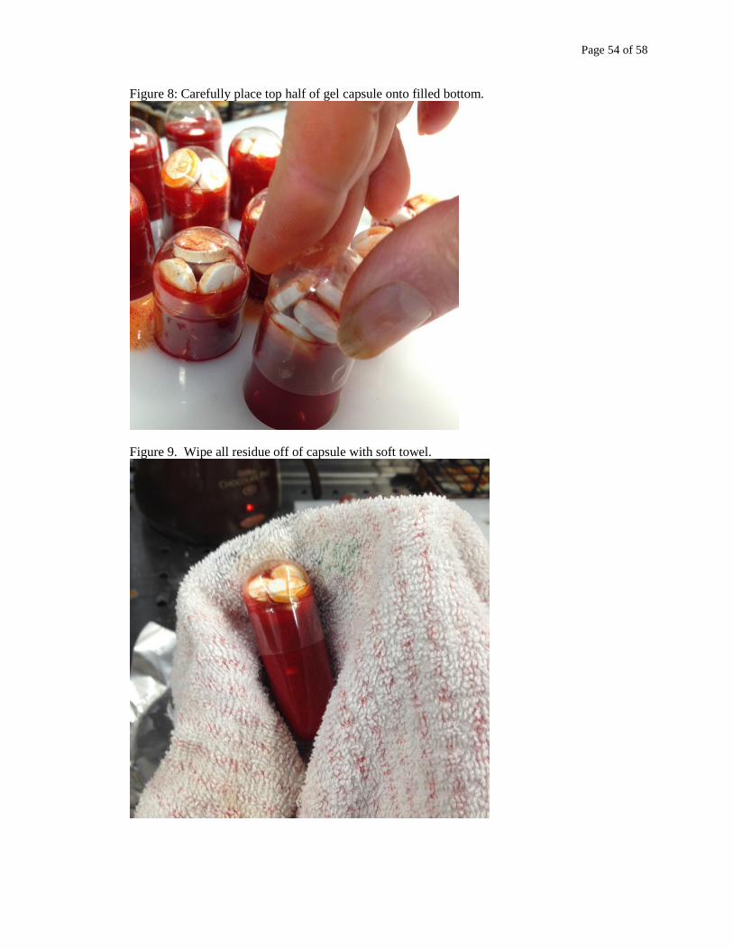
Page 54 of 58
Figure 8: Carefully place top half of gel capsule onto filled bottom.
Figure 9. Wipe all residue off of capsule with soft towel.
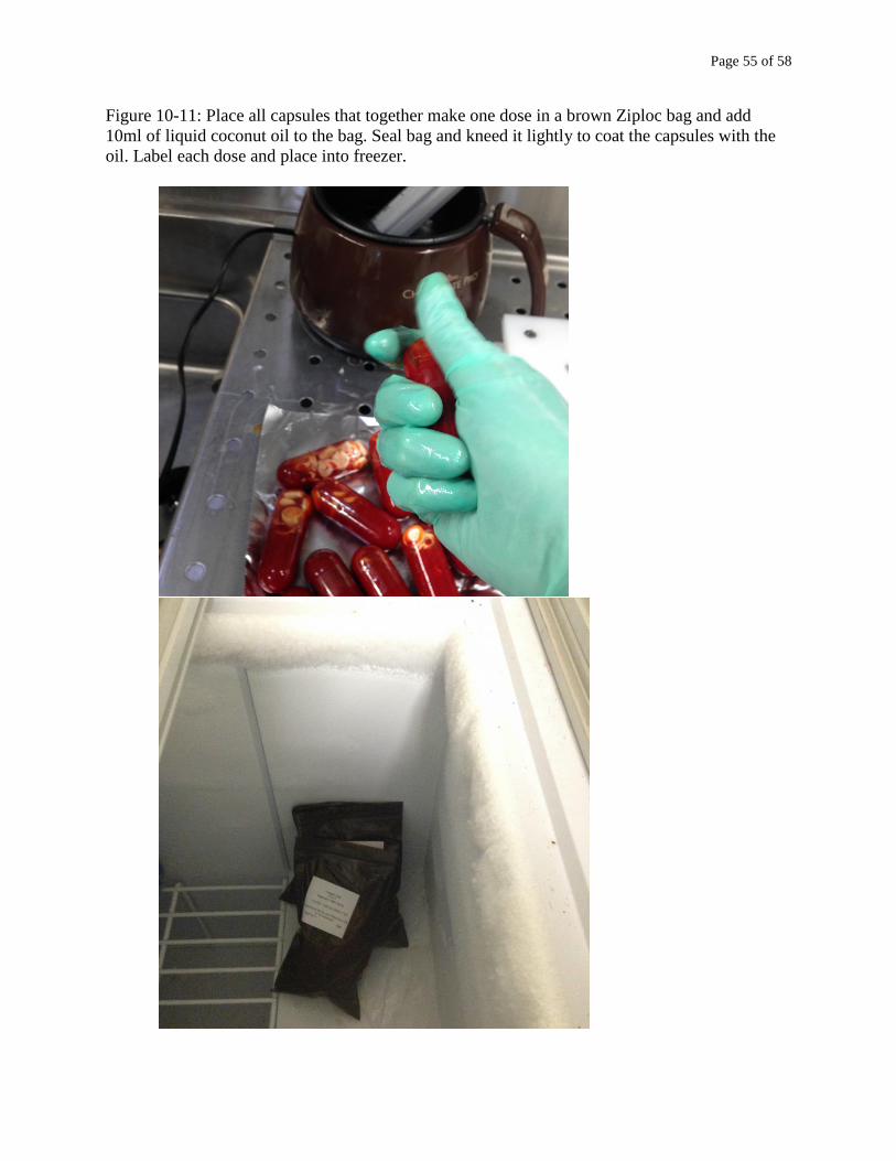
Page 55 of 58
Figure 10-11: Place all capsules that together make one dose in a brown Ziploc bag and add
10ml of liquid coconut oil to the bag. Seal bag and kneed it lightly to coat the capsules with the
oil. Label each dose and place into freezer.
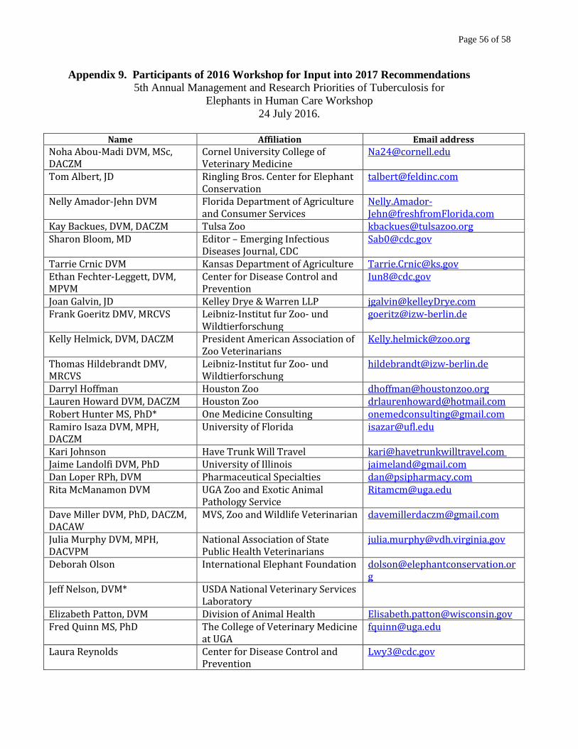
Page 56 of 58
Appendix 9. Participants of 2016 Workshop for Input into 2017 Recommendations
5th Annual Management and Research Priorities of Tuberculosis for
Elephants in Human Care Workshop
24 July 2016.
Name Affiliation Email address
Noha Abou-Madi DVM, MSc, DACZM
Cornel University College of Veterinary Medicine
Tom Albert, JD Ringling Bros. Center for Elephant Conservation
Nelly Amador-Jehn DVM Florida Department of Agriculture and Consumer Services
Kay Backues, DVM, DACZM Tulsa Zoo [email protected] Sharon Bloom, MD Editor – Emerging Infectious
Diseases Journal, CDC [email protected]
Tarrie Crnic DVM Kansas Department of Agriculture [email protected] Ethan Fechter-Leggett, DVM, MPVM
Center for Disease Control and Prevention
Joan Galvin, JD Kelley Drye & Warren LLP [email protected] Frank Goeritz DMV, MRCVS Leibniz-Institut fur Zoo- und
Wildtierforschung [email protected]
Kelly Helmick, DVM, DACZM President American Association of Zoo Veterinarians
Thomas Hildebrandt DMV, MRCVS
Leibniz-Institut fur Zoo- und Wildtierforschung
Darryl Hoffman Houston Zoo [email protected] Lauren Howard DVM, DACZM Houston Zoo [email protected] Robert Hunter MS, PhD* One Medicine Consulting [email protected] Ramiro Isaza DVM, MPH, DACZM
University of Florida [email protected]
Kari Johnson Have Trunk Will Travel [email protected] Jaime Landolfi DVM, PhD University of Illinois [email protected] Dan Loper RPh, DVM Pharmaceutical Specialties [email protected] Rita McManamon DVM UGA Zoo and Exotic Animal
Pathology Service [email protected]
Dave Miller DVM, PhD, DACZM, DACAW
MVS, Zoo and Wildlife Veterinarian [email protected]
Julia Murphy DVM, MPH, DACVPM
National Association of State Public Health Veterinarians
Deborah Olson International Elephant Foundation [email protected]
Jeff Nelson, DVM* USDA National Veterinary Services Laboratory
Elizabeth Patton, DVM Division of Animal Health [email protected] Fred Quinn MS, PhD The College of Veterinary Medicine
at UGA [email protected]
Laura Reynolds Center for Disease Control and Prevention
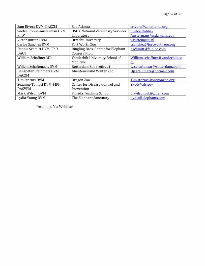
Page 57 of 58
Sam Rivera DVM, DACZM Zoo Atlanta [email protected] Suelee Robbe-Austerman DVM, PhD*
USDA National Veterinary Services Laboratory
Victor Rutten DVM Utrecht University [email protected] Carlos Sanchez DVM Fort Worth Zoo [email protected] Dennis Schmitt DVM, PhD, DACT
Ringling Bros. Center for Elephant Conservation
William Schaffner MD Vanderbilt University School of Medicine
Willem Schaftenaar, DVM Rotterdam Zoo (retired) [email protected] Hanspeter Steinmetz DVM DACZM
Abenteuerland Walter Zoo [email protected]
Tim Storms DVM Oregon Zoo [email protected] Suzanne Tomasi DVM, MPH DASVPM
Center for Disease Control and Prevention
Mark Wilson DVM Florida Teaching School [email protected] Lydia Young DVM The Elephant Sanctuary [email protected]
*Attended Via Webinar
Recommended