Clinical NEUROANATOMY
Basjiruddin ABagian Neurologi FK-Unand
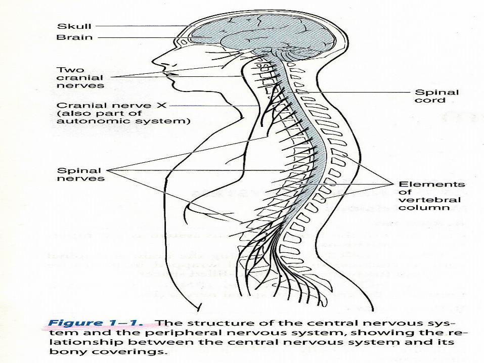
General Plan Of The Nervous System
AnatomyAnatomically, the human nervous system is a
complex of two division1. CNS-The CNS, comprising the brain and spinal
cord, is enclosed in bone and wrapped in protective covering (meninges) and fluid-filled space.
2. Peripheral Nervous System (PNS)-The PNS is formed by the cranial and spinal nerve (Fig 1.1)
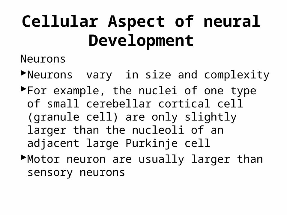
Cellular Aspect of neural Development
NeuronsNeurons vary in size and complexityFor example, the nuclei of one type of small
cerebellar cortical cell (granule cell) are only slightly larger than the nucleoli of an adjacent large Purkinje cell
Motor neuron are usually larger than sensory neurons
Cell Bodies
• The cell bodies is the metabolic and genetic center of a neuron (see fig 2-3)
Dendrites• Dendrites receive incoming synaptic
information and thus, together with the cell body, provide the receptive pole of the neuron
• Most neuron have many dendrites (see fig.2-3)
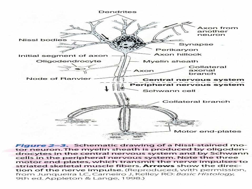

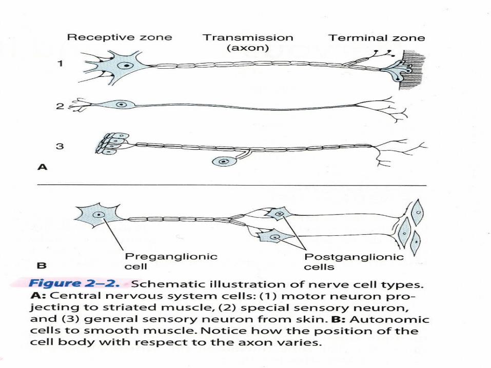
Axons• A single axon arises from the most neurons• The axon is a cylindrical tube of cytoplasm
covered by a membrane, the axolemma
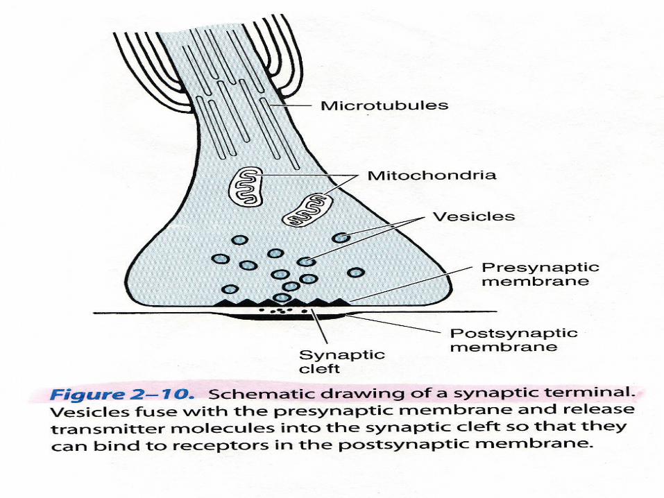
Synapses• Communication between neurons usually
occurs from the axon terminal of the transmitting neuron (presynaptic side) to the receptive region of the receiving neuron (postsinaptic side)
Neuroglia
• Neuroglial cells outnumber neurons in the brain and spinal cord
1. Macroglia2. Astrocytes3. Oligodendrocytes4. Microglia
Structural Units & Overall organization
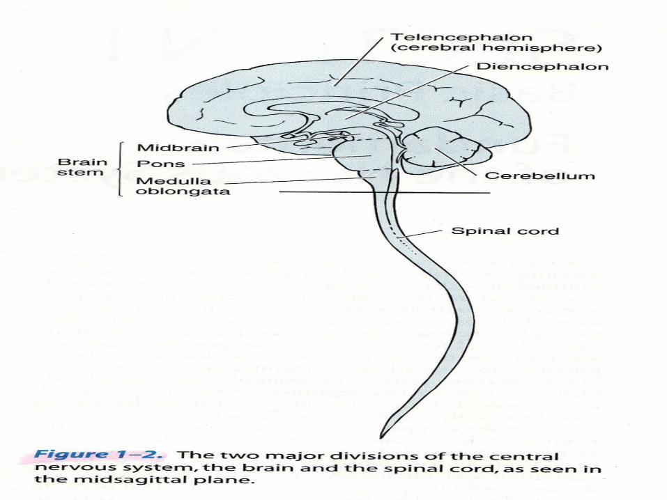
• The central portion of the nervous system consist of the brain and the elongated spinal cord (fig 1-2)
• The brain has a tiered structure and form a gross point of view, can be subdivided into the cerebrum, the brain stem, and the cerebellum
• The cerebrum (forebrain) consist of the telenchepalon and the dienchepalon
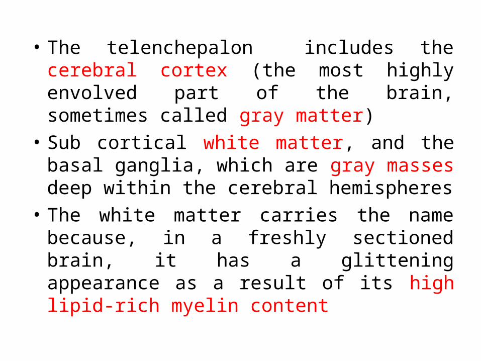
• The telenchepalon includes the cerebral cortex (the most highly envolved part of the brain, sometimes called gray matter)
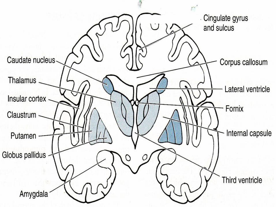
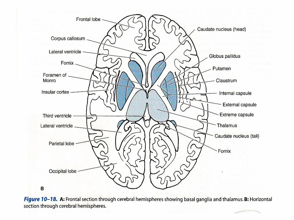
• Sub cortical white matter, and the basal ganglia, which are gray masses deep within the cerebral hemispheres
• The white matter carries the name because, in a freshly sectioned brain, it has a glittening appearance as a result of its high lipid-rich myelin content
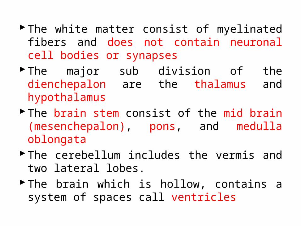
The white matter consist of myelinated fibers and does not contain neuronal cell bodies or synapses
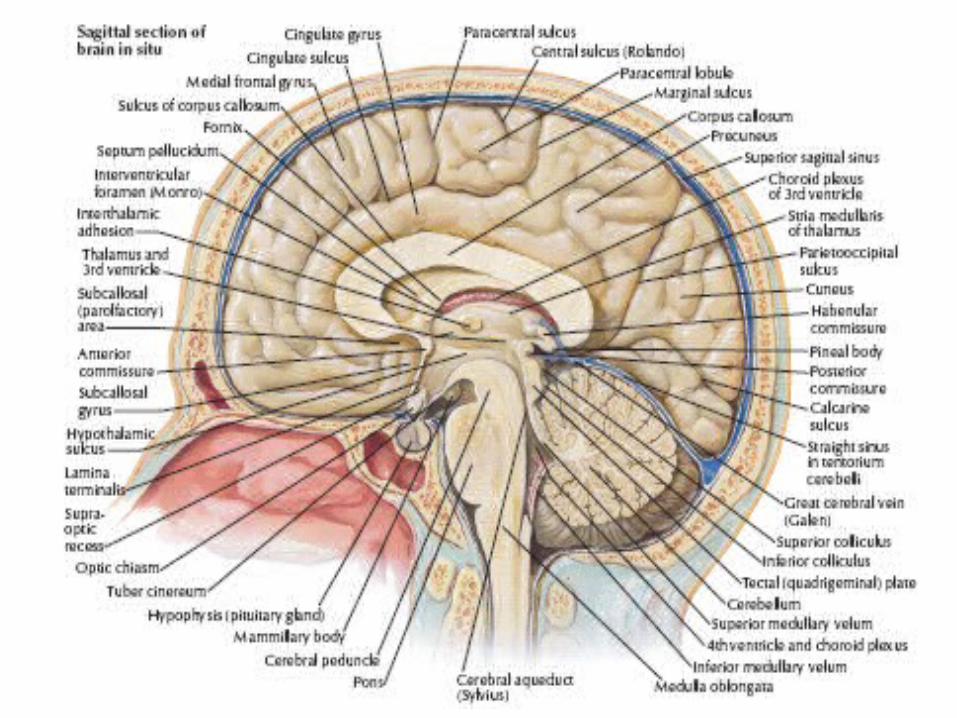
The major sub division of the dienchepalon are the thalamus and hypothalamus
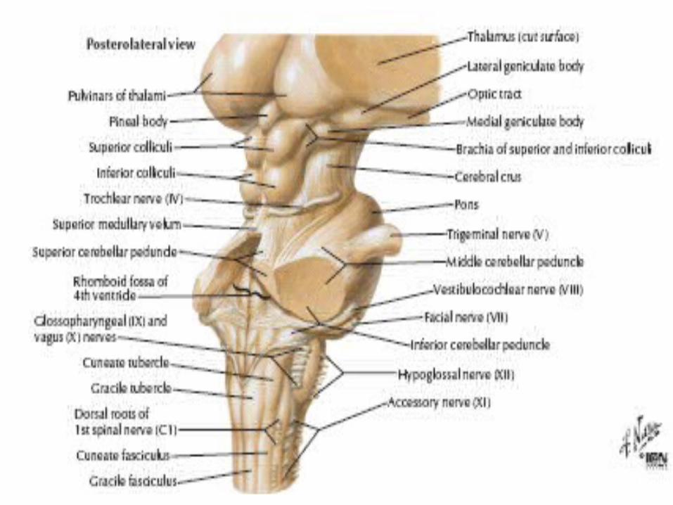
The brain stem consist of the mid brain (mesenchepalon), pons, and medulla oblongata
The cerebellum includes the vermis and two lateral lobes.
The brain which is hollow, contains a system of spaces call ventricles
Hemisfer Serebrum/Telensefalon
Anatomi Hemisfer Serebrum• Kedua hemisfer merupakan bagian terbesar
otak
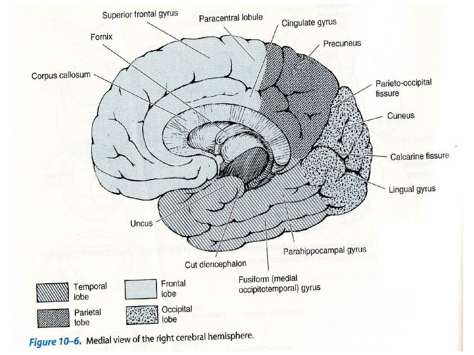
Sulkus dan Fisura Utama• Permukaan hemisfer serebrum mengandung
banyak fisura dan sulkus yang memisahkan lobus frontalis, parietalis, oksipitalis, dan temporalis dari satu sama lain dan dari insula.
Bagian-bagian otak yang terletak diantara sulkus dinamakan konvolusi atau girus
Sulkus sentralis memisahkan lobus frontalis dari lobus parietalis
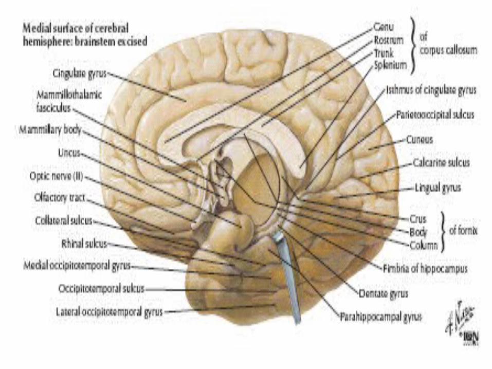
Fisura kalkarina dimulai pada permukaan medial hemisfer dekat kutub oksipitalis dan membentang ke depan ke daerah sedikit di bawah splenium dari korpus kalosum
Bagian dari korpus kalosum berbentuk busur;bagian anteriornya yang melengkung, genu berlanjut ke anteroventralis sebagai rostrum.
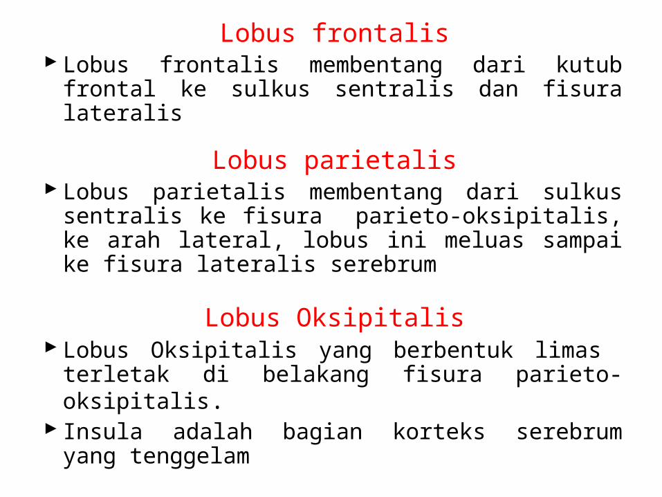
Lobus frontalisLobus frontalis membentang dari kutub frontal ke
sulkus sentralis dan fisura lateralis
Lobus parietalisLobus parietalis membentang dari sulkus sentralis ke
fisura parieto-oksipitalis, ke arah lateral, lobus ini meluas sampai ke fisura lateralis serebrum
Lobus OksipitalisLobus Oksipitalis yang berbentuk limas terletak di
belakang fisura parieto-oksipitalis.Insula adalah bagian korteks serebrum yang
tenggelam
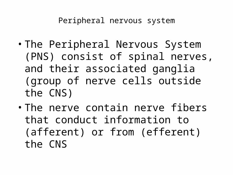
Peripheral nervous system
• The Peripheral Nervous System (PNS) consist of spinal nerves, and their associated ganglia (group of nerve cells outside the CNS)
• The nerve contain nerve fibers that conduct information to (afferent) or from (efferent) the CNS
THE SPINAL CORD AND THE ASCENDING AND DESCENDING TRACTS
VERTEBRAL COLUMNThe vertebral column is the central bony pillar of the body
The vertebral column is composed of 33 vertebrae: 7 cervical, 12 thoracic, 5 lumbar, 5 sacral (fused to form the sacrum), and 4 coccygeal (the lower 3 are commonly fused)
STRUCTURE OF THE SPINAL CORD
• The spinal cord is composed of an inner core of gray matter, which is surrounded by an outer covering of white matter; there is no indication that the cord is segmented.
• GRAY MATTER
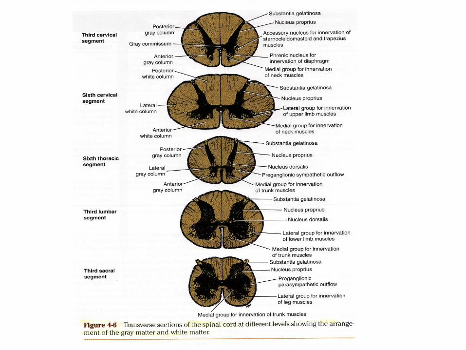
• On cross section, the gray matter is seen as an H-shaped pillar with anterior and posterior gray columns, or horns united by a thin gray commissure containing the small central canal.
WHITE MATTER
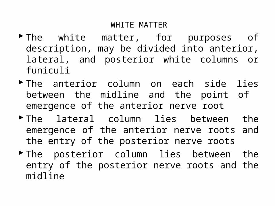
The white matter, for purposes of description, may be divided into anterior, lateral, and posterior white columns or funiculi
The anterior column on each side lies between the midline and the point of emergence of the anterior nerve root
The lateral column lies between the emergence of the anterior nerve roots and the entry of the posterior nerve roots
The posterior column lies between the entry of the posterior nerve roots and the midline
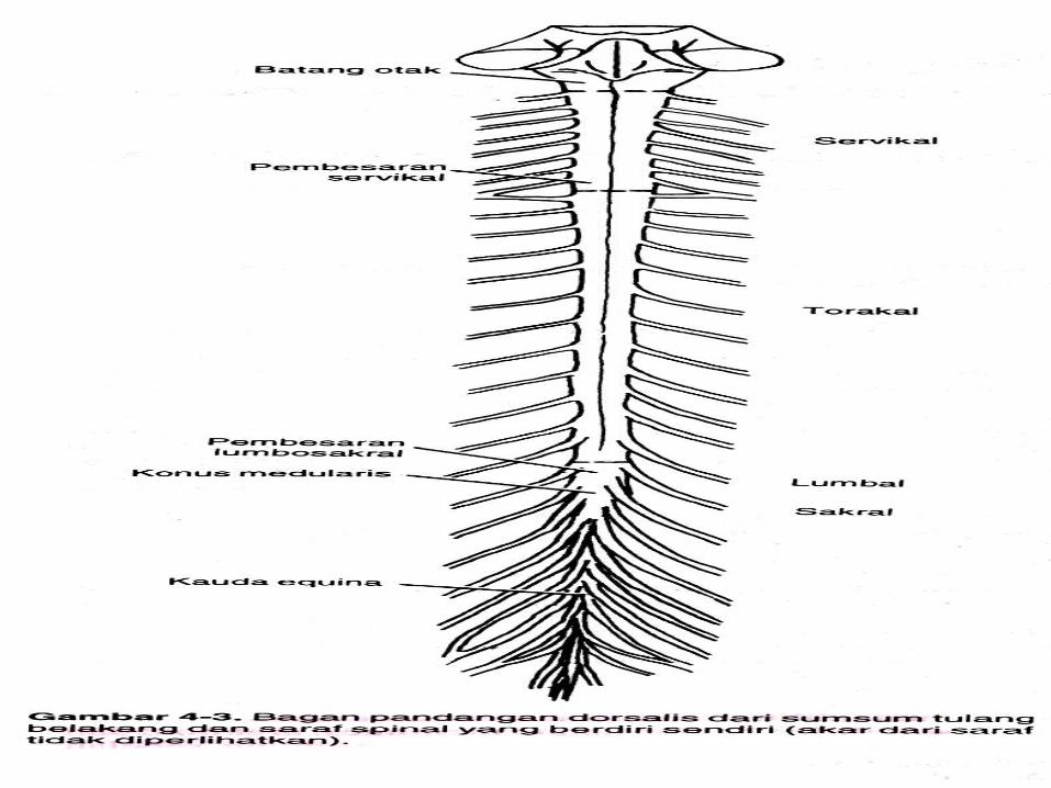
Sumsum Tulang Belakang
Anatomi luar Sumsum Tulang Belakang• Sumsum tulang belakang (medula spinalis,
atau mielyn) merupakan massa jaringan syaraf yang berbentuk silinder memanjang dan menempati dua pertiga bagian atas kanal spinal orang dewasa di dalam kolumna vertebralis
Akar dan Saraf Spinal
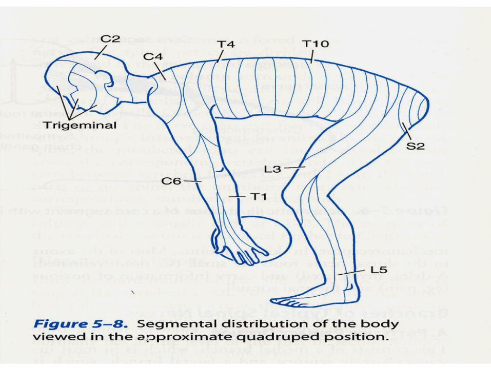
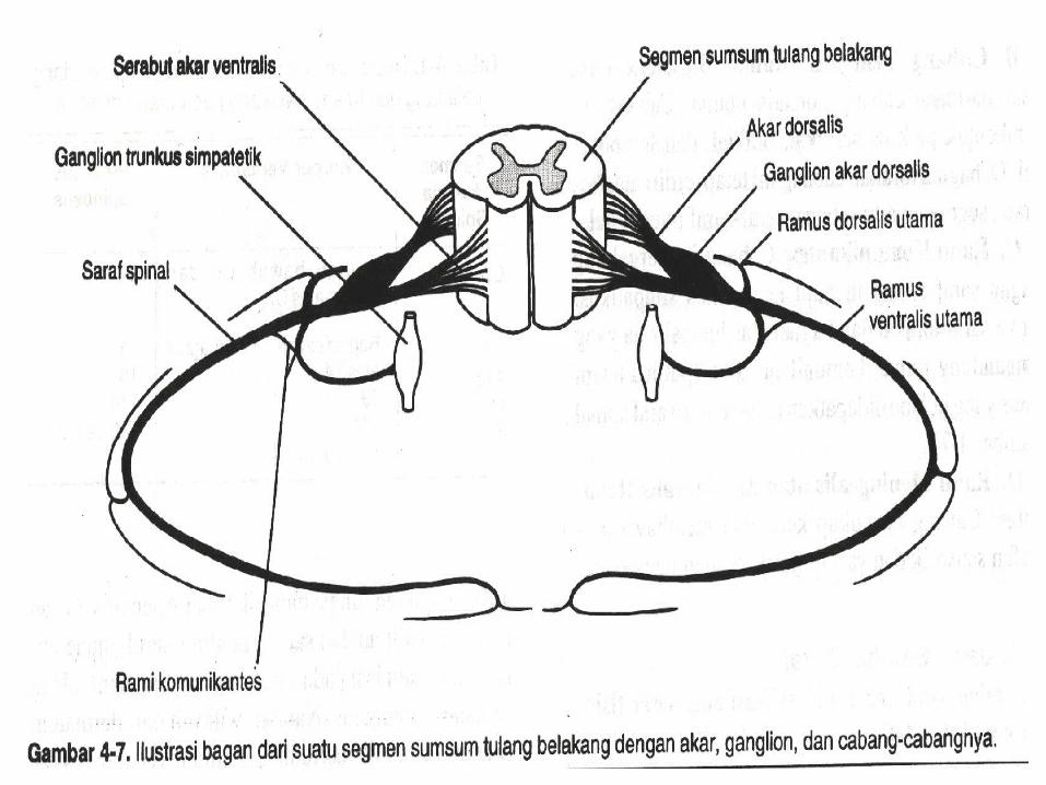
• Masing-masing segmen dari sumsum tulang belakang mempunyai 4 akar
• Ke 31 saraf spinal yang muncul dari sumsum tulang belakang mempunyai satu akar ventralis dan satu akar dorsalis
Akar ventralis
• Akar ventralis mempunyai akson neuron motorik
Akar dorsalis• Masing-masing akar dorsalis berisi serabut
aferen dari sel-sel saraf dalam ganglionnya
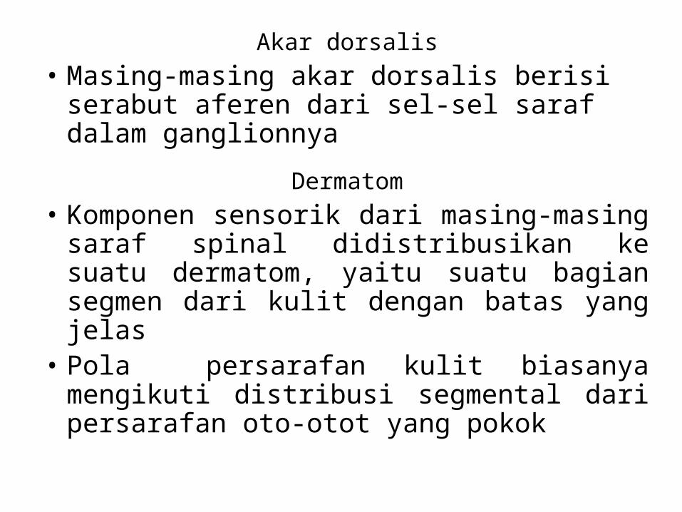
Dermatom• Komponen sensorik dari masing-masing saraf
spinal didistribusikan ke suatu dermatom, yaitu suatu bagian segmen dari kulit dengan batas yang jelas
• Pola persarafan kulit biasanya mengikuti distribusi segmental dari persarafan oto-otot yang pokok

Bagian Dalam Sumsum Tulang Belakang
Zat Kelabu (Substansia Gricea)
Kolumna : Suatu Potongan melintang dari sumsum tulang belakang yang memperlihatkan bagian dalam dari massa zat kelabu yang berbentuk huruf H yang dikelilingi oleh zat putih
Jaras Zat Putih (Substansia Alba)
Mengandung :•Sistem serabut Ascenden•Sistem serabut Descenden
THE ASCENDING TRACTS OF THE SPINAL CORD
The ascending tracts conduct afferent information, which may or may not reach consciousness. The information may be divided into two main groups :
1. Exteroceptive information, which originates from outside the body, such as pain, temperature, and touch
2. Proprioceptive information, which originates from inside the body, for example, from muscle and joints.
Pain and Temperature Pathways
Lateral Spinothalamic TractThe pain and thermal receptors in the skin and other tissues are free nerve endings
Pain ReceptionPain can be divided into two main types : fast pain and slow painFast pain is experienced whitin about 0,1 second after the pain stimulus is appliedSlow pain is felt 1,0 second or later after the stimulation
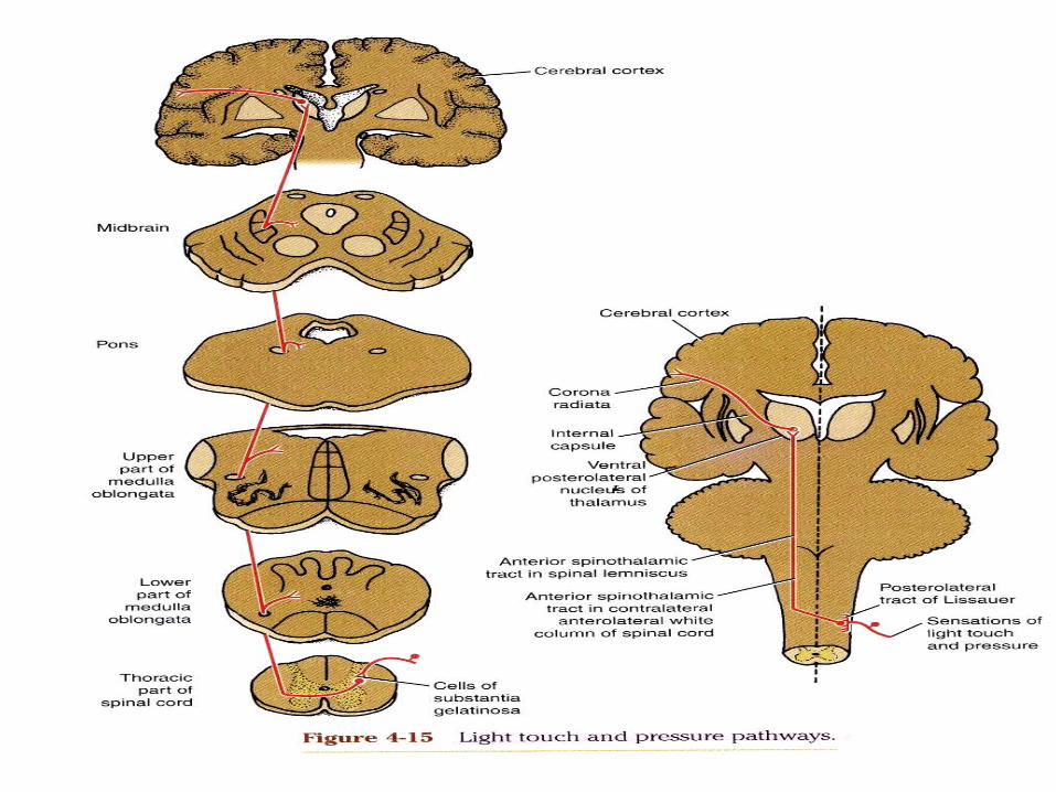
Light (Crude) Touch and Pressure PathwaysAnterior Spinothalamic Tract
The axons enter the spinal cord from the posterior root ganglion and proceed to the tip of the posterior gray column.
THE DESCENDING TRACTS OF THE SPINAL CORD
• The motor neurons situated in the anterior gray columns of the spinal cord send axons to innervate skeletal muscle through the anterior roots of the spinal nerves
• The supraspinal neurons and their tracts are sometimes referred to as the upper motor neurons, and the provide numerous separate pathways that can influence motor activity
FUNCTIONS OF THE DESCENDING TRACTS
The corticospinal tracts are the pathways corcerned with voluntary, discrete, skilled movement, especially those of the distal parts of the limbs
CORTICOSPINAL TRACTSFibers of the corticospinal tract arise as axons of
pyramidal cells situated in the fifth layer of the cerebral cortex
The descending fibers converge in the corona radiata and then pass through the posterior limb of the internal capsule
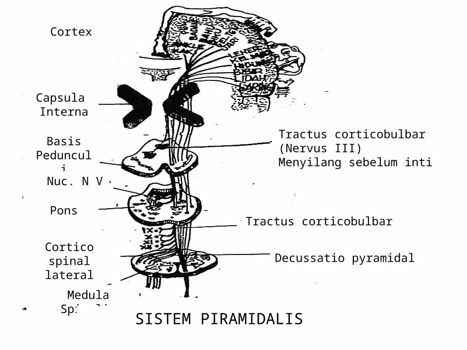
Tractus corticobulbar (Nervus III)Menyilang sebelum inti
Tractus corticobulbar
Decussatio pyramidal
Cortex
Capsula Interna
Basis Pedunculi
Nuc. N V
Pons
Cortico spinal lateral
Medula Spinalis
SISTEM PIRAMIDALIS
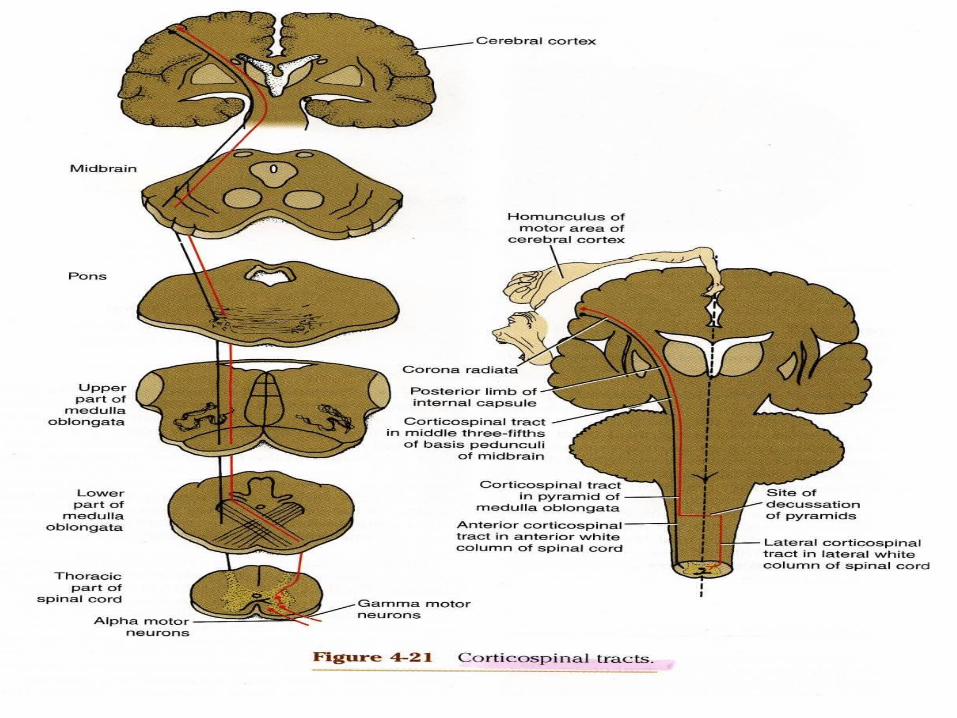
In the medulla oblongata, the bundles become grouped together along the anterior border to form a swelling known as the pyramid (alternative name pyramidal tract)
At the junction of medulla oblongata and the spinal cord, most of the fibers cross the midline at the decussation of the pyramids and enter the lateral white column of the spinal cord to form the lateral corticospinal tract
The remaining fibers do not cross in the decussation but descend in the anterior white column of the spinal cord as the anterior corticospinal tract
BLOOD SUPPLY OF THE BRAIN
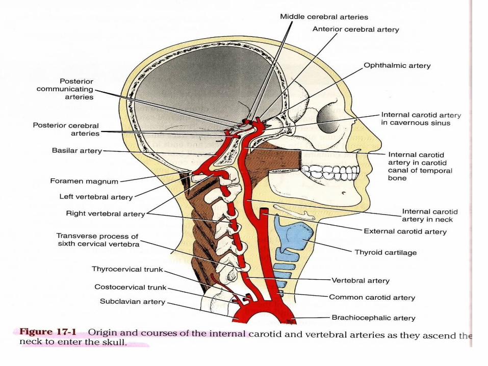
ARTERIES OF THE BRAINThe brain is supplied by the two internal carotid and
the two vertebral arteries. The arteries lie within the sub arachoid space, and their branches anastomose on the inferior surface of the brain to form the circle of Willis
Internal Carotid ArteryThe internal carotid artery begins at the bifurcation of
the common carotid artery, where it usually possesses a localized dilatation, called the carotid sinus
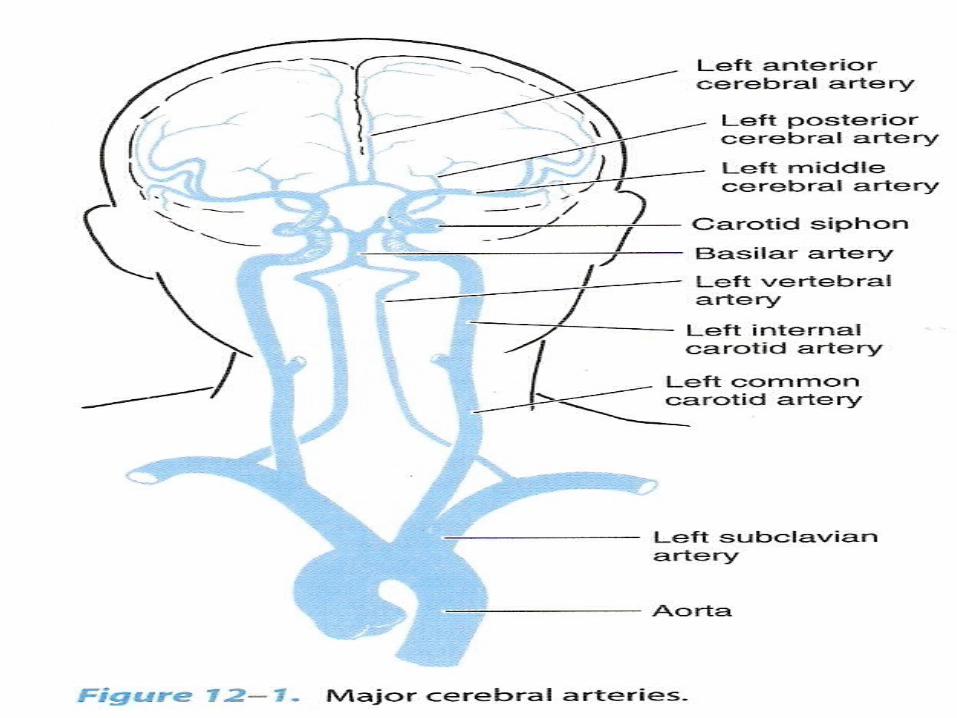
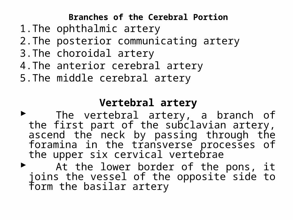
Branches of the Cerebral Portion1.The ophthalmic artery2.The posterior communicating artery3.The choroidal artery4.The anterior cerebral artery5.The middle cerebral artery
Vertebral artery The vertebral artery, a branch of the first part of
the subclavian artery, ascend the neck by passing through the foramina in the transverse processes of the upper six cervical vertebrae
At the lower border of the pons, it joins the vessel of the opposite side to form the basilar artery
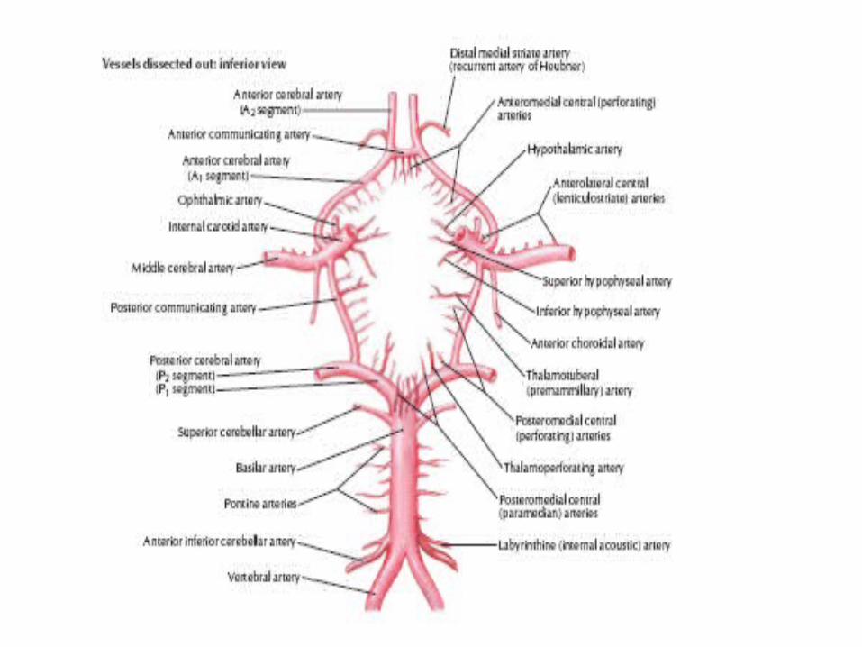
Circle of Willis The circle of Willis lies in the
interpeduncular fossa at th base of the brain
It is formed by the anastomoses between the two internal carotid arteries and the two vertebral arteries
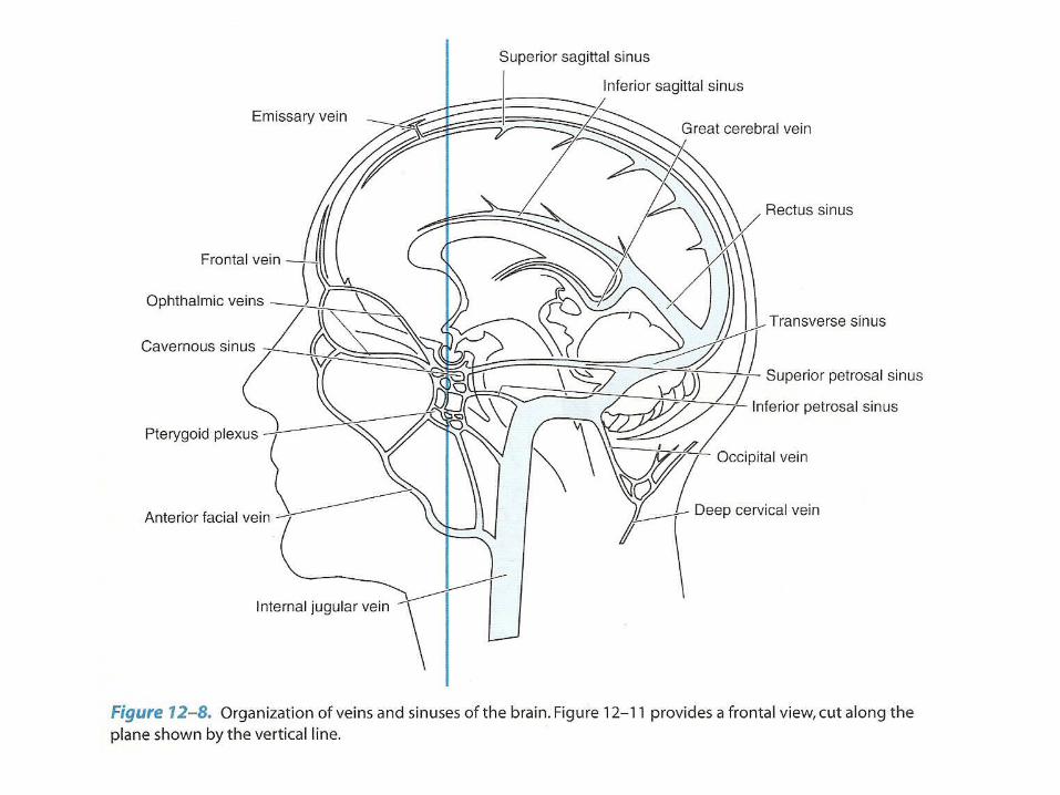
VEINS OF THE BRAIN
• The veins of the brain have no muscular tissue in their very thin walls, and the possess no valves
External Cerebral Veins
• The superior cerebral veins pass upward over the lateral surface of the cerebral hemisphere
Internal Cerebral Veins
• There are two internal cerebral veins, and they are formed by the union of the thalamostriate vein and the choroid vein

CEREBRAL CIRCULATION
• The blood flow to the brain must deliver oxygen, glucose, and other nutrients to the nervous tissue and remove carbon dioxide, lactic acid, and other metabolic by-product
BLOOD SUPPLY OF THE SPINAL CORD
Artery of the spinal cord :1. Posterior spinal artery2. Anterior spinal artery
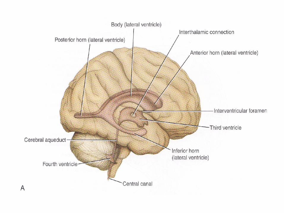
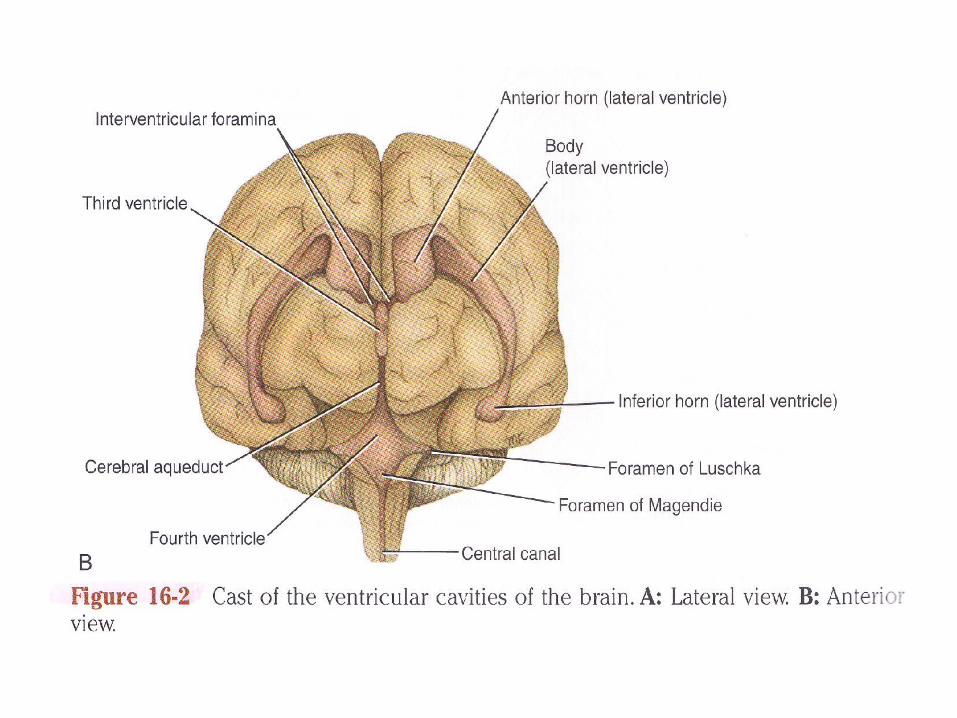
VENTRICULAR SYSTEM
• The ventricles are four fluid-filled cavities located within the brain; these are the two lateral ventricles, the third ventricle, and the fourth ventricle– Lateral Ventricle– Third Ventricle– Cerebral Aqueduct– Fourth Ventricle
Lateral VentriclesThe lateral ventricles are the largest of the ventriclesThe anterior (frontal) horn is in front of the inter
ventricular foramenThe posterior (occipital) horn extends into the
occipital lobeThe inferior (temporal) horn transverses the
temporal lobe, whose white substance forms its roofThe two inter ventricular foramens, or foramens of
Monroe, are apertures between the column of the fornix and the anterior end of the thalamus
Third ventricle• The third ventricle is a narrow ventricle cleft
between the two halves of the diencephalon
Fourth ventricle• The fourth ventricle is a pyramid-shaped
cavity bounded ventrally by the pons and medulla oblongata its floor is also known as the rhomboid fossa
Referensi• Chusid. JG, Correlative neuroanatomy and
functional neurology• Jack de Groot, Neuroanatomy correlative• Snell, RS. Neuroanatomi klinik• Sukardi. E, Neuroanatomi medica• Waxman. SG, Clinical Neuroanatomy
Recommended