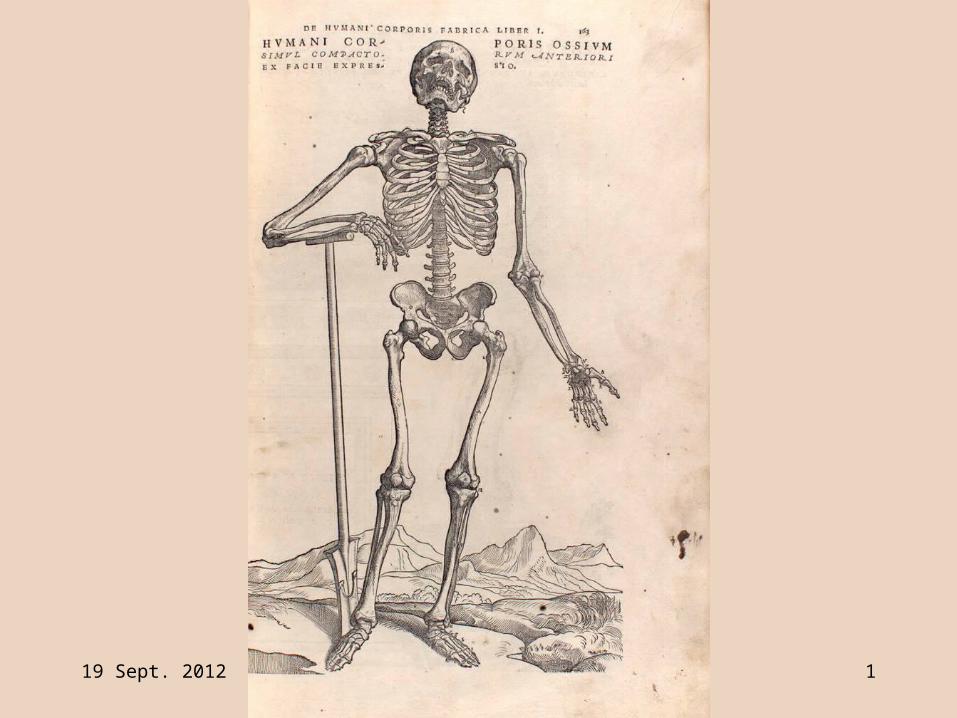
19 Sept. 2012 Bone_tissue.ppt 1

19 Sept. 2012 Bone_tissue.ppt 2
BONES and SKELETAL TISSUES
Skeletal System:
a framework, foundation for body &
solid support for soft organs
19 Sept. 2012 Bone_tissue.ppt 3
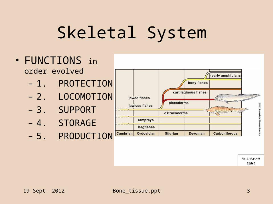
Skeletal System
• FUNCTIONS in order evolved
– 1. PROTECTION
– 2. LOCOMOTION
– 3. SUPPORT
– 4. STORAGE
– 5. PRODUCTION
Slide 6
Fig. 27.5, p. 458
19 Sept. 2012 Bone_tissue.ppt 4
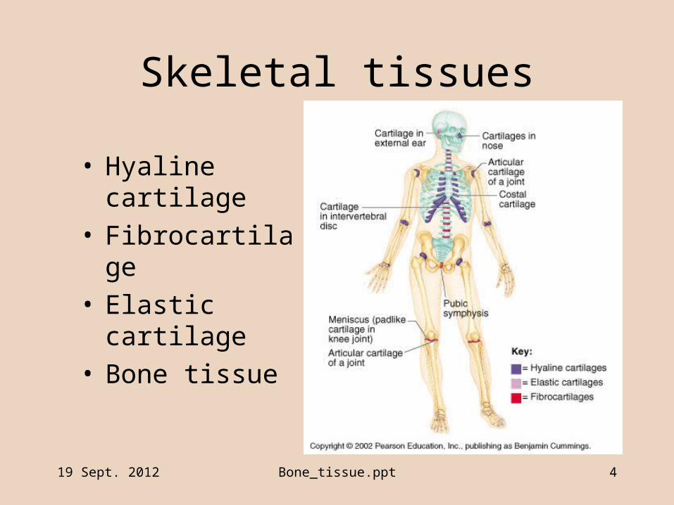
Skeletal tissues
• Hyaline cartilage• Fibrocartilage• Elastic cartilage• Bone tissue

19 Sept. 2012 Bone_tissue.ppt 5
Structure of Bone Tissue • Bone as connective tissue
– Cells, osteocytes, separated by extracellular matrix
– Collagen fibers, • provide tensile strength
– Extracellular matrix• solid mineral salts (Calcium phosphate)
– “hydroxyapatite”
• Provides hardness, resistance to bending
• about 65% of bone weight
19 Sept. 2012 Bone_tissue.ppt 6
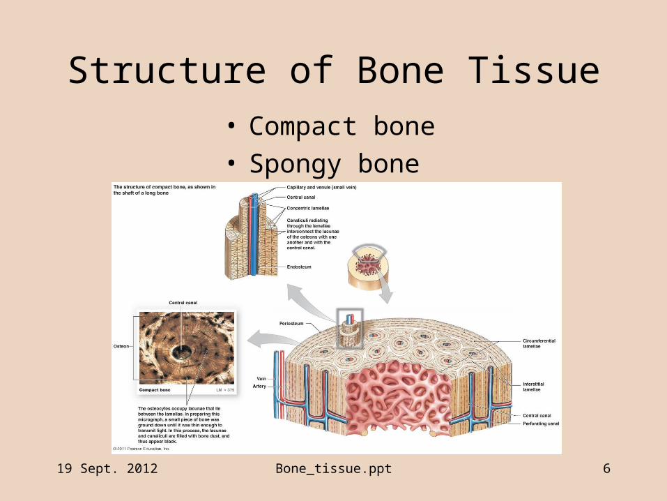
Structure of Bone Tissue • Compact bone• Spongy bone
19 Sept. 2012 Bone_tissue.ppt 7
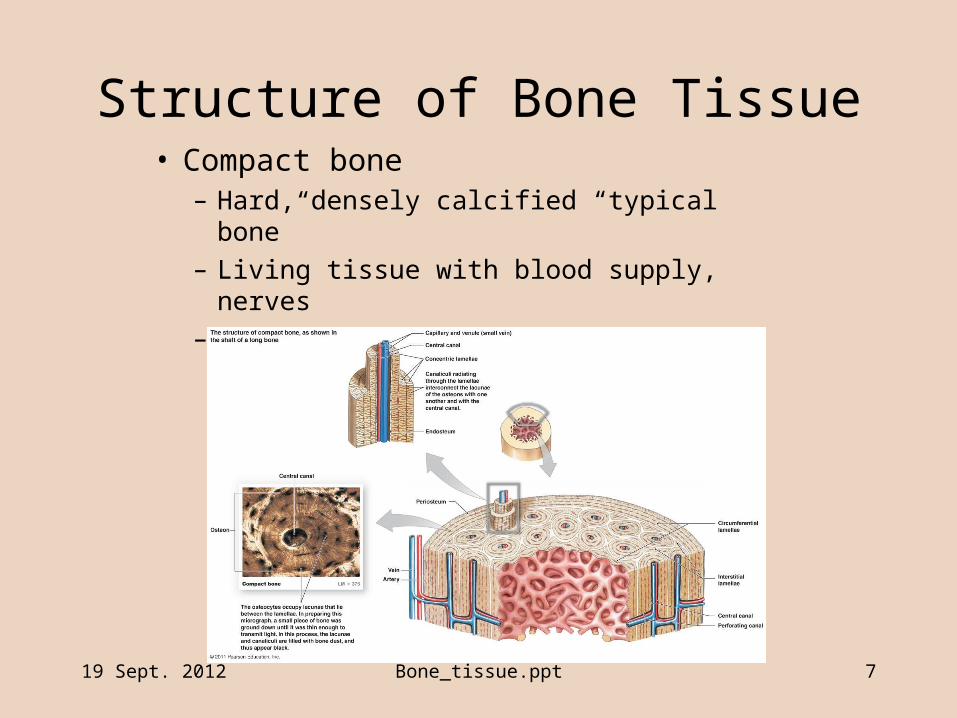
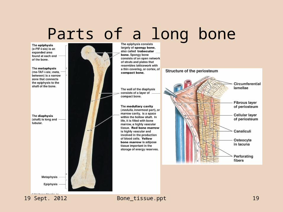
Structure of Bone Tissue• Compact bone
– Hard, densely calcified “typical bone”
– Living tissue with blood supply, nerves
– Organized of osteons

19 Sept. 2012 Bone_tissue.ppt 8
Structure of Bone Tissue
• Compact bone– Osteon
• Central (Haversian) canal at center
• Osteocytes in lacunae surrounding Haversian canal
• Lamellae of bone matrix between rings of osteocytes

19 Sept. 2012 Bone_tissue.ppt 9
Structure of Compact Bone– Osteons can’t fit together
• Interstitial lamellae fill space to make solid structure
• Circumferential lamellae fill space to shape outer surface of bone
19 Sept. 2012 Bone_tissue.ppt 10
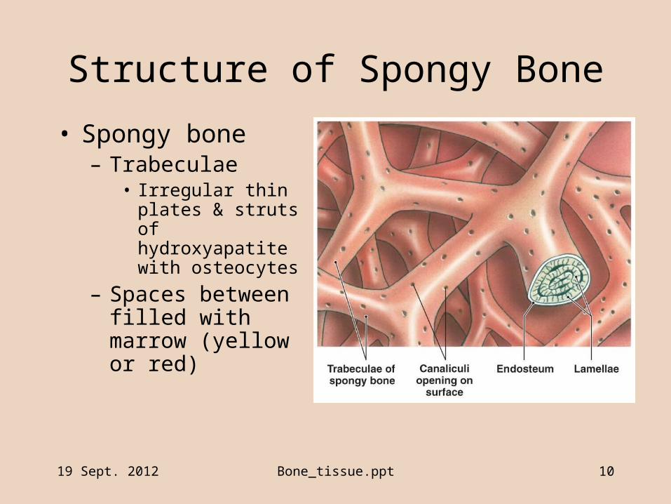
Structure of Spongy Bone
• Spongy bone– Trabeculae
• Irregular thin plates & struts of hydroxyapatite with osteocytes
– Spaces between filled with marrow (yellow or red)
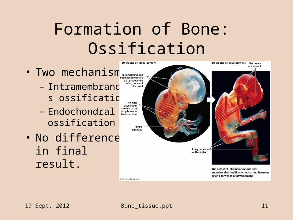
Formation of Bone: Ossification
• Two mechanisms– Intramembranous
ossification
– Endochondral ossification
• No difference in final result.
19 Sept. 2012 Bone_tissue.ppt 11
19 Sept. 2012 Bone_tissue.ppt 12
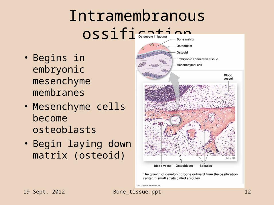
Intramembranous ossification
• Begins in embryonic mesenchyme membranes
• Mesenchyme cells become osteoblasts
• Begin laying down matrix (osteoid)

19 Sept. 2012 Bone_tissue.ppt 13
Intramembranous ossification
• Layer of “woven bone” and periosteum
• Remodeling to form compact bone on surfaces
• Cranial & facial bones, mandible, clavicles.
19 Sept. 2012 Bone_tissue.ppt 14
Endochondral ossification
• Embryonic mesenchyme differentiates to cartilage – Chondrocytes– Perichondrium
• “Cartilage model” is starting point for endochondral ossification– (endo- = within, chondr- = cartilage)
19 Sept. 2012 Bone_tissue.ppt 15
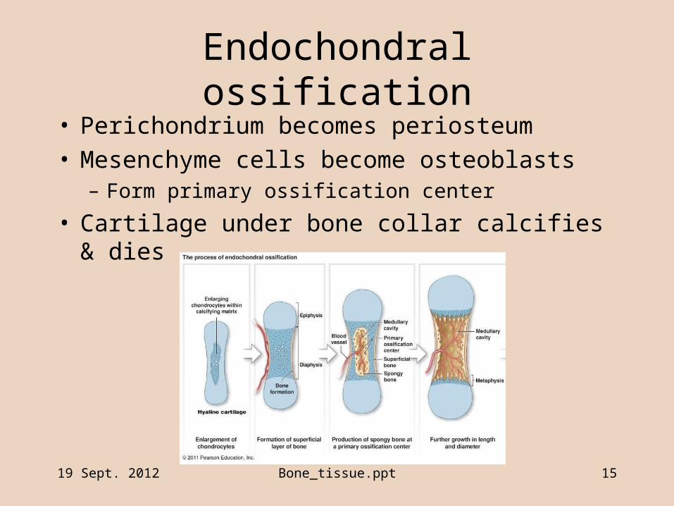
Endochondral ossification
• Perichondrium becomes periosteum• Mesenchyme cells become osteoblasts
– Form primary ossification center
• Cartilage under bone collar calcifies & dies
19 Sept. 2012 Bone_tissue.ppt 16
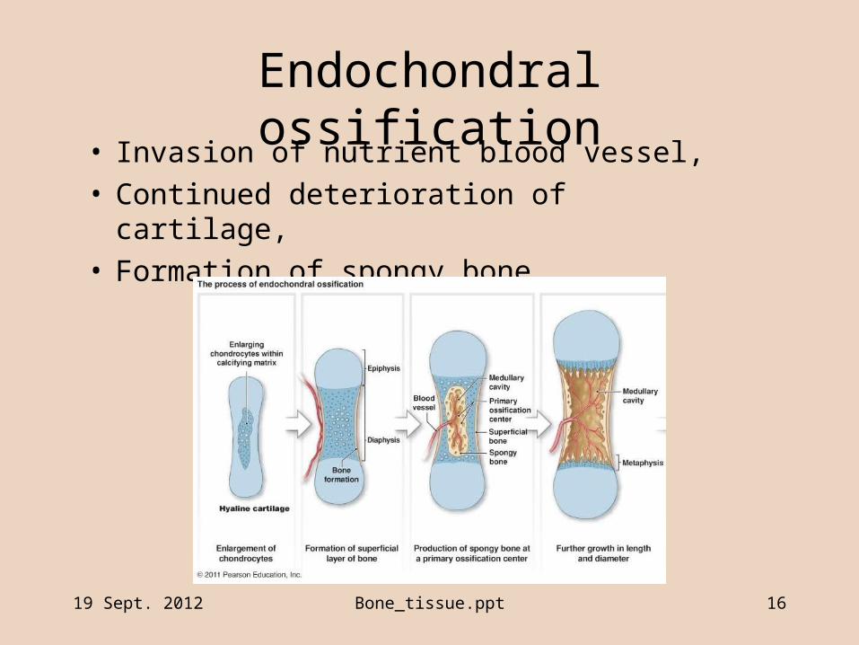
Endochondral ossification• Invasion of nutrient blood vessel,• Continued deterioration of cartilage,• Formation of spongy bone
19 Sept. 2012 Bone_tissue.ppt 17
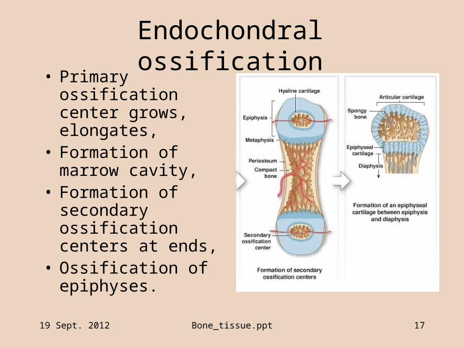
Endochondral ossification• Primary ossification
center grows, elongates,• Formation of marrow
cavity,• Formation of secondary
ossification centers at ends,
• Ossification of epiphyses.
19 Sept. 2012 Bone_tissue.ppt 18
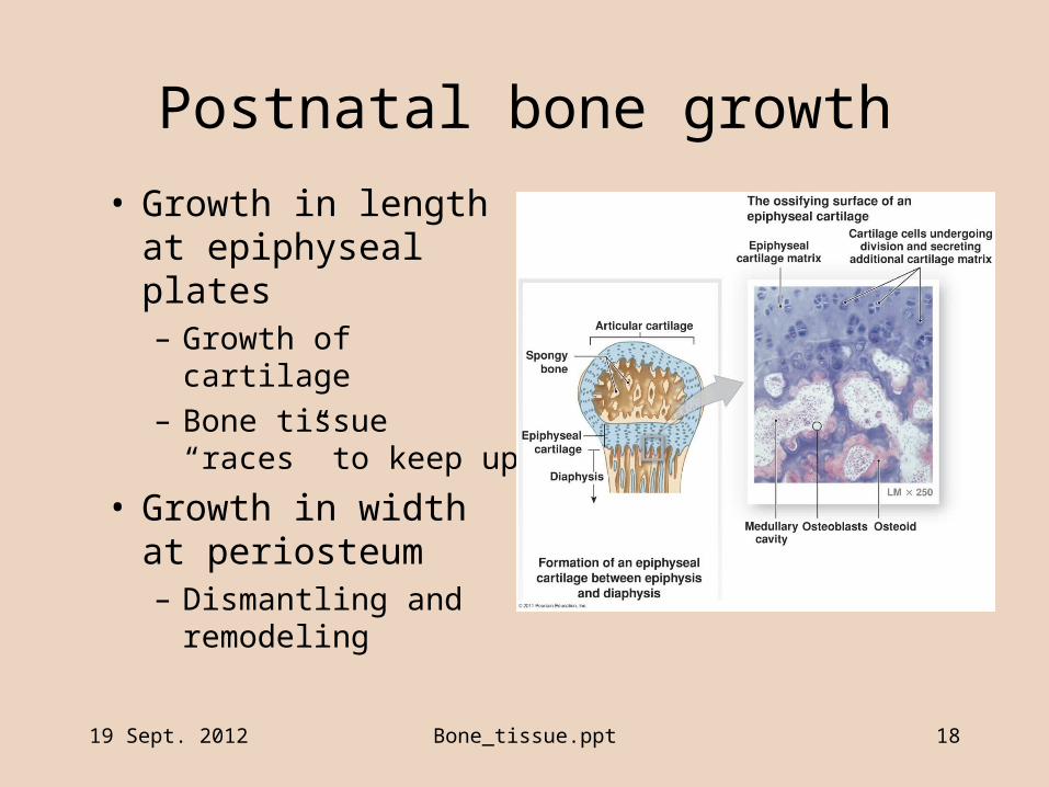
Postnatal bone growth
• Growth in length at epiphyseal plates– Growth of cartilage
– Bone tissue “races” to keep up
• Growth in width at periosteum– Dismantling and
remodeling
19 Sept. 2012 Bone_tissue.ppt 19
Parts of a long bone
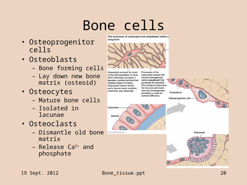
Bone cells
19 Sept. 2012 Bone_tissue.ppt 20
• Osteoprogenitor cells• Osteoblasts
– Bone forming cells– Lay down new bone
matrix (osteoid)
• Osteocytes– Mature bone cells– Isolated in lacunae
• Osteoclasts– Dismantle old bone
matrix– Release Ca2+ and
phosphate

19 Sept. 2012 Bone_tissue.ppt 21
Remodeling
• Normal continuous process– Osteoclasts dismantle old matrix– Osteoblasts rebuild bone matrix
• Allows– Maintenance of bone tissue– Change in shape with age & weight– Reinforcing of parts subject to stress & strain
19 Sept. 2012 Bone_tissue.ppt 22
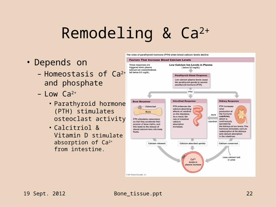
Remodeling & Ca2+
• Depends on– Homeostasis of Ca2+
and phosphate
– Low Ca2+
• Parathyroid hormone (PTH) stimulates osteoclast activity
• Calcitriol & Vitamin D stimulate absorption of Ca2+ from intestine.

19 Sept. 2012 Bone_tissue.ppt 23
Remodeling & Ca2+
• Depends on– Homeostasis of Ca2+
and phosphate
– High Ca2+
• Calcitonin stimulates deposition of Ca2+ into bone tissue.
• Excess Ca2+ excreted.
19 Sept. 2012 Bone_tissue.ppt 24
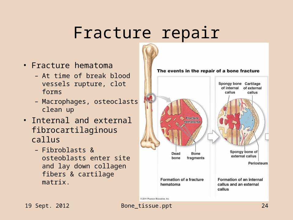
Fracture repair
• Fracture hematoma– At time of break blood
vessels rupture, clot forms
– Macrophages, osteoclasts clean up
• Internal and external fibrocartilaginous callus– Fibroblasts & osteoblasts
enter site and lay down collagen fibers & cartilage matrix.

19 Sept. 2012 Bone_tissue.ppt 25
Fracture repair
• Bony callus– Osteoblasts lay down
spongy bone trabeculae
– Join broken ends
• Remodeling– Osteoblasts continue
forming bone
– Compact bone replaces spongy bone
Recommended