10/02/08Biochemistry: Nucleic Acid Chem&Struct
Nucleic AcidStructure
Andy HowardIntroductory Biochemistry
7 October 2008
10/02/08 Biochemistry: Nucleic Acid Chem&Struct p. 2 of 38
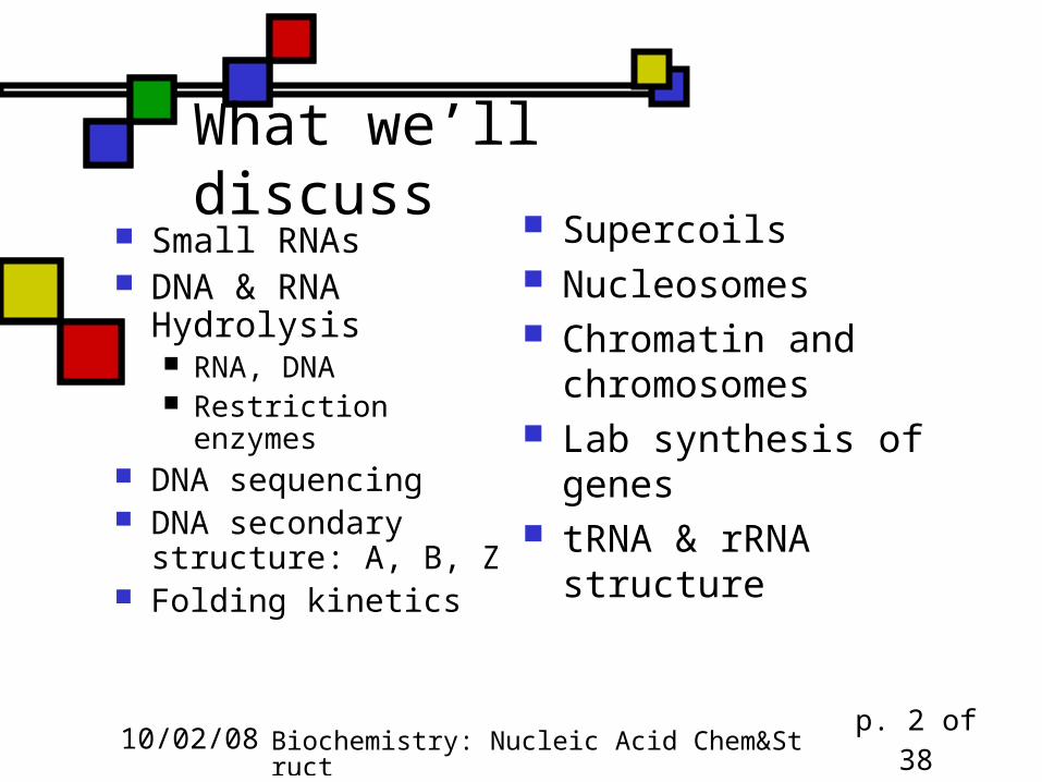
What we’ll discuss Small RNAs DNA & RNA
Hydrolysis RNA, DNA Restriction enzymes
DNA sequencing DNA secondary
structure: A, B, Z Folding kinetics
Supercoils Nucleosomes Chromatin and
chromosomes Lab synthesis of genes tRNA & rRNA structure
10/02/08 Biochemistry: Nucleic Acid Chem&Struct p. 3 of 38
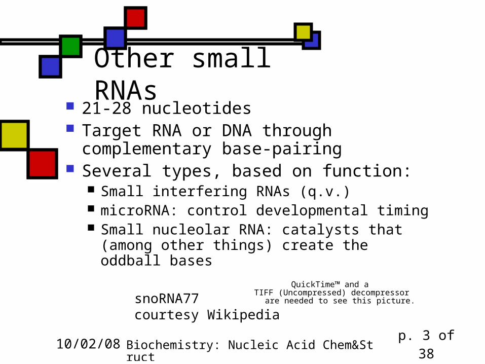
Other small RNAs 21-28 nucleotides Target RNA or DNA through
complementary base-pairing Several types, based on function:
Small interfering RNAs (q.v.) microRNA: control developmental timing Small nucleolar RNA: catalysts that (among
other things) create the oddball basesQuickTime™ and a
TIFF (Uncompressed) decompressorare needed to see this picture.snoRNA77
courtesy Wikipedia
10/02/08 Biochemistry: Nucleic Acid Chem&Struct p. 4 of 38
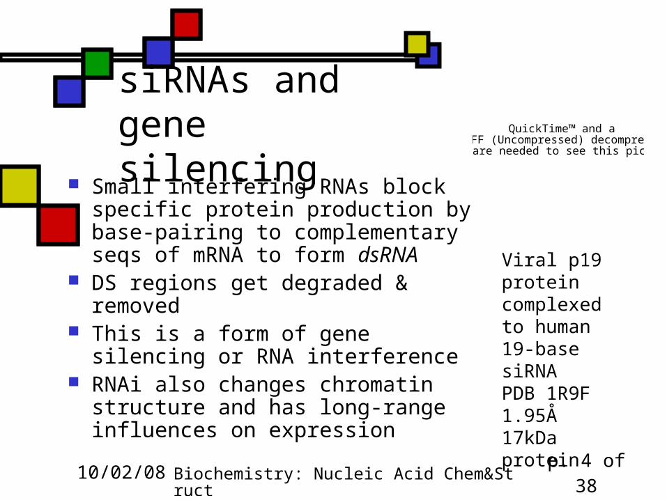
siRNAs and gene silencing
Small interfering RNAs block specific protein production by base-pairing to complementary seqs of mRNA to form dsRNA
DS regions get degraded & removed This is a form of gene silencing or RNA
interference RNAi also changes chromatin structure
and has long-range influences on expression
QuickTime™ and aTIFF (Uncompressed) decompressor
are needed to see this picture.
Viral p19 protein complexed to human 19-base siRNAPDB 1R9F1.95Å17kDa protein
10/02/08 Biochemistry: Nucleic Acid Chem&Struct p. 5 of 38
Do the differences between RNA and DNA matter? Yes! DNA has deoxythymidine, RNA has uridine:
cytidine spontaneously degrades to uridine dC spontaneously degrades to dU
The only dU found in DNA is there because of degradation: dT goes with dA
So when a cell finds dU in its DNA, it knows it should replace it with dC or else synthesize dG opposite the dU instead of dA
10/02/08 Biochemistry: Nucleic Acid Chem&Struct p. 6 of 38
Ribose vs. deoxyribose Presence of -OH on 2’ position makes the 3’
position in RNA more susceptible to nonenzymatic cleavage than the 3’ in DNA
The ribose vs. deoxyribose distinction also influences enzymatic degradation of nucleic acids
I can carry DNA in my shirt pocket, but not RNA
10/02/08 Biochemistry: Nucleic Acid Chem&Struct p. 7 of 38
Backbone hydrolysis of nucleic acids in base(fig. 10.29)
Nonenzymatic hydrolysis in base occurs with RNA but not DNA, as just mentioned
Reason: in base, RNA can form a specific 5-membered cyclic structure involving both 3’ and 2’ oxygens
When this reopens, the backbone is cleaved and you’re left with a mixture of 2’- and 3’-NMPs
10/02/08 Biochemistry: Nucleic Acid Chem&Struct p. 8 of 38
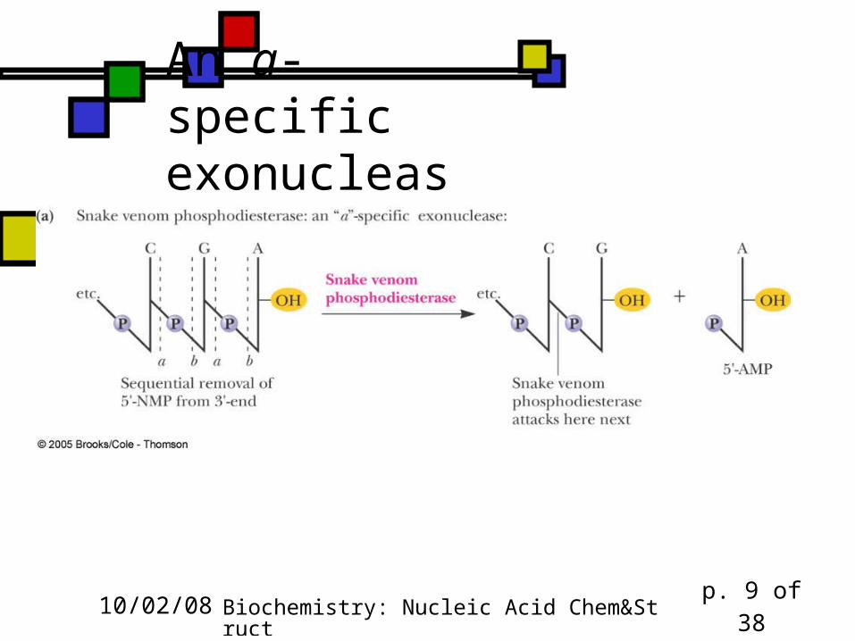
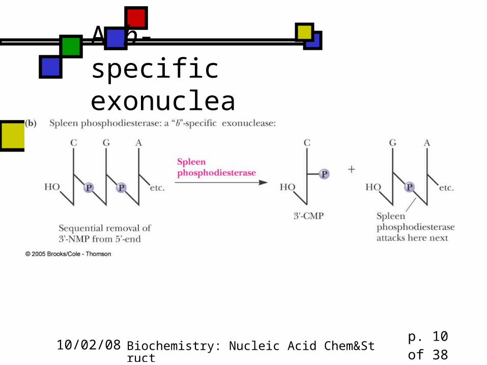
Enzymatic cleavage of oligo- and polynucleotides Enzymes are phosphodiesterases Could happen on either side of the P 3’ cleavage is a-site; 5’ is b-site. Endonucleases cleave somewhere on
the interior of an oligo- or polynucleotide Exonucleases cleave off the terminal
nucleotide
10/02/08 Biochemistry: Nucleic Acid Chem&Struct p. 9 of 38
An a-specific exonuclease
10/02/08 Biochemistry: Nucleic Acid Chem&Struct p. 10 of 38
A b-specific exonuclease
10/02/08 Biochemistry: Nucleic Acid Chem&Struct p. 11 of 38
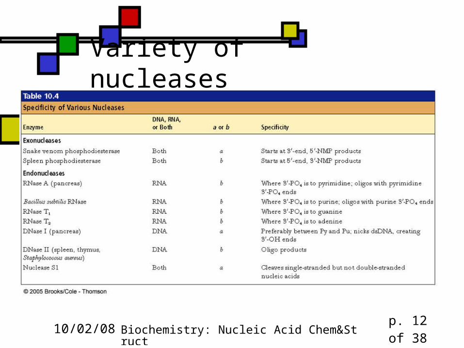
Specificity in nucleases Some cleave only RNA, others only DNA,
some both Often a preference for a specific base or
even a particular 4-8 nucleotide sequence (restriction endonucleases)
These can be used as lab tools, but they evolved for internal reasons
10/02/08 Biochemistry: Nucleic Acid Chem&Struct p. 12 of 38
Variety of nucleases
10/02/08 Biochemistry: Nucleic Acid Chem&Struct p. 13 of 38
Restriction endonucleases Evolve in bacteria as antiviral tools “Restriction” because they restrict the
incorporation of foreign DNA into the bacterial chromosome
Recognize and bind to specific palindromic DNA sequences and cleave them
Self-cleavage avoided by methylation Types I, II, III: II is most important I and III have inherent methylase activity; II has
methylase activity in an attendant enzyme
10/02/08 Biochemistry: Nucleic Acid Chem&Struct p. 14 of 38
What do we mean by palindromic?
In ordinary language, it means a phrase that reads the same forward and back: Madam, I’m Adam. (Genesis 3:20) Eve, man, am Eve. Able was I ere I saw Elba. (Napoleon) A man, a plan, a canal: Panama!
(T. Roosevelt) With DNA it means the double-stranded
sequence is identical on both strands
10/02/08 Biochemistry: Nucleic Acid Chem&Struct p. 15 of 38
Quirky math question to ponder Numbers can be palindromic: 484, 1331, 727, 595… Some numbers that are palindromic have
squares that are palindromic… 222 = 484, 1212 = 14641, . . . Question: if a number is perfect square
and a palindrome, is its square root a palindrome? (answer will be given orally)
10/02/08 Biochemistry: Nucleic Acid Chem&Struct p. 16 of 38
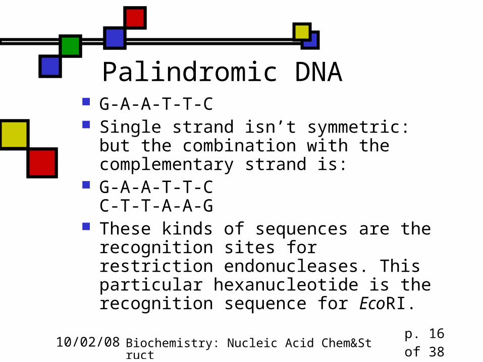
Palindromic DNA G-A-A-T-T-C Single strand isn’t symmetric: but the
combination with the complementary strand is:
G-A-A-T-T-CC-T-T-A-A-G
These kinds of sequences are the recognition sites for restriction endonucleases. This particular hexanucleotide is the recognition sequence for EcoRI.
10/02/08 Biochemistry: Nucleic Acid Chem&Struct p. 17 of 38
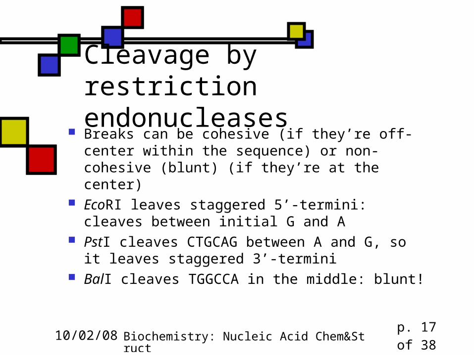
Cleavage by restriction endonucleases
Breaks can be cohesive (if they’re off-center within the sequence) or non-cohesive (blunt) (if they’re at the center)
EcoRI leaves staggered 5’-termini: cleaves between initial G and A
PstI cleaves CTGCAG between A and G, so it leaves staggered 3’-termini
BalI cleaves TGGCCA in the middle: blunt!
10/02/08 Biochemistry: Nucleic Acid Chem&Struct p. 18 of 38
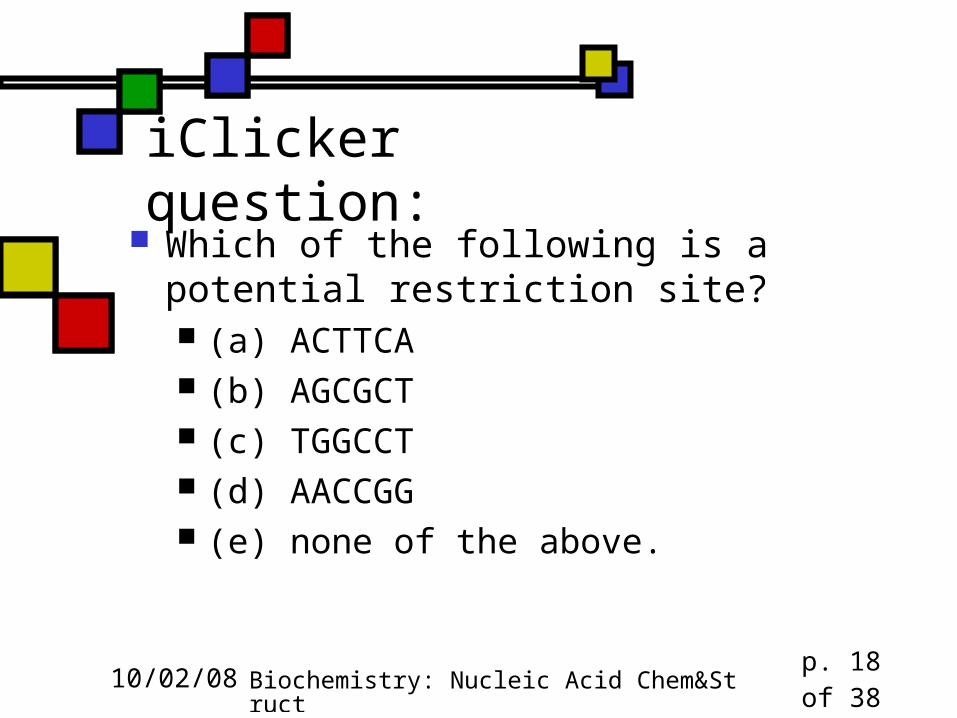
iClicker question: Which of the following is a potential
restriction site? (a) ACTTCA (b) AGCGCT (c) TGGCCT (d) AACCGG (e) none of the above.
10/02/08 Biochemistry: Nucleic Acid Chem&Struct p. 19 of 38
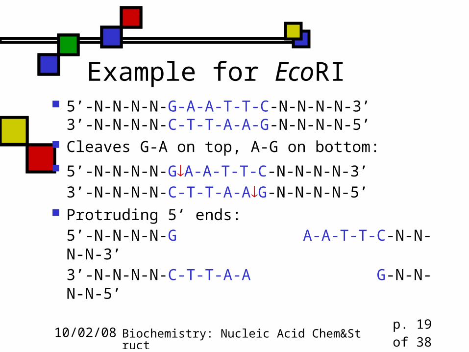
Example for EcoRI 5’-N-N-N-N-G-A-A-T-T-C-N-N-N-N-3’
3’-N-N-N-N-C-T-T-A-A-G-N-N-N-N-5’ Cleaves G-A on top, A-G on bottom: 5’-N-N-N-N-GA-A-T-T-C-N-N-N-N-3’
3’-N-N-N-N-C-T-T-A-AG-N-N-N-N-5’ Protruding 5’ ends:
5’-N-N-N-N-G A-A-T-T-C-N-N-N-N-3’3’-N-N-N-N-C-T-T-A-A G-N-N-N-N-5’
10/02/08 Biochemistry: Nucleic Acid Chem&Struct p. 20 of 38
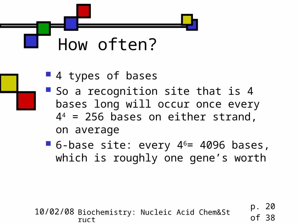
How often?
4 types of bases So a recognition site that is 4 bases long
will occur once every 44 = 256 bases on either strand, on average
6-base site: every 46= 4096 bases, which is roughly one gene’s worth
10/02/08 Biochemistry: Nucleic Acid Chem&Struct p. 21 of 38
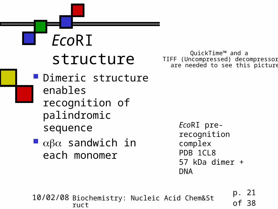
EcoRI structure
Dimeric structure enables recognition of palindromic sequence
sandwich in each monomer
QuickTime™ and aTIFF (Uncompressed) decompressor
are needed to see this picture.
EcoRI pre-recognition complexPDB 1CL857 kDa dimer + DNA
10/02/08 Biochemistry: Nucleic Acid Chem&Struct p. 22 of 38
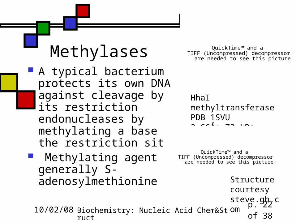
Methylases A typical bacterium protects
its own DNA against cleavage by its restriction endonucleases by methylating a base in the restriction site
Methylating agent is generally S-adenosylmethionine
QuickTime™ and aTIFF (Uncompressed) decompressor
are needed to see this picture.
HhaI methyltransferasePDB 1SVU2.66Å; 72 kDa dimer
QuickTime™ and aTIFF (Uncompressed) decompressor
are needed to see this picture.
Structure courtesy steve.gb.com
10/02/08 Biochemistry: Nucleic Acid Chem&Struct p. 23 of 38
Use of restriction enzymes Nature made these to protect bacteria; we use
them to cleave DNA in analyzable ways Similar to proteolytic digestion of proteins Having a variety of nucleases means we can get
fragments in multiple ways We can amplify our DNA first
Can also be used in synthesis of inserts that we can incorporate into plasmids that enable us to make appropriate DNA molecules in bacteria
10/02/08 Biochemistry: Nucleic Acid Chem&Struct p. 24 of 38
Sanger dideoxy method Incorporates DNA replication as an analytical
tool for determining sequence Uses short primer that attaches to the 3’ end of
the ssDNA, after which a specially engineered DNA polymerase
Each vial includes one dideoxyXTP and 3 ordinary dXTPs; the dideoxyXTP will be incorporated but will halt synthesis because the 3’ position is blocked.
See figs. 11.3 & 11.4 for how these are read out
10/02/08 Biochemistry: Nucleic Acid Chem&Struct p. 25 of 38
Automating dideoxy sequencing Laser fluorescence detection allows for
primer identification in real time An automated sequencing machine can
handle 4500 bases/hour That’s one of the technologies that has
made large-scale sequencing projects like the human genome project possible
10/02/08 Biochemistry: Nucleic Acid Chem&Struct p. 26 of 38
DNA secondary structures If double-stranded DNA were simply a straight-
legged ladder: Base pairs would be 0.6 nm apart Watson-Crick base-pairs have very uniform
dimensions because the H-bonds are fixed lengths But water could get to the apolar bases
So, in fact, the ladder gets twisted into a helix. The most common helix is B-DNA, but there are
others. B-DNA’s properties include: Sugar-sugar distance is still 0.6 nm Helix repeats itself every 3.4 nm, i.e. 10 bp
10/02/08 Biochemistry: Nucleic Acid Chem&Struct p. 27 of 38
Properties of B-DNA Spacing between base-pairs along helix
axis = 0.34 nm 10 base-pairs per full turn So: 3.4 nm per full turn is pitch length Major and minor grooves, as discussed
earlier Base-pair plane is almost perpendicular
to helix axis
10/02/08 Biochemistry: Nucleic Acid Chem&Struct p. 28 of 38
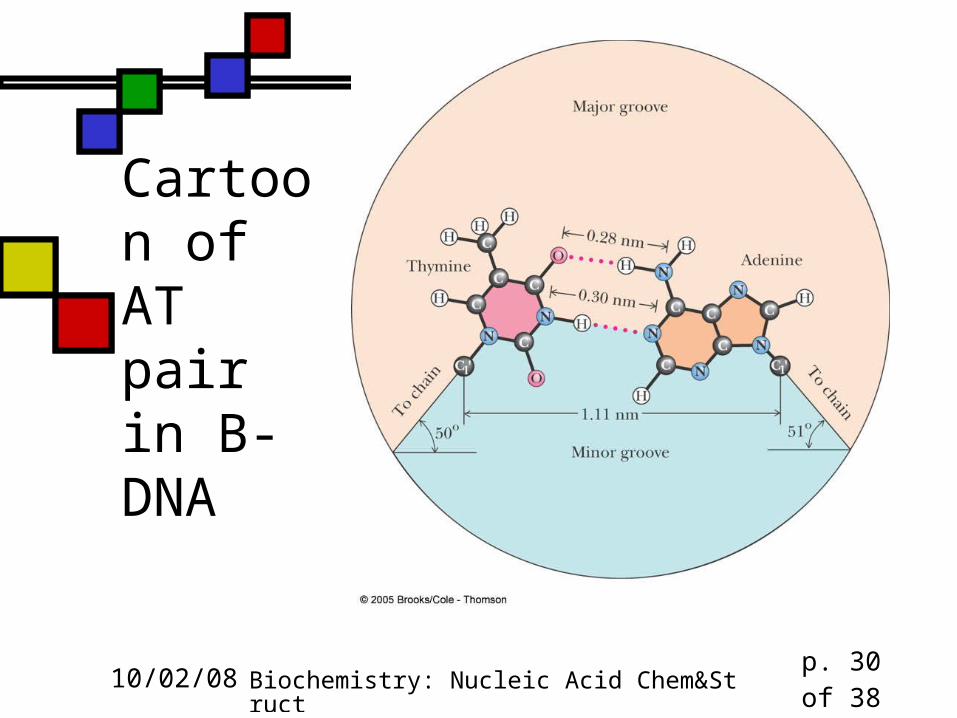
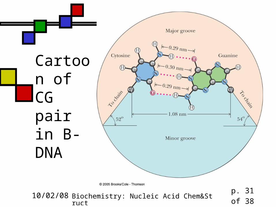
Major groove in B-DNA
H-bond between adenine NH2 and thymine ring C=O
H-bond between cytosine amine and guanine ring C=O
Wide, not very deep
10/02/08 Biochemistry: Nucleic Acid Chem&Struct p. 29 of 38
Minor groove in B-DNA
H-bond between adenine ring N and thymine ring NH
H-bond between guanine amine and cytosine ring C=O
Narrow but deep
10/02/08 Biochemistry: Nucleic Acid Chem&Struct p. 30 of 38
Cartoon of AT pair in B-DNA
10/02/08 Biochemistry: Nucleic Acid Chem&Struct p. 31 of 38
Cartoon of CG pair in B-DNA
10/02/08 Biochemistry: Nucleic Acid Chem&Struct p. 32 of 38
What holds duplex B-DNA together?
H-bonds (but just barely) Electrostatics: Mg2+ –PO4
-2
van der Waals interactions - interactions in bases Solvent exclusion
Recognize role of grooves in defining DNA-protein interactions
10/02/08 Biochemistry: Nucleic Acid Chem&Struct p. 33 of 38
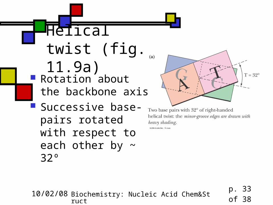
Helical twist (fig. 11.9a)
Rotation about the backbone axis
Successive base-pairs rotated with respect to each other by ~ 32º
10/02/08 Biochemistry: Nucleic Acid Chem&Struct p. 34 of 38
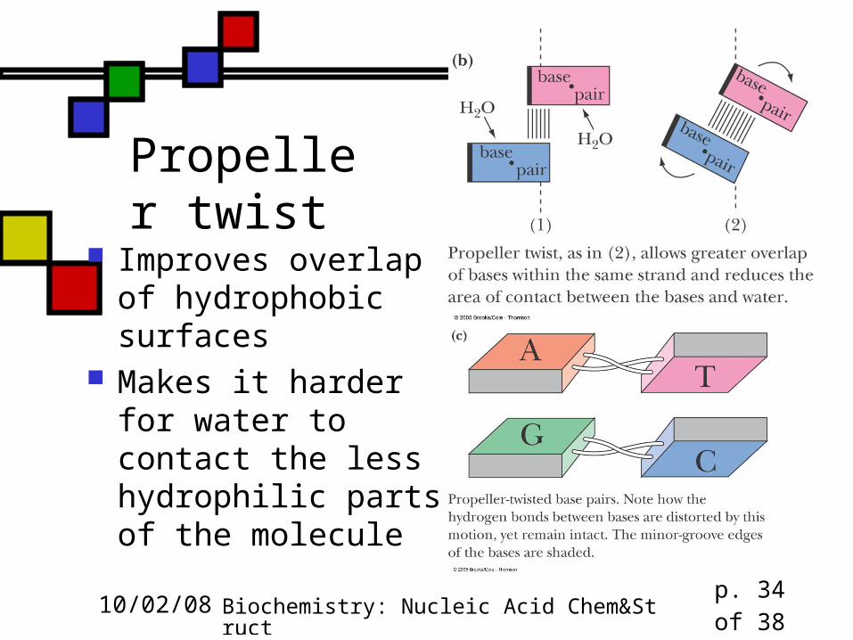
Propeller twist
Improves overlap of hydrophobic surfaces
Makes it harder for water to contact the less hydrophilic parts of the molecule
10/02/08 Biochemistry: Nucleic Acid Chem&Struct p. 35 of 38
A-DNA (figs. 11.10) In low humidity this forms naturally Not likely in cellular duplex DNA, but it does form
in duplex RNA and DNA-RNA hybrids because the 2’-OH gets in the way of B-RNA
Broader 2.46 nm per full turn 11 bp to complete a turn
Base-pairs are not perpendicular to helix axis:tilted 19º from perpendicular
10/02/08 Biochemistry: Nucleic Acid Chem&Struct p. 36 of 38
Z-DNA (figs. 11.10)
Forms in alternating Py-Pu sequences and occasionally in PyPuPuPyPyPu, especially if C’s are methylated
Left-handed helix rather than right Bases zigzag across the groove
10/02/08 Biochemistry: Nucleic Acid Chem&Struct p. 37 of 38
Getting from B to Z
Can be accomplished without breaking bonds
… even though purines have their glycosidic bonds flipped (anti -> syn) and the pyrimidines are flipped altogether!
10/02/08 Biochemistry: Nucleic Acid Chem&Struct p. 38 of 38
DNA is dynamic
Don’t think of these diagrams as static The H-bonds stretch and the torsions
allow some rotations, so the ropes can form roughly spherical shapes when not constrained by histones
Shape is sequence-dependent, which influences protein-DNA interactions
Recommended