#1
mGluR5 regulates autophagy in Alzheimer’s disease in a sex-specific manner
Khaled S. Abd-Elrahman, Alison Hamilton, Awatif Albaker and Stephen S. G. Ferguson
1. University of Ottawa Brain and Mind Research Institute and Department of Cellular and Molecular
2. Medicine, University of Ottawa, Ottawa, Ontario K1H 8M5, Canada
Background: Alzheimer’s disease is characterized by neurotoxicity due to accumulation of beta
amyloid (Aβ) oligomers, causing progressive cognitive decline. We have previously shown that genetic
deletion or chronic pharmacological inhibition of mGluR5 by the negative allosteric modulator CTEP,
rescued cognitive function and reduced Aβ aggregation in male APPswe/PS1ΔE9 (APPswe) mice via
the activation of novel ZBTB16-medaited autophagic mechanism (1, 2).
Objectives: To show that CTEP mitigates AD pathology and regulates ZBTB16 autophagic pathways in
females APPswe similar to male mice
Methods: We tested brain samples from male and female APPswe mice treated for 12 weeks with either
vehicle or CTEP (2mg/kg) for changes in the autophagic clearance of Aβ oligomers via ZBTB16
pathway.
Results: Both male and female APPswe mice exhibited higher Aβ oligomers levels at 6 months of age.
To our surprise, CTEP significantly reduced Aβ burden and improved cognitive function in male mice
yet, it exacerbated Aβ oligomers accumulation and did not improve cognitive function in female
APPswe mice. mGluR5 blockade activated ZBTB16 autophagic pathway to reduce Aβ burden in male
but not female APPswe mice. Interestingly, a significant reduction in the surface expression of mGluR5
was detected in female APPswe compared to male mice.
Conclusions: We also provide evidence, for the first time, that mGluR5 signaling is not conserved
between males and females and there are sex-specific differences that must be considered when
embracing mGluR5 as a drug target for neurodegenerative disease.
1. Abd-Elrahman et al., 2017. Sci. Signal.
2. Abd-Elrahman et al., 2018. Mol. Brain.
Keywords: GPCR, autophagy, Alzheimer’s disease, mGluR5, sex
#2
Evaluating the Use of the Lymphocyte Toxicity Assay for Diagnosis of Delayed-Type Allergy to
Amoxicillin.
Awatif M. Abuzgaia1, Michael J Rieder1
1. Department of Pediatrics, Schulich School of Medicine and Dentistry, Western University, London,
Ontario
Background: Drug allergy especially to beta-lactam antibiotics (BLAs) is a major health problem. The
prevalence of self-reported allergy to BLAs in the general population is very high (~10%), however,
true, immune-mediated reactions are over 10 times less. The most popular course of action in such cases
is drug avoidance, which result in exposing the patient to less effective, more expensive alternatives
with inferior safety profiles. This is mainly due to lack of any reliable and safe diagnostic tool for this
type of reactions.
Objectives: The objective of this study was to evaluate the performance of an in vitro test (the
lymphocyte toxicity assay, LTA) as a diagnostic tool for BLAs-induced delayed type allergic reactions.
Methods: We recruited 8 patients (5 females and 3 males), age range 18 months to 16 years, who
developed symptoms (skin rash with or without other systemic symptoms) highly suggestive of delayed
allergy to amoxicillin after 6 to 10 days of exposure to the drug. Blood samples were obtained from the
patients as well as another 8 healthy control volunteers. Peripheral blood monocytes (PBMCs) were
isolated and the LTA test was performed on all subjects.
Results: Cells from the 8 patients exhibited significantly more toxicity (p<0.05) as compared to cells
from the healthy controls.
Conclusions: This is a very small sample size; however, this preliminary data highlights the potential of
using the in vitro toxicity test as safe diagnostic and research tool in patients suspected of having
delayed allergic reactions to BLAs.
Keywords: in vitro toxicity assay, beta-lactam antibiotics, amoxicillin, drug allergy, hypersensitivity
reactions

#3
Xenobiotic metabolism and tobacco-associated cancers – A case of CYP1A1, ALDH3A1 and
GSTM1 gene signature?
Nasir A Afsar1, HR Ahmad2*, Ingolf Cascorbi3
1. Jinnah Medical and Dental College, Sohail University, 22-23 Shaheed-e-Millat Road, Karachi-
75400, Pakistan.
2. Department of Biological and Biomedical Sciences, The Aga Khan University, Karachi, Pakistan
3. Institute of Experimental and Clinical Pharmacology, Christian Albrechts University Kiel,
Hospitalstr. 4, Kiel-24105, Germany.
Background: Tobacco smoke contains numerous pro-inflammatory and carcinogenic substances, such
as polycyclic aromatic hydrocarbons (PAHs) which are activated by CYP1A1 and detoxified by GSTM1
and ALDH3A1 within respiratory tract, from lips to lungs. Genetic polymorphism of these enzymes may
influence development of conditions like chronic obstructive pulmonary disease as well as
oropharyngeal and lung cancer. Indeed, Pakistan has one of the highest incidence of such cancers in the
world. Although the role of CYP1A1 and GSTM1 is known with varying severity, new evidence
regarding ALDH3A1 has renewed the interest in this regard in global perspective.
Objectives: To estimate the prevalence pattern of SNVs of CYP1A1, GSTM1 and ALDH3A1 in
Pakistani population and evaluate its role as a genetic signature predisposing to the increased incidence
of tobacco-associated cancers.
Method: In this study, 155 healthy adults (99 females) were included from all districts of Karachi. DNA
was extracted from saliva and genotyped for SNVs either through PCR (GSTM1), RFLP (CYP1A1) or
pyrosequencing (ALDH3A1).
Results: About 64% of the participants were born to parents who were unrelated to each other. There
was generally a higher prevalence (p<0.05) of variant alleles of CYP1A1*2B, ALDH3A1, and GSTM1
in this study cohort than in other ethnicities reported in the HapMap database (Table 1). When viewed as
a gene signature (Figure 1), 68% population had high risk to develop tobacco related COPD and cancers.
Conclusions: Karachiites have a significantly different prevalence of xenobiotic metabolizing gene
signature, which could have putative clinical consequences on gene-environment interaction and
carcinogenesis.
Key Words: Lung cancer, Oro-pharyngeal cancer, PAH, Tobacco, Environmental toxicity
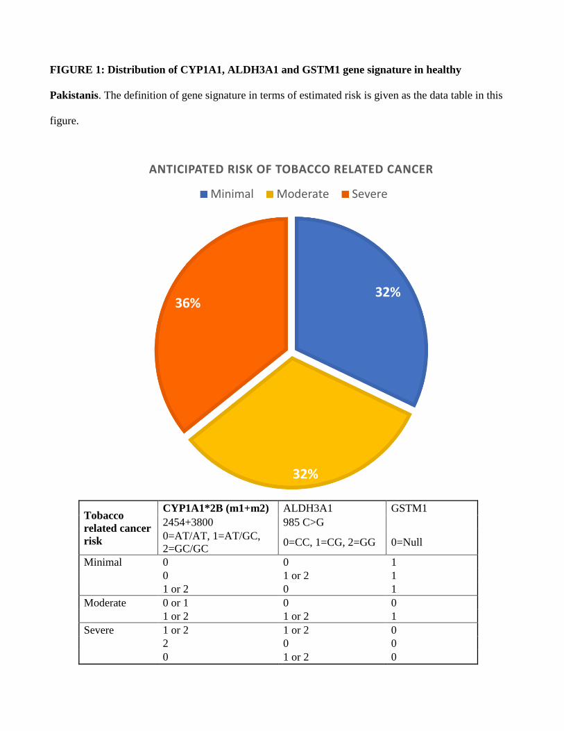
FIGURE 1: Distribution of CYP1A1, ALDH3A1 and GSTM1 gene signature in healthy
Pakistanis. The definition of gene signature in terms of estimated risk is given as the data table in this
figure.
Tobacco
related cancer
risk
CYP1A1*2B (m1+m2) ALDH3A1 GSTM1
2454+3800 985 C>G
0=AT/AT, 1=AT/GC,
2=GC/GC 0=CC, 1=CG, 2=GG 0=Null
Minimal 0 0 1
0 1 or 2 1
1 or 2 0 1
Moderate 0 or 1 0 0
1 or 2 1 or 2 1
Severe 1 or 2 1 or 2 0
2 0 0
0 1 or 2 0
32%
32%
36%
ANTICIPATED RISK OF TOBACCO RELATED CANCER
Minimal Moderate Severe

TABLE 1: Comparison of variant allele frequency with other ethnic groups. The Chi square value
was computed with df=1.
Genotype KHI
Sample
HapMap
Ethnicities
Variant
Allele % ² Value p-value
CYP1A1*2 11.07 CHIN 25.6 7.05 0.008
CAUC 3.1 4.83 0.028
GUJ 10.2 0.04 0.84
AFR 0 6.18 0.013
CYP1A1*2A 32.64 CHIN 37.5 0.52 0.47
CAUC 10 15.28 <0.001
GUJ - -
AFR 14.4 9.25 0.002
ALDH3A1
985C>G 66.89 CHIN 44.4 10.25 0.001
CAUC 29.2 28.46 <0.001
GUJ - -
AFR 44.1 10.5 0.001
GSTM1 30.13 CHIN - -
CAUC 0 20.4 <0.001
GUJ - -
AFR 0 20.4 <0.001
KHI, Karachi sample; CHIN, Chinese of Han ancestry; CAUC, Caucasian of Northern and Western
European ancestry, GUJ, Gujrati Indians in Houston Texas; AFR, African of Yoruba Nigerian ancestry.

#4
Physiologically-based pharmacokinetic modeling of amantadine and acetylamantadine
metabolites for potential applications as cancer biomarker.
Eman A. Alraddadi, Donald W. Miller
1. Department of Pharmacology and Therapeutics, The Rady Faculty of Health Sciences, University of
Manitoba, Winnipeg, Canada
Background: Cancer is the second leading cause of death globally. Despite advances in treatment, there
is a need for faster and economical screening tests for early diagnosis of cancer. Spermidine/spermine
N1-acetyltransferase (SSAT-1) is overexpressed in many cancers. Recent studies suggest that that
SSAT-1 based acetylation of amantadine could serve as a biomarker for lung cancer. However the
potential use of amantadine for detection of tumor in other tissues is unclear.
Objectives: Use physiologically-based pharmacokinetic modeling to determine whether appropriate
amounts of amantadine and acetylamantadine could be achieved in other tissue/organs.
Methods: Physiologically-based pharmacokinetic modeling was performed using GastroplusTM. Plasma
and tissue compartment kinetics for amantadine (200 mg PO) was simulated and prediction of
acetylamantadine metabolite in urine under normal conditions and following increases in SSAT-1
enzyme expression was performed.
Results: The simulated plasma Cmax of 750 ng/ml and half-life of 12 hours was similar to reported
values in healthy subjects. Modeling of the acetylamantadine metabolite concentrations in urine under
low SATT-1 expression were consistent with levels observed in healthy controls (approximately 3
ng/ml). When the expression of SATT-1 was increased 3-fold simulating increased expression in lung
tumor, the levels of acetylamantadine in urine increased to levels comparable to patients with lung
cancer (around 10 ng/ml).
Conclusions: Physiologically-based pharmacokinetic modeling of amantadine indicates significant
accumulation of the drug in tissue including the brain. Acetylamantadine metabolite formation may be
useful for cancer detection and therapeutic monitoring in various solid organ tumors.
Keywords: Amantadine, Acetylamantadine, Lung cancer, PBPK modeling

#5
Paracrine effects of perivascular adipose tissue on atherogenesis: Role of exosomal intercellular
communications
Noura N. Ballasy,1,2,3 Anshul S. Jadli,1,2, Tishani M. Wijesuriya,1,2 Darrell D. Belke,2,4 Yong X. Chen,2,4
Edward O’Brien,2,4 and Vaibhav B. Patel1,2
1. Department of Physiology and Pharmacology, Cumming School of Medicine, University of Calgary
2. Libin Cardiovascular Institute of Alberta (LCIA), University of Calgary
3. Department of Physiology, Benha University
4. Departement of Cardiac Sciences, Cumming School of Medicine, University of Calgary
Background: Development of atherosclerosis depends on the interaction between various factors. Type
2 diabetes accelerates these interactions and predisposes to rapid progression of atherosclerosis.
Diminution of vasoprotective effects of perivascular adipose tissue (PVAT) in metabolic disorders
suggests a molecular link between diabetes and atherosclerosis.
Objectives: To assess the paracrine role of the PVAT on the progression of diabetic atherosclerosis, via
intercellular communications between PVAT and the underlying vasculature.
Methods: Periaortic adipose tissue from Type 2 diabetic (db/db) mice were transplanted around the
right common carotid arteries of ApoE-/- mice, followed by 16 weeks of atherogenic diet. Carotid
arteries and adipose tissues were assessed for lesion formation and inflammatory markers, respectively.
Adipose stem cells (ASCs) from PVAT were treated with lipopolysaccharide (1µg/ml), palmitate
(200µM), and high-glucose (42 mM) for 24 hrs (denoted as P-ASCs) to mimic type 2 diabetes-
associated metabolic alterations. Exosomes were isolated from the conditioned media. Aortic vascular
smooth muscle cells (SMCs) were incubated with ASC-derived exosomes (25µg/ml). The migratory
potential of SMCs was evaluated by wound healing assay.
Results: Histological analysis displayed atherogenic plaque formation with transplantation of type 2
diabetic PVAT around the carotid arteries of ApoE-/- mice, that is otherwise resistant to plaque
formation.. Pro-inflammatory markers were significantly increased in periaortic adipose tissue from
db/db compared to WT. Exosomes secreted from P-ASCs greatly enhanced the SMC migration when
compared with the control ASCs.
Conclusions: Our data shows type 2 diabetes-accelerated progression of atherosclerosis is mediated by
PVAT-derived exosomes.
Keywords: Exosomes, Diabetes, perivascular adipose tissue, Atherosclerosis
#6
Management of Marfan aortic root remodelimg with ARBs: blood pressure or endothelial
function?
Arash Y Therani1, Pascal Bernatchez1
1. UBC Department of Anesthesiology, Pharmacology & Therapeutics.
Background: Marfan syndrome (MFS) causes accelerated aortic root widening and aneurysm. The anti-
hypertensive angiotensin II (AngII) receptor type 1 (ATR1) blocker (ARB) Losartan has shown
underwhelming efficacy in MFS patients, along with the other guideline approve beta blocker atenolol.
We have reported that Losartan’s anti-aortic root remodeling effects are likely BP-independent, but
rather endothelial function and nitric oxide (NO)-dependent. Whether other ARBs can provide greater
protection against MFS aortic root widening is unknown.
Objectives: To study the anti-aortic root remodeling properties of other ARBs, some at doses that do
not reduce BP.
Methods: Aortic root dilation, aortic vessel remodeling, and vessel contractility were compared
following treatment with Losartan, Telmisartan and Valsartan between MFS and wild-type (WT) mice
with normal and blunted ATR1 expression.
Results: Loss of ATR1 expression does not interfere with the activity of ARBs in MFS aortic widening.
Although Valsartan had no effect on BP, all ARBs reduced MFS aortic root widening, medial
thickening, and elastic fiber fragmentation with Telmisartan reaching >95% inhibition efficacy. All
ARBs decreased vascular contractility ex vivo between 60-80% in both MFS and WT aortas in a NO-
sensitive fashion.
Conclusions: ARBs likely mediate their effect in an ATR1 independent fashion. Telmisartan was the
most effective ARB at blocking MFS aortic root widening and activating endothelial function, whereas,
Valsartan showed a high degree of efficacy with little to no BP-lowering effects. Aortic root protection
could be achieved with lesser side effects for MFS patients, addressing key compliance issues. Future
clinical trials should focus on other ARBs than Losartan for the management of MFS-associated
aortopathies.
Keywords: aorta, nitric oxide, angiotensin, Marfan.

#7
A tri-peptide IRW (Ile-Arg-Trp) from egg white protein ovotransferrin improves mitochondrial
status in metabolic disease models
Khushwant S. Bhullar1,2, Wang Liao1, Myoungjin Son1 and Jianping Wu1.
1. Department of Agricultural, Food & Nutritional Science, University of Alberta, Canada
2. Department of Pharmacology, University of Alberta, Canada
Background: Obesity and hypertension are critical unfavorable health metrics that have a disastrous
impact on health and contribute to other metabolic diseases such as type 2 diabetes mellitus,
dyslipidemia, and certain cancers. However, appropriate nutritional and pharmacological interventions
with feasible potential to delay such diseases and promote healthspan remain rare.
Objectives: The objectives of the current study were to evaluate the ability of a tri-peptide IRW (Ile-
Arg-Trp) to improve mitochondrial status towards mitigation of metabolic disorders.
Methods: The ability to tri-peptide IRW was evaluated in different mammalian cell lines followed by
measurement of mitochondrial mass and activity. Animal models of diet-induced obesity (C57BL/6J)
and hypertension (SHRs) were used to examine the impact of IRW at different dosage (15 and 45 mg/kg
BW). Further, the target identification of peptide was completed using MS/MS studies.
Results: Administration of IRW at a moderate dose (15 and 45 mg/kg BW) improves mitochondrial
status in C57BL/6J and SHRs in vivo. Further, it enhances mitochondrial mass in mammalian cells and
induces mitochondrial biogenesis as indicated by elevated expression of TFAM and COXIV. Also, the
target identification indicated the interaction of IRW with FAM120B, a transcriptional co-activator of
PPARƴ and a newly established regulator of DNA repair. The cas9 guided KO of FAM120B, lowered
the efficacy of IRW in 293T cells.
Conclusions: To the best of our knowledge, IRW is the first peptide which improves mitochondrial
status in both in vitro and in vivo models.
Keywords: Obesity, Hypertension, mitochondria, biogenesis, peptide

#8
The Pharmacogenomics of Clozapine-Induced Myocarditis (PROCLAIM) Consortium
Chad A. Bousman1, Steven C. Greenway1, Maja Tarailo-Graovac1, Quan Long1, Rory Sellmer1, David
Crockford1, Kathlyn Ronaldson2, John McNeil2, Dan Siskind3, Karl Winckel3, Kevin Li4, Lynn DeLisi4,
Gary Remington5, Murray Cairns6, Katherine Aitchison7, Robert Stowe8, Shailesh Nadkarni9, Amlan
Das9, Charles Ohene-Darkoh9, Cynthia Shannon Weickert10, Mahesh Jayaram11, Naveen Thomas11,
Christos Pantelis11
1. University of Calgary, Calgary, AB, Canada
2. Monash University
3. Clayton, VIC, Australia
4. University of Queensland QLD, Australia
5. Harvard University, MA, USA
6. University of Toronto, ON, Canada
7. University of Newcastle, NSW, Australia
8. University of Alberta, AB, Canada
9. University of British Columbia, BC, Canada
10. William Osler Health System, ON, Canada
11. Neuroscience Research Australia, NSW, Australia
12. University of Melbourne, VIC, Australia
Background: Clozapine has proven efficacy in treatment-resistant schizophrenia (TRS). However, the
occurrence of clozapine-induced myocarditis, an unpredictable and often fatal adverse drug reaction, has
substantially limited its use. Markers for and the mechanism by which clozapine induces myocardial
inflammation and damage is unknown.
Objectives: (1) To identify clinical and genomic factors associated with clozapine-induced myocarditis,
and (2) develop a novel in vitro model system using patient-derived induced pluripotent stem cells
(iPSCs) to explore how clozapine induces myocardial inflammation and damage and ultimately identify
a mechanism.
Methods: Recruitment of adult TRS patients aged 18-65 with and without a history of clozapine-
induced myocarditis is on-going at 11 participating sites. Patients provide DNA samples, which are used
for genomic analysis. In a subset of participants, blood is used to generate iPSCs that are differentiated
into cardiomyocytes for in vitro functional studies to identify abnormal cellular pathways and responses
to clozapine.
Results: To date, 42 treatment-resistant schizophrenia individuals with a history of clozapine-induced
myocarditis and 68 without a history have been recruited. Details related to recruitment rates at
participating sites and inclusion/exclusion criteria as well as preliminary results of genomic and patient
iPSCs analyses will be presented.
Conclusions: The PROCLAIM consortium is an innovative initiative that if successful will lead to safer
and expanded use of clozapine. In addition, success will provide a novel and clinically-relevant in vitro
model for elucidating the etiology of clozapine-induced myocarditis and for the study of other drug-
induced cardiotoxicities.
Keywords: pharmacogenomics, adverse drug reaction, precision medicine

#9
Characterizing T-cell phenotype in patients with hypersensitivity reactions to sulfonamides and
beta-lactam antibiotics
Christine C Caron1,4, Abdelbaset A Elzagallaai2,3,4, Michael J Rieder1,2,3,4,5
1. Department of Pathology and Laboratory Medicine, Western University, London Ontario
2. Department of Pediatrics, Western University, London, Ontario, Canada
3. Department of Physiology and Pharmacology, Western University, London, Ontario, Canada
4. Robarts Research Institute, Western University, London, Ontario, Canada
5. Division of Clinical Pharmacology, Department of Medicine, Schulich School of Medicine and
Dentistry, London, Ontario, Canada
Background: Delayed drug hypersensitivity reactions (DHRs) are idiosyncratic and can present days
after exposure to the culprit drug. There is currently no consensus on how T-cell activation occurs.
Previous research groups different resulting skin reactions, often without accounting for rash type or
severity, although some may have different cytokine profiles and T-cell subset involvement. We
hypothesize that differences in activated T cell subsets lead to the different clinical presentations
observed in DHRs to sulfamethoxazole and beta-lactam antibiotics.
Objectives: This project will address this issue and determine DHR mechanisms in the context of both
drug type and clinical presentation.
Methods: Peripheral blood mononuclear cells (PBMCs) are isolated from participants with DHRs to
sulfamethoxazole or beta-lactam antibiotics. PBMCs are stimulated in vitro with the culprit drug or anti-
CD3. T-cell subset proliferation is assessed by T-cell specific surface markers using flow cytometery
and 3H-thymidine incorporation, and secreted effector cytokines are measured by Luminex.
Results: Proliferation and surface staining of isolated T-cells has been optimized. 3H-thymidine for
measuring T-cell proliferation and flow cytometry for T-cell CD69 expression/activation using control
blood samples have provided satisfactory results. Participants with DHRs are being recruited to
participate. Preliminary results have suggested that different clinical presentations occur due to response
of different T-cell subsets and effector cytokines.
Conclusions: When completed, this study will provide evidence into the underlying pathophysiology of
DHRs. Identifying differences in cytokine profiles between skin rashes in DHRs to sulfamethoxazole
and beta-lactam antibiotics can help with developing reliable, minimally-invasive, diagnostic and
predictive tests.
Keywords: Hypersensitivity, beta-lactam, sulfamethoxazole, pathomechanism, T-cell
#10
Stevens-Johnson Syndrome and Toxic Epidermal Necrolysis Caused by Antibiotics-
an International cohort study
Wan-Chun Chang1,2, Reo Tanoshima1,2, Colin Ross2,3, Galen Wright1,2, Britt Drögemöller2,3, Shuen-Iu
Hung4, Wen-Hung Chung5,6,7, Bruce C. Carleton1,2
1. Department of Pediatrics, Faculty of Medicine, University of British Columbia, Vancouver, British
Columbia, Canada.
2. British Columbia Children’s Hospital Research Institute, Vancouver, British Columbia, Canada.
3. Faculty of Pharmaceutical Sciences, University of British Columbia, Vancouver, British Columbia,
Canada.
4. Cancer vaccine and immune cell therapy Center, Chang Gung Memorial Hospital, Linkou, Taiwan.
5. Department of Dermatology, Drug Hypersensitivity Clinical and Research Center, Chang Gung
Memorial Hospital, Linkou, Taipei and Keelung, Taiwan.
6. Chang Gung Immunology Consortium, Chang Gung Memorial Hospital and Chang Gung University,
Taiwan.
7. Whole-Genome Research Core Laboratory of Human Diseases, Chang Gung Memorial Hospital,
Keelung, Taiwan.
Background: Antibiotics have saved millions of lives; however, antibiotics are also one of the most
common causes to induce potentially fatal adverse events, such as severe cutaneous adverse reaction
(SCAR)- Stevens-Johnson syndrome (SJS) and toxic epidermal necrolysis (TEN).
Objectives: To investigate the epidemiological characteristics of antibiotic-associated SCAR and to
identify the genetic susceptibility to specific antibiotic-SCAR
Methods: We retrospectively reviewed the Canadian Pharmacogenomics Network for Drug Safety
(CPNDS) database and Taiwan Chang Gung Memorial Hospital system database over a 14-year period
(2004-2018).
Results: From a review of 6,450 individuals in CPNDS database, 3,828 (59.3%) participants were
prescribed at least one antibiotic treatments. Among them, twenty-three percent of patients experienced
antibiotic-related cutaneous adverse reactions, ranging from common rash to serious systemic reactions
(i.e. SCARs). Thirty severe patients met the criteria of the SJS/TEN definitions. Among all antibiotics,
β-lactams, particularly amoxicillin, is the leading cause in the Canadian cohort and is responsible for
41% of all SJS/TEN, following by sulfonamides (i.e. sulfamethoxazole). Consistently, β-lactam
accounts for half (50.1%) of all SCAR cases in the Taiwanese cohort. Our next step is to identify genetic
variants associated with SJS/TEN induced by β-lactams and sulfonamides in different ancestries.
Conclusions: β-lactams are the most common culprit antibiotics to induce SJS/TEN in the both
Canadian and Taiwanese cohorts. Given the widespread use of antibiotics, understanding the
pathogenesis of antibiotic-associated SCAR can develop interventions to prevent its onset, resulting in
improving the safety of antibiotics use.
Keywords: antibiotics, ADR, SCAR, SJS/TEN

#11
Severity of Celiac disease and oral felodipine pharmacokinetics: Comparison to the interaction
with grapefruit juice.
Marc L Chretien 1, David G Bailey 1,4, Linda Asher 1, Jeremy Parfitt 2, David Driman 2, Jamie Gregor 3,
George K Dresser 1,4
1. Division of Clinical Pharmacology, Faculty of Medicine, Western University
2. Division of Pathology, Faculty of Medicine, Western University
3. Division of Gastroenterology, Faculty of Medicine, Western University
4. Lawson Health Research Institute, London, Ontario, Canada
Background: Celiac disease is a hypersensitivity reaction to gluten-containing foods in genetically
susceptible individuals. It is characterized by damage to the small intestinal mucosa that ranges from
inflammation to villous atrophy. CYP3A4 is constitutively expressed in human small intestinal villi and
accounts for first-pass prehepatic metabolism of drugs. Celiac patients with severe disease have low
duodenal CYP3A4 expression.
Objectives: To determine whether oral felodipine bioavailability would be dependent on celiac disease
severity and be caused by a grapefruit-like mechanism.
Methods: Celiac patients were histologically stratified into three categories: Group A (n=15, normal),
B+C (n=16, intraepithelial cell invasion with/without mild villous blunting) and D (n=16,
moderate/severe villous blunting). Single dose oral pharmacokinetics of felodipine 10 mg were assessed.
Healthy volunteers (n = 68) undergoing similar testing in prior felodipine–grapefruit juice interaction
crossover studies served as negative and positive controls.
Results: Groups A, B+C and D had linear trends of increasing felodipine AUC0-8 (mean + SEM, 14.4+
2.1, 17.6+2.8, 25.7+5.0; p<0.05) and Cmax (3.5+0.5, 4.0+0.6, 6.4+1.1; p<0.02), respectively. Healthy
subjects receiving water had lower felodipine AUC0-8 (11.9+0.9 vs 26.9+0.9, p=0.0001) and Cmax
(2.9+0.2 vs 7.7+0.2, p=0.0001) versus those receiving grapefruit juice. Group A and D had similar
felodipine pharmacokinetics for healthy subjects with water and grapefruit juice, respectively.
Conclusions: Patients with severe celiac disease had increased oral felodipine bioavailability like that
with grapefruit juice from low small intestinal CYP3A4 protein expression. They could be at risk of
serious overdose toxicities with numerous grapefruit-affected drugs and should be considered for altered
pharmacotherapy.
Keywords: Celiac disease, prehepatic metabolism, CYP3A4, felodipine, pharmacokinetics

#12
Genomic analyses of L-asparaginase-induced Pancreatitis in Pediatric Cancer Patients
Britt I. Drögemöller1,2 Galen E.B. Wright2,3, Shahrad R. Rassekh2,3, Shinya Ito4, Bruce C. Carleton2,3,5,
Colin J.D. Ross1,2 and The Canadian Pharmacogenomics Network for Drug Safety Consortium
1. Faculty of Pharmaceutical Sciences, University of British Columbia, Vancouver, BC, Canada
2. BC Children’s Hospital Research Institute, University of British Columbia, Vancouver, BC, Canada
3. Department of Pediatrics, Faculty of Medicine, University of British Columbia, Vancouver, BC,
Canada
4. Clinical Pharmacology and Toxicology, The Hospital for Sick Children, University of Toronto,
Toronto, ON, Canada
5. Pharmaceutical Outcomes Programme, BC Children’s Hospital, Vancouver, BC, Canada
Background: L-asparaginase is highly effective in the treatment of pediatric acute lymphoblastic
leukemia. Unfortunately, the use of this treatment is limited by the occurrence of pancreatitis, a severe
and potentially lethal adverse drug reaction, which occurs in 2-18% of patients. As previous studies have
been unable to identify strong associations between clinical variables and susceptibility to L-
asparaginase-induced pancreatitis, genetic factors are expected to play an important role this adverse
drug reaction.
Objectives: We sought to explore the role of these genetic susceptibility factors to L-asparaginase-
induced pancreatitis in pediatric cancer patients.
Methods: Patients who were treated with L-asparaginase were recruited from 13 pediatric oncology
units across Canada (n=284) and extensive clinical data were collected for all patients. Genotyping was
performed using the Illumina HumanOmniExpress and Global Screening Arrays and pancreatic gene
expression profiles were imputed in these individuals using GTEx v7 and S-PrediXcan. Genome- and
transcriptome-wide associations (GWAS and TWAS) were performed to identify associations with L-
asparaginase-induced pancreatitis.
Results: GWAS analyses identified significant associations between genetic variants in HLA-DQA1 and
–DRB1 and pancreatitis, while TWAS revealed that individuals experiencing L-asparaginase-induced
pancreatitis exhibited lower expression levels of HLA-DRB5. Further interrogation of the TWAS data
revealed an enrichment in genes involved in the somatic diversification of immune receptors.
Conclusions: These analyses uncovered an association between genetic variation in immune-related
genes and the development of L-asparaginase-induced pancreatitis. These associations mirror previous
associations with the HLA region and (i) pancreatitis induced by other drugs and (ii) L-asparaginase-
induced hypersensitivity.
Keywords: Genome-wide association study, L-asparaginase, Pancreatitis, Pharmacogenomics

#13
Sex Differences in the Role of Pannexin-1 in Neuropathic Pain
Churmy Fan1, Rebecca Dalgarno2, Charlie HT Kwok3, Tuan Trang4
1. University of Calgary, Faculty of Medicine, Calgary, Alberta, Canada
2. University of Calgary, Faculty of Medicine, Calgary, Alberta, Canada
3. University of Calgary, Faculty of Veterinary Medicine, Calgary, Alberta, Canada
4. University of Calgary, Faculty of Medicine/Veterinary Medicine, Calgary, Alberta, Canada
Background: Neuropathic pain is among the most debilitating types of chronic pain conditions and its
clinical management is difficult because the underlying causes are poorly understood. Pannexin-1
(Panx1) channels have recently been implicated in the modulation of neuropathic pain. Our preliminary
findings indicate that Panx1 channels expressed in the central nervous system differentially modulate
neuropathic pain in male and female rats.
Objectives: To investigate the role of Panx1 in the expression of neuropathic pain in male and female
rats.
Methods: Nerve injury was induced in male and female Sprague Dawley rats using the spared nerve
injury (SNI) model. Mechanical threshold was assessed using the von Frey filament test and in a subset
of animals, thermal threshold was tested using the Tail Flick test. We investigated the importance of
Panx1 in the expression of neuropathic pain by intrathecally administering 10panx, a Panx1 blocking
mimetic peptide, into male and female rats with SNI.
Results: Peripheral nerve injury produced robust mechanical allodynia in male and female rats. A single
intrathecal injection of 10panx transiently reversed mechanical allodynia in male rats on day 7 after SNI.
In contrast, 10panx produced a partial reversal of the established mechanical allodynia in females. We
confirmed that in sham control rats, intrathecal administration of 10panx did not alter baseline thermal
threshold.
Conclusions: Our results suggest that Panx1 expressed in the central nervous system is necessary for the
ongoing expression of neuropathic pain, and that there may be sex differences in the contribution of
spinal Panx1 in the expression of neuropathic pain.
Keywords: Pannexin-1, neuropathic pain, chronic pain, sex difference

#14
Bend Beauty, Inc.’s oral skincare supplement, Anti-Aging Formula, protects skin cells from
ultraviolet light and reactive oxygen species
Steven R. Hall1,3, Anna-Jean Reid3, Marc St-Onge3, Kerry B. Goralski1,2
1. College of Pharmacy, Dalhousie University
2. Department of Pharmacology, Dalhousie University
3. Bend Beauty, Inc., Halifax, Nova Scotia, Canada
Background: Ultraviolet (UV) light overexposure is associated with multiple health risks. The ingestion
of natural product photoprotectors can increase skin’s UV-resistance. Bend Beauty, Inc.’s skincare
supplement, marketed as “Anti-Aging Formula” (AAF), contains daily doses of eicosapentaenoic acid
(1050 mg), docosahexaenoic acid (350 mg), gamma-linolenic acid (120 mg), zeaxanthin (2.5 mg), lutein
(5 mg), and vitamin D3 (1,000 IU), and reduces UV-induced skin erythema (sunburn) clinically.
Objective: To determine AAF’s photoprotective and antioxidant properties in dermal fibroblasts.
Methods: Primary human dermal fibroblasts were treated with 0.005% AAF or vehicle for up to 14
days. (i) Photoprotection assays: AAF-treated fibroblasts were exposed to UVA (365 nm; 100-216
J/cm2) or UVB (312 nm; 15.6-8,000 mJ/cm2); cell viability quantified via methyltetrazolium (MTT)
assays or confocal microscopy (dyed with phalloidin, propidium iodide, and Hoechst 32258). (ii)
Reactive oxygen species (ROS): AAF-treated fibroblasts were exposed to H2O2 (11.1-2,700 M); cell
viability and ROS activity quantified via MTT assays and ROS-probe (CM-DCFH2-DA) fluorescence,
respectively.
Results: (i) The viability (MTTs) of UVA- or UVB-irradiated fibroblasts was increased by 4.2- and 3.2-
fold, respectively, by AAF treatment vs. control (P<0.05). AAF-treated fibroblasts were protected
against structural damage as visualized with confocal microscopy (Figure 1). (ii) The viability of H2O2-
treated fibroblasts was increased up to 2.5-fold, and H2O2-induced ROS activity reduced by 38%, by
AAF treatment vs. control (P<0.05).
Conclusions: AAF protects human dermal fibroblasts from the damaging effects of UVA, UVB, and
H2O2, demonstrating its cellular photoprotective and antioxidant properties.
Keywords: Photoprotection, lipids, essential fatty acids, skin health, academic-industry collaboration
Figure 1

#15
Abstract title: Contribution of Cyclic AMP and β-arrestin-2-dependent Mechanisms to β2-
adrenoceptor-mediated Gene Expression Changes in Human Airway Epithelial Cells
Omar Hamed1, Aubrey Michi1, Mahmoud M Mostafa1, Mark A Giembycz1
1. Department of Physiology & Pharmacology, University of Calgary, Calgary, Alberta, Canada
Background: β2-adrenoceptor (β2-AR) agonists are used routinely in asthma management. However,
chronic use as a monotherapy is associated with adverse events (AEs). Previous studies have reported
that β2-AR agonists promote a significant transcriptional response in BEAS-2B airway epithelial cells,
which could contribute to their AEs. However, the signalling mechanism(s) responsible is unclear.
Objectives: To investigate the extent to which canonical (G-protein/cAMP/protein kinase A [PKA]) and
non-canonical (β-arrestin/extracellular signal-regulated kinase [ERK]) signalling regulate gene
expression in human primary bronchial epithelial cells (HBECs).
Methods: The effect of different β2-AR agonists on ERK phosphorylation quantified by using western
blotting was used as an index of β-arrestin-dependent signalling. Adenovirus-mediated over-expression
of an inhibitor of PKA (PKIα) and silencing of dual specificity phosphatase-1 (DUSP1) were used to
interrogate the role of canonical signalling. To evaluate the genomic effect of β2-AR agonists, HBECs
from five normal subjects were treated with vehicle, formoterol, forskolin and tumor necrosis factor-α
(TNFα) alone and in their various combinations. Total RNA was extracted, quantified, and sequenced.
Results: In BEAS-2B cells and HBECs, β2-AR agonists dephosphorylated basal ERK expression. This
effect was time-dependent and inhibited in cells treated with a β2-AR antagonist (ICI 118,551), PKIα
and siRNAs that target DUSP1. In HBECs, formoterol promoted a significant number of gene
expression changes in absence and presence of TNFα which were replicated by forskolin highlighting
the cAMP dependency of this genomic effect.
Conclusions: These data indicate that cAMP/PKA signalling plays a dominant role in regulating
LABA-induced gene expression changes in HBECs and BEAS-2B cells.
Keywords: β2-receptor, β-arrestin, Cyclic AMP.
#16
TRPV1 channels modulate the activity of Opioid receptor via β-Arrestin2 nuclear shuttling.
Author(s) and Affiliations: Lilian Basso1, Reem. Aboushousha1, Mircea C. Iftinca1, Robyn E. Flynn1,
Ahmed T. Hassan1 and Christophe Altier1.
1. Department of Physiology and Pharmacology, Inflammation Research Network-Snyder Institute for
Chronic Diseases and Alberta Children’s Hospital Research Institute, Cumming School of Medicine,
University of Calgary, Calgary, Alberta Canada.
Background: Inflammation enhances the analgesic properties of opioids, and there is much interest in
determining the mechanisms by which inflammatory mediators prime opioid receptor signaling in
afferent nociceptors. Here, we report that the Transient Receptor Potential Vanilloid type 1 (TRPV1)
channel, a key transducer of inflammatory signals, stimulates a mitogen-activated protein kinase
(MAPK) signaling pathway that was accompanied by the shuttling of the scaffold protein β-arrestin2 to
the nucleus. We investigated whether the nuclear translocation of β-arrestin2 could prevent its
recruitment to the agonist-bound µ opioid receptor (MOR), the subsequent internalization of MOR, and
the suppression of its activity that occurs upon receptor desensitization.
Objectives: To assess the role of TRPV1 activation in 1. ERK 1/2 phosphorylation, 2. β-arrestin2
cellular localization and 3. the regulation on opioid receptor activity.
Methods: 1. western blotting to assess ERK1/2 Phosphorylation
2. Confocal microscopy to identify the MOR1 and β-arrestin2 cellular localization
3. BRET assay to assess MOR-β-arrestin2 interaction
4. complete Freund's adjuvant (CFA) inflammatory pain model to examine the role of TRPV1 in
regulating endogenous opioid analgesia
Results: 1. The activation of TRPV1 channels induced the shuttling of β-arrestin2 to the nucleus.
2. Resiniferatoxin activation of TRPV1 induced ERK1/2 phosphorylation
3. TRPV1 activation disrupts MOR-β-arrestin2 interaction and subsequent receptor internalization
4. TRPV1 is essential to endogenous opioid mediated regulation of inflammatory pain.
Conclusions: Activation of TRPV1 mediates β-arrestin2 nuclear translocation. thus preventing β-
arrestin2 -mediated MOR internalization and desensitization and enhancing endogenous opioid
analgesia during inflammation.
Keywords: TRPV1, Beta-Arrestin2, μ-opioid receptors, inflammatory Pain.
#17
The Effect of Vitamin C on the Vasodilator Response to Nitroglycerin in those with and without
ALHD-2 polymorphism
Jerry D. He BSc 1, Kangbin Zhou PhD 3, John D. Parker, MD 1, 2, 3
1. Department of Pharmacology and Toxicology, University of Toronto
2. Division of Cardiology, Department of Medicine, Sinai Health System and the Peter Munk Cardiac
3. Centre, University Health Network, Toronto
4. The Lunenfeld-Tanenbaum Research Institute, Sinai Health System, Toronto
Background: Humans with the ALDH-2 polymorphism who have little ALDH-2 activity have been
reported to have blunted responses to nitroglycerin (GTN). We hypothesized that lack of ALDH-2
activity leads to accumulation of reactive aldehydes, which impair the bioactivation of GTN.
Objectives: To test the hypothesis that supplemental Vitamin C would increase vascular responses to
GTN in humans with the ALDH-2 polymorphism.
Methods: East Asian subjects with and without the ALDH-2 polymorphism received 2, sequential intra-
arterial infusions of GTN at 5, 11 and 22 nmol/min, separated by a 30-minute recontrol. Both infusions
were carried out in the presence and absence of vitamin C using a randomized crossover design. Venous
occlusion plethysmography was used to measure forearm blood flow responses to GTN.
Results: During the first GTN infusion there was no difference in the blood flow response to GTN
between subjects with and without functional ALDH-2. During the first infusion, vitamin C did not
modify GTN responses in either group (RM-ANOVA, P=NS). In subjects with the ALDH-2
polymorphism, the response to GTN was significantly blunted during the second GTN infusion while
the response of the wildtype group was unchanged (RM-ANOVA, effect of Genotype P=0.03). The co-
administration of vitamin C restored blood flow responses to GTN during the second GTN infusion.
Conclusions: Subjects with the ALDH-2 polymorphism developed acute tolerance to GTN, possibly
due to lack of ROS defense mechanisms, since the antioxidant vitamin C, prevented loss of GTN
responses during the second exposure to GTN (P=NS, vs first infusion).
Keywords: Nitroglycerin, Aldehyde Dehydrogenase 2, Vitamin C, Oxidative stress, Tolerance

#18 Design, Synthesis, and Antifungal Activity of New Triazole Derivatives
Khadije Hoseinpour1, Hossein Sadeghpour1, Kamiar Zomorodian2 , Zahra Rezaei1
1. Department of Medicinal Chemistry, School of Pharmacy, Shiraz University of Medical Sciences
2. Department of Medical Mycology and Parasitology, School of Medicine, Shiraz University of
Medical Sciences
Background: Fungal diseases are a menace to human life. They are a major problem, not only for
individuals suffering from primary infection, but also for individuals suffering from fungal infection as a
secondary infection and for immunocompromised patients suffering from other disorders. Among the
antifungal agents, azoles were used widely in treatment of fungal infections.
Objectives: This study describes the design, synthesis and evaluation of a novel series of fluconazole
derivatives bearing nitrotriazole (series A) or piperazine ethanol (series B) side chain.
Methods: It docked in the active site of lanosterol 14α-demethylase enzyme (1EA1) using the Autodock
4.2 program (The scripps research institute, La Jolla, CA, USA). The structures of synthesized
compound were confirmed by various methods including elemental and spectral (NMR, CHN, and
Mass) analyses. Then antifungal activities of the synthesized compound were tested against several
natural and clinical strains of fungi using a broth microdilution assay against several standard and
clinical fungi.
Results: Nitrotriazole derivatives showed excellent and desirable antifungal activity against most of the
tested fungi. Among the synthesized compounds, 5a–d and 5g, possessing nitrotriazole moiety, showed
maximum antifungal activity, in particular against several fluconazole-resistant fungi.
Conclusions: Here two series of novel fluconazole-derivatives containing nitrotriazole or 2-(piperazin-
1-yl) ethanol moieties were synthesized. All the synthesized derivatives except 5i, 5j and 5h exhibited
moderate to high in vitro antifungal activities. Compound 5b was the most active antifungal agents.
Keywords: Fluconazole, Lanosterol 14α-demethylase, Docking, Nitrotriazole

#19
Pharmacological inhibition of mitochondrial fission prevents development of abdominal aortic
aneurysm in mouse model
Anshul S. Jadli,1,2 Noura N. Ballasy,1,2,3 Tishani M. Wijesuriya,1,2 Darrell D. Belke,2,4 Paul W.M.
Fedak,1,2,4 and Vaibhav B. Patel1,2
1. Department of Physiology and Pharmacology, Cumming School of Medicine, University of Calgary
2. Libin Cardiovascular Institute of Alberta (LCIA), University of Calgary
3. Department of Physiology, Benha University
4. Department of Cardiac Sciences, Cumming School of Medicine, University of Calgary
Background: Abdominal Aortic Aneurysm (AAA) is progressive dilatation of aorta due to abnormal
alterations in structural integrity of aorta. Complex pathophysiology and limited therapeutic
interventions warrant identification of novel mechanisms of pathogenesis or therapeutic agents.
Objective: To study the role of mitochondrial dynamics in the development of AAA.
Methods: Mitochondrial division inhibitor 1 (mDivi-1) (1.2 mg/kg/day) was administered to 8-10
weeks old male ApoEKO mice model of angiotensin II (Ang II)-induced AAA. The structural
alterations in abdominal aorta and mitochondria were assessed by histology staining and transmission
electron microscopy (TEM). In-vitro protective effects of mdivi-1 (50 µM) on staurosporine (1 µM)
treated vascular smooth muscle cells (VSMCs) were evaluated by mitochondria labelling and flow
cytometric analysis for apoptosis and mitochondrial permeability transition pore (mPTP) opening.
Mitochondrial metabolism in treated VSMCs was assessed using Seahorse analyser. The data was
compared using one-way ANOVA and P value <0.05 was considered statistically significant.
Results: Using TEM, increased mitochondrial fission was observed in abdominal aorta of AAA mouse
model. Treatment of VSMCs with mDivi-1 showed reduced apoptosis, mPTP opening, and elongated
mitochondrial structure. Metabolic profile of VSMCs showed higher oxygen consumption rate,
extracellular acidification rate, and metabolic potential in response to mDivi-1. mDivi-1 significantly
attenuated the dilatation of abdominal aorta in AAA mice model. Immunostaining and histological
assessment showed reduced matrix remodeling and VSMCs apoptosis along with normal mitochondrial
morphology.
Conclusions: The data indicates inhibiting mitochondrial fission protects against development of AAA,
suggesting a novel pharmacological strategy.
Keywords: Abdominal aortic aneurysm, mitochondrial fission, mDivi-1
#20
Human Red Blood Cell Transport of Selenite in the Presence of Arsenite
Warda Javed1, Gurnit Kaur2, Diane Swanlund1, Emmanuelle Cordat1, Elaine Leslie1,2
1. Department of Physiology, University of Alberta
2. Department of Laboratory Medicine and Pathology, University of Alberta
Background: Over 200 million people worldwide are exposed to the proven human carcinogen arsenic,
due to contaminated drinking water. Animal studies have shown that arsenic and the essential trace
element selenium can undergo mutual detoxification through the formation of the seleno-bis(S-
glutathionyl) arsinium ion [(GS)2AsSe]- which undergoes biliary excretion, resulting in fecal elimination
of both metalloids. [(GS)2AsSe]- has been shown to form in animal red blood cells (RBCs), resulting in
the sequestration of arsenic and selenium. In rat RBCs, selenite (SeIV) uptake is inhibited by 4,4'-
diisothiocyano-2,2'-stilbenedisulfonic acid (DIDS), suggesting uptake is mediated by the erythrocyte
anion-exchanger 1 (eAE1, or Band 3).
Objectives: In human RBCs (hRBCs), the influence of arsenic on selenium accumulation is largely
unknown. We hypothesized that the presence of arsenite (AsIII) would increase radioactive 75SeIV
accumulation in hRBCs by means of Band 3.
Methods: Uptake was quantified using 75SeIV transport assays ± AsIII ± DIDS ± bovine serum albumin
(BSA).
Results: SeIV uptake by hRBCs was inhibited by ~90% in the presence of DIDS (50 µM). AsIII was able
to increase SeIV accumulation by approximately two-fold after 10 minutes in the presence of BSA (36
mg/mL), and pre-loaded RBCs effluxed 75SeIV in the presence of BSA.
Conclusions: Under physiological conditions AsIII is able to increase SeIV accumulation in hRBCs, with
SeIV uptake mediated by Band 3. Consistent with animal RBCs, human RBCs can sequester arsenic in
the presence of selenium and this likely has a protective function.
Keywords: Arsenic, Selenium, Band 3, Toxicity
#21
Comparative analysis of the transcriptomic changes produced by a long acting β2 adrenoceptor
agonist and a prostanoid EP4 receptor agonist in airway epithelial cells
Radhika Joshi1, Dong Yan1, and Mark Giembycz1
1. Department of Physiology & Pharmacology, University of Calgary
Background: Patients with severe, chronic obstructive pulmonary disease (COPD) often respond
suboptimally to mainstay therapies including long-acting β2-adrenoceptor agonists (LABAs) and inhaled
corticosteroids (ICS). It has been proposed that selective agonists of the EP4-receptor, which typically
elevate cAMP, could be alternative therapeutic candidates as they exhibit bronchodilator and anti-
inflammatory activity. We have reported previously that LABAs promote significant gene expression
changes in human airway epithelial cells by activating a canonical Gsα-cAMP-PKA pathway and that
this could contribute to their beneficial effects in COPD. However, there is literature precedent for
compartmentalized cAMP signalling whereby the cAMP generated by different GPCRs leads to
different functional outcomes. Accordingly, we hypothesized that the transcriptomic signatures of a
LABA and an EP4-receptor agonist could be fundamentally different and that this could be exploited to
therapeutic advantage.
Objectives: We compared the transcriptomic changes produced by the LABA, vilanterol and the EP4-
receptor agonist, ONO-AE1-329 in BEAS-2B airway epithelial cells.
Methods: Changes in global gene expression was determining by RNA-sequencing.
Results: Gene expression changes produced by vilanterol and ONO-AE1-329 were highly correlated
suggesting that the β2-adrenoceptor and the EP4-receptor shared a common mechanism of action.
However, whereas vilanterol was a full agonist on all gene expression changes, ONO-AE1-329 was a
partial agonist with intrinsic activity values that varied from 0.1 to 0.9.
Conclusions: Contrary to our hypothesis, LABAs and EP4-receptor agonists generated similar genomic
signatures in BEAS-2B cells. Thus, if genomic mechanisms are important, targeting the EP4-receptor
might not offer an advantage over the β2-adrenoceptor.
Keywords: Prostanoid EP4 receptor, β2-adrenoceptor, RNA-seq, Transcriptome, Airway epithelium
#22
Development and Characterization of Magnetic Nanoparticles for Treating Glioblastomas
Su Hyun Kim,1 Vinith Yathindranath,1 Mathew Worden,2 James Thliveris,3 Torsten Hegmann,2 Donald
W. Miller1
1. Department of Pharmacology and Therapeutics, University of Manitoba
2. Department of Chemical Physics, Liquid Crystal Institute, Kent State University
3. Department of Anatomy, University of Manitoba
Background: Glioblastoma multiforme (GB) is the most aggressive brain cancer with a median survival
time of 12 months. One of the main impediments for early stage GB therapy is the highly restrictive
BBB. Nanoparticles are widely investigated as drug carriers for cancer applications. Among
nanoparticles, non-spherical shapes are of interest as they have been shown to exhibit higher cell
internalization than spherical nanoparticles. Here we investigate a novel non-spherical brick shaped iron
oxide nanoparticle (IONB_EDT) as a potential drug carrier for GB.
Objectives: To investigate the usefulness of brick-shaped magnetic iron oxide nanoparticles for GB
therapy and identify nanoparticle formulations for further in vivo assessment.
Methods: Two human GB cell lines, U-87 MG and U-251 MG were cultured in Dulbecco’s Modified
Eagles’ F12 medium. The cellular uptake of IONB_EDT in these cell lines was estimated with and
without magnetic field at 4⁰C and 37⁰C. The cytotoxicity of IONB_EDT and doxorubicin loaded
IONB_EDT in GB cells were determined using MTT assay.
Results: IONB_EDT were not toxic at 50µg/mL to bEnd3 and N9 cells over 24h exposure. The cellular
uptake of IONB_EDT was higher at 37⁰C, indicating an energy-dependent pathway, predominantly
caveolae-mediated endocytosis. The IONB_EDT_Dox produced over 50% cell death in both U87 and
U251 cells, which was further enhanced to over 80% in the presence of lysosome modulator
desloratadine
Conclusions: IONB_EDT_DOX was promising as a carrier for GB cells. Studies using BBB and animal
models are warranted to further establish its compatibility and usefulness for brain drug delivery.
Keywords: (iron oxide nanoparticles, glioblastoma, caveolae)

#23
Prevention of hyperglycaemia-induced endothelial dysfunction by metformin: A novel role for
Orphan Nuclear Receptor, NR4A1/Nur77
Vivek Krishna Pulakazhi Venu1, Mahmoud Saifeddine1, Majid Motahhary1, Laurie A. Alston1, Hong
Ding3, Chris R. Triggl3, Simon A Hirota1, Morley D. Hollenberg1,2
1. Inflammation Research Network, Libin Cardiovascular Institute of Alberta and Snyder Institute for
Chronic Disease, Department of Physiology & Pharmacology
2. Department of Medicine, University of Calgary Cumming School of Medicine, Calgary AB Canada
3. Departments of Pharmacology and Medical Education, Weill Cornell Medicine in Qatar, Al-Rayyan,
Qatar
Background: Metformin is known to improve hyperglycaemia-impaired endothelial function by an as-
yet-unknown mechanism. Since metformin and the nuclear-orphan-receptor-NR4A1, are both involved
in AMPKinase activation, we hypothesised that metformin and NR4A1 are linked to metformin’s
endothelial hyperglycaemia-protective actions.
Objectives: We tested this hypothesis using both murine aorta-ring organ cultures and aorta-ring-tissue-
derived endothelial cell (EC) cultures from wild-type(WT) and NR4A1-null mice.
Methods: Isolated aorta rings from WT and NR4A1-null mice were cultured [48h/37°C/48h in DMEM
containing either 25 or 10mM glucose(G)], without or with 1-5 µM metformin. Acetylcholine and 2-fLI
(PAR2 agonist)-mediated endothelium-induced vasodilation was then evaluated by bioassay with
endothelium-intact aorta rings. Cultured ECs from the same rings, maintained under 10 or 25 mM
glucose for 48h without or with metformin(1-5µM), were assessed for eNOS levels and mitochondrial
function (Seahorse-XFe24-measured oxygen-consumption-rate(OCR).
Results: Under hyperglycaemic culture conditions (25 mM-G), metformin(0.5-5 µM) preserved the
wild-type-tissue ACh- and 2fLI- vasodilator responses (95±6% and 58±7%, respectively. P<0.05 vs no
metformin), compared with endothelium-impaired metformin-untreated-tissue vasodilator responses
(22±4% and 18±3%, respectively). Metformin did not preserve hyperglycaemia-induced endothelial
dysfunction in Nr4a1-null tissues (30±4% vasodilator responses to ACh/2fLI in all tissues/25 mM-G).
Further, 25 mM-Glucose-cultured Nr4a1-WT ECs exhibited an increased mitochondrial oxygen-
consumption-rate(OCR) that was diminished by metformin (5 µM) treatment, whilst hyperglycaemia-
cultured-Nr4a1-null ECs showed no reduction in mitochondrial-OCR in the presence of metformin.
Conclusions: Metformin (0.5-to-5 µM) can directly preserve endothelial function by a process
involving the essential participation of Nr4a1.
Keywords: Hyperglycaemia, organ culture, metformin, mitochondria.

#24
The role that G protein-coupled receptors play in mast cell functions
Background: Mast cells are tissue-resident immune cells that are involved in inflammation and fibrosis
but also serve beneficial roles including tissue maintenance, angiogenesis, pathogen clearance, and
immunoregulation. As a result, mast cells have become an important target for drug discovery and
diagnostic research. Recent work has focused on applying novel nanotechnologies to explore mast cell
biology.
Objectives: Our lab has developed and designed several nanomaterials to target specific receptors on
mast cells and thereby manipulate their function.
Methods: Microfluidic platforms have been used to create lipid nanoparticles for DNA encapsulation.
Self-assembled protein nanomaterials have been designed to specifically target G protein couple
receptors expressed by mast cells.
Results: Nanomaterials can be used successfully to either activate or inhibit mast cell functions, either
by specifically targeting surface receptors or by targeting gene expression.
Conclusions: Nanomaterials can be customized to manipulate mast cell functions by targeting specific
receptors or by modulating gene expression through DNA delivery.
Keywords: Human mast cells, Nanomaterials, GPCR
#25
Varied mechanisms and sites of action of anti-epileptic potassium channel activator compounds
Harley T. Kurata1
1. University of Alberta, Edmonton, Canada
Background: Kv7 (KCNQ) voltage-gated potassium channels are targeted by a variety of activating
compounds that shift the voltage-dependence of activation. The underlying pharmacology of these
activator compounds is of growing interest for the treatment of epilepsy, pain, and other diseases.
Retigabine/flupirtine is the first Kv channel activator approved for human use, but this drug class
remains poorly understood.
Objectives: The objective of our research program has been to accelerate the development of Kv7
activators by understanding the molecular mechanism(s) of action of these drugs.
Methods: We use electrophysiological recording of Kv7 channel mutants and concatenated tetrameric
channels with known subunit stoichiometry to investigate the mechanism of action of Kv7 channel
activators.
Results: Retigabine is the prototype member of a class of pore-targeted activators that influence all non-
cardiac Kv7 channels (Kv7.2-7.5) isoforms with little specificity. A single binding site is required for
maximal effectiveness of pore-targeted activators. A second sent of activators binds exclusively to the
activated state of the voltage-sensing domain (VSD). These VSD-targeted drugs, including ICA-069673,
discriminate between different Kv7 subtypes and require four drug-sensitive subunits for maximal
effectiveness. Specific amino acids have been identified that selectively abolish sensitivity to either
pore- or VSD-targeted activators, suggesting they act at separate binding sites.
Conclusions: Our findings provide a framework for the classification of Kv7 potassium channel
activators. Kv7 activators can be clustered into at least two subtypes that differ in their binding site,
stoichiometry, and mechanism of action.
Keywords: potassium channel, epilepsy, electrophysiology, M-current
#26
Injury-responsive dermal dermal fibroblasts acquire divergent fates dependent on their location
within the wound
Labit E1, Sinha S1, Abbasi S, Biernaskie J1
1. Biernaskie Lab, Comparative Biology and Experimental Medicine, University of Calgary, Calgary,
AB, Canada
Background: Few adult mammals can completely regrow a damaged tissue – a process known as tissue
regeneration. Instead, humans and most other mammals repair injuries by producing scar tissue, which
has different properties compared to the original tissue it replaces. In mice, an interesting model
combines regeneration and scar; After large wounds, regeneration occurs in the center zone whereas scar
is formed in the periphery. However, the mechanisms driving regeneration in the central zone is unclear.
Objectives: We hypothesize that the skin progenitor cells recruited are different in center and periphery.
Methods: To test this hypothesis, we used Hic1-tdTomato-lineage to trace the mesenchymal
progenitors. We compared centre and peripheral cells using single-cell-RNA-sequencing 14 days after
injury.
Results: Our data showed that fibroblasts from regenerative zone reacquired some development cells
fate (CRABP1) and some specific transcription factor (RUNX1). Conversely, scar fibroblasts expressed
some specific scar transcription factor (DlK1). However, center and periphery fibroblasts share a lot of
common markers. Indeed, their inducibility to become regenerative or scar fibroblasts seems to be
dependent on the micro-environment signals. We found that epithelial and immune compartment are
very different in the two zones and could be responsible for the specific signal in the centre by switching
the fibroblasts in regenerative fibroblasts. Neutrophils and ROS production are two times higher in the
regenerative part than in the scar part.
Conclusions: Ongoing studies are examining the functional role of neutrophils by blocking its
infiltration into healing wounds. Hence, our work identifies novel strategies to improve skin
regeneration by modulating the microenvironment during wound healing.
Keywords: Skin regeneration, Neutrophils, ROS production, Micro-environment, single-cell-RNA-
sequencing

#27
Metabolic Alterations in a Mouse Model of Cisplatin-Induced AKI
Yong (James) Lim1, Emily Hartjes1, Nicholas Tonial1, Brad Urquhart1
1. Department of Physiology and Pharmacology, University of Western Ontario
Background: Cisplatin-induced acute kidney injury (AKI) occurs in 1/3 of cisplatin-treated patients.
Cisplatin-AKI is diagnosed by elevated serum creatinine (SCr), but nephrotoxicity develops before
measurable changes to SCr. Novel diagnostic/predictive markers of AKI may explain why only certain
cisplatin-treated patients get AKI. FVB/N mice are more susceptible to cisplatin-AKI than C57BL/6.
These two strains were used to model the interindividual variability of cisplatin nephrotoxicity.
Objectives: 1) Measure expression of renal transporters/enzymes involved in cisplatin disposition in
FVB/N and C57BL/6 mice.
2) Investigate metabolic differences between FVB/N and C57BL/6 mice using metabolomics.
Methods: Mice were administered 15mg/kg cisplatin or saline by intraperitoneal injection and sacrificed
1,3, and 4 days post-treatment. AKI severity was assessed by plasma creatinine quantification and
histological analysis. Gene expression was assessed using RT-PCR. LC-MS was used for untargeted
metabolomics.
Results: Renal mRNA expression of transporters Oct2 and Oat1, and metabolizing enzyme Ggt1 were
higher (+20%,+38%,+45%;p<0.05) in untreated FVB/N mice compared to C57BL/6. Principal
component analysis (PCA) of untreated plasma samples showed separation based on strain. PCA of day
4 plasma samples separated cisplatin and saline groups for both strains. LysoPC(16:0), taurine, indoxyl
sulfate, phenyl sulfate and p-cresyl sulfate were metabolites altered in cisplatin-AKI.
Conclusions: Compared to C75BL/6, FVB/N mice exhibited higher expression of various renal
transporters/enzymes involved in cisplatin disposition. PCA clustering of plasma samples from untreated
mice indicates baseline metabolic differences between strains; separation by treatment suggests that
cisplatin alters the metabolic profiles of the mice. Future work will further characterize metabolic
changes associated with cisplatin-AKI.
Keywords: Metabolomics, acute kidney injury, cisplatin, biomarkers

#28
Optimizing chronic Hepatitis C treatment in a Canadian cohort using precision medicine based
methods
Jennifer J. Lin1, Catrina M. Loucks2, Jessica N. Trueman2, Eric M. Yoshida3, Samuel S. Lee4, Edward
Tam5, Alnoor Ramji3, Britt I. Drögemöller2,6, Galen E.B. Wright2,1, Colin J. Ross6, & Bruce C.
Carleton2,1,6, on behalf of the Canadian Pharmacogenomics Network for Drug Safety Consortium
1. Department of Medical Genetics, University of British Columbia
2. Department of Pediatrics, University of British Columbia
3. Division of Gastroenterology, University of British Columbia
4. Division of Gastroenterology and Hepatology, University of Calgary Liver Unit
5. LAIR Centre, Vancouver, BC
6. Faculty of Pharmaceutical Sciences, University of British Columbia
Background: Chronic Hepatitis C virus (HCV) infection is a major cause of cirrhosis and liver disease,
making it a leading indicator for liver transplantation. In the era of direct acting antiviral therapy for
HCV, ribavirin is still used to improve outcomes and shorten therapy length. However, ribavirin can
cause serious anemia. This compromises patient health, and treatment success, resulting in longer
treatment periods and higher costs.
Objectives: We aim to validate previously identified genomic variants, discover novel variants
associated with ribavirin-induced anemia in Canadian patients and develop a genomic prediction model
identifying patients most likely to develop anemia due to ribavirin-containing treatments.
Methods: We have recruited patients treated with ribavirin-containing regimens from five sites across
Canada. A genome-wide association study will then be conducted to validate the role that previously
associated variants play in current ribavirin treatment regimens and uncover novel genomic predictors of
ribavirin-induced anemia.
Results: We have recruited 191 patients treated with ribavirin-containing regimens. Of these, 98 cases
were identified. Cases had a median hemoglobin decline of 38g/L during treatment and are comprised of
a higher proportion of viral genotype 1 infected patients and lower proportion of viral genotype 3. We
are currently assessing patients for ~700,000 variants genome-wide for genomic analyses which will be
completed by May 2019.
Conclusions: Knowing in whom serious anemia to ribavirin is likely to occur, via precision medicine,
will allow for tailoring of therapy to individual patients to increase the probability of treatment success
and minimize the likelihood of developing serious anemia.
Keywords: Hepatitis C, ribavirin, pharmacogenomics

#29
Delay in Venous Thromboprophylaxis Initiation: Evaluation of the main causes
Noushin Mashatan,1 Farzaneh Dastan,2 Jamshid Salamzadeh,2 Babak Sharif-Kashani3
1. Student Research Committee, School of Pharmacy, Shahid Beheshti University of Medical Sciences,
Tehran, Iran.
2. Department of Clinical Pharmacy, School of Pharmacy, Shahid Beheshti University of Medical
Sciences, Tehran, Iran.
3. Department of Cardiology, School of Medicine, Shahid Beheshti University of Medical Sciences,
Tehran, Iran.
Background: VTE1 includes DVT2 and PE3 which causes morbidity and mortality. Most hospitalized
patients are at risk of VTE. An accurate prevention of VTE is vital to avoid acute complications and
even mortality. It is also important to avoid unjustified anticoagulation therapy due to the risk of
bleeding.
Objectives: This research was done to determine the rate of delay in VTE prophylaxis initiation and
evaluation its causes.
Methods: In this study which was done in 2017, medical records of 742 patients, who were admitted to
internal medicine and CCU wards of Masih Daneshvari hospital, were gathered through a valid
checklist; it was developed based on the expert’s opinion which was consisted of some sections such as
demographic data, past medical history, Caprini score table, bleeding risk factors and main reasons of
delay in the VTE prophylaxis initiation. According to guidelines, first six hours after admission is the
golden time for thromboprophylaxis initiation. The medical records were reviewed by investigators and
were analysed by SPSS software version 23.
Results: The patients had the same distribution pattern in term of demographic parameters (P > 0.05).
%34.3 and %50.1 of patients had high and
very high VTE risk4 respectively. Also,
%4.9 of patients had high bleeding risk5, so
were excluded from the study. More details
about results:
Conclusions:
This study revealed that delay’s main
reason was the delay in prescribing
medication by the physicians. Hence, by
implementing an up to date hospital
guideline, involving clinical pharmacist’s
consultation and making sure all the
doctors are aware of and have access to the
guidelines, VTE and its complications will decrease significantly.
Keywords: VTE, DVT, PE, Thromboprophylaxis
1 Venous thromboembolism 2 Deep Vein Thrombosis 3 Pulmonary Embolism 4 Based on Caprini score 5 Based on HAS-BLED score
#30
Individualized Amiodarone Maintenance Doses Prediction Using Response to a Loading Dose and
a Two-Compartment Pharmacokinetics-Based Decision Rule
P. Timothy Pollak1, Steven Shafer2
1. University of Calgary, Calgary, AB, Canada
2. Stanford University, Palo Alto, CA, USA.
Background: One-dose-fits-all is not Personalized Medicine, but adjusting amiodarone (AM) dose is
challenging (55d half-life). Stable AM personal-PK, makes maintenance dose proportional to
accumulated [AM] at first point that good clinical effect is observed during loading. Since measuring
[AM] is not always available, we examined a model to pharmacokinetically predict optimum individual
AM maintenance dose from loading dose history prior to good effect (e.g. sinus rhythm).
Objectives: Develop a pop-PK decision rule using information from a 77 pt population.
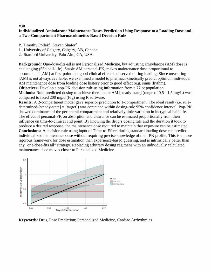
Methods: Rule-predicted dosing to achieve therapeutic AM [steady-state] (range of 0.5 - 1.5 mg/L) was
compared to fixed 200 mg/d (Fig) using R software.
Results: A 2-compartment model gave superior prediction to 1-compartment. The ideal result (i.e. rule-
determined-[steady-state] = [target]) was contained within dosing-rule 95% confidence interval. Pop-PK
showed dominance of the peripheral compartment and relatively little variation in its typical half-life.
The effect of personal-PK on absorption and clearance can be estimated proportionally from their
influence on time-to-clinical end point. By knowing the drug’s dosing rate and the duration it took to
produce a desired response, the maintenance dose required to maintain that exposure can be estimated.
Conclusions: A decision rule using input of Time-to-Effect during standard loading dose can predict
individualized maintenance dose without requiring precise knowledge of their PK profile. This is a more
rigorous framework for dose estimation than experience-based guessing, and is intrinsically better than
any "one-dose-fits all" strategy. Replacing arbitrary dosing regimens with an individually calculated
maintenance dose moves closer to Personalized Medicine.
Keywords: Drug Dose Prediction; Personalized Medicine, Cardiac Arrhythmias
#31
Ionization status of drugs has poor association with their transmembrane diffusion
Mohammad A Randhawa1, Salman A Malik2
1. Department of Pharmacology, HBS Medical and Dental College, Islamabad, Pakistan
2. Department of Biological Sciences, Quaid-i-Azam University, Islamabad, Pakistan (Retired)
Background: Ionization of drugs in solution can be calculated from Henderson-Hasselbalch Equation
(HHEq), knowing their pKa and pH of medium. HHEq reveals that drugs should have similar
transmembrane diffusion when pKa and pH of medium are same. But drugs with similar pKa/ionization
had poor correlation with their pH dependent buccal absorption (BA) and urine excretion (UE).
Similarly, quaternary amines (QA), ionized, had similar effects as tertiary amines (TA), not ionized, on
pupil size.
Objectives: To demonstrate that ionization of drugs does not affect their transmembrane diffusion.
Methods: (a) Ratio unionized/ionized (U/I) at pH4 calculated from HHEq was correlated with %BA at
pH4 (Beckett & Triggs, 1967) for 7 acidic drugs with similar pKa (3.5-5.6). (b) Ratio U/I at pH9 was
correlated with %BA at pH9 for 10 basic drugs with similar pKa (9-9.6). (c) Ratio acid/alkaline (Ac/Al)
for 24-hour urine excretion (UE), (Randhawa & Turner, 1988), of 10 basic drugs was correlated with
their pKa. (d) Onset and duration of action of 10 QA and TA was determined on pupil size of rabbit eye.
Results: Correlation (R2) between parameters mentioned above in methods (a-c) was 0.0527, 0.0583
and 0.0192, respectively. QA and TA had similar onset and duration of action on pupil size.
Conclusions: Poor correlation between BA and UE of drugs with their pKa values, ratio U/I, or Ac/Al;
and similar effects of QA and TA on pupil size indicate that ionization of drugs in not associated with
their transmembrane diffusion.
Keywords: Ionization, acidic/basic drugs, poor correlation, transmembrane diffusion.
#32
Long term benefits of a new life-style change for the management of GERD – A case report
Mohammad A Randhawa1, Mohammad Tariq Baqai2
1. Department of Pharmacology, HBS Medical and Dental College, Islamabad, Pakistan
2. Department of Medicine, HBS Medical and Dental College, Islamabad, Pakistan
Background: Gastro-Esophageal Reflux Disease (GERD) is a challenge for medical profession. Proton
pump inhibitors (PPIs) are prescribed but have many risks, especially with prolonged use. In earlier pilot
study, benefits of short-term practice of new life-style change, two meals a day with only soft-drinks in
between, were reported for management of GERD. Present case-report demonstrates benefits of its long-
term practice.
Objectives: To report long-term benefits of new life-style change in a patient with severe GERD.
Methods: A 61 year old patient complaining of night refluxes was endoscopically diagnosed to suffer
from severe GERD, with ulcerations at gastroesophageal junction, Barrett’s esophagus and hiatus
hernia. Besides, he had mild erosions in stomach. He practiced suggested dietary regimen, two meals a
day with only soft drinks in between (Water, fruit juice, tea and milk), whenever he felt hungry or
thirsty. His reflux symptoms improved within fortnight. During this period he took only antacid mixture
for 4-5 days and continued suggested dietary regimen.
Results: For 7 years he had no complaint, except that whenever he took solid food during day suffered
from mild reflux symptoms, which were relieved by antacid mixture for 1-2 days. Recently, his
endoscopy was done to see prognosis. His gastroesophageal junction was clear without any ulcers or
inflammation. However, he had mild sliding hiatus hernia and erosions in stomach.
Conclusions: Two meals a day with only soft drinks in between is very useful dietary regimen for
management of GERD.
Keywords: Case report, severe GERD, new life-style modification, long-term benefits.

#34
The effect of chronic kidney disease on CYP2B expression and activity in male Wistar rats
Adrien AE. RaoPeters1, Andrew S. Kucey1, Thomas J. Velenosi1, Nicholas C. Tonial1, Alvin Tieu1,
Brad L. Urquhart1
1. Department of Physiology and Pharmacology, Schulich School of Medicine and Dentistry
Background: Chronic Kidney Disease (CKD) is characterized by a progressive reduction in kidney
function over time. CKD affects greater than 10% of the population and its incidence is on the rise due
to the growing prevalence of its risk factors. Previous studies demonstrated CKD alters non-renal
clearance of drugs in addition to reducing renal clearance. While CKD has been shown to decrease
hepatic CYP3A and CYP2C mediated metabolism, it is unknown whether it alters CYP2B mediated
metabolism.
Objective: To assess the function and expression of hepatic CYP2B enzymes using a rat model of CKD.
Methods: CKD was induced in Wistar rats by supplementing their chow with adenine and confirmed by
measuring creatinine, urea and other uremic toxins in plasma. Bupropion was used as a probe substrate
for hepatic CYP2B function using rat liver microsomes. The resulting metabolite, hydroxy-bupropion,
and bupropion were quantified by ultra-performance liquid chromatography coupled to quadrupole time-
of-flight mass spectrometry. Levels of CYP2B mRNA and protein were determined by RT-PCR and
Western Blot, respectively.
Results: CYP2B1 mRNA level was significantly decreased (88%, p<0.001) in CKD relative to control.
Similarly, maximal enzymatic velocity (Vmax) for CYP2B was decreased by 46% in CKD relative to
control (p<0.0001).
Conclusions: Previous studies involving patients with CKD demonstrated altered bupropion
pharmacokinetics compared to control. Hence, our results suggest that these alterations may be mediated
by attenuated CYP2B hepatic metabolism. This finding may partially explain the alterations in
pharmacokinetics and non-renal drug clearance frequently observed in patients with CKD.
Keywords: chronic kidney disease, uremic toxins, non-renal clearance, hepatic CYP metabolism

#35
Human pluripotent stem cell-derived liver organoid model to assess metabolism and cardiac
function in mental health disease
Leili Rohani1,2, Derek Toms2,3, Shiying Liu1, Chad Bousman2,4,5, Mark Ungrin3,6, and Derrick E.
Rancourt1,2,7
1. Department of Biochemistry and Molecular Biology, Cumming School of Medicine, University of
Calgary
2. Alberta Children’s Hospital Research Institute, University of Calgary
3. Department of Comparative Biology and Experimental Medicine, Faculty of Veterinary Medicine,
University of Calgary
4. Hotchkiss Brain Institute, Cumming School of Medicine, University of Calgary
5. Departments of Medical Genetics, Psychiatry, and Physiology & Pharmacology, Cumming School of
Medicine, University of Calgary
6. Alberta Diabetes Institute, University of Alberta
7. Department of Oncology and Medical Genetics, Cumming School of Medicine
Background: Current treatments for mental health disease often show adverse drug reactions (ADRs),
most commonly within the context of heart inflammation. Organoids derived from human pluripotent
stem cells (hPSCs) are quickly becoming powerful in vitro models for pharmacological testing and may
prove effective at predicting ADRs.
Objectives: Here we present a scalable system for the production of liver organoids and the utility of
this system to elucidate the effects of liver-mediated metabolism of psychotropics on hPSC-derived
cardiomyocytes.
Methods: Liver organoids were generated from aggregated hPSCs through a step-wise differentiation
procedure. Expression of drug-metabolizing CYP enzymes were assessed at both a protein and
transcriptional level. After three weeks, liver organoids were treated with diazepam for one day.
Cardiomyocytes were then incubated with liver-conditioned media containing metabolites for an
additional day. Organoids, cardiomyocytes and media were analyzed for pro-inflammatory markers.
Results: We were successful in generating tens of thousands of liver organoids in parallel using our
novel differentiation protocol. By the end of differentiation, we detected robust expression of CYP3A4,
a key metabolizing enzyme of diazepam. Conditioned media from liver organoids treated with diazepam
prevented upregulation of the pro-inflammatory marker CCL2 in cardiomyocytes, compared to treating
cardiomyocytes with diazepam directly. This data suggests that our liver organoid system is able to
convert diazepam to the heart-safe metabolites temazepam and nordazepam.
Conclusions: As we continue to characterize our hPSC-derived liver organoids, this platform provides
an attractive starting point for precision screening of ADRs.
Keywords: Human pluripotent stem cell, liver organoid, cardiac organoid, adverse drug reaction
#36
Equilibrative nucleobase transporter 1 mediates the cellular efflux of 6-mercaptopurine
Nicholas M Ruel1, Khanh Hoa Nguyen1, James R Hammond1
1. Department of Pharmacology, University of Alberta, Edmonton, Alberta
Background: 6-mercaptopurine (6-MP) is a nucleobase analog drug used in inflammatory bowel
diseases and acute lymphoblastic leukemia. We have established that transfection of cells with
SLC43A3, which encodes for equilibrative nucleobase transporter 1 (ENBT1), increases both 6-MP
influx and its cytotoxicity. This system may also contribute to 6-MP resistance if it is capable of
mediating the transport of 6-MP out of cells.
Objectives: To determine if ENBT1 is capable of mediating the efflux of 6-MP, and how this compares
with established 6-MP efflux by multidrug resistance proteins MRP4 and MRP5, using a recombinant
expression model.
Methods: HEK293 cells stably transfected with SLC43A3 and un-transfected HEK293 cells were
loaded with 100 µM [14C]6-MP. Cells were then pelleted and the supernatant removed, and efflux
measurements initiated upon suspension of the cells in buffer containing different concentrations of
adenine with or without the MRP inhibitors, ceefourin-1 (MRP4) and zaprinast (MRP5). Aliquots of
cells were taken at times from 10 – 180 sec and assessed for intracellular [14C]6-MP.
Results: Un-transfected cells displayed minimal efflux with ~80% of initial 6-MP remaining after 3
min. Transfected cells displayed rapid efflux of 6-MP (rate constant >0.35 s-1) with maximum release
achieved within 10 sec. Adenine (100 µM or 1 mM) decreased the rate of efflux significantly (rate
constants: 100 µM; 0.15±0.02s-1, 1 mM; 0.02±0.002s-1). Ceefourin-1 also decreased the efflux of 6-MP
from these cells, while zaprinast did not.
Conclusions: ENBT1 mediates the efflux of 6-MP. MRP4 also contributes to 6-MP efflux in this model
but is less efficient in that regard than ENBT1.
Keywords: nucleobase, 6-mercaptopurine, efflux, MRP4, MRP5

#37
Transcriptome-wide association study of anthracycline-induced cardiotoxicity in pediatric cancer
patients
Erika N. Scott,1,2 Galen E.B. Wright,1,2 Britt I. Drögemöller,2,3 Jafar S. Hasbullah,1,2 Bruce C.
Carleton,1,2,3,4,5 and Colin J.D. Ross1,2,3
1. Department of Medical Genetics, Faculty of Medicine, University of British Columbia (UBC),
Vancouver, BC
2. BC Children’s Hospital Research Institute, Vancouver, BC
3. Faculty of Pharmaceutical Sciences, UBC, Vancouver, BC
4. Pharmaceutical Outcomes Programme, BC Children’s Hospital, Vancouver, BC
5. Division of Translational Therapeutics, Department of Pediatrics, Faculty of Medicine, UBC,
Vancouver, BC
Background: Anthracyclines are highly effective chemotherapeutic agents; however, their clinical
utility is limited by severe anthracycline-induced cardiotoxicity (ACT). Genome-wide association
studies (GWAS) have uncovered several genetic variants associated with ACT, but these findings
explain only a portion of its heritability.
Objectives: To provide an in-depth examination of the genetics underlying ACT.
Methods: We conducted a transcriptome-wide association study (TWAS) using our previous GWAS
summary statistics (n=280 patients) to identify gene expression-related associations with ACT. Next, we
conducted gene set enrichment and pathway analyses to investigate biological mechanisms underlying
ACT.
Results: Three genes expressed in heart/arterial tissues were significantly associated with ACT in the
TWAS: GDF5 (Z-score = -4.30, P = 1.70 x 10⁻⁵), FRS2 (Z-score = 4.07, P = 4.67 x 10⁻⁵), and HDDC2
(Z-score = 4.01, P = 6.08 x 10⁻⁵). Significantly enriched gene sets consisted of genes essential in: mice
(P = 2.0 x 10⁻⁷), culture (P = 0.036), and humans (P = 1.36 x 10⁻⁵), as well as genes differentially
expressed upon treatment with the cardioprotectant all-trans retinoic acid (ATRA; P = 1.53 x 10⁻⁴) and
genes up-regulated at lower doses of anthracyclines (P = 3.60 x 10⁻⁴). Pathway analyses revealed an
overrepresentation of genes involved in ribosomal, spliceosomal, and cardiomyopathy pathways.
Conclusions: In summary, we have identified three novel genetic associations with ACT; determined
that genes essential for survival, cardioprotection, and response to anthracyclines are important for
development of ACT; and uncovered pathways dysregulated in patients with ACT, providing further
insight into the mechanism of ACT.
Keywords: Anthracycline-induced cardiotoxicity; Pharmacogenomics; Bioinformatics; Gene
expression/regulation; Transcriptome-wide association study
#38
Development and assessment of novel HEK293 cell lines for the study of equilibrative nucleoside
transporter regulation
Nayiar Shahid1, Khanh Hoa Nguyen1, Chris Cromwell1, Basil P Hubbard1, James Hammond1
1. Department of Pharmacology, Faculty of Medicine & Dentistry, University of Alberta
Background: Equilibrative nucleoside transporters (ENT1, ENT2) mediate the transmembrane flux of
endogenous nucleosides along with nucleoside-analogue drugs used in anti-cancer and anti-viral
therapies. An understanding of how ENT function is regulated is critical to controlling/exploiting its
impact on therapies using nucleoside analogues.
Objectives: Previous work indicates that ENT1 is regulated by PKC and may involve interactions
between ENT1 and ENT2. To assess underlying mechanisms, we have developed novel HEK293
mutants (using CRISPR/cas9) lacking both ENT1 and ENT2 (HEK293-NTD), or just lacking ENT2
(HEK293-E2KO). We now report on the characteristics of these cell lines, and the effects of PKC
activator-PMA thereon.
Methods: ENT1 function was assessed by measuring the initial rates of [3H]2-chloroadenosine uptake
(1 – 150 µM) ± PMA (100nM, 15 min, 37°C). Protein expression was assessed by immunoblotting using
ENT-specific antibodies.
Results: HEK293-E2KO had a similar level of ENT1 as wild-type HEK293 cells (Km-38.7±6.9 µM,
Vmax-5.4±0.6 pmol/µl/s). Treatment of HEK293 cells with PMA resulted in a significant decrease in
ENT1 (Vmax 4.7±1.0 to 1.9±0.7 pmol/µl/s). ENT2 activity (Vmax) also decreased from 0.7±0.2 to
0.2±0.3 pmol/µl/s. PMA had no effect on the Km of 2-chloroadenoisne in HEK293 cells. In contrast,
treatment of the HEK293-E2KO cells with PMA had no significant effect on the Vmax of ENT1-
mediated 2-chloroadenosine transport (Vmax, 2.06±0.15 and 2.164±0.13 pmol/µl/s, ± PMA), but did
cause a significant 2-fold increase in the Km.
Conclusions: These data suggest that ENT2 contributes to the effect of PKC on ENT1. Further
exploitation of these new cell models may reveal further mechanisms of ENT regulation.
Keywords: nucleoside transporter, PKC, adenosine, CRISPR/Cas9

#39
Vancomycin Pharmacokinetics in Pediatric Patients with Febrile Neutropenia
Yuko Shimamoto1, Sholeh Ghayoori1, Shinya Ito1, 2
1. Division of Clinical Pharmacology and Toxicology, The Hospital for Sick Children
2. Department of pediatrics, University of Toronto
Background: Vancomycin is recommended in guidelines for febrile neutropenia following
Hematopoietic Stem Cell Transplantation (HSCT). Recent published studies suggest that empirical
vancomycin dosing 60 mg/kg/day does not reach serum trough concentration targets of 15-20 mg/L for
serious or complicated infection and higher dosing regimen is required in pediatric patients. However,
vancomycin pharmacokinetics in pediatric patients with febrile neutropenia has not been fully
characterized.
Objectives: The purpose of this study was to investigate the pharmacokinetics of vancomycin in
pediatric patients with febrile neutropenia following HSCT.
Methods: The inclusion criteria were pediatric patients (age 0-17) and treatment with vancomycin for
febrile neutropenia following HSCT from 2009 to 2014. Population pharmacokinetics analysis was
performed with NONMEM 7.4 to evaluate potential covariates including demographic information and
laboratory parameters.
Results: A total of 122 patients had 255 vancomycin concentrations were used in the population
pharmacokinetics analysis. A one compartment structure model best described data and objective
function value (OFV) decreased in the model includes body weight using allometry and maturation
factor. Regarding maturation factor, TM50 which describes the maturation half-time was 47.4 weeks and
the value approximates TM50 of GFR; 47.7 weeks. Estimated vancomycin clearance was 0.18 (0.07-
0.28) L/h/kg that demonstrated enhanced clearance compared with previous reports.
Conclusions: Vancomycin clearance in pediatric patients with febrile neutropenia was enhanced
probably due to augmented renal clearance. In addition, age-appropriated vancomycin dosage based on
develop and maturation is required for neonates and infants
Keywords: vancomycin, population pharmacokinetics, children, febrile neutropenia

#40
Investigation of TMAP as a novel biomarker of kidney function
Nicole A. Sidor1, Thomas J. Velenosi1, Gilles A. Lajoie2, Brad L. Urquhart1
1. Department of Physiology and Pharmacology, Schulich School of Medicine and Dentistry
2. Department of Biochemistry, University of Western Ontario, London, Ontario, Canada
Background: Although kidney function is critical for determining drug dosing and diagnosing chronic
kidney disease (CKD), there is a lack of sensitive biomarkers for detecting early changes in kidney
function. While the current gold standard is creatinine, serum levels of creatinine do not change until
approximately 50% of kidney function has been lost. We previously used metabolomics to discover new
biomarkers and identified N,N,N-trimethyl-L-alanyl-L-proline betaine (TMAP) as a novel biomarker of
kidney function.
Objectives: Develop a liquid chromatography coupled to mass spectrometry (LCMS) method for the
absolute quantitation of TMAP in CKD patient and control plasma samples, and evaluate changes in
plasma concentration as CKD progresses.
Methods: Plasma samples were spiked with varying concentrations of TMAP to generate a standard
curve. Liquid chromatography coupled to quadrupole – time of flight (QToF) mass spectrometry was
used to determine the concentration of TMAP in plasma of healthy controls and patients with CKD.
Results: The standard curve of TMAP was linear (R2 = 0.99971) within the range of 1.9 and 500 ng/mL.
Preliminary data indicate plasma TMAP is 4.9- fold higher in CKD patient plasma when compared to
controls with normal kidney function.
Conclusions: TMAP has previously been shown to be a more sensitive indicator of decreased renal
function than serum creatinine, the current standard for renal function evaluation. Quantitation of
absolute levels of TMAP in plasma shows that as CKD progresses, TMAP plasma concentration
increases.
Keywords: chronic kidney disease, metabolomics, biomarker

#41
Equilibrative nucleoside transporter-4 (ENT4) mediated efflux of adenine nucleosides: a potential
contributor to ischaemia-reperfusion injury
David Tandio1, Gonzalo Vilas1, James R Hammond1
1. Department of Pharmacology, University of Alberta
Background: Cardiac ischaemia-reperfusion injury remains a life-threatening condition with no
effective treatments. Though the mechanisms remain unknown, we know that ischaemic tissues are
acidic and that adenosine is protective. Equilibrative nucleoside transporter 4 (ENT4), expressed in
cardiomyocytes and the vasculature, transports adenosine specifically under acidic conditions.
Therefore, we hypothesise that ENT4 activity in ischaemic tissues regulates the protective actions of
adenosine and may mediate the loss of intracellular adenosine during reperfusion.
Objectives: To investigate whether ENT4 is capable of mediating nucleoside efflux, and whether ENT4
activity/expression changes under ischaemic conditions.
Methods: hENT4-transfected pig kidney epithelial nucleoside transporter deficient (PK15-ENT4) cells
were loaded with [3H]2-chloroadenosine for 12 min at pH 6.0, and efflux was initiated by exposing the
cells to a substrate-free buffer. HL-1 cardiomyocytes were exposed to ischaemia-mimetic conditions for
4 hours and reperfused with culture media for 2 hours before assessing [3H]nucleoside uptake. mRNA
levels were analysed using real-time quantitative PCR.
Results: ENT4 mediated 2-chloroadenosine efflux under acidic conditions in PK15-ENT4 cells (k =
0.050±0.009 min-1). HL-1 cardiomyocytes exhibited pH-dependent uptake of 10 μM 2-chloroadenosine
(k = 0.218±0.185 min-1) and 100 μM adenosine (k = 0.280±0.042 min-1), characteristic of ENT4.
Following 4 hr simulated ischaemia, this pH-dependent uptake was reduced, but recovered with 2 hr
reperfusion. ENT4 mRNA levels were also reduced with ischemia, and remained suppressed even after
reperfusion.
Conclusions: While ENT4 appears to be down-regulated under ischaemic conditions, our efflux data
suggests that ENT4 might contribute to the loss of intracellular adenosine upon reperfusion.
Keywords: Nucleoside transporters, ischaemia-reperfusion

#42
Thresholds of evidence required for the prioritization of robust and actionable pharmacogenomic
biomarkers.
Reo Tanoshima1,2, Galen E.B. Wright2,3, Britt I. Drögemöller2,4, Philip Khayat1,2, Stephanie Dean5,
Colin J.D. Ross3,4, Bruce C. Carleton1,2,3,4.
1. Division of Translational Therapeutics, Department of Pediatrics, Faculty of Medicine, The
University of British Columbia, Vancouver, British Columbia, Canada.
2. BC Children's Hospital Research Institute, Vancouver, British Columbia, Canada.
3. Department of Medical Genetics, University of British Columbia, Vancouver, British Columbia,
Canada.
4. Faculty of Pharmaceutical Sciences, The University of British Columbia, Vancouver, British
Columbia, Canada.
5. Department of Pharmacy, Children’s Hospital of Eastern Ontario, Ottawa, Ontario, Canada.
Background: Pharmacogenomic studies have identified genetic variants related to adverse drug
reactions (ADRs). However, many pharmacogenomic biomarkers have weak associations or conflicting
results between the drug outcome and genetic variant, or are at the margin of clinical relevance. The
identification of actionable biomarkers in the prediction of ADRs is therefore important. Some
investigators have suggested that randomized controlled trials are necessary to determine clinical
relevance. However, randomized controlled trials may not be feasible due to the low incidence of the
ADRs or ethical concerns with placing patients at unnecessary risk of ADRs.
Objectives: The objective of this presentation is to propose thresholds of evidence for the prioritization
of clinically actionable pharmacogenomic biomarkers for the prediction of serious ADRs.
Methods: A group of experts in medical genetics, regulatory science, clinical pharmacology and clinical
medicine was formed. The relevant literature, regulatory actions, and clinical practice guidelines were
assessed to determine the most relevant thresholds for the identification of clinically actionable variants.
Results: Levels of evidence were selected according to published evidence, and we concluded a
pharmacogenomic biomarker should meet one of the three following criteria to be considered clinically
relevant:
1. Drug label recommendations by regulatory agencies.
2. Clinical practice guidelines by expert consortia published in peer-reviewed journals.
3. Evidence of a drug outcome-gene association that is replicated in at least three independent patient
populations with an odds ratio of at least three.
Conclusions: Establishing a high threshold for clinical action is essential when bringing a new
technology into clinical care.
Keywords: pharmacogenomics, clinical implementation, biomarker, drug outcome

#43
Proteinase Activated Receptor 4 (PAR4) agonist peptide structure and conformational dynamics
elucidated by 2D NMR and molecular dynamics simulations
Pierre Thibeault1, Peter Stathopulos1, Rithwik Ramachandran1
1. Department of Physiology and Pharmacology, University of Western Ontario
Background: Proteinase-activated receptor 4 (PAR4) is a member of the proteolytically-activated PAR
family of GPCRs and is activated through limited proteolysis by enzymes such as thrombin to unmask a
receptor activating tethered-ligand sequence. PAR4 can also be activated by tethered-ligand mimicking
synthetic peptides such as AYPGKF-NH2. Since PAR4 is a key regulator of platelet activation, there is
much interest in discovering novel compounds targeting PAR4 for development as anti-thrombotic
agents. In a previous study we determined structure activity relationships (SAR) of PAR4 with a library
of novel peptide agonists to determine peptide properties that enable PAR4 activation.
Objectives: To understand peptide properties enabling PAR4 activation, we investigate structural and
conformational dynamics of the PAR4 agonist peptide AYPGKF-NH2.
Methods: 2D nuclear magnetic resonance spectroscopy was utilized to determine the structure of
AYPGKF-NH2. The structures were then used as input for molecular dynamics simulations (MD) using
gromacs with AMBER99sb-ILDN forcefield for simulation and internal routines for analysis.
Results: 2D NMR reveals two distinct conformations that we have termed major and minor. Both
conformations were used as inputs for 100ns MD simulation. Data analyses comparing major and minor
conformations reveal significant differences in hydrogen bonding, conformational dynamics, and
diffusion coefficients.
Conclusions: These data represent what we believe are two distinct conformations of AYPGKF-NH2 in
solution that may possess varying degrees of efficacy. Current studies are aimed at characterizing
peptides from our SAR study that may adopt similar conformations to major or minor AYPGKF-NH2 to
elucidate how the peptide structure contributes to PAR4 activation.
Keywords: Proteinase Activated Receptor 4 (PAR4), 2D NMR, MD Simulation

#44
Efficient, multi-dimensional optimization of organoid production from pluripotent stem cells
enables combinatorial drug screening
Derek Toms1,2, Leili Rohani2,3, Shiying Liu3, Derrick Rancourt3,4, and Mark Ungrin1,2,5
1. Department of Comparative Biology and Experimental Medicine, Faculty of Veterinary Medicine,
University of Calgary
2. Alberta Children’s Hospital Research Institute, University of Calgary
3. Department of Biochemistry and Molecular Biology, Cumming School of Medicine, University of
Calgary
4. Department of Oncology and Medical Genetics, Cumming School of Medicine
5. Alberta Diabetes Institute, University of Alberta
Background: Effective and economically viable organoid-based drug screening will depend on the
establishment of optimized protocols for the production of the necessary cell types. Our ability to do this
will depend in turn on the capacity to efficiently screen multiple variables in a timely and cost-effective
manner.
Objectives: Here, we present a generalizable, integrated strategy employing statistical design of
experiments to efficiently produce optimized hepatic organoids from pluripotent stem cells and in turn
use them to screen for drug metabolic activity.
Methods: We have developed a liquid-handing and mathematical modeling approach to enable us to
screen an arbitrary number of soluble factors using liquid-handling robotics to permit complex
experimental designs and minimize errors. Importantly, we are able to identify factor-factor interactions
that can be missed in typical single-factor high-throughput screening approach. We used this pipeline to
optimize the differentiation of hepatic organoids for drug metabolizing activity.
Results: Initial validation of this platform resulted in the establishment of a novel 3D hepatocyte
differentiation protocol. We further demonstrated that organoids differentiated in this manner express
the drug-metabolizing cytochrome P450 enzymes CYP3A4 and CYP2D6 and are able to metabolize the
anti-anxiety medication diazepam. Further rounds of optimization identified factor combinations that
were able to increase expression of key hepatic functional markers. With these optimized organoids we
plan to perform a combinatorial screen to identify significant drug-drug interactions.
Conclusions: This broadly applicable approach to multifactorial, high-throughput optimization and
screening provides a foundation to develop functional organoid models and improve precision
pharmacology.
Keywords: Organoids; High-throughput screening; Drug metabolism

#45
Effect of chronic kidney disease on drug disposition in rodent models
Nicholas C. Tonial1, James Lim1, Brad L. Urquhart1
1. Department of Physiology and Pharmacology, University of Western Ontario
Background: Over 40% of chronic kidney disease (CKD) patients experience adverse drug reactions.
Cytochrome P450 (CYP) metabolism and elimination by transporters are major components influencing
drug disposition. Although rodent models of CKD exhibit reduced CYP activity, the impact of CKD on
transporters, namely organic anion transporting polypeptides (OATPs), is not well elucidated. In
addition, cannabinoid metabolism by CYP enzymes has not been investigated in the setting of CKD.
Objectives: 1) To study the effects of CKD on drug transporter activity, in a humanized OATP1B1/1B3
mouse model. 2) To investigate cannabinoid metabolism in a rat model of CKD. It was hypothesized
that CKD would decrease the activity of OATP transporters and reduce cannabinoid metabolism.
Methods: 1) FVB/N mice humanized for OATP1B1/1B3 were fed chow supplemented with 0.2%
adenine (n=6) to induce CKD. Plasma creatinine levels and the liver to plasma ratio of the OATP probe
drug, fexofenadine (L:PFEX), were measured using ultra performance liquid chromatography coupled to
mass spectrometry (UPLC-MS). 2) Hepatic microsomal fractions, isolated from CKD-induced rats, will
be incubated with cannabinoids and metabolite formation will be measured by UPLC-MS.
Results: Plasma creatinine was doubled in adenine-fed mice compared to controls (p=0.0011), and there
was a modest 15% decrease in L:PFEX (p=0.1870).
Conclusions: Induction of CKD did not significantly alter the liver distribution of fexofenadine. Further
analysis of OATP expression levels, as well as cannabinoid metabolism, in rodent models will help to
elucidate the impact of CKD on drug disposition.
Keywords: Chronic Kidney Disease, Drug Disposition, Drug Transporters, Cytochrome P450s
#46
Urinary diacetylspermine as a metabolic biomarker of doxorubicin effectiveness in triple negative
breast cancer patient-derived xenografts
Thomas J. Velenosi1, Kristopher W. Krausz1, Frank J. Gonzalez1
1. Laboratory of Metabolism, Center for Cancer Research, National Cancer Institute, National Institutes
of Health, Bethesda, MD
Background: Triple negative breast cancer (TNBC) patients have the worst clinical outcomes and
highest rate of recurrence compared to other breast cancer molecular subtypes. Treatment of TNBC
requires non-specific chemotherapy and therefore, early detection of drug effectiveness would be highly
beneficial to improve outcomes.
Objectives: The objective of this study was to identify metabolic biomarkers of doxorubicin
effectiveness using TNBC patient-derived xenografts (TNBC-PDXs).
Methods: NSG mice were engrafted with a doxorubicin sensitive or a doxorubicin resistant TNBC-PDX
and administered intravenous doxorubicin (2 mg/kg) or vehicle control once weekly for three weeks.
Longitudinal urine samples were collected after each dose followed by a final 24-hour urine collection
in the absence of drug (n=8-9).
Results: Untargeted metabolomics revealed diacetylspermine as the most significantly altered
metabolite in urine samples by two-way repeated measures ANOVA (p=1.26x10⁻¹¹, q=4.82 x 10⁻⁸).
Diacetylspermine correlated with tumor growth in the doxorubicin sensitive model. Interestingly, when
these mice were treated with doxorubicin, there was a further increase in diacetylspermine despite the
decrease in tumor size, which persisted for one week following the final dose. Spermine/spermidine
acetyl-transferase 1 (SAT1) expression, which is responsible for spermidine and spermine acetylation,
was significantly elevated in the doxorubicin sensitive tumor and further increased with doxorubicin
treatment. Diacetylspermine, N-acetylspermine, and N1-acetylspermidine were also increased in tumor
samples when doxorubicin sensitive mice were treated with doxorubicin compared to vehicle.
Conclusions: These data suggest that doxorubicin increases diacetylspermine in doxorubicin sensitive
tumors and may be a useful non-invasive urinary biomarker for evaluating doxorubicin effectiveness
during treatment.
Keywords: metabolomics, polyamines, triple negative breast cancer, drug resistance

#47
A pharmacogenomic investigation of the cardiac safety profiles of 5-HT3 antagonists in children
and pregnant women
Galen E.B. Wright1,2,*; Britt I. Drögemöller2,3,*; Jessica N. Trueman1; Fudan Miao2; Colin J.D.
Ross2,3,**; Bruce C. Carleton1,2,**
1. Division of Translational Therapeutics, Department of Pediatrics, University of British Columbia,
Vancouver, British Columbia, Canada.
2. BC Children's Hospital Research Institute, Vancouver, British Columbia, Canada.
3. Faculty of Pharmaceutical Sciences, University of British Columbia, Vancouver, British Columbia,
Canada *Contributed equally; **Jointly supervised
Background: 5-HT3 receptor antagonists such as ondansetron are highly effective medications for the
treatment of nausea and vomiting. However, these medications are also associated with severe cardiac
adverse drug reactions. Pharmacogenomic information for response to ondansetron exists, particularly
pertaining to CYP2D6, but no study has been performed on genetic factors that influence the cardiac
safety of this medication.
Objectives: Determine ondansetron-related cardiac safety profiles in three diverse cohorts and assess
whether pharmacogenomic information can be used to predict at risk patients.
Methods: Three patient groups who received ondansetron for the prevention of nausea and vomiting
were recruited via active surveillance (pediatric surgery n=100; pediatric oncology n=96; pregnant
women n=61). Electrocardiograms were conducted at baseline and post-ondansetron administration.
Pharmacogenomic associations were then assessed via analyses of comprehensive CYP2D6 genotyping
data and imputed genome wide information.
Results: In the entire cohort, 62 patients (24.1%) were defined as cases based on Bazett-corrected QTc
values. The most significant shift from baseline occurred at five minutes post-administration (P=9.8 x10-
4). Although CYP2D6 activity score was not associated with case-control status, genome-wide analyses
identified novel candidate genes for this drug-induced phenotype. The candidate genes that were
identified through these analyses (rs5896569 MPDZ and rs73111553 PTPRG) have been previously
implicated in serotonin-related traits and therefore represent biologically relevant findings.
Conclusions: The results of this study provide the first step towards pharmacogenomically-informed
therapeutic decision-making regarding the use of 5-HT3 antagonists in children and obstetric
populations, with the overall goal to improve the safety of these commonly used antiemetic medications.
Keywords: CYP2D6; cardiac safety; GWAS; ondansetron; pharmacogenomics

#48
Modulation of SSAT1 Expression for Cancer Cell Sensitization and Therapy
Vinith Yathindranath,1 Donald W. Miller1
1. Department of Pharmacology and Therapeutics, University of Manitoba
Background: The regulation of polyamine levels in cells is highly regulated and dependent upon both
synthesis and catabolism processes within the cell. The enzyme Spermine/spermidine acetyl transferase
1 (SSAT1) is critical for the metabolism of polyamines and is overexpressed in many cancers including
glioblastoma. Preliminary studies suggest SSAT1 overexpression in glioblastoma (GB) may contribute
to radiation resistance. Whether similar advantages are present for chemotherapeutic responses is
unclear. The present study examined the effects of, lipid nanoparticles (LNPs) to selectively knockdown
(KD) SSAT1 expression in GB cells.
Objectives: To identify the survival advantages of SSAT1 in GB cells and develop a cellular "tuned"
nanoparticle-based delivery system tailored to modulate SSAT1 expression.
Methods: SSAT1 specific siRNA was loaded in 1,2-dioleoyl-3-trimethylammonium-propane (DOTAP)
LNPs prepared using the microfluidics method. The SSAT1 knockdown efficiency of LNPs was
quantified in the U251 GB cell line using qPCR and WB. The effects of SSAT1 knockdown on both
DNA double strand breaks and cytotoxic response to doxorubicin (DOX) and carmustine (BCNU) was
evaluated using MTT assay.
Results: A SSAT1 knockdown in U251 cells of approximately 70% was achieved at the messenger
level and 50% at the protein level using LNPs. An enhancement of toxicity of around 15% towards Dox
and BCNU was observed in U251 cells following SSAT1 knockdown.
Conclusions: Using LNPs to deliver siRNA is a promising strategy for selectively knocking down
SSAT1 and sensitize GB cells towards chemotherapy.
Keywords: (SSAT1, Lipid nanoparticles, glioblastoma)

#49
Crataegus songarica Causes Endothelium-dependent Relaxation via a Redox-sensitive Src- and
Akt-dependent Activation of Endothelial NO Synthase mediated by Estrogen Receptors
Waqas Younis1,2,3, Alamgeer1, Valerie B Schini-Kerth2, Richard E. Roberts3
1. Laboratory of Cardiovascular Research and Integrative Pharmacology, College of Pharmacy,
University of Sargodha, Sargodha 40100, Pakistan
2. UMR CNRS 7213, Laboratory of Biophotonics and Pharmacology, Faculty of Pharmacy, University
of Strasbourg, Illkirch, France.
3. Pharmacology Research Group, School of Life Sciences, University of Nottingham, Medical School,
Queen's Medical Centre, Nottingham NG7 2UH, United Kingdom
Background: Fruits of Crataegus songarica are commonly used in folk medicine for the treatment of
hypertension, vascular insufficiency and heart problems.
Objectives: The aim of current study was to determine the mechanisms underlying the vasorelaxant
properties of C. songarica.
Methods: Segments of porcine distal coronary artery were set up in a wire myograph for isometric force
measurements. Extract/fractions of C. songarica were tested for vasodilator activity by measurement of
changes in tone after pre-contraction with the thromboxane mimetic U46619 in the presence or absence
of inhibitors of intracellular signaling cascades. The formation of reactive oxygen species was accessed
by dihydroethidine staining and the level of eNOS and AKT phosphorylation was measured by
immunohistochemical staining in sections of coronary arteries.
Results: Vasorelaxant effect of C. songarica was reduced by pretreatment with L-NAME (a competitive
inhibitor of NO synthase), ODQ (soluble guanylyl cyclase inhibitor), KCl And tetraethylammonium
(non-selective K+ channel inhibitor). Relaxations were also attenuated by LY249002 (PI3-kinase
inhibitor), PP2 (Src tyrosine kinase inhibitor) and intracellular ROS scavengers, PEG-catalase (hydrogen
peroxide inhibitor), diethyldithiocarbamate (superoxide dismutase inhibitor) and MnTMPyP. Inhibitors
of muscarinic, bradykinin B2 and tachykinin NK1 receptors had no effect on the relaxation response but
both estrogen receptor antagonists (tamoxifen and ICI 182,782) reduced the AS-CS mediated
vasorelaxation. C. songarica stimulated the endothelial formation of ROS and phosphorylation of Akt
and eNOS in coronary artery sections.
Conclusions: Current data validated that C. songarica produces a noteworthy, endothelium-dependent
vasorelaxation of coronary artery rings owing to activation of estrogen receptors leading to initiation of
ROS/Src/PI3K/NO/cGMP pathway. Stimulation of this pathway possibly lead to inhibition of calcium
influx, either directly or through activation of K+ channels.
Keywords: Crataegus, endothelium-dependent relaxation, PI3-kinase/Akt, coronary artery
#50
Establishing the HepaRG cell line as a model for studying the influence of selenium on arsenic
hepatobiliary transport
Janet R. Zhou1, Gurnit Kaur1, Brooke Comrie2, and Elaine M. Leslie1,2
1. Division of Analytical and Environmental Toxicology, Department of Laboratory Medicine and
Pathology, University of Alberta, Canada
2. Department of Physiology, University of Alberta, Canada
Background: Worldwide, >200 million people are exposed to the proven human carcinogen arsenic at
levels exceeding the World Health Organization guideline (10 µg/L). In animal models selenium and
arsenic are mutually protective via the formation and biliary excretion of the seleno-bis (S-glutathionyl)
arsinium ion [SeAs(GS)2]-. Despite ongoing human selenium-supplementation trials in arsenic-endemic
regions, the influence of selenium on human hepatic handling of arsenic is not adequately understood.
We hypothesized that selenium would increase the biliary excretion of arsenite (AsIII) from human
HepaRG cells, an immortalized cell line used as a surrogate for primary human hepatocytes (PHH).
Objectives: To study the influence of selenite (SeIV) and selenide (SeII) on arsenic efflux from HepaRG
cells.
Methods: The formation of canalicular networks and function of multidrug resistance protein 2 (MRP2)
(the canalicular transporter of [SeAs(GS)2]-) were evaluated using fluorescence imaging of cells after
treatment with 5(6)-carboxy-2’,7’-dichlorofluorescein (CDF) diacetate. 73AsIII (1 µM) efflux was
measured across sinusoidal and canalicular membranes using B-CLEAR® technology in the presence or
absence of SeIV or SeII- (1 µM). Biliary excretion indices (BEIs) were calculated to quantify biliary
excretion.
Results: CDF accumulated in canalicular networks, suggesting the presence of functional MRP2.
Biliary excretion of 73AsIII at 10 min was 4%. In the presence of SeIV and SeII-, BEIs were increased to
22% and 26%, respectively. Therefore, selenium increased arsenic biliary excretion.
Conclusions: This work will lead to a better understanding of the influence of selenium on arsenic
handling by human liver and provide valuable information for ongoing selenium-supplementation trials.
Keywords: Arsenic, selenium, hepatobiliary transport, HepaRG, multidrug resistance protein 2
Recommended