Anatomy 1
Histology 4
ANATOMY
Knowledge of nervous system anatomy is essential for success in surgical neuro-
pathology. Familiarity with native cellular elements will often predict the appear-
ances of diverse tumors within the brain but, perhaps more importantly,
recognition of normal or reactive processes will help avoid the pitfall of diagnos-
ing malignancy when, in fact, a nonneoplastic reactive process or even normal
tissue is present. Many surgical neuropathology specimens originate in the brain
and spinal cord coverings, cranial and spinal nerve roots, blood vessels, and bone
and soft tissue surrounding the nervous system; thus recognition of the normal
brain is not enough. As with general surgical pathology, knowledge of diseases
common to these locations and the ages at which they typically occur is essential.
The human brain can be described in many ways. However, for the purpose
of surgical neuropathology, this description will emphasize different surgical
compartments and cytoarchitectural areas that are especially associated with
tumors or other pathological processes (Figure 1.1). The central nervous system
(CNS) is often divided into the supratentorial and infratentorial compartments by
the dural tentorium, which separates the cerebral hemispheres from the brainstem
and cerebellum. The spinal cord, roots, and distalmost cauda equina and filum
terminale (Figure 1.2) are often considered separately, especially in view of the
paraspinal soft tissue pathology, which can affect the integrity of the spinal cord.
Brain tissue is divided into gray and white matter. This distinction may be
useful in the differential diagnosis of neoplasms, but is more important in other
pathological processes such as infection or neurodegeneration. The surgical
neuropathologist is often interested in the relation of a tumor to the ventricles,
including the ventricular spaces themselves and their periventricular regions.
The base of the skull, including the pituitary gland-bearing sella turcica, is
another particular region defined by the propensities of certain tumors for this
region, both sellar and suprasellar.
The gross anatomy of the nervous system also reflects a level of complex and
interrelated structure and function enviable to say the least of visceral organs.
1
1 NORMAL ANATOMY AND HISTOLOGY
OF THE CNS
© Cambridge University Press www.cambridge.org
Cambridge University Press978-0-521-88161-6 - Nervous SystemHannes VogelExcerptMore information

The nervous system possesses a wide array of sometimes overlapping but clin-
ically distinct functions, thus forming the basis of a vast array of clinical symp-
toms attributable to the regions affected, not to mention their pace of onset.
Thereby, seizures imply injury to gray matter; lesions involving given white
matter tracts cause neurological deficits reflecting the normal function of the
tract, including weakness, altered sensation, visual deficits, cranial neuropathies,
or other motor and sensory deficits.
Mass lesions first compromise local blood supply, then intrude into ventric-
ular spaces, sometimes causing obstruction of flow of cerebrospinal fluid at
various narrow passages such as the foramina of Munro, cerebral aqueduct, or
fourth ventricle, and finally compression of brain tissue via paths of least resist-
ance. Expanding lesions are ultimately confronted by the rigid resistance of the
dura and skull. These cerebral herniations cause clinical symptoms that herald the
serious effects of mass lesions from the supratentorial compartment as they press
the medial temporal lobes against the edge of the tentorium. A greater or diffuse
swelling of the supratentorial brain will cause the brainstem to herniate down-
ward, often resulting in hemorrhages within the pons, termed Duret hemorrhages
Figure 1.1. Midsagittal
magnetic resonance imagingof the brain with regions often
associated with particularneuropathological processes.
Blue = supratentorialhemispheres with lateral and
third ventricles.Red = cerebellum.Brainstem = midbrain
(light green) pons (tan) andmedulla (pink). Yellow = sella
turcica and optic pathways.Dark green = spinal cord.
Brown = pineal gland region.
NERVOUS SYSTEM
2
© Cambridge University Press www.cambridge.org
Cambridge University Press978-0-521-88161-6 - Nervous SystemHannes VogelExcerptMore information

representing the rupture of penetrating blood vessels that are otherwise tethered
to the basilar artery. The most serious and life-threatening herniation involves
downward pressure of the contents of the posterior fossa leading to compression
of the medulla and vital respiratory control centers by the cerebellar tonsils.
Vascular pathology is inextricably related to virtually the entire spectrum of
neuropathology. While the brain may only account for 2 percent of body weight,
it accounts for 20 percent of oxygen consumption and thus normally receives 20
percent of cardiac output. The circle of Willis is an interconnected structure
supplying arterial blood to the brain through the merging of paired sources in
the carotid arteries and vertebral arteries at the base of the brain. The branching of
the brain’s arterial blood supply has been likened to that of an oak tree with right
angles whereas that of the venous drainage is like the tapering confluence of elm
branches. This is of more than botanical significance, since metastatic tumors,
infections, or other arterial microemboli have a propensity to become lodged at a
point of critical narrowing and angulation occurring at the grey–white junction
such that this is often the location of the smallest of such lesions.
Figure 1.2. The spinal cord,roots, and distalmost cauda
equina and filum terminale(arrow) are often considered
separately, especially in viewof the paraspinal soft tissue
pathology, which can affectthe integrity of the spinal
cord.
NORMAL ANATOMY AND HISTOLOGY OF THE CNS
3
© Cambridge University Press www.cambridge.org
Cambridge University Press978-0-521-88161-6 - Nervous SystemHannes VogelExcerptMore information

The brain is covered by successive layers. The most intimately associated is
the pia mater, which tightly adheres to the entire surface of the brain and invests
large penetrating blood vessels. Investing the pia mater is the arachnoid mem-
brane, which is so intimately associated as to be combined through the term pia-
arachnoid. The arachnoid membrane divests from the pia in the lower spinal
canal, resulting in an expanded subarachnoid space that is amenable to lumbar
puncture below lumbar vertebral body L2, which corresponds to the lower
extent of the cauda equina.
HISTOLOGY
Surgical neuropathology requires the knowledge of normal histology in the imma-
ture, adult, and aged brain and how the cellular constituents vary accordingly.
Neurons are the functional unit of the nervous system and display a number
of different and distinctive morphologies, which are of both functional and
anatomical significance. Neurons of all types generally contain a round nucleus
with a prominent nucleolus and a cell cytoplasm or perikaryon with Nissl
substance. Motor neurons tend to be large trapezoidal or triangular cells (Figure
1.3) while sensory neurons have a more globular shape (Figure 1.4). A third type
of neurons that are abundant in the cerebellum and dentate gyrus of the hippo-
campus are granular cell neurons (Figure 1.5). These are significantly smaller
than most cortical neurons and do not show obvious cell processes in routine
sections. The importance in recognizing normal neuronal morphology for the
surgical neuropathologist lies in distinguishing normal ganglionic or neuronal
cells from dysplastic or neoplastic ganglion cells, to recognize their appearance
in gray matter structures or spinal or cranial nerve ganglia that are infiltrated by
neoplasms, and to carefully distinguish normal granular cell neurons from
‘‘small blue cell’’ neoplasms or lymphocytes.
Neuronsmay be identified immunohistochemically with either one or a com-
bination of three different antibodies. Synaptophysin is a useful marker of the
neuronal cell surface or cytoplasm (Figure 1.6a), although the immunostaining
results in infiltrative tumors or other processes can be difficult to interpret
because of background normal staining for synaptophysin. Neurofilament is
the characteristic intermediate filament of neurons. Antibodies to the nonphos-
phorylated neurofilament of the neuronal cell body (perikaryon) and to the
phosphorylated neurofilament of neuronal processes can be used to distinguish
these compartments accordingly. However, mixed neurofilament antibodies are
commonly used in current surgical neuropathology and label both the cell body
and its neurites (Figure 1.6b). A third marker of neuronal differentiation is anti-
neuN, which offers the advantage of nuclear staining (Figure 1.6c). Not all
neurons stain positively for neuN, including Purkinje cells, most neurons of
the internal nuclear layer of the retina, and the sympathetic chain ganglia.
Neurons are the most quintessential cellular component of the CNS. How-
ever, astrocytes are the most plentiful cell type and exhibit the broadest range
NERVOUS SYSTEM
4
© Cambridge University Press www.cambridge.org
Cambridge University Press978-0-521-88161-6 - Nervous SystemHannes VogelExcerptMore information

of normal and reactive morphologies. Two predominant types of astrocytes
may be found in the brain: fibrous astrocytes in white matter and subpial and
perivascular gray matter, which are detectable by glial fibrillary acidic protein
(GFAP) immunohistochemistry, and protoplasmic astrocytes in gray matter,
which contain little detectable GFAP. Reactive astrocytes often show eosino-
philic cytoplasm and profusion of delicate cell processes detectable
Figure 1.4. Sensory neuronshave a more globular shape.
Figure 1.3. Large motorneurons tend to be large
trapezoidal or triangular cells.Note cytoplasmic Nissl
substance and eccentricaggregation of golden brown
lipofuscin pigment. Corporaamylacea (arrows) are within
slender glial cell processes notdiscernable in H&E sections.
NORMAL ANATOMY AND HISTOLOGY OF THE CNS
5
© Cambridge University Press www.cambridge.org
Cambridge University Press978-0-521-88161-6 - Nervous SystemHannes VogelExcerptMore information

by hematoxylin – eosin defined (H&E) stain or especially by GFAP immuno-
histochemistry (Figure 1.7). This may be useful in distinguishing white matter
astrocytes from oligodendrocytes. A particular type of chronic gliosis is rec-
ognizable as dense subpial gliosis often seen in chronic epilepsy resection
specimens, known as Chaslin’s gliosis (Figure 1.8). Distinguishing reactive
Figure 1.6.Immunohistochemistry for
neuronal differentiation:(a) synaptophysin; (b)
neurofilament; (c) neuN.
Figure 1.5. Granular cellneurons of the cerebellum are
seen in the upper field.Infiltrating neoplastic cells in
this medulloblastoma are seento be considerably larger,
although cytologicalpreparations and cryosections
of cerebellar tissue in whichgranule cells predominate
may be mistaken for a ‘‘smallblue cell’’ tumor.
NERVOUS SYSTEM
6
© Cambridge University Press www.cambridge.org
Cambridge University Press978-0-521-88161-6 - Nervous SystemHannes VogelExcerptMore information

from neoplastic astrocytes may represent a significant challenge, and this
challenge is discussed under ‘‘Astrocytic Tumors.’’
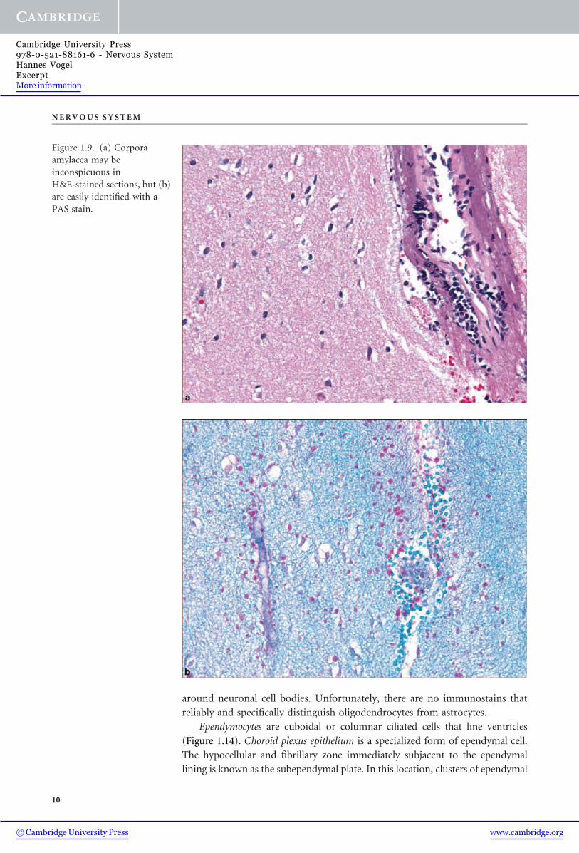
Corpora amylacea are age-related inclusions in astrocytes, which should not
be confused with other pathological inclusions or microorganisms such as
fungi. These are easily identified with a periodic acid – Schiff (PAS) stain and
Figure 1.6. continued.
NORMAL ANATOMY AND HISTOLOGY OF THE CNS
7
© Cambridge University Press www.cambridge.org
Cambridge University Press978-0-521-88161-6 - Nervous SystemHannes VogelExcerptMore information

usually signify chronic gliosis (Figure 1.9). Another feature of reactive, and
especially indolent and chronic gliosis are Rosenthal fibers. These are brightly
eosinophilic elongated or beaded structures, which are also intracellular inclu-
sions in astrocytes although they appear as solitary structures within the brain
background (Figure 1.10).
Figure 1.7.Immunohistochemistry for
glial differentiation:(a) reactive fibrillary
astrocytes; (b) GFAPimmunohistochemistry.
NERVOUS SYSTEM
8
© Cambridge University Press www.cambridge.org
Cambridge University Press978-0-521-88161-6 - Nervous SystemHannes VogelExcerptMore information

Reactive astrocytes may undergo mitotic activity and one feature is the
multinucleated Creutzfeldt astrocyte with its characteristic ‘‘micronuclei.’’
These may be a conspicuous feature of demyelinating disease (Chapter 6,
Figure 6.1). The gemistocytic astrocyte is one with abundant eosinophilic
cytoplasm causing nuclear displacement. This may be a striking feature of
reactive processes, such as that seen in the wall of a cerebral infarction or
abscess, and is sometimes difficult to distinguish from a neoplastic gemisto-
cytic astrocyte.
Perhaps one of the most striking examples of abnormal cellular morphol-
ogies among reactive astrocytes may be seen by the surgical neuropathologist in
examples of progressive multifocal leukoencephalopathy. These are markedly
enlarged cells with bizarre nuclei (Figure 1.11).
Another more subtle form of reactive astrocytosis is in the form of metabolic
astrocytes, the most typical of which are designated the Alzheimer Type 2 astro-
cytes (Figure 1.12). This is not usually a primary diagnostic issue in surgical
neuropathology; however, they should not be confused with other types of infil-
trating, particularly neoplastic cells such as there in oligodendroglioma in gray
matter.
Oligodendrocytes are especially plentiful in CNS white matter being the
myelinating cell of the CNS, which includes the optic nerves. Their inconspic-
uous round nuclei are scattered evenly or in vague rows throughout white
matter, a tendency that can become more pronounced in the atrophic process
that affects the aged brain (Figure 1.13). This may create an unsettling degree of
hypercellularity that may even be misdiagnosed as a diffuse glioma. Oligoden-
drocytes may also be noted in gray matter as innocuous cells loosely arranged
Figure 1.8. Dense subpial orinterface (Chaslin’s) gliosis.
NORMAL ANATOMY AND HISTOLOGY OF THE CNS
9
© Cambridge University Press www.cambridge.org
Cambridge University Press978-0-521-88161-6 - Nervous SystemHannes VogelExcerptMore information
around neuronal cell bodies. Unfortunately, there are no immunostains that
reliably and specifically distinguish oligodendrocytes from astrocytes.
Ependymocytes are cuboidal or columnar ciliated cells that line ventricles
(Figure 1.14). Choroid plexus epithelium is a specialized form of ependymal cell.
The hypocellular and fibrillary zone immediately subjacent to the ependymal
lining is known as the subependymal plate. In this location, clusters of ependymal
Figure 1.9. (a) Corporaamylacea may be
inconspicuous inH&E-stained sections, but (b)
are easily identified with aPAS stain.
NERVOUS SYSTEM
10
© Cambridge University Press www.cambridge.org
Cambridge University Press978-0-521-88161-6 - Nervous SystemHannes VogelExcerptMore information
Recommended