1
CNS Basic AnatomyCNS Basic Anatomy

2
SLP and NeuroscienceSLP and Neuroscience• Speech-Language Pathology
– Study of developmental and acquired disorders of human cognition, language and speech
– Complete neurolinguistic assessments and management

3
NeuroscienceNeuroscience• Neurology• Neurosurgery• Neuroanatomy• Neuroradiology• Neuroembryology• Neurophysiology• Neuropathology

4
The Nervous SystemThe Nervous System• Central Nervous System (CNS)
– Brain + Spinal Cord
• Peripheral Nervous System(PNS)– Spinal Nerves– Cranial Nerves– All nerves to muscles and sensory reception
sites

5
Terms for Fiber TractsTerms for Fiber Tracts• Fiber tracts like the internet – sending
information across distances– Bundle - a group of fibers– Column - a pillar of fibers– Fasciculus - a small bundle – Funiculus - a cord; a cord of nerve fibers in a nerve
trunk– Lemniscus - a ribbon of fibers– Tract - a large group of fibers, a pathway
6
OrganizationOrganization• CNS
– Relays incoming and outgoing messages– Integrates Information– Higher mental functions (language, cognition)– Regulates

7
The two hemispheresThe two hemispheres• Bilateral Anatomical Symmetry• Connected by Corpus Callosum• Unilateral Functional Differences• Little lateralization of function at birth• Gradual development of specialization
• Left hemisphere is dominant for language and handedness
• Right hemisphere is dominant for music, emotion, and spatial processing

8
Laterality and FunctionLaterality and Function• Sensory information projects to opposite
hemisphere– Object felt in right hand, Information processed by left
hemisphere– Pain felt in left foot, Information processed by right
hemisphere
• Motor functions are also contralateralMotorFunctionsSensory
Functions
9
Types of Brain TissueTypes of Brain Tissue• Gray Matter: The neurons or cells
which have specialized neurologic functions (motor or sensory)
• White Matter: Axons which form pathways for conducting different types of information.
10
Distinct PathwaysDistinct Pathways• Connections are not random – specific.
organization of connections.• Carry information from peripheral body
parts to specific areas of the brain - project to particular cortex (outside bark) of the brain
• Each peripheral body part has a receptive area of the brain responsible for processing or receiving input
• Example: visual cortex

11
Plasticity of the BrainPlasticity of the Brain• Brain injury is permanent, but individuals
can show recovery.• Plasticity refers to the brain’s ability to
reorganize and modify functions and adapt to internal and external changes– Important for learning– Important for rehabilitation– Younger brains tend to be more plastic
12
How do we learn about brain How do we learn about brain function?function?
• Classically, examine deficits following brain injury, infer that damaged brain area is required for task.
• Today, most studies of brain function utilize neuroimaging techniques such as fMRI (functional Magnetic Resonance Imaging) or PET(Positron Emission Tomography) – These studies usually focus on normal brains

13
MRI scanMRI scanThis image is in
radiological orientation (left is shown on right).
Images can also be in neurological orientation (left on left)
These structural scans can show abnormalities and injury.
L
14
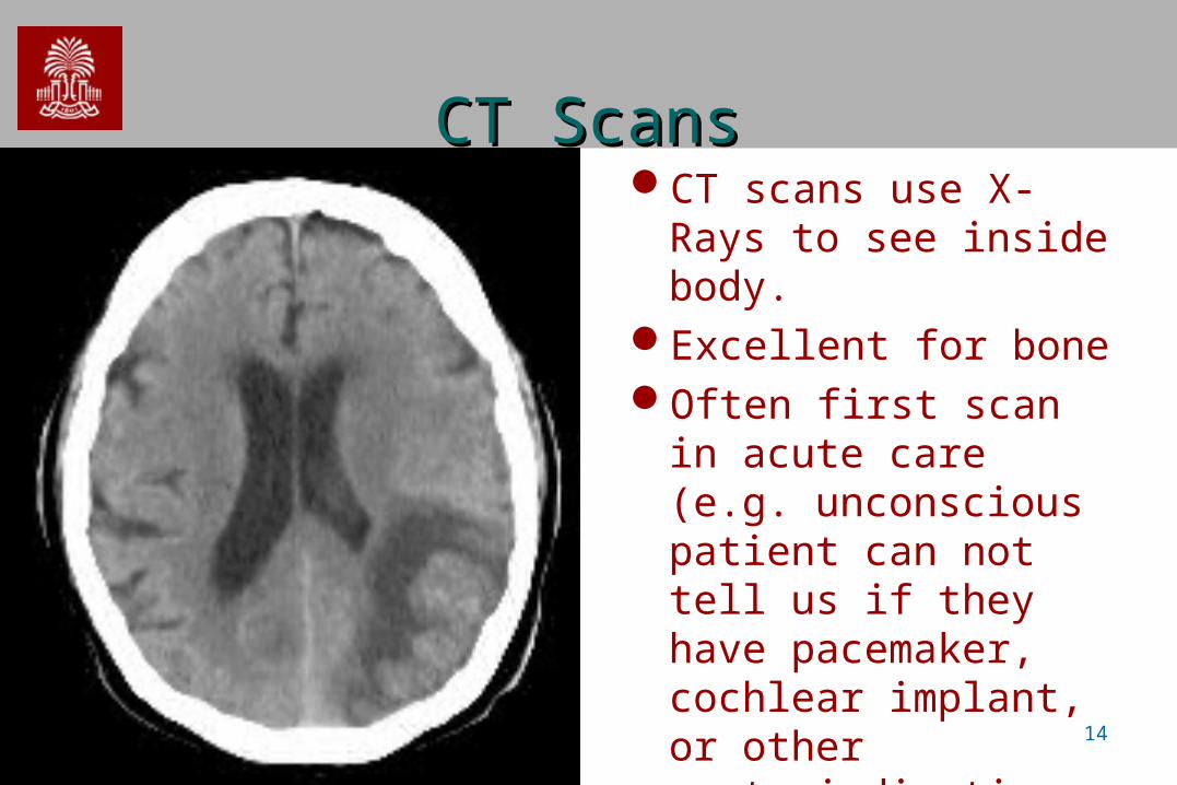
CT ScansCT ScansCT scans use X-Rays to
see inside body.Excellent for boneOften first scan in acute
care (e.g. unconscious patient can not tell us if they have pacemaker, cochlear implant, or other contraindications to MRI).

15
PET/SPECT ImagesPET/SPECT ImagesMeasures of blood flow can help
determine brain metabolism. PET: Inject radioactively labeled glucose.
Note: reduced uptake in posterior region.

16
Combining anatomy and Combining anatomy and metabolismmetabolism
Anatomical scans (T2 MRI) have excellent spatial resolution.
Metabolic scans can identify abnormalities (e.g. tumor).Combining takes advantage of complementary strengths

17
Relative CoordinatesRelative Coordinates• On the globe we talk about North, South,
East and West.• Lets explore the coordinates for the brain.

18
OrientationOrientation• Human anatomy
described as if person is standing
• If person is lying down, we would still say the head is superior to feet.
19
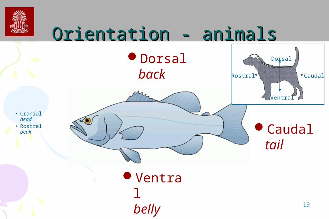
Orientation - animalsOrientation - animals
• Cranialhead
• Rostralbeak Caudal
tail
Dorsalback
Ventralbelly
Rostral Caudal
Ventral
Dorsal

20
Coordinates – Coordinates – Dorsal VentralDorsal Ventral• Human dorsal/ventral and rostral/caudal differ for brain and spine.
– Head/Foot, Superior/Inferior, Anterior/Posterior not ambiguous.
DorsalVentral
Dorsal
Ventral
Dorsal
Ventral

21
Coordinates – Coordinates – HumanHuman• Human dorsal/ventral and rostral/caudal differ for brain and spine.
– Head/Foot, Superior/Inferior, Anterior/Posterior not ambiguous.
C
PosteriorAnterior
R CR
C
R
22
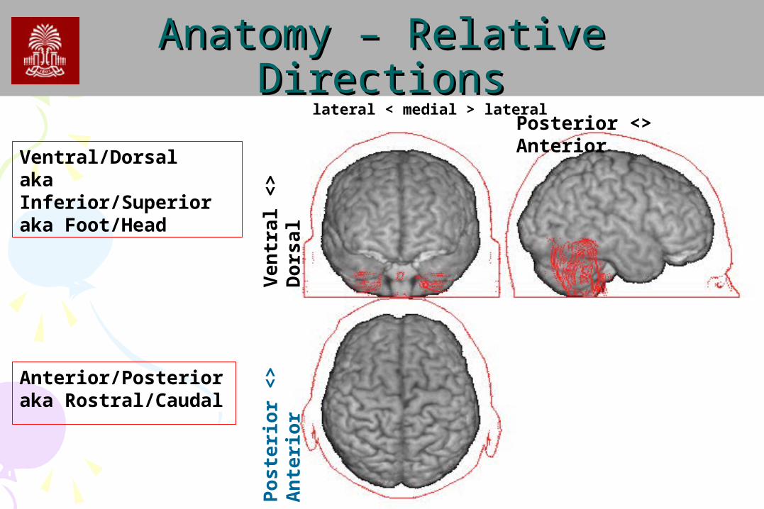
Anatomy – Relative DirectionsAnatomy – Relative DirectionsPosterior <> Anterior
Pos
teri
or <
> A
nte
rior
Ven
tral
<>
Dor
sal
lateral < medial > lateral
Anterior/Posterioraka Rostral/Caudal
Ventral/Dorsalaka Inferior/Superioraka Foot/Head
23
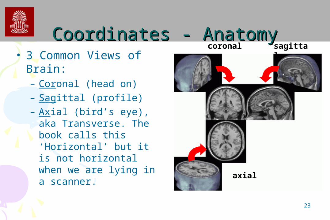
Coordinates - AnatomyCoordinates - Anatomy• 3 Common Views of
Brain:– Coronal (head on)– Sagittal (profile)– Axial (bird’s eye), aka
Transverse. The book calls this ‘Horizontal’ but it is not horizontal when we are lying in a scanner.
sagittalcoronal
axial

24
CoronalCoronal• Corona: ‘crown’ a coronal plane is parallel
to crown that passes from ear to ear– Coronal cut creates anterior, posterior
portions

25
TransverseTransverse• Transverse: perpendicular to the long axis
– These cuts are also referred to as Axial.
Example: cucumber slices are transverse to long axis.

26
SagittalSagittal• Sagittal – ‘arrow like’
– Sagittal cut divides object into left and right
– sagittal suture looks like an arrow. top view

27
Sagittal and MidsagittalSagittal and Midsagittal• A Sagittal slice down the
midline is called the ‘midsagittal’ view.midsagittal sagittal

28
Oblique SlicesOblique Slices• Slices that are not cut parallel to an orthogonal plane
are called ‘oblique’.• The oblique blue slice is neither Coronal nor Axial.
Ax
Cor
Oblique
29
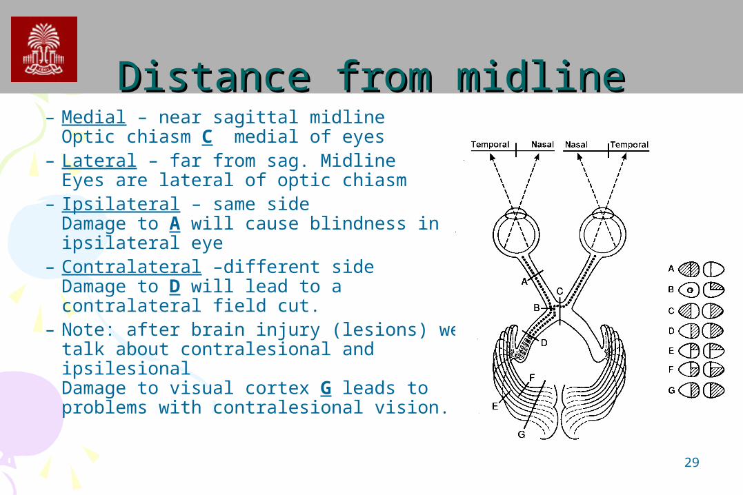
Distance from midlineDistance from midline– Medial – near sagittal midline
Optic chiasm C medial of eyes– Lateral – far from sag. Midline
Eyes are lateral of optic chiasm– Ipsilateral – same side
Damage to A will cause blindness in ipsilateral eye
– Contralateral –different sideDamage to D will lead to a contralateral field cut.
– Note: after brain injury (lesions) we talk about contralesional and ipsilesionalDamage to visual cortex G leads to problems with contralesional vision.

30
Relative positionsRelative positions• Distance From Body
– Proximal, Central: near center of body• Think ‘proximity’• Shoulders are proximal parts of arms
– Distal,peripheral: away from body• Think distant• Fingers are distal parts of the arms
• Distance from Surface– Superficial, external: near surface
• The bump bruised superficial tissue.
– Profound, deep: far from surface• The car crash injured deep organs.

31
MovementsMovements
AbductionAdduction
Supination
Pronation
Flexion
Extension

32
Types of cells in the brainTypes of cells in the brain• Neuron: Cell which is
responsible for receiving, transmitting and synthesizing information– cell body: contains organelles for
metabolism and a nucleus
• Glial Cells: Support cells for Neurons (CNS: oligodendrocytes, astrocytes, ependymal cells, radial glial; PNS : Satellite and Schwann cells)

33
Neuron TypesNeuron Types• Neurons come in different types – some only
communicate locally, while others have very long axons that communicate with distant regions.

34
Glial CellsGlial Cells• Glial cells have crucial functions
www.mult-sclerosis.org/glialcells.html
– Repair, maintenance and cleaning. They produce new myelin when it become damaged, lay down scar tissue, and remove dead cells and other debris.
– Physical support. They have hairlike filaments which hold the neurons in place and allow the central nervous system to retain its structural integrity.
– CNS development. Help migration of neurons.– Chemical regulation. Supply chemicals such as potassium and
calcium and regulate neurotransmitter levels.
• Ten times as many glial cells as neurons• Glial cells involved with many tumours (gliomas)

35
The Central Nervous SystemThe Central Nervous System
• Telencephalon (Cerebrum)– Cortex– Basal Ganglia
• Diencephalon – Thalamus– Hypothalamus)
• Mesencephalon (Midbrain)
• Rhombencephalon– Cerebellum– Pons– Medulla
36
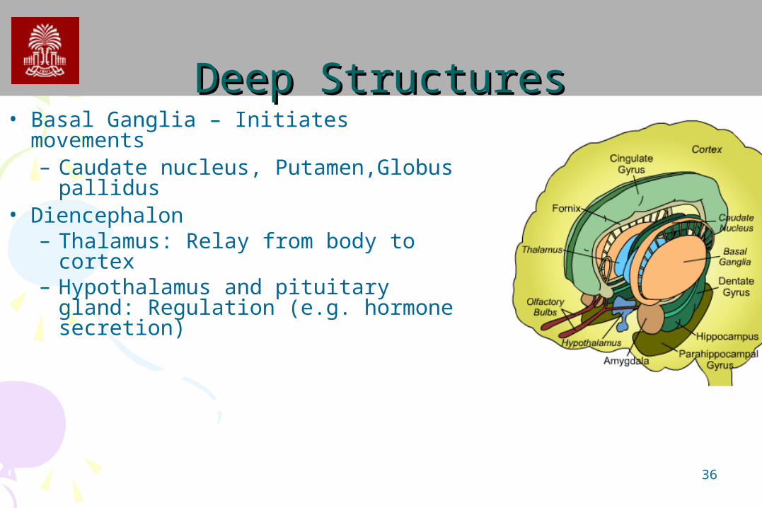
Deep StructuresDeep Structures• Basal Ganglia – Initiates movements
– Caudate nucleus, Putamen,Globus pallidus
• Diencephalon– Thalamus: Relay from body to
cortex– Hypothalamus and pituitary gland:
Regulation (e.g. hormone secretion)

37
Deep StructuresDeep Structures• Basal Ganglia – Initiates movements
– Caudate nucleus(red)
– Putamen (green)
– Globus pallidus (blue)• Diencephalon
– Thalamus: (yellow)– Hypothalamus: (not shown)

38
Brain StemBrain Stem• Midbrain
– Early auditory/visual processing– Dopamine for movement
control– CN III and IV emerge
• Pons– CN V, VI, VII VIII
• Medulla Oblongata– Pyramidal decussation:
nerves from left cross to right side and vise versa
– CN IX, X, XI, XII

39
The cortexThe cortex• Cortex – ‘Bark’ shell of brain – mostly gray
matter~80% of human brain~20% of squirrel brain

40
Cortical foldingCortical folding• Cortical folding increases surface area.• Ridges are called Gyri (singular = Gyrus)
– Greek gyros = circle, hence a coil of brain cortex
• Valleys are called Sulci (singular = Sulcus).– Latin = a groove.Gyri
Sulci

41
Gray and White MatterGray and White Matter• The outer surface of the cortex is gray
matter: lots of interconnected neurons (like cities)
• Underneath is the white matter – the highways connecting regions.

42
Functional ClassificationsFunctional Classifications• Some neurons transmit general information
– Pain and Temperature– Originate in surface structures
• Other neurons transmit specialized information– Specialized receptors– Hearing and vision
• Somatic: Skeletal muscles• Visceral: Refer to internal vital body organs• Can be either
– Afferent: Sensory– Efferent: Motor

43
Cortical layersCortical layers• Neurons are in six layers
I. Molecular layerII. External granular layerIII. External pyramidal layerIV. Internal granular layerV.Internal pyramidal layerVI. Fusiform layer
• Functions– Superficial layers (I-III): inter-cortical
connections– IV: input from thalamus– V,VI: outputs to leave cortex

44
The big foldsThe big folds• The folds of your brain are like a
fingerprint – there are a few general patterns, with individual variability.
• Two main folds– Central Sulcus
Fissure of RolandoRolandic sulcus
– Lateral sulcusSylvian fissure
45
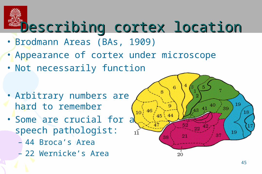
Describing cortex locationDescribing cortex location• Brodmann Areas (BAs, 1909)• Appearance of cortex under microscope• Not necessarily function
• Arbitrary numbers are hard to remember
• Some are crucial for a speech pathologist:– 44 Broca’s Area– 22 Wernicke’s Area
46
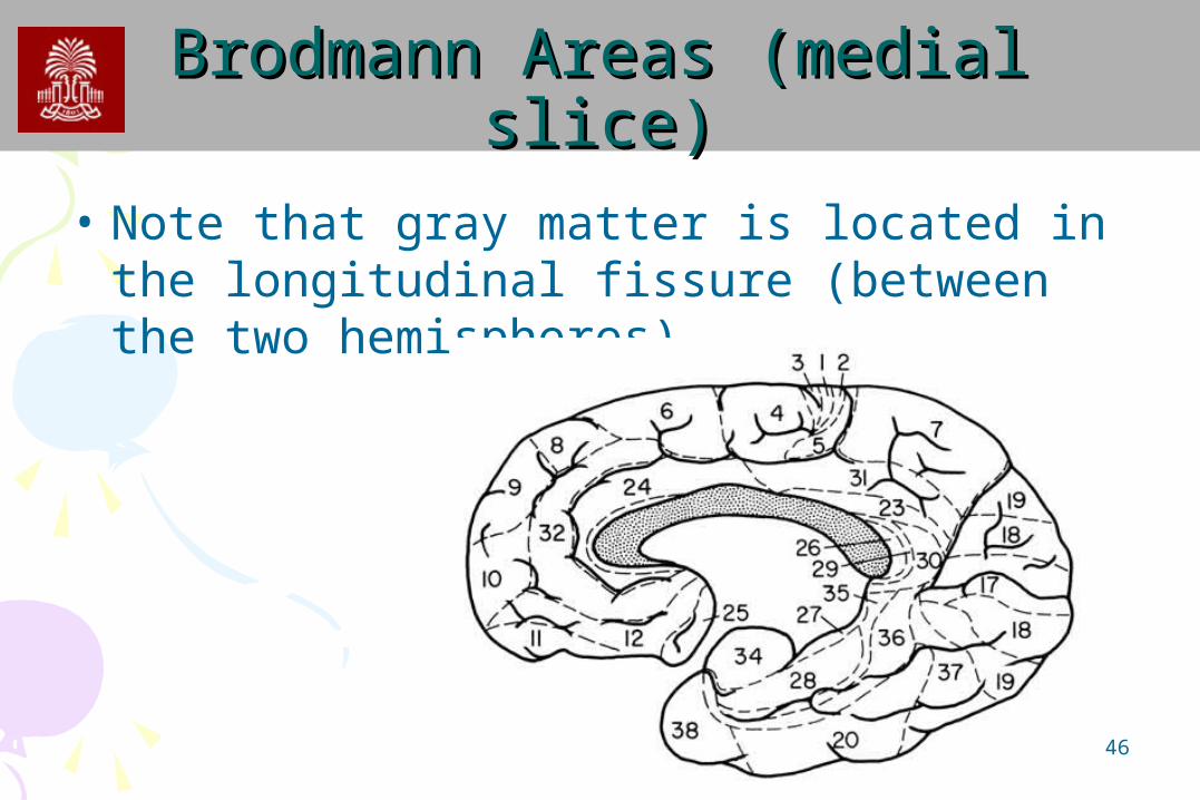
Brodmann Areas (medial slice)Brodmann Areas (medial slice)• Note that gray matter is located in the
longitudinal fissure (between the two hemispheres)

47
Cortical NamesCortical Names• Much of cortex referred
to by combination of coordinate+lobe+gyrus
• E.G. Superior Temporal Gyrus (STG)
• Middle Temporal Gyrus(MTG)
• Lateral Occipital Gyrus (LOG)

48
Cortical namesCortical names• Tip of an object called a ‘pole’• Frontal Pole• Temporal Pole

49
Sulci namesSulci names• Many of sulci referred to by combination
of coordinate+lobe+sulcus– Superior temporal sulcus (STS)– Inferior frontal sulcus (IFS)
– Precentral and postcentral sulciare just anterior and posterior to the central sulcus.
50
Brain functionBrain function• Anatomy is interested with
the structure of an organism.
• Physiology is interested in the function of the structure.
• We are still learning about brain function
• Modern maps of brain function are primitive…

51
Brain functionBrain function• Much of the primate cortex
devoted to vision.• In some monkeys, up to
50% of neocortex is devoted to vision.

52
Brain functionBrain function• Two striking features
of human brain1. Lots of cortex ‘left
over’ (yellow)not devoted to specific task – we are flexible
2. Not much of the cortex is solely devoted to language.
Recommended