Embed Size (px)
Citation preview
Zurich Open Repository andArchiveUniversity of ZurichMain LibraryStrickhofstrasse 39CH-8057 Zurichwww.zora.uzh.ch
Year: 2019
Management of brainstem haemorrhages
Wang, Sophie S ; Yang, Yang ; Velz, Julia ; Keller, Emanuela ; Luft, Andreas R ; Regli, Luca ; Neidert,Marian C ; Bozinov, Oliver
Abstract: Among spontaneous intracranial haemorrhages, primary non-traumatic brainstem haemor-rhages are associated with the highest mortality rate. Patients classically present with rapid neurologicaldeterioration. Previous studies have found that the severity of initial neurological symptoms and hydro-cephalus are predictors of poor outcomes. In addition, radiological parameters aim to classify brainstemhaematomas according to volume, extension and impact on prognosis. However, previous studies havefailed to agree on a differentiated radiological classification for outcome and functional recovery. Electro-physiology, including motor, auditory and somatosensory evoked potentials, is used to estimate the extentof the initial injury and predict functional recovery. The current management of brainstem haematomasremains conservative, focusing on initial close neurocritical care monitoring. Surgical treatment conceptsexist, but similarly to general intracranial haemorrhage management, they continue to be controversialand have not been sufficiently investigated. This is especially the case for haematomas in the posteriorfossa, as these are excluded from most current clinical trials. Existing studies were mostly carried outbefore the present millennium began, and limitations are evident in the adaptation of those results andrecommendations to current management, with todayrsquo;s technological and diagnostic possibilities.We therefore recommend the re-evaluation of brainstem haemorrhages in the modern neurosurgical andintensive care environment.
DOI: https://doi.org/10.4414/smw.2019.20062
Posted at the Zurich Open Repository and Archive, University of ZurichZORA URL: https://doi.org/10.5167/uzh-176671Journal ArticlePublished Version
The following work is licensed under a Creative Commons: Attribution-NonCommercial-NoDerivatives4.0 International (CC BY-NC-ND 4.0) License.
Originally published at:Wang, Sophie S; Yang, Yang; Velz, Julia; Keller, Emanuela; Luft, Andreas R; Regli, Luca; Neidert,Marian C; Bozinov, Oliver (2019). Management of brainstem haemorrhages. Swiss Medical Weekly,149:w20062.DOI: https://doi.org/10.4414/smw.2019.20062
Review article: Biomedical Intelligence | Published 05 April 2019 | doi:10.4414/smw.2019.20062
Cite this as: Swiss Med Wkly. 2019;149:w20062
Management of brainstem haemorrhages
Wang Sophie S.a, Yang Yanga, Velz Juliaa, Keller Emanuelaa, Luft Andreas R.bc, Regli Lucaa, Neidert Marian C.a, Bozinov
Olivera
a Department of Neurosurgery, University Hospital Zurich, University of Zurich, Switzerlandb Department of Neurology, University Hospital Zurich, University of Zurich, Switzerlandc Cereneo, Centre for Neurology and Rehabilitation, Vitznau, Switzerland
Summary
Among spontaneous intracranial haemorrhages, primary
non-traumatic brainstem haemorrhages are associated
with the highest mortality rate. Patients classically present
with rapid neurological deterioration. Previous studies
have found that the severity of initial neurological symp-
toms and hydrocephalus are predictors of poor outcomes.
In addition, radiological parameters aim to classify brain-
stem haematomas according to volume, extension and im-
pact on prognosis. However, previous studies have failed
to agree on a differentiated radiological classification for
outcome and functional recovery. Electrophysiology, in-
cluding motor, auditory and somatosensory evoked po-
tentials, is used to estimate the extent of the initial injury
and predict functional recovery. The current management
of brainstem haematomas remains conservative, focusing
on initial close neurocritical care monitoring. Surgical treat-
ment concepts exist, but similarly to general intracranial
haemorrhage management, they continue to be contro-
versial and have not been sufficiently investigated. This is
especially the case for haematomas in the posterior fos-
sa, as these are excluded from most current clinical tri-
als. Existing studies were mostly carried out before the
present millennium began, and limitations are evident in
the adaptation of those results and recommendations to
current management, with today’s technological and diag-
nostic possibilities. We therefore recommend the re-evalu-
ation of brainstem haemorrhages in the modern neurosur-
gical and intensive care environment.
Keywords: neurosurgery, brainstem, brainstem haemor-
rhage, spontaneous intracranial haemorrhage, neurocriti-
cal care
Introduction
Terminology
The American Heart Association/American Stroke Asso-
ciation (AHA/ASA) has defined an intracerebral haemor-
rhage (ICH) as a focal collection of blood within the brain
parenchyma or ventricular system that is not the result of
trauma. ICHs are often referred to as “spontaneous”, “non-
traumatic” or “primary” [1]. The same terminology ap-
plies to brainstem haemorrhages. Some terms focus on the
pons only, and therefore the label “primary pontine haem-
orrhage” is applied. Others also include tegmental parts.
However, we have noticed an inconsistency: some groups
use the term “pontine haemorrhage” for bleedings that also
expand into the tegmentum (mesencephalon). Therefore,
we suggest using the term “brainstem haemorrhage” to ad-
dress the brainstem as one functional and anatomical unit.
Epidemiological features – incidence, risk factors and
mortality
In developed countries, stroke remains one of the leading
causes of death and morbidity. Spontaneous ICH is the un-
derlying cause of up to 19.6% of all strokes [2, 3]. Six to
ten percent of spontaneous ICHs are localised in the brain-
stem. The risk of ICH increases continuously with age.
However, brainstem haemorrhages occur in a younger pa-
tient group, with the highest incidence occurring in pa-
tients between 40 years and 60 years old [4, 5]. The most
common cause of primary bleeding into the brainstem is
hypertension [4]. Previous studies have noted a higher dis-
tribution of ICHs in cocaine users, but no significance was
accorded to the relationship between this risk factor and lo-
calisation to the brainstem [6] (fig. 1 presents an illustra-
tive case).
Vascular malformations, mostly cavernomas and arteriove-
nous malformations (AVMs), can be secondary causes of
brainstem haemorrhages [7]. Brainstem haemorrhages are
associated with the highest mortality rate among sponta-
neous ICHs [8]. Their reported mortality rate varies be-
tween 47% and 80% [8, 9].
Clinical presentation
Brainstem haemorrhage is an acute neurological illness
with a very sudden onset. It is associated with early pri-
mary coma, motor disturbances (e.g. tetraplegia, hemiple-
gia or extensor posturing), respiratory disturbance, hyper-
thermia and pupillary abnormalities (e.g. pinpoint pupils,
anisocoria) resulting from the destruction of the base or
the tegmentum [4]. In the computed tomography (CT) and
post-magnetic resonance imaging (MRI) era, even minis-
cule haemorrhages can be identified, resulting in the possi-
ble presentation of a broader spectrum of symptoms. How-
ever, in contrast to pontine infarctions, pontine bleedings
rarely evolve slowly enough to present initial symptoms
that reflect the presence of progressive damage to the cra-
nial nerves and their nuclei [4, 10]. Previous retrospec-
Correspondence:
Sophie S. Wang, Depart-
ment of Neurosurgery, Uni-
versity Hospital Zurich,
Frauenklinikstrasse 10,
CH-8091 Zurich, so-
phie.wang[at]usz.ch
Swiss Medical Weekly · PDF of the online version · www.smw.ch
Published under the copyright license “Attribution – Non-Commercial – No Derivatives 4.0”.
No commercial reuse without permission. See http://emh.ch/en/services/permissions.html.
Page 1 of 9

tive studies have emphasised that severe initial neurologi-
cal deficits, especially early coma, the need for mechanical
ventilation and hydrocephalus, are associated with poor
prognosis [9, 11]. Pupillary abnormalities do not show a
significant correlation with outcome [11]. Patients bleed-
ing secondarily into the brainstem due to cavernomas or
arteriovenous malformations usually show less severe ini-
tial symptoms and have more favourable outcomes [7, 12].
These secondary haemorrhages have different dynamics to
primary bleedings as they are more expanding than disrup-
tive. Therefore, we have excluded them from this review.
Radiological parameters and their impact onoutcome
Brainstem ICH is a medical emergency, and therefore di-
agnostics must be acquired quickly. MRI has proven to
be more accurate than CT imaging at differentiating be-
tween haemorrhage ages and therefore at diagnosing older
ICHs. For acute ICHs, they offer the same degree of accu-
racy [13]. Due to its rapid availability, the CT scan remains
the imaging modality of choice for evaluating brainstem
haemorrhages. In addition, unstable patients in acute dis-
tress represent a challenge where MRI is concerned: MRI
is risky for them due to its long duration and the restricted
access to the patient. Therefore, previous studies have tried
to predict outcome depending on radiological CT findings
[14–19]. However, MRI can serve as a follow-up diagnos-
tic tool that is not only helpful for prognostication, but also
for identifying the precise extension of the lesion, as well
as the long-term effects of brainstem bleeding, e.g. olivary
degeneration [20] (see also fig. 1).
Different kinds of classifications have been proposed for
brainstem haemorrhages, mostly based on the axial CT fea-
tures of the exact localisation and anatomical spread [11,
14, 15, 21]. Some classifications include tegmental parts
(mesencephalon), and others concentrate solely on pon-
tine haemorrhages (fig. 2). In a study of 62 cases in 1992,
Chung et al. classified brainstem haematomas into four
types: (1) the “massive” type, which they defined by its bi-
lateral spread into both the basis pontis and the tegmen-
tum; (2) the “bilateral tegmental” type, which occupied the
bilateral tegmentum only; (3) the “basal-tegmental” type,
which was localised in the junction between the basis pon-
tis and the tegmentum bilaterally; and (4) the “small uni-
lateral tegmental” type, which referred to haematomas lo-
cated exclusively in the unilateral tegmentum [14]. The
survival rates for the types were shown to be 7.1%, 14.3%,
26.1% and 94.1%, respectively [14]. Other groups (Rabin-
stein et al. and Balci et al.) tried to simplify this classifi-
cation into three types by combining the basal and bilater-
al tegmental types in one group [16, 19]. However, a 1999
study by Fong et al. with 39 cases found a survival rate
of 30.8% for the bilateral tegmental type and a 100% sur-
vival rate for the basal-tegmental type. This suggests that
grouping the two locations together may not be justified
[17]. In general, previous studies agree that basal-tegmen-
tum and small unilateral tegmentum haemorrhages are as-
sociated with more favourable outcomes, while almost all
Figure 1: A 37-year-old female patient collapsed after alcohol and drug (cocaine) abuse. The “bilateral tegmental” haematoma was more pro-
nounced on the right, with minimal ventricular extension. The patient’s initial GCS (Glasgow Coma Scale) was 6, with no pupil abnormality,
and the patient was intubated in the ED. The patient was a regular cocaine user with a known history of hypertension. On day +4, extubation
was unsuccessful due to bulbar palsy. Therefore, tracheotomy was performed on day +6. After decannulation, the patient was discharged to a
neurological rehabilitation centre at GCS15 with a horizontal gaze palsy, tetra spasticity, left hypo sensitivity and left facial palsy. A. shows
sagittal view and shows axial view of initial CT scan. B. shows initial MRI (axial FLAIR on the left-hand side and sagittal T2 on the right-hand
side. Arrow (→) indicates the haemorrhage. 1 indicates cerebellum, 2 mesencephalon, 3 pons and 4 medulla oblongata. C. shows five-month
follow-up MRI (axial FLAIR) with hypertrophic olivary degeneration (double arrows →→) caused by the brainstem haemorrhage. The inferior
olivaries are part of the dentato-rubro-olivary tract – known as the Triangle of Guillain-Mollaret – connecting the brainstem and the deep cere-
bellar nuclei. A lesion like hypertrophic olivary degeneration at this triangle may cause modulation problems in spinal cord motor activity and
myoclonus.
Review article: Biomedical Intelligence Swiss Med Wkly. 2019;149:w20062
Swiss Medical Weekly · PDF of the online version · www.smw.ch
Published under the copyright license “Attribution – Non-Commercial – No Derivatives 4.0”.
No commercial reuse without permission. See http://emh.ch/en/services/permissions.html.
Page 2 of 9
massive haemorrhages and most bilateral tegmental haem-
orrhage cases are prognostically poor [16–18].
Russell et al. proposed a different approach. They divided
brainstem haematomas into three types: central, dorsolater-
al tegmental and tegmentobasilar [21]. Large haematomas
resulting from arterial hypertension generally occupied the
central pons, leading to a rapid, fatal clinical course due
to the involvement of the reticular system. Partial pontine
haematomas, mostly occurring due to the rupture of cryptic
vascular malformations, were restricted to the lateral half
of the pons, sparing the reticular system. These could be
either dorsolateral tegmental or tegmentobasilar.
In a study of 19 patients with pontine haemorrhages in
2012, Nishizaki et al. combined the classifications of Rus-
sel et al. and Chung et al. with some modifications. The re-
sulting classifications are massive, tegmentobasilar, trans-
verse oval and small unilateral [22]. Nishizaki et al.
defined the transverse oval haematoma as an elliptical
haematoma with the bilateral involvement of the basis,
tegmentum or basal-tegmental junction. This type was
similar to one type in Chung’s classification, for which the
basal-tegmental junction between the basis pontis and the
tegmentum was involved bilaterally. Nishizaki et al. found
a mortality rate of 25% in cases with transverse oval or
small unilateral haematomas and a mortality rate of 65% in
cases with massive or tegmentobasilar haematomas [22].
Wessels et al. reviewed the clinical data of 29 consecutive
patients with primary pontine haemorrhages (PPHs) in
2004, and divided PPHs into three new types without men-
tioning unilateralism or bilateralism: (1) dorsal, (2) ventral
and (3) massive [11]. Studies using this classification re-
ported that ventral or massive haematomas were related to
high mortality, while dorsal haematomas had a favourable
outcome [11, 23, 24]. Jang et al. conducted an outcome
study involving 281 patients and using Wessels’ classifi-
cation. It found 30-day mortality rates of 66.9, 24.7 and
1.5%. The study also investigated 90-day functional recov-
ery and found rates of 3.8%, 14.8% and 14.9% for massive,
ventral and dorsal PPHs respectively [25].
In another study of 39 consecutive patients by Dziewas
et al., trans-axial pontine haemorrhages (PHs) were divid-
ed into three subtypes according to Kase and Caplan: (1)
large paramedian PH, (2) unilateral basotegmental PH and
(3) lateral tegmental PH. Dziewas et al. reported that para-
median PH was related to a fatal outcome, while lateral
tegmental PH was associated with a favourable prognosis
[15].
Despite all these variations in anatomical classification
systems, the literature agrees on the following points:
haematoma size (usually, the threshold values for predict-
ing outcome range between 4 and 5 ml for volume or
20 and 27 mm for trans-axial diameter) and radiological
signs of acute hydrocephalus correlate with poor outcomes
[11, 15, 21, 23–27]. All studies of brainstem haemorrhages
in the past two decades showed that unilateral tegmental
haemorrhages had favourable outcomes, whereas bilateral
basal bleedings and haemorrhages, including anterior seg-
ments (so-called massive), had the worst outcomes [9, 11,
14] (fig. 3). For all patients with radiological findings be-
tween these two extremes, survival outcome was very dif-
ficult to predict based on CT alone.
Electrophysiology
The importance of determining good prediction parameters
for functional recovery in this patient group, in which sur-
vivors are often described as being in a severely disabled
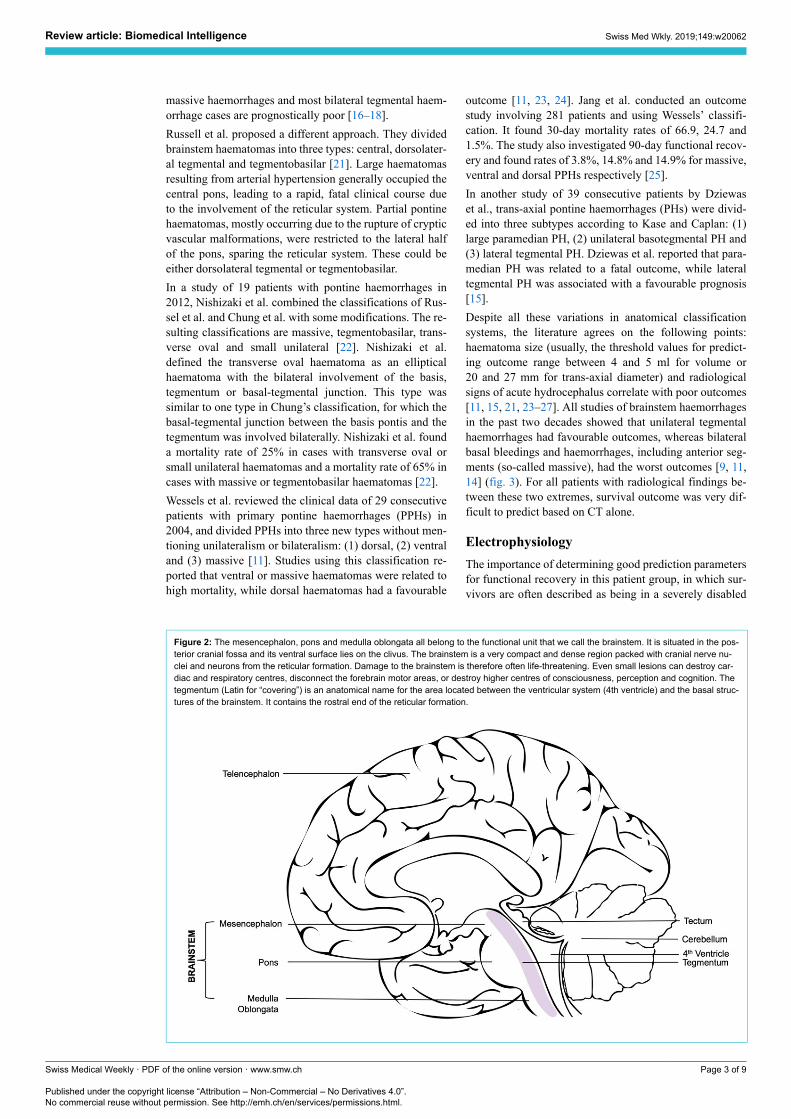
Figure 2: The mesencephalon, pons and medulla oblongata all belong to the functional unit that we call the brainstem. It is situated in the pos-
terior cranial fossa and its ventral surface lies on the clivus. The brainstem is a very compact and dense region packed with cranial nerve nu-
clei and neurons from the reticular formation. Damage to the brainstem is therefore often life-threatening. Even small lesions can destroy car-
diac and respiratory centres, disconnect the forebrain motor areas, or destroy higher centres of consciousness, perception and cognition. The
tegmentum (Latin for “covering”) is an anatomical name for the area located between the ventricular system (4th ventricle) and the basal struc-
tures of the brainstem. It contains the rostral end of the reticular formation.
Review article: Biomedical Intelligence Swiss Med Wkly. 2019;149:w20062
Swiss Medical Weekly · PDF of the online version · www.smw.ch
Published under the copyright license “Attribution – Non-Commercial – No Derivatives 4.0”.
No commercial reuse without permission. See http://emh.ch/en/services/permissions.html.
Page 3 of 9

state, is clear [11, 25]. Imaging parameters do not convey
any real-time information on the functional integrity of the
corticospinal tract and somatosensory pathway. The use of
neuromonitoring is widely recognised as an objective tool
in terms of predicting post-stroke functional recovery. Lit-
tle literature is available on the use of neuromonitoring to
predict outcome and to guide therapeutic decision-making
in patients with brainstem haemorrhages [28, 29].
The role of electroencephalogram (EEG) in brainstem
monitoring remains highly disputed. It has been shown that
the stimulation of the brainstem (e.g. the reticular forma-
tion) evokes changes in the EEG [30]. However, perform-
ing an EEG as part of brainstem monitoring, and especially
in brainstem death diagnostics, is claimed to be more re-
assuring for the patients’ relatives than practical enough to
guide clinical decision-making, for the obvious reason that
recording an EEG from the scalp can hardly test brainstem
function [31] (fig. 4 presents an illustrative case).
Electrophysiological examinations such as somatosensory
evoked potentials (SSEPs), brainstem auditory evoked po-
tentials (BAEPs) and motor evoked potentials (MEPs in
limb muscles and corticobulbar MEPs (CoMEPs) in cra-
nial nerve innervated muscles) are available to monitor the
functional integrity of the motor pathways passing through
the brainstem. Before the introduction of MEPs, BAEPs
and SSEPs were the only two standard tools in surgical
interventions in and around the brainstem. However, it
was only possible to monitor approximately 20% of the
brainstem with just these two neuromonitoring modalities
(BAEPs and SSEPs) [32]. To obtain real-time information
on the functional integrity of the motor pathways passing
through the brainstem (corticospinal tract, CST), transcra-
nial electrical stimulation (TES) is applied [33]. Multipulse
TES allows MEP recording under general anaesthesia [32].
MEP responses are preferentially recorded from the distal
muscles of the hand and foot. For CoMEPs, TES is applied
to the motor strip (M1) for the cranial motor nerves, and
CoMEPs are recorded in the appropriate muscles, i.e. those
innervated by the cranial motor nerves [33, 34] (fig. 5).
Seong et al. showed that the combined use of SSEPs and
MEPs in the Neurocritical Care Unit was a reliable and
useful tool for predicting functional recovery in patients
with brainstem haemorrhages [35]. Furthermore, Seong et
al. insisted that the combination of MEPs and SSEPs was
a more powerful tool than measuring either the transverse
diameter or the volume using CT. In their study, 14 pa-
tients with primary pontine haemorrhages were divided in-
to good-outcome and poor-outcome groups according to
the modified Rankin Score (mRS). When the MEP and
SEP scores were summed (EP sum), the mRS and func-
tional ambulatory category had the highest value [35].
Treatment
Conservative treatment
Prehospital and emergency department management for
brainstem ICH includes close monitoring, securing the air-
way, provision of cardiovascular support and correction of
the underlying haemostatic abnormalities. Hyperglycaemia
and hypoglycaemia should be avoided. In 2015, Hemphill
et al. added a new management recommendation, the treat-
Figure 3: A-D show an axial view of the pontine part of the Brainstem. A. shows a “basal tegmental” haemorrhage, B. a “massive” haemor-
rhage, C. “bilateral tegmental” haemorrhage and D. “unilateral tegmental” haemorrhage. “Massive” brainstem haemorrhages have the poorest
and “unilateral tegmental” the best clinical prognoses according to our literature review.
Review article: Biomedical Intelligence Swiss Med Wkly. 2019;149:w20062
Swiss Medical Weekly · PDF of the online version · www.smw.ch
Published under the copyright license “Attribution – Non-Commercial – No Derivatives 4.0”.
No commercial reuse without permission. See http://emh.ch/en/services/permissions.html.
Page 4 of 9
ment of fever, to the well-cited AHA/ASA guidelines for
the management of spontaneous. This was because the du-
ration of fever has been associated with poor prognosis and
haematoma growth [36–38]. Blood pressure (BP) manage-
ment in ICH has been extensively discussed recently. The
current AHA/ASA guidelines, mostly influenced by the
INTERACT2 study, suggest that early intensive lowering
of BP to 140 mm Hg is safe and may be effective for pa-
tients presenting with a Glasgow Coma Score (GCS) >5
and a 150–220 mm Hg systolic BP. However, ATACH-2,
a 2017 meta-analysis that included five studies and 4360
patients, subsequently confirmed that intensive, acute low-
ering of BP is safe, but found that it has no clinical benefit
in terms of mortality or functional outcome [39–41].
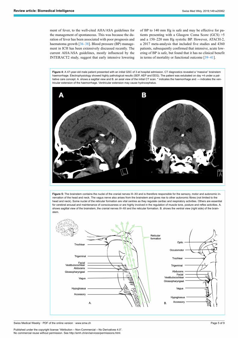
Figure 4: A 47-year-old male patient presented with an initial GSC of 3 at hospital admission. CT diagnostics revealed a “massive” brainstem
haemorrhage. Electrophysiology showed highly pathological results (SEP, AEP and EEG). The patient was extubated on day +4 under a pal-
liative care concept. A. shows a sagittal view and B. an axial view of the initial CT scan. * indicates the haemorrhage and → indicates the ven-
tricular extension of the haemorrhage. Ventricular extension may cause hydrocephalus.
Figure 5: The brainstem contains the nuclei of the cranial nerves III–XII and is therefore responsible for the sensory, motor and autonomic in-
nervation of the head and neck. The vagus nerve also arises from the brainstem and gives rise to other autonomic fibres (not limited to the
head and neck). Some nuclei of the reticular formation are vital centres as they regulate cardiac and respiratory activities. Others are essential
for cerebral arousal and maintenance of consciousness or are highly involved in the regulation of muscle tone, posture and reflex activities. A.
shows sagittal view of the brainstem, the cranial nerves III–XII and the reticular formation. B. shows the ventral view (right side) of the brain-
stem.
Review article: Biomedical Intelligence Swiss Med Wkly. 2019;149:w20062
Swiss Medical Weekly · PDF of the online version · www.smw.ch
Published under the copyright license “Attribution – Non-Commercial – No Derivatives 4.0”.
No commercial reuse without permission. See http://emh.ch/en/services/permissions.html.
Page 5 of 9
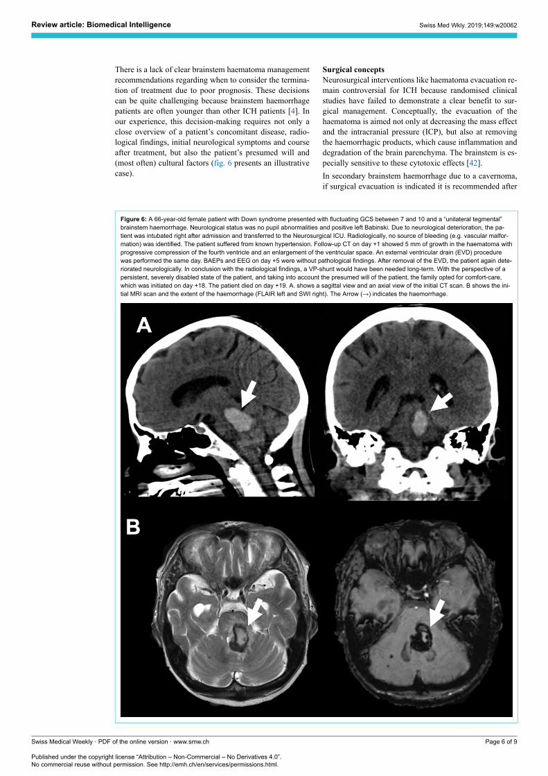
There is a lack of clear brainstem haematoma management
recommendations regarding when to consider the termina-
tion of treatment due to poor prognosis. These decisions
can be quite challenging because brainstem haemorrhage
patients are often younger than other ICH patients [4]. In
our experience, this decision-making requires not only a
close overview of a patient’s concomitant disease, radio-
logical findings, initial neurological symptoms and course
after treatment, but also the patient’s presumed will and
(most often) cultural factors (fig. 6 presents an illustrative
case).
Surgical concepts
Neurosurgical interventions like haematoma evacuation re-
main controversial for ICH because randomised clinical
studies have failed to demonstrate a clear benefit to sur-
gical management. Conceptually, the evacuation of the
haematoma is aimed not only at decreasing the mass effect
and the intracranial pressure (ICP), but also at removing
the haemorrhagic products, which cause inflammation and
degradation of the brain parenchyma. The brainstem is es-
pecially sensitive to these cytotoxic effects [42].
In secondary brainstem haemorrhage due to a cavernoma,
if surgical evacuation is indicated it is recommended after
Figure 6: A 66-year-old female patient with Down syndrome presented with fluctuating GCS between 7 and 10 and a “unilateral tegmental”
brainstem haemorrhage. Neurological status was no pupil abnormalities and positive left Babinski. Due to neurological deterioration, the pa-
tient was intubated right after admission and transferred to the Neurosurgical ICU. Radiologically, no source of bleeding (e.g. vascular malfor-
mation) was identified. The patient suffered from known hypertension. Follow-up CT on day +1 showed 5 mm of growth in the haematoma with
progressive compression of the fourth ventricle and an enlargement of the ventricular space. An external ventricular drain (EVD) procedure
was performed the same day. BAEPs and EEG on day +5 were without pathological findings. After removal of the EVD, the patient again dete-
riorated neurologically. In conclusion with the radiological findings, a VP-shunt would have been needed long-term. With the perspective of a
persistent, severely disabled state of the patient, and taking into account the presumed will of the patient, the family opted for comfort-care,
which was initiated on day +18. The patient died on day +19. A. shows a sagittal view and an axial view of the initial CT scan. B shows the ini-
tial MRI scan and the extent of the haemorrhage (FLAIR left and SWI right). The Arrow (→) indicates the haemorrhage.
Review article: Biomedical Intelligence Swiss Med Wkly. 2019;149:w20062
Swiss Medical Weekly · PDF of the online version · www.smw.ch
Published under the copyright license “Attribution – Non-Commercial – No Derivatives 4.0”.
No commercial reuse without permission. See http://emh.ch/en/services/permissions.html.
Page 6 of 9
a period of about two weeks to allow the patient to stabilise
and the haematoma to organise [7]. For primary
haematomas in the posterior fossa, however, the AHA/
ASA Guidelines recommend considering the surgical re-
moval of cerebellar haemorrhages, but not of brainstem
haemorrhages. The guidelines even clearly advise against
the surgical evacuation of brainstem haematomas [38].
Ongoing clinical trials plan to deliver new data on hemi-
craniectomy (SWITCH) and minimal invasive approaches
(MISTIE III and MISTICH) to surgical ICH management.
However, infratentorial ICH has, without exception, been
excluded from all these studies. In 1984, De Pian et al.
brought together 50 brainstem haematoma cases from 22
Italian centres, and showed that there was some value to
surgical evacuation in chronic cases [43]. Two Japanese
groups (Takahama et al. and Hara et al.) proposed stereo-
tactic aspiration. They found a favourable functional out-
come in surgically treated patients compared to patients
with conservatively managed haematomas [44, 45].
A more commonly performed surgical procedure in the
case of brainstem haematoma is the external ventricular
drain (EVD) procedure. An EVD is indicated in cases
that present with clinical and radiological signs of hydro-
cephalus. This is often the case when the bleeding ex-
tends to the ventricular system [46]. However, as men-
tioned above, hydrocephalus has been associated with poor
outcomes in brainstem haemorrhages, and one can argue
that its indication should therefore be thoroughly evalu-
ated. Murata et al. found no significant improvement in
outcome when hydrocephalus was treated with ventricular
drainage [9]. In a retrospective, observational, single-cen-
tre study that focused on the comparison of decompressive
craniectomy, medical therapy and EVD placement in pa-
tients with haemorrhages in the posterior fossa (focusing
on cerebellar haemorrhages with and without brainstem in-
volvement), Luney et al. found a significant increase in hy-
drocephalus and intraventricular bleeds in patients treated
with EVD placement [47].
Conclusion
Primary brainstem haemorrhages remain associated with
poor outcomes compared to other forms of spontaneous
ICH. Negative prognostic factors are coma on admission,
the need for mechanical ventilation, haematoma volume
and the ventral extension of the haemorrhage. Many
groups have tried to classify brainstem haemorrhages ac-
cording to radiological parameters such as anatomical
spread, haematoma size and signs of hydrocephalus. To
summarise their findings, they all agree on the association
of poor outcome with so-called “massive” haemorrhages
and the association of good prognosis with unilateral
tegmental haemorrhages. All cases between these two ex-
tremes show large variation in survival and functional re-
covery rates (see bottom of fig. 7). Therefore, clinical pre-
sentation and other surrogate markers must be considered
to enable better early prognosis for the patient and their
family, and to guide clinical decision-making.
In our opinion, besides close neurological examinations,
the combined use of SSEPs, BAEPs, MEPs and CoMEPs
provides the most comprehensive approach to predicting
functional recovery in patients with brainstem haemor-
rhages. In the future, a prospective study on neuromonitor-
Figure 7: As it is a very heterogeneous disease, clear manage-
ment guidelines concerning brainstem haemorrhage do not exist.
An initial clinical and neuroradiological evaluation must be ob-
tained rapidly in the ER, including thorough neurological exams,
the acquisition of the patient’s medical history, risk factors, a cra-
nial CT scan and an angio CT. If there are signs of hydrocephalus,
the decision on an EVD must be made after CT. Management
should continue in the ICU (Intensive Care Unit) with close clinical
assessments, neuroprotective treatment (airway protection, tight
control of blood pressure, temperature, glucose and electrolytes,
correction of coagulopathy) and electrophysiology. Ultimately,
whether to rehabilitate or to initiate palliative care is an individual
decision. Clinical assessment and electrophysiological testing can
guide the making of this decision.
ing in patients with brainstem haemorrhages will be neces-
sary to define specific parameters that correlate with good
and poor outcomes.
Currently, treatment is still conservative neurocritical care.
Well-known guidelines clearly advise against surgical in-
tervention in primary brainstem haemorrhages. However,
the dataset supporting this recommendation remains small
because ICH in the posterior fossa has been excluded from
large ICH surgical intervention trials in the past, as well as
from those that are planned for the future. EVD is formal-
ly indicated in cases which present with hydrocephalus.
However, its benefit for those presenting with brainstem
haemorrhages has not been proven yet.
Generally, there have been few studies concerning the neu-
rosurgical management of brainstem haemorrhages, and
most of the existing ones were carried out before the pre-
sent millennium began. Therefore, we see limitations in
the adaptation of those results and recommendations to
today’s clinical practice. When comparing the numbers,
newer studies differ from older studies, showing better
survival rates for the same radiological haemorrhage sub-
types.
Brainstem haemorrhage management needs re-evaluation
and reinvestigation to update the numbers for incidence,
mortality, outcome, prognostic factors, standardised man-
Review article: Biomedical Intelligence Swiss Med Wkly. 2019;149:w20062
Swiss Medical Weekly · PDF of the online version · www.smw.ch
Published under the copyright license “Attribution – Non-Commercial – No Derivatives 4.0”.
No commercial reuse without permission. See http://emh.ch/en/services/permissions.html.
Page 7 of 9
agement and the indication of surgical procedures. How-
ever, the number of cases is limited. Therefore, we recom-
mend a national or international registry that will collect
data and shed light on the current state of brainstem haem-
orrhage treatment. This could be an important step for fu-
ture clinical trials investigating new treatment options for
brainstem haemorrhages.
Disclosure statement
No financial support and no other potential conflicts of interest rele-
vant to this article was reported.
References
1 Sacco RL, Kasner SE, Broderick JP, Caplan LR, Connors JJ, Culebras
A, et al.; American Heart Association Stroke Council, Council on Car-
diovascular Surgery and Anesthesia; Council on Cardiovascular Radiol-
ogy and Intervention; Council on Cardiovascular and Stroke Nursing;
Council on Epidemiology and Prevention; Council on Peripheral Vascu-
lar Disease; Council on Nutrition, Physical Activity and Metabolism. An
updated definition of stroke for the 21st century: a statement for health-
care professionals from the American Heart Association/American
Stroke Association. Stroke. 2013;44(7):2064–89. doi: http://dx.doi.org/
10.1161/STR.0b013e318296aeca. PubMed.
2 Sacco S, Marini C, Toni D, Olivieri L, Carolei A. Incidence and 10-year
survival of intracerebral hemorrhage in a population-based registry.
Stroke. 2009;40(2):394–9. doi: http://dx.doi.org/10.1161/STROKEA-
HA.108.523209. PubMed.
3 Sarti C, Rastenyte D, Cepaitis Z, Tuomilehto J. International trends in
mortality from stroke, 1968 to 1994. Stroke. 2000;31(7):1588–601. doi:
http://dx.doi.org/10.1161/01.STR.31.7.1588. PubMed.
4 Dinsdale HB. Spontaneous hemorrhage in the posterior fossa. A study of
primary cerebellar and pontine hemorrhages with observations on their
pathogenesis. Arch Neurol. 1964;10(2):200–17. doi: http://dx.doi.org/
10.1001/archneur.1964.00460140086011. PubMed.
5 van Asch CJ, Luitse MJ, Rinkel GJ, van der Tweel I, Algra A, Klijn CJ.
Incidence, case fatality, and functional outcome of intracerebral haemor-
rhage over time, according to age, sex, and ethnic origin: a systematic
review and meta-analysis. Lancet Neurol. 2010;9(2):167–76. doi:
http://dx.doi.org/10.1016/S1474-4422(09)70340-0. PubMed.
6 Martin-Schild S, Albright KC, Hallevi H, Barreto AD, Philip M, Misra
V, et al. Intracerebral hemorrhage in cocaine users. Stroke.
2010;41(4):680–4. doi: http://dx.doi.org/10.1161/STROKEA-
HA.109.573147. PubMed.
7 Bozinov O, Hatano T, Sarnthein J, Burkhardt JK, Bertalanffy H. Current
clinical management of brainstem cavernomas. Swiss Med Wkly.
2010;140:. PubMed.
8 Nilsson OG, Lindgren A, Brandt L, Säveland H. Prediction of death in
patients with primary intracerebral hemorrhage: a prospective study of a
defined population. J Neurosurg. 2002;97(3):531–6. doi:
http://dx.doi.org/10.3171/jns.2002.97.3.0531. PubMed.
9 Murata Y, Yamaguchi S, Kajikawa H, Yamamura K, Sumioka S, Naka-
mura S. Relationship between the clinical manifestations, computed to-
mographic findings and the outcome in 80 patients with primary pontine
hemorrhage. J Neurol Sci. 1999;167(2):107–11. doi: http://dx.doi.org/
10.1016/S0022-510X(99)00150-1. PubMed.
10 Masiyama S, Niizuma H, Suzuki J. Pontine haemorrhage: a clinical
analysis of 26 cases. J Neurol Neurosurg Psychiatry.
1985;48(7):658–62. doi: http://dx.doi.org/10.1136/jnnp.48.7.658.
PubMed.
11 Wessels T, Möller-Hartmann W, Noth J, Klötzsch C. CT findings and
clinical features as markers for patient outcome in primary pontine hem-
orrhage. AJNR Am J Neuroradiol. 2004;25(2):257–60. PubMed.
12 Choi JH, Mast H, Sciacca RR, Hartmann A, Khaw AV, Mohr JP, et al.
Clinical outcome after first and recurrent hemorrhage in patients with
untreated brain arteriovenous malformation. Stroke. 2006;37(5):1243–7.
doi: http://dx.doi.org/10.1161/01.STR.0000217970.18319.7d. PubMed.
13 Kidwell CS, Chalela JA, Saver JL, Starkman S, Hill MD, Demchuk
AM, et al. Comparison of MRI and CT for detection of acute intracere-
bral hemorrhage. JAMA. 2004;292(15):1823–30. doi: http://dx.doi.org/
10.1001/jama.292.15.1823. PubMed.
14 Chung CS, Park CH. Primary pontine hemorrhage: a new CT classifica-
tion. Neurology. 1992;42(4):830–4. doi: http://dx.doi.org/10.1212/
WNL.42.4.830. PubMed.
15 Dziewas R, Kremer M, Lüdemann P, Nabavi DG, Dräger B, Ringelstein
B. The prognostic impact of clinical and CT parameters in patients with
pontine hemorrhage. Cerebrovasc Dis. 2003;16(3):224–9. doi:
http://dx.doi.org/10.1159/000071120. PubMed.
16 Balci K, Asil T, Kerimoglu M, Celik Y, Utku U. Clinical and neuroradi-
ological predictors of mortality in patients with primary pontine hemor-
rhage. Clin Neurol Neurosurg. 2005;108(1):36–9. doi: http://dx.doi.org/
10.1016/j.clineuro.2005.02.007. PubMed.
17 Fong C. Factors affecting the outcome in pontine hemorrhage. Acta
Neurol Taiwan. 1999;8:6–11.
18 Wijdicks EF, St Louis E. Clinical profiles predictive of outcome in pon-
tine hemorrhage. Neurology. 1997;49(5):1342–6. doi: http://dx.doi.org/
10.1212/WNL.49.5.1342. PubMed.
19 Rabinstein AA, Tisch SH, McClelland RL, Wijdicks EF. Cause is the
main predictor of outcome in patients with pontine hemorrhage. Cere-
brovasc Dis. 2004;17(1):66–71. doi: http://dx.doi.org/10.1159/
000073900. PubMed.
20 Uchino A, Hasuo K, Uchida K, Matsumoto S, Tsukamoto Y, Ohno M, et
al. Olivary degeneration after cerebellar or brain stem haemorrhage:
MRI. Neuroradiology. 1993;35(5):335–8. doi: http://dx.doi.org/10.1007/
BF00588362. PubMed.
21 Russell B, Rengachary SS, McGregor D. Primary pontine hematoma
presenting as a cerebellopontine angle mass. Neurosurgery.
1986;19(1):129–33. doi: http://dx.doi.org/10.1227/
00006123-198607000-00023. PubMed.
22 Nishizaki T, Ikeda N, Nakano S, Sakakura T, Abiko M, Okamura T.
Factors Determining the Outcome of Pontine Hemorrhage in the Ab-
sence of Surgical Intervention. Open Journal of Modern Neurosurgery.
2012;02(02):17–20. doi: http://dx.doi.org/10.4236/ojmn.2012.22004.
23 Ye Z, Huang X, Han Z, Shao B, Cheng J, Wang Z, et al. Three-year
prognosis of first-ever primary pontine hemorrhage in a hospital-based
registry. J Clin Neurosci. 2015;22(7):1133–8. doi: http://dx.doi.org/
10.1016/j.jocn.2014.12.024. PubMed.
24 Matsukawa H, Shinoda M, Fujii M, Takahashi O, Murakata A. Risk fac-
tors for mortality in patients with non-traumatic pontine hemorrhage.
Acta Neurol Scand. 2015;131(4):240–5. doi: http://dx.doi.org/10.1111/
ane.12312. PubMed.
25 Jang JH, Song YG, Kim YZ. Predictors of 30-day mortality and 90-day
functional recovery after primary pontine hemorrhage. J Korean Med
Sci. 2011;26(1):100–7. doi: http://dx.doi.org/10.3346/
jkms.2011.26.1.100. PubMed.
26 Behrouz R. Prognostic factors in pontine haemorrhage: A systematic re-
view. European Stroke Journal. 2018???:1-9.
27 Shin S, Lim DJ, Kim SD. Primary pontine hemorrhage. an analysis of
35 cases and research in prognostic factors. Kor J Cerebrovasc Surg.
2007;9:41–5.
28 Guérit JM. Neurophysiological testing in neurocritical care. Curr Opin
Crit Care. 2010;16(2):98–104. doi: http://dx.doi.org/10.1097/
MCC.0b013e328337541a. PubMed.
29 Lee SY, Lim JY, Kang EK, Han MK, Bae HJ, Paik NJ. Prediction of
good functional recovery after stroke based on combined motor and so-
matosensory evoked potential findings. J Rehabil Med.
2010;42(1):16–20. doi: http://dx.doi.org/10.2340/16501977-0475.
PubMed.
30 Moruzzi G, Magoun HW. Brain stem reticular formation and activation
of the EEG. Electroencephalogr Clin Neurophysiol. 1949;1(4):455–73.
doi: http://dx.doi.org/10.1016/0013-4694(49)90219-9. PubMed.
31 Pallis C. ABC of brain stem death. The arguments about the EEG. Br
Med J (Clin Res Ed). 1983;286(6361):284–7. doi: http://dx.doi.org/
10.1136/bmj.286.6361.284. PubMed.
32 Deletis V, Sala F. Intraoperative neurophysiological monitoring of the
spinal cord during spinal cord and spine surgery: a review focus on the
corticospinal tracts. Clin Neurophysiol. 2008;119(2):248–64. doi:
http://dx.doi.org/10.1016/j.clinph.2007.09.135. PubMed.
33 Deletis V, Fernández-Conejero I. Intraoperative monitoring and map-
ping of the functional integrity of the brainstem. J Clin Neurol.
2016;12(3):262–73. doi: http://dx.doi.org/10.3988/jcn.2016.12.3.262.
PubMed.
34 Slotty PJ, Abdulazim A, Kodama K, Javadi M, Hänggi D, Seifert V, et
al. Intraoperative neurophysiological monitoring during resection of in-
fratentorial lesions: the surgeon’s view. J Neurosurg.
2017;126(1):281–8. doi: http://dx.doi.org/10.3171/2015.11.JNS15991.
PubMed.
35 Seong JW, Kim MH, Shin HK, Lee HD, Park JB, Yang DS. Usefulness
of the combined motor evoked and somatosensory evoked potentials for
the predictive index of functional recovery after primary pontine hemor-
rhage. Ann Rehabil Med. 2014;38(1):13–8. doi: http://dx.doi.org/
10.5535/arm.2014.38.1.13. PubMed.
36 Schwarz S, Häfner K, Aschoff A, Schwab S. Incidence and prognostic
significance of fever following intracerebral hemorrhage. Neurology.
2000;54(2):354–61. doi: http://dx.doi.org/10.1212/WNL.54.2.354.
PubMed.
Review article: Biomedical Intelligence Swiss Med Wkly. 2019;149:w20062
Swiss Medical Weekly · PDF of the online version · www.smw.ch
Published under the copyright license “Attribution – Non-Commercial – No Derivatives 4.0”.
No commercial reuse without permission. See http://emh.ch/en/services/permissions.html.
Page 8 of 9
37 Rincon F, Lyden P, Mayer SA. Relationship between temperature,
hematoma growth, and functional outcome after intracerebral hemor-
rhage. Neurocrit Care. 2013;18(1):45–53. doi: http://dx.doi.org/10.1007/
s12028-012-9779-9. PubMed.
38 Hemphill JC, 3rd, Greenberg SM, Anderson CS, Becker K, Bendok BR,
Cushman M, et al.; American Heart Association Stroke Council; Coun-
cil on Cardiovascular and Stroke Nursing; Council on Clinical Cardiolo-
gy. Guidelines for the management of spontaneous intracerebral hemor-
rhage: a guideline for healthcare professionals from the American Heart
Association/American Stroke Association. Stroke. 2015;46(7):2032–60.
doi: http://dx.doi.org/10.1161/STR.0000000000000069. PubMed.
39 Anderson CS, Heeley E, Huang Y, Wang J, Stapf C, Delcourt C, et al.;
INTERACT2 Investigators. Rapid blood-pressure lowering in patients
with acute intracerebral hemorrhage. N Engl J Med.
2013;368(25):2355–65. doi: http://dx.doi.org/10.1056/NEJ-
Moa1214609. PubMed.
40 Qureshi AI, Palesch YY, Barsan WG, Hanley DF, Hsu CY, Martin RL,
et al.; ATACH-2 Trial Investigators and the Neurological Emergency
Treatment Trials Network. Intensive blood-pressure lowering in patients
with acute cerebral hemorrhage. N Engl J Med. 2016;375(11):1033–43.
doi: http://dx.doi.org/10.1056/NEJMoa1603460. PubMed.
41 Boulouis G, Morotti A, Goldstein JN, Charidimou A. Intensive blood
pressure lowering in patients with acute intracerebral haemorrhage: clin-
ical outcomes and haemorrhage expansion. Systematic review and meta-
analysis of randomised trials. J Neurol Neurosurg Psychiatry.
2017;88(4):339–45. doi: http://dx.doi.org/10.1136/jnnp-2016-315346.
PubMed.
42 Broderick JP. The STICH trial: what does it tell us and where do we go
from here? Stroke. 2005;36(7):1619–20. doi: http://dx.doi.org/10.1161/
01.STR.0000170714.43167.34. PubMed.
43 Da Pian R, Bazzan A, Pasqualin A. Surgical versus medical treatment of
spontaneous posterior fossa haematomas: a cooperative study on 205
cases. Neurol Res. 1984;6(3):145–51. doi: http://dx.doi.org/10.1080/
01616412.1984.11739680. PubMed.
44 Takahama H, Morii K, Sato M, Sekiguchi K, Sato S. [Stereotactic aspi-
ration in hypertensive pontine hemorrhage: comparative study with con-
servative therapy]. No Shinkei Geka. 1989;17(8):733–9. PubMed.
45 Hara T, Nagata K, Kawamoto S, Sashida J, Abe T, Wada A, et al. [Func-
tional outcome of primary pontine hemorrhage: conservative treatment
or stereotaxic surgery]. No Shinkei Geka. 2001;29(9):823–9. PubMed.
46 Nieuwkamp DJ, de Gans K, Rinkel GJ, Algra A. Treatment and out-
come of severe intraventricular extension in patients with subarachnoid
or intracerebral hemorrhage: a systematic review of the literature. J Neu-
rol. 2000;247(2):117–21. doi: http://dx.doi.org/10.1007/PL00007792.
PubMed.
47 Luney MS, English SW, Longworth A, Simpson J, Gudibande S, Matta
B, et al. Acute posterior cranial fossa hemorrhage-is surgical decompres-
sion better than expectant medical management? Neurocrit Care.
2016;25(3):365–70. doi: http://dx.doi.org/10.1007/s12028-015-0217-7.
PubMed.
Review article: Biomedical Intelligence Swiss Med Wkly. 2019;149:w20062
Swiss Medical Weekly · PDF of the online version · www.smw.ch
Published under the copyright license “Attribution – Non-Commercial – No Derivatives 4.0”.
No commercial reuse without permission. See http://emh.ch/en/services/permissions.html.
Page 9 of 9