Embed Size (px)
Citation preview
Zoë Rebecca Hunter
Plasticity of the adult human brain and motor recovery after stroke
PICS Publications of the Institute of Cognitive Science
Volume 5-2005
ISSN: 1610-5389 Series title: PICS Publications of the Institute of Cognitive Science Volume: 5-2005 Place of publication: Osnabrück, Germany Date: March 2005 Editors: Kai-Uwe Kühnberger Peter König Petra Ludewig Cover design: Thorsten Hinrichs
© Institute of Cognitive Science
1
Bachelor’s Thesis
Plasticity of the adult human brain
and motor recovery after stroke
Zoë Rebecca Hunter
Cognitive Science, University of Osnabrück
August 2004
Supervisors
PhD Jacqueline Griego, Cognitive Science, University of Osnabrück
Magister Timo Steffens, Cognitive Science, University of Osnabrück
Zoë Rebecca Hunter Plasticity of the adult human brain and motor recovery after stroke
2
Abstract
Stroke may cause a major destruction of brain tissue through a loss of blood supply and leads to a functional impairment of the lesioned area. This paper looks at the usefulness of neural networks in providing neuroclinical recovery schemes, with plasticity as seen in the motor cortex following a stroke as the paradigm case. The effects of stroke on the motor cortex of the adult human brain and the possibility of functional recovery through mechanisms of plasticity are reviewed. Plasticity as seen in the uninjured brain, for example during normal motor learning or peripheral deafferentation, provides profound evidence of the mechanisms underlying plastic reorganization and helps to develop an understanding of the neural changes that are involved. Upon this knowledge a definition of the different types of plasticity that come into operation after damage to the motor cortex, such as within-system plasticity, including substitution of parallel pathways, and cross-modal plasticity, is possible. Artificial neural networks that implement these different types of plasticity offer valuable clues on the factors that influence plastic reorganization of the brain. A neural network that has been partly destroyed through lesion strongly depends on guided, targeted input to be able to recover functionally. The evidence that is derived from neural network models then helps to define the factors that influence plasticity. Additionally it aides the development of appropriate neuroclinical recovery schemes for patients affected by a lesion of the motor cortex, such as constraint-induced therapy, leading to a better functional and behavioral outcome after stroke.
Zoë Rebecca Hunter Plasticity of the adult human brain and motor recovery after stroke
3
Table of Contents
Introduction…………………………………………………………………… 4
I. Plasticity………………………………………………………………. 6
Mechanisms of Plasticity…………………………………………….. 6 Peripheral deafferentation……………………………………. 7 Motor Learning……………………………………………… 7 Blind Plasticity………………………………………………. 8
Summary……………………………………………………………. 9
II. Motor Cortex Damage through Stroke……………………………… 10
Anatomy of Motor Cortex……………………………………………. 10
Pathology of Stroke………………………………………………….. 13 Post-stroke Brain Processes…………………………………... 13
III. Plasticity of Motor Cortex after Stroke……………………………... 13
Mechanisms of Recovery…………………………………………….. 13
Functional Imaging Studies…………………………………………... 14 PET and fMRI………………………………………………..15 TMS, MEG and EEG………………………………………… 16
Within-System Plasticity……………………………………………... 17 Parallel Pathways……………………………………………. 18 Cortex Reorganization……………………………………….. 19
Cross-Modal Plasticity……………………………………………….. 21
Animal experiments…………………………………………………..21 Motor Cortex Lesions in Mice………………………………... 22
IV. Modelling Plasticity with Artificial Neural Networks……………... 23
Hebbian Learning……………………………………………………. 23
Computational Models……………………………………………….. 24 Lesion and Recovery of a Motor Model………………………. 24
Theoretical Models of Recovery……………………………………… 26 Autonomous Recovery………………………………………..27 Guided Recovery…………………………………………….. 28
Neuroclinical Recovery Schemes…………………………………….. 29
V. Conclusion…………………………………………………………….. 30
References……………………………………………………………………... 35
Zoë Rebecca Hunter Plasticity of the adult human brain and motor recovery after stroke
4
Introduction
The brain is made up of functionally
specialized neural networks, which are
designed to perform specific tasks individually.
Almost always a neural network involved in a
specific function is found in the same part of
the human brain, as the location is strongly
genetically determined. Therefore the brain can
be divided into areas like the motor cortex,
sensory cortex or visual cortex, brain regions
that are anatomically and functionally identical
across human brains.
However, if these regions or networks
within these regions are partly damaged or
even completely destroyed, as can occur after
stroke, the specific function they were involved
in will be impaired or lost. No other part of the
brain ‘knows’ how to perform the task
formerly carried out by the damaged neurons.
If for example a certain part of the motor
cortex is lesioned this may result in the
inability to swallow, paralysis of one arm or
paralysis of the contralateral side of the body,
known as hemiplegia. In particular, skilled use
of the hands and the performance of fine tuned
movements are often impaired (Hoffman &
Strick, 1995).
Fortunately the brain is ‘plastic’ and able to
reorganize itself up to a certain degree of
damage. This is especially true for the motor
cortex, which can be modified by sensory
input, experience and learning, as well as in
response to brain lesions. Thus many people
can partly or even totally recover after stroke
and regain some or most of the lost function
after a period of recovery (Hallett, 2001). The
degree to which recovery is possible and the
time period involved depend strongly on
factors like lesion site and lesion size, as well
as individual variations in anatomical and
functional connections (Chen, Cohen &
Hallett, 2002). Another very important factor is
the exogenous application of treatment, either
through behavioural therapy or
pharmacological treatment. These and other
factors strongly influence the brains ability of
plasticity and functional reorganization
(Robertson & Murre, 1999).
Studying the modulation of cortical
representations in response to activity,
behaviour and skill acquisition leads to a
deeper understanding of the mechanisms
involved in plasticity. As these mechanisms are
identical to the mechanisms underlying the
functional reorganization of the brain after
injury through stroke (Donoghue et al., 1996),
a thorough understanding is necessary for
developing appropria te treatment to improve
functional outcome.
With the help of functional imaging
techniques it can be actively examined how the
brain may compensate for the injuries caused
by stroke (Buckner & Petersen, 2000). These
non-invasive methods allow the comparison
between activity of the undamaged brain
during a specific task and post-stroke neural
activity in the brains of recovered stroke
patients performing an identical task.
Differences in intensity and location of
activation offer valuable clues to the
Zoë Rebecca Hunter Plasticity of the adult human brain and motor recovery after stroke
5
reorganizational processes of the motor cortex
during recovery (for example Cao et al., 1994;
Rossini et al., 1998; Azari & Seitz, 2000; Ward
et al. 2003).
A more theoretical approach towards the
plasticity processes involved in recovery is
modelling these with artificial neural networks.
Theoretical models help to gain more
knowledge about the influence of various
factors on successful recovery of a lesioned
neural network. Especially, exogenous
influences on plasticity can be tested, with the
goal of deriving effective neuroclinical
recovery schemes.
Several recovery schemes have been
proposed, including movement-therapy
(Johansen-Berg et al., 2002) and constraint-
induced therapy (Liepert et al., 1998), which
lead to improved functional outcome after
stroke.
How plastic is the brain when recovering
from a lesion to the motor cortex after stroke?
Which different types of plasticity can be
distinguished and what effects do they have on
functional performance? What kinds of factors
influence the degree of plasticity and therefore
the degree of recovery? How can theoretical
and practical experimental findings help to
develop successful therapeutic approaches for
recovery schemes? These questions will be
investigated and tried to be answered in this
paper.
The overall goal will be to find out how
recovery can be positively influenced by
neuroclinical recovery schemes, leading to a
better functional outcome after stroke, and
evaluate how ‘plastic’ the brain really is.
Finding appropriate recovery schemes requires
that the mechanisms underlying plasticity of
the brain are understood, different forms of
plasticity are defined and factors that influence
cortical reorganization are determined. To do
so practical approaches such as clinical patient
studies and animal experiments will be
reviewed, as well as looking at theoretical
approaches in form of modelling lesions and
recovery processes with artificial neural
networks. Viewing such practical and
theoretical studies, with plasticity in the motor
cortex as the paradigm case, will provide the
basis for pursuing the above given goal.
To give an overview, in the first part basic
facts about plasticity in the uninjured brain will
be introduced. In the second part the motor
cortex will be described briefly and
additionally brain processes during and
immediately after stroke will be explained. The
third part will present the different types of
plasticity involved in recovery of motor
function after stroke, including results of
functional imaging studies and animal
experiments. The forth part will provide
examples for modelling plasticity with
artificial neural networks, thereby looking at
theoretical approaches that help to gain more
knowledge about the mechanisms involved in
plasticity and provide a basis for possible
recovery-schemes.
Zoë Rebecca Hunter Plasticity of the adult human brain and motor recovery after stroke
6
I. Plasticity
It was long thought that the brain only
changed during development and that the adult
brain was fixed in its functional organization,
with specific areas allocated to specific
functions. Today there is no doubt that the
brain is reorganizing itself constantly, for
example every time new knowledge is stored
or a new motor skill learned. The study of
mechanisms underlying brain plasticity during
‘normal’ learning and reorganization provides
a good basis for understanding how functional
reorganization of the brain takes place after
damage through stroke.
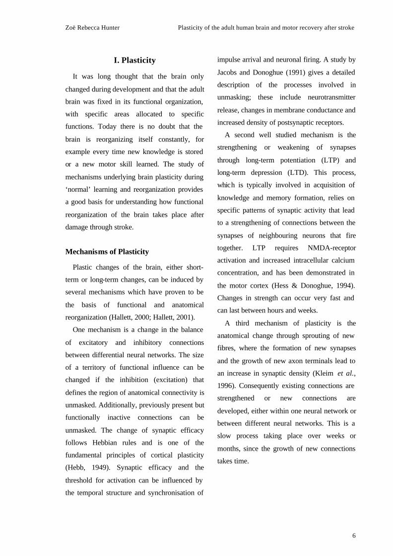
Mechanisms of Plasticity
Plastic changes of the brain, either short-
term or long-term changes, can be induced by
several mechanisms which have proven to be
the basis of functional and anatomical
reorganization (Hallett, 2000; Hallett, 2001).
One mechanism is a change in the balance
of excitatory and inhibitory connections
between differential neural networks. The size
of a territory of functional influence can be
changed if the inhibition (excitation) that
defines the region of anatomical connectivity is
unmasked. Additionally, previously present but
functionally inactive connections can be
unmasked. The change of synaptic efficacy
follows Hebbian rules and is one of the
fundamental principles of cortical plasticity
(Hebb, 1949). Synaptic efficacy and the
threshold for activation can be influenced by
the temporal structure and synchronisation of
impulse arrival and neuronal firing. A study by
Jacobs and Donoghue (1991) gives a detailed
description of the processes involved in
unmasking; these include neurotransmitter
release, changes in membrane conductance and
increased density of postsynaptic receptors.
A second well studied mechanism is the
strengthening or weakening of synapses
through long-term potentiation (LTP) and
long-term depression (LTD). This process,
which is typically involved in acquisition of
knowledge and memory formation, relies on
specific patterns of synaptic activity that lead
to a strengthening of connections between the
synapses of neighbouring neurons that fire
together. LTP requires NMDA-receptor
activation and increased intracellular calcium
concentration, and has been demonstrated in
the motor cortex (Hess & Donoghue, 1994).
Changes in strength can occur very fast and
can last between hours and weeks.
A third mechanism of plasticity is the
anatomical change through sprouting of new
fibres, where the formation of new synapses
and the growth of new axon terminals lead to
an increase in synaptic density (Kleim et al.,
1996). Consequently existing connections are
strengthened or new connections are
developed, either within one neural network or
between different neural networks. This is a
slow process taking place over weeks or
months, since the growth of new connections
takes time.
Zoë Rebecca Hunter Plasticity of the adult human brain and motor recovery after stroke
7
Peripheral deafferentation One typical
condition where short-term plasticity of the
motor cortex can be viewed is after
deafferentation of a limb. Studies with
primates (Donoghue, 1995), as well as
transcranial magnetic stimulation studies with
patients who have lost one arm (for example
Hallett et al. 1993) show that the deafferented
cortex undergoes reorganizational changes.
These changes in cortical representation
reflect a functional output reorganization of the
involved neural networks. Mostly the now
functionally unused part of the cortex, the
territory of the amputated limb, is taken over
by the expanding motor cortex of the muscles
proximal to the amputation. Therefore a
change in cortical representation during the use
of proximal muscles leads to the conclusion
that a functional reorganization of the motor
cortex must have taken place.
A study examining the time needed for
motor cortical representations to change and
the modulation of motor output to occur has
been conducted with reversible deafferentation
in humans (Brasil-Neto et al., 1992). A blood
pressure cuff above the elbow was used to
produce ischemia in the lower part of the arm
and hand. Additionally, sensory input was
inhibited by regional anaesthesia to produce
the overall impression of a deafferented limb.
This was determined by the absence of
movement, the disappearance of motor evoked
potentials (MEP’s) from the muscle ipsilateral
to the cuff and absence of tactile sensation
below the cuff. The amplitudes of the MEP’s
to magnetic stimulation from muscles proximal
to the temporarily deafferented limb were then
measured.
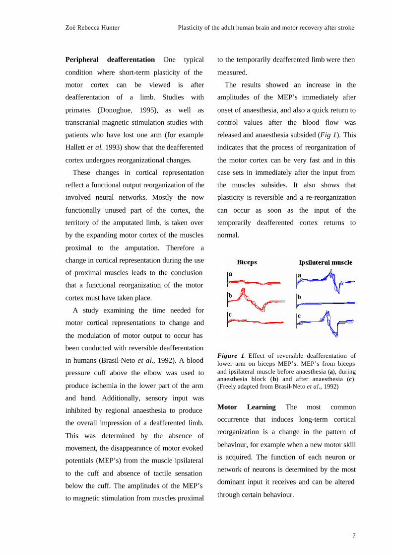
The results showed an increase in the
amplitudes of the MEP’s immediately after
onset of anaesthesia, and also a quick return to
control values after the blood flow was
released and anaesthesia subsided (Fig 1). This
indicates that the process of reorganization of
the motor cortex can be very fast and in this
case sets in immediately after the input from
the muscles subsides. It also shows that
plasticity is reversible and a re-reorganization
can occur as soon as the input of the
temporarily deafferented cortex returns to
normal.
Figure 1: Effect of reversible deafferentation of lower arm on biceps MEP’s. MEP’s from biceps and ipsilateral muscle before anaesthesia (a), during anaesthesia block (b) and after anaesthesia (c). (Freely adapted from Brasil-Neto et al., 1992)
Motor Learning The most common
occurrence that induces long-term cortical
reorganization is a change in the pattern of
behaviour, for example when a new motor skill
is acquired. The function of each neuron or
network of neurons is determined by the most
dominant input it receives and can be altered
through certain behaviour.
Zoë Rebecca Hunter Plasticity of the adult human brain and motor recovery after stroke
8
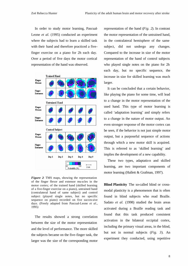
In order to study motor learning, Pascual-
Leone et al. (1995) conducted an experiment
where the subjects had to learn a skilled task
with their hand and therefore practiced a five-
finger exercise on a piano for 2h each day.
Over a period of five days the motor cortical
representation of the hand was observed.
Figure 2: TMS maps, showing the representation of the finger flexor and extensor mu scles in the motor cortex; of the trained hand (skilled learning of a five-finger exercise on a piano), untrained hand (contralateral hand of same subject) and control subject (played single notes, but no specific sequence on piano) recorded on five succes sive days. (Freely adapted from Pascual-Leone et al., 1995)
The results showed a strong correlation
between the size of the motor representation
and the level of performance. The more skilled
the subjects became on the five-finger task, the
larger was the size of the corresponding motor
representation of the hand (Fig. 2). In contrast
the motor representation of the untrained hand,
in the contralateral hemisphere of the same
subject, did not undergo any changes.
Compared to the increase in size of the motor
representation of the hand of control subjects
who played single notes on the piano for 2h
each day, but no specific sequence, the
increase in size for skilled learning was much
larger.
It can be concluded that a certain behavior,
like playing the piano for some time, will lead
to a change in the motor representation of the
used hand. This type of motor learning is
called ‘adaptation learning’ and simply refers
to a change in the nature of motor output. An
even stronger response of the motor cortex can
be seen, if the behavior is not just simple motor
output, but a purposeful sequence of actions
through which a new motor skill is acquired.
This is referred to as ‘skilled learning’ and
implies the development of a new capability.
These two types, adaptation and skilled
learning, are two important components of
motor learning (Hallett & Grafman, 1997).
Blind Plasticity The so-called blind or cross-
modal plasticity is a phenomenon that is often
found in blind subjects who read Braille.
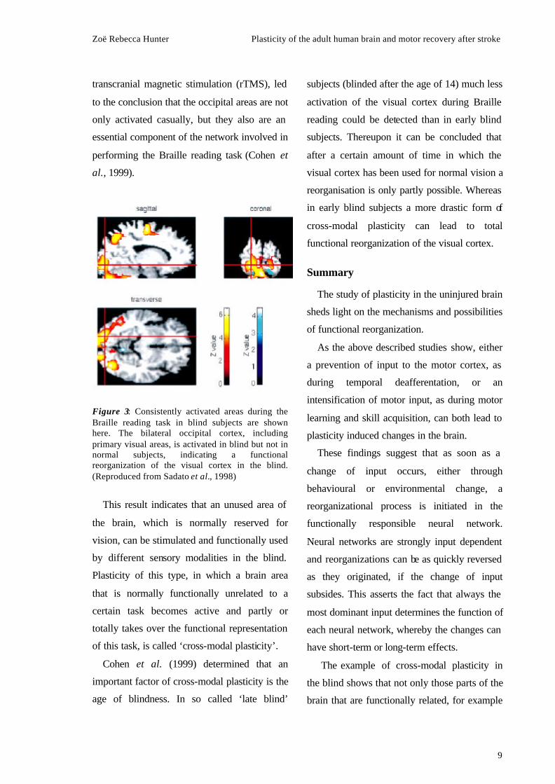
Sadato et al. (1998) studied the brain areas
activated during a Braille reading task and
found that this task produced consistent
activation in the bilateral occipital cortex,
including the primary visual areas, in the blind,
but not in normal subjects (Fig. 3). An
experiment they conducted, using repetitive
Zoë Rebecca Hunter Plasticity of the adult human brain and motor recovery after stroke
9
transcranial magnetic stimulation (rTMS), led
to the conclusion that the occipital areas are not
only activated casually, but they also are an
essential component of the network involved in
performing the Braille reading task (Cohen et
al., 1999).
Figure 3: Consistently activated areas during the Braille reading task in blind subjects are shown here. The bilateral occipital cortex, including primary visual areas, is activated in blind but not in normal subjects, indicating a functional reorganization of the visual cortex in the blind. (Reproduced from Sadato et al., 1998)
This result indicates that an unused area of
the brain, which is normally reserved for
vision, can be stimulated and functionally used
by different sensory modalities in the blind.
Plasticity of this type, in which a brain area
that is normally functionally unrelated to a
certain task becomes active and partly or
totally takes over the functional representation
of this task, is called ‘cross-modal plasticity’.
Cohen et al. (1999) determined that an
important factor of cross-modal plasticity is the
age of blindness. In so called ‘late blind’
subjects (blinded after the age of 14) much less
activation of the visual cortex during Braille
reading could be detected than in early blind
subjects. Thereupon it can be concluded that
after a certain amount of time in which the
visual cortex has been used for normal vision a
reorganisation is only partly possible. Whereas
in early blind subjects a more drastic form of
cross-modal plasticity can lead to total
functional reorganization of the visual cortex.
Summary
The study of plasticity in the uninjured brain
sheds light on the mechanisms and possibilities
of functional reorganization.
As the above described studies show, either
a prevention of input to the motor cortex, as
during temporal deafferentation, or an
intensification of motor input, as during motor
learning and skill acquisition, can both lead to
plasticity induced changes in the brain.
These findings suggest that as soon as a
change of input occurs, either through
behavioural or environmental change, a
reorganizational process is initiated in the
functionally responsible neural network.
Neural networks are strongly input dependent
and reorganizations can be as quickly reversed
as they originated, if the change of input
subsides. This asserts the fact that always the
most dominant input determines the function of
each neural network, whereby the changes can
have short-term or long-term effects.
The example of cross-modal plasticity in
the blind shows that not only those parts of the
brain that are functionally related, for example
Zoë Rebecca Hunter Plasticity of the adult human brain and motor recovery after stroke
10
different parts of the motor cortex, are involved
in reorganizational changes among each other,
but also functionally unrelated brain areas,
such as the visual cortex and the sensorymotor
cortex, can take over additional tasks which
they are normally not connected with.
Accordingly, plasticity can not only occur
within the same neural network, but between
different neural networks. The pathways
usually connected to one network are then re-
routed to another network and regions
originally reserved for other processes will be
functionally invaded. A re-routing of function
of this kind is an age dependent process.
II. Motor Cortex Damage through
Stroke
Anatomy of Motor Cortex
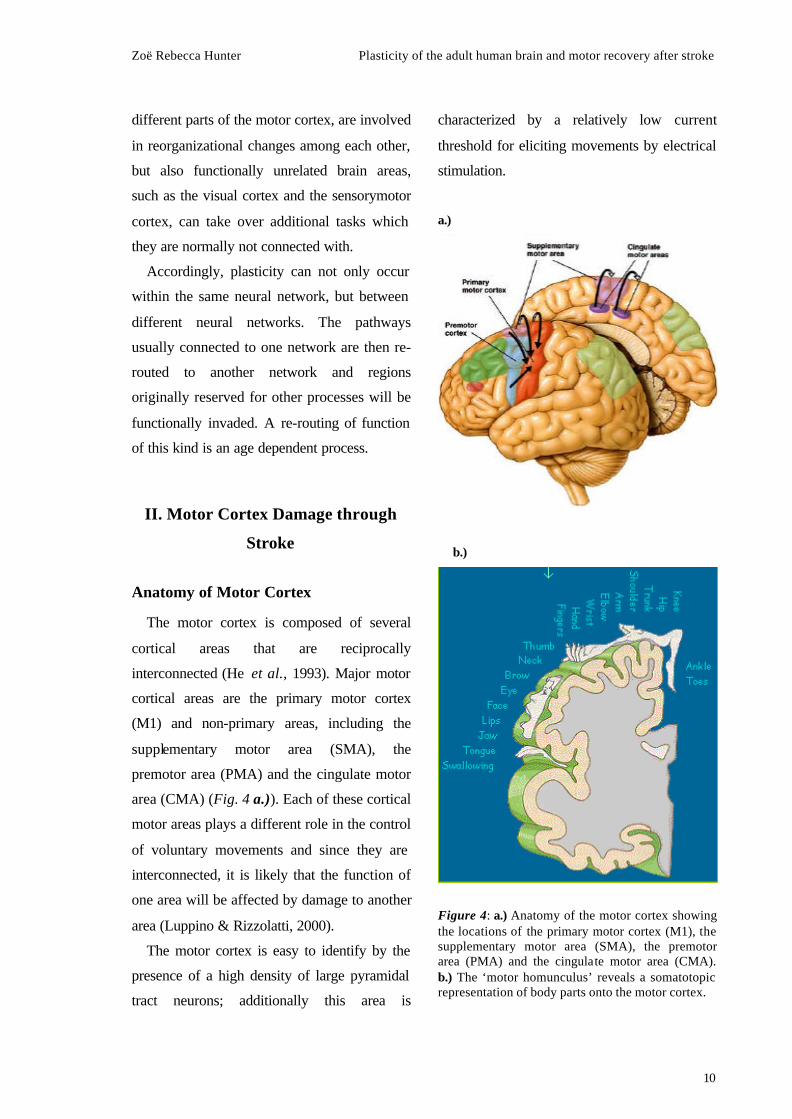
The motor cortex is composed of several
cortical areas that are reciprocally
interconnected (He et al., 1993). Major motor
cortical areas are the primary motor cortex
(M1) and non-primary areas, including the
supplementary motor area (SMA), the
premotor area (PMA) and the cingulate motor
area (CMA) (Fig. 4 a.)). Each of these cortical
motor areas plays a different role in the control
of voluntary movements and since they are
interconnected, it is likely that the function of
one area will be affected by damage to another
area (Luppino & Rizzolatti, 2000).
The motor cortex is easy to identify by the
presence of a high density of large pyramidal
tract neurons; additionally this area is
characterized by a relatively low current
threshold for eliciting movements by electrical
stimulation.
a.)
b.)
Figure 4: a.) Anatomy of the motor cortex showing the locations of the primary motor cortex (M1), the supplementary motor area (SMA), the premotor area (PMA) and the cingulate motor area (CMA). b.) The ‘motor homunculus’ reveals a somatotopic representation of body parts onto the motor cortex.
Zoë Rebecca Hunter Plasticity of the adult human brain and motor recovery after stroke
11
This was utilised by Penfield and Boldrey
(1937) some decades ago to derive maps of the
motor cortex using cortical stimulation
techniques. Their motor maps created a basis
for the somatotopic output organisation of the
motor cortex and resembled a body projection,
the so called ‘motor homunculus’ (Fig. 4 b.)).
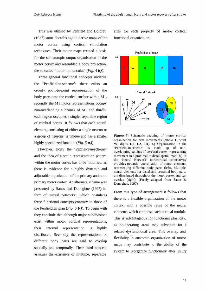
Three general functional concepts underlie
the ‘Penfieldian-scheme’: there exists an
orderly point-to-point representation of the
body parts onto the cortical surface within M1,
secondly the M1 motor representations occupy
non-overlapping subzones of M1 and thirdly
each region occupies a single, separable region
of cerebral cortex. It follows that each neural
element, consisting of either a single neuron or
a group of neurons, is unique and has a single,
highly specialized function (Fig. 5 a.)).
However, today the ‘Penfieldian-scheme’
and the idea of a static representation pattern
within the motor cortex has to be modified, as
there is evidence for a highly dynamic and
adjustable organization of the primary and non-
primary motor cortex. An alternate scheme was
presented by Sanes and Donoghue (1997) in
form of ‘neural networks’, which postulates
three functional concepts contrary to those of
the Penfieldian plan (Fig. 5 b.)). To begin with
they conclude that although major subdivisions
exist within motor cortical representations,
their internal representation is highly
distributed. Secondly the representations of
different body parts are said to overlap
spatially and temporally. Their third concept
assumes the existence of multiple, separable
sites for each property of motor cortical
functional organization.
Figure 5: Schematic d rawing of motor cortical organization for arm movements (elbow E, wrist W, digits D1, D2, D4) a.) Organization in the ‘Penfieldian-scheme’ is made up of non-overlapping patches of cerebral cortex, representing movement in a proximal to distal spatial map. b.) In the ‘Neural Network’ intracortical connectivity provides potential coordination of neural elements representing different body parts (left). Multiple neural elements for distal and proximal body parts are distributed throughout the motor cortex and can overlap (right). (Freely adapted from Sanes & Donoghue, 1997) From this type of arrangement it follows that
there is a flexible organization of the motor
cortex, with a possible reuse of the neural
elements which compose each cortical module.
This is advantageous for functional plasticity,
as co-operating areas may substitute for a
related dysfunctional area. This overlap and
flexibility in anatomic organization of motor
maps may contribute to the ability of the
system to reorganize functionally after injury
Zoë Rebecca Hunter Plasticity of the adult human brain and motor recovery after stroke
12
and provides the basis for recovery of motor
function after stroke.
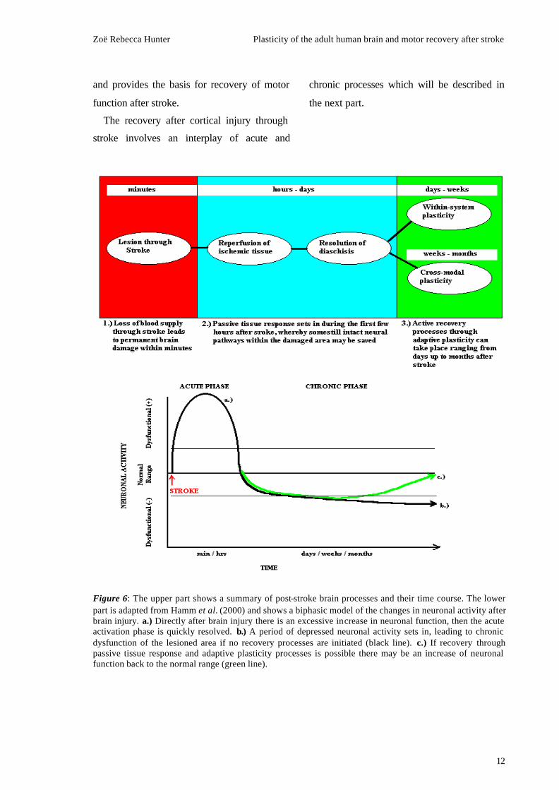
The recovery after cortical injury through
stroke involves an interplay of acute and
chronic processes which will be described in
the next part.
Figure 6: The upper part shows a summary of post-stroke brain processes and their time course. The lower part is adapted from Hamm et al. (2000) and shows a biphasic model of the changes in neuronal activity after brain injury. a.) Directly after brain injury there is an excessive increase in neuronal function, then the acute activation phase is quickly resolved. b.) A period of depressed neuronal activity sets in, leading to chronic dysfunction of the lesioned area if no recovery processes are initiated (black line). c.) If recovery through passive tissue response and adaptive plasticity processes is possible there may be an increase of neuronal function back to the normal range (green line).
13
Pathology of Stroke
The occurrence of stroke is defined as a
sudden loss of blood supply to a certain brain
region, leading to permanent tissue damage.
This may be caused by multiple etiologies such
as atherosclerosis, haemorrhage, cerebral
embolism, artery occlusion or local
thrombosis.
As soon as the blood flow is disrupted the
area affected by ischemia undergoes changes in
neural activity and undersupplied neurons or
neural networks start to degenerate. The
process of destruction happens fast and can
only be prevented or reduced in its extent
through reperfusion of the ischemic tissue
during the first few hours and days after stroke.
After this initial passive tissue response,
whereby some still intact neural pathways
within the damaged neural system may be
saved, the anatomical extent of the damage that
has been caused to the brain area is permanent
and not reversible any more.
Post-stroke Brain Processes Recovery
processes set in immediately and during the
first few hours and days after stroke the passive
tissue response leads to a reperfusion of
ischemic tissue and a cessation of
inflammatory processes, which are secondary
to brain damage. Thereby the extent and time
of initiation of reperfusion ultimately
determine the degree of persistent damage and
consequently the degree of possible later
recovery. Pharmacological treatment within the
first hours after stroke may be able to actively
support these processes and prevent the loss of
neuronal tissue to some extent (Hamm et al.,
2000).
Also the initially induced ‘shock’ of those
neurons which are connected to the lesion site
is resolved during the first few days after
stroke. In these sites, which are connected to
the ischemic area, a functional change in
neuronal activity may lead to metabolic
changes, which cause temporarily and
reversible dysfunction. This condition is
known as diaschis is and its resolution may
explain some motor recovery after stroke (Seitz
et al., 1999).
The above described passive recovery
mechanisms are initiated in the first few hours
and days after the stroke occurred. Then the
damage that was caused can not be reduced
through passive tissue response any more and
the extent of the lesion becomes clear.
Subsequently, active recovery through
reorganizational processes sets in to provide a
basis for functional recovery. These processes
involve adaptive plasticity and take place over
a much longer time course, ranging from days
to months or even years (Fig. 6 summarises
these events). Active recovery through plastic
reorganization will be discussed in the next
section.
III. Plasticity of Motor Cortex after
Stroke
Mechanisms of Recovery
Much of the recovery after the initial days is
likely due to active recovery of the injured
Zoë Rebecca Hunter Plasticity of the adult human brain and motor recovery after stroke
14
motor cortex through mechanisms of adaptive
plasticity. The functions previously performed
by the damaged regions are now taken over by
some areas of the brain that have not been
damaged through stroke.
Several different mechanisms of plastic
recovery have been proposed by Lee and Van
Donkelaar (1995), including redundancy of
brain circuitry, where a parallel pathway
performing a similar function as the damaged
pathway may be able to functionally take over
after brain injury. A second mechanism, which
they have described, leads to functional
recovery after stroke through unmasking of
previously existing but functionally inactive
pathways, a process defined earlier. A third
possibility is the sprouting of new fibres from
surviving neurons, leading to the formation of
new synapses.
Mechanisms such as unmasking and the
substitution of pathways may explain why
functional reorganization is possible, although
most cortical circuits are local and normally
are anatomically restricted in their
functionality.
Apart from several possible mechanisms for
plastic reorganization the extent of functional
recovery also depends on factors such as
location and size of the lesion, the age of the
patient and individual variations in anatomical
and functional connections (Chen, Cohen &
Hallett, 2002).
Basically the recovery mechanisms that will
be activated after stroke depend on the extent
of the injury and have to be distinguished into
at least two different types of plastic recovery.
‘Within-system plasticity’ is possible if some
pathways within the lesioned neural system
have survived undamaged and can be recruited
for recovery processes. If, on the other hand,
there is complete damage to a neural system,
there may still be the possibility to recruit an
alternative system which can compensate for
the functional loss through so called ‘cross-
modal plasticity’ (Seitz & Freund, 1997).
Functional imaging makes it possible to
investigate the different types of plasticity after
stroke in vivo.
Functional Imaging Studies
Studying brain plasticity with non-invasive
functional imaging methods has led to a much
better insight on anatomical reorganization.
Different types of techniques are available,
such as fMRI (functional Magnetic Resonance
Imaging) and PET (Positron Emission
Tomography) or EEG (Electro Encephalo
Graphy), MEG (Magneto Encephalo Graphy)
and TMS (Transcranial Magnetic Stimulation).
Functional imaging is an essential method for
studying plasticity of the human brain, as it
allows the observation of neural activity in the
intact and in the damaged brain. At first a brain
area that represents a specific function in the
normal brain has to be localized, and then it
can be compared to areas that are active in the
post-stroke brain during the same functional
task. The goal of functional imaging is to
obtain direct evidence for a relationship
between cortex reorganization, time course of
recovery and clinical outcome in patients.
Zoë Rebecca Hunter Plasticity of the adult human brain and motor recovery after stroke
15
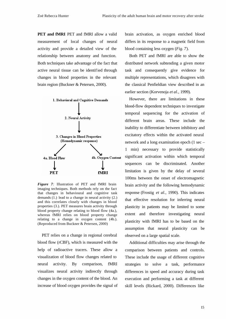
PET and fMRI PET and fMRI allow a valid
measurement of local changes of neural
activity and provide a detailed view of the
relationship between anatomy and function.
Both techniques take advantage of the fact that
active neural tissue can be identif ied through
changes in blood properties in the relevant
brain region (Buckner & Petersen, 2000).
Figure 7: Illustration of PET and fMRI brain imaging techniques. Both methods rely on the fact that changes in behavioural and cognitive task demands (1.) lead to a change in neural activity (2.) and this correlates closely with changes in blood properties (3.). PET measures brain activity through blood property change relating to blood flow (4a.), whereas fMRI relies on blood property change relating to a change in oxygen content (4b.). (Reproduced from Buckner & Petersen, 2000)
PET relies on a change in regional cerebral
blood flow (rCBF), which is measured with the
help of radioactive tracers. These allow a
visualization of blood flow changes related to
neural activity. By comparison, fMRI
visualizes neural activity indirectly through
changes in the oxygen content of the blood. An
increase of blood oxygen provides the signal of
brain activation, as oxygen enriched blood
differs in its response to a magnetic field from
blood containing less oxygen (Fig. 7).
Both PET and fMRI are able to show the
distributed network subtending a given motor
task and consequently give evidence for
multiple representations, which disagrees with
the classical Penfieldian view described in an
earlier section (Korvenoja et al., 1999).
However, there are limitations in these
blood-flow dependent techniques to investigate
temporal sequencing for the activation of
different brain areas. These include the
inability to differentiate between inhibitory and
excitatory effects within the activated neural
network and a long examination epoch (1 sec –
1 min) necessary to provide statistically
significant activation within which temporal
sequences can be discriminated. Another
limitation is given by the delay of several
100ms between the onset of electromagnetic
brain activity and the following hemodynamic
response (Frostig et al., 1990). This indicates
that effective resolution for inferring neural
plasticity in patients may be limited to some
extent and therefore investigating neural
plasticity with fMRI has to be based on the
assumption that neural plasticity can be
observed on a large spatial scale.
Additional difficulties may arise through the
comparison between patients and controls.
These include the usage of different cognitive
strategies to solve a task, performance
differences in speed and accuracy during task
execution and performing a task at different
skill levels (Rickard, 2000). Differences like
Zoë Rebecca Hunter Plasticity of the adult human brain and motor recovery after stroke
16
these lead to functional imaging results that
show differential neural activation patterns, but
are not an evidence for plastic changes. To
avoid this, a comparison is sometimes made
between the damaged and undamaged
hemisphere of the same subject during
performances of the stroke-affected and
unaffected hand, if this is conformable with the
task. The best case would be a comparison of
brain activity during a certain task before and
after the stroke in the same patient, but this is
only seldom possible.1
To conduct an ideal functional imaging
study, three criteria should be fulfilled
(Rickard, 2000). First of all the patient must
show evidence for significant behavioural
recovery of function. Secondly, the task
performed should lead to equivalent execution
of cognitive or information processing steps in
patients and controls. And thirdly there must be
statistically significant activation in the
plasticity area for the patient, but not for the
controls.
Experiments using PET have been
conducted by Chollet et al. (1991), who found
that, compared with movement of the
unaffected hand, movement of the recovered
stroke-affected hand was associated with
increased activation of multiple regions
bilaterally, including cerebellum, primary
sensory motor cortex and premotor cortex.
In a second PET study, Weiller et al. (1993)
compared recovered stroke patients with
1 This is true for human studies, whereas in animal studies the problems due to using a control group do not exist, as will be described later on.
controls and were able to show increased
activation, during movement of the recovered
hand, in the premotor cortex, sensorymotor
cortex and cerebellum of the undamaged
hemisphere, as well as in the bilateral anterior
inferior parietal cortex and the supplementary
motor area.
Both studies give evidence of a role in
recovery for bihemispheric activation,
recruitment of motor-related networks and
cortical map reorganization.
TMS, MEG and EEG The other group of
functional imaging techniques are TMS, MEG
and EEG, which analyse electromagnetic
properties of the brain neurons. TMS allows
painless excitation of the neural structures
underlying a certain motor output by creating a
brief but intense magnetic field. When applied
to the scalp regions corresponding to the motor
cortex, TMS can trigger transient
electromyographic responses in the target
muscles, so called MEP’s, motor evoked
potentials, which allow an examination of the
threshold of excitability (Rossini & Rossi,
1998). With the help of TMS individual motor
output maps can be generated and maps of the
undamaged motor cortex can be compared to
motor maps of the lesioned and recovered
cortex, to detect plasticity induced changes.
These changes show two main characteristics,
either an enlargement or restriction of the
excitable area due to the recruitment or
derecruitment of adjacent neurons, or a
migration of the responsive area outside its
usual boundaries (Rossini & Pauri, 2000).
Zoë Rebecca Hunter Plasticity of the adult human brain and motor recovery after stroke
17
MEG is able to spatially identify the
synchronous firing of neurons in restricted
cortical areas in response to an external
stimulus and due to its physical properties
allows a precise 3D-localisation of the firing
neuronal pool (Williamson & Kaufman, 1990).
Combining MEG and TMS makes it possible
to examine and evaluate the long and short-
term effects on cortical motor organization and
their interhemispheric differences.
Looking at specific patient studies, where
non-invasive functional imaging techniques
were used to investigate recovery mechanisms,
within-system plasticity and cross-modal
plasticity will be further investigated in the
following part.
Within-system Plasticity
Following damage to only part of the motor
cortex or the pyramidal tract, motor recovery is
mediated by reorganization of motor functions
immediately around the stroke site (Cao et al.,
1994) or by the use of alternative cortical areas
somewhere within the motor system, either in
the same hemisphere as the lesion or in the
opposite hemisphere, if these can access spinal
motorneurons (Seitz et al., 1998). Activation to
motor tasks can for example occur in the
supplementary motor area or the premotor
cortex, as these areas have rich
interconnections, as well as connections with
subcortical structures and the primary motor
cortex (Thirumala, Hier & Patel, 2002).
There are three possibilities how within-
system plasticity can be realised: Either
parallel, redundant pathways take over the
function of the damaged pathways or a new
region of the contralateral or ipsilateral
hemisphere2 is functionally reorganized to
compensate for the lost function of the
damaged system.
The third possibility allows a rebuilding of
neuronal connections up to a certain degree
through neuronal sprouting, if some original
connections have survived undamaged.
In a PET study conducted with stroke
patients, Azari and Seitz (2000) tried to find an
answer to the question, which neural networks
in the brain take over the place of the networks
damaged through stroke.
Seven patients had sustained damage to the
primary motor cortex after stroke and
subsequently showed symptoms of paralysis of
the contralateral hand. After a recovery period
of six months the patients had regained the
ability to use the stroke-affected hand. They
then had to perform a sequential finger-
manipulation task with the stroke-affected
hand and with the unaffected hand. During
both tasks the neural activity of the brain was
visualised with PET (Fig. 8).
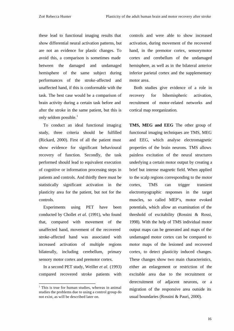
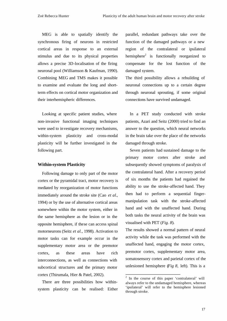
The results showed a normal pattern of neural
activity while the task was performed with the
unaffected hand, engaging the motor cortex,
premotor cortex, supplementary motor area,
somatosensory cortex and parietal cortex of the
unlesioned hemisphere (Fig 8, left). This is a
2 In the course of this paper ‘contralateral’ will always refer to the undamaged hemisphere, whereas ‘ipsilateral’ will refer to the hemisphere lesioned through stroke.
Zoë Rebecca Hunter Plasticity of the adult human brain and motor recovery after stroke
18
normal activation pattern as expected during
this type of task.
a.)
b.)
Figure 8: a.) A sequential finger-manipulation task was used to assess neural activity of recovering stroke patients. Activity of the brain was measured with PET while the patients performed the task with the stroke-affected hand and the unaffected hand. b.) PET images reveal an abnormal activation pattern during task performance of the stroke-affected hand (right) and a normal pattern of activity during task performance of the unaffected hand (left). (Reproduced from Azari & Seitz, 2000)
In contrast, the performance of the same task
with the stroke-affected hand revealed a very
different pattern of neural activation, showing
activity in regions such as the premotor cortex
and the supplementary cortex of both
hemispheres and the prefrontal cortex of the
lesioned hemisphere. This abnormal pattern of
activity leads to the suggestion that the
recovery of the stroke-affected hand is based
on the recruitment of ‘new’ cortex, which is
part of the same neural system.
Parallel Pathways Within the motor system
several parallel motor pathways have been
identified. Not only the primary motor area but
also premotor cortex, supplementary motor
area and the cingulate motor cortex contain
somatotopic representations, and all these
motor areas contribute to the pyramidal tract.
Therefore these parallel pathways can
substitute for each other functionally in
recovery from stroke (Fries et al., 1993).
In the recovered brains of stroke patients
different pathways than in the normal brain are
used to control the stroke-affected hand. In the
normal brain long projections are sent from
neurons in the motor cortex to the pyramidal
tract and excite the spinal motor neurons that
have an effect on muscle contractions of the
hand. However, this normal activation route is
compromised through the lesion and a different
pathway must be used to achieve recovery.
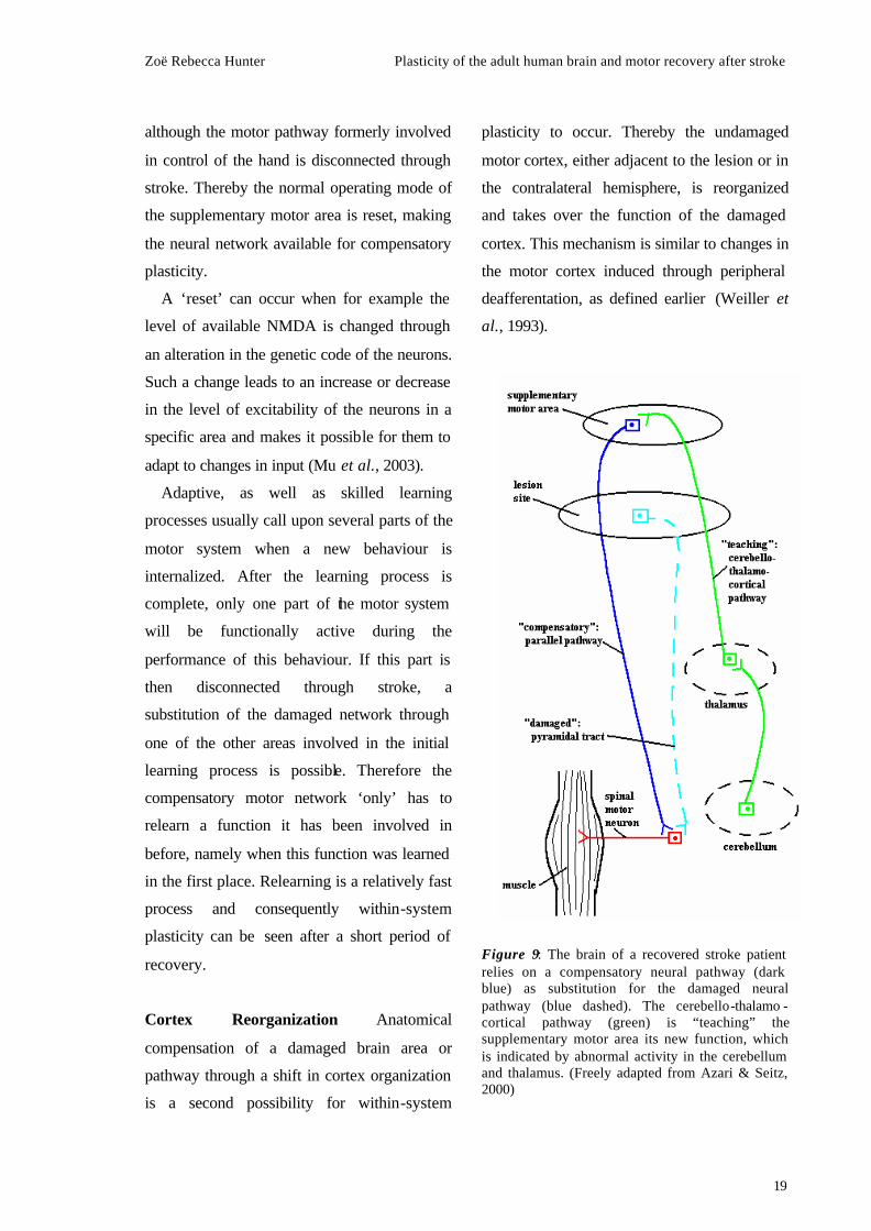
Azari and Seitz (2000) examined neural
activity in patients recovered from paralysis of
one hand after stroke. They were able to detect
the activation of a compensatory pathway
during motor tasks, which led from the
supplementary motor area to the spinal cord
(Fig. 9).
This unusual activation was accompanied by
abnormally enhanced connections between the
thalamus and the cerebellum. In this case the
usage of a strong cerebello-thalamo-cortical
pathway serves as a resetting mechanism for
the compensatory neural processes and is
involved in ‘teaching’ the supplementary
motor area its new role. A ‘detour’ of motor
control allows the brain to function normally,
Zoë Rebecca Hunter Plasticity of the adult human brain and motor recovery after stroke
19
although the motor pathway formerly involved
in control of the hand is disconnected through
stroke. Thereby the normal operating mode of
the supplementary motor area is reset, making
the neural network available for compensatory
plasticity.
A ‘reset’ can occur when for example the
level of available NMDA is changed through
an alteration in the genetic code of the neurons.
Such a change leads to an increase or decrease
in the level of excitability of the neurons in a
specific area and makes it possible for them to
adapt to changes in input (Mu et al., 2003).
Adaptive, as well as skilled learning
processes usually call upon several parts of the
motor system when a new behaviour is
internalized. After the learning process is
complete, only one part of the motor system
will be functionally active during the
performance of this behaviour. If this part is
then disconnected through stroke, a
substitution of the damaged network through
one of the other areas involved in the initial
learning process is possible. Therefore the
compensatory motor network ‘only’ has to
relearn a function it has been involved in
before, namely when this function was learned
in the first place. Relearning is a relatively fast
process and consequently within-system
plasticity can be seen after a short period of
recovery.
Cortex Reorganization Anatomical
compensation of a damaged brain area or
pathway through a shift in cortex organization
is a second possibility for within-system
plasticity to occur. Thereby the undamaged
motor cortex, either adjacent to the lesion or in
the contralateral hemisphere, is reorganized
and takes over the function of the damaged
cortex. This mechanism is similar to changes in
the motor cortex induced through peripheral
deafferentation, as defined earlier (Weiller et
al., 1993).
Figure 9: The brain of a recovered stroke patient relies on a compensatory neural pathway (dark blue) as substitution for the damaged neural pathway (blue dashed). The cerebello-thalamo -cortical pathway (green) is “teaching” the supplementary motor area its new function, which is indicated by abnormal activity in the cerebellum and thalamus. (Freely adapted from Azari & Seitz, 2000)
Zoë Rebecca Hunter Plasticity of the adult human brain and motor recovery after stroke
20
One situation where there is strong evidence
for the activation of the motor cortex
contralateral to the lesion is in recovery from
dysphagia. Swallowing problems affect one in
three patients immediately after stroke, but in
most cases complete recovery occurs within
the first few weeks. A study by Hamdy and
Rothwell (1998) relates the qualitative and
quantitative very good recovery results to how
the area of the motor cortex concerned with
swallowing is organized. The bilateral but
asymmetric inter-hemisphere representation
within the motor and premotor cortex allows
for a good compensation of lost function after
stroke.
If there is a lesion in the swallowing motor
cortex of the hemisphere with the greater
swallowing output, dysphagia is likely to
occur. However, as additional substrate for
swallowing is available in the contralateral
undamaged hemisphere, a functional
reorganization becomes possible. The
contralateral motor cortex is now able to take
over the function of the damaged swallowing
area and increases the capacity for
compensatory reorganization, as well as the
chances of good functional recovery.
A comparison between non-dysphagic
patients and recovered dysphasic patients
shows an increased activation of the
proportionally smaller swallowing motor
cortex in the undamaged hemisphere of the
recovered dysphagic patients. This indicates
that the contralateral hemisphere contributes to
the recovery process by giving of a
functionally identical, formerly unused part of
the motor cortex for reorganizational purposes.
Cortical map reorganization within the
motor system of the ipsilateral damaged
hemisphere, often directly along the lesion rim,
is another mechanism which contributes to
recovery of motor function after stroke. This
theory is supported by the results of Cramer
and Bastings (2000), who measured the
reorganization of multiple cortical map
elements along the lesion rim using fMRI. The
intact cortical regions surrounding the lesion
were able to take over the function of the
damaged region through a shift in cortex
representation. Changes that led to this shift
include a local increase in dendrites, synapses
and levels of proteins related to axonal growth.
A shift in, for example, hand motor
representation may be medial, anterolateral,
ventral or posterior. A study by Weiller et al.
(1993) showed that after damage to the hand
motor cortex movement of the recovered hand
leads to motor cortex activation that extends
laterally to the face area, suggesting that the
hand representation may shift towards the
motor representation of the face. This gives
evidence for a relationship between shifts in
cortical activation and an improvement of
functional performance.
Plastic changes in the ipsilateral damaged
hemisphere generally seem to be more efficient
in producing good recovery, compared to
reorganizational changes involving the
contralateral hemisphere. As demonstrated by
Rossini et al. (1998) the recovering muscles
have enlarged and relocated cortical map
Zoë Rebecca Hunter Plasticity of the adult human brain and motor recovery after stroke
21
representations around the lesion site. These
lead to good functional performance and can
be viewed after only a short period of recovery,
indicating that ipsilateral plasticity is better and
faster than contralateral plasticity in producing
improvement.
Cross-modal Plasticity
In some cases, if the motor cortex is
extensively damaged, patients recruit networks
in areas of the brain that are not normally
involved in the performance of a particular
motor task and which are not part of the
original functional system. Often the
recruitment of an alternative network outside
the damaged system happens in addition to
recruitment of cortex within the damaged
system. However, after complete destruction of
a functional system, substitution by other
systems remains the only alternative (Seitz &
Freund, 1997). Cross-modal plasticity after
stroke is similar to that seen in blind patients,
who engage the visual cortex during a tactile
Braille reading task.
In the above described study by Azari and
Seitz (2000) some recovered stroke patients
were observed, who also recruited networks in
areas of the brain that are not normally
involved in the performance of a sequential
finger-manipulation task. When these patients
moved the stroke affected hand, neural activity
could be viewed in parts of the visual cortex,
although they were not receiving any visual
input, as they were blindfolded as a control.
The visual cortex seemed to subserve a motor
function and the active areas within the visual
cortex could be functionally associated through
PET scan with the motor task that had to be
performed. The recruitment of visual cortex
happened in the late stages of recovery (after
several months) and involved cross-modal
adaptive plasticity.
A temporal distinction was found between
cross-modal and within-system plasticity
(Azari & Seitz, 2000). Patients who recruited
an alternative network had been recovering for
at least six months, while the recovery process
of patients using within-system plasticity took
only a few weeks. This leads to the suggestion
that there must be a distinct time course in
recovery processes. Within-systems seem to be
easy to access and are recruited fairly early,
whereas alternative cross-modal networks are
difficult to access and need a longer time to be
effective. The alternative network is naïve to
the task it is supposed to perform, so that the
process of recovery does not only involve a
relearning of the task, as during within-system
recovery, but the system has to learn what to
do in the first place, and this takes more time.
Animal Experiments
In conducting animal experiments for studying
the effects of stroke, some of the difficulties
which affect patient studies can be avoided.
For example it is possible to study brain
activity before and after stroke in the same
animal, not having to refer to data of
unaffected control subjects for comparison.
Another advantage is that the lesion can be
induced intentionally in exact the area of the
animals brain involved in the performance of a
Zoë Rebecca Hunter Plasticity of the adult human brain and motor recovery after stroke
22
specific task, which has been trained
beforehand. The brain area that has been
identified to functionally represent this task is
lesioned and can then be studied during
performance of the same task directly after the
lesion and after a period of recovery. Thereby
data derived from only one animal provides a
good basis for the direct comparison of activity
before and after stroke and can reveal
information about recovery induced plasticity.
Motor Cortex Lesions in Mice Skilled
reaching movements, which are an important
aspect of human motor behaviour, are typically
impaired after a stroke to the motor cortex. To
study the effects underlying the impairment
and subsequent functional recovery, Farr &
Whishaw (2002) developed a ‘mouse- model’
of human motor stroke.
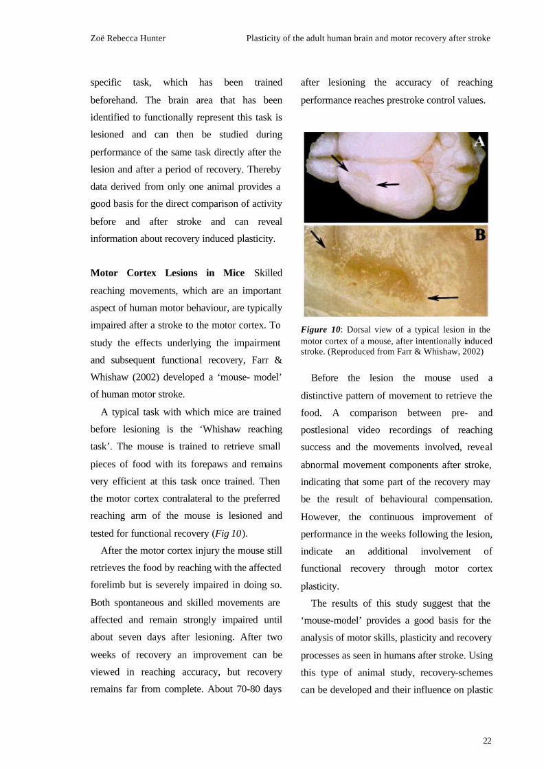
A typical task with which mice are trained
before lesioning is the ‘Whishaw reaching
task’. The mouse is trained to retrieve small
pieces of food with its forepaws and remains
very efficient at this task once trained. Then
the motor cortex contralateral to the preferred
reaching arm of the mouse is lesioned and
tested for functional recovery (Fig 10).
After the motor cortex injury the mouse still
retrieves the food by reaching with the affected
forelimb but is severely impaired in doing so.
Both spontaneous and skilled movements are
affected and remain strongly impaired until
about seven days after lesioning. After two
weeks of recovery an improvement can be
viewed in reaching accuracy, but recovery
remains far from complete. About 70-80 days
after lesioning the accuracy of reaching
performance reaches prestroke control values.
Figure 10: Dorsal view of a typical lesion in the motor cortex of a mouse, after intentionally induced stroke. (Reproduced from Farr & Whishaw, 2002)
Before the lesion the mouse used a
distinctive pattern of movement to retrieve the
food. A comparison between pre- and
postlesional video recordings of reaching
success and the movements involved, reveal
abnormal movement components after stroke,
indicating that some part of the recovery may
be the result of behavioural compensation.
However, the continuous improvement of
performance in the weeks following the lesion,
indicate an additional involvement of
functional recovery through motor cortex
plasticity.
The results of this study suggest that the
‘mouse-model’ provides a good basis for the
analysis of motor skills, plasticity and recovery
processes as seen in humans after stroke. Using
this type of animal study, recovery-schemes
can be developed and their influence on plastic
Zoë Rebecca Hunter Plasticity of the adult human brain and motor recovery after stroke
23
reorganization can be tested, to help to provide
appropriate schemes for human recovery after
stroke.
IV. Modelling Plasticity with
Artificial Neural Networks
In order to understand how the brain
recovers from stroke to the motor cortex and
how the mechanisms underlying this recovery
work, traditionally either clinical studies with
stroke patients or animal models have been
pursued, as described above. An alternative
approach is the use of computational models,
to investigate the reorganizational capacities of
the motor cortex following a lesion. As
computational models are very useful for the
analysis of complex systems in general, they
seem to be ideal for examining the complex
events occurring in the brain during and after
stroke (Reggia et al., 2000).
Hebbian Learning
Although brain functions are strongly
genetically determined, not all details of the
brain networks and their interactions are
specified from the onset. This gives neural
networks the opportunity to adapt functionally
to changes in input. An important adaptation
mechanism is synaptic plasticity, which can be
implemented in artificial neural networks using
biologically realistic learning rules
(Trappenberg, 2002).
‘Hebbian learning’ is the principle idea
behind such rules, based on a theory outlined
by Hebb (1949). It provides a framework for
investigating the interactions between neural
and behavioural levels of analysis and relies on
the following assumption: The synaptic
strength and therefore the weight between two
connected neural elements increases when both
elements are active that is there is a correlation
between presynaptic and postsynaptic activity,
otherwise it decreases.
The ability of networks of neurons to form
associations between co-occurrences of stimuli
is the basis for many information processing
mechanisms in the brain. Implementing these
abilities in the brain, by using rules governing
synaptic plasticity, enables networks of
neurons to efficiently engage in local learning
mechanisms through LTP, leading to a change
in their response and a reorganization of
functional connectivity (Wolters et al., 2003).
Local changes between two synapses cause a
change in the neural network they are involved
in, which in turn may lead to a reorganization
of connecting areas, indicating that the basic
principle of ordinary learning can lead to
functional reorganization on a large scale (Hess
& Donoghue, 1994).
Two groups of neurons that have been
disconnected by a lesion can reconnect, if they
are activated at the same time through an
external circuit whose neurons are functionally
interconnected. The activation of this neural
network leads to simultaneous activation of the
disconnected neurons, which may become
reconnected through repetition of this process.
Using this knowledge to implement brain
functions in artificial neural networks can help
to reveal many details of synaptic plasticity
and possibilities of manipulation. Therefore
Zoë Rebecca Hunter Plasticity of the adult human brain and motor recovery after stroke
24
using artificial neural network modelling and
the relatively simple mechanisms involved in
Hebbian learning is very useful for studying
the plastic changes occurring in the brain after
stroke.
Computational Models
One goal of developing a computational
model of stroke is to understand the changes
the lesioned tissue, the connected areas and the
undamaged parts of the brain undergo. Upon
this knowledge those factors that may lead to a
better recovery can be determined and then are
used to derive recovery-schemes that can
improve functional outcome after stroke.
Several computational models of cortical
map self-organization and map refinement
have been developed to achieve this goal
(Ritter et al., 1992). Typically these models are
constructed by using a two-layer network and
an unsupervised Hebbian learning method,
often involving competitive learning. These
studies help to derive some plausible
assumptions about network architecture and
synaptic modifications due to plastic changes
of the brain.
Lesion and Recovery of a Motor Model To
be able to demonstrate how brain damage can
be modeled computationally, Reggia et al.
(2000) developed models which were
explicitly intended to simulate plasticity
following a small stroke to the motor cortex
and examine compensatory mechanisms in
areas immediately surrounding the lesioned
tissue.
Their model consisted of two parts: a
simulated arm able to move in three-
dimensional space and a closed-loop of neural
elements, each representing a group of real
neurons, responsible for controlling and
sensing the arms position through
proprioceptive input and motor output. If the
lower motor neuron elements are activated they
position the model arm in a specific spatial
position and the arm then generates input
signals to the cortex via proprioceptive neuron
elements.
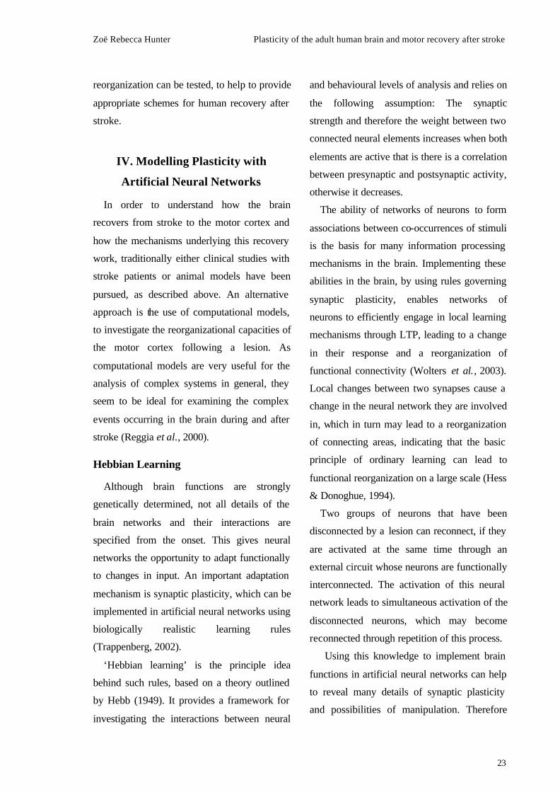
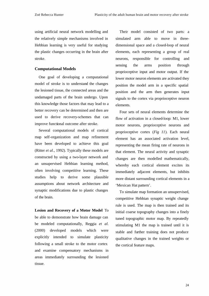
Four sets of neural elements determine the
flow of activation in a closed-loop: M1, lower
motor neurons, proprioceptive neurons and
proprioceptive cortex (Fig 11). Each neural
element has an associated activation level,
representing the mean firing rate of neurons in
that element. The neural activity and synaptic
changes are then modelled mathematically,
whereby each cortical element excites its
immediately adjacent elements, but inhibits
more distant surrounding cortical elements in a
‘Mexican Hat pattern’.
To simulate map formation an unsupervised,
competitive Hebbian synaptic weight change
rule is used. The map is then trained and its
initial coarse topography changes into a finely
tuned topographic motor map. By repeatedly
stimulating M1 the map is trained until it is
stable and further training does not produce
qualitative changes in the trained weights or
the cortical feature maps.
25
After training the M1 motor map develops
clusters of elements representing the same
muscle group. When this stage is reached the
model is lesioned through permanently setting
the activation levels of a set of cortical
elements at zero and severing the connections
to and from the lesioned elements. Then the
effect of the lesion on the trained motor cortex
is examined directly after lesioning and after a
period of continually retraining the lesioned
network.
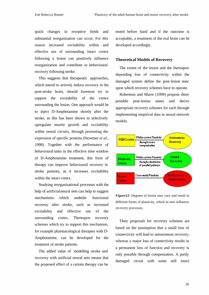
After a structural lesion to M1,
reorganization can be viewed in both the M1
sensory and motor output maps. The results
show a two-phase model of recovery.
Immediately after the lesion the M1 maps
adjusted and the number and excitability of
responsive elements in the normal cortex near
the lesion edge increased. Additionally, overall
rates of responsiveness in the M1 sensory map
and relative activity in the M1 motor map
increased. These changes in activation
dynamics form the first, very rapid phase of
recovery, which is then followed by a slow,
second phase due to synaptic plasticity.
The increased excitability following the
lesion is necessary for map reorganization to
be initiated in the cortex surrounding the
lesion, which consistently participates in the
reorganization process and achieves a higher-
density feature map than before the lesion. This
can be explained by the synaptic modification
rule that underlies map formation. Changes in
the receptive field of a cortical element happen
through a shift in the receptive field, to become
more like the pattern of input elements that
activate that cortical element. Therefore low
activity following a lesion leads to very slow
changes of the receptive fields and only limited
reorganization. Whereas high activity induces
Figure 11: Structure of the closed-loop of neural elements: 12 proprioceptiv neuron elements form the input layer and are fully connected to the proprioceptive cortex (P1). P1 and the primary motor cortex (M1) are two-dimensional arrays of neural elements, with a partial projection from P1 to M1 in a coarse topographic order. M1 is connected to six lower motor neuron elements. The simulated arm model transforms activity in lower motor neurons into proprioceptive input. (Freely adapted from Reggia et al., 2000)
Zoë Rebecca Hunter Plasticity of the adult human brain and motor recovery after stroke
26
quick changes in receptive fields and
substantial reorganization can occur. For this
reason increased excitability within and
effective use of surrounding intact cortex
following a lesion can positively influence
reorganization and contribute to behavioural
recovery following stroke.
This suggests that therapeutic approaches,
which intend to actively induce recovery in the
post-stroke brain, should foremost try to
support the excitability of the cortex
surrounding the lesion. One approach would be
to inject D-Amphetamine shortly after the
stroke, as this has been shown to selectively
upregulate neurite growth and excitability
within neural circuits, through promoting the
expression of specific proteins (Stroemer et al.,
1998). Together with the performance of
behavioural tasks in the effective time window
of D-Amphetamine treatment, this form of
therapy can improve behavioural recovery in
stroke patients, as it increases excitability
within the intact cortex.
Studying reorganizational processes with the
help of artificial neural nets can help to suggest
mechanisms which underlie functional
recovery after stroke, such as increased
excitability and effective use of the
surrounding cortex. Thereupon recovery
schemes which try to support this mechanism,
for example pharmacological therapies with D-
Amphetamine, can be developed for the
treatment of stroke patients.
The added value of modelling stroke and
recovery with artificial neural nets means that
the proposed effect of a certain therapy can be
tested before hand and if the outcome is
acceptable, a treatment of the real brain can be
developed accordingly.
Theoretical Models of Recovery
The extent of the lesion and the thereupon
depending loss of connectivity within the
damaged system define the post-lesion state
upon which recovery schemes have to operate.
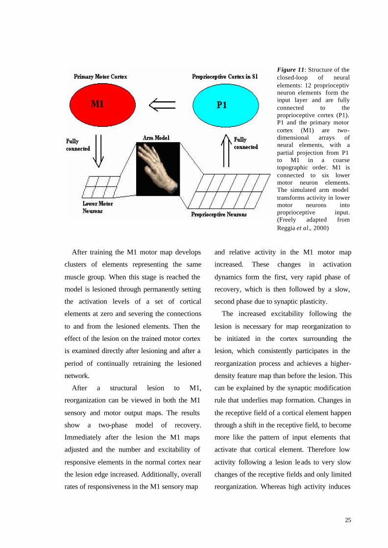
Robertson and Murre (1999) propose three
possible post-lesion states and derive
appropriate recovery schemes for each through
implementing empirical data in neural network
models.
Figure12: Degrees of lesion may vary and result in
different forms of plasticity, which in turn influence
recovery processes.
Their proposals for recovery schemes are
based on the assumption that a small loss of
connectivity will lead to autonomous recovery,
whereas a major loss of connectivity results in
a permanent loss of function and recovery is
only possible through compensation. A partly
damaged circuit with some still intact
Zoë Rebecca Hunter Plasticity of the adult human brain and motor recovery after stroke
27
connections may be saved through principles
of guided recovery (Fig. 12).
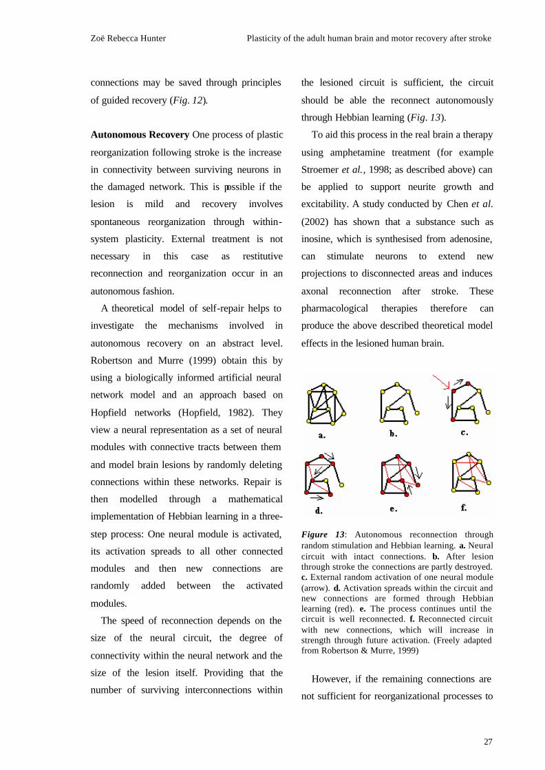
Autonomous Recovery One process of plastic
reorganization following stroke is the increase
in connectivity between surviving neurons in
the damaged network. This is possible if the
lesion is mild and recovery involves
spontaneous reorganization through within-
system plasticity. External treatment is not
necessary in this case as restitutive
reconnection and reorganization occur in an
autonomous fashion.
A theoretical model of self-repair helps to
investigate the mechanisms involved in
autonomous recovery on an abstract level.
Robertson and Murre (1999) obtain this by
using a biologically informed artificial neural
network model and an approach based on
Hopfield networks (Hopfield, 1982). They
view a neural representation as a set of neural
modules with connective tracts between them
and model brain lesions by randomly deleting
connections within these networks. Repair is
then modelled through a mathematical
implementation of Hebbian learning in a three-
step process: One neural module is activated,
its activation spreads to all other connected
modules and then new connections are
randomly added between the activated
modules.
The speed of reconnection depends on the
size of the neural circuit, the degree of
connectivity within the neural network and the
size of the lesion itself. Providing that the
number of surviving interconnections within
the lesioned circuit is sufficient, the circuit
should be able the reconnect autonomously
through Hebbian learning (Fig. 13).
To aid this process in the real brain a therapy
using amphetamine treatment (for example
Stroemer et al., 1998; as described above) can
be applied to support neurite growth and
excitability. A study conducted by Chen et al.
(2002) has shown that a substance such as
inosine, which is synthesised from adenosine,
can stimulate neurons to extend new
projections to disconnected areas and induces
axonal reconnection after stroke. These
pharmacological therapies therefore can
produce the above described theoretical model
effects in the lesioned human brain.
Figure 13: Autonomous reconnection through random stimulation and Hebbian learning. a. Neural circuit with intact connections. b. After lesion through stroke the connections are partly destroyed. c. External random activation of one neural module (arrow). d. Activation spreads within the circuit and new connections are formed through Hebbian learning (red). e. The process continues until the circuit is well reconnected. f. Reconnected circuit with new connections, which will increase in strength through future activation. (Freely adapted from Robertson & Murre, 1999)
However, if the remaining connections are
not sufficient for reorganizational processes to
Zoë Rebecca Hunter Plasticity of the adult human brain and motor recovery after stroke
28
occure due to the extent of the lesion, the
reconnection process can be supported by a
principle of guided recovery.
Guided Recovery Whereas autonomous
recovery is independent of exogenous
behavioural influences, guided recovery
depends on specific external stimulation. There
are various methods available for this purpose,
including non-specific stimulation, which can
be further classified into bottom-up targeted
stimulation and top-down targeted stimulation
(Robertson & Murre, 1999).
It has been shown that environmental and
behavioural factors have strong elevating
effects on synaptic connectivity and dendritic
sprouting in animal neural circuits (for
example Will & Kelche, 1992). In the above
described model of reconnection through
Hebbian learning, non-specific environmental
stimulation should then facilitate synaptic
connectivity through a greater number of
coactivations between the disconnected nodes.
As a result the reconnection processes of the
model should be faster and even more
successful in producing good functional
recovery.
One example for non-specific environmental
stimulation that can be tested in the real brain
is multimodal stimulation. In an experimental
approach Volpe et al. (2000) tested the effect
of multidisciplinary rehabilitation activity on
functional outcome in stroke patients. They
used a novel therapeutic strategy where
additional training of the stroke affected limb
was delivered by a robotic device, through
interacting with the patient in real-time and
guiding the affected limb through a stereotyped
movement pattern. This robot aided therapy
leads to enhanced motor outcome, as it aids
reconnectional processes through non-specific
stimulation.
One draw back of non-specific stimulation is
that it may cause maladaptive connections.
This occurs if the stimulation leads to a co-
activation of the lesioned circuit with some
other neural network, instead of co-activation
with disconnected parts of the lesioned
network. The other neural network then
increases its strength and region of
functionality (Fig.14). A reconnection of the
lesioned network is therefore inhibited or still
existing connections are further reduced, as
there is a competition for connectivity induced
through non-specific stimulation.
Consequently, it is important to stimulate the
target area, namely the lesioned neural circuit,
directly.
For stroke patients needing a stimulative
therapy the theoretical principle of maladaptive
plasticity indicates that the patients may be
deprived of the opportunity for recovery if the
rehabilitation inadvertently activates circuits
that competitively inhibit the impaired neural
network (Fitzsimonds et al., 1997).
Guided, targeted stimulation can either be
realized in a bottom-up or top-down fashion.
Bottom-up processes should provide a
sequence of cued inputs, which are able to
precisely boost connections within the lesioned
network through repeated stimulation. Such
repetitive training would allow a faster
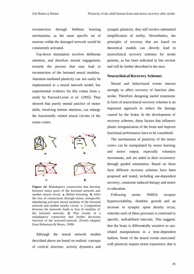
Zoë Rebecca Hunter Plasticity of the adult human brain and motor recovery after stroke
29
reconnection through Hebbian learning
mechanisms, as the same specific set of
neurons within the damaged network would be
consistently activated.
Top-down stimulation involves deliberate
attention, and therefore mental engagement,
towards the process that may lead to
reconnection of the lesioned neural modules.
Attention-mediated plasticity can not easily be
implemented in a neural network model, but
experimental evidence for this comes from a
study by Pascual-Leone et al. (1995). They
showed that purely mental practice of motor
skills, involving intense attention, can enlarge
the functionally related neural circuits of the
motor cortex.
Figure 14: Maladaptive connections that develop between intact parts of the lesioned network and another neural circuit. a. Before lesioning. b. After the loss of connections through lesion, nonspecific stimulating activates neural modules of the lesioned network and another nearby circuit. c. Competition between the networks leads to loss of modules of the lesioned network. d. This results in a maladaptive connection that further decreases function of the lesioned network. (Freely adapted from Robertson & Murre, 1999)
Although the neural network models
described above are based on realistic concepts
of cortical structure, activity dynamics and
synaptic plasticity, they still involve substantial
simplification of reality. Nevertheless, the
principles of recovery that are based on
theoretical models can directly lead to
neuroclinical recovery schemes for stroke
patients, as has been indicated in this section
and will be further described in the next.
Neuroclinical Recovery Schemes
Neural and behavioural events interact
strongly to affect recovery of function after
stroke. Therefore designing useful treatments
in form of neuroclinical recovery schemes is an
important approach to reduce the damage
caused by the lesion. In the development of
recovery schemes, those factors that influence
plastic reorganization of the brain and improve
functional performance have to be considered.
The mechanisms of plasticity of the motor
cortex can be manipulated by motor learning
and motor output, especially voluntary
movements, and are aided in their occurrence
through guided stimulation. Based on these
facts different recovery schemes have been
proposed and tested, including use-dependent
recovery, constraint-induced therapy and motor
re-education.
Following stroke NMDA receptor
hyperexcitability, dendritic growth and an
increase in synaptic spine density occur,
whereby each of these processes is restricted to
specific, well-defined intervals. This suggests
that the brain is differentially sensitive to use-
related manipulations in a time-dependent
fashion. Some of the neural events associated
with plasticity require motor experience, that is
Zoë Rebecca Hunter Plasticity of the adult human brain and motor recovery after stroke
30
they are use-dependent. The experience of the
appropriate use necessarily has to occur
coincidentally with the optimal time-window
for the related neural event (Schallert et al.,
2000).
Neural events have been shown to be
modifiable by disuse, use and overuse. One
example for disuse is the ‘learned nonuser’ that
can be viewed in the stroke affected limb after
a period of recovery (Taub et al., 1994). In
order to compensate for impaired functioning
of this limb after stroke, alternative
behavioural strategies are often adopted
leading to non-use of the affected limb. The
reestablishment of normal motor patterns, even
if functionally possible, is therefore inhibited.
Liepert et al. (1998) studied the effect of
constraint-induced movement therapy in the
brains of stroke patients. They immobilized the
functionally unaffected arm of patients several
months after stroke, and in combination with
motor re-education in form of physical therapy
(such as mechanical stimulation, massage and
passive movement) were able to reverse the
effect of learned non-use, leading to significant
functional improvement in the stroke affected
arm. Whereas total disuse of the affected limb
over an extended period of time causes the
region of the lesion to expand, forced overuse
of the affected limb, especially very early after
stroke, has the same effect of inhibiting
functional improvement (Kozlowski et al.,
1996). Consequently it is important to give the
right amount of stimuli at the most appropriate
time point.
A fMRI study examining the effects of
constraint-induced movement therapy on the
functionally related areas of the brain
(Johansen-Berg et al., 2002), showed an
increase in brain activity. Patients took part in
a two-week home-based therapy programme,
combining restraint of the unaffected limb with
progressive exercise of the affected limb. As a
result, improvement in hand function after
therapy could be viewed, which correlated with
increases in fMRI activity in the premotor
cortex and secondary somatosensory cortex of
the lesioned hemisphere. These changes in
activity are associated with successful motor
rehabilitation, thus providing evidence for
recovery after specific input, as has been
predicted above by the neural network model
of guided, targeted stimuli.
IV. Conclusion
Several questions were proposed in the
introduction and have been thoroughly
investigated in the course of this paper with the
goal of finding appropriate answers.
The first question was, ‘How plastic is the
brain when recovering from stroke?’
When the brain recovers from a lesion to the
motor cortex after stroke, it nearly always
engages in reorganizational processes, which
then lead to better functional recovery.
Therefore plasticity of the brain during
recovery can be seen in most cases. The degree
of plasticity and consequently the degree of
recovery depend strongly on the extent of the
lesion.
Zoë Rebecca Hunter Plasticity of the adult human brain and motor recovery after stroke
31
The second question asked, ‘Which different
types of plasticity can be distinguished and
what effects do they have on functional
performance?’
A very severe lesion may completely
destroy a neural network representing a
specific function and makes recovery through
reorganization of the lesioned area impossible.
In this case the only possible plasticity process
that can support recovery of function is cross-
modal adaptive plasticity. A different area of
the cortex that operates independent from the
lesioned area is functionally invaded and
makes recovery possible through allocating an
undamaged neural network towards regaining
the functionally impaired behaviour. Recovery
in this case is limited in its quality of
functional compensation and as the substituted
system is extrinsic to the task, it is also a fairly
slow process. This type of plasticity is similar
to the cross-modal plasticity seen in blind
patients during a tactile Braille reading task.
In contrast, if the lesion is moderate to mild,
a reorganization of the lesioned area and the
surrounding cortex is possible through within-
system plasticity and may lead to a fast and
very good recovery. Neural modules that have
survived the lesion undamaged can be
reconnected and neural networks belonging to
the same functional system can functionally
reorganize. This type of plasticity seems
essential for achieving optimal recovery after
stroke.
‘What kind of factors influence the degree of
plasticity and therefore the degree of
recovery?’ was the third question proposed in
the introduction.
A patient who is not able to move his arm
after stroke is fairly impaired in daily life,
especially as skilled use of the hands and the
performance of fine tuned movements are often
the most difficult. During rehabilitation two
different approaches are possible, either a
passive movement therapy is initiated and the
patient is encouraged to use the stroke-affected
arm, with the goal of providing conditions
under which voluntary movement can be
obtained. Or the patient must learn to cope
with daily challenges without using the stroke-
affected arm, therefore engaging in strategies
of behavioural compensation.
Changes in the cortex are always experience
dependent and plasticity of the brain is a key
mechanism underlying both normal learning
and recovery of function following brain
damage. This is a very important fact with
respect to initiating recovery processes, as a
variation in experience and motor input to
damaged neural circuits is a factor that will
strongly shape synaptic interconnections and
hence influence recovery. Therefore supporting
the brain’s capacity for plastic reorganization
through stimulative input to the motor cortex
has shown to have the same effects as motor
learning in general, namely an expansion of the
motor cortex representation of the involved
body part. This is the main pre-requisite for
good functional recovery.
Knowledge of the physiological mechanisms
of plasticity can help to develop new therapies
for stroke patients. Especially the observation
Zoë Rebecca Hunter Plasticity of the adult human brain and motor recovery after stroke
32
that disuse, use and overuse can strongly
modify neural events is important in this
respect.
As the example of peripheral deafferentation
of a limb shows, there is a strong competition
between adjacent muscles for areas of
functional representation in the motor cortex.
Immediately after the input from a certain
muscle subsides, a reorganizational process
sets in. The neighbouring muscles will start to
expand their territory of functional
representation and invade the now functionally
unused part of the cortex. A further tissue loss
can be prevented through purposefully
retraining the affected muscle and forced non-
use of the adjacent muscles. Consequently,
when developing a rehabilitative training
program, usage of the muscles that are close to
the stroke-affected muscle should be
prevented, for example through anaesthesia.
Apart from exogenous, therapeutic
influences on recovery, other endogenous
factors play an important role. The way in
which the stroke-affected part of the body is
represented in the brain provides the working
basis for processes of plasticity.
For example the swallowing motor cortex
reorganization shows how plasticity strongly
depends on the cortex organization of the
lesioned area. The bilateral representation of
the swallowing motor cortex allows for a good
compensation of the lost function, as additional
substrate for network reorganization is
available in the undamaged hemisphere.
The fourth question was, ‘How can
theoretical and practical experimental findings
help to develop successful therapeutical
approaches for recovery schemes?’
Analysing the determinants of normal short-
and long-term plasticity in the undamaged
motor cortex, as in skilled motor learning,
provides a useful basis for determining whether
and how functional recovery can be guided
through the input of appropriate recovery
schemes.
It seems that the continual use of a limb is
critical for achieving and maintaining an
appropriate cortical representation. Improving
function of the stroke-affected limb therefore
depends on actively using it. Increased
excitability and effective use of the cortex
surrounding the lesion can be exploited
through therapeutic techniques that stimulate
movement. Thereby optimal adaptation can be
reached through skilled use of the stroke
affected limb, rather than just increasing use.
To intensify the training effect and to prevent a
competition between input of the stroke-
affected and the unaffected limb, constraint-
induced therapy combined with skilled motor
re-education seems to produce the best results
in functional recovery.
As has been predicted by the neural network
model, the functional input following a lesion
has to be a guided, targeted stimulus.
Constraining the unaffected limb and in doing
so preventing competitive input, as well as
intensively training the affected limb, leads to a
functionally much better reorganization of the
lesioned neural network than could be achieved
Zoë Rebecca Hunter Plasticity of the adult human brain and motor recovery after stroke
33
through non-specific input. Additionally, the
constraint-induced therapy can produce
reorganizational effects not only in acute stroke
patients, but also in chronic stroke patients,
suggesting that the motor cortex retains a
capacity for recovery through plasticity over a
long period of time after the lesion occurred.
Using neuroimaging techniques provides a
basis for bridging the gap between clinical
practice and the neural representation of
recovery mechanisms in the brain, leading to
new physical rehabilitative therapies. In
providing information on the excitability,
extension, and localisation of motor cortex
areas during recovery, functional imaging
plays an important role for viewing
possibilities of functional reorganization.
Nevertheless, the specificity with which
brain reorganization relates to good clinical
outcome is still not completely understood.
This is due to the fact that most patients that
have been studied show good functional
recovery. Furthermore, additional factors that
may influence recovery, such as age, lesion
location and prestroke neurological status
make it difficult to generalize experimental
findings.
In this paper reorganization capabilities of
the brain due to mechanisms of plasticity are
looked at referring to relatively low-level
functioning of the motor cortex. Many
functional imaging studies have been
conducted in this area, as the motor circuits in
the cortex are well-understood and the precise
topographical maps that exist in this area easily
reveal any reorganization that takes place.
Nevertheless, there are also several
neuroimaging studies that give evidence for the
reorganization of circuits that underlie higher-
level cognitive functions, such as memory and
language, which occurs in a similar fashion to
reorganization of the motor cortex (for
example Weiller et al., 1995).
Through dealing with and answering the
questions that were set out in the beginning, it
was possible to fulfil the main goal of the
thesis, which was to find out how recovery can
be positively influenced by neuroclinical
recovery schemes, leading to a better
functional outcome after stroke, and evaluate
how ‘plastic’ the brain really is.
Recovery of function after a lesion remains
very difficult to predict in individual cases, as
many factors that vary in individuals have an
effect on the outcome. Therefore a
generalisation of post-lesion training effects
over time or across patients and their
individual situation is not always possible.
Individual variations in functional connectivity
of the prestroke brain have a large influence on
the ability of the affected neural network to
recover from the lesion. Strong cortical
connectivity between neural modules offers a
better basis for reconnection and
reorganization than low cortical connectivity.
Consequently, the functional outcome is not
decided by the number of neurons left, but how
they function and which connections they are
able to make.
The principle of Hebbian learning
implemented in neural networks provides a
Zoë Rebecca Hunter Plasticity of the adult human brain and motor recovery after stroke
34
model for how partly lesioned neural circuits
can regain their original pattern of connectivity
and reinvolve in the cortical functions they
subserve.
The novel approach of using computational
modelling for the study of neuroplasticity can
complement traditional studies, such as clinical
patient studies and animal experiments, and
provides a guide for future studies. One very
useful aspect of computational models is their
heuristic value in suggesting novel
experimental investigations, such as
amphetamine therapy or principles of
multimodal stimulation, and to shed light on
the effectiveness of therapeutic intervention
and new recovery schemes for recovery after
stroke.
With the help of neural network models the
main factors that influence plastic
reorganization can be defined as the extent of
the lesion, the amount of intact connections
between neural modules that have survived the
lesion and the availability of specific, targeted
input during recovery. These factors determine
how plastic the brain is while recovering from
lesion through stroke.
Zoë Rebecca Hunter Plasticity of the adult human brain and motor recovery after stroke
35
References
Azari, N.P. & Seitz, R.J. (2000). Brain plasticity and recovery from stroke. American Scientist, Vol. 88 (5), pp. 426-431.
Brasil-Neto, J.P., Cohen, L.G., Pascual-Leone, A., Jabir, F.K., Wall, R.T. & Hallett, M. (1992) Rapid reversible modulation of human motor outputs after transient deafferentation of the forearm: A study with transcranial magnetic stimula tion. Neurology, Vol. 42, pp. 1302-1306.
Buckner, R.L. & Petersen, S.E. (2000). Neuroimaging of functional recovery. In H.S. Levin & J. Grafman (Eds.) Cerebral Reorganization of Function after Brain Damage, Oxford University Press.
Byrne, J.H., Berry, W.O. (1989). Neural Models of Plasticity: Experimental and Theoretical Approaches. Academic Press.
Calautti, C., Warburton, E.A. & Baron, J.-C. (2003). Functional neuroimaging and recovery of function following brain damage in adults. In F. Boller & J. Grafman (Eds.) Handbook of Neuropsychology, 2nd Edition, Vol. 9. Elsevier.
Cao, Y., Vikingstad, E.M., Huttenlocher, P.R., Towle, V.L. & Levin, D.N. (1994). Functional magnetic resonance studies of the reorganization of the human hand sensorymotor area after unila teral brain injury in the perinatal period. Proc. Natl. Acad. Sci., Vol. 91, pp. 9612-9616.
Chen, R., Cohen, G. & Hallett, M. (2002). Nervous system reorganization following injury. Neuroscience, Vol. 111 (4), pp. 761-773.
Chen, P., Goldberg, D.E., Kolb, B., Lanser, M. & Benowitz, L.I. (2002). Inosine induces axonal rewiring and improves behavioral outcome after stroke. Proc. Natl. Acad. Sci., Vol. 99, pp. 9031-9036.
Chollet, F., DiPiero, V., Wise, R.J.S., Brooks, D.J., Dolan, R.J. & Frackowiak, R.S.J. (1991). The functional anatomy of motor recovery after stroke in humans: A study with positron emission
tomography. Annual Neurology, Vol. 29, pp. 63-71.
Cohen, L.G., Weeks, R.A., Sadato, N., Celnik, P., Ishii, K. & Hallett, M. (1999). Period of susceptibility for cross-modal plasticity in the blind. Annual Neurology, Vol. 45, pp. 451-460.
Cramer, S.C. & Bastings, E.P. (2000). Mapping clinically relevant plasticity after stroke. Neuropharmacology, Vol. 39, pp. 842-851.
Donoghue, J.P. (1995). Plasticity of adult sensorymotor representations. Current Opinion Neurobiology, Vol. 5, pp. 749-754.
Donoghue, J.P., Hess, G. & Sanes, J.N. (1996). Motor cortical substrates and mechanisms for learning. In J.R. Bloedel, T.J. Ebner & S.P. Wise (Eds.) Acquisition of Motor Behavior in Vertebrates. MIT, Cambridge.
Farr, T.D. & Whishaw, I.Q. (2002). Quantitative and qualitative impairments in skilled reaching in the mouse after a focal motor cortex stroke. Stroke, Vol. 33, pp. 1869-1875.
Fitzsimonds, R.M., Song, H.J. & Poo, M.M. (1997). Propagation of activity-dependent synaptic depression in simple neural networks. Nature, Vol. 388, pp. 439-448.
Fries, W., Danek, A., Scheidtmann, K. & Hamburger, C. (1993). Motor recovery following capsular stroke. Role of descending pathways from multiple motor areas. Brain, Vol. 116, pp. 369-382.
Frostig, R.D., Lieke, E.E., Ts’o, Dy. &Grinvald, A. (1990). Cortical functional architecture and local coupling between neuronal activity and the microcirculation revealed by in vivo high-resolution optical imaging of intrinsic signals. Proc. Natl. Acad. Sci., Vol. 87, pp. 6082-6086.
Hallett, M., Cohen, L.G., Pascual-Leone, A., Brasil-neto, J., Wassermann, E.M. & Cammarota, A.N. (1993). Plasticity of the human motor cortex. In A.F. Thilmann, D.J. Burke & W.Z. Rymer (Eds.) Spasticity: Mechanisms and Management. Springer-Verlag, Berlin.
Zoë Rebecca Hunter Plasticity of the adult human brain and motor recovery after stroke
36
Hallett, M. & Grafman, J. (1997). Executive function and motor skill learning. In J.D. Schmahmann (Ed.) The Cerebellum and Cognition, Vol. 41, pp. 297-323. Academic Press, San Diego.
Hallett, M. (2000). Plasticity. In J.C. Mazziotta, A.W. Toga, R.S.J. Frachowiak (Eds.) Brain Mapping: The Disorders. Academic Press.
Hallett, M. (2001). Plasticity of the human motor cortex and recovery from stroke. Brain Research Reviews, Vol. 36, pp. 169-174.
Hamdy, S. & Rothwell, J.C. (1998). Gut feelings about recovery after stroke: the organization and reorganization of human swallowing motor cortex. Trends in Neuroscience, Vol. 21, pp. 278-282.
Hamm, J.R., Temple, M.D., Buck, D.L., Deford, S.M. & Floyd, C.L. (2000). Cognitive recovery from traumatic brain injury: Results of posttraumatic experimental interventions. In H.S. Levin & J. Grafman (Eds.) Cerebral Reorganization of Function after Brain Damage. Oxford University Press.
He, S.Q., Dum, R.P. & Strick, P.L. (1993). Topographic organization of corticospinal projections from the frontal lobe: motor areas on the lateral surface of the hemisphere. Journal of Neuroscience, Vol. 13, pp. 952-980.
Hebb, D.O. (1949). The Organization of Behavior. Wiley, New York.
Hess, G. & Donoghue, J.P. (1994). Long-term potentiation of horizontal connections provides a mechanism to reorganize cortical maps. Journal of Neurophysiology, Vol. 71, pp. 2543-2547.
Hoffman, D. & Strick, P. (1995). Effects of a primary motor cortex lesion on step-tracking movements of the wrist. Journal of Neurophysiology, Vol. 73, pp. 891-895.
Hopfield, J.J. (1982). Neural networks and physical systems with emergent collective computational abilities. Proceedings of the National Academy of Sciences USA, Vol. 79, pp. 2554-2558.
Jacobs, K. & Donoghue, J. (1991). Reshaping the cortical map by unmasking latent intracortical
connections. Science, Vol. 251, pp. 944-947.
Johansen-Berg, H., Dawes, H., Guy, C., Smith, S. M., Wade, D. T. & Matthews, P. M. (2002). Correlation between motor improvements and altered fMRI activity after rehabilitative therapy. Brain , Vol. 125 (12), pp. 2731-2742.
Johansson, B.B. (2000). Brain plasticity and stroke rehabilitation. The Willis Lecture. Stroke, Vol. 31, pp. 223-230.
Kleim, J.A., Lussing, E., Schwarz, E.R., Comery, T.A. & Greenough, W.T. (1996). Synaptogenesis and fos expression in the motor cortex of the adult rat after motor skill learning. Journal of Neuroscience, Vol. 16, pp. 4529-4535.
Kolb, B. & Whishaw, I.Q. (2000). Reorganization of function after cortical lesions in rodents. In H.S. Levin & J. Grafman (Eds.) Cerebral Reorganization of Function after Brain Damage. Oxford University Press.
Kolb, B., Gibb, R. & Robinson, T.E. (2003). Brain plasticity and behaviour. Current Directions In Psychological Science, Vol. 12 (1), pp. 1-5.
Kolb, B. & Metz, G.A (2003) Animal models of brain plasticity and behavioural change. In F. Boller & J. Grafman (Eds.) Handbook of Neuropsychology, 2nd Edition, Vol. 9. Elsevier.
Korvenoja, A., Huttunen, J., Salli, E., Pohjonen, H., Martinkauppi, S., Palva, J.M., Lauronen, L., Virtanen, J., Ilmoniemi, R.J. & Aronen, H. (1999). Activation of multiple cortical areas in response to somatosensory stimulation: combined magneto encephalographic and functional magnetic resonance imaging. Human Brain Mapping, Vol. 8, pp. 13-27.
Kozlowski, D.A., James, D.C. & Schallert, T. (1996). Use-dependent exaggeration of neuronal injury following unilateral sensorymotor cortex lesion. Journal of Neuroscience, Vol. 16, pp. 4776-4786.
Lee, R.G. & Van Donkelaar, P. (1995). Mechanisms underlying functional recovery following stroke. Can. Journal of Neurological Science, Vol. 22, pp. 257-263.
Zoë Rebecca Hunter Plasticity of the adult human brain and motor recovery after stroke
37
Levin, H.S., Grafman, J. (2000) Cerebral Reorganization of Function after Brain Damage. Oxford University Press.
Liepert, J., Miltner, W.H.R., Bauder, H., Sommer, M., Dettmers, C., Taub, E. & Weiller, C. (1998). Motor cortex plasticity during constraint-induced movement therapy in stroke patients. Neuroscience Letters, Vol. 259, pp. 5-8.
Luppino, G. & Rizzolatti, G. (2000). The organization of the frontal motor cortex. News Physiol Sci, Vol. 15, pp. 219-224.
Mazziotta, J.C. & Frackowiak, R.S.J (2000). The study of human disease with brain mapping methods. In J.C. Mazziotta, A.W. Toga, R.S.J. Frachowiak (Eds.) Brain Mapping: The Disorders. Academic Press.
Mu, Y., Otsuka, T., Horton, A.C., Scott, D.B. & Ehlers, M.D. (2003). Activity-Dependent mRNA Splicing Controls ER Export and Synaptic Delivery of NMDA Receptors. Neuron, Vol. 40, pp. 581-594.
Pascual-Leone, A., Dang, N., Cohen, L.G., Brasil-Neto, J.P., Cammarota, A. & Hallett, M. (1995). Modulation of muscle responses evoked by transcranial magnetic stimulation during the acquisition of new fine motor skills. Journal of Neurophysiology, Vol. 74, pp. 1037-1045.
Penfield, W. & Boldrey, E. (1937). Somatic motor and sensory representation in the cerebral cortex of man as studied by electrical stimulation. Brain , Vol. 60, pp. 389-443.
Reggia, J.A., Goodall, S., Revett, K. & Ruppin, E. (2000). Computational modelling of the cortical response to focal damage. In H.S. Levin & J. Grafman (Eds.) Cerebral Reorganization of Function after Brain Damage. Oxford University Press.
Rickard, T.C. (2000). Methodological issues in functional magnetic resonance imaging studies of plasticity following brain injury. In H.S. Levin & J. Grafman (Eds.) Cerebral Reorganization of Function after Brain Damage. Oxford University Press.
Ritter, H., Martinetz, T. & Schulten, K. (1992). Neural Computation and Self -
Organizing Maps. Reading, MA: Addison-Wesley.
Robertson, I.H. & Murre, J.M.J. (1999). Rehabilitation of brain damage: Brain plasticity and principles of guided recovery. Psychological Bulletin, Vol. 125 (5), pp. 544-575.
Robinson, R.G. (1998). The Clinical Neuropsychiatry of Stroke: Cognitive, behavioural and emotional disorders following vascular brain injury. Cambridge University Press.
Rossini, P.M., Caltagirone, C., Castriota-Scanderbeg, A., Cicinelli, P., Del Gratta, C., Dermartin, M., Pizzella , V., Traversa, R. & Romani, G.L. (1998). Hand motor cortical area reorganization in stroke: a study with fMRI, MEG and TCS maps. NeuroReport, Vol. 9, pp. 2141-2146.
Rossini, P.M. & Rossi, S. (1998). Clinical application of motor evoked potentials. Electroencep. Clinical Neurophysiology, Vol. 106, pp. 180-194.
Rossini, P.M. & Pauri, F. (2000). Neuromagnetic integrated methods tracking human brain mechanisms of sensorymotor areas ‘plastic’ reorganisation. Brain Research Reviews, Vol. 33, pp. 131-154.
Sadato, N., Pascual-Leone, A., Grafman, J., Deiber, M.-P., Ibanez, V. & Hallett, M. (1998). Neural networks for Braille reading by the blind. Brain, Vol. 121, pp. 1213-1229.
Sanes, J.N. & Donoghue, J.P. (1997). Static and dynamic organization of motor cortex. In H.-J. Freund, B.A. Sabel & O.W. Witte (Eds.) Brain Plasticity, Advances in Neurology, Vol. 73. Lippincott-Raven Publishers, Philadelphia.
Schallert, T., Bland, S.T., Leasure, J.L., Tillerson, J., Gonzales, R., Williams, L., Aronowski, J. & Grotta, J. (2000). Motor rehabilitation, use-related neural events, and reorganization of the brain after injury. In H.S. Levin & J. Grafman (Eds.) Cerebral Reorganization of Function after Brain Damage. Oxford University Press.
Seitz, R.J. & Freund, H.-J. (1997). Plasticity of the human motor cortex. In H.-J. Freund, B.A. Sabel & O.W. Witte
Zoë Rebecca Hunter Plasticity of the adult human brain and motor recovery after stroke
38
(Eds.) Brain Plasticity, Advances in Neurology, Vol. 73. Lippincott-Raven Publishers, Philadelphia.
Seitz, R.J., Hoflich, P., Binkofski, F., Tellmann, L., Herzog, H. & Freund, H.J. (1998). Role of the premotor cortex in recovery from middle cerebral artery infarction. Arch Neurol, Vol. 55, pp. 1081-1088.
Seitz, R.J., Azari, N.P., Knorr, U., Binkofski, F., Herzog, H. & Freund, H.-J. (1999). The role of diaschisis in stroke recovery. Stroke, Vol. 30, pp. 1844-1850.
Stroemer, R.P., Kent, T.A. & Hulsebosch, C.E. (1998). Enhanced neocortical sprouting, synaptogenesis, and behavioural recovery with D-Amphetamine Therapy after neocortical infarction in rats. Stroke, Vol. 29, pp. 2381-2395.
Stuss, D.T., Winocur, G., Robertson, I.H. (1999). Cognitive Neurorehabilitation. Cambridge University Press.
Taub, E., Crago, J.E., Burgio, L.D., Groomes, T.E., Cook, E.W., DeLuca, S.C. & Miller, N.E. (1994). An operant approach to rehabilitation medicine: Overcoming learned non-use by shaping. Journal of Experimental Analysis of Behaviour, Vol. 61, pp. 281-293.
Thirumala, P., Hier, D.B. & Patel, P. (2002). Motor recovery after stroke: Lessons from functional brain imaging. Neurological Research, Vol. 24, pp. 453-458.
Trappenberg, T.P. (2002). Fundamentals of Computational Neuroscience. Oxford University Press.
Volpe, B.T., Krebs, H.I., Hogan, N., Edelstein, L., Diels, C. & Aisen, M. (2000). A novel approach to stroke rehabilitation. Robot-aided sensorimotor stimulation. Neurology, Vol. 54, pp. 1938-1944.
Ward, N.S., Brown, M.M., Thompson, A.J. & Frackowiak, R.S.J. (2003). Neural correlates of motor recovery after stroke: a longitudinal fMRI study. Brain , Vol. 126 (11), pp. 2476-2496.
Weiller, C., Ramsay, S.C., Wise, R.J.S., Friston, K.J. & Frackowiak, R.S.J. (1993). Individual patterns of functional
reorganization in the human cerebral cortex after capsular infarction. Annual Neurology, Vol. 33, pp. 181-189.
Weiller, C., Isensee, C., Rijntjes, M., Huber, W., Muller, S., Bier, D., Dutschka, K., Woods, R.P., Noth, J. & Diener, H.C. (1995). Recovery from Wernicke’s aphasia: a positron emission tomographic study. Annual Neurology, Vol. 37, pp. 723-732.
Will, B. & Kelche, C. (1992). Environmental approaches to recovery of function from brain damage: a review of animal studies (1981-1991). In F.D. Rose & D.A. Johnson (Eds.) Recovery from brain damage: Reflections and directions. New York, Plenum.
Williamson, S.J. & Kaufman, L. (1990). Evolution of neuromagnetic topographic mapping. Brain Topography, Vol. 3, pp. 113-127.
Wolters, A., Sandbrink, F., Schlottmann, A., Kunesch, E., Stefan, K., Cohen, L.G., Benecke, R. & Classen, J. (2003). A temporally asymmetric Hebbian rule governing plasticity in the human motor cortex. Journal of Neurophysiology, Vol. 89, pp. 2339-2345.
Ziemann, U., Iliac, T.V., Pauli, C., Meintzschel, F. & Ruge, D. (2004). Learning modifies subsequent induction of long-term potentiation-like and long-term depression-like plasticity in human motor cortex. The Journal of Neuroscience, Vol. 24 (7), pp. 1666-1672.
Zoë Rebecca Hunter Plasticity of the adult human brain and motor recovery after stroke
39
Statement I, Zoë Rebecca Hunter, hereby confirm that this paper “Plasticity of the adult human brain and
motor recovery after stroke“ is exclusively my own work, and that I used only the given references
and resources.
Erklärung
Hiermit erkläre ich, Zoë Rebecca Hunter, die vorliegende Arbeit „Plasticity of the adult human
brain and motor recovery after stroke“ selbständig verfasst zu haben und keine anderen Quellen
oder Hilfsmittel als die angegebenen verwendet zu haben.
Osnabrück, den 12.08.2004
______________________
Zoë Rebecca Hunter
(Matrikelnummer: 904857)