Embed Size (px)
Citation preview
AUSTRALIAN BIOCHEMIST
SHOWCASE ON RESEARCHSHOWCASE ON RESEARCHSHOWCASE ON RESEARCH
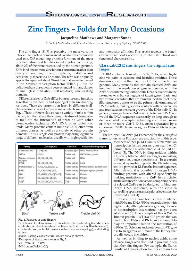
The zinc finger (ZnF) is probably the most versatileintracellular protein domain and it is certainly a commonlyused one. ZnF-containing proteins form one of the mostprevalent structural families in eukaryotes, comprising,about 2% of the proteins encoded by the human genome.ZnFs bind one or more zinc ions in a structural (rather thancatalytic) manner, through cysteine, histidine andoccasionally aspartate side chains. The term was originallyapplied to repeats of about 30 residues that were discoveredin the Xenopus transcription factor TFIIIA (1), but thedefinition has subsequently been extended to many classesof small (less than about 100 residues) zinc-ligatingdomains.
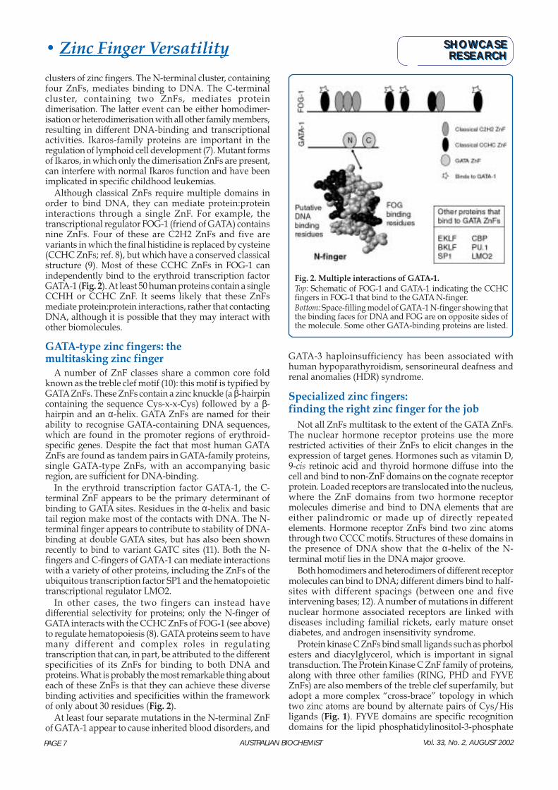
Different classes of ZnFs differ by structure and function,as well as by the identity and spacing of their zinc-bindingresidues. There are currently at least 14 different well-characterised classes known, some of which are shown inFig. 1. These different classes have a variety of roles withinthe cell, but they share the common feature of being ableto mediate the interaction of proteins with otherbiomolecules, including DNA, RNA, other proteins orlipids. Many proteins contain multiple ZnFs, often fromdifferent classes, as well as a variety of other proteindomains. Thus, a single ZnF protein may bring together arange of different molecules, usually with high specificities
Zinc Fingers – Folds for Many OccasionsJacqueline Matthews and Margaret Sunde
School of Molecular and Microbial Biosciences, University of Sydney, NSW 2006
and interaction affinities. This article reviews the better-characterised ZnFs according to their structural andfunctional characteristics.
Classical/C2H2 zinc fingers: the original zincfinger
TFIIIA contains classical (or C2H2) ZnFs, which ligatezinc via pairs of cysteine and histidine residues. Thesedomains constitute the majority of ZnFs in the humangenome. Many proteins that contain classical ZnFs areinvolved in the regulation of gene expression, with theZnFs often interacting with specific DNA sequences in thepromoter or enhancer regions of target genes. Basic andhydrophobic residues that are found in the α-helix of theseββα structures appear to be the primary determinants ofDNA-binding, making specific contacts with between twoand four bases in the major groove of the DNA helix (2). Ingeneral, a single classical ZnF is not able to bind DNA, norwould the DNA sequence necessarily be long enough todefine a useful transcriptional binding site. Instead, seriesof three or more C2H2 ZnFs, often separated by aconserved TGEKP linker, recognise DNA motifs in targetgenes.
The Krüppel-like ZnFs (KLFs, named for the Drosophilatranscription factor Krüppel) are a subfamily of this type.
Members of the Sp/KLF family of mammaliantranscription factors possess, at or near their C-termini, three KLFs that bind to GC or CACCCboxes (3). The DNA-binding residues withinKLFs vary between different fingers, impartingdifferent sequence specificities. To a certainextent, it is possible to predict the DNA-bindingsite of a particular KLF on the basis of sequence.Alternatively, it is possible to design DNA-binding proteins with altered specificity bymaking mutations in a ZnF. In principle,artificial transcription factors comprising seriesof selected ZnFs can be designed to bind anytarget DNA sequence, with the view tocontrolling specific transcriptional events in vivo(reviewed in ref. 4).
Classical ZnFs have been shown to interactwith RNA and DNA/RNA heteroduplexes withhigh affinity, although no biological significanceof heteroduplex interactions has yet beenestablished (5). One example of this is Wilm’sTumour protein 1 (WT1), a KLF protein that canbind to both DNA and RNA, and is thought toplay an important role in the maturation ofmRNA (6). Deletions and mutations in WTI giverise to an aggressive tumour of the kidney thatusually occurs in children.
As well as binding to nucleic acids, someclassical fingers can also bind to proteins, oftenvia other zinc fingers. For example, the Ikarosfamily of transcription factors contain two
Fig. 1. Features of zinc fingers.Top: Classes of ZnFs reviewed in this article with zinc-binding ligands (whereapplicable, A refers to residues that ligate the first zinc ion, B to the second),structural class (treble clef (x) refers to the cross-brace topology), and bindingtargets.Bottom: Examples of structural classes are also shown.1Examples of structures shown in Fig. 32ZnF from TFIIIA (19)3NF from mGATA-1 (20)
PAGE 6 Vol. 33, No. 2, AUGUST 2002
Vol. 33, No. 2, AUGUST 2002AUSTRALIAN BIOCHEMIST
clusters of zinc fingers. The N-terminal cluster, containingfour ZnFs, mediates binding to DNA. The C-terminalcluster, containing two ZnFs, mediates proteindimerisation. The latter event can be either homodimer-isation or heterodimerisation with all other family members,resulting in different DNA-binding and transcriptionalactivities. Ikaros-family proteins are important in theregulation of lymphoid cell development (7). Mutant formsof Ikaros, in which only the dimerisation ZnFs are present,can interfere with normal Ikaros function and have beenimplicated in specific childhood leukemias.
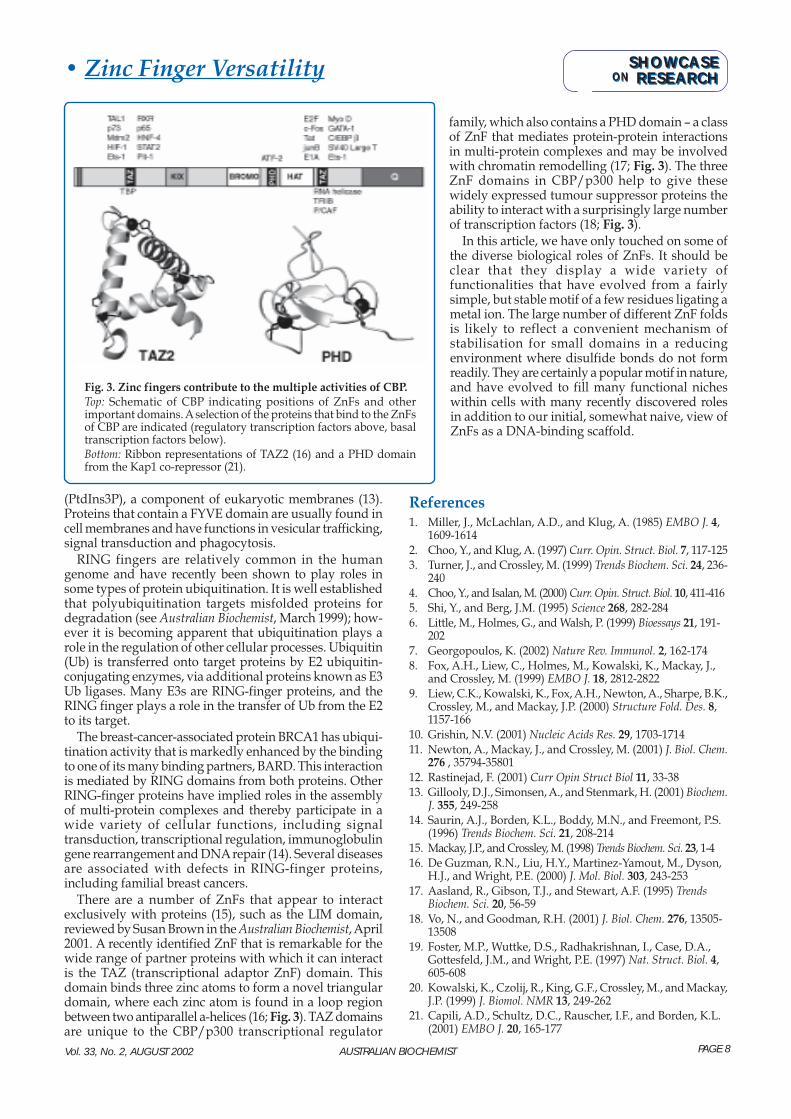
Although classical ZnFs require multiple domains inorder to bind DNA, they can mediate protein:proteininteractions through a single ZnF. For example, thetranscriptional regulator FOG-1 (friend of GATA) containsnine ZnFs. Four of these are C2H2 ZnFs and five arevariants in which the final histidine is replaced by cysteine(CCHC ZnFs; ref. 8), but which have a conserved classicalstructure (9). Most of these CCHC ZnFs in FOG-1 canindependently bind to the erythroid transcription factorGATA-1 (Fig. 2). At least 50 human proteins contain a singleCCHH or CCHC ZnF. It seems likely that these ZnFsmediate protein:protein interactions, rather that contactingDNA, although it is possible that they may interact withother biomolecules.
GATA-type zinc fingers: themultitasking zinc finger
A number of ZnF classes share a common core foldknown as the treble clef motif (10): this motif is typified byGATA ZnFs. These ZnFs contain a zinc knuckle (a β-hairpincontaining the sequence Cys-x-x-Cys) followed by a β-hairpin and an α-helix. GATA ZnFs are named for theirability to recognise GATA-containing DNA sequences,which are found in the promoter regions of erythroid-specific genes. Despite the fact that most human GATAZnFs are found as tandem pairs in GATA-family proteins,single GATA-type ZnFs, with an accompanying basicregion, are sufficient for DNA-binding.
In the erythroid transcription factor GATA-1, the C-terminal ZnF appears to be the primary determinant ofbinding to GATA sites. Residues in the α-helix and basictail region make most of the contacts with DNA. The N-terminal finger appears to contribute to stability of DNA-binding at double GATA sites, but has also been shownrecently to bind to variant GATC sites (11). Both the N-fingers and C-fingers of GATA-1 can mediate interactionswith a variety of other proteins, including the ZnFs of theubiquitous transcription factor SP1 and the hematopoietictranscriptional regulator LMO2.
In other cases, the two fingers can instead havedifferential selectivity for proteins; only the N-finger ofGATA interacts with the CCHC ZnFs of FOG-1 (see above)to regulate hematopoiesis (8). GATA proteins seem to havemany different and complex roles in regulatingtranscription that can, in part, be attributed to the differentspecificities of its ZnFs for binding to both DNA andproteins. What is probably the most remarkable thing abouteach of these ZnFs is that they can achieve these diversebinding activities and specificities within the frameworkof only about 30 residues (Fig. 2).
At least four separate mutations in the N-terminal ZnFof GATA-1 appear to cause inherited blood disorders, and
• Zinc Finger Versatility
GATA-3 haploinsufficiency has been associated withhuman hypoparathyroidism, sensorineural deafness andrenal anomalies (HDR) syndrome.
Specialized zinc fingers:finding the right zinc finger for the job
Not all ZnFs multitask to the extent of the GATA ZnFs.The nuclear hormone receptor proteins use the morerestricted activities of their ZnFs to elicit changes in theexpression of target genes. Hormones such as vitamin D,9-cis retinoic acid and thyroid hormone diffuse into thecell and bind to non-ZnF domains on the cognate receptorprotein. Loaded receptors are translocated into the nucleus,where the ZnF domains from two hormone receptormolecules dimerise and bind to DNA elements that areeither palindromic or made up of directly repeatedelements. Hormone receptor ZnFs bind two zinc atomsthrough two CCCC motifs. Structures of these domains inthe presence of DNA show that the α-helix of the N-terminal motif lies in the DNA major groove.
Both homodimers and heterodimers of different receptormolecules can bind to DNA; different dimers bind to half-sites with different spacings (between one and fiveintervening bases; 12). A number of mutations in differentnuclear hormone associated receptors are linked withdiseases including familial rickets, early mature onsetdiabetes, and androgen insensitivity syndrome.
Protein kinase C ZnFs bind small ligands such as phorbolesters and diacylglycerol, which is important in signaltransduction. The Protein Kinase C ZnF family of proteins,along with three other families (RING, PHD and FYVEZnFs) are also members of the treble clef superfamily, butadopt a more complex “cross-brace” topology in whichtwo zinc atoms are bound by alternate pairs of Cys/Hisligands (Fig. 1). FYVE domains are specific recognitiondomains for the lipid phosphatidylinositol-3-phosphate
Fig. 2. Multiple interactions of GATA-1.Top: Schematic of FOG-1 and GATA-1 indicating the CCHCfingers in FOG-1 that bind to the GATA N-finger.Bottom: Space-filling model of GATA-1 N-finger showing thatthe binding faces for DNA and FOG are on opposite sides ofthe molecule. Some other GATA-binding proteins are listed.
SHOWCASERESEARCH
SHOWCASERESEARCHONON
PAGE 7
Vol. 33, No. 2, AUGUST 2002 AUSTRALIAN BIOCHEMIST
(PtdIns3P), a component of eukaryotic membranes (13).Proteins that contain a FYVE domain are usually found incell membranes and have functions in vesicular trafficking,signal transduction and phagocytosis.
RING fingers are relatively common in the humangenome and have recently been shown to play roles insome types of protein ubiquitination. It is well establishedthat polyubiquitination targets misfolded proteins fordegradation (see Australian Biochemist, March 1999); how-ever it is becoming apparent that ubiquitination plays arole in the regulation of other cellular processes. Ubiquitin(Ub) is transferred onto target proteins by E2 ubiquitin-conjugating enzymes, via additional proteins known as E3Ub ligases. Many E3s are RING-finger proteins, and theRING finger plays a role in the transfer of Ub from the E2to its target.
The breast-cancer-associated protein BRCA1 has ubiqui-tination activity that is markedly enhanced by the bindingto one of its many binding partners, BARD. This interactionis mediated by RING domains from both proteins. OtherRING-finger proteins have implied roles in the assemblyof multi-protein complexes and thereby participate in awide variety of cellular functions, including signaltransduction, transcriptional regulation, immunoglobulingene rearrangement and DNA repair (14). Several diseasesare associated with defects in RING-finger proteins,including familial breast cancers.
There are a number of ZnFs that appear to interactexclusively with proteins (15), such as the LIM domain,reviewed by Susan Brown in the Australian Biochemist, April2001. A recently identified ZnF that is remarkable for thewide range of partner proteins with which it can interactis the TAZ (transcriptional adaptor ZnF) domain. Thisdomain binds three zinc atoms to form a novel triangulardomain, where each zinc atom is found in a loop regionbetween two antiparallel a-helices (16; Fig. 3). TAZ domainsare unique to the CBP/p300 transcriptional regulator
• Zinc Finger Versatility
family, which also contains a PHD domain – a classof ZnF that mediates protein-protein interactionsin multi-protein complexes and may be involvedwith chromatin remodelling (17; Fig. 3). The threeZnF domains in CBP/p300 help to give thesewidely expressed tumour suppressor proteins theability to interact with a surprisingly large numberof transcription factors (18; Fig. 3).
In this article, we have only touched on some ofthe diverse biological roles of ZnFs. It should beclear that they display a wide variety offunctionalities that have evolved from a fairlysimple, but stable motif of a few residues ligating ametal ion. The large number of different ZnF foldsis likely to reflect a convenient mechanism ofstabilisation for small domains in a reducingenvironment where disulfide bonds do not formreadily. They are certainly a popular motif in nature,and have evolved to fill many functional nicheswithin cells with many recently discovered rolesin addition to our initial, somewhat naive, view ofZnFs as a DNA-binding scaffold.
References1. Miller, J., McLachlan, A.D., and Klug, A. (1985) EMBO J. 4,
1609-16142. Choo, Y., and Klug, A. (1997) Curr. Opin. Struct. Biol. 7, 117-1253. Turner, J., and Crossley, M. (1999) Trends Biochem. Sci. 24, 236-
2404. Choo, Y., and Isalan, M. (2000) Curr. Opin. Struct. Biol. 10, 411-4165. Shi, Y., and Berg, J.M. (1995) Science 268, 282-2846. Little, M., Holmes, G., and Walsh, P. (1999) Bioessays 21, 191-
2027. Georgopoulos, K. (2002) Nature Rev. Immunol. 2, 162-1748. Fox, A.H., Liew, C., Holmes, M., Kowalski, K., Mackay, J.,
and Crossley, M. (1999) EMBO J. 18, 2812-28229. Liew, C.K., Kowalski, K., Fox, A.H., Newton, A., Sharpe, B.K.,
Crossley, M., and Mackay, J.P. (2000) Structure Fold. Des. 8,1157-166
10. Grishin, N.V. (2001) Nucleic Acids Res. 29, 1703-171411. Newton, A., Mackay, J., and Crossley, M. (2001) J. Biol. Chem.
276 , 35794-3580112. Rastinejad, F. (2001) Curr Opin Struct Biol 11, 33-3813. Gillooly, D.J., Simonsen, A., and Stenmark, H. (2001) Biochem.
J. 355, 249-25814. Saurin, A.J., Borden, K.L., Boddy, M.N., and Freemont, P.S.
(1996) Trends Biochem. Sci. 21, 208-21415. Mackay, J.P., and Crossley, M. (1998) Trends Biochem. Sci. 23, 1-416. De Guzman, R.N., Liu, H.Y., Martinez-Yamout, M., Dyson,
H.J., and Wright, P.E. (2000) J. Mol. Biol. 303, 243-25317. Aasland, R., Gibson, T.J., and Stewart, A.F. (1995) Trends
Biochem. Sci. 20, 56-5918. Vo, N., and Goodman, R.H. (2001) J. Biol. Chem. 276, 13505-
1350819. Foster, M.P., Wuttke, D.S., Radhakrishnan, I., Case, D.A.,
Gottesfeld, J.M., and Wright, P.E. (1997) Nat. Struct. Biol. 4,605-608
20. Kowalski, K., Czolij, R., King, G.F., Crossley, M., and Mackay,J.P. (1999) J. Biomol. NMR 13, 249-262
21. Capili, A.D., Schultz, D.C., Rauscher, I.F., and Borden, K.L.(2001) EMBO J. 20, 165-177
Fig. 3. Zinc fingers contribute to the multiple activities of CBP.Top: Schematic of CBP indicating positions of ZnFs and otherimportant domains. A selection of the proteins that bind to the ZnFsof CBP are indicated (regulatory transcription factors above, basaltranscription factors below).Bottom: Ribbon representations of TAZ2 (16) and a PHD domainfrom the Kap1 co-repressor (21).
SHOWCASERESEARCH
SHOWCASERESEARCHONON
PAGE 8
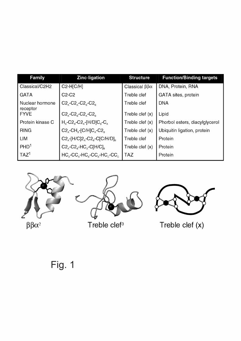
Fig. 1
Fig. 2
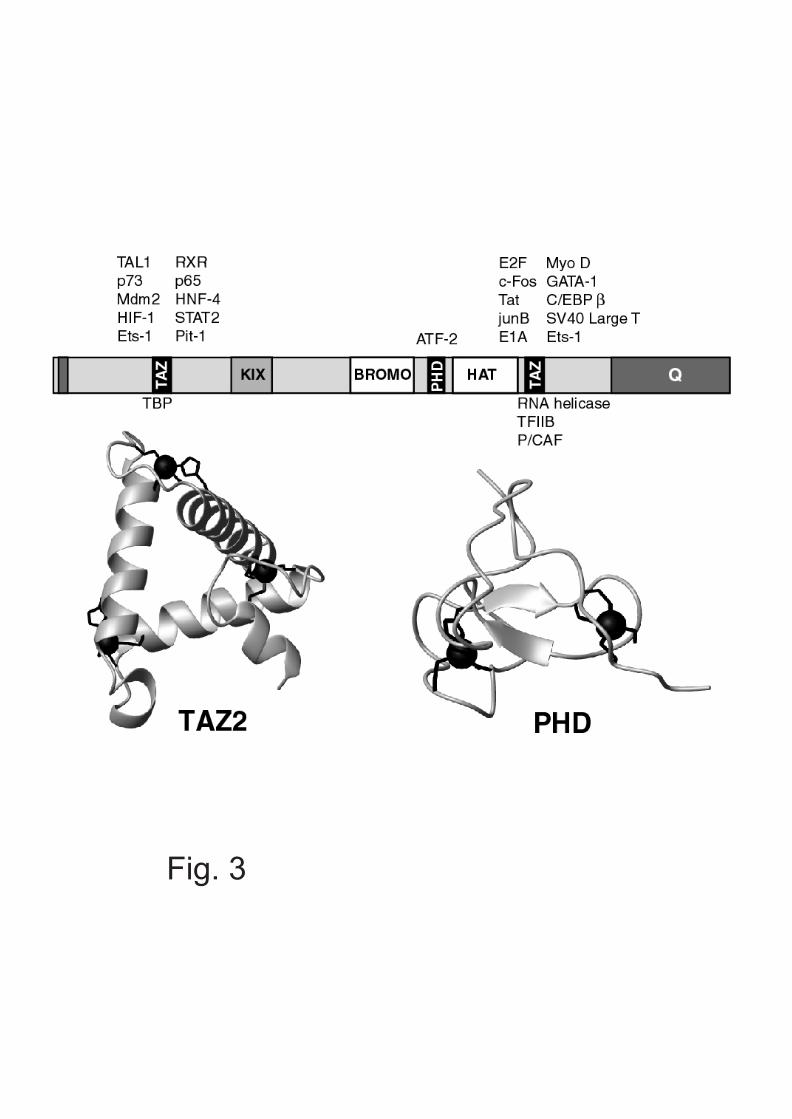
Fig. 3