Embed Size (px)
Citation preview

The Effect of Transcription Factor Zhangfei/CREBZF on
Osteosarcoma Cells and the Mechanisms Responsible
A Thesis Submitted to the College of
Graduate Studies and Research
In Partial Fulfillment of the Requirements
For the Degree of Doctor of Philosophy
In the Department of Veterinary Microbiology
Western College of Veterinary Medicine
University of Saskatchewan, Saskatoon
By
Rui Zhang
© Copyright Rui Zhang, June 2014. All rights reserved

i
PERMISSION TO USE
In presenting this thesis/dissertation in partial fulfillment of the requirements for a
Postgraduate degree from the University of Saskatchewan, I agree that the Libraries of
this University may make it freely available for inspection. I further agree that permission
for copying of this thesis/dissertation in any manner, in whole or in part, for scholarly
purposes may be granted by the professor or professors who supervised my
thesis/dissertation work or, in their absence, by the Head of the Department or the Dean
of the College in which my thesis work was done. It is understood that any copying or
publication or use of this thesis/dissertation or parts thereof for financial gain shall not be
allowed without my written permission. It is also understood that due recognition shall be
given to me and to the University of Saskatchewan in any scholarly use which may be
made of any material in my thesis/dissertation.
Requests for permission to copy or to make other uses of materials in this
thesis/dissertation in whole or part should be addressed to:
Head of the Department of Veterinary Microbiology
Western College of Veterinary Medicine
University of Saskatchewan
Saskatoon, Saskatchewan, S7N 5B4
Canada
OR
Dean
College of Graduate Studies and Research
University of Saskatchewan
107 Administration Place
Saskatoon, Saskatchewan, S7N 5A2
Canada

ii
ABSTRACT
Osteosarcoma (OS) is the most common primary malignant bone tumour in humans and
dogs. Although medicine has made dramatic progress in treating osteosarcoma by surgery,
with chemotherapy given before and after surgery, drug resistance and highly metastatic
spread are often responsible for the failure of current therapies. Thus, more effective
therapeutic approaches for treating osteosarcoma are needed. Previous results from our
laboratory and others had shown that the basic-leucine zipper (bLZip) containing
transcription factor, Zhangfei/CREBZF is a potent inhibitor of a variety of other
transcription factors and has a dramatic effect on the growth of several cancer cell lines,
including dog OS and human medulloblastoma cells. The objective of the studies
described in this thesis was to determine the molecular mechanisms by which Zhangfei
exerts its effect on dog and human OS cells.
Several stressors in the microenvironment of cancer cells directly or indirectly perturb the
endoplasmic reticulum (ER), which then activates the Unfolded Protein Response (UPR).
The UPR modulates the effects of stress and allows tumours to survive, develop,
metastasize and escape therapy. The UPR is regulated by three bLZip transcription
factors—ATF6, ATF4 and Xbp1s. Since Zhangfei inhibits Luman/CREB3, a bLZip
structurally similar to and closely related to ATF6 and ATF4, I initially focused my
efforts on this pathway. I hypothesized that Zhangfei interacts with UPR-related bLZip
transcription factors and inhibits their ability to activate the UPR signaling pathways,
thereby suppressing the growth of cancer cells and increasing their susceptibility to ER
stressors.
To test this hypothesis, we monitored cell growth as well as levels of UPR gene
transcripts and proteins in several dog and human osteosarcoma cell lines infected with
adenovirus vectors expressing Zhangfei, and studied the interactions between Zhangfei
and the UPR-mediator, Xbp1s. The results showed that the ectopic expression of
Zhangfei in cell lines derived from dog osteosarcomas potently suppressed cell growth
and inhibited their ability to activate the UPR. Further studies demonstrated that Zhangfei
inhibited the UPR, at least partially, by binding to Xbp1s and suppressing its ability to

iii
activate transcription from a promoter containing unfolded protein response elements
(UPRE). The leucine zipper of Zhangfei was required for this interaction, which led to
the subsequent proteasomal degradation of Xbp1s. However, we also found that the
effects of Zhangfei were not universal. While Zhangfei had a profound effect on the
growth and UPR in some OS cell lines, it either had only a partial effect, or no effect on
others. This suggested that susceptibility (or resistance) to Zhangfei may be an inherent
property of OS cell lines.
Since the suppressive effects of Zhangfei were not universal, and it had no obvious
effects on untransformed cells and some cancer cell lines, I proposed that Zhangfei
mediates its effect on cell growth and the UPR through an intermediary that is either not
induced or is defective in cells that are unaffected by Zhangfei. I found that this
intermediary was the tumour suppressor protein p53. The inhibitory effects of Zhangfei
were only observed in the wild-type p53 expressing OS cell line U2OS while Zhangfei
had no effect on the p53-null OS cell line MG63. In cells with functional p53, the ectopic
expression of Zhangfei caused it to displace the ubiquitin ligase mdm2 and stabilize p53.
Suppression of p53 by siRNA partially inhibited the effects of Zhangfei on the UPR and
cell growth. In contrast, OS cells lacking functional p53 could be made to respond to
Zhangfei if they were transfected to express wild-type p53. These results explain why
Zhangfei has a profound effect on some cancer cells while having no obvious effect on
others. I also characterized the interaction of Zhangfei and p53 by mapping the
interacting domains on both proteins, showing that the bLZip domain of Zhangfei and the
N-terminal transactivation domain (NTD) of p53 were required for their interactions.
My findings reveal the profoundly inhibitory effects of Zhangfei on OS growth and the
UPR, a stress-response known to promote tumour survival. I also show how Zhangfei
may exert its effects. My work suggests an alternative modality for the therapy of certain
types of OS, and perhaps other tumours with functional p53.
Key words: Zhangfei/CREBZF, osteosarcoma, cell growth, UPR, p53

iv
ACKNOWLEDGEMENT
Foremost, I would like to express my sincere gratitude to my supervisor Prof. Vikram
Misra for his continuous support and encouragement for my Ph.D study and research; for
his patience, motivation, enthusiasm, and immense knowledge; for his unforgettable and
constant smiles and greetings. This thesis would not have been possible without him.
I would like to express my appreciation to my thesis committee: Drs. Bruce Wobeser,
Deborah Haines, Janet Hill, Jim Xiang, and Valerie MacDonald-Dickinson, for their
encouragement and insightful comments.
My sincere thanks also goes to our technician Noreen Rapin, for her assistance and
friendship. To my past and present lab members: Iran, Tim, Andy, Kirsten, Zhengxin and
Arinjay, as well as to my office members: Yanyun, Isha, Teenus, Aline and Matheus, for
the good conversation, sharing, fun and encouragement they brought into my everyday
life. A thank you to all the friends I met and had the opportunity to share the last four
years in Saskatoon with.
Last but not least, my eternal thank you to my family: my parents Yuhuan Wang and
Fengxiang Zhang, and my sister Wei Zhang, for giving me their unequivocal support
throughout, as always, and sharing each happy and sad moment in my life with me.

v
Table of Contents
PERMISSION TO USE ..................................................................................................... i
ABSTRACT ....................................................................................................................... ii
ACKNOWLEDGEMENT ............................................................................................... iv
List of Figures .................................................................................................................... x
List of Tables .................................................................................................................. xiii
List of Abbreviations ..................................................................................................... xiv
1. Introduction ................................................................................................................... 1
1.1 Osteosarcoma (OS) ................................................................................................. 1
1.1.1 Canine and human OS ....................................................................................... 1
1.1.2 Current therapeutic approaches of OS ............................................................... 1
1.1.3 Genetic alternation in OS ................................................................................... 2
1.2 Zhangfei/CREBZF .................................................................................................. 3
1.2.1 Overview ............................................................................................................ 3
1.2.2 Structure and function ........................................................................................ 3
1.3 Unfolded Protein Response (UPR) ........................................................................ 5
1.3.1 Endoplasmic reticulum (ER) stress .................................................................... 5
1.3.2 UPR signaling pathways .................................................................................... 5
1.3.3 UPR and cancer ................................................................................................ 11
1.4 p53 signaling pathways and cancer therapy ....................................................... 12
1.4.1 Structure and function of p53 .......................................................................... 12
1.4.2 p53-mediated cell cycle arrest and apoptosis ................................................... 13
1.4.3 p53 mutation and cancer .................................................................................. 16
1.4.4 Activation of p53 as a therapeutic strategy ...................................................... 17
1.5 Rationale, Hypothesis and Objectives ................................................................. 19
2. The Effect of Zhangfei on the Unfolded Protein Response (UPR) and Growth of
Cells Derived from Canine and Human Osteosarcomas ............................................. 22
2.1 Abstract .................................................................................................................. 23
2.2 Introduction ........................................................................................................... 24

vi
2.3 Materials and Methods ......................................................................................... 26
2.3.1 Cell Culture ...................................................................................................... 26
2.3.2 Quantitative Real-Time PCR ........................................................................... 27
2.3.3 Antibodies, Microscopy and Immunofluorescence ......................................... 28
2.3.4 Detection of proteins by immunoblotting ........................................................ 29
2.3.5 Detection of apoptotic cells ............................................................................. 29
2.3.6 Statistics ........................................................................................................... 29
2.4 Results .................................................................................................................... 30
2.4.1 Detecting the UPR in dog and human osteosarcoma cells .............................. 30
2.4.2 Suppression of UPR by Zhangfei .................................................................... 33
2.4.3 Effect of Zhangfei on UPR related proteins .................................................... 35
2.4.4 Effect of Zhangfei on the growth of dog and human osteosarcoma cells ........ 37
2.5 Discussion .............................................................................................................. 41
2.6 Acknowledgements ............................................................................................... 43
3. Zhangfei/CREB-ZF – a potential regulator of the Unfolded Protein Response ... 44
3.1 Abstract .................................................................................................................. 45
3.2 Introduction ........................................................................................................... 46
3.3 Materials and Methods ......................................................................................... 48
3.3.1 Cell Culture ...................................................................................................... 48
3.3.2 Immunofluorescence ........................................................................................ 48
3.3.3 Plasmids ........................................................................................................... 48
3.3.4 Adenovirus vectors expressing Zhangfei and β-galactosidase (LacZ) ............ 49
3.3.5 mRNA purification and cDNA synthesis ........................................................ 49
3.3.6 qRT-PCR arrays and PCR confirmation .......................................................... 49
3.3.7 Co-immunoprecipitation .................................................................................. 51
3.3.8 Adult DRG culture ........................................................................................... 51
3.4 Results .................................................................................................................... 53
3.4.1 Does the ectopic expression of Zhangfei influence the UPR? ......................... 53
3.4.2 Can Zhangfei suppress the ability of Xbp1s to activate transcription and is its
leucine-zipper required? ............................................................................................ 59
3.4.3 How does Zhangfei suppress Xbp1? ................................................................ 62

vii
3.4.4 Does Zhangfei interact with Xbp1s? ................................................................ 66
3.4.5 Can endogenous Zhangfei suppress the UPR in sensory neurons? ................. 68
3.5 Discussion .............................................................................................................. 71
4. Effects of Cyclic AMP Response Element Binding Protein – Zhangfei (CREBZF)
on the Unfolded Protein Response and cell growth are exerted through the tumour
suppressor p53 ................................................................................................................. 75
4.1 Abstract .................................................................................................................. 76
4.2 Introduction ........................................................................................................... 77
4.3 Materials and Methods ......................................................................................... 79
4.3.1 Cells and tissue culture .................................................................................... 79
4.3.2 Plasmids ........................................................................................................... 79
4.3.3 Transfection and CAT Assays ......................................................................... 80
4.3.4 RNA interference ............................................................................................. 80
4.3.5 Adenovirus Vectors Expressing Zhangfei (Adeno-ZF) and β-galactosidase
(Adeno-LacZ) ........................................................................................................... 80
4.3.6 Antibodies, immunoblotting and immunofluorescence ................................... 81
4.3.7 Quantitative real-time PCR .............................................................................. 81
4.3.8 Co-immunoprecipitation .................................................................................. 82
4.3.9 Statistical analysis ............................................................................................ 82
4.4 Results .................................................................................................................... 83
4.4.1 Leucine-Zipper is required for the suppressive effects of Zhangfei on both cell
growth and UPR. ....................................................................................................... 83
4.4.2 Zhangfei regulates p53 at a post-translational level and promotes p53 nuclear
retention. ................................................................................................................... 86
4.4.3 Basic-region leucine zipper domain (bLZip) of Zhangfei is required for the
regulation of p53. ...................................................................................................... 89
4.4.4 p53 is the key molecule responsible for mediating suppressive regulation of
Zhangfei on D-17 cell growth and the UPR. ............................................................ 91
4.4.5 Zhangfei suppresses the growth and UPR in p53-expressing, but not in p53-
null human osteosarcoma cells. ................................................................................ 94
4.4.6 Zhangfei interacts with p53. .......................................................................... 100

viii
4.4.7 Zhangfei displaces mdm2 from p53, protecting it from proteolysis. ............. 100
4.5 Discussion ............................................................................................................ 103
4.6 Acknowledgements ............................................................................................. 107
5. Structural domains responsible for p53-Zhangfei interaction ............................. 108
5.1 Abstract ................................................................................................................ 109
5.2 Introduction ......................................................................................................... 110
5.3 Materials and Methods ....................................................................................... 111
5.3.1 Cells and tissue culture .................................................................................. 111
5.3.2 Plasmids ......................................................................................................... 111
5.3.3 Transfection and CAT Assays ....................................................................... 112
5.3.4 Co-immunoprecipitation ................................................................................ 112
5.3.5 Antibodies and immunofluorescence ............................................................. 112
5.4 Results .................................................................................................................. 114
5.4.1 p53 forms a complex with Zhangfei via its N-terminal transactivation domain
(NTD) ...................................................................................................................... 114
5.4.2 N-terminal transactivation domain (NTD) is required for Zhangfei-mediated
nuclear retention of p53 .......................................................................................... 116
5.4.3 Zhangfei enhances p53-mediated transactivation through the N-terminal
transactivation domain (NTD) of p53 ..................................................................... 118
5.5 Discussion ............................................................................................................ 120
6. The effect of Zhangfei/CREBZF on cell growth, differentiation, apoptosis,
migration, and the UPR in several canine osteosarcoma cell lines ........................... 122
6.1 Abstract ................................................................................................................ 123
6.2 Introduction ......................................................................................................... 124
6.3 Materials and Methods ....................................................................................... 125
6.3.1 Cells and tissue culture .................................................................................. 125
6.3.2 Adenovirus Vectors Expressing Zhangfei and β-galactosidase (LacZ) ......... 125
6.3.3 WST-1 Cell Proliferation and Viability Assay .............................................. 125
6.3.4 Annex V-apoptosis assay ............................................................................... 125
6.3.5 Scratch wound healing assay ......................................................................... 126
6.3.6 Quantitative real-time PCR (qPCR) ............................................................... 126

ix
6.3.7 PCR and sequencing of p53 genes ................................................................. 126
6.3.8 Plasmids and chloramphenicol acetyl transferase (CAT) assay .................... 127
6.3.9 Antibodies, immunoblotting and immunofluorescence ................................. 127
6.4 Results .................................................................................................................. 128
6.4.1 All four canine OS cells lines express functional p53 ................................... 128
6.4.2 Cellular outcome following ectopic expression of Zhangfei: growth cessation,
apoptosis and differentiation ................................................................................... 131
6.4.3 Expression of Zhangfei suppresses migration of canine osteosarcoma cells 135
6.4.4 Zhangfei negatively regulates the UPR in canine osteosarcomas ................. 137
6.4 Discussion ............................................................................................................ 139
7. General discussion and conclusions ........................................................................ 141
8. Reference ................................................................................................................... 147

x
List of Figures
Figures Page
Fig 1.1 Structure, domains and functions of Zhangfei protein. 21
Fig 1.2 The three signaling pathways (IRE1, PERK and ATF6) of the Unfolded Protein Response (UPR).
24
Fig 1.3 Mechanism of ATF4 translation during phosphorylation of the α subunit of eukaryotic initiation factor 2 (eIF2α-P).
27
Fig 1.4 Structure domains of p53 protein. 29
Fig 1.5 Schematic representation of the p53-dependent apoptotic pathways
31
Fig 2.1 Design of primers for detecting and quantifying spliced and unspliced Xbp-1 cDNA.
47
Fig 2.2 The UPR-related transcripts in response to treatment with thapsigargin.
48
Fig 2.3 Effect of Zhangfei or LacZ-expression on thapsigargin-induced changes in Xbp-1, HERP, CHOP and GRP78/Bip transcript levels in canine D-17 and human Saos-2 cells.
50
Fig 2.4 Effect of Zhangfei on stable HERP and GRP78 in cells treated with thapsigargin.
52
Fig 2.5 Effect of Zhangfei on the growth of canine D-17 and human Saos-2 cells.
55
Fig 3.1 Suppression of UPR genes by Zhangfei in ONS-76 medulloblastoma cells treated with thapsigargin.
70
Fig 3.2 Effect of Zhangfei on HERP and GRP78 transcripts and proteins in cells treated with thapsigargin.
73
Fig 3.3 Zhangfei suppresses the ability of Xbp1s to activate transcription and requires its leucine zipper to do so.
76
Fig 3.4 Zhangfei directs Xbp1 for proteasomal degradation and requires its leucine zipper to do so.
79
Fig 3.5 Immunofluorescent images showing the absence of Xbp1 in cells expressing Zhangfei but not Zhangfei with a mutated
81

xi
leucine zipper.
Fig 3.6 Zhangfei co-immunoprecipitates with Xbp1s in MG132-treated cells.
83
Fig 3.7 Endogenous Zhangfei suppresses the activation of UPR genes in rat peripheral neurons.
85
Fig 4.1 Spontaneous mutation of leucine residues in the bLZip domain of Zhangfei in D-17 cells stably expressing the protein in the presence of tetracycline.
100
Fig 4.2 Zhangfei regulates p53 in a post-translational level and induces p53 nuclear localization.
103
Fig 4.3 The basic-region leucine zipper domain (bLZip) of Zhangfei is required for its effect on p53.
106
Fig 4.4 Zhangfei regulates p53-mediated cell growth and UPR. 108
Fig 4.5 Zhangfei suppresses cell growth and UPR in wild-type p53-expressing U2OS cells, but not in p53-null MG63 cells.
111
Fig 4.6 p53 mediates the suppressive effects of Zhangfei on cell growth and UPR in human osteosarcoma cells.
114
Fig 4.7 In vitro interaction of Zhangfei and p53. 117
Fig 4.8 Zhangfei and ER stress have opposing effects on p53. 122
Fig 5.1 Complex formation between Zhangfei and p53. 131
Fig 5.2 Co-localization of Zhangfei and p53 (or its deletion mutants).
133
Fig 5.3 Zhangfei activates p53-dependent transactivation via interaction with its 92 amino acids in N-terminal.
135
Fig 6.1 p53 in dog osteosarcoma cell lines. 147
Fig 6.2 Ectopic expression of Zhangfei suppresses cell growth in canine osteosarcomas.
149
Fig 6.3 Zhangfei induces differentiation of canine osteosarcoma cells.
151
Fig 6.4 Zhangfei causes canine osteosarcoma cells to commit apoptosis.
152

xii
Fig 6.5 Ectopic expression of Zhangfei causes decreased cell motility in canine osteosarcoma cells.
154
Fig 6.6 Zhangfei negatively regulates the Unfolded Protein Responses (UPR) in canine osteosarcomas.
156

xiii
List of Tables
Tables Page
Table 2.1 Sequence of primers used for qRT-PCR. 43
Table 3.1 Oligonucleotides primers used for Real Time PCR 66
Table 5.1 The sequence of primers for p53 and its deletion mutant amplification
127

xiv
List of Abbreviations
• AdLZ/Adeno-LacZ – adenovirus vector expressing beta-galactosidase
• AdZF/Adeno-ZF – adenovirus vector expressing Zhangfei
• ANOVA – analysis of variance
• ATF4 – activation transcription factor 4
• ATF6 – activation transcription factor 6
• β-gal – beta-Galactosidase
• bLZip – basic region, leucine zipper domain
• CAT – Chloramphenicol-acetyl-transferase
• CBP – Cyclic AMP responsive element binding protein-binding protein
• CEBPB – CCAAT enhancer binding protein-beta,
• CHOP – CAAT enhancer-binding protein homology protein
• CREBH – Cyclic AMP responsive element binding protein, hepatocyte specific
• CREBZF – Cyclic AMP response element binding protein, Zhangfei
• CREB3 – Cyclic AMP response element binding protein 3, also known as Luman
(see below)
• CTp53 – Carboxyl terminus of 53,000 molecular weight tumour suppressor
protein
• DDIT3 – DNA damage inducible transcript -3
• DNAJB9 – homologue of DNAJ/ 40 kD heat shock protein
• EDEM – ER degradation enhancer mannosidase alpha-like 1
• EDTA – ethylenediaminetetraacetic acid
• eIF2α – eukaryotic translation initiation factor 2alpha
• ER – endoplasmic reticulum
• ERAD – ER-associated protein degradation
• ERN1 – ER to nucleus signaling
• FBS – foetal bovine serum
• GAPDH – glyceraldehyde 6 phosphate dehydrogenase
• GADD34 – Growth arrest and DNA damage-inducible protein

xv
• GRP78/Bip – glucose regulated protein 78,000 MW
• HBX – hepatitis B virus X protein
• HCF – host cell factor
• HERP – homocysteine-induced endoplasmic reticulum protein
• HERPUD1 – homocysteine-inducible ER stress inducible ubiquitin-like domain
member 1
• HIFs – hypoxia inducible family of transcription factors
• HSPA1B – heat shock protein A1B, also called HSP70 kD protein 1B
• HSV-1 – Herpes simplex virus type 1
• K-bLZip – Kaposi sarcoma herpes virus bLZip protein
• ING2 – inhibitor of growth protein 2
• INSIG1 – insulin-induced gene 1
• IRE1 – inositol requiring enzyme
• IRF1 – host interferon response factor 1
• L – leucine
• LacZ – Escherichia coli protein beta-galactosidase
• Luman/CREB3 – Cyclic AMP response element binding protein – Luman
• MafB – host transcription factor maculoaponeurotic fibroma homologue B
• MAPK – mitogen-activated protein kinase
• mdm2 – mouse double minute homologue 2
• MEM – Eagles minimal essential medium
• MI – mock-infected
• mTOR – the kinase mammalian target of rapamycin
• NOTCH1 – Drosophila Notch family protein 1
• NTD – N-terminal transactivation domain of p53
• OS – osteosarcoma
• p53 – protein 53,000 molecular weight
• p53C – DNA-binding core domain of p53
• PCR – polymerase chain reaction
• PERK – putative receptor protein kinase

xvi
• PIG3 – p53-inducible protein 3
• PRR – proline-rich region of p53
• SDS – sodium dodecyl sulfate
• siRNA – small interfering RNA
• SMAD – small body size – mothers against decapentaplegic
• SMILE – small heterodimer partner interacting leucine zipper protein, also known
as Zhangfei or CREBZF
• TAD – transactivation domain of p53
• TET – tetramerization domain of p53
• UPR – Unfolded Protein Response
• UPRE – Unfolded Protein Response Element
• VP16 – virion protein #16
• Xbp1 – X-factor binding protein 1
• Xbp1s – Xbp1 derived from cytoplasmic spliced mRNA

1
1. Introduction
1.1 Osteosarcoma (OS)
1.1.1 Canine and human OS
Osteosarcoma (OS) is the most frequent primary malignant bone tumour in children and
adolescents, and its incidence in dogs is ten times greater than in humans. The high
incidence of spontaneous canine OS makes the dog an attractive model candidate to study
in relation to their human counterparts in a large group. In addition, dog OS bears many
similarities with the OS in humans, which are difficult to replicate in other models. For
instance: a) Dogs share a common environment with humans and are exposed to similar
environmental contributors to tumorigenesis. b) Canine and human OS have similar
microenvironments (Rankin et al. 2012). c) Compared to the immunodeficient rodents,
the role of an intact immune system in the initiation and development of OS, as well as its
contribution to therapeutic strategies, can be investigated in dogs. d) Similar biological
and clinical features such as, male sex predilection, large patient size, appendicular site,
metaphyseal location, generally unknown etiology, high-grade histology, high local
aggressiveness, high genomic instability, rapid metastasizing potential, lung metastasis,
and similar response to conventional therapies (reviewed by (Withrow et al. 1991;
Ambron and Walters 1996; Mueller et al. 2007; Paoloni and Khanna 2008; Khanna et al.
2009). Thus, spontaneous dog osteosarcomas are excellent models for studying signaling
pathways that regulate human tumour growth and for the development of therapeutic
agents.
1.1.2 Current therapeutic approaches of OS
Current therapeutic approaches for OS consist of surgical resection, multi-agent
chemotherapy and radiotherapy. Although long-term survival in localized OS has been
dramatically improved in both humans and dogs by conventional treatments, recurrence
and metastatic spread of OS always leads to a poor patient prognosis (Ando et al. 2013).
For example, over 80% of patients with localized OS developed lung metastases after

2
amputation alone, and the mean survival time after recurrence is less than 12 months
(Marina et al. 2004). Thus, innovative drugs and alternative approaches are needed to
further improve outcomes for OS patients. At present, several new therapeutic agents
being studied target the signal transduction pathways that are activated, inhibited or
mutated in OS. These relevant signal pathways include nonreceptor tyrosine kinase Src,
Mammalian Target of Rapamycin (mTOR), Hedgehog (Hh) signaling pathway, tyrosine
kinase receptors (e.g receptors for vascular endothelial growth factor [VEGF], platelet-
derived growth factor [PDGF], insulin-like growth factor [IGF], human epidermal growth
factor receptor-2 [HER2] and hepatocyte growth factor receptor [HGFR]), as well as
tumour suppressor p53 (reviewed by (Ambron and Walters 1996; Ando et al. 2013; Gill
et al. 2013). In addition to these intracellular signal pathways, immunomodulators and
signaling of bone metabolism are also considered as the alternative targets of the novel
therapeutic agents.
1.1.3 Genetic alternation in OS
Nearly 70% of osteosarcoma tumours have genetic and molecular alternations, including
abnormalities of chromosomal regions, mutation of tumour suppression genes, activation
of oncogenes, as well as deregulation of signaling pathways (Tang et al. 2008). The most
frequently altered genes and signaling pathways in both dogs and humans include: p53,
retinoblastoma protein (Rb), Phosphatase and tensin homolog (PTEN), phosphoinositide
3-kinase/AKT (PI3K/AKT) pathways, and Mitogen-activated protein kinases (MAPK)
cascade (reviewed by (Mueller et al. 2007). Many studies consider OS as a differentiation
disease, because the genetic and epigenetic changes (e.g. activation of oncogenes or
inactivation of p53 and RB tumour suppressor genes) disrupt osteoblast differentiation
from mesenchymal stem cells and may contribute to OS development (Haydon et al.
2007). One of the most frequently detected genetic alternations involved in osteogenic
differentiation and bone tumorigenesis is tumour suppressor protein p53. The association
of p53 mutations with significant numbers of OS suggests that a lack of functional p53
increases susceptibility of OS. (Andreassen et al. 1993; Bodey et al. 1997; Varley 2003).
Therefore, the mutation status of p53 could serve as a valuable indicator for early
diagnosis, prognosis, and for predicting chemoresistance of OS (Goto et al. 1998).

3
1.2 Zhangfei/CREBZF
1.2.1 Overview
Zhangfei, also known as CREBZF or SMILE (Small Heterodimer Partner [SHP]-
interacting leucine zipper protein), is a neuronal cellular transcription factor identified in
our laboratory through its interaction with Host Cell Factor 1 (HCF-1) (Lu and Misra
2000b), a cellular protein required for the initiation of the herpes simplex virus type 1
(HSV-1) replicative cycle. Zhangfei belongs to the cyclic AMP response element-binding
(CREB) protein family. Zhangfei is believed to play a role in the initiation of HSV-1
latency by inhibiting viral replication in unstressed neurons (Varley 2003; Akhova et al.
2005; Misra et al. 2005). While Zhangfei is present in mature neurons, it is not detected
in developing neurons or in neuronal tumours.
1.2.2 Structure and function
Zhangfei, an important member of the basic domain-leucine zipper (bLZip) family of
transcription factors, possess a basic region and a hydrophobic leucine zipper region
containing multiple leucine residues at approximately 7-residue intervals. The bLZip
motif of Zhangfei exhibits sequence homology with other members of the bLZip family,
and like them, may form homo- and hetero-dimeric complexes through pairing of bLZip
motifs, creating a DNA contact surface capable of binding to diverse cis-acting
regulatory elements in gene promoters (Hogan et al. 2006). Zhangfei may interact with
other transcription regulators through an acidic activation domain, an HCF-1-binding
motif (HBM) and nuclear receptor binding motifs (Lu and Misra 2000b) (Fig 1.1).
Zhangfei differs from other bLZip proteins in that while it can homo- or hetero-dimerize
through its leucine zipper, it is incapable of binding any consensus bLZip response
elements as a homodimer. This is likely due to the absence of a critical asparagine residue
in the basic region, which in other bLZip transcription factors is considered crucial to
their ability to recognize response elements in gene promoters (Cockram et al. 2006).
Zhangfei can, however, bind to and regulate some other bLZip transcription factors as a
heterodimer with other proteins such as Luman/CREB3 (Misra et al. 2005), SMAD 1,5,8

4
(Lee et al. 2012a), CREBH (Misra et al. 2012), and ATF4 (Hogan et al. 2006). Zhangfei
has been reported to regulate the activity of the HSV co-activator VP16 through HBM
(Lu and Misra 2000b) and to act as a transcriptional co-repressor of nuclear receptors
glucocorticoid receptor, constitutive androstane receptor, and hepatocyte nuclear factor
4α together with SHP (Xie et al. 2008; Xie et al. 2009a; Xie et al. 2009b). In addition,
curcumin, a natural polyphenolic compound, can induce the expression of the Zhangfei
gene through the activation of AMPK (Misra et al. 2012). Treatment of INS-1 rat
insulinoma cells with high concentrations of glucose and palmitate (Lee et al. 2012b) as
well as treatment of canine MDCK cells with amino acid deprivation (Zhang et al. 2010)
can also increase Zhangfei expression (Fig 1.1). Based on the previous studies of our
group, Zhangfei was implicated in cell cycle arrest and apoptosis of cancer cells through
the observation that ectopic expression of Zhangfei in medulloblastoma (ONS76, UW228)
and osteosarcoma (D-17) cells caused them to cease growth and eventually die
(Valderrama et al. 2009; Bergeron et al. 2013).
Figure 1.1 Structure, domains and functions of Zhangfei protein. The Zhangfei
protein consists of an activation domain, a nuclear receptor binding motif, a basic region-
leucine zipper domain (bLZip), an HCF1-binding motif (HBM), and a proline-rich region.
The functions of each domain are described in 1.2.2.

5
1.3 Unfolded Protein Response (UPR)
1.3.1 Endoplasmic reticulum (ER) stress
The endoplasmic reticulum (ER) is an essential eukaryotic organelle where secretary and
membrane proteins are synthesized and undergo a series of post-translational
modifications, notably glycosylation and the formation of disulfide bonds. The ER is also
responsible for the storage and regulation of calcium, the metabolism of steroids, and for
the detoxification of some lipid-soluble drugs and various harmful compounds produced
by metabolism in liver cells (Zhao and Ackerman 2006; Anelli and Sitia 2008).
In eukaryotic cells, newly synthesized proteins are transported into the lumen of the ER
as unfolded polypeptide chains, and are folded by ER-resident chaperones. If the amount
of nascent or unfolded proteins exceeds the capacity of the ER for protein maturation, the
normal physiological state of the organelle is perturbed, creating ER stress (Ron and
Walter 2007).
ER stress can also be induced by hypoxia, glucose deprivation, altered calcium regulation,
alterations in the function of important membrane and secretory proteins, viral infection,
obesity, and protein-inclusion-body diseases (Kim et al. 2008). Under these ER stress
conditions, unfolded or misfolded proteins accumulate and activate adaptive intracellular
stress responses that aim to alleviate ER stress and allow the cell to adapt to the new
conditions (Malhotra and Kaufman 2007). One of the most important stress responses is
the unfolded protein response (UPR).
1.3.2 UPR signaling pathways
The UPR is an adaptive cellular stress response that strives to alleviate ER stress and
maintain ER homeostasis, and it is conserved between all mammalian species, as well as
yeast and some worm organisms, such as Caenorhabditis elegans (Uccelletti et al. 2008;
Natarajan et al. 2013). As a homeostatic mechanism, the UPR alleviates ER stress by two
primary intracellular events: 1) Decreasing the demand for protein folding by the down-
regulation of new protein synthesis; this is followed by: 2) Transcriptional induction of
ER-resident molecular chaperone genes that are involved in protein folding, as well as the

6
activation of the ER-associated degradation (ERAD) system that enhances the
degradation of the misfolded proteins (Meusser et al. 2005). The UPR modulates these
processes by asserting control at both transcriptional and translational levels. If these
processes are not achieved within a certain time or if the disruption is prolonged, UPR
signaling eventually induces cell death by apoptosis (Malhotra and Kaufman 2007).
The UPR consists of three main signaling pathways initiated by three distinct ER stress
sensors: inositol-requiring protein 1 (IRE1), protein kinase RNA (PKR), (PKR)-like ER
kinase (PERK, also known as EIF2AK3), and activating transcription factor 6 (ATF6).
They are integral ER membrane proteins that deliver signals from the ER to the cytosol
and the nucleus following ER stress. The activation of each of these sensors of the UPR
is dictated by their interaction with the ER luminal chaperone, glucose-regulated protein
78 kDa (GRP78; also called BiP) (Bertolotti et al. 2000). GRP78 is critical to the
regulation of the ER stress response and considerable redundancy exists between
different signaling pathways. Under homeostatic conditions, GRP78 binds the luminal
domain of ATF6, IRE1 and PERK, suppressing their activation. During ER stress,
GRP78 dissociates from these transmembrane signaling proteins, and preferentially binds
to unfolded proteins (Fig 1.2). Dissociation of GRP78 allows sensor oligomerization and
thereby initiates the UPR.

7
Figure 1.2 The three signaling pathways (IRE1, PERK and ATF6) of the Unfolded
Protein Response (UPR). Under homeostatic conditions, GRP78 (also called BiP) binds
the luminal domain of ATF6, IRE1 and PERK, suppressing their activation. During ER
stress, GRP78 dissociates from these transmembrane signaling proteins, and
preferentially binds to unfolded proteins. The dissociation of GRP78 from the three UPR
senssors promotes PERK-dependent phosphorylation of eIF2α, which leads to the
activation of the transcription factor ATF4. Concomitantly, IRE1 is phosphorylated and
activated to mediate the splicing and activation of Xbp1, and ATF6 is proteolytically
processed by two Golgi resident enzymes (S1P/S2P). The activated transcription factors
ATF4, Xbp1 and ATF6 migrate to nucleus and drive a global transcriptional induction of
chaperones and quality control factors that regulate protein systhesis and degradation.

8
1.3.2.1 IRE1-Xbp1 pathway
Inositol-requiring enzyme 1 (IRE1) is a type I transmembrane serine/threonine protein
kinase receptor. The accumulation of unfolded proteins in the ER can induce the
oligomerization of IRE1, which further activates Ser/Thr kinase activity of IRE1 and
results in the autophosphorylation of IRE1 on Serine residue 724. Phosphorylation of
IRE1 activates its mRNA endoribonuclease activation, catalyzing a unique splicing event
that generates a shorter spliced form of mRNA encoding X-box-binding protein 1 (Xbp1;
HAC1 in yeast) (Fig 1.2) (Calfon et al. 2002). Xbp1 is a transcription factor that belongs
to the basic leucine zipper (bLZip) family. Under the normal conditions, Xbp1 exists as
an inactive protein with 261 amino acids; once IRE1 is activated during the UPR, an
intron of 26 nucleotides of Xbp1 will be spliced by IRE1, creating an active Xbp1 protein.
The spliced Xbp1 is a transcription factor that can translocate into the nucleus and
activate a variety of UPR relevant genes by binding to the UPR promoter element
(UPRE). These target genes are usually required for protein folding and modification,
ER-Golgi transport, and ER-assisted degradation (ERAD) (Yoshida et al. 2001; Schroder
and Kaufman 2005; Yamamoto et al. 2007).
1.3.2.2 PERK/eIF2α/ATF4 pathway
PERK is also a type I transmembrane Ser/Thr protein kinase receptor, the catalytic
domain of which shares substantial homology to other eukaryotic initiation factor 2α
(eIF2α) family kinases. Dissociation of GRP78 (also known as Bip) from PERK in ER
membranes induces its auto-phosphorylation and kinase domain activation, resulting in
phosphorylation of serine-51 of eIF2α in the cytosol (Fig 1.2). eIF2α phosphorylation
shuts off mRNA translation, leading to a general inhibition of global protein synthesis,
thereby indirectly inhibiting the accumulation of toxic misfolded proteins and reducing
the protein load on the ER (Schroder and Kaufman 2005). In addition, phosphorylated
eIF2α also mediates the specific and selective translation of certain mRNAs, including
the mRNA encoding transcription factor ATF4. ATF4 is translated by a special
mechanism controlled by two upstream open reading frames (uORF): uORF1 and uORF2
(Fig 1.3). The 5’-proximal uORF1 is a positive-acting element that always gets translated

9
first, then facilitates the 40S ribosomal subunit to reinitiate translation at a downstream
start codon, depending on the levels of eukaryotic initiation factor (eIF) 2-GTP. Under
non-stressful conditions, the phosphorylated eIF2α (eIF2α-P) is low and eIF2-GTP levels
are abundant to bind to methionyl-tRNA (Met-tRNA), allowing the ribosome to readily
acquire the eIF2-GTP-Met-tRNA complex and reinitiate translation of uORF2. The
uORF2 overlaps with the coding sequence of ATF4 and prevents its translation. Once the
uORF2 is translated, the ribosome will dissociate from the ATF4 mRNA. However,
under the ER stress, the level of eIF2-GTP is low with an increase in eIF2α-P. As a
consequence, the 40S ribosome needs more time to reacquire the eIF2-GTP-Met-tRNA
complex, allowing it to scan through the uORF2 initiation codon and subsequently obtain
the limiting eIF2-GTP-Met-tRNA complex and translate the ATF4 (Fig 1.3). The ATF4
protein is a member of the bLZip family of transcription factors, which contributes to the
regulation of a variety of genes involved in amino acid metabolism and transport,
antioxidative stress responses, and ER chaperone synthesis (Bernales et al. 2006). These
target genes can further increase the levels of chaperones, restore cellular redox
homeostasis, and help the ER to either fold proteins or degrade them.
In addition, the PERK pathway also up-regulates a pro-apoptotic transcription factor, the
C/EBP homologous protein (CHOP/GADD153), under conditions of extensive or
prolonged ER stress, which plays an important role in ER stress-induced cell death.
CHOP can exacerbate ER stress by increasing the ER load and by inducing the
expression of ER oxidase ERO1α, which makes the ER lumen more oxidative (Marciniak
et al. 2004). On the other hand, induction of CHOP in turn causes phosphatase growth
arrest and DNA damage 35 (GADD34) expression. When interacted with protein
phosphatase 1, GADD34 dephosphorylates eIF2a, relieving its inhibitory effects on
eIF2β. Thus, protein translation recovers, and a negative feedback loop is completed
(Wang et al. 1996).

10
Figure 1.3 Mechanism of ATF4 translation during phosphorylation of the α subunit
of eukaryotic initiation factor 2 (eIF2α-P). Under non-stressful conditions, the
concentration of phosphorylated eIF2α (eIF2α-P) is low and eIF2-GTP levels are
abundant. It then binds methionyl-tRNA (Met-tRNA), allowing the ribosome to readily
acquire the eIF2-GTP-Met-tRNA complex and initiate translation of uORF2. The uORF2
overlaps with the coding sequence of ATF4 and prevents its translation. Once the uORF2
is translated, the ribosome will dissociate from the ATF4 mRNA. When the ER is
stressed eIF2a is phosphorylated by PERK (see Fig 1.2). This leads to a decrease in levels
of eIF2-GTP and therefore the eIF2-GTP-Met-tRNA complex. As a consequence, the
40S ribosome needs more time to reacquire the eIF2-GTP-Met-tRNA complex, allowing
it to scan through the uORF2 initiation codon and subsequently obtain the limiting eIF2-
GTP-Met-tRNA complex and translate the ATF4.

11
1.3.2.3 ATF6 pathway
Activating transcription factor 6 (ATF6) is also an ER-localized transmembrane protein,
which includes two isoforms (ATF6α and ATF6β). ER stress triggers a different
mechanism of protein activation for ATF6 proteins compared with PERK and IRE1.
Instead of oligomerization, activation of ATF6 involves regulated intramembrane
proteolysis, which liberates the cytoplasmic portion of ATF6 from the ER membrane
under conditions of ER stress (Haze et al. 1999). The release of ATF6 proteins from
GRP78 allows them to translocate to the Golgi apparatus, where the full-length 90-kDa
ATF6 is proteolytically processed by two Golgi resident enzymes: site-1 protease (S1P)
and site-2 protease (S2P), thus releasing a 50-kDa cytosolic basic leucine zipper (bLZip)
transcription factor (Haze et al. 1999). The cleaved ATF6 migrates into the nucleus and
binds to several different promoter elements to promote the transcription of ER stress-
related genes (Fig 1.2). The target genes of the ATF6 pathway that have been identified
include genes encoding CHOP, ER-resident chaperones (e.g., GRP78, GRP94, PDI,
among others), ERAD components, and ER degradation-enhancing α-mannosidase-like
protein 1 (EDEM1) (Yamamoto et al. 2007), resulting in increased ER chaperone activity
and degradation of misfolded proteins.
1.3.3 UPR and cancer
The importance of the UPR in cancer development is becoming increasing clear. Rapid
growth of tumour cells coupled with inadequate vascularization often leads to hypoxia,
nutrient deprivation, and metabolic dysregulation that cause ER stress and the UPR.
Under the mild to modest stresses, the adaptive and anti-apoptotic pathways of the UPR
are activated to protect tumour cells from apoptosis, and to allow them to survive,
migrate and escape therapy in unfriendly tumour microenvironment.
Accumulating evidence has demonstrated that the UPR pathways are activated in several
tumour types, including the overexpression of Xbp1, phosphorylation of eIF-2α,
induction of ATF4 and CHOP, activation of ATF6, and up-regulation of ER chaperones
(reviewed by (Davenport et al. 2008; Li et al. 2011). Therefore, the repression of UPR
pathways may direct an alternative strategy for cancer therapy.
12
1.4 p53 signaling pathways and cancer therapy
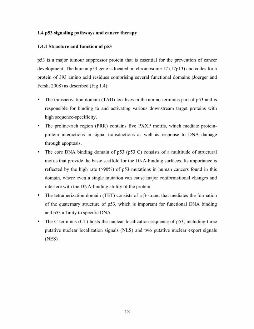
1.4.1 Structure and function of p53
p53 is a major tumour suppressor protein that is essential for the prevention of cancer
development. The human p53 gene is located on chromosome 17 (17p13) and codes for a
protein of 393 amino acid residues comprising several functional domains (Joerger and
Fersht 2008) as described (Fig 1.4):
• The transactivation domain (TAD) localizes in the amino-terminus part of p53 and is
responsible for binding to and activating various downstream target proteins with
high sequence-specificity.
• The proline-rich region (PRR) contains five PXXP motifs, which mediate protein-
protein interactions in signal transductions as well as response to DNA damage
through apoptosis.
• The core DNA binding domain of p53 (p53 C) consists of a multitude of structural
motifs that provide the basic scaffold for the DNA-binding surfaces. Its importance is
reflected by the high rate (>90%) of p53 mutations in human cancers found in this
domain, where even a single mutation can cause major conformational changes and
interfere with the DNA-binding ability of the protein.
• The tetramerization domain (TET) consists of a β-strand that mediates the formation
of the quaternary structure of p53, which is important for functional DNA binding
and p53 affinity to specific DNA.
• The C terminus (CT) hosts the nuclear localization sequence of p53, including three
putative nuclear localization signals (NLS) and two putative nuclear export signals
(NES).

13
Figure 1.4 Structure domains of p53 protein. The p53 protein consists of an N-
terminal transactivation domain (TAD, 1-60 amino acids), a proline-rich region (PRR,
62-97 aa), the core DNA binding domain (p53 C, 102-292 aa), the tetramerization
domain (TET, 320-356 aa) and the C terminus (CT, 363-393 aa). There are several
nuclear localization signals (NLS) and nuclear export signals (NES) in the C terminus of
p53.
1.4.2 p53-mediated cell cycle arrest and apoptosis
In normal cells, p53 is a short-lived protein and functions to control excessive cell
proliferation. Low intranuclear concentrations of p53 protein are maintained by its
binding to the E3 ubiquitin-ligase mdm2, which keeps p53 in check by continuous
ubiquitylation and subsequent degradation by the 26S Proteasome.
When the cell is stressed by DNA damage, hypoxia, cytokines, metabolic changes, viral
infection, or oncogenes, the activation of p53 can trigger cell cycle arrest, thereby
providing time for DNA repair before replicating the genome. Hence, p53 prevents the
transmission of damaged genetic information from one cell generation to the next. If the
damage is too severe to be repaired, p53 will initiate cell apoptosis and work as an
emergency brake on cancer development by killing cells that attempt to proliferate in
harsh microenvironments, such as those caused by hypoxia, nutrient deprivation, UV
radiation and chemotherapy.
p53 mainly functions as a transcription factor. Under the stress conditions, p53 becomes
phosphorylated on multiple sites, such as Ser15, Thr18, Ser 20 or Ser46. A potential
outcome of such phosphorylation is the stabilization of p53 through disruption of mdm2
association and inhibition of p53 ubiquitination and degradation. Once activated, p53
represses or enhances transcription of a variety of target genes involved in DNA repair,

14
cell cycle control and/or apoptosis. These include p21, PIG3, NOTCH1, E2F, bax and
PUMA. p21 for example, functions as an important inhibitor of a number of cyclin-
dependent kinases (CDKs) that are active in the late G1, S, G2 and M phases of the cell
cycle. Thus, by stimulating the transcription of the p21 gene, p53 prevents cell
proliferation at many points of cell cycle (Amaral et al. 2010; Wei et al. 2012).
p53 initiates apoptosis directly (through interaction of the Bax) or indirectly (through
transcription of genes involved in apoptosis). Once p53 becomes activated, a wide
network of apoptotic signals, such as Bax, the Fas receptor, and the IGFBP3 protein, are
activated and expressed. These signals act through two major apoptotic pathways:
intrinsic pathways and extrinsic pathways (Fig 1.5).
The intrinsic apoptotic pathway is controlled by the proteins involved in Bcl2 family,
which govern the release of Cytochrome-C (CytoC) and other pro-apoptotic proteins
from the mitochondria. Interestingly, p53 can target and promote expression of genes
encoding a key subset of Bcl2 family proteins, including Bax, Noxa, and PUMA. These
proteins work to open the mitochondrial channels and release CytoC, which activates the
apoptotic pathway by activating Caspases (Caspase-9, 3, 6 and 7).
In addition, an alternative route of p53 to trigger apoptosis is initiated outside the cell by
activating pro-apoptotic cell surface receptors, such as the Fas receptor and IGFBP-3. p53
induces the transcription of these ‘death’ receptors, leading to a cascade of activation of
Caspases (Caspase8 and Caspase3), which in turn induce apoptosis (Amaral et al. 2009;
Sharp et al. 2010; Ozaki and Nakagawara 2011).

15
Figure 1.5 Schematic representation of the p53-dependent apoptotic pathways
(Intrinsic and Extrinsic pathways). Under normal conditions, the intranuclear
concentration of p53 is tightly controlled by its negative regulator mdm2, which binds to
p53 and translocates it out of the nucleus for preteasomal degradation via the ubiquitin-
dependent pathways. Once p53 is phosphorylated and activated, it induces the expression
of proteins that target both the mitochondrial-induced apoptotic pathways (intrinsic
pathway) and the death-receptor-induced apoptotic pathways (extrinsic pathway).

16
1.4.3 p53 mutation and cancer
Given the important role of p53 in tumour suppression, tumour development is often
accompanied by high rate of mutations or deletions of the p53 gene, with >50% of human
tumours exhibiting some impairment of p53 function. Compromised p53 activity
promotes the accumulation of DNA damage in cells, which increases the possibility for
malignant transformation (Petitjean et al. 2007). In many cancers containing wild-type
p53, the p53 signaling pathways can be altered by other oncogenic events, suggesting that
a decrease in p53 function may be a common feature in many cancers.
The fast growth rate and stressful microenvironment of most tumours lead to
physiological stresses which would normally lead to p53-triggered apoptosis. However,
selection of cells with impaired p53 function during the early phases of tumour
development permits these cells to survive under stress conditions that usually lead to the
death of normal cells and accumulate mutations at higher rates. This in turn increases the
chances of oncogene activation and tumour suppressor gene inactivation.
p53 mutations mainly include the germinal mutation and somatic mutation:
• Germinal mutation:
Germinal mutation of p53 gene causes Li-Fraumeni syndrome, which is an extremely
rare inherited disease that greatly increases susceptibility to sarcomas (including
osteosarcoma), leukemia and breast cancer. The mutations can be inherited, or can arise
de novo early in embryogenesis or in one of the parent's germ cells (Varley 2003).
• Somatic mutation:
Somatic mutations in the p53 gene are one of the most frequent alterations in cancers.
More than half of human cancers of breast, bone, colon, lung, liver, prostate, bladder, and
skin have the mutated p53 gene. Over 90% of all known tumorigenic mutations are
located in the DNA-binding domain, thus preventing p53 from activating the
transcription of its target genes. These p53 mutants have different effects on tumour
development. One of outcomes of p53 mutations is the loss of tumour suppressor

17
functions of the wild-type protein, characteristically manifested as a total lack of p53
expression or production of unstable or truncated mutant proteins. On the other hand, the
cancer-associated p53 mutations also endow the mutant protein with new activities that
can contribute actively to various stages of tumour progression and to increased
resistance to anticancer treatments. These activities are referred to as mutant p53 gain-of-
function (GOF) (Oren and Rotter 2010). For example, transfection of p53-null cells with
mutant p53 enhanced tumour formation in mice (Wolf et al. 1984).
1.4.4 Activation of p53 as a therapeutic strategy
Since p53 activation induces apoptosis in response to physiologic stresses, such as DNA
damage, hypoxia, cytokines, metabolic changes, viral infection, or oncogene signaling,
there are several therapeutic drugs that might be used in cancer therapy.
1.4.4.1 DNA-damaging agents in cancer chemotherapy
Many chemotherapeutic drugs are DNA-damaging agents that can activate p53-mediated
apoptosis in cancers. However, studies have shown that the ability of many DNA-
damaging agents to induce p53 is not due to their ability to damage DNA. For many of
these compounds, p53 is activated through the inhibition of transcription that results in
nucleolar disruption. Nucleolar disruption is related to defects in ribosome biogenesis and
further leads to the release of free ribosomal proteins. These proteins can bind to mdm2
and inhibit its interaction with p53, thus activating the p53 response (Zhang and Lu 2009;
Lane et al. 2010). The agents that activate p53 through this route include the CDK
inhibitors, ribonucleotide production inhibitors and the RNA polymerase inhibitors
(reviewed by (Lane et al. 2010).
1.4.4.2 Gene therapy-based approaches
1.4.4.2.1 Introduction of wild-type p53 in tumours
The re-introduction of the wild-type p53 gene into a variety of human tumour cells has
been shown to induce apoptosis and growth inhibition. Especially in the p53-defective
tumour cells, the expression of wild-type p53 has a profound antitumour activity. The

18
first p53-based gene therapy was performed by injecting a retroviral vector containing the
wild-type p53 gene into tumours of patients with lung cancer (Roth et al. 1996). Then,
several virus delivery systems were established for p53 gene therapy; the most commonly
used was adenovirus. The obvious problems with this approach are the control of the
level of p53 expression and the effective delivering of the p53 gene to tumour cells.
1.4.4.2.2 Introduction of siRNA against the negative regulator of p53 in tumours
The overexpression of p53-negative regulatory proteins in tumour cells may result in the
inactivation of wild-type p53, so siRNA can be used against these proteins to activate p53
response. For example, in tumours where p53 is inactivated by ubiquitin-E3 ligase mdm2,
the introduction of siRNA specific to mdm2 induced an effective p53 response (Jiang and
Milner 2002).
1.4.4.3 Reactivation of mutant p53
Because of the high frequency of p53 mutation in tumour cells, the restoration of p53 has
been considered an attractive cancer therapeutic strategy. Some compounds activate p53
response by reactivating the wild-type p53 functions of mutant p53. For instance, the
small molecule CP-31398 induces specific p53 response and apoptosis in tumour cells by
stabilizing the active conformation of newly synthesized p53. PRIMA-1 (p53 reactivation
and induction of massive apoptosis) is another small molecule that restores sequence-
specific DNA binding and p53-dependent apoptosis. MIRA-1 (mutant p53-dependent
induction of rapid apoptosis) has been shown to reactivate DNA binding and preserve the
active conformation and transcriptional function of mutant p53 (Romer et al. 2006;
Levesque and Eastman 2007; Lane et al. 2010).
1.4.4.4 Inhibition of nuclear export
p53 is usually inactivated by nuclear export and degradation; thus, the inhibition of
nuclear export might be a feasible route to activate p53 response in tumour cells. Crm-1
is an exportin that mediates the transport of proteins from the nucleus to the cytoplasm,
while Leptomcin B (LMB) has been known as a potent inhibitor of crm-1. LMB can
effectively kill tumour cells in culture by stabilizing p53 from mdm2-mediated

19
degradation and activating p53-dependent transcription (Lain et al. 1999; Menendez et al.
2003).
1.4.4.5 Inhibition of p53-mdm2 interaction
Mdm2 plays a significant role in the negative regulation of p53. It interacts with p53 and
translocates it out of the nucleus for degradation via ubiquitin-dependent pathways. So
targeting mdm2 for p53 stabilization with small-molecule antagonists is a promising
approach for activating p53. For example, the nutlins act as antagonists of the mdm2-p53
interaction, which can bind in the pocket of mdm2 and prevent it from interacting with
p53 (Vassilev et al. 2004).
1.4.4.6 Inhibition of p53 activity after genotoxic stress
Conventional chemotherapeutic agents (such as DNA-damaging agents, antimetaolities,
and proteasome inhibitors) and radiotherapy can induce apoptosis following activation of
p53 not only in tumour cells, but also in normal cells. Thus, the compound that can
inhibit p53 activity is an option to protect normal cells from the severe side effects of
cancer treatment, such as the cytoprotective agent Pifihrin-a (Sinn et al. 2010). However,
it must be noted that the inhibition of p53 in normal tissue following cancer treatment
might increase the incidence of tumours; therefore, the combination of drugs that activate
p53 through targeting multiple molecule pathways could be an alternative way to induce
apoptosis without genotoxic response on normal and tumour tissues.
1.5 Rationale, Hypothesis and Objectives
One of the main goals of our research group is to determine the role of transcription
factor Zhangfei in the growth of cancer cells. In the previous studies, we have
demonstrated that Zhangfei dramatically suppressed cell growth in several cancer cell
lines, including canine osteosarcoma cells and human medulloblastoma cells (Valderrama
et al. 2009; Bergeron et al. 2013). To obtain a clear picture of the functions and
characteristics of Zhangfei, I attempted to answer two questions: first, does Zhangfei
have a universally inhibitory influence on the growth of different OS cell lines, especially

20
the canine and human osteosarcoma cell lines? Second, what are the molecular
mechanisms by which Zhangfei suppresses the growth of OS cells?
Based on the following observations:
• Zhangfei suppresses cell growth and induces apoptosis in several tumour cells
(Valderrama et al. 2009).
• The UPR is an adaptive stress response activated in cancer cells, allowing tumours to
survive, develop, metastasize and escape therapy.
• Zhangfei possesses a bLZip motif that exhibits sequence homology with the UPR-
related bLZip transcription factors Xbp1, ATF4 and ATF6. Zhangfei can interact with
ATF4 through the bLZip region (Hogan et al. 2006).
• Luman/CREB3 is a transcription factor that is structurally similar to and closely
related to UPR mediator ATF6 (Raggo et al. 2002).
• Zhangfei suppresses the ability of Luman/CREB3 to activate gene expression (Misra
et al. 2005).
My first hypothesis was:
Zhangfei interacts with the UPR-related bLZip transcription factors and inhibits
their ability to activate the UPR signaling pathways, thereby suppressing the growth
of cancer cells and increasing their susceptibility to ER stressors. Since our
laboratory had previously shown that Zhangfei does not have a very suppressive effect on
ATF6, I concentrated my efforts on Zhangfei-Xbp1 interactions.
My objectives were to:
• Determine the effect of Zhangfei on the growth and differentiation of several canine
and human tumour cell lines.
• Examine the effect of Zhangfei on the UPR signaling pathways and determine if
Zhangfei interacts with the UPR-related bLZip transcription factor Xbp1.
• Determine the molecular mechanisms through which Zhangfei exerts its effects in
cancer cells.
21
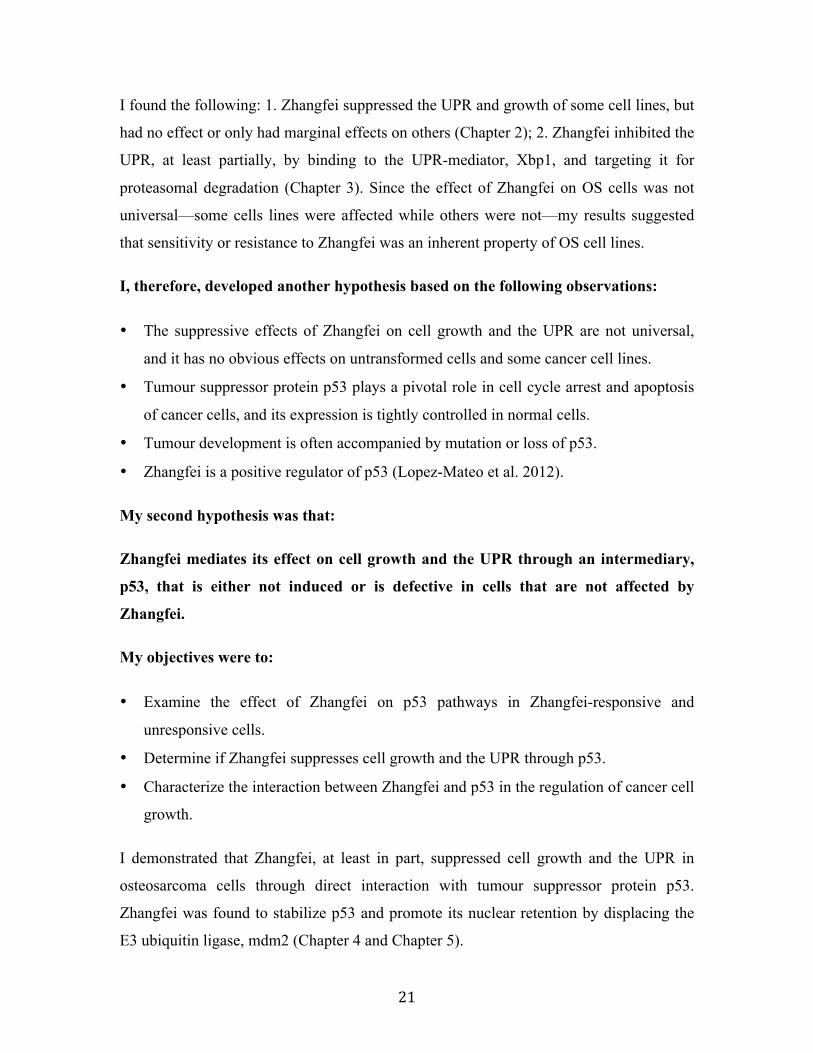
I found the following: 1. Zhangfei suppressed the UPR and growth of some cell lines, but
had no effect or only had marginal effects on others (Chapter 2); 2. Zhangfei inhibited the
UPR, at least partially, by binding to the UPR-mediator, Xbp1, and targeting it for
proteasomal degradation (Chapter 3). Since the effect of Zhangfei on OS cells was not
universal—some cells lines were affected while others were not—my results suggested
that sensitivity or resistance to Zhangfei was an inherent property of OS cell lines.
I, therefore, developed another hypothesis based on the following observations:
• The suppressive effects of Zhangfei on cell growth and the UPR are not universal,
and it has no obvious effects on untransformed cells and some cancer cell lines.
• Tumour suppressor protein p53 plays a pivotal role in cell cycle arrest and apoptosis
of cancer cells, and its expression is tightly controlled in normal cells.
• Tumour development is often accompanied by mutation or loss of p53.
• Zhangfei is a positive regulator of p53 (Lopez-Mateo et al. 2012).
My second hypothesis was that:
Zhangfei mediates its effect on cell growth and the UPR through an intermediary,
p53, that is either not induced or is defective in cells that are not affected by
Zhangfei.
My objectives were to:
• Examine the effect of Zhangfei on p53 pathways in Zhangfei-responsive and
unresponsive cells.
• Determine if Zhangfei suppresses cell growth and the UPR through p53.
• Characterize the interaction between Zhangfei and p53 in the regulation of cancer cell
growth.
I demonstrated that Zhangfei, at least in part, suppressed cell growth and the UPR in
osteosarcoma cells through direct interaction with tumour suppressor protein p53.
Zhangfei was found to stabilize p53 and promote its nuclear retention by displacing the
E3 ubiquitin ligase, mdm2 (Chapter 4 and Chapter 5).

22
2. The Effect of Zhangfei on the Unfolded Protein Response (UPR) and Growth of
Cells Derived from Canine and Human Osteosarcomas
Tania Bergeron1, Rui Zhang1, Kirsty Elliot2, Noreen Rapin1, Valerie MacDonald2,
Kathleen Linn2, Elemir Simko3 and Vikram Misra1*
Departments of Microbiology1, Small Animal Clinical Sciences2 and Pathology3,
Western College of Veterinary Medicine, University of Saskatchewan, Saskatoon,
Saskatchewan, S7N5B4, Canada
This Chapter determined the effects of transcription factor Zhangfei on the growth and
the UPR of canine and human osteosarcoma cell lines. The manuscript “The Effect of
Zhangfei on the Unfolded Protein Response and Growth of Cells Derived from Canine
and Human Osteosarcomas” has been published in Veterinary and Comparative
Oncology by Bergeron, T., Zhang, R., et al. 2013. 11(2):140-150 and is reproduced here
with the permission of the copyright owner.
My contributions to this manuscript: I examined the effects of Zhangfei on the growth
and programmed cell death (apoptosis) of canine and human osteosarcoma cell lines by
cell proliferation assay and Annexin V Apoptosis detection, respectively (Fig 2.5). In
addition, I also performed the immunoblotting detection to determine that the suppressive
effect of Zhangfei on transcripts for UPR-related genes was reflected in a decrease in
UPR proteins as well as in cells treated with calcium ionophore thapsigagin (Fig 2.4).
Bergeron, T., and others performed the rest of the experiments and obtained the data in
Fig 2.1-2.3 and Fig 2.5D.

23
2.1 Abstract
The objective of this study was to determine whether the protein Zhangfei could suppress
the unfolded protein response (UPR) and growth of osteosarcoma cells. Dog (D-17) and a
human (Saos-2) osteosarcoma cells were infected with adenovirus vectors expressing
either Zhangfei or the control protein β-galactosidase. We monitored the growth rate of
the cells by using the WST-1 cell proliferation reagent and designed oligonucleotide
primers that could be used to measure relative levels of transcripts for selected UPR
genes from dogs and humans. Two of the down-stream UPR proteins—HERP and
GRP78—were detected by immunoblots. The mechanism of programmed cell death was
determined by monitoring cells for the markers of apoptosis, autophagy and
macropinocytosis. We found that Zhangfei suppressed the growth of both D-17 and Saos-
2 cells. Zhangfei-expressing D-17 cells displayed large vacuoles containing pinocytosed
culture medium and expressed phosphatidylserine on their external surface suggesting
that Zhangfei induced macropinocytosis and apoptosis in these cells. While Zhangfei
inhibited the growth of both D-17 and Saos-2 cells, it inhibited thapsigargin-induced
UPR, as detected by a decrease in transcripts for UPR genes, and HERP and GRP78
proteins, only in D-17 cells, suggesting that the ability of Zhangfei to suppress the UPR
and tumour cells growth may not be linked.
Key words: unfolded protein response, osteosarcoma, Zhangfei, CREB-ZF, basic leucine-
zipper, apoptosis, macropinocytosis

24
2.2 Introduction
The fast growth rate of most cancer cells subjects them to stresses such as hypoxia, lack
of nutrients and accumulation of metabolic by-products. These stresses force cells to
activate responses that induce angiogenesis, metastasis and resistance to treatment by
radiation and chemotherapy (reviewed by (Dewhirst et al. 2008)). Three interconnected
signalling pathways regulate cellular response to stress (reviewed by (Wouters and
Koritzinsky 2008)). These are: hypoxia inducible family of transcription factors (HIFs),
the kinase mammalian target of rapamycin (mTOR) and the unfolded protein response
(UPR). Several chemotherapeutic agents are directed towards suppressing HIF (reviewed
by (Poon et al. 2009)) and mTOR. However, awareness of the involvement of UPR in the
stress response of cancer cells is relatively recent (Feldman et al. 2005) and apart from
Geldanamycin and Nelfinavir (an anti-HIV drug), few effective drugs are directed against
this pathway.
The UPR is activated by three sensors embedded in the endoplasmic reticulum (ER) of
cells. These include inositol requiring enzyme (IRE1), putative receptor protein kinase
(PERK) and activation transcription factor (ATF)6. The three sensors, IRE1, PERK and
ATF6, are held in a quiescent state until they are activated by a variety of stressors in the
ER such as the presence of unfolded proteins, changes in glycosylation of proteins, redox
status, glucose availability, calcium homeostasis or hypoxia. IRE1 and PERK, in turn,
activate the basic leucine-zipper motif (bLzip) transcription factors—Xbp1 and ATF4.
ATF6, which is itself a bLzip transcription factor, is translocated upon activation from
the ER, via the Golgi apparatus to the nucleus. Xbp1, ATF4 and ATF6 then activate the
expression of genes that relieve ER stress. If they are successful, as yet little understood
mechanisms turn them off. If ER stress is not relieved, the cells are directed to commit
suicide (apoptosis). The UPR is intimately linked to HIF and mTOR pathways (Wouters
and Koritzinsky 2008).
The protein Zhangfei is expressed in mature neurons (Akhova et al. 2005) but is not
detected in immature neurons or in cells derived from medulloblastomas and
neuroblastomas (Valderrama et al. 2009). Recently, we demonstrated that ectopic

25
expression of Zhangfei in medulloblastoma cells caused them to cease growth and
eventually die (Valderrama et al. 2009). While Zhangfei possesses a bLzip motif, its
basic domain lacks an asparagine residue that in other bLzip transcription factors is
considered crucial to their ability to recognize response elements in gene promoters.
Likely as a consequence of this, Zhangfei, as a homodimer, cannot bind known response
elements for bLzip transcription factors (Lu and Misra 2000b). Zhangfei can, however,
suppress the ability of some other bLzip transcription factors such as Luman/CREB3—a
transcription factor closely related to ATF6—(Misra et al. 2005), as well as Xbp1, ATF4
and ATF6 (our unpublished observations) to activate gene expression.
Many spontaneous dog cancers have similar histological characteristics, behaviour and
genetics to their human counterparts and respond in similar ways to therapeutics.
Spontaneous dog tumours therefore provide opportunities for studying signalling
pathways that regulate canine and human tumour growth and for the development of
therapeutic agents that might prevent their growth (reviewed by (Mueller et al. 2007;
Paoloni and Khanna 2008; Khanna et al. 2009). There is little information on the role of
ER stress in canine cancers although Klopfleisch and colleagues have recently shown that
metastatic and intra-lymphatic canine mammary tumour cells contain higher levels of
Derlin-1, a component of the UPR and a transporter for unfolded proteins from the ER
(Klopfleisch et al.). This suggests that the ER is stressed in these cells and that the UPR
may aid cells to metastasize.
Since the UPR enhances the survival of some tumours (Dong et al. 2008; Adham and
Coomber 2009; Ni et al. 2009; Romero-Ramirez et al. 2009) the purpose of this study
was to develop techniques to monitor the UPR in dogs and humans and to test the
hypothesis that the ectopic expression of Zhangfei in cell lines derived from dog and
human osteosarcomas would suppress their ability to activate the UPR and, consequently,
inhibit their growth. We found that Zhangfei could dramatically inhibit the growth of
both dog and human osteosarcoma cells. However, while thapsigargin-induced UPR was
suppressed in dog osteosarcoma cells, Zhangfei had relatively little effect on the pathway
in human osteosarcoma cells. This suggested that the two effects of Zhangfei—the
suppression of the growth of tumour cells and suppression of the UPR may not be linked.

26
2.3 Materials and Methods
2.3.1 Cell Culture
The dog osteosarcoma cell line, D-17, and the human osteosarcoma cell line, Saos-2,
were obtained from the American Type Culture Collection. The human osteosarcoma cell
line U-2 OS was from Dr James Smiley (University of Alberta). D-17 cells were grown
in Eagles minimal essential medium (MEM) containing 10% foetal bovine serum (FBS),
Saos-2 cells in McCoy’s medium with 15% FBS and U-2OS cells in Dulbecco’s MEM
containing Glutamax and 10% FBS. All media contained 100 units each of Penicillin and
Streptomycin per ml. Cells were grown at 37oC in an atmosphere of 5% CO2. All media,
FBS and antibiotics were purchased from GIBCO/Invitrogen.
To determine the growth rate of cells in response to various treatments, 1×10⁴ cells in 200
µl of medium were plated per well in 96 well culture dishes (Costar, Corning Inc.).
Twenty-four hr later cells were either mock infected, infected with adenovirus vectors or
were treated with drug or the drug diluent. For an estimate of viable cells in a culture, 20
µl of WST-1 cell proliferation reagent (Roche Applied Science) were added to each well
in a set and absorbance at 420 nm measured after one hr of incubation. Cell estimates
using WST-1 were repeated at 1, 2 and 3 days after infection or treatment. Each
experiment was repeated at least twice and the results were analyzed using non-
parametric Kruskal-Wallis and Mann-Whitney tests.
To activate the UPR, cells were treated with 100 nM thapsigargin (Sigma Life Science)
for 4 hr. Stock solution of thapsigargin was 750 µM in dimethyl sulphoxide (DMSO).
Control cultures were treated with an equivalent amount of DMSO. To determine the
effect of Zhangfei on UPR, cells were infected with adenovirus vectors expressing either
Zhangfei or beta-galactosidase at a multiplicity of infection of 100 infectious units per
cell. Infected cells were treated with thapsigargin one day after infection. Adenovirus
vectors (BD Biosciences) were prepared and assayed as described earlier (Misra et al.
2005).

27
To determine if the cause of death of cells was macropinocytosis, D-17 cells were
incubated with 25 µg/ml of Alexa 488 dextran (Molecular Probes/Invitrogen) per ml of
medium for 24 hr. After further incubation for 10 min in 10 nM Lysotracker Red
(Invitrogen) cells were observed for fluorescence. To test for apoptosis cells were
collected by trypsinization and stained with fluorescence isothiocyanate labelled Annexin
V and porpidium iodide (Calbiochem). A Cyto-ID autophagy detection kit (ENZO Life
Sciences) was used to determine if cells were undergoing autophagy.
2.3.2 Quantitative Real-Time PCR
The sequences of PCR primers used in the study are in Table 1. Primers were designed
by aligning coding sequences for mRNA for the dog and human homologues of various
relevant genes using CLUSTAL (MacVector 11.1.1, MacVector Inc.) and selecting
sequences approximately 300 bases apart, which were identical or almost identical in the
dog and human genes. A maximum of two mismatches were tolerated if the two
nucleotides at the 3’ ends of the primers were conserved. The accession numbers for
sequences used for the design of primers are: Xbp1 – XM_849540 (canine), NM_005080
(human), HERP – XM_857875 (canine), DQ837586 (human), CHOP - XM_844109
(canine), NM_004083 (human), BiP/GRP78 – XM_537847 (canine), NM_005347
(human). Figure 1 describes the strategy for the design of primers that discriminated
between spliced and unspliced versions of Xbp1.
Table 2.1 Sequence of primers used for qRT-PCR.
Xbp1 spliced upstream 5’-TCTGCTGAGTCCGCAGCAGG-3’ downstream 5’-TAAGGAACTGGGTCCTTCT-3’
Xbp1 unspliced upstream 5’-TCAGACTACGTGCACCTCTGC-3’ downstream 5’-TAAGGAACTGGGTCCTTCT-3’
HERP upstream 5’-CCGAGCCTGAGCCCGTCACG-3’ downstream 5’-CTTTGGAAGCAAGTCCTTGA-3’
CHOP upstream 5’-TGGAAGCCTGGTATGAGGAC-3’ downstream 5’-TGCCACTTTCCTCTCGTTCT-3’
GRP78 upstream 5’-GGCTTGGATAAGAGGGAAGG-3’ downstream 5’-GGTAGAACGGAACAGGTCCA-3’
GAPDH upstream 5’-TGCCTCCTGCACCACCAACTGC-3’ downstream 5’-GGGCCATCCACAGTCTTCTGGG-3’

28
RNA was purified from cells using the RNEasy Plus mini kit with a genomic DNA
elimination step (Qiagen) and RNA converted to cDNA with the Quantitect Reverse
Transcription kit (Qiagen) using instructions supplied by the manufacturer. Two-step
qRT-PCR reactions used a Brilliant SYBR Green QPCR Master Mix (Stratagene) and
were performed in a Stratagene MX3005P PCR instrument. Each reaction (25 µl)
contained 150 nM of each primer, ROX as a reference dye, Brilliant SYBR Green Master
mix and 8 µl of cDNA. The thermal profile for the reactions was: 10 minutes at 95oC, 40
cycles of 30 sec at 95oC, 1 minute at 60oC and 1 minute at 72oC. All primer sets had a
reaction efficiency of over 98%. To ensure the fidelity of the reactions, the disassociation
profiles for PCR products in all reactions were examined and the PCR products of the
first reaction were analyzed by gel electrophoresis and sequence confirmed. Every cDNA
sample was also assayed for levels of GAPDH transcripts. Levels of GAPDH were used
to normalize the samples. All reactions were analyzed in duplicate and each experiment
was repeated at least once and usually twice. Relative fold changes of trancript levels
were calculated as 2DDCt where DCt was the threshold value (Ct) for a primer set
detecting GAPDH subtracted from the Ct value for a particular target and DDCt was DCt
value of sample minus the DCt for control. The veracity of the PCR products was
confirmed by electrophoresis through agarose gels and by determining their nucleotide
sequence.
2.3.3 Antibodies, Microscopy and Immunofluorescence
Mouse monoclonal antibodies against beta-galactosidase were purchased from Millipore.
Antiserum against Zhangfei was prepared in rabbits and has been described (Lu and
Misra 2000b). The antibodies have been characterized and are specific for Zhangfei
(Akhova et al. 2005). After fixing cells grown on glass coverslips in cold methanol and
blocking in phosphate buffered saline (PBS) containing 10% FBS cells were incubated
with primary antibodies against beta-galactosidase and Zhangfei. After washing off
unbound antibody cells were incubated with goat anti-mouse antibodies labelled with
Alexa 488 and goat anti-rabbit antibodies labelled with Alexa 546. After washing
coverslips were mounted in PBS containing 10% glycerol and observed using a Zeiss
29
Axiovert fluorescent microscope. Images were captured using a digital camera attached
to the microscope and Northern Eclipse software (EMPIX Imaging Inc).
2.3.4 Detection of proteins by immunoblotting
Procedure was as described in (Akhova et al. 2005). Essentially, cells were lysed in SDS-
PAGE buffer and proteins separated by electrophoresis through 10% SDS-
polyacrylamide gels. Proteins were then transferred to Hybond-LFP membranes
(Amersham, GE Healthcare) using an Amersham MiniVE Electrotransfer unit at 400
mAmps for 90 min. After incubating overnight in 5% BSA in phosphate buffered saline
(PBS) the membranes were rinsed with PBS containing 0.1% Tween20 (PBS-T) and
incubated in PBS-T containing either rabbit anti-HERP (Abcam), anti-GRP78 (Abcam),
anti-ZF (Lu and Misra 2000b) and mouse anti-GAPDH (Chemicon). After washing with
PBS-T the membranes were incubated with goat anti-mouse Alexa488 and goat anti-
rabbit Cy5 (Amersham). Fluorescent bands were detected and documented using a
Typhoon Trio variable mode imager (GE Healthcare).
2.3.5 Detection of apoptotic cells
Cells were collected by trypsinization and stained with Annexin V and propidium iodide
using an Annexin V kit (Calbiochem) and instruction supplied by the manufacturer. As a
positive control cells were treated with 50 µM etopocide (Calbiochem) for 24 hr. Cells
were analyzed in a Coulter EPICS XL flow cytometer.
2.3.6 Statistics
Analyses were performed using IBM-SPSS 19 for the Macintosh. ANOVA and Kruskal
Wallis and Mann Whitney tests were used for data with more than two groups. For data
with two groups means were compared using an independent sample T-test. Differences
with P values less than 0.05 were considered significant.

30
2.4 Results
2.4.1 Detecting the UPR in dog and human osteosarcoma cells
The ER-resident proteins IRE1, PERK and ATF6 sense cellular stress. IRE1, an
endoribonuclease, splices mRNA for transcriptionally inactive Xbp1 to a form that is
translated into a transcriptionally active factor. PERK suppresses most translation in the
cells but activates the translation of the mRNA for the transcription factor ATF4. ATF6,
which is itself a transcription factor, is translocated to the Golgi where it is released by
intra-membrane proteolysis.
To measure activation of the UPR, we designed qRT-PCR oligonucleotide primers that
could selectively detect spliced or unspliced transcripts for Xbp1 (Fig 2.1) as well as
primers that could detect representative genes activated by the three bLzip transcription
factors—Xbp1 (HERP), ATF4 (CHOP) and ATF6 (GRP78/Bip). To determine if the
primers could detect and quantitate the respective transcripts in dog and human cells, we
treated cells of dog (D-17) and human (Saos-2) osteosarcoma cells lines with the calcium
ionophore thapsigargin, which is known to activate the UPR in many cell-types. The
level of transcripts in thapsigargin-treated cells were compared with cells treated with the
drug diluent DMSO. To measure spliced and unspliced Xbp1 message, we examined
cells 4, 18 and 24 hr after treatment. In both cell types spliced Xbp1 transcripts increased
dramatically at 4 hr and then decreased over the next 20 hr (Fig 2.2A). The other UPR
transcripts were measured 4 hr after thapsigargin treatment and showed a 10 to 50-fold
increase over DMSO-treated cells (Fig 2.2B). The PCR products were of the expected
size and sequence of the dog and human gene segments amplified by the primers.
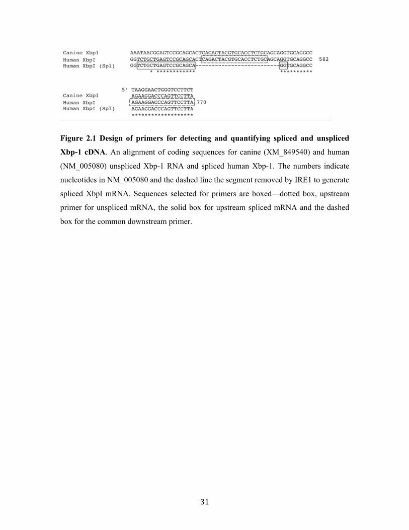
31
Figure 2.1 Design of primers for detecting and quantifying spliced and unspliced
Xbp-1 cDNA. An alignment of coding sequences for canine (XM_849540) and human
(NM_005080) unspliced Xbp-1 RNA and spliced human Xbp-1. The numbers indicate
nucleotides in NM_005080 and the dashed line the segment removed by IRE1 to generate
spliced XbpI mRNA. Sequences selected for primers are boxed—dotted box, upstream
primer for unspliced mRNA, the solid box for upstream spliced mRNA and the dashed
box for the common downstream primer.
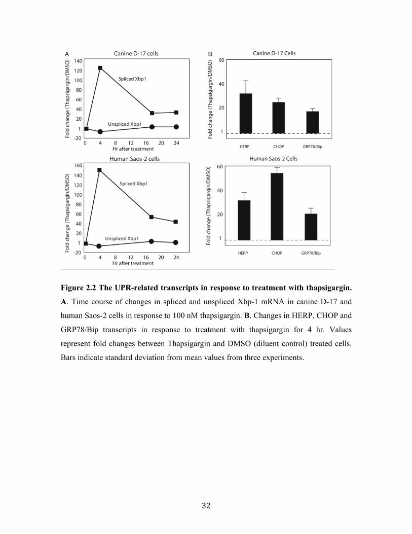
32
Figure 2.2 The UPR-related transcripts in response to treatment with thapsigargin.
A. Time course of changes in spliced and unspliced Xbp-1 mRNA in canine D-17 and
human Saos-2 cells in response to 100 nM thapsigargin. B. Changes in HERP, CHOP and
GRP78/Bip transcripts in response to treatment with thapsigargin for 4 hr. Values
represent fold changes between Thapsigargin and DMSO (diluent control) treated cells.
Bars indicate standard deviation from mean values from three experiments.

33
2.4.2 Suppression of UPR by Zhangfei
Since Zhangfei can suppress the ability of Luman, an ATF6-like ER-resident
transcriptional factor, to activate transcription (Misra et al. 2005), we determined the
effect of Zhangfei on the UPR transcripts for Xbp1, HERP, CHOP and GRP78/Bip in
thapsigargin-treated cells. Cells were mock-infected or infected with adenovirus vectors
expressing either Zhangfei or, as a control, the Escherichia coli protein beta-
galactosidase (LacZ). One day after infection, cells were treated with either DMSO or
thapsigargin, harvested 4 hr later and analyzed for UPR gene transcripts. While LacZ had
no obvious effect on the ability of thapsigagrin to activate HERP, CHOP or GRP78/Bip,
in Zhangfei-expressing D-17 cells levels of these transcripts were decreased dramatically.
Levels of spliced Xbp1 transcripts were also decreased (Fig 2.3, top panel). In contrast,
with the exception of a slight decrease in CHOP transcripts, Zhangfei had little obvious
effect on level of the UPR transcripts in thapsigargin-treated Saos-2 cells (Fig 2.3, bottom
panel).

34
Figure 2.3 Effect of Zhangfei or LacZ-expression on thapsigargin-induced changes
in Xbp-1, HERP, CHOP and GRP78/Bip transcript levels in canine D-17 and
human Saos-2 cells. Cells were mock-infected or infected with adenoviruses expressing
either LacZ or Zhangfei. Twenty four hr after infection cells were treated with either
DMSO or thapsigargin. Four hr later cells were harvested and differences in levels of the
transcripts in thapsigargin and DMSO-treated (diluent control) cells was assessed.
Standard deviations from mean are shown and P values are noted above the bars.

35
2.4.3 Effect of Zhangfei on UPR related proteins
Our results suggested that Zhangfei reduced transcripts for UPR-related genes in cells in
response to thapsigargin (Fig 2.3). To determine if this decrease in RNA was reflected in
a decrease in UPR proteins as well, we detected HERP and GRP78 in cells mock-infected
or infected with an adenovirus vector expressing Zhangfei. We examined cells that
expressed Zhangfei for 24 hr and were then treated with thapsigargin for 4 hours. This
would determine the ability of cells expressing Zhangfei to respond to the UPR. As an
alternative, we treated cells with thapsigargin for 4 hr and then infected or mock infected
cells (as in the experiment described in Fig 2.3). This would assess the ability of Zhangfei
to turn off the UPR once it had been activated. In both experiments, mock-infected cells
were an indication of the level of HERP and GRP78 without Zhangfei at the time of cell
harvest. Figure 2.4 shows that cells already expressing Zhangfei (compare lanes 1 and 2
in Fig 2.4) were unable to make detectable amounts of HERP as compared to mock-
infected cells. After 4 hr of thapsigargin treatment mock-infected cells made relatively
little GRP78 (barely visible as a faint band in lane 2) and even this was not detected in
Zhangfei-expressing cells (lane 1). When cells were treated with thapsigargin first,
infected or mock-infected, and then maintained in thapsigargin-free medium for 24 hr,
substantial amounts of GRP78 were detected in mock-infected cells (lane 4) and this was
considerably reduced in Zhangfei–expressing cells (lane 3). Zhangfei also reduced
HERP as compared to mock-infected cells (compare faint band in lane 4 with lane 3).

36
Figure 2.4 Effect of Zhangfei on HERP and GRP78 in cells treated with
thapsigargin. D-17 cells were either infected with an adenovirus vector expressing ZF
(AdZF) or were mock-infected before (lanes 1,2) or after (lanes 3,4) treatment for 4 hr
with thapsigargin. Cells were harvested 24 hr after infection, proteins separated by SDS-
PAGE and probed with antibodies against HERP, GRP78 or Zhangfei (ZF) and GAPDH.
37
2.4.4 Effect of Zhangfei on the growth of dog and human osteosarcoma cells
Ectopic expression of Zhangfei in medulloblastoma cells causes them to stop growing
(Valderrama et al. 2009). To determine if Zhangfei had a similar effect on dog and
human osteosarcoma cells, D-17 and Saos-2 cells were mock-infected or infected with
adenoviruses expressing either Zhangfei or LacZ. Growth was monitored as absorbance
at 420 nm of cultures treated for 1 hr with WST-1 cells proliferation reagent. At daily
intervals cells were also stained for Zhangfei or LacZ to ensure expression of the proteins
in all cells. While expression of LacZ had no effect on the growth of the cells, Zhangfei
suppressed the growth of D-17 cells as early as one day after infection (Fig 2.5A). All
Zhangfei-expressing cells died approximately 5-6 days after infection while most mock-
infected or LacZ-expressing cells retained their viability at that time.
All cells infected to express either LacZ or Zhangfei expressed the proteins and there was
little fluorescence because of cross-staining. We noticed that many Zhangfei-expressing
D-17 cells showed large vacuoles (Fig 2.5D). Cells die of necrosis or of three known
pathways of programmed cell death—apoptosis, autophagy and macropinocytosis (Li et
al. 2010). Zhangfei-expressing cells took up large amounts of fluorescent Alexa 488
dextran from the growth medium (Fig 2.5D). Some of the dextran containing vacuoles
co-localized with Lysotracker-staining mature lysosomes, a marker of macropinicytosis
(Li et al. 2010; Nara et al. 2010). We also determined if Zhangfei induced D-17 cells to
commit apoptosis. Intact, unfixed cells were stained with fluorescent Annexin V, 12 and
24 hour after infection. Annexin V binds to phosphatidylserine on the surface of cells, a
feature of apoptotic cells. Necrotic cells also bind Annexin V but can be distinguished
from apoptotic cells because they are permeable to propidium iodide. As a positive
control we analyzed cells treated with etopocide, a chemical that induces apoptosis.
Figure 2.5E shows that Zhangfei-expressing D-17 cells began to display markers of
apoptosis by 12 hr after infection with adenovirus vector expressing Zhangfei. By 24 hr
following infection more than 20% of cells were apoptotic. This compared with about 14%
apoptotic cells 24 hr after treatment with etopocide and less than 1% apoptotic cells in
mock-infected, Lac-Z-expressing or DMSO-treated (diluent for etopocide) cultures. The
proportion of propidium iodide staining cells (dead cells) did not exceed 1% of cells for

38
any of the samples during 24 hr of infection or treatment. The cells did not display
markers of autophagy.
While the effect of Zhangfei on Saos-2 cells was not as dramatic (Fig 2.5B) as on D-17
cells, the growth rate of these cells was significantly different from that of mock-infected
(p=0.023) and LacZ-expressing cells (p=0.026). There was no difference between mock-
infected and LacZ-expressing cells (p=0.956).

39
Figure 2.5 Effect of Zhangfei on the growth of canine D-17 and human Saos-2 cells.
Cells were mock infected or infected with adenoviruses expressing either Zhangfei or
40
LacZ. At 24 hr intervals the metabolic activity of cells was assessed by treatment with
WST. At daily intervals cells were also fixed and analyzed by immunofluorescence for
LacZ (top panel in Fig 2.5A and B) or Zhangfei (bottom panel in Fig 2.5A and B). The
difference in the growth rates of Zhangfei and LacZ-expressing D-17 cells is obvious. For
Saos-2 cells there was a statistically significant difference between the growth rates of
mock-infected or LacZ-expressing cells and Zhangfei-expressing cells, p=0.026 and
0.023, but none between mock-infected and LacZ-expressing cells, p=0.956. (C)
Immuno-blot of lysates of D-17 cells mock-infected or infected with Adenovirus vectors
expressing LacZ or ZF. The blot was developed with the rabbit antibody against Zhangfei
used in Figures A and B. (D) Zhangfei-expressing D-17 stained for macropinosomes
(green) and lysosomes (red). Top panel shows phase-contrast micrographs of LacZ and
Zhangfei-expressing cells. The cytoplasmic vacuoles in Zhangfei-expressing cells are
clearly visible. The lower panel shows Zhangfei-expressing cells grown in medium
containing fluorescent (Alexa488, green) dextran and stained for lysosomes (Lysotracker,
red). (E) AdZF or AdLacZ-infected cells, mock-infected cells and D-17 cells treated with
50µM etopocide were stained with for Annexin V-fluorescence and propidium iodide and
cells staining with either or both dyes were enumerated by FACS. Bars represent fraction
of cells undergoing apoptosis, ie. percentage of total cells in a sample that stained with
Annexin V-fluorescence but not propidium iodide. Where applicable, error bars represent
standard deviation from the mean.
41
2.5 Discussion
Our objective was to determine if Zhangfei could suppress the growth and the UPR in
tumour cells and if these two properties attributed to the protein were linked. Our results
showed that ectopic expression of Zhangfei could inhibit the growth of dog and human
osteosarcoma cells (Fig 2.5). We did not determine the mechanisms by which Zhangfei
accomplished this. However, D-17 cells appeared to undergo macropinocytosis and take
in large amounts of extracellular fluid (Fig 2.5D). Macropinocytosis is involved in
cellular processes such as antigen processing in dendritic cells (Doherty and McMahon
2009) but dysregulation has been shown to cause cell death in glioblastoma cells
(Overmeyer et al. 2008) and induction of macropinocytosis by stimulation of the
TrkA/NGF pathway causes death of medulloblastoma cells (Li et al. 2010). The
inhibition of cell division in human Saos-2 cells was not as dramatic as in D-17 cells and
we did not observe the presence of large cytoplasmic vacuoles suggesting that the
mechanism by which Zhangfei exerts its effect may be different for the two cell lines.
While Zhangfei dramatically inhibited the ability of thapsigargin to activate markers of
the UPR in D-17 cells it had no effect in Saos-2 cells (Fig 2.3) supporting a different
mechanism in these two cell lines.
Our results show (Fig 2.4) that the suppression of transcripts for UPR genes by Zhangfei
was reflected in its effect on HERP and GRP78 proteins induced by thapsigargin.
Zhangfei both prevented the synthesis of the protein in response to thapsigargin and also
reduced the proteins in cells in which they had been induced before Zhangfei expression.
When comparing the two proteins in mock-infected cells (lanes 2 and 4) we detected
more HERP in cells harvested 4 hr after thapsigargin treatment (lane 2) than in cells
harvested 24 hr after thapsigargin had been removed (lane 4). In contrast more GRP78
was detected in cells 24 hr after drug treatment (lane 4) than in cells harvested
immediately after 4 hr in thapsigargin. This suggests that after thapsigargin treatment
HERP is made before GRP78 but then degraded faster than GRP78.
Most bLzip domain-containing proteins regulate transcription as leucine-zipped homo-
dimers or as hetero-dimers with other such proteins, the properties of the partners as well

42
as the DNA motifs they recognize determining whether the dimers activate or suppress
gene expression (reviewed in (Miller 2009)). Zhangfei lacks an aspargine residue present
in the DNA-binding domain of most bLzip proteins and known to be crucial to their
ability to bind DNA (Vinson et al. 1989; Ellenberger et al. 1992). Possibly because of this,
Zhangfei does not appear to bind DNA on its own but can activate or suppress gene
expression in association with other bLzip proteins (Lu and Misra 2000b). Other studies
from our laboratory (unpublished) suggest that Zhangfei suppresses the UPR by binding
to and inhibiting the activity of XbpI, ATF4 and ATF6. It was therefore puzzling that it
inhibited the UPR in some cells (D-17, Fig 2.3) but not in others (Saos-2, Fig 2.3). This
suggests that the mechanism by which Zhangfei suppresses the UPR may be more
complex than we had envisioned. An alternative explanation may be that Zhangfei must
be post-translationally processed to be active and that the machinery required for this is
not present in Saos-2 cells.
Since Zhangfei suppressed growth but not the UPR in Saos-2 cells, our results also
suggest that the ability of Zhangfei to suppress the growth of tumour cells may not be
linked to its ability to suppress the UPR. Recently we have determined that Zhangfei
suppresses the growth of human medulloblastoma cells by activating the expression of
TrkA, the high-affinity receptor for Nerve Growth Factor (manuscript submitted). This
triggers an autocrine-signalling process that activates genes that lead to differentiation
and apoptosis in a mitogen activated protein kinase (MAPK)-dependent process. We have
not determined how Zhangfei suppresses other tumours and we are in the process of
assessing how many different kinds of human and dog tumours are susceptible to
Zhangfei. However, our results thus far suggest that cells obtained from normal tissue
may not be inhibited by the protein. This selective targeting of tumour cells is also
observed for oncolytic viruses such as reovirus, vesiculo-stomatitis virus and myxovirus
which rely on constitutively active components of the MAPK pathway in tumour cells for
their selective targeting (Smakman et al. 2006; Wang et al. 2006a; Marcato et al. 2007;
Noser et al. 2007).

43
The selective targeting of the ability of tumour cells to grow as well as the UPR (at least
in some tumour cells) may allow the therapeutic use of Zhangfei in combination with
other modalities, such as chemotherapy and radiation, that are inhibited by the UPR.
2.6 Acknowledgements
This work was supported by a Discovery grant to VM from the Natural Sciences and
Engineering Research Council (NSERC) of Canada and a grant to VMacD, KL, ES and
VM from the Western College of Veterinary Medicine (WCVM) Companion Animal
Research Fund and the Kaye Canine Foundation. TB was supported by a summer
research scholarship and KE by a fellowship from the WCVM Interprovincial fund. RZ
was supported by scholarships from the Government of China (China Scholarship
Council, RZ-2010635007) and the University of Saskatchewan Graduate Studies.

44
3. Zhangfei/CREB-ZF – a potential regulator of the Unfolded Protein Response
Rui Zhang1, Noreen Rapin1, Zhengxin Ying2, Erika Shklanka1, Timothy W. Bodnarchuk1,
Valerie M. K. Verge2 and Vikram Misra1*
1Department of Microbiology, Western College of Veterinary Medicine, 52 Campus
Road, University of Saskatchewan, Saskatoon, Saskatchewan , S7N1B4, CANADA
2Department of Anatomy and Cell Biology and Cameco Neuroscience Research Center,
University of Saskatchewan, Saskatoon, Saskatchewan, S7N5E5, CANADA
This Chapter demonstrated how Zhangfei interacted with the UPR-mediator Xbp1s and
consequently suppressed the UPR signaling pathways in cancer cell lines. The manuscript
has been published as “Zhangfei/CREB-ZF – a potential regulator of the Unfolded
Protein Response” in PLoS One. 8(10): e77256. by Zhang, R., Rapin, N., et al. and is
reproduced here with the permission of the copyright owner.
My contributions to this manuscript: I designed and performed all the experiments
mentioned in this manuscript, except the qRT-PCR arrays and PCR confirmation (Fig
3.1), which were performed by Rapin, N., as well as the adult dorsal root ganglia culture,
which was performed by Ying, X.

45
3.1 Abstract
Cells respond to perturbations in the microenvironment of the endoplasmic reticulum
(ER), and to the overloading of its capacity to process secretory and membrane-associate
proteins, by activating the Unfolded Protein Response (UPR). Genes that mediate the
UPR are regulated by three basic leucine-zipper (bLZip) motif-containing transcription
factors—Xbp1s, ATF4 and ATF6. A failure of the UPR to achieve homeostasis and its
continued stimulation leads to apoptosis. Mechanisms must therefore exist to turn off the
UPR if it successfully restores normalcy. The bLZip protein Zhangfei/CREBZF/SMILE
is known to suppress the ability of several, seemingly structurally unrelated, transcription
factors. These targets include Luman/CREB3 and CREBH, ER-resident bLZip proteins
known to activate the UPR in some cell types. Here we show that Zhangfei had a
suppressive effect on most UPR genes activated by the calcium ionophore thapsigargin.
This effect was at least partially due to the interaction of Zhangfei with Xbp1s. The
leucine zipper of Zhangfei was required for this interaction, which led to the subsequent
proteasomal degradation of Xbp1s. Zhangfei suppressed the ability of Xbp1s to activate
transcription from a promoter containing unfolded protein response elements and
significantly reduced the ability to Xbp1s to activate the UPR as measured by RNA and
protein levels of UPR-related genes. Finally, specific suppression of endogenous
Zhangfei in thapsigargin-treated primary rat sensory neurons with siRNA directed to
Zhangfei transcripts, led to a significant increase in transcripts and proteins of UPR genes,
suggesting a potential role for Zhangfei in modulating the UPR.

46
3.2 Introduction
One of the main functions of the endoplasmic reticulum (ER) is to modify, process and
fold proteins destined for secretion or insertion into membranes. The ER also plays
critical roles in maintaining intracellular calcium stores, steroid and lipid biosynthesis,
membrane regeneration and gluconeogenesis (reviewed in (Fujimoto and Hayashi 2011;
Wagner and Moore 2011)). Since protein folding and processing require an oxygen rich
microenvironment, as well as adequate nutrient and calcium levels, deficits in these
components lead to the accumulation of unfolded or inadequately modified proteins. The
ER then initiates a program of recovery called the Unfolded Protein Response (UPR,
reviewed in (Merksamer and Papa 2010; Diehl et al. 2011)). The UPR has three main
goals: the degradation of misfolded proteins, suppression of the synthesis of new proteins,
and to increase the synthesis of chaperones and other proteins required for processing.
The suppression of additional protein synthesis is achieved by the phosphorylation of the
eukaryotic translation initiation factor 2a (eIF2 a) by the ER-stress sensor—double
stranded RNA protein kinase-like ER kinase (PERK) (Harding et al. 1999). In addition,
three basic leucine-zipper motif (bLZip) containing proteins: spliced X-box binding
protein (Xbp1s), activation transcription factors ATF4 and ATF6, activate the
transcription of UPR-related genes. The protein Xbp1s results from the unique extra-
nuclear splicing of the mRNA for the transcriptionally inactive protein Xbp1u by the ER
stress sensor, inositol-requiring enzyme/ER to nucleus signaling protein (IRE1/ERN1).
Xbp1s retains the basic leucine-zipper motif (bLZip) coded by the unspliced Xbp1u
mRNA but acquires a transcription activation domain and a nuclear transport motif
(Calfon et al. 2002). A failure of the UPR to re-establish normalcy triggers apoptosis
while successful homeostasis leads to suppression of the UPR.
The UPR includes feed back mechanisms that mediate a retraction of the UPR if ER
function is restored. The proteins GADD34 (Ma and Hendershot 2003; Zhou et al. 2011),
Nck1 (Latreille and Larose 2006; Cardin et al. 2007) and p58iPK (Yan et al. 2002; van
Huizen et al. 2003) and reviewed by (Rutkowski et al. 2007) recruit protein phosphatases
that dephosphorylate eIF2a restoring protein synthesis. The protein Xbp1u, dimerizes
with Xbp1s and ATF6 and targets them for proteasomal degradation (Yoshida et al. 2006;

47
Yoshida et al. 2009). With the exception of Xbp1u, most of the UPR-modulating
mechanisms described to date are aimed at the PERK effector pathways of the UPR.
Relatively little is known about the suppression of the IRE1 and ATF6 arms of the
response.
Zhangfei/CREBZF/SMILE was first discovered as a binding partner for Host Cell Factor
(HCF), a co-activator of the herpes simplex virion transcription factor VP16 (Lu and
Misra 2000b). Translation for the protein is initiated at two alternate initiation codons
(Xie et al. 2008), although both isomers appear to have similar properties. The primary
structure of the protein contains a leucine zipper, a basic region that lacks an asparagine
residue conserved in most bLZip proteins, three potential nuclear factor binding domains
(LLXXLL, where L is a leucine residue and X is any amino acid), and a domain for
binding HCF. Zhangfei interacts with several proteins, possibly through its nuclear
receptor and HCF binding domains as well as its leucine zipper. While Zhangfei can
activate gene expression through factors such as p53 (Lopez-Mateo et al. 2012) and
ATF4 (Hogan et al. 2006), it suppresses the activity of a number of transcription factors
which include nuclear receptors (Xie et al. 2008; Xie et al. 2009a; Xie et al. 2009b),
bLZip containing proteins such as CREBH (Misra et al. 2012) and Luman/CREB3 (Misra
et al. 2005), SMAD 1,5,8 (Lee et al. 2012a) and herpes simplex virion associated VP16
(Akhova et al. 2005). We have detected Zhangfei protein in differentiated neurons, but
not in developing neurons or cells of neuronal tumours (Akhova et al. 2005). The ectopic
expression of Zhangfei in medulloblastomas and other tumours causes the cells to stop
growing and eventually to die (Valderrama et al. 2009; Bodnarchuk et al. 2012; Bergeron
et al. 2013). Zhangfei suppresses the ability of Luman/CREB3 (Misra et al. 2005) and
CREBH (Misra et al. 2012), to activate transcription. Since these proteins are known to
regulate the UPR in some cell types, we hypothesized that Zhangfei may be involved in
modulating the UPR. Here we show that Zhangfei can suppress the expression of UPR
genes activated in response to the drug thapsigargin. We further show that this effect is
mediated, at least partially, by the leucine-zipper dependent interaction of Zhangfei and
Xbp1s resulting in the proteasomal degradation of Xbp1s. Our results support the
hypothesis that Zhangfei has the capacity to modulate the UPR.
48
3.3 Materials and Methods
3.3.1 Cell Culture
The human medulloblastoma cell line ONS-76 (Tamura et al. 1989), was obtained from
Michael Taylor (University of Toronto). These cells were cultured as described
previously (Valderrama et al. 2009). Vero cells were obtained from the American Type
Tissue Culture Collection and grown in Dulbecco’s minimal essential medium containing
penicillin and streptomycin and 10% newborn calf serum. All media, serum and
antibiotics were purchased from Invitrogen. For some experiments cells were treated with
100 nM thapsigargin or with an equivalent amount of DMSO, the diluent for thapsigargin,
for 4 hr. The duration of thapsigargin treatment was based on preliminary experiments to
determine the time course of the accumulation of selected UPR-related transcripts
following thapsigargin treatment.
3.3.2 Immunofluorescence
Cells were processed for immunofluorescence as described previously (Lu and Misra
2000a). Cells were stained for Zhangfei using anti-FLAG monoclonal antibody (Sigma,
082K9164), for other proteins—rabbit anti-GRP78 serum (Abcam, ab21685), for anti-
HERP (Abcam, ab73669-100) and anti-Xbp1 (Abcan, ab37152). Secondary antibodies
were Alexa Fluor 488 linked anti-mouse and Alexa Fluor 546 linked anti-rabbit
antibodies (Invitrogen, A-11001, A-11035). Cell nuclei were stained using Hoechst
fluorescent dye (Molecular Probes, 33342).
3.3.3 Plasmids
The construction of pcZF (Lu and Misra 2000b), a plasmid that expresses Zhangfei in
mammalian cells, pCAT3BATF6 (Misra et al. 2005), with the coding sequences for
chloramphenicol acetyl transferase (CAT) and pMZF (Lu and Misra 2000b), with the
coding sequences for Zhangfei linked to the DNA-binding domain of yeast GAL4, have
been described. Plasmid pCGNATF6 (1-373), which expresses the constitutively active
truncated form of ATF6, and p5XATF6GL3, which contains 5 copies of the UPRE-
containing oligonucleotide, CTCGAGACAGGTGCTGACGTGGCATTC, were

49
obtained from Ron Prywes, Columbia University, USA (Wang et al. 2000). A plasmid
expressing the functionally active, spliced form of Xbp1 cDNA was obtained from K.
Mori, Kyoto University, Japan (Yoshida et al. 2001). The plasmid, pG5EC, a CAT
reporter plasmid with 5 copies of the yeast Gal4-UAS as well as the pM series of
plasmids for constructing Gal4 fusion proteins were obtained from I. Sadowski,
University of British Columbia, Canada (Sadowski et al. 1992). To construct pcZF(L/A),
an expression vector in which the codons for the first six consecutive leucines in the
leucine-zipper of Zhangfei were replaced by codons for alanine, a 265 bp synthetic DNA
fragment (IDT) bracketed by NotI and SgrA1sites was used to replace a fragment
between unique Not1 and SgrA1 sites in the coding sequences of Zhangfei in pcZF. An
internal PstI site within the fragment was eliminated with a silent mutation to allow for
screening of the mutant. The coding sequences in the mutant were sequenced to confirm
the mutation and to ensure that no unintended changes had been made.
3.3.4 Adenovirus vectors expressing Zhangfei and β-galactosidase (LacZ)
These vectors were constructed, grown, and purified using the Adeno-X Expression
System (Clontech, K1650-1). They were created in our laboratory as described earlier
(Misra et al. 2005). ONS-76 cells were infected with Adeno-Zhangfei, Adeno-LacZ
(expressing E. coli b-galactosidase, LacZ) or mock-infected. A multiplicity of infection
(MOI) of 100 plaque forming units (PFU) per cell was used.
3.3.5 mRNA purification and cDNA synthesis
RNA was purified using RNeasy Plus Mini Kit (Qiagen, 74136) and cDNA synthesized
using Quantitect Reverse Transcription Kit (Qiagen, 2053414). One µg of template RNA
at a time was converted to cDNA. To ensure high quality RNA for qRT-PCR array
analysis, samples were analyzed by electrophoresis on an Agilent 2100 Series
Bioanalyzer, Eukaryotic Total RNA Nano series II, version 2.0.
3.3.6 qRT-PCR arrays and PCR confirmation
Unfolded protein response qRT-PCR arrays were purchased (SABiosciences, array
number PAHS-089A-12). The array contained primers for 84 gene transcripts involved

50
in the UPR. The array also contained controls for genomic DNA contamination and
reverse transcriptase efficiency. The results from triplicate experiments were analyzed by
using a SABiosciences online resource called RT2 profiler. To confirm the results of the
qRT-PCR array we designed primers (Table 3.1) for the activated or repressed genes
using human mRNA gene sequences found on the NCBI human genome website
(http://www.ncbi.nlm.nih.gov/projects/genome/guide/human/). Sequences were designed
using the website tool Primer3 (http://frodo.wi.mit.edu/primer3/) and purchased from
Integrated DNA Technologies (IDT). For the normalizer, GAPDH, primers have been
described. Agilent Technologies’ Brilliant II SYBR Green QPCR Master Mix Kit
(catalog number 600828) was used to perform qRT-PCR (in triplicate, also repeated 3
times). The PCR machine used was a Stratagene Mx3005P model. Cycle details are as
follows: 95oC for 10 min, followed by 40 cycles (95oC for 30 sec, 55oC for 1 min, 72oC
for 1 min) and a final step (95oC for 1 min, 55oC for 30 sec, 95oC for 30 sec).
Table 3.1 Oligonucleotides primers used for Real Time PCR
Oligonucleotide primers used and their nucleotide sequences are listed in Table S1. All
qRT-PCR reactions satisfied MIQE guidelines (Bustin et al. 2009): Disassociation
profiles in reactions that yielded products contained single homogeneous peaks. In all
reactions GAPDH was used as a normalizer. In previous qRT-PCR arrays comparing
Zhangfei expressing and non-expressing cells five house keeping genes were analyzed.
The levels of GAPDH were not affected by Zhangfei expression.

51
3.3.7 Co-immunoprecipitation
Vero cells in 6-well dishes were transfected using Lipofectamine 2000 (Invitrogen,
11668-019) with plasmids expressing Xbp1s alone or in combination with a plasmid
expressing Zhangfei, tagged at its amino terminus with a FLAG epitope. Twenty-four hr
after transfection MG132 (5 µM) was added. After an additional 24 hr cells were washed
with PBS and lysed in 250 µl/well cold lysis buffer (50 mM Tris, pH7.5, 150 mM NaCl,
1 mM EDTA and 0.1% TritonX-100) containing protease inhibitor cocktail (Sigma,
P8340). After centrifugation at 13,000 ×g at 4oC mouse anti-FLAG antibody (5 µl) was
added to the supernatant and the sample incubated for 12 hr with constant gentle agitation.
Protein A/G agarose beads (100 µl, Fisher Scientific, 20421) was added and the samples
were incubated for an additional 4 hr at 4oC. Agarose beads were collected by
centrifugation at 13,000 ×g and washed 4 times in lysis buffer before boiling in SDS-
PAGE sample buffer. Proteins in samples of the unfractionated cell lysate or
immunoprecipitates were separated by SDS-PAGE, transferred to membranes and probed
with either rabbit anti-Xbp1 (Abcam, ab37152) or anti-ZF antisera. Antibodies were
visualized after incubation with Alexa488-labelled anti-rabbit antibody (Invitrogen, A-
11001).
3.3.8 Adult DRG culture
The study was carried out in strict accordance with the Canadian Council on Animal Care
and the University of Saskatchewan animal care committee guidelines. The protocol
(Role of neurotropic molecules in intact and injured neurons, protocol #19920164) was
approved by the University of Saskatchewan Committee on Animals Care (UCACS) and
the Animal Research Ethics Board (AREB). Dorsal root ganglia (DRG) were removed
from adult male Wistar rats, treated with 0.25% collagenase (Sigma, C-0130) for 1 h at
37°C and then dissociated with 2.5 % trypsin (Sigma) for 30 min at 37°C before being
plated on laminin (1 mg/mL, BD Biosciences, 354232 ) and poly-D-lysine-coated (25
mg/mL, Sigma) coverslips at 104 cells per well in a 6-well plate (BD biosciences) in
DMEM (Sigma Life Science) supplemented with 10 ng/ml of NGF (Cedarlane,
CLMNET-005.1). Cytosine β-D-arabinofuranoside (Ara-C, 10 mM; Sigma, C-6880) was

52
included to inhibit proliferation of non-neuronal cells. Rat neurons were transfected using
Lipofectamine RNAiMAX (Invitrogen, 13778-150) with plasmids expressing either
siRNA against Zhangfei or control siRNA. In previous experiments we had shown that
this siRNA reduces Zhangfei protein levels by almost 50% and abrogates the ability of
the protein to suppress the transcription factor Luman/CREB3 (Valderrama et al. 2008).
Twenty four hr after transfection, cells were treated with 100 nM thapsigargin (Sigma, T-
9033) for 4 hr. Cells were then harvested, RNA purified and UPR-related transcripts as
well as ZF transcripts assessed by qRT-PCR as described above. To assess the efficiency
with which primary neurons could be transfected with the procedure used, DRG neurons
were transfected at a final concentration of 10 nM with the TYE 563 DS Transfection
Control duplex (Integrated DNA Technologies) using Lipofectamine RNAiMAX
Reagent (Life Technologies) according to the manufacturer's instructions. Cells were
imaged 24 hours post transfection.

53
3.4 Results
3.4.1 Does the ectopic expression of Zhangfei influence the UPR?
We used qRT-PCR arrays designed to assess genes related to the UPR to determine if the
expression of Zhangfei in cells influenced the response. Since we have shown that
Zhangfei has a profound influence when ectopically expressed in ONS-76 human
medulloblastoma cells (Valderrama et al. 2008; Valderrama et al. 2009), we compared
RNA purified from ONS-76 cells infected with adenoviruses expressing either Zhangfei
(AdZF) or the control protein β-galactosidase (AdLZ, Fig 3.1A). Except for two
transcripts that were present in a two–fold excess in Zhangfei-expressing cells, we found
no significant differences between the two samples. Since cells in the laboratory normally
grow in unstressed conditions, we then determined if the calcium ionophore thapsigargin
would induce UPR genes. Cells were treated with 100 nM thapsigargin or with an
equivalent amount of DMSO, the diluent for thapsigargin, for 4 hr. The drug was then
removed and cells infected with AdLZ. Forty eight hr later RNA was purified, converted
to cDNA and analyzed with UPR qRT-PCR arrays (Fig 3.1B). In thapsigargin-treated
cells transcripts for several UPR-related genes were significantly increased. We next
compared thapsigargin-treated cells infected with either AdZF or AdLZ. Figure 3.1C
shows that many UPR-related gene transcripts were reduced in Zhangfei-expressing cells.
To confirm these results we independently designed primers to amplify portions of the
mRNAs for the genes identified by the arrays. Results using these primers confirmed the
results of the arrays (Fig 3.1D).

54
Figure 3.1 Suppression of UPR genes by Zhangfei in ONS-76 medulloblastoma cells
treated with thapsigargin. A,B,C – “Volcano” plots from data derived from qRT-PCR
arrays designed to monitor transcripts of genes associated with the UPR. The horizontal
axes in these graphs represent log2 fold differences between the samples indicated. The
vertical axis represents –log10 of P values for data derived from three pairs of arrays. The
horizontal line (-1.3) represents a P value of 0.05. Transcripts that changed more than 2
fold (<-2 or >2, vertical lines) and had a P value <0.05 were considered significant and

55
are indicated by solid spots with gene designations. (A) Difference in UPR transcripts
between ONS-76 cells expressing either Zhangfei or LacZ. Cells were infected with
adenoviruses expressing either Zhangfei or LacZ and harvested 48 hr later for RNA
extraction and analysis. (B) Differences between LacZ-expressing cells treated with
either thapsigargin or DMSO (solvent for thapsigargin). Cells were treated for 4 hr then
infected with adenovirus vector expressing LacZ and harvested for RNA extraction and
analysis 48 hr later. (C) Effect of Zhangfei on UPR genes activated by thapsigargin. Cells
were treated with DMSO or thapsigargin for 4 hr then infected with adenovirus vectors
expressing either Zhangfei or LacZ and harvested 48 hr later for RNA extraction and
analysis. (D) A comparison of data from the qRT-PCR arrays (C) with data from qRT-
PCR experiments using primers designed “in-house”. ATF4 – activation transcription
factor 4CEBPB – CCAAT enhancer binding protein-beta, DDIT3 – DNA damage
inducible transcript -3, DNAJB9 – homologue of DNAJ/ 40 kD heat shock protein,
EDEM – ER degradation enhancer mannosidase alpha-like 1, ERN1 – ER to nucleus
signaling, HERPUD1 – homocysteine-inducible ER stress inducible ubiquitin-like
domain member 1, HSPA1B – heat shock 70 kD protein 1B, INSIG1 – insulin-induced
gene 1, MAPK10 – mitogen-activated protein kinase 10, Xbp1 – X box binding protein 1.
56
To determine whether a decrease in the transcripts for UPR genes was reflected in a
decrease in proteins as well, we simultaneously detected in both ONS76 and Vero cells,
protein (Fig 3.2A) and RNA (Fig 3.2B) for HERP and GRP78, two components of the
UPR. Cells were mock-infected (MI) or infected with an adenovirus vector expressing
Zhangfei. We examined cells that expressed Zhangfei for 24 hr and then treated with
thapsigargin for 4 hours (lanes 1, 2, 5 and 6). This would determine the ability of cells
expressing Zhangfei to respond to the UPR. As an alternative, we treated cells with
thapsigargin for 4 hr and then infected or mock infected cells (lanes 3, 4, 7 and 8). This
would assess the ability of Zhangfei to turn off the UPR once it had been activated. In
both experiments, mock-infected cells were an indication of the level of HERP and
GRP78 without Zhangfei at the time of cell harvest. Cells already expressing Zhangfei
(compare lane 1 with 2, and 5 with 6 in Fig 3.2A) were unable to make detectable
amounts of HERP as compared to mock-infected cells. After 4 hr of thapsigargin
treatment mock-infected cells made relatively little GRP78 (barely visible as a faint band
in lanes 2 and 6) and even this was not detected in Zhangfei-expressing cells (lanes 1 and
5). When cells were treated with thapsigargin before infection or mock-infection, and
then maintained in thapsigargin-free medium for 24 hr, substantial amounts of GRP78
was detected in mock-infected cells (lanes 4 and 8) and this was considerably reduced in
Zhangfei–expressing cells (lanes 3 and 7—the decrease for Vero cells was less than for
ONS76 cells). Zhangfei also reduced HERP as compared to mock-infected cells
(compare faint band in lane 4 with lane 3). Figure 3.2B shows that in this experiment
Zhangfei, before or after thapsigargin treatment, reduced levels of HERP and GRP78
transcripts. To confirm the effects of Zhangfei on GRP78 we transfected ONS76 and
Vero cells with a plasmid expressing Zhangfei and then visualized ZF and GRP78 by
immunofluorescence (Fig 3.2C). Both cell lines cells expressing Zhangfei lacked
detectable GRP78.
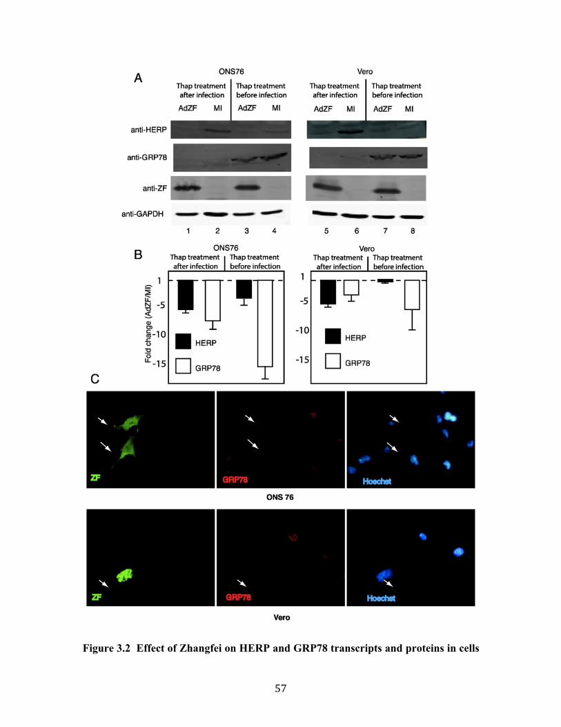
57
Figure 3.2 Effect of Zhangfei on HERP and GRP78 transcripts and proteins in cells

58
treated with thapsigargin. ONS76 and Vero cells were treated for 4 hr with thapsigargin
either after infection, or mock infection, with an adenovirus vector expressing ZF (lanes
1,2, 5 and 6) or before infection (lanes 3,4, 7 and 8). Cells were harvested 24 hr after
infection. Proteins in samples were separated by SDS-PAGE and probed with antibodies
against HERP, GRP78 or Zhangfei (ZF) and GAPDH (A) and RNA was extracted from
parallel duplicate cultures and assayed for HERP and GRP78 transcripts by qRT-PCR (B).
(C) ONS-76 and Vero cells were transfected with plasmids expressing FLAG epitope
linked to the coding sequences of Zhangfei. Cells were fixed and incubated with a
mixture of mouse monoclonal antibody against FLAG and rabbit antibody against GRP78
followed by Alexa 488 linked anti-mouse and Alexa 546-linked anti-rabbit antibodies.
Cells were also stained with Hoechst to stain nuclei. Arrows identify Zhangfei (ZF)
expressing cells.
59
3.4.2 Can Zhangfei suppress the ability of Xbp1s to activate transcription and is its
leucine-zipper required?
Since the published literature suggests that at least in some cells Zhangfei is relatively
ineffective against ATF6 (Misra et al. 2012), we examined the effect of Zhangfei on the
UPR-regulating transcription factor Xbp1s. In transiently transfected cells Xbp1s can
activate transcription of reporter genes linked to unfolded protein response elements
(UPRE). To determine if Zhangfei could suppress this ability of Xbp1s, Vero cells were
transfected with a CAT-reporter plasmid linked to multiple copies of UPRE (Fig 3.3 A
and B). Cells were also transfected with plasmids expressing the spliced form of Xbp1,
Xbp1s, and increasing amounts of a plasmid expressing Zhangfei (Fig 3.3A). Zhangfei
inhibited the activity of Xbp1s in a dose-dependent manner (solid circles). The
immunoblot in Figure 3.3D shows that increasing the amount of the Zhangfei-expressing
plasmid in the transfection mixtures also led to increased amounts of the protein in the
cells. To determine if the leucine-zipper of Zhangfei was required for its suppressive
activity, parallel cultures in the experiments described above were transfected with a
plasmid expressing a mutant of Zhangfei in which the first 6 consecutive leucine residues
in the bLZip domain were replaced with alanine residues (ZF(L/A)). Unlike Zhangfei,
ZF(L/A) did not suppress Xbp1s (Fig 3.3, A,B). Figure 3.3D shows that the mutations in
ZF(L/A) did not affect its stability. Interestingly, not only did ZF(L/A) not suppress
Xbp1s, it consistently appeared to enhance the activities of Xbp1s almost 2 fold (Fig
3.3A and B). This enhancement did not reflect an increased ability of ZF(L/A) to activate
transcription as neither Zhangfei nor ZF(L/A) on their own, had any effect on a UPRE
containing promoter (Fig 3.3B).
To confirm that Zhangfei required its leucine zipper to interact with Xbp1s, we conducted
two-hybrid assays in which activation of a reporter gene depended on interaction of
Xbp1s with Zhangfei or ZF(L/A) tethered to a promoter by the DNA-binding domain of
the yeast transcription factor Gal4 (UAS). Cells were transfected with a reporter plasmid
with a Gal4 UAS containing promoter and either Gal-ZF or Gal-ZF(L/A) with or without
plasmids expressing Xbp1s. The results in Figure 3C show that the reporter activity in the
presence of Gal-ZF was significantly greater than the activity in the presence of Gal-

60
ZF(L/A).
Figure 3.3 Zhangfei suppresses the ability of Xbp1s to activate transcription and
requires its leucine zipper to do so. (A) and (B). Vero cells were transfected with a
plasmid containing the coding sequence for CAT linked to a promoter with three copies
of the unfolded protein response element as well as a plasmid expressing Xbp1s and
varying amounts of plasmids expressing either Zhangfei (ZF) or a mutant, ZF(L/A) in
which all leucine residues in the LZip domain had been replaced with alanines. All
samples also contained, as a control, a plasmid expressing b-galactosidase. The CAT
activity in each sample was normalized to this internal control and expressed as a
percentage of the activity in samples containing no vector expressing either ZF or ZF
(L/A). The total amount of DNA in each transfection was made up to 5 µg with “empty”
expression vector (pcDNA3). Bars indicate standard deviation from the mean. (B)
ZF(L/A) does not activate a promoter containing UPRE but enhances the activity of
Xbp1s. (C) ZF interacts with Xbp1 with its leucine zipper. Cells were transfected with a
vector with the coding sequence for CAT linked to three copies of a sequence, UAS, that
61
binds the DNA-binding domain of the yeast protein GAL4. Cells also received plasmids
expressing either ZF or ZF(L/A) linked to the Gal4 DNA-binding domain and either an
“empty” expression vector or vectors expressing Xbp1s. Bars represent the ratio of the
relative CAT activity (normalized to the internal control, b-galactosidase) of samples
with Xbp1s to samples with no activator (“empty” vector). (D) An immunoblot showing
that vectors with cloned ZF or ZF (L/A) express the proteins in a dose-dependent manner.
The results represent the averages of three experiments assayed in duplicate. Bars in all
figures represent standard deviation from the mean and P values are indicated on the
figures.

62
3.4.3 How does Zhangfei suppress Xbp1?
Zhangfei may suppress Xbp1s by either causing its degradation, inhibiting its ability to
bind to cognate promoters or by sequestering it from the nucleus. To test the first
possibility we examined Xbp1s in cells expressing Zhangfei. Vero cells were transfected
with either a plasmid expressing Xbp1s alone or with a plasmid expressing Zhangfei. In
cells expressing both proteins we were unable to detect Xbp1s (Fig 3.4A, compare lane 2
with lane 3). In contrast, the co-expression of both proteins did not lead to an appreciable
decrease in Zhangfei (compare lanes 1 and 3). The proteasome inhibitor, MG132 restored
Xbp1s levels in Zhangfei-expressing cells (lane 6). To determine whether the leucine
zipper of Zhangfei was required for the induced degradation of Xbp1s, we expressed
Xbp1s with increasing amounts of plasmid expressing either Zhangfei or ZF(L/A). Figure
3.4B (Fig 3.4C shows densitometer measurements of repeated experiments) shows that at
low concentrations protein (0.5 and 1 µg expression plasmid) there was an obvious
difference in the ability of Zhangfei and its LZip mutant to induce the degradation of
Xbp1s. At higher concentrations (2 µg of plasmid, not shown) the differences between
the mutant and wild-type Zhangfei were less pronounced. Figure 3.5, which shows
intracellular proteins detected by immunofluorescence, supports these data—we were
unable to detect Xbp1s in cells expressing Zhangfei, while the LZip mutant, ZF(L/A),
had no effect. When Xbp1s and ZF(L/A) were expressed together both proteins
colocalize in the nucleus (Fig 3.5, bottom row) with higher concentrations in nuclear
structures. This suggests that nuclear localization of Xbp1s does not rely on association
with Zhangfei.
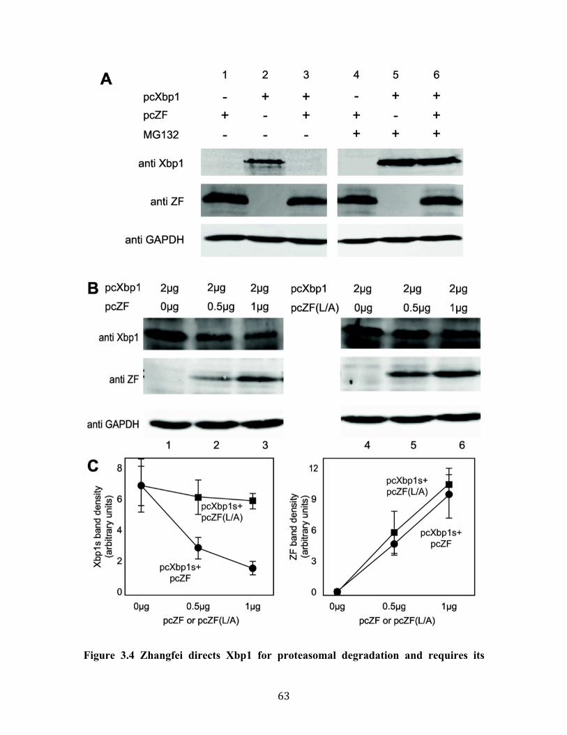
63
Figure 3.4 Zhangfei directs Xbp1 for proteasomal degradation and requires its

64
leucine zipper to do so. (A) Vero cells were transfected as indicated with plasmids
expressing Xbp1s and an empty vector or plasmid expressing Zhangfei. Cells either
received 5 µM MG132 or an equivalent volume of carrier, DMSO. Cells were harvested
24 hr later and proteins detected by immunoblotting. (B) Vero cells were transfected with
a plasmid expressing Xbp1s alone or increasing amounts of a plasmid expressing
Zhangfei or a mutant in which all leucine residues in the zipper had been changed to
alanine - pcZF(L/A). Cells were harvested 24 hr later and proteins detected by
immunoblotting. The density of each band on the immunoblot was estimated by
densitometry and normalized to the density of the GAPDH band in the sample. Average
values and standard deviation from three experiments are shown in (C).

65
Figure 3.5 Immunofluorescent images showing the absence of Xbp1 in cells
expressing Zhangfei but not Zhangfei with a mutated leucine zipper. Vero cells were
transfected with plasmids expressing either Xbp1 alone or in combination with either
Zhangfei or ZF(L/A). Cells were then fixed and incubated with a mixture of rabbit anti-
Zhangfei and mouse anti-Xbp1 antibodies. Antibodies were visualized by staining with
Alexa546 (red) anti mouse and Alexa488 (green) anti-rabbit antibodies.
66
3.4.4 Does Zhangfei interact with Xbp1s?
To determine if Zhangfei and Xbp1s interacted, we co-expressed the proteins in Vero
cells. Zhangfei coding sequences included a FLAG epitope. Since our previous
experiments indicated that interactions between the two proteins might lead to the
proteasomal degradation of Xbp1s (Fig 3.4A), we treated cells with MG132 to suppress
degradation. From the lysates of these cells we precipitated Zhangfei and associated
proteins with monoclonal antibodies against FLAG and then detected Xbp1 or Zhangfei
in the immunoprecipitates using immunoblots antisera against either Xbp1 or Zhangfei.
Figure 3.6A shows that in cells expressing both proteins they were in a stable association
(lane 6). In a similar experiment, Xbp1 did not precipitate with ZF(L/A) (Fig 3.6B,
compare lanes 7 and 8) confirming our results that the leucine-zipper of Zhangfei was
required for the interaction.

67
Figure 3.6 Zhangfei co-immunoprecipitates with Xbp1s in MG132-treated cells. (A)
Vero cells were transfected to express Xbp1s and FLAG-tagged Zhangfei either on their
own alone or together. The cells were treated with MG132 to reduce proteasomal
degradation and, 24 hr later, cells lysates were immunoprecipitated with mouse anti-
FLAG antibody. The precipitates were separated by SDS-PAGE and Xbp1 and Zhangfei
detected in immunoblots with rabbit anti-Xbp1 or anti-Zhangfei antisera. Lanes 1-3,
represent cell lysates without immunoprecipitation while lanes 4-6 are immunoblots of
material precipitated by anti-FLAG antibodies. (B) A similar experiment as in A,
showing that Xbp1s was not co-precipitated with the Zhangfei mutant ZF(L/A).

68
3.4.5 Can endogenous Zhangfei suppress the UPR in sensory neurons?
We had previously detected Zhangfei in mature neurons of the central nervous system
and in sensory neurons in trigeminal ganglia (Akhova et al. 2005). To determine if
endogenous Zhangfei could suppress the UPR in these cells, disassociated neurons from
adult rat dorsal root ganglia were transfected with plasmids expressing either siRNA
against Zhangfei or control siRNA (Valderrama et al. 2008). The UPR was then induced
in these cells with thapsigargin. Levels of transcripts for Xbp1s, unspliced Xbp1
(Xbp1us), CHOP, GRP78 and Zhangfei as well as protein levels for Xbp1s, GRP78,
HERP, GAPDH and Zhangfei were then measured using qRT-PCR, and immunoblotting
and densitometry. The primers used for qRT-PCR were directed against conserved
regions of the coding sequences of these genes. The results (Fig 3.7A and 3.7B) show
that in cells in which siRNA against Zhangfei had reduced its endogenous transcripts and
protein (Fig 3.7B and 3.7C), transcripts and proteins for several UPR-related genes were
increased. There was a trend towards an increase in Xbp1us transcripts although the
difference from control siRNA-expressing cells was not significant. These results
suggested that endogenous Zhangfei has the capacity to modulate the UPR. To
demonstrate that most cells in the primary sensory neuron culture could be transfected we
transfected the cells with fluorescent double stranded RNA. Figure 3.7D shows that most
cells in the culture were capable of taking up transfected DNA.
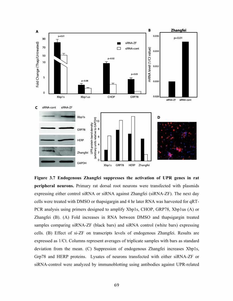
69
Figure 3.7 Endogenous Zhangfei suppresses the activation of UPR genes in rat
peripheral neurons. Primary rat dorsal root neurons were transfected with plasmids
expressing either control siRNA or siRNA against Zhangfei (siRNA-ZF). The next day
cells were treated with DMSO or thapsigargin and 4 hr later RNA was harvested for qRT-
PCR analysis using primers designed to amplify Xbp1s, CHOP, GRP78, Xbp1us (A) or
Zhangfei (B). (A) Fold increases in RNA between DMSO and thapsigargin treated
samples comparing siRNA-ZF (black bars) and siRNA control (white bars) expressing
cells. (B) Effect of si-ZF on transcripts levels of endogenous Zhangfei. Results are
expressed as 1/Ct. Columns represent averages of triplicate samples with bars as standard
deviation from the mean. (C) Suppression of endogenous Zhangfei increases Xbp1s,
Grp78 and HERP proteins. Lysates of neurons transfected with either siRNA-ZF or
siRNA-control were analyzed by immunoblotting using antibodies against UPR-related

70
proteins. Bands on immunoblots of the left were scanned and band densities relative to
the internal standard GAPDH are on the graph on the right. (D) Transfection efficiency
test of siRNA. Primary rat dorsal root neurons were transfected at a final concentration of
10 nM with the TYE 563 DS Transfection Control duplex, and were imaged 24 hours
post transfection (Red: marked siRNA; blue: nuclear).

71
3.5 Discussion
The inability of the UPR to restore normal ER function results in apoptosis. However, a
successful homeostatic response should result in modulation of the UPR and resumption
of normal cellular function. To do this several UPR induced proteins such as p58IPK
(Rutkowski et al. 2007), NCK1 (Latreille and Larose 2006; Cardin et al. 2007) and
GADD34 (Ma and Hendershot 2003; Zhou et al. 2011) feed-back to relieve the inhibition
of protein synthesis mediated by PERK. These act by recruiting phosphatases that
dephosphorylate eIF2 a. In addition, the product of unspliced Xbp1 mRNA binds Xbp1s
and ATF6 through bLZip interactions and targets the proteins for proteasomal destruction
(Yoshida et al. 2006; Yoshida et al. 2009).
Our qRT-PCR array data revealed a global significant decrease in transcripts for genes
activated by the UPR inducer thapsigargin in ONS-76 human medulloblastoma cells
when Zhangfei was expressed in these cells (Fig 3.1). In contrast, Zhangfei had no effect
on the UPR gene transcripts in cells growing normally under favourable non-stressful
conditions. This suggests that Zhangfei did not globally suppress transcription in cells but
directly or indirectly targeted the expression of certain genes. Mature neurons are one of
the few cell types in which we have been able to detect endogenous Zhangfei (Akhova et
al. 2005). In support of our data, we also showed that selective suppression of Zhangfei
by siRNA in adult primary sensory neurons from rat dorsal root ganglia increased levels
of transcripts and proteins for UPR-related genes (Fig 3.7) confirming the ability of the
protein to suppress the UPR once it had been activated, by thapsigargin in this case.
To determine how Zhangfei might modulate the UPR, we focused on the ability of
Zhangfei to suppress the ability of Xbp1s, one of the main bLZip mediators of the UPR.
We found that in cells transfected to transiently express Xbp1s as well as Zhangfei, the
ability of Xbp1s to activate transcription was suppressed (Fig 3.3). This was confirmed
by a corresponding decrease in the level of the corresponding UPR proteins (Fig 3.2).
While this can be explained, at least to some extent, by the effect of Zhangfei on the
ability of Xbp1s to initiate transcription of UPR genes, we are puzzled how Zhangfei
suppressed levels of the spliced Xbp1 transcripts themselves. The bLZip transcription

72
factor responsible for Xbp1 transcription has not been unambiguously identified however
our earlier data (Bergeron et al. 2013) as well as that of others show that activation of the
UPR leads to an increase in spliced Xbp1 mRNA without a corresponding increase in
unspliced transcripts. In addition Figure 3.4 shows that while suppression of Zhangfei by
siRNA decreased levels of spliced Xbp1 mRNA it did not have a significant effect on
unspliced transcripts. This suggests that Zhangfei may have an effect on the IRE1-
mediated cytoplasmic splicing process in addition to suppressing transcription by Xbp1s.
Basic leucine-zipper motif containing proteins are known to interact with other bLZip
proteins through their leucine zippers and can be categorized according to whether they
strongly favour the formation of homo-dimers, hetero-dimers or both (Vinson et al. 2002).
In addition, Newman and Keating (Newman and Keating 2003) using peptide arrays and
fluorescent bLZip probes, measured interactions between the bLZip regions of all human
and yeast bLZip proteins. According to their results the Xbp1 and ATF6 domains were
the only ones among the 49 bLZip proteins tested that showed a strong association with
the Zhangfei domain. Binding between Zhangfei and ATF4 could only be detected when
the fluorescent Zhangfei peptide was used to detect binding to the array.
Since bLZip proteins can act as co-activators by interacting with other transcription
factors by mechanisms that do not rely on their bLZip domains (Wardell et al. 2002), we
determined whether the suppressive effect of Zhangfei on Xbp1s required its leucine
zipper. Our data strongly suggest that it does (Figs 3.3, 3.4, 3.5 and 3.6). A mutant of
Zhangfei in which all 6 consecutive leucines in the zipper were replaced with alanine was
less efficient at suppressing Xbp1s than Zhangfei with an intact zipper. Further, our
results in the in vivo protein hybrid assay in which transcriptional activation relied upon
interaction of Gal4-linked Zhangfei or its mutant with the Xbp1s (Fig 3.3C), as well as
our inability to co-precipitate Zhangfei (L/A) and Xbp1s, support our observations.
Interestingly, not only did Zhangfei (L/A) not suppress Xbp1s, it consistently enhanced
its ability to activate transcription (Fig 3.3 A and B). A possible explanation for this
phenomenon may lie in the co-localization of the two proteins in nuclear domains (Fig
3.5, bottom row). We have observed that if proteasomal degradation is suppressed,

73
Zhangfei co-localizes with Luman/CREB3 (another bLZip protein that it suppresses) in
intranuclear promyelocytic leukemia protein-containing nuclear domains (Misra et al.
2005). These domains are sites for nuclear proteasomes (St-Germain et al. 2008) and it is
possible that, on their own, both Zhangfei and Xbp1s are normally targeted to these sites.
Expressed together, Zhangfei enhances the degradation of Xbp1s while Zhangfei (L/A),
unable to bind Xbp1s, has a suppressive effect on proteasomes.
The interaction between Zhangfei and Xbp1s results in the proteasomal degradation of
Xbp1s (Fig 3.4). Elucidating the mechanism by which Zhangfei targets Xbp1s for
proteasomal degradation will require additional work. However, several bLZip proteins
have been shown to target other proteins for such destruction. Thus, the human T-cell
leukemia virus coded bLZip protein HBZ and the host transcription factor
maculoaponeurotic fibroma (Maf) homologue B interact through their bLZip domains
following which MafB is targeted for proteasomal degradation (Mukai and Ohshima
2011). HBZ also interacts with host interferon response factor 1 (IRF1) and targets it for
degradation (Ohshima et al. 2010). Some interactions between bLZip proteins, such as
between ATF5 and nucleoplasmin (Liu et al. 2012) lead to the ubiquitination of the target
prior to degradation suggesting a mechanism for proteasomal targeting. Recently a
SUMO-conjugase, UBC9, was shown to stabilize Xbp1s by interacting with its bLZip
domain. Displacement of UBC9 from Xbp1s by Xbp1u led to decrease in the stability of
Xbp1s (Liu et al. 2012). Zhangfei may, in a similar manner, destabilize Xbp1s by
displacing UBC9 from its bLZip domain. SUMOylation of proteins prevents
ubiquitination (Johnson 2004) and subsequent proteasomal degradation. Interestingly,
UBC9 did not require its SUMOylating activity to stabilize Xbp1s (Liu et al. 2012)
suggesting that UBC9 exerts its effect by other means.
While we have shown a direct effect of Zhangfei on Xbp1s, Zhangfei may suppress the
other UPR-inducing bLZip factors as well. Although ATF6 is one of the best-
characterized of these factors, other ER-resident bLZip proteins such as Luman/CREB3
and CREBH are thought to perform this role in some cell types (reviewed in (Asada et al.
2011; Chan et al. 2011)). We have previously shown that Zhangfei can suppress the
activity of Luman/CREB3 but not ATF6. Recently, Misra and others (Misra et al. 2012)

74
showed that in hepatoma cells Zhangfei/SMILE suppresses the ability of CREBH to
induce UPR genes. While further studies about the role of Zhangfei in regulation the
UPR are clearly needed, our results and those of others show that Zhangfei has the ability
to suppress Xbp1 as well as other bLZip proteins that may substitute for the ATF6-arm of
the ER-stress sensing pathways. However, since we have only been able to detect
Zhangfei in mature, differentiated neurons, its influence is likely restricted to a few cell
types. Alternatively, given its dramatic effect on cell division (Valderrama et al. 2009;
Bergeron et al. 2013), it may be expressed in a wider array of cells but only in a very
transient manner, when it is needed.
3.6 Acknowledgements
This study was supported by a Discovery grant to VM from the Natural Sciences and
Engineering Research Council (NSERC) of Canada and a Canadian Institutes of Health
Research grant to VMKV (MOP74747). RZ and ZY were supported by scholarships from
the Government of China (China Scholarship Council, RZ-2010635007, ZY-
20106030170) and the University of Saskatchewan Graduate Studies.

75
4. Effects of Cyclic AMP Response Element Binding Protein – Zhangfei (CREBZF)
on the Unfolded Protein Response and cell growth are exerted through the tumour
suppressor p53
Rui Zhang and Vikram Misra*
Department of Microbiology, Western College of Veterinary Medicine, 52 Campus
Road, University of Saskatchewan, Saskatoon, Saskatchewan , S7N1B4, CANADA
This Chapter demonstrated the mechanisms by which Zhangfei suppressed the UPR and
cell growth in some osteosarcoma cell lines, but not in normal cells and other cancer cell
lines, based on the hypothesis that Zhangfei mediates its effect on cell growth and the
UPR through an intermediary, p53, that is either not induced or is defective in cells that
it does not affect. The manuscript has been published as “Effects of Cyclic AMP
Response Element Binding Protein – Zhangfei (CREBZF) on the Unfolded Protein
Response and cell growth are exerted through the tumour suppressor p53” in Cell Cycle.
13: 279-292. by Zhang, R. and Misra, V. and is reproduced here with the permission of
the copyright owner.
My contributions to this manuscript include: I designed and performed all the
experiments mentioned in this manuscript. I generated the first complete draft of the
submitted manuscript. Professor Vikram Misra and I worked together to revise this draft
prior to submission. We also worked together on the revision.

76
4.1 Abstract
Zhangfei/CREBZF, a basic region-leucine zipper (bLZip) transcription factor, is a potent
suppressor of growth and the Unfolded Protein Response (UPR) in some cancer cell lines,
including the canine osteosarcoma cell line, D-17. However, the effects of Zhangfei are
not universal and it has no obvious effects on untransformed cells and some cancer cell
lines (Chapter 2), suggesting that Zhangfei may act through an intermediary that is either
not induced or is defective in cells that it does not affect. Here we identify the tumour
suppressor protein p53 as this intermediary. We show the following: In cells ectopically
expressing Zhangfei the protein stabilizes p53 and co-localizes with it in cellular nuclei,
the bLZip domain of Zhangfei is required for its profound effects on cell growth and
interaction with p53. Suppression of p53 by siRNA at least partially inhibits the effects
of Zhangfei on the UPR and cell growth. The effects of Zhangfei on D-17 cells is
mirrored by its effects on the p53-expressing human osteosarcoma cell line U2OS while
Zhangfei has no effect on the p53-null osteosarcoma cell line MG63. In U2OS cells
Zhangfei displaces the E3 ubiquitin ligase mouse double minute homologue 2 (mdm2)
from its association with p53 suggesting a mechanism for the effects of Zhangfei on p53.
Key words: cell cycle, protein domains, p53, osteosarcoma, protein translocation,
Zhangfei/CREBZF, unfolded protein response, mdm2, basic-leucine zipper domain

77
4.2 Introduction
Zhangfei/CREBZF/SMILE was first discovered as a binding partner for Host Cell Factor
(HCF), a co-activator of the herpes simplex virion transcription factor VP16 (Lu and
Misra 2000b). The primary structure of the protein contains a leucine zipper, a basic
region that lacks an asparagine residue conserved in most bLZip proteins, three potential
nuclear factor binding domains (LLXXLL, where L is a leucine residue and X is any
amino acid), and a domain for binding HCF. Zhangfei interacts with several proteins,
possibly through its nuclear receptor and HCF binding domains as well as its leucine
zipper. While Zhangfei can activate gene expression through factors such as p53 (Lopez-
Mateo et al. 2012) and ATF4 (Hogan et al. 2006), it suppresses the activity of a number
of transcription factors which include nuclear receptors (Xie et al. 2008; Xie et al. 2009a;
Xie et al. 2009b), bLZip containing proteins such as CREBH (Misra et al. 2012),
Luman/CREB3 (Misra et al. 2005), Xbp1 (Zhang et al. 2013) and SMAD 1,5,8 (Lee et al.
2012a) and the HCF-binding VP16 (Akhova et al. 2005). In the previous studies (Chapter
3), we also showed that Zhangfei had a profoundly suppressive effect on the Unfolded
Protein Response (UPR) at least partly because it targets Xbp1s, an important UPR
mediator, for proteasomal degradation (Zhang et al. 2013).
We have detected Zhangfei protein in differentiated neurons, but not in developing
neurons or cells of neuronal tumours (Akhova et al. 2005), nor in osteosarcoma cell lines.
The ectopic expression of Zhangfei in several tumour cells lines derived from
medulloblastomas (ONS-76, UW228) (Valderrama et al. 2009), and osteosarcomas (D-17)
(Bergeron et al. 2013) causes the cells to stop growing and display markers of apoptosis
(Chapter 2). However, the suppressive effect of Zhangfei on the growth of cells is not
universal. While the protein has a profound effect on some cells, it has no effect on others
such as MRC5 fibroblasts (Valderrama et al. 2009). This suggests that Zhangfei may act
through an intermediary that is either not induced or is defective in cells that it does not
affect. The objective of this study was to identify such an intermediary and to determine
how Zhangfei exerted its effect. Herein, we identified that Zhangfei suppressed cell
growth and the UPR in osteosarcoma cells through direct interaction with tumour
suppressor protein p53. We demonstrated that Zhangfei stabilized p53 and promoted its

78
nuclear retention by displacing the E3 ubiquitin ligase, mdm2. Overall, our findings
reveal a novel mechanism by which Zhangfei may inhibit tumour growth and metastasis.
This may provide an alternative modality for the therapy of certain types of osteosarcoma,
and perhaps other tumours with functional p53.

79
4.3 Materials and Methods
4.3.1 Cells and tissue culture
The canine osteosarcoma D-17 cells were obtained from the American Type Tissue
Culture Collection, were grown in MEM-Alpha containing 10% fetal bovine serum.
Human MG63 and U2OS osteosarcoma cells were obtained from Dr. Douglas H. Thamm,
(Associate Professor of Oncology, Animal Cancer Center, Colorado State University) and
grown in Dulbecco’s minimal essential medium containing penicillin and streptomycin
and 10% newborn calf serum. All media, serum and antibiotics were purchased from
Invitrogen. D-17 cells were treated with doxorubicin (0.5µM, WCVM's Accredited
Veterinary Pharmacy, University of Saskachewan) for p53 induction. Stock solutions of
MG132/Z-L-L-L-A (10mM, Sigma, C2211) were prepared in dimethyl sulfoxide (DMSO)
and used at a concentration of 5µM. Equivalent volumes of DMSO were added to
untreated cultures.
4.3.2 Plasmids
The construction of pcZF (Lu and Misra 2000b), a plasmid that expresses Zhangfei in
mammalian cells, has been described. The plasmids expressing Zhangfei mutant pcZF
Zip(L>A), in which all leucines in the leucine-zipper domain were replaced with alanines,
was constructed by subcloning a 265 bp synthetic DNA fragment (IDT) bracketed by
NotI and SgrA1sites into the corresponding coding sequences of Zhangfei between
unique Not1 and SgrA1 sites in pcZF. The plasmids expressing Zhangfei mutant pcZF
Basic del, in which basic region was deleted, was constructed by site directed loop-out
mutagenesis using 36 base complementary oligonucleotides that bracketed the 25 codons
for the basic region of Zhangfei. The CAT reporter plasmid pCAT3B-p53RE was
constructed by transferring two copies of p53 responsive element,
GGTCAAGTTGGGACACGTCCaaGAGCTAAGTCCTGACATGTCT, to pCAT3Basic
(Promega). Oligonucleotides representing the p53 responsive elements with overhanging
5’ terminal KpnI and 3’ terminal BglII sites were purchased from IDT company. The
oligonucleotides were annealed and ligated pCAT3Basic cut with the same enzymes. The
plasmids expressing the full length of wild-type p53 were constructed by subcloning the

80
HindIII – XhoI cDNA fragment containing the entire coding sequence of p53 from U2OS
cells between the HindIII and XhoI sites in the multiple restriction site cloning regions of
the mammalian expression vector pcDNA3 (Invitrogen, V79020). The specific primers
for p53 amplification are: forward: 5'-GACACGCTTCCCTGGATTGGC-3', reverse: 5'-
TCAGTCTGAGTCAGGCCCTT-3'. The veracity of all cloned wild-type and mutants’
coding sequences were confirmed by sequencing
4.3.3 Transfection and CAT Assays
Cells were transfected with plasmids using Lipofectamine 2000 (Invitrogen, 11668-019)
as described in manufacturer’s instructions. Five µg of DNA was used for 5 × 10⁵ cells
for chloramphenicol acetyl transferase (CAT) assays. For CAT assays, 250 ng of
pCMVBGal, a plasmid specifying β-galactosidase, were added to each transfection.
Lysates were assayed for β-galactosidase (Sambrook and Russell 2001) and for CAT
using an enzyme-linked immunosorbent assay kit (Roche Applied Science, 11-363-727-
001). CAT values were adjusted for transfection efficiency using β-galactosidase values.
In figures for CAT assays, each datum point is the average of duplicate transfections with
the bar representing the range. The data are representative of several (at least two)
independent experiments that gave the same results.
4.3.4 RNA interference
p53-specific siRNA (Invitrogen, TP53VHS40366) or Stealth RNAi™ siRNA negative
control (Invitrogen, 12935-300) (1pmol/5000 cells) were transfected into cells with
Lipofectamine 2000 (Invitrogen, 11668-019) as described in manufacturer’s instructions.
These p53-specific siRNAs are homologous to human as well as canine p53, which was
confirmed by comparing the sequence of human or canine p53 with siRNA sequence.
4.3.5 Adenovirus Vectors Expressing Zhangfei (Adeno-ZF) and β-galactosidase
(Adeno-LacZ)
These vectors were constructed, grown, and purified using the Adeno-X Expression
System (Clontech, K1650-1) as described earlier (Misra et al. 2005). Cells were infected
with adenovirus vectors expressing either Zhangfei (Adeno-ZF) or a control protein—E.
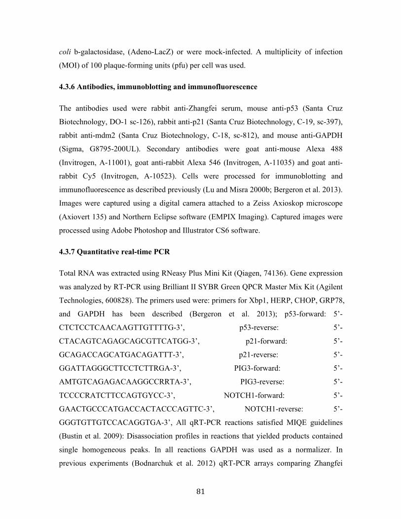
81
coli b-galactosidase, (Adeno-LacZ) or were mock-infected. A multiplicity of infection
(MOI) of 100 plaque-forming units (pfu) per cell was used.
4.3.6 Antibodies, immunoblotting and immunofluorescence
The antibodies used were rabbit anti-Zhangfei serum, mouse anti-p53 (Santa Cruz
Biotechnology, DO-1 sc-126), rabbit anti-p21 (Santa Cruz Biotechnology, C-19, sc-397),
rabbit anti-mdm2 (Santa Cruz Biotechnology, C-18, sc-812), and mouse anti-GAPDH
(Sigma, G8795-200UL). Secondary antibodies were goat anti-mouse Alexa 488
(Invitrogen, A-11001), goat anti-rabbit Alexa 546 (Invitrogen, A-11035) and goat anti-
rabbit Cy5 (Invitrogen, A-10523). Cells were processed for immunoblotting and
immunofluorescence as described previously (Lu and Misra 2000b; Bergeron et al. 2013).
Images were captured using a digital camera attached to a Zeiss Axioskop microscope
(Axiovert 135) and Northern Eclipse software (EMPIX Imaging). Captured images were
processed using Adobe Photoshop and Illustrator CS6 software.
4.3.7 Quantitative real-time PCR
Total RNA was extracted using RNeasy Plus Mini Kit (Qiagen, 74136). Gene expression
was analyzed by RT-PCR using Brilliant II SYBR Green QPCR Master Mix Kit (Agilent
Technologies, 600828). The primers used were: primers for Xbp1, HERP, CHOP, GRP78,
and GAPDH has been described (Bergeron et al. 2013); p53-forward: 5’-
CTCTCCTCAACAAGTTGTTTTG-3’, p53-reverse: 5’-
CTACAGTCAGAGCAGCGTTCATGG-3’, p21-forward: 5’-
GCAGACCAGCATGACAGATTT-3’, p21-reverse: 5’-
GGATTAGGGCTTCCTCTTRGA-3’, PIG3-forward: 5’-
AMTGTCAGAGACAAGGCCRRTA-3’, PIG3-reverse: 5’-
TCCCCRATCTTCCAGTGYCC-3’, NOTCH1-forward: 5’-
GAACTGCCCATGACCACTACCCAGTTC-3’, NOTCH1-reverse: 5’-
GGGTGTTGTCCACAGGTGA-3’, All qRT-PCR reactions satisfied MIQE guidelines
(Bustin et al. 2009): Disassociation profiles in reactions that yielded products contained
single homogeneous peaks. In all reactions GAPDH was used as a normalizer. In
previous experiments (Bodnarchuk et al. 2012) qRT-PCR arrays comparing Zhangfei

82
expressing and non-expressing cells five housekeeping genes were analyzed. The levels
of GAPDH were not affected by Zhangfei expression.
4.3.8 Co-immunoprecipitation
U2OS and MG63 cells in 6-well dishes were infected with Adeno-ZF or mock infected.
Twenty-four hr after infection, cells were washed with cold PBS and lysed in 250µl/well
cold lysis buffer (50mM Tris, pH7.5, 150mM NaCl, 1mM EDTA and 0.1% TritonX-100)
containing protease inhibitor cocktail (Sigma, P8340). After centrifugation at 13,000 xg
at 4 oC, 20µl of cell lysate supernatant were frozen as pre-immunoprecipitated sample,
and mouse anti-p53 antibody (2µg, Santa Cruz Biotechnology, DO-1 sc-126) was added
to the remaining supernatant (230µl) and the sample incubated for 12hr with constant
gentle agitation. Protein A/G agarose beads (100µl, Pierce, Fisher Scientific, 20421) was
added and the samples were incubated for an additional 4hr at 4 oC. Agarose beads were
collected by centrifugation at 13,000 xg at 4 oC and washed 4 times in lysis buffer before
boiling in SDS-PAGE sample buffer. Proteins in samples of the unfractionated cell lysate
or immunoprecipitates were separated by SDS-PAGE, transferred to membranes and
probed with rabbit anti-Zhangfei antisera, mouse anti-p53, or rabbit anti-mdm2 (Santa
Cruz Biotechnology, C-18 sc-821). Antibodies were visualized after incubation with
Alexa488-labelled anti-rabbit or anti-mouse antibody.
4.3.9 Statistical analysis
Statistical analysis was preformed by T-test or ANOVA test using IBM SPSS statistics
version 21.0.0 software. ANOVA tests with LSDpost hoc comparison was used to
analyze the differences between multi-group means and their associated procedures by
adding individuals as a treatment variable, and a paired t-test was used to evaluate the
effects of one treatment compared with no treatment/control. A P value of less than 0.05
was considered to be statistically significant for ANOVA tests and t-tests.

83
4.4 Results
4.4.1 Leucine-Zipper is required for the suppressive effects of Zhangfei on both cell
growth and UPR.
We have previously shown that Zhangfei has a profound effect on the growth and the
induction of the UPR in the canine osteosarcoma cell line, D-17 (Chapter 2) (Bergeron et
al. 2013). We have also shown that Zhangfei requires its leucine zipper to suppress the
UPR in some cells (Chapter 3) (Zhang et al. 2013). To better characterize the molecular
mechanism by which Zhangfei suppresses the growth of these cells, we attempted to
generate clones of D-17 cells in which Zhangfei could be induced. Six clones that
produced stable and detectable Zhangfei only in the presence of the inducer, doxycycline
(Fig 4.1A shows evidence from representative clones), were analyzed further. While the
original D-17 cells responded to the ectopic expression of Zhangfei by complete
cessation of growth [Fig 4.1B, compares cells infected with adenovirus vectors
expressing either Zhangfei (Adeno-ZF) or the control protein β-galactosidase (Adeno-
LacZ) and mock-infected cells (MI)] the induction of Zhangfei had little or no effect on
most of the clones (Fig 4.1B, clones 4, 9, 11, 12 and 16). The induction of Zhangfei had a
partial effect on growth on clone 15. We next amplified the complete coding sequences
for Zhangfei by PCR from RNA purified from the induced cell clones as well as from
pTRE-tightZF (the plasmid used to develop the doxycycline-inducible clones) and
determined the nucleotide sequence of the products. While the derived amino acid
sequence of the leucine zipper (LZip) region from pTRE-TightZF was identical to the
published sequence, one or more leucines from the LZip regions of all clones had been
replaced with other amino acids (Fig 4.1C, bold). No other changes were observed in the
entire Zhangfei coding sequences suggesting that the leucine mutations were not random
changes. To determine if the Zhangfei mutants retained the ability to suppress the UPR,
we induced the UPR in the mutant cell clones with thapsigargin, with and without
doxycycline, and compared stable levels of transcripts for four UPR related genes—
Xbp1s, HERP, CHOP and GRP78. As controls, we compared the original D-17 cells,
mock-infected or infected with Adeno-ZF. While Zhangfei suppressed the UPR
transcripts in infected cells, doxycycline-induction of the protein in the cell clones had no
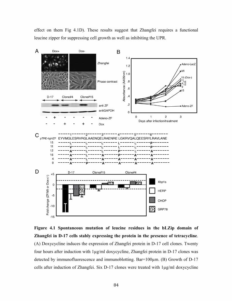
84
effect on them Fig 4.1D). These results suggest that Zhangfei requires a functional
leucine zipper for suppressing cell growth as well as inhibiting the UPR.
Figure 4.1 Spontaneous mutation of leucine residues in the bLZip domain of
Zhangfei in D-17 cells stably expressing the protein in the presence of tetracycline.
(A) Doxycycline induces the expression of Zhangfei protein in D-17 cell clones. Twenty
four hours after induction with 1µg/ml doxycycline, Zhangfei protein in D-17 clones was
detected by immunofluorescence and immunoblotting. Bar=100µm. (B) Growth of D-17
cells after induction of Zhangfei. Six D-17 clones were treated with 1µg/ml doxycycline

85
to induce Zhangfei expression. Cell growth characteristics were monitored at different
time points using WST-1 assay. Growth characteristics of the original D-17 cells, mock-
infected (MI) or infected with adenovirus vectors expressing either ZF (Adeno-ZF) or
beta-galactosidase (Adeno-LacZ) are shown for comparison. (C) Mutations in the leucine
zipper of Zhangfei-expressing D-17 clones. Amino acid sequence of the LZip region for
Zhangfei recovered by PCR from the vector used to develop the clones and the
doxycycline-inducible Zhangfei expressing clones. Dashes (-) indicate no changes.
Leucine residues (L) comprising the zipper are shown and mutations are in bold. (D)
Induction of mutant Zhangfei has no effect on the UPR. Clones expressing ZF with
mutations in the leucine zipper did not inhibit the UPR. Original D-17 cells and clones 4
and 15 were treated with 1µg/ml doxycycline for 24h, followed by 4h of treatment of
thapsigargin. Then cells were harvested and UPR transcripts were estimated by qRT-PCR.
The original D-17 cells, mock-infected or infected with Adeno-ZF were also analyzed for
comparison. The values represent fold changes in levels of transcripts between Adeno-ZF
infected cells with mock-infected cells (in original D-17 cells) or between doxycycline
treated with untreated cells (in Tet-on clones). Error bars indicated standard deviations
from means of three individual experiments. Dashed lines indicate fold changes of less or
more than two fold and changes exceeding these limits were arbitrarily regarded as
significant.

86
4.4.2 Zhangfei regulates p53 at a post-translational level and promotes p53 nuclear
retention.
Zhangfei stabilizes p53 in some cancer cells (Lopez-Mateo et al. 2012), although the
mechanisms by which Zhangfei mediates its effects on p53 have not been characterized.
To investigate if Zhangfei regulates the expression of endogenous p53 and its target
genes, we infected D-17 cells with Adeno-ZF or Adeno-LacZ. We observed that
Zhangfei increased transcripts for p21, a well-characterized p53-target gene, but had no
significant effects on p53 transcripts as well as its other target genes, PIG3 and NOTCH1
(Fig 4.2A). However, Zhangfei did increase the protein levels of both endogenous p53
and p21 comparable to that induced by the drug doxorubicin, a known inducer of p53
(Wang et al. 2004) (Fig 4.2B, compare lanes 3 and 4 with 1 and 2), suggesting that
Zhangfei increased p53 protein by post-translational mechanisms. We also assessed the
ability of Zhangfei to indirectly induce transcription from a promoter containing two
copies of p53 responsive elements (pCAT3B-p53RE). Zhangfei induced the expression
of a reporter gene, CAT, linked to the promoter by about twenty fold (Fig 4.2C).
The protein p53 possesses nuclear localization and nuclear export signals enabling it to
shuttle between the nucleus and the cytoplasm (Shaulsky et al. 1990; Zhang and Xiong
2001). To investigate the impact of Zhangfei on p53 nucleo-cytoplasmic shuttling, we
monitored the p53 localization in ZF-expressing D-17 cells. We observed that, compared
with the negative control (pcDNA3), the nuclear staining of endogenous p53 increased by
12h after transfection of the cells with a plasmid expressing Zhangfei (pcZF) with a
concomitant decrease in cytoplasmic staining (Fig 4.2D). By 36h following transfection,
endogenous p53 was predominantly in the nucleus and cells displayed features of
apoptosis (diffuse DNA staining by Hoechst and membrane blebbing).
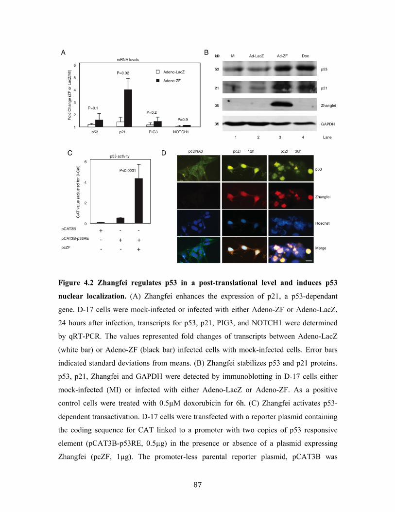
87
Figure 4.2 Zhangfei regulates p53 in a post-translational level and induces p53
nuclear localization. (A) Zhangfei enhances the expression of p21, a p53-dependant
gene. D-17 cells were mock-infected or infected with either Adeno-ZF or Adeno-LacZ,
24 hours after infection, transcripts for p53, p21, PIG3, and NOTCH1 were determined
by qRT-PCR. The values represented fold changes of transcripts between Adeno-LacZ
(white bar) or Adeno-ZF (black bar) infected cells with mock-infected cells. Error bars
indicated standard deviations from means. (B) Zhangfei stabilizes p53 and p21 proteins.
p53, p21, Zhangfei and GAPDH were detected by immunoblotting in D-17 cells either
mock-infected (MI) or infected with either Adeno-LacZ or Adeno-ZF. As a positive
control cells were treated with 0.5µM doxorubicin for 6h. (C) Zhangfei activates p53-
dependent transactivation. D-17 cells were transfected with a reporter plasmid containing
the coding sequence for CAT linked to a promoter with two copies of p53 responsive
element (pCAT3B-p53RE, 0.5µg) in the presence or absence of a plasmid expressing
Zhangfei (pcZF, 1µg). The promoter-less parental reporter plasmid, pCAT3B was

88
included as a control to show basal CAT activity. All samples also contained, as a control,
a plasmid expressing b-galactosidase. 24h after transfection, the CAT activity was
determined. Values represented the relative CAT activity (normalized to the internal
control, b-galactosidase) of different treatments. Standard deviations from means of three
individual experiments are shown and the significant P values from ANOVA tests were
noted above the bars. (D) Zhangfei alters the subcellular localization of p53. D-17 cells
were transfected with 1µg of pcZF or a control (pcDNA3), and 12h and 36h after
transfection, endogenous p53 as well as Zhangfei were visualized by
immunofluorescence with anti-p53 and anti-ZF antibody. The nucleus was detected by
Hoechst staining (bar=10µm). The means and standard deviations of representative
experiments (n=3) were shown. P<0.05 were considered to be significant.
89
4.4.3 Basic-region leucine zipper domain (bLZip) of Zhangfei is required for the
regulation of p53.
Given that the bLZip domain plays an important role in the inhibitory ability of Zhangfei
on cell growth and the UPR as described above, we next sought to examine whether this
domain was also required for the regulation of p53. We found that transfection of
plasmids expressing Zhangfei with a deleted basic region (pcZF Basic del) or a mutated
leucine zipper (pcZF Zip(L>A)) (Fig 4.3A) did not increase the protein levels of either
p53 or p21 (Fig 4.3B, compare lane 2 with lanes 3 and 4). The increase of p53
transcriptional activity induced by wild-type Zhangfei was also significantly reduced in
cells expressing the mutated proteins (Fig 4.3C). In addition, the mutant Zhangfei
proteins were unable to increase nuclear localization of p53 (Fig 4.3D). These results
indicate that bLZip domain is an important functional region of Zhangfei, required for its
regulatory effects on cell growth, the UPR, as well for its interaction with p53.

90
Figure 4.3 The basic-region leucine zipper domain (bLZip) of Zhangfei is required
for its effect on p53. (A) Schematic representation of the structures of Zhangfei (ZF) and
Zhangfei mutants: ZF Basic del, basic region was deleted; ZF Zip(L>A), all leucines in
the leucine-zipper domain were replaced with alanines. (B) The bLZip domain of
Zhangfei is required for stabilization of p53 and p21 proteins. D-17 cells were transfected
with 1µg of plasmid expressing Zhangfei (pcZF) or mutants (pcZF Zip(L>A) or pcZF
Basic del). 24h after transfection endogenous p53 and p21 proteins were detected by
immunoblotting. (C) The bLZip domain of Zhangfei is required for p53-dependent
transactivation. D-17 cells were transfected with 0.5µg of p53 response element
containing reporter plasmid pCAT3B-p53RE and 1µg of pcZF or mutants (pcZF
Zip(L>A) or pcZF Basic del). Twenty four hours after transfection, the CAT activity was
determined. The means and standard deviations of experiments (n=3) were shown.
P<0.05 were considered to be significant. (D) The bLZip domain of Zhangfei is required
for p53 nuclear retention. D-17 cells were treated as described in (A), and endogenous
p53 as well as Zhangfei were visualized by immunofluorescence. The nucleus was
detected by Hoechst staining (bar=10µm).

91
4.4.4 p53 is the key molecule responsible for mediating suppressive regulation of
Zhangfei on D-17 cell growth and the UPR.
The tumour suppressor p53 limits cellular proliferation by inducing cell cycle arrest and
apoptosis in response to cellular stresses such as DNA damage, hypoxia, nutrient
deprivation and oncogene activation (reviewed by (Sharpless and DePinho 2002;
Vousden and Lu 2002)), and these stresses also activate the UPR. The results shown
above demonstrated that Zhangfei down-regulated cell growth and UPR, but up-regulated
p53 through its bLZip domain. To explore whether Zhangfei expression influence cell
proliferation and the UPR through p53, we used p53-specific siRNA to knock down p53
transcripts and protein. While Zhangfei enhanced transcription from a promoter with p53
response elements, siRNA against p53 suppressed its effects (Fig 4.4A). Suppression of
p53 also partially restored the growth of D-17 cells inhibited by Zhangfei (Fig 4.4B) and
partially restored the suppression of UPR transcripts (Fig 4.4C). These results indicate
that the inhibitory influences of Zhangfei on D-17 cells that we have described previously,
at least in part, are mediated by p53.

92
Figure 4.4 Zhangfei regulates p53-mediated cell growth and UPR. (A) Zhangfei-
induced transcription from a promoter with p53 response elements is mediated by p53. D-
17 cells were transfected with 0.5µg of p53 response element containing promoter
(pCAT3B-p53), 1µg of plasmid expressing Zhangfei (pcZF) and 100pM of si-RNA
against p53 (si-p53) or non-targeting siRNA control (si-cont), 48h after transfection, cells
were analyzed for CAT activity or p53, Zhangfei and GAPDH proteins (inset). (B)
Zhangfei-mediated suppression of cell growth is mediated through p53. D-17 cells were
transfected with 100pM of si-RNA against p53 (siRNA-p53) or non-targeting siRNA
control (siRNA-cont). Eight hours after transfection, cells were either mock-infected or

93
infected with Adeno-ZF, and growth characteristics were measured using WST-1 assay at
0h, 24h, 48h and 72h after infection. (C) Zhangfei-mediated suppression of UPR genes is
mediated by p53. D-17 cells were transfected 100pM of siRNA-p53 or non-targeting
siRNA-control. 48h after transfection, cells were treated with thapsigargin for 4h and
harvested. Differences in levels of transcripts (Xbp1s, CHOP, and GRP78) were
determined by qRT-PCR. The results were expressed as fold changes relative to mock-
infected cells. The means and standard deviations of experiments (n=3) are shown.
P<0.05 were considered to be significant.
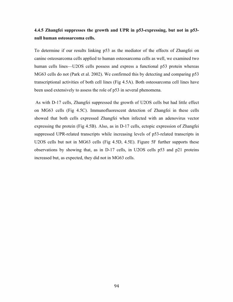
94
4.4.5 Zhangfei suppresses the growth and UPR in p53-expressing, but not in p53-
null human osteosarcoma cells.
To determine if our results linking p53 as the mediator of the effects of Zhangfei on
canine osteosarcoma cells applied to human osteosarcoma cells as well, we examined two
human cells lines—U2OS cells possess and express a functional p53 protein whereas
MG63 cells do not (Park et al. 2002). We confirmed this by detecting and comparing p53
transcriptional activities of both cell lines (Fig 4.5A). Both osteosarcoma cell lines have
been used extensively to assess the role of p53 in several phenomena.
As with D-17 cells, Zhangfei suppressed the growth of U2OS cells but had little effect
on MG63 cells (Fig 4.5C). Immunofluorescent detection of Zhangfei in these cells
showed that both cells expressed Zhangfei when infected with an adenovirus vector
expressing the protein (Fig 4.5B). Also, as in D-17 cells, ectopic expression of Zhangfei
suppressed UPR-related transcripts while increasing levels of p53-related transcripts in
U2OS cells but not in MG63 cells (Fig 4.5D, 4.5E). Figure 5F further supports these
observations by showing that, as in D-17 cells, in U2OS cells p53 and p21 proteins
increased but, as expected, they did not in MG63 cells.

95
Figure 4.5 Zhangfei suppresses cell growth and UPR in wild-type p53-expressing
U2OS cells, but not in p53-null MG63 cells. (A) p53-dependent transactivation in

96
U2OS and MG63 cells. Cells were transfected with 0.5µg of p53 reporter (pCAT3B-
p53RE) for 24h, then p53 transcriptional activity was determined by CAT ELISA assay.
(B) U2OS and MG63 cells express Zhangfei when infected with adenovirus vector
expressing the protein. Cells were infected with Adeno-ZF, and 24h later Zhangfei
protein was visualized by immunofluorescence (bar=10µm). (C) Ectopic expression of
ZF suppresses cell growth in p53-wild type U2OS cells, but has no effect in p53-null
MG63 cells. U2OS and MG63 human osteosarcoma cells were mock-infected or infected
with Adeno-ZF or Adeno-LacZ and their growth rates were assessed at different time
points after infection as absorbance at 405 nm after treatment with WST-1. (D) Zhangfei
negatively regulates the UPR in U2OS cells but not in MG63 cells. U2OS and MG63
human cells were either mock-infected or infected with Adeno-ZF or Adeno-LacZ. 24h
after infection, cells were treated with thapsigargin for 4h, and then harvested. The
differences in levels of transcripts for UPR genes (Xbp1s, CHOP, and GRP78) were
determined by qRT-PCR. (E) Zhangfei enhances p53-dependent transcripts in U2OS
cells but not in MG63 cells. Transcripts for p53, p21, PIG3, and NOTCH1 in U2OS and
MG63 cells either mock-infected or infected with Adeno-LacZ or Adeno-ZF were
measured by pRT-PCR. (F) Zhangfei increases p53 and p21 proteins in U2OS cells but
not in MG63 cells. p53 and p21 proteins were detected by immunoblotting in U2OS and
MG63 cells either mock-infected or infected with Adeno-ZF. The values in (D) and (E)
represented fold changes of transcripts between Adeno-LacZ (white bar) or Adeno-ZF
(black bar) infected cells with mock infected cells. The means and standard deviations of
experiments (n=3) were shown. P<0.05 were considered to be significant. Error bars
indicate standard deviations from means. Horizontal dotted line indicated a two-fold
change. A change of more than 2-fold was arbitrarily regarded as significant.
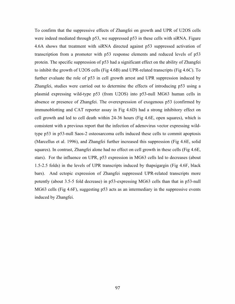
97
To confirm that the suppressive effects of Zhangfei on growth and UPR of U2OS cells
were indeed mediated through p53, we suppressed p53 in these cells with siRNA. Figure
4.6A shows that treatment with siRNA directed against p53 suppressed activation of
transcription from a promoter with p53 response elements and reduced levels of p53
protein. The specific suppression of p53 had a significant effect on the ability of Zhangfei
to inhibit the growth of U2OS cells (Fig 4.6B) and UPR-related transcripts (Fig 4.6C). To
further evaluate the role of p53 in cell growth arrest and UPR suppression induced by
Zhangfei, studies were carried out to determine the effects of introducing p53 using a
plasmid expressing wild-type p53 (from U2OS) into p53-null MG63 human cells in
absence or presence of Zhangfei. The overexpression of exogenous p53 (confirmed by
immunoblotting and CAT reporter assay in Fig 4.6D) had a strong inhibitory effect on
cell growth and led to cell death within 24-36 hours (Fig 4.6E, open squares), which is
consistent with a previous report that the infection of adenovirus vector expressing wild-
type p53 in p53-null Saos-2 osteosarcoma cells induced these cells to commit apoptosis
(Marcellus et al. 1996), and Zhangfei further increased this suppression (Fig 4.6E, solid
squares). In contrast, Zhangfei alone had no effect on cell growth in these cells (Fig 4.6E,
stars). For the influence on UPR, p53 expression in MG63 cells led to decreases (about
1.5-2.5 folds) in the levels of UPR transcripts induced by thapsigargin (Fig 4.6F, black
bars). And ectopic expression of Zhangfei suppressed UPR-related transcripts more
potently (about 3.5-5 fold decrease) in p53-expressing MG63 cells than that in p53-null
MG63 cells (Fig 4.6F), suggesting p53 acts as an intermediary in the suppressive events
induced by Zhangfei.

98
Figure 4.6 p53 mediates the suppressive effects of Zhangfei on cell growth and UPR
in human osteosarcoma cells. (A) Zhangfei-mediated transcription from a p53 response
element-containing promoter is mediated by p53 in U2OS cells. U2OS cells were
transfected with 0.5µg of p53 reporter plasmid (pCAT3B-p53RE) and 1µg of plasmids
expressing Zhangfei (pcZF) in the presence of 100pM of siRNA-p53 or non-targeting
siRNA-control. Forty eight hours after transfection CAT reporter activity and p53 protein
were detected by ELISA and immunoblotting (inset). (B) Suppression of p53 by siRNA
in U2OS cells restored Zhangfei-induced suppression of cell growth. U2OS were
transfected with 100pM of siRNA-p53 or non-targeting siRNA-control. Eight hours after

99
transfection, cells were either mock-infected or infected with Adeno-ZF and measured
growth rates as absorbance at 405 nm after treatment with WST-1. (C) Suppression of
p53 prevents Zhangfei-induced inhibition of UPR genes. U2OS cells were transfected
with 100pM of siRNA-p53 or non-targeting siRNA-control. Forty eight hours after
transfection, cells were treated with thapsigargin for 4h and harvested. Differences in
levels of transcripts for UPR genes (Xbp1s, CHOP, and GRP78) were determined by
qRT-PCR. The values represented fold changes of transcripts between Adeno-LacZ
(white bar) or Adeno-ZF (black bar) infected cells with mock infected cells. (D)
Transactivation activity and expression of p53 protein in p53-null MG63 cells. p53-null
MG63 cells were transfected with 0.5µg of p53 reporter plasmid and 1µg of plasmids
expressing Zhangfei (pcZF) in the presence of 1µg of plasmids expressing wild-type p53
(pc-p53, from U2OS cells) for 24h, and then CAT reporter activity and p53 protein were
detected by ELISA and immunoblotting. (E) Overexpression of p53 in p53-null MG63
cells suppressed cell growth and activated growth arrest induced by Zhangfei. MG63
cells were transfected with 3µg of pcZF in the absence or presence of 3µg of pc-p53 and
measured growth rates as absorbance at 405 nm after treatment with WST-1. (F)
Overexpression of p53 in p53-null MG63 cells activated the UPR suppression induced by
Zhangfei. MG63 cells were transfected with 3µg of pcZF in the absence or presence of
3µg of pc-p53. Twenty four hours after transfection, cells were treated with thapsigargin
for 4h and harvested. Differences in levels of transcripts for UPR genes (Xbp1s, CHOP,
and GRP78) were determined by qRT-PCR. The values represented fold changes of
transcripts between pcZF (white bar), pc-p53 (black bar), or pcZF plus pc-p53 (grey bar)
transfected cells with pcDNA3 transfected cells. The total amount of DNA in each
transfection above was made up to 5 µg with pcDNA3. The means and standard
deviations of experiments (n=3) were shown. *P<0.05 were considered to be significant.
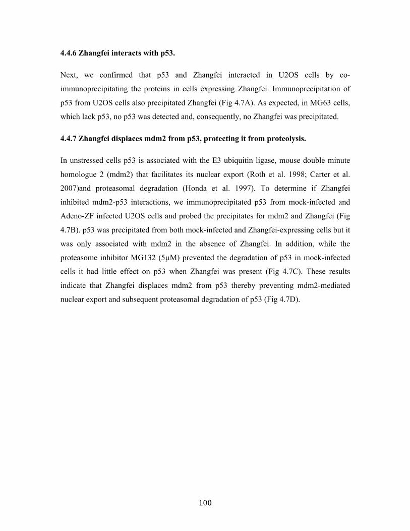
100
4.4.6 Zhangfei interacts with p53.
Next, we confirmed that p53 and Zhangfei interacted in U2OS cells by co-
immunoprecipitating the proteins in cells expressing Zhangfei. Immunoprecipitation of
p53 from U2OS cells also precipitated Zhangfei (Fig 4.7A). As expected, in MG63 cells,
which lack p53, no p53 was detected and, consequently, no Zhangfei was precipitated.
4.4.7 Zhangfei displaces mdm2 from p53, protecting it from proteolysis.
In unstressed cells p53 is associated with the E3 ubiquitin ligase, mouse double minute
homologue 2 (mdm2) that facilitates its nuclear export (Roth et al. 1998; Carter et al.
2007)and proteasomal degradation (Honda et al. 1997). To determine if Zhangfei
inhibited mdm2-p53 interactions, we immunoprecipitated p53 from mock-infected and
Adeno-ZF infected U2OS cells and probed the precipitates for mdm2 and Zhangfei (Fig
4.7B). p53 was precipitated from both mock-infected and Zhangfei-expressing cells but it
was only associated with mdm2 in the absence of Zhangfei. In addition, while the
proteasome inhibitor MG132 (5µM) prevented the degradation of p53 in mock-infected
cells it had little effect on p53 when Zhangfei was present (Fig 4.7C). These results
indicate that Zhangfei displaces mdm2 from p53 thereby preventing mdm2-mediated
nuclear export and subsequent proteasomal degradation of p53 (Fig 4.7D).

101

102
Figure 4.7 In vitro interaction of Zhangfei and p53. (A) Zhangfei associates with p53
in U2OS cells. Cell lysate from U2OS and MG63 human cells previously infected with
adenovirus vector expressing Zhangfei (Ad-ZF) or mock infected (MI) were incubated
with 2µg of mouse anti-p53 antibody for 12 hours at 4 oC, followed by an additional 4
hours incubation with 100µl of Protein A/G agarose beads (IP). The associated proteins
were detected by immune-blotting (IB) using rabbit anti-Zhangfei antibody and mouse
anti-p53 antibody. Input represented 1/10 of lysate used for immunoprecipitation. (B)
Zhangfei displaces mdm2 from p53 in U2OS cells. Cell lysates as in (A) were
immunoprecipitated with anti-p53 antibody followed by immunoblotting with either anti-
mdm2 or anti-Zhangfei antibody. (C) Inhibition of proteasomal degradation reverses
degradation of p53 in the absence of Zhangfei. Cells infected as in (A) were either treated
with the proteasomal inhibitor MG132 (5µM) or left untreated. Proteins in the Cell
lysates were separated by SDS-PAGE and p53, Zhangfei and GAPDH (loading control)
were detected by immunoblotting. (D) Schematic diagram of a model for the proposed
mechanism by which Zhangfei inhibits cell growth and UPR. Under normal conditions,
mdm2 translocates the p53 protein out of the nucleus for degradation via the ubiquitin-
dependent pathway. In Zhangfei-expressing cells, Zhangfei interacts with p53 and
prevents it from binding to mdm2. This results in p53 stabilization and nuclear
accumulation, which, in turn, activates its target genes and suppresses tumour cell growth
and the UPR. These results illustrate how Zhangfei controls the activity of p53 toward the
cell growth and the UPR, and offer an explanation to why p53 is upregulated by Zhangfei
in tumour cells.
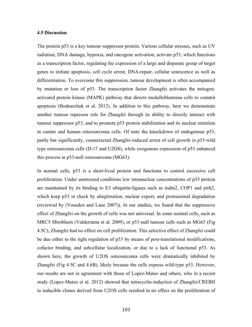
103
4.5 Discussion
The protein p53 is a key tumour suppressor protein. Various cellular stresses, such as UV
radiation, DNA damage, hypoxia, and oncogene activation, activate p53, which functions
as a transcription factor, regulating the expression of a large and disparate group of target
genes to initiate apoptosis, cell cycle arrest, DNA-repair, cellular senescence as well as
differentiation. To overcome this suppression, tumour development is often accompanied
by mutation or loss of p53. The transcription factor Zhangfei activates the mitogen-
activated protein kinase (MAPK) pathway that directs medulloblastoma cells to commit
apoptosis (Bodnarchuk et al. 2012). In addition to this pathway, here we demonstrate
another tumour repressor role for Zhangfei through its ability to directly interact with
tumour suppressor p53, and to promote p53 protein stabilization and its nuclear retention
in canine and human osteosarcoma cells. Of note the knockdown of endogenous p53,
partly but significantly, counteracted Zhangfei-induced arrest of cell growth in p53-wild
type osteosarcoma cells (D-17 and U2OS), while exogenous expression of p53 enhanced
this process in p53-null osteosarcoma (MG63).
In normal cells, p53 is a short-lived protein and functions to control excessive cell
proliferation. Under unstressed conditions low intranuclear concentrations of p53 protein
are maintained by its binding to E3 ubiquitin-ligases such as mdm2, COP1 and pirh2,
which keep p53 in check by ubiqitination, nuclear export, and proteasomal degradation
(reviewed by (Vousden and Lane 2007)). In our studies, we found that the suppressive
effect of Zhangfei on the growth of cells was not universal. In some normal cells, such as
MRC5 fibroblasts (Valderrama et al. 2009), or p53-null tumour cells such as MG63 (Fig
4.5C), Zhangfei had no effect on cell proliferation. This selective effect of Zhangfei could
be due either to the tight regulation of p53 by means of post-translational modifications,
cofactor binding, and subcellular localization, or due to a lack of functional p53. As
shown here, the growth of U2OS osteosarcoma cells were dramatically inhibited by
Zhangfei (Fig 4.5C and 4.6B), likely because the cells express wild-type p53. However,
our results are not in agreement with those of Lopez-Mateo and others, who in a recent
study (Lopez-Mateo et al. 2012) showed that tetracyclin-induction of Zhangfei/CREBH
in inducible clones derived from U2OS cells resulted in no effect on the proliferation of

104
the cells, nor did the cells display obvious morphological changes. We speculate that
there could be two reasons for the differences in our results: 1) Leaky expression of
tetracycline-inducible Zhangfei in U2OS cells (Lopez-Mateo et al. 2012) may have
resulted in the selection of clones with structural and functional mutations in Zhangfei.
This would be consistent with our observations (Fig 4.1) that suggest that the growth
suppressive effects of Zhangfei in cells expressing functional p53 exert strong selective
pressure and lead to the selection of cells expressing non-functional Zhangfei. 2) The cell
line studied by Lopez and co-workers may have spontaneously lost its functional p53.
We confirmed that the U2OS cell line we used in this study does indeed have a functional
p53 (Fig 4.5A).
The protein p53 has been reported to antagonize the UPR and inhibit ground glass
hepatocyte development during ER stress (Dioufa et al. 2012). In the present study, we
determined that p53, at least in part, mediates Zhangfei-induced suppression of the UPR
(Fig 4.4 C, 4.6 C and F). Induction of UPR is a protective mechanism utilized by cells to
adapt to ER stress. In normal cells, the UPR is related to tissue preservation or organ
protection against ER stress. In neoplastic cells, however, the adaptive function of the
UPR is implicated in immune resistance, cancer progression, and drug resistance
(reviewed by (Moenner et al. 2007; Ron and Walter 2007)). Therefore, the inhibition of
UPR, mediated by p53, represents a potential strategy by which Zhangfei could be used
to inhibit cancer. We show that Zhangfei inhibits the UPR and induces apoptosis by
promoting p53 stabilization and nuclear retention. Interestingly, in keeping with our
results, ER stress, and the resulting activation of the UPR induces p53 cytoplasmic
localization and prevents p53-dependent apoptosis (Qu et al. 2004), suggesting the
existence of an ER stress-p53-UPR regulatory loop (see model in Fig 4.8). Although the
precise relationship between p53 stabilization and UPR remains unclear, glycogen
synthase kinase-3 is shown to be a mediator of ER-stress induced p53 cytoplasmic
localization (Qu et al. 2004), and the ubiquitination and importin-a3 binding of p53 are
also associated with stress-mediated p53 translocalization (Becker et al. 2007;
Marchenko et al. 2012). In the case of Zhangfei, improved understanding of how exactly
p53 suppresses the UPR still should be further investigated.

105
We had previously shown that ectopic expression of Zhangfei in medulloblastoma
(Valderrama et al. 2009) and osteosarcoma cells (Chapter 2) (Bergeron et al. 2013)
suppressed the cell growth and the UPR. In addition, the effects of this protein were also
observed in four other canine osteosarcoma cell lines (not published, see Chapter 6). To
study the mechanisms involved we tried to develop cell lines in which the expression of
Zhangfei could be induced by doxycycline. We were successful in developing several
clones that expressed Zhangfei protein, as detected by immunofluorescence and
immunoblots, only in the presence of the inducer (Fig 4.1A). However, none of the
clones behaved as did cells in which the ectopic expression of Zhangfei was induced by
infection with adenovirus vectors (Fig 4.1B and D). On further examination we found
that in the clones one to as many as all six leucines had been replaced with other amino
acids (Fig 4.1C). The clone with only one substitution had growth properties most similar
to the cells expressing adenovirus-Zhangfei. No other mutations in the entire Zhangfei
coding sequences were observed in any of the clones. We interpret this to mean that even
small amounts of intact Zhangfei, that might be expressed in the absence of the inducer,
were sufficient to block cell division; only cells in which the zipper had been disabled
survived.
While our results clearly identify the leucine zipper of Zhangfei as important for its
interaction with p53 and subsequent p53-mediated suppression of the UPR, we have also
shown (Chapter 3) that Zhangfei interacts with the bLZip UPR transcription factor Xbp1s,
targeting it for proteosomal degradation (Zhang et al. 2013), and that the leucine zipper of
Zhangfei is also important for this interaction. Hence, modulation of UPR by Zhangfei
may require a combination of several coordinated events, including direct interaction
with UPR mediators and indirect mediation through p53.
Like Zhangfei several other bLZip proteins such as ATF3 (Yan et al. 2005; Wang et al.
2010), ING2 (Wang et al. 2006b ), K-bLZip (Park et al. 2000), and CREB (Okoshi et al.
2011), are known to interact with p53 through their leucine zippers. The nature of these
interactions as well as their consequences, however, varies. Interactions between p53 and
Zhangfei, ATF3 and CREB lead to enhanced transcriptional activity and stabilization of
p53. Our results show that Zhangfei displaced mdm2 from p53 (Fig 4.7B) and prevented

106
its degradation to a similar extent as the proteasomal inhibitor MG132 (Fig 4.7C). The
effects of MG132 and Zhangfei were not additive. This may reflect the toxic effects of
the combined inhibitors leading to a decrease in a rapidly turned over protein like p53. In
contrast to the effects of Zhangfei on p53, ATF3 neither displaces mdm2 nor does it
suppress its activity. ATF3 binds directly to the carboxyl terminus of p53 and prevents
ubiquitination of key lysine residues in the region (Yan et al. 2005). CREB binds to the
amino terminus of p53 and acts as a bridge directing CREB binding protein to p53
responsive promoters (Giebler et al. 2000) and suppresses transcription of mdm2 (Okoshi
et al. 2008). Unlike the bLZip-p53 interactions mentioned above, which enhance p53
activity, the association of the Kaposi sarcoma herpesvirus protein K-bLZip with p53
suppresses its activity and may contribute to tumorigenesis by the virus (Park et al. 2000).
Figure 4.8 Zhangfei and ER stress have opposing effects on p53. In Zhangfei-
expressing cells, p53 is targeted for nuclear localization and stabilization, and further to
prevent cell growth, induce cell apoptosis, as well as inhibit the UPR in response to ER
stress. In contrast, ER stress promotes the cytoplasmic localization and degradation of
p53.
107
4.6 Acknowledgements
The authors thank Noreen Rapin for technical assistance. This work was supported by a
Discovery grant to VM from the Natural Sciences and Engineering Research Council
(NSERC) of Canada; and a research grant from the Western College of Veterinary
Medicine, Companion Animal Health Fund. RZ was supported by scholarships from the
Government of China (China Scholarship Council, RZ-2010635007) and the University
of Saskatchewan College of Graduate Studies and Research.

108
5. Structural domains responsible for p53-Zhangfei interaction
Rui Zhang and Vikram Misra*
Department of Microbiology, Western College of Veterinary Medicine, 52 Campus
Road, University of Saskatchewan, Saskatoon, Saskatchewan , S7N1B4, CANADA
This Chapter described the characterization of the interaction of Zhangfei and p53 by
mapping the interacting domains of p53. It showed that p53 formed a complex with
Zhangfei via its N-terminal Transactivation Domain (NTD), which was required for
Zhangfei-mediated nuclear retention and transcriptional activity of p53. This Chapter will
be submitted in the future with further study for publication as “Characteristics of the
interaction of p53 and Zhangfei/CREBZF.”
My contributions to this Chapter include: To date, I constructed several plasmids
expressing p53 deletion mutants containing its different functional domains, respectively.
I also used these constructed plasmids to determine the interacting and transactivating
domain of p53 with Zhangfei. The specific amino acid sites of p53 that function as the
binding sites of Zhangfei will be detected in future studies using direct-site mutagenesis
of p53. I generated all the final figures and wrote the first draft, which was revised by
Professor Vikram Misra.

109
5.1 Abstract
We described (Chapter 4) that basic-region leucine zipper (bLZip) transcriptional factor
Zhangfei has the ability to stabilize tumour suppressor p53 and activate its transcription
activity by competing with the negative regulator mdm2 which, when bound to p53,
targets it for degradation. The bLZip domain of Zhangfei is required for its interaction
with p53. In this study, we identified the domains of p53 which are responsible for
complex formation with Zhangfei. Immunoprecipitation assay revealed that p53
interacted with Zhangfei through its N-terminal transactivation domain (NTD), which is
also the mdm2-binding domain and transcription activation region of p53.
Overexpression of Zhangfei enhanced the nuclear retention and increased the
transcription activity of both wild-type p53 and the 92 amino acid residues at its N-
terminus (NTD region). In contrast, with the mutant form of p53 lacking an NTD region,
we could not observe any changes in its cellular localization or transcription activity
between the absence and presence of Zhangfei. Our results suggested that Zhangfei
positively regulates p53 by occupying the NTD region and thereby preventing its
association with negative regulator mdm2.
Key words: p53, Zhangfei, N-terminal transactivation domain, interaction
110
5.2 Introduction
Tumour suppressor protein p53 plays an important role in cell fate determination in
response to oncogenic or other cellular stress signals. It exerts its function as a nuclear
sequence-specific transcription factor, and induces or represses the expression of various
target genes involved in cell cycle arrest and apoptosis (Joerger and Fersht 2008). p53
comprises several functional regions: the N-terminal transactivation region consists of a
transactivation domain (TAD) and a proline-rich region (PRR), followed by the DNA-
binding core domain (p53C), a tetramerization domain (TET) and a C terminus (CT) (Fig.
1A). Under physiological conditions, p53 is a short-lived protein that is strictly regulated
by several other proteins, and one of the most important of these is E3 ubiquitin-ligase
mdm2. The N-terminal sequence-specific transactivation domain (NTD) of p53 is
responsible for its transcriptional and pro-apoptotic activities. Thus, mdm2 keeps p53 in
check by binding to its NTD, inhibiting its transcriptional and pro-apoptotic activity
(Momand et al., 1992), and further targeting p53 for proteasomal degradation via
ubiquitin-dependent pathways (Kulikov et al. 2010). In addition, p53 can up-regulate the
expression of mdm2 by an auto-regulatory feedback loop to prevent inappropriate
apoptosis. Under cellular stress conditions, p53 is rapidly phosphorylated on multiple
sites, such as Ser15, Thr18 or Ser20. A potential outcome of such phosphorylation is the
stabilization of p53 through disruption of its binding with mdm2 and inhibition of p53
ubiquitination and degradation. Once p53 becomes activated, it functions as a
transcription factor and drives the expression of a wide network of apoptotic signals.
Recently, our lab and others have found that the activity of p53 was enhanced by a basic-
region leucine zipper (bLZip) transcriptional factor, Zhangfei. Further studies
demonstrated that Zhangfei stabilized p53 and promoted its nuclear retention by
disrupting its association of mdm2 and preventing p53 from degradation. The bLZip
domain of Zhangfei contributed to its profound effects on p53 (Chapter 4) (Lopez-Mateo
et al. 2012; Zhang and Misra 2014). The objective of this study is to identify structural
domain of p53 responsible for the complex formation with Zhangfei. Our results suggest
that p53 forms a complex with Zhangfei via its NTD region, which is required for
Zhangfei-mediated nuclear retention and transcriptional activity of p53.

111
5.3 Materials and Methods
5.3.1 Cells and tissue culture
Human MG63 and U2OS osteosarcoma cells were obtained from Dr. Douglas H. Thamm,
(Associate Professor of Oncology, Animal Cancer Center, Colorado State University).
Vero cells were obtained from the American Type Tissue Culture Collection. All cell
lines were grown in Dulbecco’s minimal essential medium containing penicillin and
streptomycin and 10% newborn calf serum. All media, serum and antibiotics were
purchased from Invitrogen.
5.3.2 Plasmids
The construction of pcZF (Lu and Misra 2000b), a plasmid that expresses Zhangfei in
mammalian cells, has been described. The CAT reporter plasmid pCAT3B-p53RE was
constructed as described before (Chapter 4) (Zhang and Misra 2014). The construction of
the plasmids expressing the full length of wild-type p53 as well as its several deletion
mutants was performed using an In-Fusion HD Cloning Kit (Clontech, 639648) as
described in the manufacturer’s instructions. All of these p53-plasmids contained three
copies of the FLAG-coding sequence. The cDNA fragment containing the entire coding
sequence of p53 was recovered from U2OS cells and its sequence confirmed. The
specific primers for p53 and its deletion mutant amplification are summarized in Table
5.1.
Table 5.1 The sequence of primers for p53 and its deletion mutant amplification
Full length of wild-type p53
Forward 5’- GCGGCCGCGAATTCATGGAGGAGCCGCAGTCAGAT-3’ Reverse 5’- ATCTATCGATGAATTCTCAGTCTGAGTCAGGCCCTTCT-3’
p53 (1-356aa) Forward 5’- GCGGCCGCGAATTCAATGGAGGAGCCGCAGTCAGAT-3’ Reverse 5’- ATCTATCGATGAATTCTCACCCAGCCTGGGCATC -3’
p53 (1-292aa) Forward 5’- GCGGCCGCGAATTCATGGAGGAGCCGCAGTCAGAT-3’ Reverse 5’- ATCTATCGATGAATTCTCATTTCTTGCGGAGATTCT-3’
p53 (92-393aa) Forward 5’- TTGCGGCCGCGAATTCACTGTCATCTTCTGTCCCTT-3’ Reverse 5’- ATCTATCGATGAATTCTCAGTCTGAGTCAGGCCCTTCT-3’
p53 (92-292aa) Forward 5’- TTGCGGCCGCGAATTCACTGTCATCTTCTGTCCCTT-3’ Reverse 5’- ATCTATCGATGAATTCTCATTTCTTGCGGAGATTCT -3’
p53 (1-92aa) Forward 5’- GCGGCCGCGAATTCATGGAGGAGCCGCAGTCAGAT -3’ Reverse 5’- ATCTATCGATGAATTCTCAGGGCCAGGAGGGGGC -3’

112
5.3.3 Transfection and CAT Assays
Vero cells were transfected with plasmids using Lipofectamine 2000 (Invitrogen, 11668-
019) as described in the manufacturer’s instructions. MG63 cells were transfected using
MG-63 Transfection Kit (for Osteosarcoma Cells, CRL-1427) (Cedarlane, 6848) as
described in the manufacturer’s instructions. Chloramphenicol acetyl transferase (CAT)
assays have been described earlier (Zhang and Misra 2014). CAT values were detected
using an enzyme-linked immunosorbent assay kit (Roche Applied Science, 11-363-727-
001).
5.3.4 Co-immunoprecipitation
Vero cells in 6-well dishes were transfected with pcZF. Cells were also co-transfected
with plasmid expressing wild-type p53 or its deletion mutants. Twenty-four hr after
transfection, cells were washed with cold PBS and lysed in 250µl/well cold lysis buffer
(50mM Tris, pH7.5, 150mM NaCl, 1mM EDTA and 0.1% TritonX-100) containing a
protease inhibitor cocktail (Sigma, P8340). After centrifugation at 13,000 xg at 4 oC, 20µl
of cell lysate supernatant were frozen as a pre-immunoprecipitated sample, and a mouse
anti-flag antibody (5µg, Sigma, F4042) was added to the remaining supernatant (230µl).
The sample was incubated for 12hr with constant gentle agitation. Protein A/G agarose
beads (100µl, Pierce, Fisher Scientific, 20421) were added and the samples were
incubated for an additional 4hr at 4 oC. Agarose beads were collected by centrifugation at
13,000 xg at 4 oC and washed 4 times in a lysis buffer before boiling in an SDS-PAGE
sample buffer. Proteins in samples of the unfractionated cell lysate or immunoprecipitates
were separated by SDS-PAGE, transferred to membranes and probed with rabbit anti-
Zhangfei antisera or rabbit anti-flag (Sigma, F7025). Antibodies were visualized after
incubation with Alexa488-labelled anti-rabbit antibody.
5.3.5 Antibodies and immunofluorescence
The antibodies used were rabbit anti-Zhangfei serum (Lu and Misra 2000b), mouse anti-
flag antibody (Sigma, F4042), rabbit anti-flag (Sigma, F7025) and mouse anti-GAPDH
(Sigma, G8795-200UL). Secondary antibodies were goat anti-mouse Alexa488

113
(Invitrogen, A-11001), goat anti-rabbit Alexa546 (Invitrogen, A-11035) and goat anti-
rabbit Cy5 (Invitrogen, A-10523). Cells were processed for immunofluorescence as
described previously (Bergeron et al. 2013). Images were captured using a digital camera
attached to an Olympus BX51 microscope and DPmanager software. Captured images
were processed using Adobe Photoshop and Illustrator CS6 software.

114
5.4 Results
5.4.1 p53 forms a complex with Zhangfei via its N-terminal transactivation domain
(NTD)
To map the region(s) of p53 required for complex formation with Zhangfei, we assessed
the ability of various p53 domains to bind to Zhangfei. FLAG-labeled p53 deletion
mutants as well as wild-type p53 (Fig 5.1A) were inserted into expression plasmids
(pcDNA3), and were co-expressed with plasmid expressing Zhangfei (pcZF) in Vero
cells by transfection. 24h after transfection, cell lysates were prepared and
immunoprecipitated with an anti-flag antibody followed by immunoblotting with an anti-
Zhangfei antibody. As shown in Fig. 1B, wild-type p53 and some p53 deletion mutants
(mutant lanes 2, 3 and 6) co-precipitated with Zhangfei, but other p53 deletion mutants
(mutant lanes 4 and 5) were not. These results suggested that only 92 amino acid residues
in the N-terminal of p53 (the N-terminal transactivation domain (NTD), which comprises
two parts: the transactivation domain (TAD) and the proline rich region (PRR)) were
sufficient and required for the interaction with Zhangfei (Fig 5.1B lane 6), while those
p53 mutants lacking these regions (Fig 5.1B lane 4) could not bind to Zhangfei.

115
Figure 5.1 Complex formation between Zhangfei and p53. (A) Schematic drawing of
the structures of wild-type p53 and its deletion mutants. TAD, transactivation domain;
PRR, proline rich region; p53C, central DNA-binding domain; TET, oligomerization
domain; CT, basic C-terminal domain. The flag represents the FLAG tag. (B) p53
interacts with Zhangfei through its 92 amino acid residues of N-teminal. Cell lysate from
Vero cells previously co-transfected with 1µg of plasmid expressing Zhangfei (pcZF) and
1 µg of plasmid expressing p53 (or its deletion mutants) were incubated with 2 µg of
mouse anti-FLAG antibody for 12 hr at 4oC, followed by an additional 4 hr of incubation
with 100 µl of Protein A/G agarose beads (IP). The associated proteins were detected by
immune-blotting (IB) using an rabbit anti-Zhangfei antibody and a rabbit anti-FLAG
antibody.

116
5.4.2 N-terminal transactivation domain (NTD) is required for Zhangfei-mediated
nuclear retention of p53
In previous studies (Chapter 4), we reported that Zhangfei stabilized p53 and promoted
its nuclear retention by displacing the E3 ubiquitin ligase mdm2. Mdm2 is a negative
regulator of p53, which binds to the NTD domain of p53 and further translocates it out of
the nucleus for degradation (Moll and Petrenko 2003). Therefore, we supposed that NTD
is also required for the Zhangfei-mediated nuclear retention of p53. To test this
hypothesis, we examined the co-localization of Zhangfei and p53 deletion mutants (Fig
5.2 A) by immnofluorescence assay. Consistent with our previous observations (Chapter
4), the expression of Zhangfei enhanced the nuclear localization of wild-type p53 (Fig
5.2B). Similarly, the immunofluorescence showed that Zhangfei expression resulted in an
increased nuclear staining of the p53 mutant that only contained the NTD region (1-92
aa), whereas it had no effect on the localization of the fragment containing the remainder
of p53 (92-393 aa) (Fig 5.2B). Because this p53 mutant (92-393 aa) was short of the
mdm2-binding domain, it was not transported out of the nucleus, and was localized in the
nucleus in both the absence and presence of Zhangfei. These results further suggest that
the Zhangfei-binding domain in p53 is also the mdm2-binding domain, and Zhangfei
achieves its functions through replacing mdm2 and interacting with the NTD region of
p53.

117
Figure 5.2 Co-localization of Zhangfei and p53 (or its deletion mutants). (A)
Schematic drawing of the structures of wild-type p53 and its deletion mutants. TAD,
transactivation domain; PRR, proline rich region; p53C, central DNA-binding domain;
TET, oligomerization domain; CT, basic C-terminal domain. The flag represents the
FLAG tag. (B) Vero cells were transfected with 1 µg of pcZF or a control (pcDNA3).
Cells were also co-transfected with a plasmid expressing wild-type p53 or its deletion
mutants. 24 h after transfection, p53 as well as Zhangfei were visualized by
immunofluorescence with anti-FLAG and anti-ZF antibody. The nucleus was stained
with Hoechst dye. Zhangfei alters the subcellular localization of wild-type p53 and the
p53 mutant that only contains 92 amino acids in the N-terminus.

118
5.4.3 Zhangfei enhances p53-mediated transactivation through the N-terminal
transactivation domain (NTD) of p53
We then asked whether the NTD region was also required for the effect of Zhangfei on
p53-mediated transcriptional activation. To this end, we performed a CAT reporter assay
to assess the ability of Zhangfei to indirectly induce transcription from a promoter
containing two copies of p53 responsive elements (pCAT3B-p53RE) in the presence of
p53 deletion mutants or wild-type p53. As shown in Fig 5.3, Zhangfei enhanced the
expression of a reporter gene, CAT, linked to the promoter in the cells expressing wild-
type p53 or several p53 mutants that contain NTD region, like p53 (1-356 aa), p53 (1-292
aa) and p53 (1-92 aa), but had no significant effect on the transcription activation of the
p53 mutant (92-393 aa). Also, due to the deletion of the transactivation domain, the p53
mutant (92-393 aa) itself didn’t activate transcription of its downstream genes.

119
Figure 5.3 Zhangfei activates p53-dependent transactivation via interaction with its
92 amino acids in N-terminal. p53-null MG63 cells were transfected with a reporter
plasmid containing the coding sequence for CAT linked to a promoter with two copies of
a p53 responsive element (pCAT3B-p53RE, 0.5µg) in the presence or absence of a
plasmid expressing Zhangfei (pcZF, 1µg). All samples also contained, as a control, a
plasmid expressing b-galactosidase. 24h after transfection, CAT activity was determined.
Values represented the relative CAT activity (normalized to the internal control, b-
galactosidase) of different treatments. Each datum point is the average of duplicate
transfections with the bar representing the range. Standard deviations from means of three
independent experiments are shown, and the significant P values from t-tests were noted
above the bars.

120
5.5 Discussion
Inactivation of the p53 gene in cancer cells is usually mediated through mutations, direct
interaction and transcriptional repression. Mdm2 acts as an E3 ubiquitin protein ligase for
p53 and binds to amino acids Phe19, Trp23 and Leu26 in the N-terminal transactivation
domain (NTD) of p53; consequently, this suppresses its transcriptional and pro-apoptotic
activity (Momand et al. 1992; Moll and Petrenko 2003). Therefore, the elimination of the
negative effect of mdm2 on p53 has been wildly considered to effectively induce
apoptosis of cancer cells. According to our previous results (Chapter 4), transcription
factor Zhangfei, also known as cAMP Response Element Binding Protein ZF (CREBZF),
was a profound positive regulator of p53, which induced the growth arrest and apoptosis
of cancer cells by displacing mdm2 and stabilizing p53 (Zhang and Misra 2014). Here,
we found that Zhangfei enhanced nuclear retention and increased transcriptional activity
of p53 by binding to its N-terminal transactivation domain, providing supportive
evidence for our previous conclusions (Zhang and Misra 2014).
In addition to Zhangfei, several other p53 N-terminal binding proteins have been shown
to repress or activate the transcriptional activity and stabilization of p53 by masking or
uncovering the NTD of p53. These include the p53 negative regulator hepatitis B virus X
protein (HBX) (Lee and Rho 2000) and p53 transcriptional coactivators (p300/CBP)
(Teufel et al., 2007). HBX represses the transcription of the p53 gene through the E-box
element and simultaneously interacts with the p53 protein through the NTD,
consequently inhibiting its functions (Wang et al. 1994; Lee and Rho 2000). Like
Zhangfei, the transcriptional coactivator p300 and the CREB-binding protein (CBP)
interact with N-terminal transactivation domain of p53 and may compete with mdm2 for
binding to p53 (Teufel et al. 2007). But, unlike the very tight binding between Zhangfei
and p53 (shown in Fig 4.7, Chapter 4), p300/CBP could be outcompeted by other high-
affinity p53 N-terminal binding proteins, such as mdm2, leading to the formation of
mixed complexes consisting of p53, p300 and mdm2 (Kobet et al. 2000; Teufel et al.
2007). Moreover, mdm2 has been reported to inhibit p300-mediated p53 activation by
forming a ternary complex with the two proteins (Kobet et al. 2000).

121
The competition between Zhangfei and mdm2 for binding to p53 depends on their
affinity to the NTD of p53. The mdm2-binding residues of p53 have been identified at 19,
23, and 26 amino acid in the N-terminal (Moll and Petrenko 2003). The occupation of
these sites by mdm2 covers the phosphorylation sites of p53, such as Ser15, Thr18 or
Ser20, and prevents its activation. To replace mdm2 from p53, Zhangfei may also bind to
the same sites with mdm2, or occupy more amino acid residues in the NTD for a higher
affinity. Thus, the direct-site mutagenesis of p53 will explore these in future studies.

122
6. The effect of Zhangfei/CREBZF on cell growth, differentiation, apoptosis, migration, and the UPR in several canine osteosarcoma cell lines
Rui Zhang, Douglas H. Thamm1 and Vikram Misra*
Department of Microbiology, Western College of Veterinary Medicine, 52 Campus
Road, University of Saskatchewan, Saskatoon, Saskatchewan , S7N1B4, CANADA
1. Colorado State University Flint Animal Cancer Center, 300 West Drake Road, Fort
Collins, CO 80523, USA
The purpose of this Chapter was to determine if the effects of Zhangfei on D-17 cells in
Chapter 2, 3 and 4 applied more universally to canine OS. I examined three other
independently isolated canine OS cell lines—Abrams, McKinley and Gracie, and found
that, like D-17, the three cell lines expressed p53 proteins that were capable of activating
promoters with p53 response elements on their own, and synergistically with Zhangfei.
Furthermore, as with D-17 cells, Zhangfei suppressed the growth and UPR-related
transcripts in the OS cell lines. Zhangfei also induced the activation of osteocalcin
expression, a marker of osteoblast differentiation and triggered programmed cell death.
This Chapter will be submitted for publication as “The effect of Zhangfei/CREBZF on
cell growth, differentiation, apoptosis, migration, and the UPR in several canine
osteosarcoma cell lines.”
My contributions to this Chapter include: I designed and performed all the
experiments mentioned in this manuscript. I generated all the final figures and wrote the
first draft, which was revised by Professor Vikram Misra. The canine Abrams, Mckinley
and Gracie cell lines were kindly provided by Dr. Douglas H. Thamm (Associate
Professor of Oncology, Animal Cancer Center, Colorado State University).

123
6.1 Abstract
We have shown that the bLZip domain-containing transcription factor,
Zhangfei/CREBZF inhibits the growth and the unfolded protein response (UPR) in cells
of the D-17 canine osteosarcoma (OS) line (Chapter 2) and that the effects of Zhangfei
are mediated by it stabilizing the tumour suppressor protein p53 (Chapter 4). To
determine if our observations with D-17 cells applied more universally to canine OS, we
examined three other independently isolated canine OS cell lines—Abrams, McKinley
and Gracie. Like D-17, the three cell lines expressed p53 proteins that were capable of
activating promoters with p53 response elements on their own, and synergistically with
Zhangfei. Furthermore, as with D-17 cells, Zhangfei suppressed the growth and UPR-
related transcripts in the OS cell lines. Zhangfei also induced the activation of osteocalcin
expression, a marker of osteoblast differentiation and triggered programmed cell death.

124
6.2 Introduction
Osteosarcoma (OS) is the most common primary malignant bone tumour in children and
adolescents, although its incidence in dogs is ten times greater than in humans (Withrow
2003). Spontaneously occurring osteosarcomas in dogs are an ideal model for cancer
research due to their anatomical and physiological similarities with human counterparts
(reviewed by (Mueller et al. 2007; Paoloni and Khanna 2008; Khanna et al. 2009).
We have shown that the bLZip domain-containing transcription factor,
Zhangfei/CREBZF/SMILE inhibits the growth and the unfolded protein response (UPR)
in the D-17 canine osteosarcoma (OS) cell line (Chapter 2) and that the effects of
Zhangfei are mediated by stabilizing the tumour suppressor protein p53 (Chapter 4). To
determine if our observations with D-17 cells applied more universally to canine OS, we
examined three other independently isolated canine OS cell lines—Abrams, McKinley
and Gracie. The purpose of this study was to determine the inhibitory role of Zhangfei in
these OS cell lines by exploring its potential involvement in growth, differentiation,
apoptosis, and metastasis.
Zhangfei was initially identified through its interaction with the host cell factor (HCF1) a
protein required for the initiation of herpes simplex virus gene expression (Lu and Misra
2000b). Unlike other bLZip transcription factors, Zhangfei appears to be incapable of
binding to consensus bLZip response elements as a homodimer (Cockram et al. 2006).
Instead, it fulfills its role in transcriptional regulation by hetero-dimerizing with and
modulating other transcription factors or signaling molecules, such as Luman/CREB3
(Misra et al. 2005), Xbp1 (Zhang et al. 2013), ATF4 (Hogan et al. 2006), SMAD 1,5,8
(Lee et al. 2012a), herpes simplex virus VP16 (Akhova et al. 2005), and p53 (Lopez-
Mateo et al. 2012).
125
6.3 Materials and Methods
6.3.1 Cells and tissue culture
Canine osteosarcoma D-17 cells, obtained from the American Type Tissue Culture
Collection, were grown in MEM-Alpha containing 10% fetal bovine serum (FBS).
Canine Abrams, Mckinley and Gracie cell lines were obtained from Dr. Douglas H.
Thamm (Associate Professor of Oncology, Animal Cancer Center, Colorado State
University), and grown in Dulbecco’s minimal essential medium containing penicillin,
streptomycin and 10% newborn calf serum. All media, serum and antibiotics were
purchased from Invitrogen (Carlsbad, California).
6.3.2 Adenovirus Vectors Expressing Zhangfei and β-galactosidase (LacZ)
Adenovirus Vectors Expressing Zhangfei and β-galactosidase (LacZ) were constructed,
grown, and purified using the Adeno-X Expression System (Clontech). They were
created in our laboratory as described earlier (Misra et al. 2005). Cells were infected with
Adeno-Zhangfei, Adeno-LacZ (expressing E. coli b-galactosidase, LacZ) or mock-
infected. A multiplicity of infection (MOI) of 100 plaque-forming units (pfu) per cell was
used.
6.3.3 WST-1 Cell Proliferation and Viability Assay
To determine the growth rate of cells, 104 cells/well were seeded into 96-well plates. 24h
later cells were either mock infected or infected with adenovirus vectors expressing
Zhangfei (Adeno-ZF) or b-galactosidase (Adeno-LacZ). Cell proliferation was assessed
using Cell Proliferation Reagent WST-1 (Roche, Mannheim, Germany) according to the
manufacturer’s specifications.
6.3.4 Annex V-apoptosis assay
Cells were collected after trypsinization and stained with Annexin V and propidium
iodide using an Annexin V kit (Calbiochem) following manufacturer’s instructions. As a
positive control cells were treated with 50 µM etopocide (Calbiochem) for 24 hr. Cells
were analyzed in a Coulter EPICS XL flow cytometer.

126
6.3.5 Scratch wound healing assay
Scratch wounds more than 5mm in length and of equal thickness were made in 100%
confluent cultures of D-17 or Abrams cells mock-infected or infected with Adeno-ZF or
Adeno-LacZ with a 10 µl disposable eppendorf tip. Phase contrast images were taken at 0,
4, 8, 12, and 24 hours after infection from identical regions. The wound size at each time
point after infection relative to the starting wound size was measured using Photoshop
software in three independent experiments.
6.3.6 Quantitative real-time PCR (qPCR)
Total RNA was extracted using RNeasy Plus Mini Kit from Qiagen (Mississauga, ON,
Canada). Gene expression was analyzed by RT-PCR using Brilliant II SYBR Green
QPCR Master Mix Kit (Agilent Technologies). The primers used were: Xbp1 spliced-
forward: 5’-TCTGCTGAGTCCGCAGCAGG-3’, Xbp1 5’-spliced-reverse:
TAAGGAACTGGGTCCTTCT-3’, HERP-forward: 5’-CCGAGCCTGAGCCCGTCACG
-3’, HERP-reverse: 5’-CTTTGGAAGCAAGTCCTTGA-3’, CHOP-forward: 5’-
TGGAAGCCTGGTATGAGGAC-3’, CHOP-reverse: 5’-TGCCACTTTCCTCTCGTTC
T-3’, GRP78-forward: 5’-GGCTTGGATAAGAGGGAAGG-3’, GRP78-reverse: 5’-
GGTAGAACGGAACAGGTCCA-3’, osteocalcin-forward: 5’-AAGCRGGAGGGCAGC
AGGT-3’, osteocalcin-reverse: 5’-CYGRTARGCYTCCTGRAAGC-3’.
6.3.7 PCR and sequencing of p53 genes
The sequences of PCR primers used for canine p53 amplification were: canine p53-
forward: GGTGACTGCAATGGAGGAGTCGCA, canine p53-reverse:
TCAGTCTGAGTCAAGCCCTTCTCT. RNA was purified from cells using the RNEasy
Plus mini kit with a genomic DNA elimination step (Qiagen) and RNA converted to
cDNA with the Quantitect Reverse Transcription kit (Qiagen) using instructions supplied
by the manufacturer. Two-step RT-PCR reactions used TopTaq emzyme (Qiagen) and
were performed in a PCR machine.

127
6.3.8 Plasmids and chloramphenicol acetyl transferase (CAT) assay
The construction of pcZF (Lu and Misra, 2000), a plasmid that expresses Zhangfei in
mammalian cells, has been described. The CAT reporter plasmid pCAT3B-p53RE was
constructed by transferring oligonucleotides containing two copies of p53 responsive
element, GGTCAAGTTGGGACACGTCCaaGAGCTAAGTCCTGACATGTCT (IDT,
Coralville, Iowa), to pCAT3Basic (Promega), which contains the coding sequence for
CAT linked to a basal promoter. Oligonucleotides representing the p53 responsive
elements with overhanging 5’ terminal KpnI and 3’ terminal BglII sites were annealed
and ligated to pCAT3Basic cut with the same enzymes.
In CAT assay, D-17, Abrams, Gracie and McKinley cells were transfected with 0.5µg of
pCAT3B-p53RE, in the presence or absence of a plasmid expressing Zhangfei (pcZF,
1µg), using Lipofectamine 2000 (Invitrogen) as described in the manufacturer’s
instructions. The promoter-less parental reporter plasmid, pCAT3B was included as a
control to show basal CAT activity. 250 ng of pCMVBGal, a plasmid specifying β-
galactosidase, were added to each transfection as an internal control. 24h after
transfection, the CAT activity was determined by ELISA. CAT values were normalized
to β-galactosidase.
6.3.9 Antibodies, immunoblotting and immunofluorescence
The antibodies used were mouse anti-FLAG (Sigma), rabbit anti-Zhangfei serum, rabbit
anti-Xbp1 (Abcam, Cambridge, MA), rabbit anti-HERP (Abcam, Cambridge, MA),
rabbit anti-GRP78 (Abcam, Cambridge, MA), and mouse anti-GAPDH (Chemicon,
Billerica, MA). Secondary antibodies were goat anti-mouse Alexa488, goat anti-rabbit
Alexa546 and goat anti-rabbit Cy5 (Invitrogen). Cells were processed for
immunoblotting and immunofluorescence as described previously (Lu and Misra 2000b;
Bergeron et al. 2013).
128
6.4 Results
6.4.1 All four canine OS cells lines express functional p53
To determine the effects of Zhangfei we had observed in D-17 OS cells we examined
three other canine OS cell lines. We have shown that Zhangfei exerts its effect on cell
growth and the UPR by stabilizing p53 (Chapter 4, Zhang and Misra, 2014) and it
therefore has no effect on cancer cells that do not possess functional p53. To assess the
status of p53 in the canine cell lines I amplified p53 transcripts from the cells using PCR
and determined the nucleotide sequences of the products. Figure 6.1B shows the derived
amino acid sequences of p53 from the cell lines and the reference sequence from the
canine genome database. All four cell lines contained transcripts for p53 that, with the
exception of a few amino acid variations, were identical to the reference sequence. None
of the amino acid polymorphisms in the sequences were at positions identified as
important for p53 function (Joerger and Fersht 2008)(Fig 6.1A).
To determine if the p53 proteins in the cell lines were functionally active, I transfected
the cells with a plasmid that expressed the reporter protein chloramphenicol acetyl
transferase (CAT) regulated by a promoter with two copies of a p53 response element
(pCATp53RE). As a negative control, cells were transfected with a plasmid (pCAT3B)
without the response elements. Parallel cultures were transfected with a plasmid
expressing Zhangfei. Figure 6.1C shows that expression of CAT was activated in all four
cell lines in a p53 response element–dependent manner and that the presence of Zhangfei
enhanced expression.

129

130
Figure 6.1. p53 in dog osteosarcoma cell lines. (A) Schematic structure of full-length
p53. TAD: N-terminal transactivation domain; PRR: proline-rich region; p53C: central
DNA-binding domain; TET: tetramerization domain; CT: extreme C terminus. p53C is
the domain where most cancer-associated p53 mutations are located. The numbers above
the diagram represent the residues with highest frequency of oncogenic missense
mutations (Joerger and Fersht 2008). (B) Derived amino acid sequence alignment of p53s
from 4 dog osteosarcoma cell lines and dog wild-type p53. The amino acid sequences
were aligned using MacVector software. The residues that have high mutant frequency
were marked above the diagram. (C) p53s of dog osteosarcoma cells have transcription
activity, and Zhangfei enhances p53-dependent transactivation. D-17, Abrams, McKinley,
and Gracie cells were transfected with 0.5µg of pCAT3B or pCAT3B-p53RE, in the
presence or absence of 1µg of pcZF. 24h after transfection, the CAT activity was
determined. Values represented the relative CAT activity (adjusted by β-galactosidase) of
different treatments. Standard deviations from means of three individual experiments are
shown (*P<0.05, **P<0.01).

131
6.4.2 Cellular outcome following ectopic expression of Zhangfei: growth cessation,
apoptosis and differentiation
We next compared the effect of Zhangfei on the growth characteristics of Abrams,
Mckinley, and Gracie cells with its effect on D-17 cells. The cells were infected with
adenovirus expressing either Zhangfei (Adeno-ZF) or the control protein b-galactosidase
(Adeno-LacZ). Cell growth was monitored by the WST-1 Cell Proliferation Assay. In
agreement with previous results, all four Adeno-ZF-infected cells failed to divide as early
as day 1 after infection as determined by their ability to convert WST-1 Cell Proliferation
reagent and absorb light at 405 nm. Mock infected cells continued to grow for three days
and the growth of Adeno-LacZ-infected cells was indistinguishable from mock-infected
cells (Fig 6.2).

132
Figure 6.2. Ectopic expression of Zhangfei suppresses cell growth in canine
osteosarcomas. D-17, Abrams, McKinley, and Gracie canine OS cells were mock-
infected or infected with adenovirus vectors expressing either Zhangfei (Adeno-ZF) or
beta-galactosidase (Adeno-LacZ) and measured growth rates by absorbance at 405 nm
with WST-1 at different time points after infection.
Since Zhangfei may stop cell growth by inducing differentiation and/or causing apoptosis,
we further performed a transcript level analysis of the osteosarcoma differentiation
marker—osteocalcin (Ciovacco et al. 2009) and a flow cytometric analysis of Annexin
V-stained apoptotic cells in D-17 and Abrams cells infected with either Adeno-ZF or
Adeno-LacZ. Compared with LacZ-expressing and even vitamin D3-treated cells
(negative and positive controls, respectively), Zhangfei significantly increased the
expression of osteocalcin transcripts in a time-dependent manner (Fig 6.3). Meanwhile,
we also found both D-17 and Abrams cells began to undergo apoptosis upon expression

133
of Zhangfei as the percentage of cells with Annexin V stain increased to ~10% by 12h
after infection and to >22% by 24h, while the mock infected and LacZ-expressing cells
had obviously lower percentages of apoptotic cells (Fig 6.4).
Figure 6.3. Zhangfei induces differentiation of canine osteosarcoma cells. D-17 and
Abrams cells were either mock-infected or infected with Adeno-ZF or Adeno-LacZ. The
positive control cells were treated with 10-5 mM of vitamin D3. The mRNA levels of
osteoblast differentiation marker (osteocalcin) were estimated by qRT-PCR.
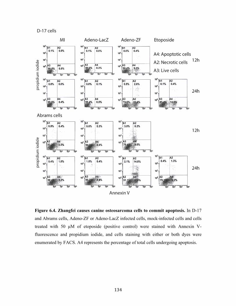
134
Figure 6.4. Zhangfei causes canine osteosarcoma cells to commit apoptosis. In D-17
and Abrams cells, Adeno-ZF or Adeno-LacZ infected cells, mock-infected cells and cells
treated with 50 µM of etoposide (positive control) were stained with Annexin V-
fluorescence and propidium iodide, and cells staining with either or both dyes were
enumerated by FACS. A4 represents the percentage of total cells undergoing apoptosis.

135
6.4.3 Expression of Zhangfei suppresses migration of canine osteosarcoma cells
Migratory/metastatic behaviour in cancer cells is a typical hallmark of malignancy. To
investigate whether ectopic expression of Zhangfei correlated with altered migratory
behaviour, we performed cell motility assays on the canine osteosarcoma cultures.
Following scratch wounding, wound closure was significantly slower in cultures (D-17
and Abrams canine cells) infected with Adeno-ZF compared to cultures infected with
Adeno-LacZ or mock-infected cells (Fig 6.5), showing that the ectopic expression of
Zhangfei indeed causes decreased cell motility in canine osteosarcoma cells.

136
Figure 6.5 Ectopic expression of Zhangfei causes decreased cell motility in canine
osteosarcoma cells. (A) Scratch wounds were made in 100% confluent cultures of D-17
or Abrams cells mock-infected or infected with Adeno-ZF or Adeno-LacZ. Phase
contrast images were taken at 0, 4, 8, 12, and 24 hours after infection from identical
regions. (B) The wound size relative to the starting wound size was measured at each
time point after infection in three independent experiments and expressed as a percentage
reduction in wound size + standard deviation (s.d.).
137
6.4.4 Zhangfei negatively regulates the UPR in canine osteosarcomas
The unfolded protein response (UPR) is an adaptive cellular stress response that
alleviates ER stress or, failing, induces apoptosis. In previous studies (Chapter 2), we
found Zhangfei was a negative regulator of the UPR in D-17 canine osteosarcoma cells
(Bergeron et al. 2013). To investigate if Zhangfei could consistently suppress the UPR in
other canine osteosarcoma cells, the four canine osteosarcoma cell lines infected with
either Adeno-ZF or Adeno-LacZ were treated with the UPR pharmacological inducer
thapsigargin, or were deprived of glucose. The latter treatment is a known physiological
inducer of the UPR. The levels of UPR transcripts (Xbp1s, HERP, CHOP and GRP78)
activated by thapsigargin (Fig 6.6A) and glucose deprivation (Fig 6.6B) were
dramatically decreased in Zhangfei-expressing canine cell lines, in contrast, LacZ had no
obvious effect on the ability of thapsigargin or glucose deprivation to activate the UPR.
In addition, this decrease in mRNA was reflected in a decrease in UPR proteins (Xbp1s,
HERP, and GRP78) in thapsigargin-treated D-17 (shown by Chapter 1) and Abrams (Fig
6.6C) cells. Fig 6.6D, which showed intracellular proteins detected by
immunofluorescence, also supported these data—the Xbp1s protein was undetectable in
D-17 and Abrams cells expressing Zhangfei.
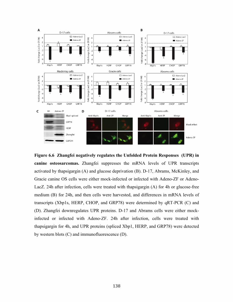
138
Figure 6.6 Zhangfei negatively regulates the Unfolded Protein Responses (UPR) in
canine osteosarcomas. Zhangfei suppresses the mRNA levels of UPR transcripts
activated by thapsigargin (A) and glucose deprivation (B). D-17, Abrams, McKinley, and
Gracie canine OS cells were either mock-infected or infected with Adeno-ZF or Adeno-
LacZ. 24h after infection, cells were treated with thapsigargin (A) for 4h or glucose-free
medium (B) for 24h, and then cells were harvested, and differences in mRNA levels of
transcripts (Xbp1s, HERP, CHOP, and GRP78) were determined by qRT-PCR (C) and
(D). Zhangfei downregulates UPR proteins. D-17 and Abrams cells were either mock-
infected or infected with Adeno-ZF. 24h after infection, cells were treated with
thapsigargin for 4h, and UPR proteins (spliced Xbp1, HERP, and GRP78) were detected
by western blots (C) and immunofluorescence (D).

139
6.4 Discussion
Canine OS is an aggressive tumour that accounts for approximately 85% of primary bone
tumours in dog (Selvarajah and Kirpensteijn 2010). OS causes local skeletal destruction
resulting in osteoproductive and osteolytic lesions, and it is highly metastatic to the lungs.
Although there has been dramatic progress in the standard treatments of OS, including
amputation, chemotherapy, and palliative radiation, these therapies often fail, leading to
recurrence of the tumour and metastatic spread (Brodey and Abt 1976; Tomlin et al. 2000;
Walter et al. 2005; Fan et al. 2009). Over the years, combined therapies, such as
chemotherapy combined with immune modulators, have been practiced on dog
osteosarcoma (Dow et al. 2005), although with poor overall survival times.
In previous studies (Chapter 2), we found that the transcription factor CREBZF/Zhangfei
suppressed the growth of D-17 dog OS cells (Bergeron et al. 2013). Herein, we further
discovered that the growth suppressive effects of Zhangfei were applicable to three other
independently isolated canine OS cell lines as well.
The unfolded Protein Response (UPR) is an adaptive response induced by endoplasmic
reticulum (ER) stress, which alleviates ER stress by up-regulating the expression of ER-
resident chaperons (Kaufman et al. 2002), inducing ER-associated protein degradation
(ERAD), and down-regulating the synthesis of new proteins (Meusser et al. 2005; Lai et
al. 2007). If these mechanisms are not sufficient to alleviate ER stress, then an apoptosis
program is initiated to induce cell death. My results (Chapter 3) (Zhang et al. 2013)
suggest that Zhangfei is a potential regulator of the UPR, and it might accelerate UPR
feedback mechanisms by interacting with the UPR mediator-Xbp1 and targeting it for
proteasomal degradation. In the present study, the strong inhibitory effects of Zhangfei on
both pharmacological (thapsigargin) and physiological inducer (glucose deprivation) –
induced UPR was also observed in the four canine osteosarcoma cell lines we examined.
Zhangfei suppresses the UPR and cell growth by stabilizing the tumour suppressor
protein p53 (Chapter 4). All four canine OS cell lines we examined express functional
p53 (Figure 6.1). These results suggest that the induction of Zhangfei expression in
canine OS may be an effective strategy for suppressing cell growth and metastasis.

140
However, the strategy would likely only be successful with OS that had functional p53. A
large proportion of human cancers have deleted or otherwise non-functional p53. At
present we do not know the proportion of canine OS that have inactive p53. Some studies
(Mendoza et al. 1998) suggest that most canine OS do not have deletions or major
rearrangements in the gene for p53. Although 30-50% of p53 coding sequences in canine
OS have polymorphisms (van Leeuwen et al. 1997; Johnson et al. 1998; Mendoza et al.
1998) the effect of these changes on p53 functionality is unknown. It is therefore difficult
to determine how universally applicable Zhangfei would be as a modality for treating
canine OS. The role of p53 in canine OS is also controversial. The expression of ectopic
p53 in canine OS cells, both in vitro and in vivo models leads to reduced tumour growth
and an increase in apoptotic cells (Yazawa et al. 2003; Kanaya et al. 2011; Bergeron et al.
2013). In contrast, other studies in both humans (Fu et al. 2013) and dogs (Sagartz et al.
1996; Loukopoulos et al. 2003) suggest that increased p53 expression in OS correlates
with more aggressive tumours and decreases survival time.
141
7. General discussion and conclusions
Zhangfei is a cellular transcriptional repressor identified in our laboratory through its
interaction with a cellular co-factor (HCF1) that is required for initiating Herpes Simplex
Virus-1 gene expression during infection (Lu and Misra 2000b). In the previous studies,
we found that Zhangfei was implicated in the differentiation and death of human
medulloblastoma cells (Bergeron et al. 2013). The main goal of this study was to further
determine the effect of transcription factor Zhangfei on the growth of osteosarcoma cells
and the molecular mechanisms responsible. Given the important role of the UPR in
maintaining malignancy in cancer as well as the suppressive ability of Zhangfei on many
other cellular regulators (Lu and Misra 2000b; Misra et al. 2005; Xie et al. 2008; Misra et
al. 2012), we developed techniques to monitor the UPR in dog and human osteosarcoma
cells. We tested the hypothesis that the ectopic expression of Zhangfei in these cells
would suppress their ability to activate the UPR and, consequently, inhibit cell growth.
As described in Chapter 2, we revealed that while Zhangfei inhibited both the UPR
pathways and the growth of D-17 dog cells, it had no effect on the UPR and relatively
modest, but significant, effect on the growth of Saos-2 human cells. Although these data
suggested there might not be a direct link between the UPR suppression and cell growth
arrest induced by Zhangfei, it was foundational to the investigation presented in this
thesis since it demonstrated the selectively suppressive effects of Zhangfei on both cell
growth and the UPR in OS cell lines.
The UPR pathways are mediated by three bLZip-containing transcription factors—Xbp1,
ATF4 and ATF6. The similarity of the bLZip domains of these UPR mediators to
Zhangfei, suggested the possibility that Zhangfei might associate with them influencing
their ability to mediate the UPR. Thus, we hypothesized that Zhangfei may interact with
Xbp1, ATF6 and ATF4 by its leucine zipper and prevent these transcription factors from
activating UPR genes. As described in Chapter 3, we showed that Zhangfei had a
suppressive effect on most UPR genes activated in response to the drug thapsigargin, and
this effect was mediated, at least partially, by the leucine zipper dependent interaction of
Zhangfei and Xbp1s, which resulted in the subsequent proteasomal degradation of Xbp1s
(Zhang et al. 2013). It should be emphasized that while we showed the direct effect of

142
Zhangfei on Xbp1, Zhangfei may suppress the other UPR-inducing bLZip factors as well.
We have previously shown that Zhangfei can suppress the activity of ER-resident bLZip
protein Luman/CREB3 but not ATF6 (Lu and Misra 2000b; Misra et al. 2005). Recently,
Misra and others (Misra et al. 2012) also showed that Zhangfei/SMILE suppresses the
ability of CREBH to induce UPR genes in hepatoma cells. While the findings in Chapter
3 and those of others showed that Zhangfei had the ability to suppress the UPR pathways
by interacting with Xbp1 as well as other bLZip proteins, it was still unclear why the
effects of Zhangfei were not universal—Zhangfei had no obvious effects on
untransformed cells and some cancer cell lines (as showed in Chapter 2). Thus, more
detailed understandings about the role of Zhangfei in regulation of both the UPR and cell
growth are clearly needed.
Neoplastic transformation occurs when gene mutations in a single cell make it unable to
repair DNA damage and trigger programmed cell death. The most common examples of
this are the inactivation of tumour suppressor genes and the up-regulation of oncogenes,
leading to uncontrolled proliferation of the cells with damaged genetic information.
Among the many genetic lesions in cancer, the inactivation of p53 function is the most
universal event and more than 50% of human cancers contain p53 mutation that alter or
abrogate its tumour suppressor functions (Lane et al. 2010). The high frequency of p53
inactivation in cancer points to its importance in preventing oncogenesis and makes this
gene a good target for the development of new cancer therapies. Gene therapy using
wild-type p53 has been demonstrated to restore p53 functions and lead to tumour
regression in several animal models (Martins et al. 2006; Ventura et al. 2007; Xue et al.
2007). With the growing understanding of the structure and regulation of p53, several
gene therapeutic strategies have been employed in the attempt to activate p53 in cancers
that still retain a functional protein. For example, treatment of colorectal adenocarcinoma
cells that have functional p53 with siRNA against mdm2 inhibited the growth of the cells
and rendered them more sensitive to cisplatin (Yu et al. 2006). Crystal structure analysis
showed that nutlin, an antagonist of the mdm2-p53 interaction, binds to mdm2 and
prevents its interaction with p53. This allows the activation of p53 pathways inducing cell
cycle arrest and apoptosis in mice xenograft tumours (Vassilev et al. 2004).

143
Based on the observations that Zhangfei has profound inhibitory effects on cell growth
and the UPR in some cancer cells, but not in normal cells and other tumour cells, I
hypothesized that Zhangfei acts through an intermediary that is either not activated or is
defective in cells that it does not affect. I also hypothesized that this intermediary was
p53. The investigation described in Chapter 4 demonstrated that Zhangfei enhanced the
activation of wild-type p53 by interacting with p53 and displacing its negative regulator
mdm2 from association with p53, thereby leading to inhibition of the UPR pathways and
growth arrest of osteosarcoma cells (Zhang and Misra 2014). Based on the observations
that Zhangfei only suppressed the growth and the UPR in wild-type p53-expressing, but
not in p53-null human osteosarcoma cells (in Chapter 4), we speculated that the
differential influences on the UPR and growth of Saos-2 cells, described in Chapter 2,
could be because these cells possess mutated p53 (Smardova et al. 2005), which only
partially inhibited its functions. Moreover, these findings are important, not only because
they provided a reasonable explanation for the “selective suppression” phenomenon of
Zhangfei, but that they demonstrated a novel mechanism to reactivate wild-type p53 in
cancer cells and provided a “bigger picture” on the interaction between p53, the UPR and
Zhangfei in the regulation of cancer cell growth.
As discussed in Chapter 4, the induction of the UPR is a protective mechanism utilized
by neoplastic cells to adapt to ER stress and to promote immune resistance, cancer
progression, and drug resistance (Moenner et al. 2007; Ron and Walter 2007). Therefore,
in addition to the cell cycle arrest and apoptosis directly induced by the activated p53, the
inhibition of the UPR, mediated by p53, represents another potential strategy by which
Zhangfei could be used to inhibit cancer. Further, the fact that ER stress, and the resulting
activation of the UPR induces p53 cytoplasmic localization and prevents p53-dependent
apoptosis (Qu et al. 2004) suggests the existence of an ER stress-p53-UPR regulatory
loop (Fig 4.8, Chapter 4). However, I was not able to define the specific mechanisms by
which p53 conversely inhibits the UPR pathways. p53 and mdm2 form an autoregulatory
negative feedback loop aimed at controlling levels of nuclear p53 (Moll and Petrenko
2003). Mdm2 is an E3 ubiquitin ligase that recongnizes its target protein and mediates its
ubiquitylation and degradation. Mdm2 is principal cellular antagonist of p53 and
promotes p53 degradation through an ubiquitin-dependent pathway on 26S proteasomes

144
(Honda et al. 1997); p53, in turn, stimulates the expression of mdm2, and thus operates in
a negative feedback loop to prevent the overactivation of p53 (Wu et al. 1993). However,
under the stress conditions, mdm2 induced by the activated p53 accumulates in cells but
is not able to interact with p53 due to its phosphorylation. Given the previous
observations that Zhangfei stabilizes p53 by displacing mdm2 (in Chapter 4), as well as
Zhangfei suppresses UPR mediator Xbp1 by targeting it for proteasomal degradation (in
Chapter 3), we speculate that Zhangfei and p53 may mediate the inhibition of UPR by
stimulating and recruiting mdm2 that subsequently recognizes the UPR-related proteins
and target them for degradation. To test this hypothesis further investigations are needed.
Chapter 5 revealed that the N-terminal transactivation domain (NTD) of p53 was
responsible for the complex formation with Zhangfei. This work was significant because
it further confirmed that the Zhangfei-binding domain of p53 was closely associated with
its mdm2-binding domain and transcription activation region. Together with the
observations in Chapter 4, therefore, reactivation of p53 by Zhangfei-mediated
displacement of mdm2 from the p53-mdm2 complex may represent a novel mechanism
for cancer gene therapy.
Taken together, the general conclusions of this thesis are:
1. Zhangfei profoundly inhibits the UPR and cell growth in some tumour cells, but not
in normal cells or in some other tumour cells.
2. Zhangfei is a negative regulator of the UPR mediator Xbp1s.
3. Zhangfei is able to activate p53 signaling; consequently, suppresses the UPR
pathways and induces cell growth arrest only in the osteosarcoma cells with
functional p53.
The specific conclusions of each chapter are:
Chapter 2: Effect of Zhangfei on Canine/Human Osteosarcoma Cells
• Zhangfei dramatically suppresses the growth of canine D-17 osteosarcoma cells.
While the effect of Zhangfei on Saos-2 human cells is not as dramatic as on D-17

145
cells, the growth rate of these cells is significantly suppressed compared with the
negative controls.
• Zhangfei induces macropinocytosis and apoptosis in D-17 cells.
• Zhangfei inhibits thapsigargin-induced UPR only in canine D-17 cells, but not in
human Saso-2 cells, suggesting that the ability of Zhangfei to suppress the UPR and
tumour cells growth may not be linked.
Chapter 3: Zhangfei is a Negative Regulator of the UPR Mediator Xbp1s
• Zhangfei has a suppressive effect on most UPR genes activated by the calcium
ionophore thapsigargin.
• Zhangfei suppresses the UPR, at least partially, due to the interaction of Zhangfei
with Xbp1s, which leads to the subsequent proteasomal degradation of Xbp1s. The
bLZip domain of Zhangfei is required for this interaction.
• Zhangfei suppresses the ability of Xbp1s to activate transcription from a promoter
containing unfolded protein response elements and significantly reduces the ability of
Xbp1s to activate the UPR.
Chapter 4: Effects of Zhangfei on the UPR and Cell Growth are Exerted Through the
Tumour Suppressor p53
• Zhangfei is a positive regulator of p53. In cells ectopically expressing Zhangfei, the
protein stabilizes p53 and promotes its nuclear retention.
• Suppression of p53 by siRNA partially restores cell growth and the UPR that are
inhibited by Zhangfei, indicating that the inhibitory influences of Zhangfei, at least in
part, are mediated by p53.
• Zhangfei interacts with p53 and displaces the E3 ubiquitin ligase mdm2 from its
association with p53, thereby preventing mdm2-mediated nuclear export and
subsequent proteasomal degradation of p53.
• The bLZip domain of Zhangfei is required for its profound effects on cell growth, the
UPR and interaction with p53.
Chapter 5: Characteristics of the Interaction of p53 and Zhangfei

146
• p53 forms a complex with Zhangfei via its N-terminal transactivation domain (NTD),
which is also the mdm2-binding domain and transcription activation region of p53.
• NTD is required for Zhangfei-mediated nuclear retention and transcriptional
activation of p53.
Chapter 6: The Effect of Zhangfei/CREBZF on Cell Growth, Differentiation, Apoptosis,
Migration, and the UPR in Several Canine Osteosarcoma Cell Lines
• Zhangfei suppresses the growth and UPR-related transcripts in several dog OS cell
lines that express p53 proteins capable of activating promoters with p53 response
elements, both on their own and synergistically with Zhangfei.
• Zhangfei induces the activation of osteocalcin expression, a marker of osteoblast
differentiation and triggers programmed cell death in these dog OS cell lines.

147
8. Reference Adham SA, Coomber BL. 2009. Glucose is a key regulator of VEGFR2/KDR in human
epithelial ovarian carcinoma cells. Biochemical and biophysical research communications 390: 130-135.
Akhova O, Bainbridge M, Misra V. 2005. The neuronal host cell factor-binding protein Zhangfei inhibits herpes simplex virus replication. Journal of virology 79: 14708-14718.
Amaral JD, Castro RE, Steer CJ, Rodrigues CM. 2009. p53 and the regulation of hepatocyte apoptosis: implications for disease pathogenesis. Trends in molecular medicine 15: 531-541.
Amaral JD, Xavier JM, Steer CJ, Rodrigues CM. 2010. The role of p53 in apoptosis. Discovery medicine 9: 145-152.
Ambron RT, Walters ET. 1996. Priming events and retrograde injury signals. A new perspective on the cellular and molecular biology of nerve regeneration. Mol Neurobiol 13: 61-79.
Ando K, Heymann MF, Stresing V, Mori K, Redini F, Heymann D. 2013. Current therapeutic strategies and novel approaches in osteosarcoma. Cancers 5: 591-616.
Andreassen A, Oyjord T, Hovig E, Holm R, Florenes VA, Nesland JM, Myklebost O, Hoie J, Bruland OS, Borresen AL et al. 1993. p53 abnormalities in different subtypes of human sarcomas. Cancer research 53: 468-471.
Anelli T, Sitia R. 2008. Protein quality control in the early secretory pathway. The EMBO journal 27: 315-327.
Asada R, Kanemoto S, Kondo S, Saito A, Imaizumi K. 2011. The signalling from endoplasmic reticulum-resident bZIP transcription factors involved in diverse cellular physiology. J Biochem 149: 507-518.
Becker K, Marchenko ND, Maurice M, Moll UM. 2007. Hyperubiquitylation of wild-type p53 contributes to cytoplasmic sequestration in neuroblastoma. Cell Death Differ 14: 1350-1360.
Bergeron T, Zhang R, Elliot K, Rapin N, MacDonald V, Linn K, Simko E, Misra V. 2013. The effect of Zhangfei on the unfolded protein response and growth of cells derived from canine and human osteosarcomas. Veterinary and comparative oncology 11: 140-150.
Bernales S, Papa FR, Walter P. 2006. Intracellular signaling by the unfolded protein response. Annual review of cell and developmental biology 22: 487-508.
Bertolotti A, Zhang Y, Hendershot LM, Harding HP, Ron D. 2000. Dynamic interaction of BiP and ER stress transducers in the unfolded-protein response. Nature cell biology 2: 326-332.
Bodey B, Groger AM, Bodey B, Jr., Siegel SE, Kaiser HE. 1997. Immunohistochemical detection of p53 protein overexpression in primary human osteosarcomas. Anticancer research 17: 493-498.
Bodnarchuk TW, Napper S, Rapin N, Misra V. 2012. Mechanism for the induction of cell death in ONS-76 medulloblastoma cells by Zhangfei/CREB-ZF. J Neurooncol 109: 485-501.
Brodey RS, Abt DA. 1976. Results of surgical treatment in 65 dogs with osteosarcoma. Journal of the American Veterinary Medical Association 168: 1032-1035.

148
Bustin SA, Benes V, Garson JA, Hellemans J, Huggett J, Kubista M, Mueller R, Nolan T, Pfaffl MW, Shipley GL et al. 2009. The MIQE guidelines: minimum information for publication of quantitative real-time PCR experiments. Clin Chem 55: 611-622.
Calfon M, Zeng H, Urano F, Till JH, Hubbard SR, Harding HP, Clark SG, Ron D. 2002. IRE1 couples endoplasmic reticulum load to secretory capacity by processing the XBP-1 mRNA. Nature 415: 92-96.
Cardin E, Latreille M, Khoury C, Greenwood MT, Larose L. 2007. Nck-1 selectively modulates eIF2alphaSer51 phosphorylation by a subset of eIF2alpha-kinases. FEBS J 274: 5865-5875.
Carter S, Bischof O, Dejean A, Vousden KH. 2007. C-terminal modifications regulate MDM2 dissociation and nuclear export of p53. Nature cell biology 9: 428-435.
Chan CP, Kok KH, Jin DY. 2011. CREB3 subfamily transcription factors are not created equal: Recent insights from global analyses and animal models. Cell Biosci 1: 6.
Ciovacco WA, Goldberg CG, Taylor AF, Lemieux JM, Horowitz MC, Donahue HJ, Kacena MA. 2009. The role of gap junctions in megakaryocyte-mediated osteoblast proliferation and differentiation. Bone 44: 80-86.
Cockram GP, Hogan MR, Burnett HF, Lu R. 2006. Identification and characterization of the DNA-binding properties of a Zhangfei homologue in Japanese pufferfish, Takifugu rubripes. Biochemical and biophysical research communications 339: 1238-1245.
Davenport EL, Morgan GJ, Davies FE. 2008. Untangling the unfolded protein response. Cell cycle 7: 865-869.
Dewhirst MW, Cao Y, Moeller B. 2008. Cycling hypoxia and free radicals regulate angiogenesis and radiotherapy response. Nature reviews Cancer 8: 425-437.
Diehl JA, Fuchs SY, Koumenis C. 2011. The cell biology of the unfolded protein response. Gastroenterology 141: 38-41, 41 e31-32.
Dioufa N, Chatzistamou I, Farmaki E, Papavassiliou AG, Kiaris H. 2012. p53 antagonizes the unfolded protein response and inhibits ground glass hepatocyte development during endoplasmic reticulum stress. Exp Biol Med (Maywood) 237: 1173-1180.
Doherty GJ, McMahon HT. 2009. Mechanisms of endocytosis. Annual review of biochemistry 78: 857-902.
Dong D, Ni M, Li J, Xiong S, Ye W, Virrey JJ, Mao C, Ye R, Wang M, Pen L et al. 2008. Critical role of the stress chaperone GRP78/BiP in tumor proliferation, survival, and tumor angiogenesis in transgene-induced mammary tumor development. Cancer research 68: 498-505.
Dow S, Elmslie R, Kurzman I, MacEwen G, Pericle F, Liggitt D. 2005. Phase I study of liposome-DNA complexes encoding the interleukin-2 gene in dogs with osteosarcoma lung metastases. Human gene therapy 16: 937-946.
Ellenberger TE, Brandl CJ, Struhl K, Harrison SC. 1992. The GCN4 basic region leucine zipper binds DNA as a dimer of uninterrupted alpha helices: crystal structure of the protein-DNA complex. Cell 71: 1223-1237.
Fan TM, Charney SC, de Lorimier LP, Garrett LD, Griffon DJ, Gordon-Evans WJ, Wypij JM. 2009. Double-blind placebo-controlled trial of adjuvant pamidronate with palliative radiotherapy and intravenous doxorubicin for canine appendicular
149
osteosarcoma bone pain. Journal of veterinary internal medicine / American College of Veterinary Internal Medicine 23: 152-160.
Feldman DE, Chauhan V, Koong AC. 2005. The unfolded protein response: a novel component of the hypoxic stress response in tumors. Molecular cancer research : MCR 3: 597-605.
Fu HL, Shao L, Wang Q, Jia T, Li M, Yang DP. 2013. A systematic review of p53 as a biomarker of survival in patients with osteosarcoma. Tumour biology : the journal of the International Society for Oncodevelopmental Biology and Medicine 34: 3817-3821.
Fujimoto M, Hayashi T. 2011. New insights into the role of mitochondria-associated endoplasmic reticulum membrane. Int Rev Cell Mol Biol 292: 73-117.
Giebler HA, Lemasson I, Nyborg JK. 2000. p53 recruitment of CREB binding protein mediated through phosphorylated CREB: a novel pathway of tumor suppressor regulation. Molecular and cellular biology 20: 4849-4858.
Gill J, Ahluwalia MK, Geller D, Gorlick R. 2013. New targets and approaches in osteosarcoma. Pharmacology & therapeutics 137: 89-99.
Goto A, Kanda H, Ishikawa Y, Matsumoto S, Kawaguchi N, Machinami R, Kato Y, Kitagawa T. 1998. Association of loss of heterozygosity at the p53 locus with chemoresistance in osteosarcomas. Japanese journal of cancer research : Gann 89: 539-547.
Harding HP, Zhang Y, Ron D. 1999. Protein translation and folding are coupled by an endoplasmic-reticulum-resident kinase. Nature 397: 271-274.
Haydon RC, Luu HH, He TC. 2007. Osteosarcoma and osteoblastic differentiation: a new perspective on oncogenesis. Clinical orthopaedics and related research 454: 237-246.
Haze K, Yoshida H, Yanagi H, Yura T, Mori K. 1999. Mammalian transcription factor ATF6 is synthesized as a transmembrane protein and activated by proteolysis in response to endoplasmic reticulum stress. Molecular biology of the cell 10: 3787-3799.
Hogan MR, Cockram GP, Lu R. 2006. Cooperative interaction of Zhangfei and ATF4 in transactivation of the cyclic AMP response element. FEBS letters 580: 58-62.
Honda R, Tanaka H, Yasuda H. 1997. Oncoprotein MDM2 is a ubiquitin ligase E3 for tumor suppressor p53. FEBS letters 420: 25-27.
Jiang M, Milner J. 2002. Selective silencing of viral gene expression in HPV-positive human cervical carcinoma cells treated with siRNA, a primer of RNA interference. Oncogene 21: 6041-6048.
Joerger AC, Fersht AR. 2008. Structural biology of the tumor suppressor p53. Annual review of biochemistry 77: 557-582.
Johnson AS, Couto CG, Weghorst CM. 1998. Mutation of the p53 tumor suppressor gene in spontaneously occurring osteosarcomas of the dog. Carcinogenesis 19: 213-217.
Johnson ES. 2004. Protein modification by SUMO. Annual review of biochemistry 73: 355-382.
Kanaya N, Yazawa M, Goto-Koshino Y, Mochizuki M, Nishimura R, Ohno K, Sasaki N, Tsujimoto H. 2011. Anti-tumor effect of adenoviral vector-mediated p53 gene transfer on the growth of canine osteosarcoma xenografts in nude mice. The

150
Journal of veterinary medical science / the Japanese Society of Veterinary Science 73: 877-883.
Kaufman RJ, Scheuner D, Schroder M, Shen X, Lee K, Liu CY, Arnold SM. 2002. The unfolded protein response in nutrient sensing and differentiation. Nature reviews Molecular cell biology 3: 411-421.
Khanna C, London C, Vail D, Mazcko C, Hirschfeld S. 2009. Guiding the optimal translation of new cancer treatments from canine to human cancer patients. Clinical cancer research : an official journal of the American Association for Cancer Research 15: 5671-5677.
Kim I, Xu W, Reed JC. 2008. Cell death and endoplasmic reticulum stress: disease relevance and therapeutic opportunities. Nature reviews Drug discovery 7: 1013-1030.
Klopfleisch R, Schutze M, Linzmann H, Brunnberg L, Gruber AD. 2010. Increased Derlin-1 expression in metastases of canine mammary adenocarcinomas. J Comp Pathol 142: 79-83.
Kobet E, Zeng X, Zhu Y, Keller D, Lu H. 2000. MDM2 inhibits p300-mediated p53 acetylation and activation by forming a ternary complex with the two proteins. Proceedings of the National Academy of Sciences of the United States of America 97: 12547-12552.
Kulikov R, Letienne J, Kaur M, Grossman SR, Arts J, Blattner C. 2010. Mdm2 facilitates the association of p53 with the proteasome. Proceedings of the National Academy of Sciences of the United States of America 107: 10038-10043.
Lai E, Teodoro T, Volchuk A. 2007. Endoplasmic reticulum stress: signaling the unfolded protein response. Physiology 22: 193-201.
Lain S, Midgley C, Sparks A, Lane EB, Lane DP. 1999. An inhibitor of nuclear export activates the p53 response and induces the localization of HDM2 and p53 to U1A-positive nuclear bodies associated with the PODs. Experimental cell research 248: 457-472.
Lane DP, Cheok CF, Lain S. 2010. p53-based cancer therapy. Cold Spring Harbor perspectives in biology 2: a001222.
Latreille M, Larose L. 2006. Nck in a complex containing the catalytic subunit of protein phosphatase 1 regulates eukaryotic initiation factor 2alpha signaling and cell survival to endoplasmic reticulum stress. The Journal of biological chemistry 281: 26633-26644.
Lee JH, Lee GT, Kwon SJ, Jeong J, Ha YS, Kim WJ, Kim IY. 2012a. CREBZF, a novel Smad8-binding protein. Molecular and cellular biochemistry.
Lee KM, Seo YJ, Kim MK, Seo HA, Jeong JY, Choi HS, Lee IK, Park KG. 2012b. Mediation of glucolipotoxicity in INS-1 rat insulinoma cells by small heterodimer partner interacting leucine zipper protein (SMILE). Biochemical and biophysical research communications 419: 768-773.
Lee SG, Rho HM. 2000. Transcriptional repression of the human p53 gene by hepatitis B viral X protein. Oncogene 19: 468-471.
Levesque AA, Eastman A. 2007. p53-based cancer therapies: Is defective p53 the Achilles heel of the tumor? Carcinogenesis 28: 13-20.

151
Li C, Macdonald JI, Hryciw T, Meakin SO. 2010. Nerve growth factor activation of the TrkA receptor induces cell death, by macropinocytosis, in medulloblastoma Daoy cells. J Neurochem 112: 882-899.
Li X, Zhang K, Li Z. 2011. Unfolded protein response in cancer: the physician's perspective. Journal of hematology & oncology 4: 8.
Liu X, Liu D, Qian D, Dai J, An Y, Jiang S, Stanley B, Yang J, Wang B, Liu DX. 2012. Nucleophosmin (NPM1/B23) interacts with activating transcription factor 5 (ATF5) protein and promotes proteasome- and caspase-dependent ATF5 degradation in hepatocellular carcinoma cells. The Journal of biological chemistry 287: 19599-19609.
Lopez-Mateo I, Villaronga MA, Llanos S, Belandia B. 2012. The transcription factor CREBZF is a novel positive regulator of p53. Cell cycle 11: 3887-3895.
Loukopoulos P, Thornton JR, Robinson WF. 2003. Clinical and pathologic relevance of p53 index in canine osseous tumors. Veterinary pathology 40: 237-248.
Lu R, Misra V. 2000a. Potential role for Luman, the cellular homologue of herpes simplex virus VP16 (alpha gene trans-inducing factor), in herpesvirus latency. Journal of virology 74: 934-943.
-. 2000b. Zhangfei: a second cellular protein interacts with herpes simplex virus accessory factor HCF in a manner similar to Luman and VP16. Nucleic Acids Res 28: 2446-2454.
Ma Y, Hendershot LM. 2003. Delineation of a negative feedback regulatory loop that controls protein translation during endoplasmic reticulum stress. The Journal of biological chemistry 278: 34864-34873.
Malhotra JD, Kaufman RJ. 2007. The endoplasmic reticulum and the unfolded protein response. Seminars in cell & developmental biology 18: 716-731.
Marcato P, Shmulevitz M, Pan D, Stoltz D, Lee PW. 2007. Ras transformation mediates reovirus oncolysis by enhancing virus uncoating, particle infectivity, and apoptosis-dependent release. Mol Ther 15: 1522-1530.
Marcellus RC, Teodoro JG, Charbonneau R, Shore GC, Branton PE. 1996. Expression of p53 in Saos-2 osteosarcoma cells induces apoptosis which can be inhibited by Bcl-2 or the adenovirus E1B-55 kDa protein. Cell Growth Differ 7: 1643-1650.
Marchenko ND, Hanel W, Li D, Becker K, Reich N, Moll UM. 2012. Stress-mediated nuclear stabilization of p53 is regulated by ubiquitination and importin-alpha3 binding. Cell Death Differ 17: 255-267.
Marciniak SJ, Yun CY, Oyadomari S, Novoa I, Zhang Y, Jungreis R, Nagata K, Harding HP, Ron D. 2004. CHOP induces death by promoting protein synthesis and oxidation in the stressed endoplasmic reticulum. Genes & development 18: 3066-3077.
Marina N, Gebhardt M, Teot L, Gorlick R. 2004. Biology and therapeutic advances for pediatric osteosarcoma. The oncologist 9: 422-441.
Martins CP, Brown-Swigart L, Evan GI. 2006. Modeling the therapeutic efficacy of p53 restoration in tumors. Cell 127: 1323-1334.
Mendoza S, Konishi T, Dernell WS, Withrow SJ, Miller CW. 1998. Status of the p53, Rb and MDM2 genes in canine osteosarcoma. Anticancer research 18: 4449-4453.

152
Menendez S, Higgins M, Berkson RG, Edling C, Lane DP, Lain S. 2003. Nuclear export inhibitor leptomycin B induces the appearance of novel forms of human Mdm2 protein. British journal of cancer 88: 636-643.
Merksamer PI, Papa FR. 2010. The UPR and cell fate at a glance. J Cell Sci 123: 1003-1006.
Meusser B, Hirsch C, Jarosch E, Sommer T. 2005. ERAD: the long road to destruction. Nature cell biology 7: 766-772.
Miller M. 2009. The importance of being flexible: the case of basic region leucine zipper transcriptional regulators. Curr Protein Pept Sci 10: 244-269.
Misra J, Chanda D, Kim DK, Li T, Koo SH, Back SH, Chiang JY, Choi HS. 2012. Curcumin differentially regulates endoplasmic reticulum stress through transcriptional corepressor SMILE (small heterodimer partner-interacting leucine zipper protein)-mediated inhibition of CREBH (cAMP responsive element-binding protein H). The Journal of biological chemistry 286: 41972-41984.
Misra V, Rapin N, Akhova O, Bainbridge M, Korchinski P. 2005. Zhangfei is a potent and specific inhibitor of the host cell factor-binding transcription factor Luman. The Journal of biological chemistry 280: 15257-15266.
Moenner M, Pluquet O, Bouchecareilh M, Chevet E. 2007. Integrated endoplasmic reticulum stress responses in cancer. Cancer research 67: 10631-10634.
Moll UM, Petrenko O. 2003. The MDM2-p53 interaction. Molecular cancer research : MCR 1: 1001-1008.
Momand J, Zambetti GP, Olson DC, George D, Levine AJ. 1992. The mdm-2 oncogene product forms a complex with the p53 protein and inhibits p53-mediated transactivation. Cell 69: 1237-1245.
Mueller F, Fuchs B, Kaser-Hotz B. 2007. Comparative biology of human and canine osteosarcoma. Anticancer research 27: 155-164.
Mukai R, Ohshima T. 2011. Dual effects of HTLV-1 bZIP factor in suppression of interferon regulatory factor 1. Biochemical and biophysical research communications 409: 328-332.
Nara A, Aki T, Funakoshi T, Uemura K. 2010. Methamphetamine induces macropinocytosis in differentiated SH-SY5Y human neuroblastoma cells. Brain Res 1352: 1-10.
Natarajan B, Gaur R, Hemmingsson O, Kao G, Naredi P. 2013. Depletion of the ER chaperone ENPL-1 sensitizes C. elegans to the anticancer drug cisplatin. Worm 2: e24059.
Newman JR, Keating AE. 2003. Comprehensive identification of human bZIP interactions with coiled-coil arrays. Science 300: 2097-2101.
Ni M, Zhou H, Wey S, Baumeister P, Lee AS. 2009. Regulation of PERK signaling and leukemic cell survival by a novel cytosolic isoform of the UPR regulator GRP78/BiP. PLoS One 4: e6868.
Noser JA, Mael AA, Sakuma R, Ohmine S, Marcato P, Lee PW, Ikeda Y. 2007. The RAS/Raf1/MEK/ERK signaling pathway facilitates VSV-mediated oncolysis: implication for the defective interferon response in cancer cells. Mol Ther 15: 1531-1536.
Ohshima T, Mukai R, Nakahara N, Matsumoto J, Isono O, Kobayashi Y, Takahashi S, Shimotohno K. 2010. HTLV-1 basic leucine-zipper factor, HBZ, interacts with

153
MafB and suppresses transcription through a Maf recognition element. J Cell Biochem 111: 187-194.
Okoshi R, Kubo N, Nakashima K, Shimozato O, Nakagawara A, Ozaki T. 2011. CREB represses p53-dependent transactivation of MDM2 through the complex formation with p53 and contributes to p53-mediated apoptosis in response to glucose deprivation. Biochemical and biophysical research communications 406: 79-84.
Okoshi R, Ozaki T, Yamamoto H, Ando K, Koida N, Ono S, Koda T, Kamijo T, Nakagawara A, Kizaki H. 2008. Activation of AMP-activated protein kinase induces p53-dependent apoptotic cell death in response to energetic stress. The Journal of biological chemistry 283: 3979-3987.
Oren M, Rotter V. 2010. Mutant p53 gain-of-function in cancer. Cold Spring Harbor perspectives in biology 2: a001107.
Overmeyer JH, Kaul A, Johnson EE, Maltese WA. 2008. Active ras triggers death in glioblastoma cells through hyperstimulation of macropinocytosis. Molecular cancer research : MCR 6: 965-977.
Ozaki T, Nakagawara A. 2011. p53: the attractive tumor suppressor in the cancer research field. Journal of biomedicine & biotechnology 2011: 603925.
Paoloni M, Khanna C. 2008. Translation of new cancer treatments from pet dogs to humans. Nature reviews Cancer 8: 147-156.
Park J, Seo T, Hwang S, Lee D, Gwack Y, Choe J. 2000. The K-bZIP protein from Kaposi's sarcoma-associated herpesvirus interacts with p53 and represses its transcriptional activity. Journal of virology 74: 11977-11982.
Park YB, Park MJ, Kimura K, Shimizu K, Lee SH, Yokota J. 2002. Alterations in the INK4a/ARF locus and their effects on the growth of human osteosarcoma cell lines. Cancer Genet Cytogenet 133: 105-111.
Petitjean A, Mathe E, Kato S, Ishioka C, Tavtigian SV, Hainaut P, Olivier M. 2007. Impact of mutant p53 functional properties on TP53 mutation patterns and tumor phenotype: lessons from recent developments in the IARC TP53 database. Human mutation 28: 622-629.
Poon E, Harris AL, Ashcroft M. 2009. Targeting the hypoxia-inducible factor (HIF) pathway in cancer. Expert Rev Mol Med 11: e26.
Qu L, Huang S, Baltzis D, Rivas-Estilla AM, Pluquet O, Hatzoglou M, Koumenis C, Taya Y, Yoshimura A, Koromilas AE. 2004. Endoplasmic reticulum stress induces p53 cytoplasmic localization and prevents p53-dependent apoptosis by a pathway involving glycogen synthase kinase-3beta. Genes & development 18: 261-277.
Raggo C, Rapin N, Stirling J, Gobeil P, Smith-Windsor E, O'Hare P, Misra V. 2002. Luman, the cellular counterpart of herpes simplex virus VP16, is processed by regulated intramembrane proteolysis. Molecular and cellular biology 22: 5639-5649.
Rankin KS, Starkey M, Lunec J, Gerrand CH, Murphy S, Biswas S. 2012. Of dogs and men: comparative biology as a tool for the discovery of novel biomarkers and drug development targets in osteosarcoma. Pediatric blood & cancer 58: 327-333.
Romer L, Klein C, Dehner A, Kessler H, Buchner J. 2006. p53--a natural cancer killer: structural insights and therapeutic concepts. Angewandte Chemie 45: 6440-6460.

154
Romero-Ramirez L, Cao H, Regalado MP, Kambham N, Siemann D, Kim JJ, Le QT, Koong AC. 2009. X box-binding protein 1 regulates angiogenesis in human pancreatic adenocarcinomas. Transl Oncol 2: 31-38.
Ron D, Walter P. 2007. Signal integration in the endoplasmic reticulum unfolded protein response. Nature reviews Molecular cell biology 8: 519-529.
Roth J, Dobbelstein M, Freedman DA, Shenk T, Levine AJ. 1998. Nucleo-cytoplasmic shuttling of the hdm2 oncoprotein regulates the levels of the p53 protein via a pathway used by the human immunodeficiency virus rev protein. The EMBO journal 17: 554-564.
Roth JA, Nguyen D, Lawrence DD, Kemp BL, Carrasco CH, Ferson DZ, Hong WK, Komaki R, Lee JJ, Nesbitt JC et al. 1996. Retrovirus-mediated wild-type p53 gene transfer to tumors of patients with lung cancer. Nature medicine 2: 985-991.
Rutkowski DT, Kang SW, Goodman AG, Garrison JL, Taunton J, Katze MG, Kaufman RJ, Hegde RS. 2007. The role of p58IPK in protecting the stressed endoplasmic reticulum. Molecular biology of the cell 18: 3681-3691.
Sadowski I, Bell B, Broad P, Hollis M. 1992. GAL4 fusion vectors for expression in yeast or mammalian cells. . Gene 118 137-141.
Sagartz JE, Bodley WL, Gamblin RM, Couto CG, Tierney LA, Capen CC. 1996. p53 tumor suppressor protein overexpression in osteogenic tumors of dogs. Veterinary pathology 33: 213-221.
Sambrook J, Russell DW. 2001. Molecular Cloning, a laboratory manual. Cold Spring Harbor Laboratory Press, Cold Spring Harbor, NY.
Schroder M, Kaufman RJ. 2005. The mammalian unfolded protein response. Annual review of biochemistry 74: 739-789.
Selvarajah GT, Kirpensteijn J. 2010. Prognostic and predictive biomarkers of canine osteosarcoma. Veterinary journal 185: 28-35.
Sharp AN, Heazell AE, Crocker IP, Mor G. 2010. Placental apoptosis in health and disease. American journal of reproductive immunology 64: 159-169.
Sharpless NE, DePinho RA. 2002. p53: good cop/bad cop. Cell 110: 9-12. Shaulsky G, Goldfinger N, Ben-Ze'ev A, Rotter V. 1990. Nuclear accumulation of p53
protein is mediated by several nuclear localization signals and plays a role in tumorigenesis. Molecular and cellular biology 10: 6565-6577.
Sinn B, Schulze J, Schroeder G, Konschak R, Freyer D, Budach V, Tinhofer I. 2010. Pifithrin-alpha as a potential cytoprotective agent in radiotherapy: protection of normal tissue without decreasing therapeutic efficacy in glioma cells. Radiation research 174: 601-610.
Smakman N, van den Wollenberg DJ, Elias SG, Sasazuki T, Shirasawa S, Hoeben RC, Borel Rinkes IH, Kranenburg O. 2006. KRAS(D13) Promotes apoptosis of human colorectal tumor cells by ReovirusT3D and oxaliplatin but not by tumor necrosis factor-related apoptosis-inducing ligand. Cancer research 66: 5403-5408.
Smardova J, Pavlova S, Svitakova M, Grochova D, Ravcukova B. 2005. Analysis of p53 status in human cell lines using a functional assay in yeast: detection of new non-sense p53 mutation in codon 124. Oncology reports 14: 901-907.
St-Germain JR, Chen J, Li Q. 2008. Involvement of PML nuclear bodies in CBP degradation through the ubiquitin-proteasome pathway. Epigenetics 3: 342-349.

155
Tamura K, Shimizu K, Yamada M, Okamoto Y, Matsui Y, Park KC, Mabuchi E, Moriuchi S, Mogami H. 1989. Expression of major histocompatibility complex on human medulloblastoma cells with neuronal differentiation. Cancer research 49: 5380-5384.
Tang N, Song WX, Luo J, Haydon RC, He TC. 2008. Osteosarcoma development and stem cell differentiation. Clinical orthopaedics and related research 466: 2114-2130.
Teufel DP, Freund SM, Bycroft M, Fersht AR. 2007. Four domains of p300 each bind tightly to a sequence spanning both transactivation subdomains of p53. Proceedings of the National Academy of Sciences of the United States of America 104: 7009-7014.
Tomlin JL, Sturgeon C, Pead MJ, Muir P. 2000. Use of the bisphosphonate drug alendronate for palliative management of osteosarcoma in two dogs. The Veterinary record 147: 129-132.
Uccelletti D, Pascoli A, Farina F, Alberti A, Mancini P, Hirschberg CB, Palleschi C. 2008. APY-1, a novel Caenorhabditis elegans apyrase involved in unfolded protein response signalling and stress responses. Molecular biology of the cell 19: 1337-1345.
Valderrama X, Rapin N, Misra V. 2008. Zhangfei, a novel regulator of the human nerve growth factor receptor, trkA. J Neurovirol 14: 425-436.
Valderrama X, Rapin N, Verge VM, Misra V. 2009. Zhangfei induces the expression of the nerve growth factor receptor, trkA, in medulloblastoma cells and causes their differentiation or apoptosis. J Neurooncol 91: 7-17.
van Huizen R, Martindale JL, Gorospe M, Holbrook NJ. 2003. P58IPK, a novel endoplasmic reticulum stress-inducible protein and potential negative regulator of eIF2alpha signaling. The Journal of biological chemistry 278: 15558-15564.
van Leeuwen IS, Cornelisse CJ, Misdorp W, Goedegebuure SA, Kirpensteijn J, Rutteman GR. 1997. P53 gene mutations in osteosarcomas in the dog. Cancer letters 111: 173-178.
Varley JM. 2003. Germline TP53 mutations and Li-Fraumeni syndrome. Human mutation 21: 313-320.
Vassilev LT, Vu BT, Graves B, Carvajal D, Podlaski F, Filipovic Z, Kong N, Kammlott U, Lukacs C, Klein C et al. 2004. In vivo activation of the p53 pathway by small-molecule antagonists of MDM2. Science 303: 844-848.
Ventura A, Kirsch DG, McLaughlin ME, Tuveson DA, Grimm J, Lintault L, Newman J, Reczek EE, Weissleder R, Jacks T. 2007. Restoration of p53 function leads to tumour regression in vivo. Nature 445: 661-665.
Vinson C, Myakishev M, Acharya A, Mir AA, Moll JR, Bonovich M. 2002. Classification of human B-ZIP proteins based on dimerization properties. Molecular and cellular biology 22: 6321-6335.
Vinson CR, Sigler PB, McKnight SL. 1989. Scissors-grip model for DNA recognition by a family of leucine zipper proteins. Science 246: 911-916.
Vousden KH, Lane DP. 2007. p53 in health and disease. Nature reviews Molecular cell biology 8: 275-283.
Vousden KH, Lu X. 2002. Live or let die: the cell's response to p53. Nature reviews Cancer 2: 594-604.

156
Wagner M, Moore DD. 2011. Endoplasmic reticulum stress and glucose homeostasis. Curr Opin Clin Nutr Metab Care 14: 367-373.
Walter CU, Dernell WS, LaRue SM, Lana SE, Lafferty MH, LaDue TA, Withrow SJ. 2005. Curative-intent radiation therapy as a treatment modality for appendicular and axial osteosarcoma: a preliminary retrospective evaluation of 14 dogs with the disease. Veterinary and comparative oncology 3: 1-7.
Wang G, Barrett JW, Stanford M, Werden SJ, Johnston JB, Gao X, Sun M, Cheng JQ, McFadden G. 2006a. Infection of human cancer cells with myxoma virus requires Akt activation via interaction with a viral ankyrin-repeat host range factor. Proceedings of the National Academy of Sciences of the United States of America 103: 4640-4645.
Wang H, Mo P, Ren S, Yan C. 2010. Activating transcription factor 3 activates p53 by preventing E6-associated protein from binding to E6. The Journal of biological chemistry 285: 13201-13210.
Wang S, Konorev EA, Kotamraju S, Joseph J, Kalivendi S, Kalyanaraman B. 2004. Doxorubicin induces apoptosis in normal and tumor cells via distinctly different mechanisms. intermediacy of H(2)O(2)- and p53-dependent pathways. The Journal of biological chemistry 279: 25535-25543.
Wang XW, Forrester K, Yeh H, Feitelson MA, Gu JR, Harris CC. 1994. Hepatitis B virus X protein inhibits p53 sequence-specific DNA binding, transcriptional activity, and association with transcription factor ERCC3. Proceedings of the National Academy of Sciences of the United States of America 91: 2230-2234.
Wang XZ, Lawson B, Brewer JW, Zinszner H, Sanjay A, Mi LJ, Boorstein R, Kreibich G, Hendershot LM, Ron D. 1996. Signals from the stressed endoplasmic reticulum induce C/EBP-homologous protein (CHOP/GADD153). Molecular and cellular biology 16: 4273-4280.
Wang Y, Shen J, Arenzana N, Tirasophon W, Kaufman RJ, Prywes R. 2000. Activation of ATF6 and an ATF6 DNA binding site by the endoplasmic reticulum stress response. The Journal of biological chemistry 275: 27013-27020.
Wang Y, Wang J, Li G. 2006b. Leucine zipper-like domain is required for tumor suppressor ING2-mediated nucleotide excision repair and apoptosis. FEBS letters 580: 3787-3793.
Wardell SE, Boonyaratanakornkit V, Adelman JS, Aronheim A, Edwards DP. 2002. Jun dimerization protein 2 functions as a progesterone receptor N-terminal domain coactivator. Molecular and cellular biology 22: 5451-5466.
Wei J, Zaika E, Zaika A. 2012. p53 Family: Role of Protein Isoforms in Human Cancer. Journal of nucleic acids 2012: 687359.
Withrow SJ. 2003. Limb Sparing Trials and Canine Osteosarcoma. in Genes, Dogs and Cancer: 3rd Annual Canine Cancer Conference.
Withrow SJ, Powers BE, Straw RC, Wilkins RM. 1991. Comparative aspects of osteosarcoma. Dog versus man. Clinical orthopaedics and related research: 159-168.
Wolf D, Harris N, Rotter V. 1984. Reconstitution of p53 expression in a nonproducer Ab-MuLV-transformed cell line by transfection of a functional p53 gene. Cell 38: 119-126.

157
Wouters BG, Koritzinsky M. 2008. Hypoxia signalling through mTOR and the unfolded protein response in cancer. Nature reviews Cancer 8: 851-864.
Wu X, Bayle JH, Olson D, Levine AJ. 1993. The p53-mdm-2 autoregulatory feedback loop. Genes & development 7: 1126-1132.
Xie YB, Lee OH, Nedumaran B, Seong HA, Lee KM, Ha H, Lee IK, Yun Y, Choi HS. 2008. SMILE, a new orphan nuclear receptor SHP-interacting protein, regulates SHP-repressed estrogen receptor transactivation. Biochem J 416: 463-473.
Xie YB, Nedumaran B, Choi HS. 2009a. Molecular characterization of SMILE as a novel corepressor of nuclear receptors. Nucleic Acids Res 37: 4100-4115.
Xie YB, Park JH, Kim DK, Hwang JH, Oh S, Park SB, Shong M, Lee IK, Choi HS. 2009b. Transcriptional corepressor SMILE recruits SIRT1 to inhibit nuclear receptor estrogen receptor-related receptor gamma transactivation. The Journal of biological chemistry 284: 28762-28774.
Xue W, Zender L, Miething C, Dickins RA, Hernando E, Krizhanovsky V, Cordon-Cardo C, Lowe SW. 2007. Senescence and tumour clearance is triggered by p53 restoration in murine liver carcinomas. Nature 445: 656-660.
Yamamoto K, Sato T, Matsui T, Sato M, Okada T, Yoshida H, Harada A, Mori K. 2007. Transcriptional induction of mammalian ER quality control proteins is mediated by single or combined action of ATF6alpha and XBP1. Developmental cell 13: 365-376.
Yan C, Lu D, Hai T, Boyd DD. 2005. Activating transcription factor 3, a stress sensor, activates p53 by blocking its ubiquitination. The EMBO journal 24: 2425-2435.
Yan W, Frank CL, Korth MJ, Sopher BL, Novoa I, Ron D, Katze MG. 2002. Control of PERK eIF2alpha kinase activity by the endoplasmic reticulum stress-induced molecular chaperone P58IPK. Proceedings of the National Academy of Sciences of the United States of America 99: 15920-15925.
Yazawa M, Setoguchi A, Hong SH, Uyama R, Nakagawa T, Kanaya N, Nishimura R, Sasaki N, Masuda K, Ohno K et al. 2003. Effect of an adenoviral vector that expresses the canine p53 gene on cell growth of canine osteosarcoma and mammary adenocarcinoma cell lines. American journal of veterinary research 64: 880-888.
Yoshida H, Matsui T, Yamamoto A, Okada T, Mori K. 2001. XBP1 mRNA is induced by ATF6 and spliced by IRE1 in response to ER stress to produce a highly active transcription factor. Cell 107: 881-891.
Yoshida H, Oku M, Suzuki M, Mori K. 2006. pXBP1(U) encoded in XBP1 pre-mRNA negatively regulates unfolded protein response activator pXBP1(S) in mammalian ER stress response. J Cell Biol 172: 565-575.
Yoshida H, Uemura A, Mori K. 2009. pXBP1(U), a negative regulator of the unfolded protein response activator pXBP1(S), targets ATF6 but not ATF4 in proteasome-mediated degradation. Cell Struct Funct 34: 1-10.
Yu Y, Sun P, Sun LC, Liu GY, Chen GH, Shang LH, Wu HB, Hu J, Li Y, Mao YL et al. 2006. Downregulation of MDM2 expression by RNAi inhibits LoVo human colorectal adenocarcinoma cells growth and the treatment of LoVo cells with mdm2siRNA3 enhances the sensitivity to cisplatin. Biochemical and biophysical research communications 339: 71-78.
158
Zhang R, Misra V. 2014. Effects of cyclic AMP response element binding protein-Zhangfei (CREBZF) on the unfolded protein response and cell growth are exerted through the tumor suppressor p53. Cell cycle 13: 279-292.
Zhang R, Rapin N, Ying Z, Shklanka E, Bodnarchuk TW, Verge VMK, Misra V. 2013. Zhangfei/CREB-ZF - A Potential Regulator of the Unfolded protein Response. PLoS One 8: e77256.
Zhang Y, Jin Y, Williams TA, Burtenshaw SM, Martyn AC, Lu R. 2010. Amino acid deprivation induces CREBZF/Zhangfei expression via an AARE-like element in the promoter. Biochemical and biophysical research communications 391: 1352-1357.
Zhang Y, Lu H. 2009. Signaling to p53: ribosomal proteins find their way. Cancer cell 16: 369-377.
Zhang Y, Xiong Y. 2001. A p53 amino-terminal nuclear export signal inhibited by DNA damage-induced phosphorylation. Science 292: 1910-1915.
Zhao L, Ackerman SL. 2006. Endoplasmic reticulum stress in health and disease. Current opinion in cell biology 18: 444-452.
Zhou W, Brush MH, Choy MS, Shenolikar S. 2011. Association with endoplasmic reticulum promotes proteasomal degradation of GADD34 protein. The Journal of biological chemistry 286: 21687-21696.


![Constitutive Activation of Transcription Factor OsbZIP46 · Constitutive Activation of Transcription Factor OsbZIP46 Improves Drought Tolerance in Rice1[C][W][OA] Ning Tang, Hua Zhang,](https://img.pdfslide.us/doc/110x75/6063217b6dc5be5eac567d74/constitutive-activation-of-transcription-factor-constitutive-activation-of-transcription.jpg)