Embed Size (px)
Citation preview
HEMATOPOIESIS AND STEM CELLS
Zebrafish kidney stromal cell lines support multilineage hematopoiesisDavid L. Stachura,1 Jason R. Reyes,1 Petr Bartunek,2,3 Barry H. Paw,4 Leonard I. Zon,3 and David Traver1,5
1Division of Biological Sciences, Section of Cell and Developmental Biology, University of California at San Diego, La Jolla; 2Institute of Molecular Genetics,Prague, Czech Republic; 3Department of Medicine, Children’s Hospital Boston, MA; 4Division of Hematology, Department of Medicine, Brigham and Women’sHospital, Harvard Medical School, Boston, MA; and 5Department of Cellular and Molecular Medicine, University of California at San Diego, La Jolla
Studies of zebrafish hematopoiesis havebeen largely performed using mutagenesisapproaches and retrospective analysesbased upon gene expression patterns inwhole embryos. We previously developedtransplantation assays to test the repopula-tion potentials of candidate hematopoieticprogenitor cells. We have been impaired,however, in determining cellular differentia-tion potentials by a lack of short-term func-tional assays. To enable more precise analy-ses of hematopoietic progenitor cells, we
have created zebrafish kidney stromal (ZKS)cell lines. Culture of adult whole kidneymarrow with ZKS cells results in the mainte-nance and expansion of hematopoietic pre-cursor cells. Hematopoietic growth is depen-dent upon ZKS cells, and we show that ZKScells express many growth factors and li-gands previously demonstrated to be impor-tant in maintaining mammalian hematopoi-etic cells. In the absence of exogenousgrowth factors, ZKS cells maintain earlyhematopoietic precursors and support dif-
ferentiation of lymphoid and myeloid cells.With the addition of zebrafish erythropoi-etin, ZKS cells also support the differentia-tion of erythroid precursors. These condi-tions have enabled the ability to ascertainmore precisely the points at which hemato-poietic mutants are defective. The develop-ment of robust in vitro assays now providethe means to track defined, functional out-comes for prospectively isolated blood cellsubsets in the zebrafish. (Blood. 2009;114:279-289)
Introduction
Hematopoiesis has served as the paradigmatic tissue stem cellsystem since the pioneering studies of Till and McCulloch sug-gested the presence of rare, multipotent hematopoietic stem cells(HSCs) in murine bone marrow.1,2 The advent of bone marrowtransplantation (BMT) showed that these HSCs could sustaindonor-derived hematopoiesis for life in both mice and humans.1,3-5
The development of in vitro culture technologies subsequentlysuggested that HSCs commit to downstream myeloerythroid fatesthrough a stepwise progression of lineage-restricted progenitorcells.6-9 The lineage relationships suggested by the studies ofMetcalf and colleagues have recently been solidified through theprospective isolation of lineage-restricted progenitors at each of themajor branch points of the hematopoietic hierarchy.
The precision afforded by in vitro culture systems has led to theisolation of the common lymphoid progenitor (CLP)10 and com-mon myeloid progenitor (CMP)11 that, at the single cell levelgenerate B lymphocytes and T lymphocytes and the major myelo-erythroid fates, respectively. Similarly, oligopotent progenitors,including the megakaryocyte/erythrocyte-restricted progenitor(MEP)11 and granulocyte/monocyte-restricted progenitor (GMP),11
and monopotent precursors, such as the megakaryocyte-restrictedprecursor (MkP)12 and eosinophil-restricted precursor (EoP),13
have been demonstrated to arise downstream of these multipotentprogenitors by use of sensitive culture techniques. The long-termculture initiating cell (LTCIC) assay has served as a surrogate assayfor human HSCs in the absence of autologous transplantation.14
A variety of transformation culture assays have also been used tobetter understand the multiple steps to cancer.15-17
To enable similar approaches in the zebrafish, we have devel-oped an in vitro culture system. The zebrafish has proven an
excellent system in which to dissect the genetic bases of blood cellformation through forward mutagenesis approaches (reviewed inde Jong and Zon18). Experimental methodologies to functionallytest resulting mutants, however, have been lacking. We haverecently described the prospective isolation of the first hematopoi-etic stem and progenitor cells to arise in the zebrafish embryo.19,20
We have also developed a variety of hematopoietic cell transplanta-tion assays to provide the most stringent test of HSC function,long-term reconstitution.21,22 To complement these advances, wehave developed the means to maintain and grow hematopoieticcells in vitro through the creation of zebrafish kidney stromal(ZKS) cell lines.
We show that growth and survival of zebrafish hematopoieticcells is dependent upon coculture with ZKS cells, and that additionof recently identified zebrafish growth factors, such as zebrafisherythropoietin (zEpo), enables multilineage differentiation fromwhole kidney marrow (WKM) cells. In addition, we have analyzedgenetic mutants with erythroid defects, demonstrating that amutation in iron transport can be rescued via in vitro delivery ofmembrane-permeable iron. Together, these studies should enable abetter understanding of the biology of zebrafish hematopoietic stemand progenitor cells, and of the points along the hematopoietichierarchy at which blood mutants are defective.
Methods
Generation of ZKS cells
Kidneys were isolated from AB* WT fish, bleached for 5 minutes in0.000525% sodium hypochlorite (Fisher Scientific), then rinsed in sterile
Submitted February 4, 2009; accepted April 17, 2009. Prepublished online asBlood First Edition paper, May 11, 2009; DOI 10.1182/blood-2009-02-203638.
The online version of this article contains a data supplement.
The publication costs of this article were defrayed in part by page chargepayment. Therefore, and solely to indicate this fact, this article is herebymarked ‘‘advertisement’’ in accordance with 18 USC section 1734.
© 2009 by The American Society of Hematology
279BLOOD, 9 JULY 2009 ! VOLUME 114, NUMBER 2
phosphate-buffered saline (PBS; Mediatech). Tissue was mechanicallydissociated by trituration then filtered through a 40-!m filter (BD Bio-sciences). Flow-through was discarded, and the remaining kidney tissuewas cultured in 12.5-cm2 vented flasks (Corning Incorporated Life Sci-ences) at 32°C and 5% CO2. ZKS cells were maintained in culture mediaconsisting of 10% embryonic stem (ES) cell qualified fetal bovine serum(FBS; ATCC), 55% L-15, 32.5% Dulbecco modified Eagle medium(DMEM), and 12.5% Ham F-12 (Mediatech). Media was supplementedwith 150 mg/L sodium bicarbonate, 2% penicillin/streptomycin (10 U/mLstock), 1.5% N-2-hydroxyethylpiperazine-N"-2-ethanesulfonic acid(HEPES), 1% L-glutamine, and 0.1 mg/mL gentamycin (all from Media-tech). Cells were grown until 60%-80% confluence, then trypsinized(0.25%; Invitrogen) for 5 minutes and split 1:10 to passage. All experimentswere approved by the Institutional Animal Care and Use Committee(IACUC) at the University of California, San Diego (UCSD).
Morphologic characterization of ZKS cells
ZKS cells were grown on glass coverslips in a 24-well culture plate (BDBiosciences). After removal from culture, slides were fixed and stained withMay-Grunwald Giemsa (Sigma-Aldrich) and visualized with an OlympusBX51 microscope (Olympus). To visualize cells using transmission elec-tron microscopy, ZKS cells grown in 35-mm plastic culture dishes werefixed in 2.5% glutaraldehyde in 0.1 M Na cacodylate buffer (pH 7.3), bufferwashed, and fixed in 1% osmium tetroxide in 0.1 M Na cacodylate buffer.They were subsequently treated with 0.5% tannic acid, followed by 1%sodium sulfate in cacodylate buffer, and then dehydrated in graded ethanolseries. The cells were cleared in 2-hydroxypropyl methacrylate (HPMA;Ladd Research) and embedded in LX112 resin. After overnight polymeriza-tion at 60°C, small pieces of resin were attached to blank blocks usingSuperGlue. Thin sections (70 nm) were cut on a Reichert Ultracut E (Leica)using a diamond knife (Diatome; Electron Microscopy Sciences), mountedon parlodion-coated, copper slot grids, and stained in uranyl acetate and leadcitrate. Sections were examined on a Philips CM100 transmission electronmicroscope (TEM; FEI Company) and data-documented on a Megaview IIIcharge-coupled device (CCD) camera (Soft Imaging System Corp).
Zebrafish stocks and embryos
Zebrafish were mated, staged, and raised as described23 and maintained inaccordance with UCSD IACUC guidelines. AB* wild-type (WT) fish andtransgenic lines Tg(gata1:DsRed)21 and Tg(#actin:eGFP)21 were used.
Isolation of WKM
Zebrafish kidney was isolated under sterile conditions and mechanicallydissociated by trituration. Tissue was passaged through a 40-!m filter,rinsed in sterile PBS, then centrifuged for 10 minutes at 300g. Supernatantwas aspirated, and cells were resuspended in culture media. Cell countswere performed with the use of a bright line hemacytometer (HausserScientific), using trypan blue (Invitrogen) to assess viability.
Fluorescence-activated cell sorting
#actin:GFP WKM was isolated, resuspended in PBS with 0.9 $ FBS, andsorted on a FACSAria flow cytometer (BD Biosciences).
Cytology
Hematopoietic cells were concentrated by cytocentrifugation at 250g for5 minutes onto glass slides using a Shandon Cytospin 4 (Thermo FisherScientific). Slides were fixed and stained with May-Grunwald Giemsa(Sigma-Aldrich). Briefly, the slides were air-dried and stained in May-Grunwald solution for 10 minutes. Then, the slides were placed in a 1:5dilution of Giemsa in distilled water (dH2O) for 20 minutes, rinsed in dH2O,and allowed to dry before covering with a coverslip. For histochemicalanalysis of hemoglobin, o-dianisidine staining was performed. Cells wereconcentrated in 50 !L media, and 100 !L fresh o-dianisidine (Sigma-Aldrich)working solution (100 !L o-dianisidine solution [50 mg o-dianisidine in 35 mL100% ethanol (EtOH)], 25 !L NaOAc, pH 4.5, 5 !L 30% H2O2) was added, and
cells were incubated in the dark for 10 minutes before being cytospun. Slideswere then fixed/stained with May-Grunwald solution for 10 minutes, dried, andcovered with a coverslip.
Reverse transcription polymerase chain reaction analysis
For reverse transcription polymerase chain reaction (RT-PCR) analysis,RNA was isolated from hematopoietic cells using the RNeasy kit (QIAGEN),and cDNA was generated with a Superscript III RT-PCR kit (Invitrogen).Primers used for characterization of ZKS cells are listed in Table 1. Primersused in hematopoietic maintenance assays were pax5-forward: CTG ATTACA AAC GCC AAA AC, pax5-reverse: CTA AAT TAT GCG CAG AAACG; lck-forward: TAC GTA AAC ATG GGG AAC TG, lck-reverse: TCTTCT CCC CTT TCT CAA AC; igM-forward: AGC TTC TCT AGC TCCACC AG, igM-reverse: GCT AAA CAC ATG AAG GTT GC. Primers todetect zebrafish ef1%,19 gata3,20 cd45,20 rag2,24 mpx,25 pu.1,25 and gata125
have previously been described.
Proliferation, maintenance, and differentiation analyses
Before culture, isolated WKM was washed twice with 0.1% bovine serumalbumin (BSA) to remove FBS. Cells were suspended in 0.1% BSA, and2 !L/mL 5 mM carboxyfluorescein succinimidyl ester (CFSE; Invitrogen)were added while mixing. Cells were incubated in the dark for 10 minutes,and culture media, with an additional 10% FBS, was used to wash cellstwice. Cells were resuspended in complete ZKS media. WKM cultureswere grown at 32°C and 5% CO2 on 80%-90% confluent ZKS cells andharvested by gentle pipeting to obtain single-cell suspensions withoutdisruption of ZKS monolayer. Samples were resuspended in PBS andcounted. A fraction of the hematopoietic cells were then used for RT-PCRanalysis. Another fraction was cytospun and stained. Flow cytometryanalysis was conducted on a LSRII (BD Bioscience), using propidiumiodide (Sigma-Aldrich) to exclude dead cells. Data were analyzed usingFlowJo software (TreeStar).
Expression and purification of recombinant zEpo
The region corresponding to the mature form of zepo was reamplified byPCR from zepo cDNA26 using primers zepo-1: CGG GAT CCT CCC CATTAC GCC CCA TCT GTG, and zepo2: CGG GAT CCT CAG CTG ACACCC TGT CGA CA. The 499-bp PCR product was digested with BamHIrestriction endonuclease, isolated by gel electrophoresis, and ligated intothe prokaryotic expression vector pETH2a. This generated the expressionplasmid pHis-zEpo, which was used for transformation of T7 polymeraseexpressing BL21(DE3) cells. Induction of 19 kDa His-zEpo proteinsynthesis by isopropyl-#-D-thiogalactopyranoside (IPTG) and purificationof recombinant protein on a Ni2&-NTA agarose column was done accordingto the manufacturer’s procedure (QIAGEN) with some modifications aspublished before.27
Cytokine assay
WKM cultures were grown at 32°C and 5% CO2 with purified, recombinantzEpo, 1% carp serum, or 1:1000 ferric salicylaldehyde isonicotinoylhydrazone (Fe-SIH, catalog number I3153; Sigma-Aldrich), then harvestedby gentle pipeting to obtain single-cell suspensions without disruption ofZKS monolayer. Samples were resuspended in PBS for counting. A fractionof the hematopoietic cells were cytospun and stained with o-dianisidine andMay-Grunwald Giemsa. Another fraction was subjected to flow cytome-try analysis.
Results
ZKS cells are derived from adult zebrafish kidney stroma
Since the kidney is the main hematopoietic site in adult zebrafish,28
we generated stromal cell lines from dissociated kidney cells.Kidneys were removed from fish, gently bleached, and physically
280 STACHURA et al BLOOD, 9 JULY 2009 ! VOLUME 114, NUMBER 2
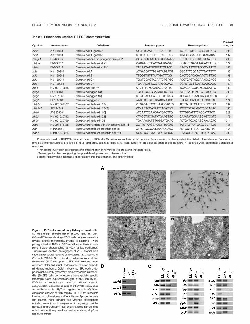
Figure 1. ZKS cells are primary kidney stromal cells.(A) Morphologic characterization of ZKS cells. (i,ii) May-Grunwald/Giemsa staining of ZKS cells on glass coverslipsreveals stromal morphology. Images in subpanel i werephotographed at 100$ at 100% confluence; those in sub-panel ii were photographed at 400$ at low confluence.Transmission electron micrographs of ZKS stromal cellsshow ultrastructural features of fibroblasts. (iii) Close-up ofZKS cell, 7900$. Note abundant mitochondria and freeribosomes. (iv) Close-up of a ZKS cell, 19 000$. Noteabundant Golgi and rough endoplasmic reticular network.n indicates nucleus; g, Golgi; r, ribosome; rER, rough endo-plasmic reticulum; ly, lysosome; f, filaments; and m, mitochon-dria. (B) ZKS cells do not express hematopoietic specifictranscripts. Gene expression analysis of ZKS cells by RT-PCR for the pan leukocytic transcript cd45 and erythroidspecific gata1. Gene names listed at left. Whole kidney usedas positive controls, dH2O as negative controls. (C) Geneexpression analysis of ZKS cells by RT-PCR for transcriptsinvolved in proliferation and differentiation of progenitor cells(left column), niche signaling and lymphoid development(middle column), and lineage-specific signaling, mainte-nance, and differentiation (right column). Gene names listedat left. Whole kidney used as positive controls, dH2O asnegative controls.
Table 1. Primer sets used for RT-PCR characterization
Cytokine Accesson no. Definition Forward primer Reverse primerProductsize, bp
zkitla AY929068 Danio rerio kit ligand a* GGATTCAATGCTTGACTTTG TGTACTATGTTGCGCTGATG 205
zkitlb AY929069 Danio rerio kit ligand b* CTTGATTGCCGTTCAGTTAG TGACCGGAGATTGTAGACAC 107
zbmp 1 DQ424857 Danio rerio bone morphogenic protein 1* GGATGGATATTGGAGGAAAG CTTTGTTCGGTCTGTAATCG 230
zil-1 la BN000717 Danio rerio interleukin-l la* GACAAGCTGAGCAATCAGAC GGAGCTGAGAAAGAGTAGGC 172
zil-1lb BN000718 Danio rerio interleukin-11b* TTGAACATTCGCTATCATCC GAGTAATCGTTCCCCAATTC 166
zdla NM 130954 Danio rerio 'A† ACGACGATTTGAGTATGACG GGGATTGGCACTTTATATCC 186
zdlb NM 130958 Danio rerio 'B† TTCCGTGTTTAATGATTTGG CACTCCACAGAAACTCTTGC 158
zdlc NM 130944 Danio rerio 'C† TGGTGGACTACAATCTGAGC ACCTCAGTAGCAAACACACG 169
zdld NM 130955 Danio rerio 'D† TGAAACATTACCAAGCCAAC GCAGTGCTTCAATAATCAGC 189
zdll4 NM 001079835 Danio rerio '-like 4† CTCTTTCAGCACACCAATTC TGAACATCCTGAGACCATTC 189
zjagla BC162468 Danio rerio jagged 1a† TGATTGGTGGATACTTCTGC AATCCATTGAGTGTGTCCTG 238
zjaglb NM 131863 Danio rerio jagged 1b† CTGTGAGCCATCTTCTTCAG AGCAAAGGAACCAGGTAGTC 213
zjag2 BC163889 Danio rerio jagged 2† AATGACTGTGTGAGCAATCC GTCATTGACCAGATCCACAC 174
zil-12a NM 001007107 Danio rerio interleukin 12a‡ GTGAGTCTGCTGAAGGAGTG AGTGACATCATTTCCTGTGC 167
zil-15–2 AB194243 Danio rerio interleukin 15–2‡ CCAAGTCCACAATTACATGC TCTTTGTAGAGCTCGCAGAC 166
zil-10 AY887900 Danio rerio interleukin 10‡ ATGAATCCAACGATGACTTG TCTTGCATTTCACCATATCC 222
zil-22 NM 001020792 Danio rerio interleukin 22‡ CTACCTGCGATATGAAGTGC GAAATATGGAAGCAGTCGTG 173
zil-26 NM 001020799 Danio rerio interleukin 26 TGAAAAGATGTGGGATGAAC ACTGATCCACAGCAAAACAC 214
zepo NM001 115128 Danio rerio erythropoietin transcript variant 1‡ ACTTGTAAGGACGATTGCAG TATCTGTAATGAGCCGATGG 156
zfgfl1 N M200760 Danio rerio fibroblast growth factor 1‡ ATACTGCGCATAAAAGCAAC AGTGGTTTTCCTCCATCTTC 154
zfgf2l N M001045324 Danio rerio fibroblast growth factor 21‡ CGGTGGTGTATGTATGTTCC GTAGCTGCACTCTGGATGAC 203
Primer sets used for RT-PCR characterization of ZKS cells. Gene names are listed at left, followed by accession number and definition listed in the database. Forward andreverse primer sequences are listed 5" to 3", and product size is listed at far right. Since not all products span exons, negative RT controls were performed alongside allreactions.
*Transcripts involved in proliferation and differentiation of hematopoietic stem and progenitor cells.†Transcripts involved in signaling, lymphoid development, and differentiation.‡Transcripts involved in lineage-specific signaling, maintenance, and differentiation.
ZEBRAFISH HEMATOPOIETIC CELL CULTURE 281BLOOD, 9 JULY 2009 ! VOLUME 114, NUMBER 2
dissociated by trituration. After trituration, the cells were passedthrough a 40-!M filter and rinsed thoroughly with media. Theflow-through contains WKM, which is comprised mainly ofhematopoietic cells.21 Non-flow-through tissue was washed fromthe filter and allowed to grow in gas vacuum plasma-treated tissueculture dishes until confluent. Upon reaching confluence severalweeks later, these cells were trypsinized and passaged for furtherculture (see “Methods”).
The optimum culture conditions for ZKS cells were determinedto be 32°C, in a humidified 5% CO2 environment. Cells dividedslower than mammalian stromal cells (once every 36-48 hours),probably due to their growth at cooler temperatures. While ZKScells appeared healthy at the physiologic temperature of zebrafish(28°C), they divided faster, and appeared similarly healthy at 32°C.Raising the temperature to 37°C for extended periods of time wastoxic to ZKS cells. Multiple lines have been maintained under theseconditions for more than 200 passages. None of the cell linesexhibited crisis at any point and were capable of repeated passagein culture. ZKS cells were density-dependent and grew optimallywhen maintained at 25%-50% confluence.
ZKS cells displayed a fibroblastic appearance when observedby May-Grunwald Giemsa staining (Figure 1Ai-ii). To confirm thatthese cells were stromal and not hematopoietic, ZKS cells wereanalyzed by TEM. ZKS cells displayed several ultrastructuralcharacteristics of teleost kidney fibroblasts.29 They were irregularlybranched, with large processes containing extensive filamentbundles (not shown), and they contained a heterochromatic nucleusand a prominent endomembrane system, including well-definedGolgi apparati, rough endoplasmic reticuli, and secretory vesicles(Figure 1Aiii-iv). We never observed cellular morphologies indica-tive of hematopoietic cell types in ZKS cultures. ZKS cells did notshow expression of the pan leukocyte marker cd45 or the red blood
cell gene gata1 by RT-PCR, confirming no hematopoietic cell typeswere present in these cultures (Figure 1B).
RT-PCR expression analyses of genes previously shown to playimportant roles in hematopoietic maintenance, development, orproliferation demonstrated that ZKS cells expressed many tran-scripts known to support hematopoietic progenitor cells (Figure 1Cleft column). In addition, ZKS cells expressed Notch ligands,known to play a role in HSC niche signaling, lymphocytedevelopment, and maturation (Figure 1C middle column). Finally,ZKS cells expressed many cytokines involved in lineage-specificsignaling, maintenance, and differentiation (Figure 1C right col-umn; see Table 1 for primer sequences).
ZKS cells support hematopoiesis
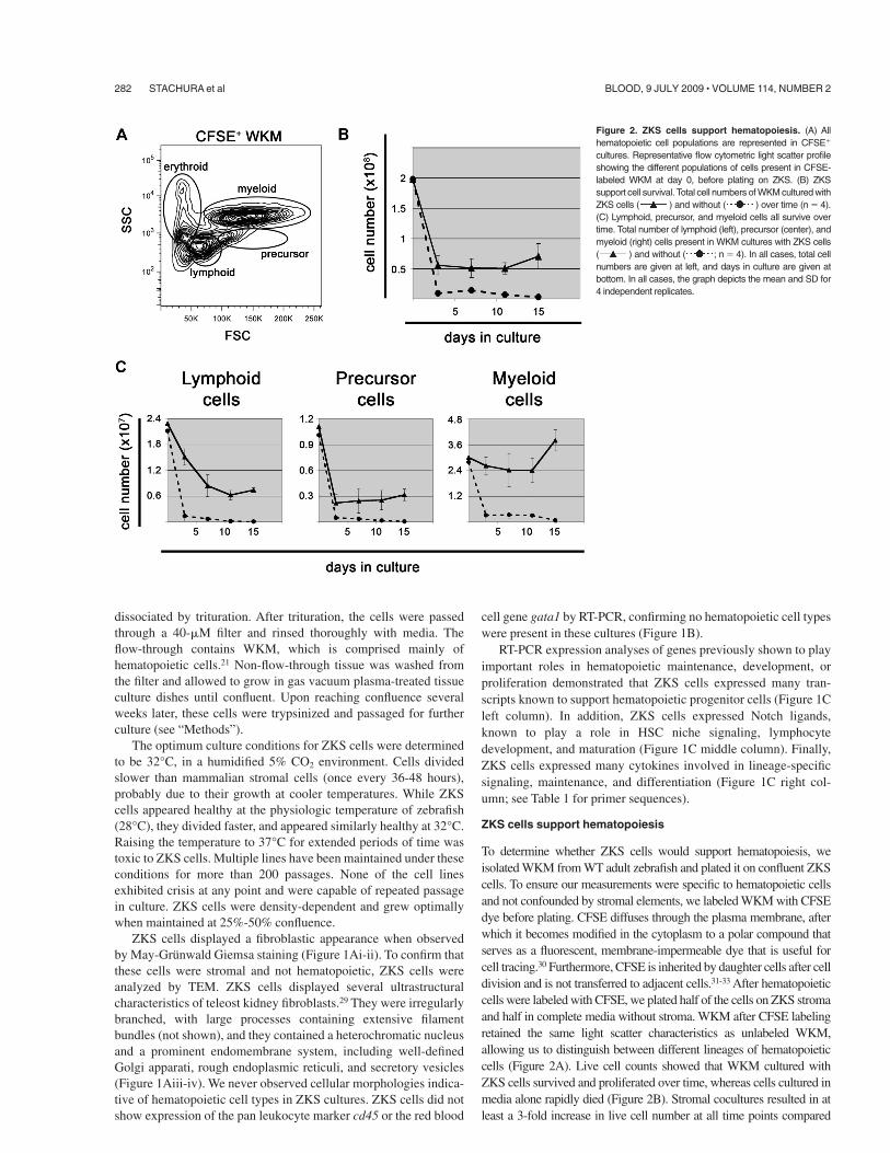
To determine whether ZKS cells would support hematopoiesis, weisolated WKM from WT adult zebrafish and plated it on confluent ZKScells. To ensure our measurements were specific to hematopoietic cellsand not confounded by stromal elements, we labeled WKM with CFSEdye before plating. CFSE diffuses through the plasma membrane, afterwhich it becomes modified in the cytoplasm to a polar compound thatserves as a fluorescent, membrane-impermeable dye that is useful forcell tracing.30 Furthermore, CFSE is inherited by daughter cells after celldivision and is not transferred to adjacent cells.31-33 After hematopoieticcells were labeled with CFSE, we plated half of the cells on ZKS stromaand half in complete media without stroma. WKM after CFSE labelingretained the same light scatter characteristics as unlabeled WKM,allowing us to distinguish between different lineages of hematopoieticcells (Figure 2A). Live cell counts showed that WKM cultured withZKS cells survived and proliferated over time, whereas cells cultured inmedia alone rapidly died (Figure 2B). Stromal cocultures resulted in atleast a 3-fold increase in live cell number at all time points compared
Figure 2. ZKS cells support hematopoiesis. (A) Allhematopoietic cell populations are represented in CFSE&
cultures. Representative flow cytometric light scatter profileshowing the different populations of cells present in CFSE-labeled WKM at day 0, before plating on ZKS. (B) ZKSsupport cell survival. Total cell numbers of WKM cultured withZKS cells ( ) and without ( ) over time (n ( 4).(C) Lymphoid, precursor, and myeloid cells all survive overtime. Total number of lymphoid (left), precursor (center), andmyeloid (right) cells present in WKM cultures with ZKS cells( ) and without ( ; n ( 4). In all cases, total cellnumbers are given at left, and days in culture are given atbottom. In all cases, the graph depicts the mean and SD for4 independent replicates.
282 STACHURA et al BLOOD, 9 JULY 2009 ! VOLUME 114, NUMBER 2
with cultures without stroma. Separate experiments confirmed thathematopoietic cultures survived on ZKS stroma over the course of 30days, while cells cultured without stroma senesced by 15 days (supple-mental Figure 1, available on the Blood website; see the SupplementalMaterials link at the top of the online article).
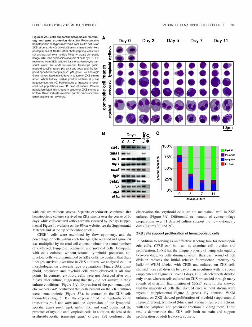
CFSE& cells were examined by flow cytometry, and thepercentage of cells within each lineage gate outlined in Figure 2Awas multiplied by the total cell counts to obtain the actual numbersof erythroid, lymphoid, precursor, and myeloid cells. Comparedwith cells cultured without stroma, lymphoid, precursor, andmyeloid cells were maintained by ZKS cells. To confirm that theselineages survived over time in ZKS cultures, we analyzed cellularmorphologies on cytocentrifuge preparations (Figure 3A). Lym-phoid, precursor, and myeloid cells were observed at all timepoints. In contrast, erythroid cells were not observed after only3 days after culture, suggesting that they did not survive in theseculture conditions (Figure 3A). Expression of the pan hematopoi-etic marker cd45 confirmed that cells present on the ZKS cultureswere hematopoietic (Figure 3B), in contrast to the ZKS cellsthemselves (Figure 1B). The expression of the myeloid-specifictranscripts pu.1 and mpx and the expression of the lymphoid-specific genes pax5, igM, gata3, lck, and rag2 confirmed thepresence of myeloid and lymphoid cells. In addition, the loss of theerythroid-specific transcript gata1 (Figure 3B) confirmed the
observation that erythroid cells are not maintained well in ZKScultures (Figure 3A). Differential cell counts of cytocentrifugepreparations over 11 days of culture support the flow cytometricdata (Figures 3C and 2C).
ZKS cells support proliferation of hematopoietic cells
In addition to serving as an effective labeling tool for hematopoi-etic cells, CFSE can be used to examine cell division andproliferation. CFSE has the unique property of being split equallybetween daughter cells during division, thus each round of celldivision reduces the initial relative fluorescence intensity byhalf.31-33 WKM labeled with CFSE and cultured on ZKS cellsshowed more cell division by day 3 than in cultures with no stroma(supplemental Figure 2). Over 11 days, CFSE-labeled cells dividedonly once, whereas cells cultured on ZKS proceeded through manyrounds of division. Examination of CFSE& cells further showedthat the majority of cells that divided once without stroma weremyeloid (supplemental Figure 2, green). By contrast, WKMcultured on ZKS showed proliferation of myeloid (supplementalFigure 2, green), lymphoid (blue), and precursor (purple) fractions,with the lymphoid and precursor fractions dividing more. Theseresults demonstrate that ZKS cells both maintain and supportproliferation of adult leukocyte subsets.
Figure 3. ZKS cells support hematopoiesis; morphol-ogy and gene expression data. (A) Representativehematopoietic cell types recovered from in vitro culture onZKS stroma. May-Grunwald/Giemsa stained cells werephotographed at 1000$. After photographing, cells werecut and pasted from multiple fields to create compositeimage. (B) Gene expression analysis of cells by RT-PCRrecovered from ZKS cultures for the panleukocytic tran-script cd45, the erythroid-specific transcript gata1,myeloid-specific transcripts pu.1 and mpx, and the lym-phoid-specific transcripts pax5, igM, gata3, lck, and rag2.Gene names listed at left, days in culture on ZKS stromaat top. Whole kidney used as positive controls, dH2O asnegative controls. (C) Percentages of lineages in recov-ered cell populations over 11 days of culture. Percentpopulation listed at left, days in culture on ZKS stroma atbottom. Green indicates myeloid; purple, precursor; blue,lymphoid; and red, erythroid.
ZEBRAFISH HEMATOPOIETIC CELL CULTURE 283BLOOD, 9 JULY 2009 ! VOLUME 114, NUMBER 2
ZKS cells support differentiation of hematopoietic precursors
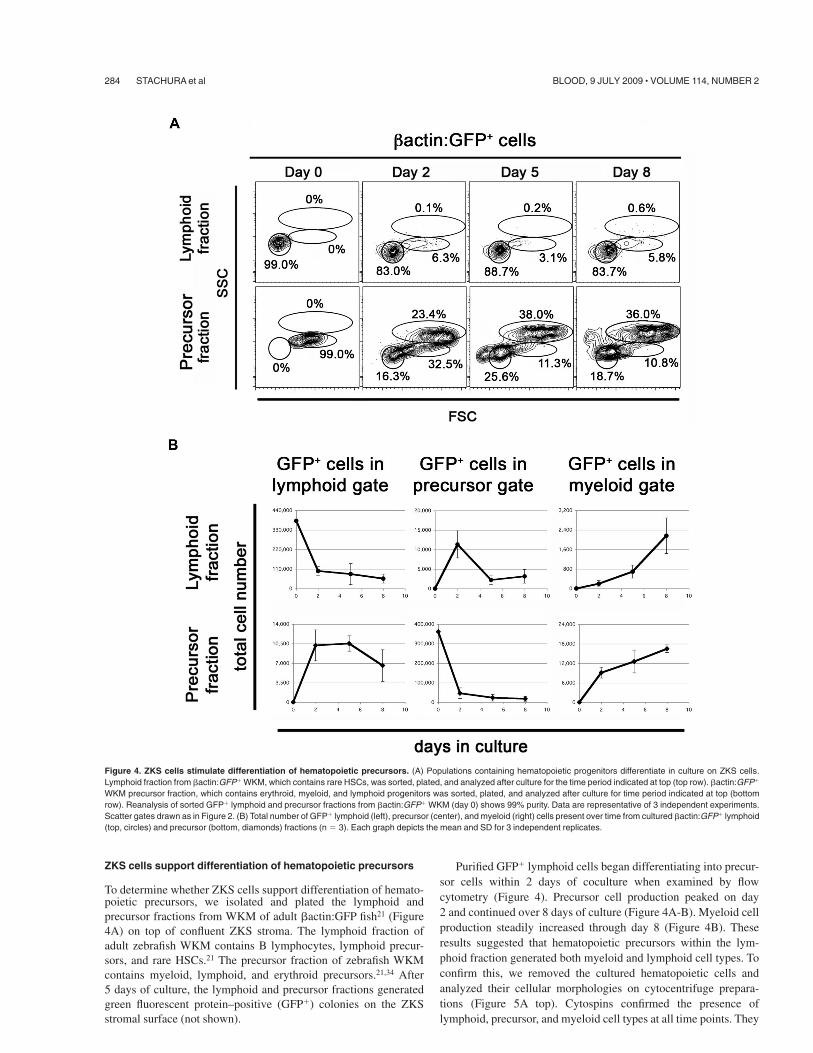
To determine whether ZKS cells support differentiation of hemato-poietic precursors, we isolated and plated the lymphoid andprecursor fractions from WKM of adult #actin:GFP fish21 (Figure4A) on top of confluent ZKS stroma. The lymphoid fraction ofadult zebrafish WKM contains B lymphocytes, lymphoid precur-sors, and rare HSCs.21 The precursor fraction of zebrafish WKMcontains myeloid, lymphoid, and erythroid precursors.21,34 After5 days of culture, the lymphoid and precursor fractions generatedgreen fluorescent protein–positive (GFP&) colonies on the ZKSstromal surface (not shown).
Purified GFP& lymphoid cells began differentiating into precur-sor cells within 2 days of coculture when examined by flowcytometry (Figure 4). Precursor cell production peaked on day2 and continued over 8 days of culture (Figure 4A-B). Myeloid cellproduction steadily increased through day 8 (Figure 4B). Theseresults suggested that hematopoietic precursors within the lym-phoid fraction generated both myeloid and lymphoid cell types. Toconfirm this, we removed the cultured hematopoietic cells andanalyzed their cellular morphologies on cytocentrifuge prepara-tions (Figure 5A top). Cytospins confirmed the presence oflymphoid, precursor, and myeloid cell types at all time points. They
Figure 4. ZKS cells stimulate differentiation of hematopoietic precursors. (A) Populations containing hematopoietic progenitors differentiate in culture on ZKS cells.Lymphoid fraction from #actin:GFP& WKM, which contains rare HSCs, was sorted, plated, and analyzed after culture for the time period indicated at top (top row). #actin:GFP&
WKM precursor fraction, which contains erythroid, myeloid, and lymphoid progenitors was sorted, plated, and analyzed after culture for time period indicated at top (bottomrow). Reanalysis of sorted GFP& lymphoid and precursor fractions from #actin:GFP& WKM (day 0) shows 99% purity. Data are representative of 3 independent experiments.Scatter gates drawn as in Figure 2. (B) Total number of GFP& lymphoid (left), precursor (center), and myeloid (right) cells present over time from cultured #actin:GFP& lymphoid(top, circles) and precursor (bottom, diamonds) fractions (n ( 3). Each graph depicts the mean and SD for 3 independent replicates.
284 STACHURA et al BLOOD, 9 JULY 2009 ! VOLUME 114, NUMBER 2
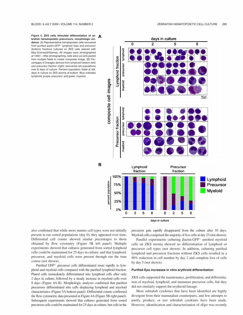
also confirmed that while more mature cell types were not initiallypresent in our sorted populations (day 0), they appeared over time.Differential cell counts showed similar percentages to thoseobtained by flow cytometry (Figure 5B left panel). Multipleexperiments showed that cultures generated from sorted lymphoidcells could be maintained for 25 days in culture, and that lymphoid,precursor, and myeloid cells were present through out the timecourse (not shown).
Purified GFP& precursor cells differentiated more rapidly to lym-phoid and myeloid cells compared with the purified lymphoid fraction.Plated cells immediately differentiated into lymphoid cells after only2 days in culture, followed by a steady increase in myeloid cells over8 days (Figure 4A-B). Morphologic analyses confirmed that purifiedprecursors differentiated into cells displaying lymphoid and myeloidcharacteristics (Figure 5A bottom panel). Differential counts confirmedthe flow cytometric data presented in Figure 4A (Figure 5B right panel).Subsequent experiments showed that cultures generated from sortedprecursor cells could be maintained for 25 days in culture, but cells in the
precursor gate rapidly disappeared from the culture after 10 days.Myeloid cells comprised the majority of live cells at day 25 (not shown).
Parallel experiments culturing #actin:GFP& purified myeloidcells on ZKS stroma showed no differentiation of lymphoid orprecursor cell types (not shown). In addition, culturing purifiedlymphoid and precursor fractions without ZKS cells resulted in a90% reduction in cell number by day 2 and complete loss of cellsby day 5 (not shown).
Purified Epo increases in vitro erythroid differentiation
ZKS cells supported the maintenance, proliferation, and differentia-tion of myeloid, lymphoid, and immature precursor cells, but theydid not similarly support the erythroid lineage.
Most zebrafish cytokines that have been identified are highlydivergent from their mammalian counterparts, and few attempts topurify, produce, or test zebrafish cytokines have been made.However, identification and characterization of zEpo was recently
Figure 5. ZKS cells stimulate differentiation of ze-brafish hematopoietic precursors; morphologic evi-dence. (A) Representative hematopoietic cells recoveredfrom purified #actin:GFP& lymphoid (top) and precursor(bottom) fractions cultured on ZKS cells stained withMay-Grunwald/Giemsa. All images were photographedat 1000$. After photographing, cells were cut and pastedfrom multiple fields to create composite image. (B) Per-centages of lineages derived from lymphoid fraction (left)and precursor fraction (right) recovered cell populationsover 8 days of culture. Percent population listed at left,days in culture on ZKS stroma at bottom. Blue indicateslymphoid; purple, precursor; and green, myeloid.
ZEBRAFISH HEMATOPOIETIC CELL CULTURE 285BLOOD, 9 JULY 2009 ! VOLUME 114, NUMBER 2
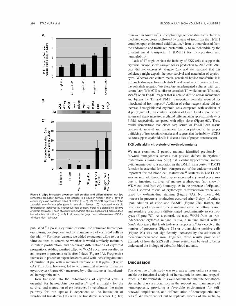
published.26 Epo is a cytokine essential for definitive hematopoi-esis during development and for maintenance of erythroid cells inthe adult.35 For these reasons, we added exogenous zEpo to our invitro cultures to determine whether it would similarly maintain,stimulate proliferation, and encourage differentiation of erythroidprogenitors. Adding purified zEpo to WKM cocultures resulted inan increase in precursor cells after 5 days (Figure 6A). Progressiveincreases in precursor expansion correlated with increasing amountsof purified zEpo, with a maximal increase at 100 !g/mL (Figure6A). This dose, however, led to only minimal increases in matureerythrocytes (Figure 6C), measured by o-dianisidine, a histochemi-cal hemoglobin stain.
Iron transport into the mitochondria of erythroid cells isessential for hemoglobin biosynthesis36 and ultimately for thesurvival and maturation of erythrocytes. In vertebrates, the majorpathway for iron uptake is dependent on the interaction ofiron-bound transferrin (Tf) with the transferrin receptor 1 (Tfr1;
reviewed in Andrews37). Receptor engagement stimulates clathrin-mediated endocytosis, followed by release of iron from the Tf/Tfr1complex upon endosomal acidification.37 Iron is then released fromthe endosome and trafficked preferentially to mitochondria by thedivalent metal transporter 1 (DMT1) for incorporation intohemoglobin.37
Lack of Tf might explain the inability of ZKS cells to support theerythroid lineage, so we assayed for its production by ZKS cells. ZKScells did not express tfa (Figure 6B), and we reasoned that thisdeficiency might explain the poor survival and maturation of erythro-cytes. Whereas our culture media contained bovine transferrin, it isextremely divergent from zebrafish Tf and is unlikely to cross-react withthe zebrafish receptor. We therefore supplemented cultures with carpserum (carp Tf is 67% similar to zebrafish Tf, while human Tf is only49%38) or an Fe-SIH reagent that is able to diffuse across membranesand bypass the Tfr and DMT1 transporters normally required formitochondrial iron import.39 Addition of either reagent alone did notincrease hemoglobinized erythroid cells compared with addition ofzEpo (Figure 6C). In contrast, addition of Fe-SIH and zEpo, or carpserum and zEpo, increased erythroid differentiation approximately 4- or6-fold, respectively, compared with zEpo alone (Figure 6C). Theseresults demonstrate that either carp serum or Fe-SIH can rescueerythrocyte survival and maturation, likely in part due to the propertrafficking of iron to mitochondria, and suggest that the inability of ZKScells to support erythroid cells is due to a lack of proper iron transport.
ZKS cells aid in vitro study of erythroid mutants
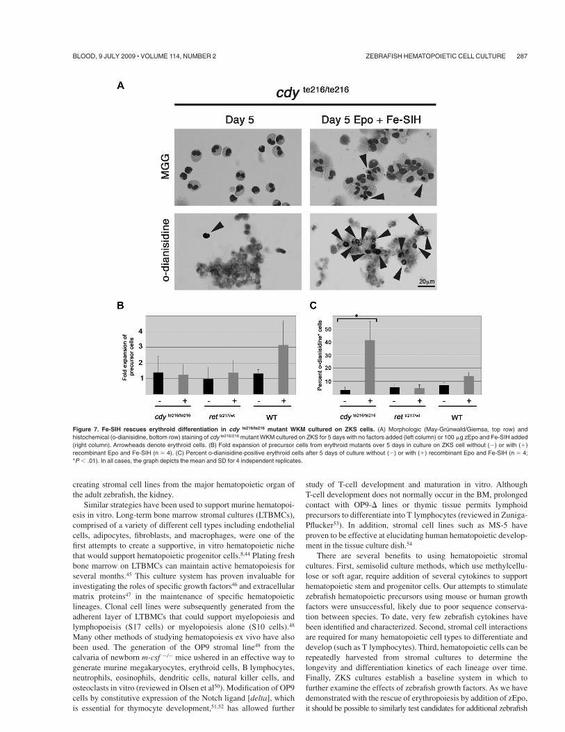
We next examined 2 genetic mutants identified previously inforward mutagenesis screens that possess defects in erythroidmaturation. Chardonnay (cdy) fish exhibit hypochromic, micro-cytic anemia due to a mutation in the DMT1 transporter.40 DMT1function is essential for iron transport out of the endosome and isimportant for red blood cell maturation.41 Mutants in DMT1 cansurvive into adulthood, but display increased erythroid precursorsdue to impaired survival of mature erythrocytes (not shown).WKM cultured from cdy homozygotes in the presence of zEpo andFe-SIH showed rescue of erythrocyte differentiation when ana-lyzed by o-dianisidine staining (Figure 7A). No measurableincrease in precursor production occurred after 5 days of cultureupon addition of zEpo and Fe-SIH (Figure 7B). Rather, theprecursor pool appeared to be maintained over the culture period,and resulting precursors differentiated predominantly to erythro-cytes (Figure 7C). As a control, we used WKM from an iron-independent erythroid mutant retsina, a mutant animal with aband3 deficiency that leads to dyserythropoiesis.42 As expected, thenumber of precursor (Figure 7B) or o-dianisidine positive cells(Figure 7C) was not significantly increased by the addition ofmembrane-permeable iron. Together, these results provide anexample of how the ZKS cell culture system can be used to betterunderstand the biology of zebrafish blood mutants.
Discussion
The objective of this study was to create a tissue culture system toenable the functional analysis of hematopoietic stem and progeni-tor cells in the zebrafish. It is well documented that the hematopoi-etic niche plays a crucial role in the support and maintenance ofhematopoiesis, providing a favorable environment for self-renewal, amplification, and differentiation of hematopoietic stemcells.43 We therefore set out to replicate aspects of the niche by
Figure 6. zEpo increases precursor cell survival and differentiation. (A) Epostimulates precursor survival. Fold change in precursor number after 5 days inculture. Cytokine conditions listed at bottom (n ( 3). (B) RT-PCR expression of thezebrafish transferrin-a (tfa) gene in zebrafish tissues. (C) Increased erythroiddifferentiation achieved by exogenous iron delivery. Percent o-dianisidine-positiveerythroid cells after 5 days of culture with erythroid stimulating factors. Factors addedto media listed at bottom (n ( 3). In all cases, the graph depicts the mean and SD for3 independent replicates.
286 STACHURA et al BLOOD, 9 JULY 2009 ! VOLUME 114, NUMBER 2
creating stromal cell lines from the major hematopoietic organ ofthe adult zebrafish, the kidney.
Similar strategies have been used to support murine hematopoi-esis in vitro. Long-term bone marrow stromal cultures (LTBMCs),comprised of a variety of different cell types including endothelialcells, adipocytes, fibroblasts, and macrophages, were one of thefirst attempts to create a supportive, in vitro hematopoietic nichethat would support hematopoietic progenitor cells.8,44 Plating freshbone marrow on LTBMCs can maintain active hematopoiesis forseveral months.45 This culture system has proven invaluable forinvestigating the roles of specific growth factors46 and extracellularmatrix proteins47 in the maintenance of specific hematopoieticlineages. Clonal cell lines were subsequently generated from theadherent layer of LTBMCs that could support myelopoiesis andlymphopoeisis (S17 cells) or myelopoiesis alone (S10 cells).48
Many other methods of studying hematopoiesis ex vivo have alsobeen used. The generation of the OP9 stromal line49 from thecalvaria of newborn m-csf )/) mice ushered in an effective way togenerate murine megakaryocytes, erythroid cells, B lymphocytes,neutrophils, eosinophils, dendritic cells, natural killer cells, andosteoclasts in vitro (reviewed in Olsen et al50). Modification of OP9cells by constitutive expression of the Notch ligand [delta], whichis essential for thymocyte development,51,52 has allowed further
study of T-cell development and maturation in vitro. AlthoughT-cell development does not normally occur in the BM, prolongedcontact with OP9-* lines or thymic tissue permits lymphoidprecursors to differentiate into T lymphocytes (reviewed in Zuniga-Pflucker53). In addition, stromal cell lines such as MS-5 haveproven to be effective at elucidating human hematopoietic develop-ment in the tissue culture dish.54
There are several benefits to using hematopoietic stromalcultures. First, semisolid culture methods, which use methylcellu-lose or soft agar, require addition of several cytokines to supporthematopoietic stem and progenitor cells. Our attempts to stimulatezebrafish hematopoietic precursors using mouse or human growthfactors were unsuccessful, likely due to poor sequence conserva-tion between species. To date, very few zebrafish cytokines havebeen identified and characterized. Second, stromal cell interactionsare required for many hematopoietic cell types to differentiate anddevelop (such as T lymphocytes). Third, hematopoietic cells can berepeatedly harvested from stromal cultures to determine thelongevity and differentiation kinetics of each lineage over time.Finally, ZKS cultures establish a baseline system in which tofurther examine the effects of zebrafish growth factors. As we havedemonstrated with the rescue of erythropoiesis by addition of zEpo,it should be possible to similarly test candidates for additional zebrafish
Figure 7. Fe-SIH rescues erythroid differentiation in cdy te216/te216 mutant WKM cultured on ZKS cells. (A) Morphologic (May-Grunwald/Giemsa, top row) andhistochemical (o-dianisidine, bottom row) staining of cdy te216/216 mutant WKM cultured on ZKS for 5 days with no factors added (left column) or 100 !g zEpo and Fe-SIH added(right column). Arrowheads denote erythroid cells. (B) Fold expansion of precursor cells from erythroid mutants over 5 days in culture on ZKS cell without ()) or with (&)recombinant Epo and Fe-SIH (n ( 4). (C) Percent o-dianisidine-positive erythroid cells after 5 days of culture without ()) or with (&) recombinant Epo and Fe-SIH (n ( 4;*P + .01). In all cases, the graph depicts the mean and SD for 4 independent replicates.
ZEBRAFISH HEMATOPOIETIC CELL CULTURE 287BLOOD, 9 JULY 2009 ! VOLUME 114, NUMBER 2
growth factor orthologs, such as thrombopoietin, granulocyte colony-stimulating factor (g-csf), and macrophage colony-stimulating factor(m-csf). Since it is often difficult to predict discovery of these moleculesby sequence homology, it should now be possible to identify these andother factors through their functions in our culture system. Progresshere, and possibly use of conditioned media from ZKS cells, shouldenable the development of semisolid culture systems for the detection ofzebrafish colony-forming cells.
Our results using WKM have shown that ZKS cells cannot onlysupport myeloid and erythroid differentiation, but can also supportlymphocyte survival and maturation. Surprisingly, we have ob-served the simultaneous differentiation and proliferation of cellsexpressing pax5, igM, gata3, rag2, and lck, likely reflecting thepresence of both B lymphocytes and T lymphocytes in our cultures.This is unexpected, since mammalian cell lines, like OP9 tend tosupport only one of these 2 lymphoid lineages. OP9 cells supportB-cell development normally, but at the apparent expense ofT cells. Transduction of the Notch ligand, *, into OP9 cellsconversely supports T-cell development at the expense of B lympho-cytes.53 ZKS cells express all of the zebrafish * ligands, which maybe responsible for differentiation of lck& T cells over time. We areworking to verify whether mature T lymphocytes and B lympho-cytes are present in ZKS cultures through development of molecu-lar probes against the T- and B-cell antigen receptors.
The remarkable conservation between mammals and ze-brafish in the lineages and functions of mature hematopoieticcells make it likely that a similar cascade of lineage commitmentproceeds downstream of HSCs as previously demonstrated inthe mouse. Using the ZKS stromal system, we can now addressthe ontogeny of the major blood lineages through identificationof defined progenitor subsets. Our current results suggest that alymphoid progenitor is present within the precursor scatterfraction, a finding not previously appreciated. Since ZKS cellsappear to support both T- and B-lymphoid development, it maybe possible to determine whether this activity is due to thepresence of a common lymphoid progenitor (CLP) equivalent inthis fraction by using prospective isolation strategies based on,for example, fluorescent transgene expression. With furtheroptimization of the ZKS system, it should be possible to performthe clonal experiments necessary to identify a zebrafish CLP andassess its full developmental potential. Similarly, identification ofclonogenic myeloid-restricted progenitors should be possible using theZKS system to determine whether zebrafish HSCs commit to the maturemyeloerythroid lineages through CMP, GMPand MEPintermediates, asdemonstrated in the mouse.11
Several large-scale, forward genetic screens have generated anarray of zebrafish mutants, many with essential defects in hemato-poiesis. Many of these mutants have not been fully characterizeddue to a lack of appropriate functional assays. Here we show byrescuing cdy mutant erythroid cell maturation that the ZKS systemmay be used to examine genetic mutants in ways not previously
possible. The ability to study and rescue mutant gene function invitro should provide additional means to elucidate normal genefunction in other mutants. Further refinement of the ZKS systemand development of semisolid culture techniques will provide moreprecision and flexibility in analysis of genetic mutants. Themajority of hematopoietic mutants were identified by visibledefects in erythrocyte maturation or survival. In vitro assays willserve to address whether any of these mutants possess additional,nonerythroid hematopoietic defects. This should greatly aid theidentification of genetic mutants possessing defects in hematopoi-etic stem and progenitor cells.
In conclusion, we have generated primary stromal cell linesfrom the adult zebrafish kidney able to support multilineagehematopoiesis. Optimization of culture conditions, along withidentification of zebrafish hematopoietic growth factors, shouldultimately enable the in vitro survival, proliferation, and differentia-tion of all blood cell lineages in the zebrafish. Furthermore, thissystem should likewise enable more precise characterization ofmutant phenotypes resulting from forward mutagenesis screensdesigned to identify erythroid, myeloid, and lymphoid mutants.
Acknowledgments
We thank Mitch Weiss, Dawne Page, and Wilson Clements forcritical evaluation of the manuscript; Shrey Purohit for technicalassistance; Malcolm Wood for electron microscopy; Roger Rain-ville, Evie Wright, and Lisa Phelps for excellent animal care; andKerstin Richter for excellent laboratory management.
This work was supported by the Prevent Cancer Foundation(D.L.S.), the LC06077 project of Ministry of Education, Youth, andSports (MSMT) and Fullbright Scholars award (P.B.), the March ofDimes Foundation (B.H.P.), the American Society of Hematology(D.T.), the Arnold and Mabel Beckman Foundation (D.T.), theSenyei Family Foundation (D.T.), and National Institutes of Health(NIH) grant nos. R01-DK074482 (D.T.), R01-DK070838, (B.H.P.),and P01-HL32262 (B.H.P.).
Authorship
Contribution: D.L.S. and J.R.R. performed the research andanalyzed the data; D.L.S. and D.T. designed the research and wrotethe article; and P.B., B.P., and L.I.Z. contributed vital reagents.
Conflict-of-interest disclosure: D.L.S., J.R.R., P.B., B.P., andD.T. declare no competing financial interests. L.Z. has stock in andis a consultant for FATE Therapeutics and Stemgent.
Correspondence: David Traver, University of California at SanDiego Division of Biological Sciences, Section of Cell andDevelopmental Biology, 9500 Gilman Dr, La Jolla, CA, 92093-0380; e-mail: [email protected].
References1. McCulloch EA, Till JE. The radiation sensitivity of
normal mouse bone marrow cells, determined byquantitative marrow transplantation into irradiatedmice. Radiat Res. 1960;13:115-125.
2. Till JE, Mc CE. A direct measurement of the radia-tion sensitivity of normal mouse bone marrowcells. Radiat Res. 1961;14:213-222.
3. Ford CE, Hamerton JL, Barnes DW, Loutit JF.Cytological identification of radiation-chimaeras.Nature. 1956;177:452-454.
4. Bach FH, Albertini RJ, Joo P, Anderson JL, Bortin
MM. Bone-marrow transplantation in a patientwith the Wiskott-Aldrich syndrome. Lancet. 1968;2:1364-1366.
5. De Koning J, Van Bekkum DW, Dicke KA, DoorenLJ, Radl J, Van Rood JJ. Transplantation of bone-marrow cells and fetal thymus in an infant withlymphopenic immunological deficiency. Lancet.1969;1:1223-1227.
6. Bradley TR, Metcalf D. The growth of mousebone marrow cells in vitro. Aust J Exp Biol MedSci. 1966;44:287-299.
7. Pluznik DH, Sachs L. The cloning of normal“mast” cells in tissue culture. J Cell Physiol. 1965;66:319-324.
8. Dexter TM, Allen TD, Lajtha LG. Conditions con-trolling the proliferation of haemopoietic stemcells in vitro. J Cell Physiol. 1977;91:335-344.
9. Allen TD, Dexter TM. Cellular interrelationshipsduring in vitro granulopoiesis. Differentiation.1976;6:191-194.
10. Kondo M, Weissman IL, Akashi K. Identification of
288 STACHURA et al BLOOD, 9 JULY 2009 ! VOLUME 114, NUMBER 2
clonogenic common lymphoid progenitors inmouse bone marrow. Cell. 1997;91:661-672.
11. Akashi K, Traver D, Miyamoto T, Weissman IL. Aclonogenic common myeloid progenitor that givesrise to all myeloid lineages. Nature. 2000;404:193-197.
12. Nakorn TN, Miyamoto T, Weissman IL. Charac-terization of mouse clonogenic megakaryocyteprogenitors. Proc Natl Acad Sci U S A. 2003;100:205-210.
13. Mori Y, Iwasaki H, Kohno K, et al. Identification ofthe human eosinophil lineage-committed progeni-tor: revision of phenotypic definition of the humancommon myeloid progenitor. J Exp Med. 2008;206:183-193.
14. Sutherland HJ, Lansdorp PM, Henkelman DH,Eaves AC, Eaves CJ. Functional characterizationof individual human hematopoietic stem cells cul-tured at limiting dilution on supportive marrowstromal layers. Proc Natl Acad Sci U S A. 1990;87:3584-3588.
15. Kraemer PM, Ray FA, Brothman AR, BartholdiMF, Cram LS. Spontaneous immortalization rateof cultured Chinese hamster cells. J Natl CancerInst. 1986;76:703-709.
16. Paran M, Sachs L, Barak Y, Resnitzky P. In vitroinduction of granulocyte differentiation in hemato-poietic cells from leukemic and non-leukemic pa-tients. Proc Natl Acad Sci U S A. 1970;67:1542-1549.
17. Lavau C, Szilvassy SJ, Slany R, Cleary ML. Im-mortalization and leukemic transformation of amyelomonocytic precursor by retrovirally trans-duced HRX-ENL. EMBO J. 1997;16:4226-4237.
18. de Jong JL, Zon LI. Use of the zebrafish systemto study primitive and definitive hematopoiesis.Annu Rev Genet. 2005;39:481-501.
19. Bertrand JY, Kim AD, Violette EP, Stachura DL,Cisson JL, Traver D. Definitive hematopoiesisinitiates through a committed erythromyeloid pro-genitor in the zebrafish embryo. Development.2007;134:4147-4156.
20. Bertrand JY, Kim AD, Teng S, Traver D. CD41&
cmyb& precursors colonize the zebrafish prone-phros by a novel migration route to initiate adulthematopoiesis. Development. 2008;135:1853-1862.
21. Traver D, Paw BH, Poss KD, Penberthy WT, LinS, Zon LI. Transplantation and in vivo imaging ofmultilineage engraftment in zebrafish bloodlessmutants. Nat Immunol. 2003;4:1238-1246.
22. Traver D, Winzeler A, Stern HM, et al. Effects oflethal irradiation in zebrafish and rescue by he-matopoietic cell transplantation. Blood. 2004;104:1298-1305.
23. Westerfield M. The Zebrafish Book. A Guide forthe Laboratory Use of Zebrafish (Danio rerio). 4thEd. Eugene, OR: University of Oregon Press;2000.
24. Tsinkalovsky O, Vik-Mo AO, Ferreira S, LaerumOD, Fjose A. Zebrafish kidney marrow containsABCG2-dependent side population cells exhibit-ing hematopoietic stem cell properties. Differen-tiation. 2007;75:175-183.
25. Hsu K, Traver D, Kutok JL, et al. The pu.1 pro-moter drives myeloid gene expression in ze-brafish. Blood. 2004;104:1291-1297.
26. Paffett-Lugassy N, Hsia N, Fraenkel PG, et al.Functional conservation of erythropoietin signal-ing in zebrafish. Blood. 2007;110:2718-2726.
27. Bartunek P, Pichlikova L, Stengl G, et al. Avianstem cell factor (SCF): production and character-ization of the recombinant His-tagged SCF ofchicken and its neutralizing antibody. Cytokine.1996;8:14-20.
28. Zapata A. Ultrastructural study of the teleost fishkidney. Dev Comp Immunol. 1979;3:55-65.
29. Meseguer J, Esteban MA, Agulleiro B. Stromalcells, macrophages and lymphoid cells in thehead-kidney of sea bass (Dicentrarchus labraxL.). An ultrastructural study. Arch Histol Cytol.1991;54:299-309.
30. Bronner-Fraser M. Alterations in neural crest mi-gration by a monoclonal antibody that affects celladhesion. J Cell Biol. 1985;101:610-617.
31. Lyons AB, Parish CR. Determination of lympho-cyte division by flow cytometry. J Immunol Meth-ods. 1994;171:131-137.
32. Nose A, Takeichi M. A novel cadherin cell adhe-sion molecule: its expression patterns associatedwith implantation and organogenesis of mouseembryos. J Cell Biol. 1986;103:2649-2658.
33. Hodgkin PD, Lee JH, Lyons AB. B cell differentia-tion and isotype switching is related to divisioncycle number. J Exp Med. 1996;184:277-281.
34. Traver D. Cellular dissection of zebrafish hemato-poiesis. Methods Cell Biol. 2004;76:127-149.
35. Fisher JW. Erythropoietin: physiology and phar-macology update. Exp Biol Med (Maywood).2003;228:1-14.
36. Napier I, Ponka P, Richardson DR. Iron traffickingin the mitochondrion: novel pathways revealed bydisease. Blood. 2005;105:1867-1874.
37. Andrews NC. Iron homeostasis: insights from ge-netics and animal models. Nat Rev Genet. 2000;1:208-217.
38. Fraenkel PG, Gibert Y, Holzheimer JL, et al.Transferrin-a modulates hepcidin expression inzebrafish embryos. Blood. 2008.
39. Ponka P, Schulman HM. Acquisition of iron from
transferrin regulates reticulocyte heme synthesis.J Biol Chem. 1985;260:14717-14721.
40. Donovan A, Brownlie A, Dorschner MO, et al. Thezebrafish mutant gene chardonnay (cdy) encodesdivalent metal transporter 1 (DMT1). Blood. 2002;100:4655-4659.
41. Gunshin H, Fujiwara Y, Custodio AO, Direnzo C,Robine S, Andrews NC. Slc11a2 is required forintestinal iron absorption and erythropoiesis butdispensable in placenta and liver. J Clin Invest.2005;115:1258-1266.
42. Paw BH, Davidson AJ, Zhou Y, et al. Cell-specificmitotic defect and dyserythropoiesis associatedwith erythroid band 3 deficiency. Nat Genet.2003;34:59-64.
43. Dexter TM. Stromal cell associated haemopoi-esis. J Cell Physiol Suppl. 1982;1:87-94.
44. Dexter TM, Testa NG. Differentiation and prolif-eration of hemopoietic cells in culture. MethodsCell Biol. 1976;14:387-405.
45. Lichtman MA. The ultrastructure of the hemopoi-etic environment of the marrow: a review. ExpHematol. 1981;9:391-410.
46. Harigaya K, Cronkite EP, Miller ME, ShadduckRK. Murine bone marrow cell line producingcolony-stimulating factor. Proc Natl Acad SciU S A. 1981;78:6963-6966.
47. Zuckerman KS, Wicha MS. Extracellular matrixproduction by the adherent cells of long-term mu-rine bone marrow cultures. Blood. 1983;61:540-547.
48. Landreth KS, Dorshkind K. Pre-B cell generationpotentiated by soluble factors from a bone mar-row stromal cell line. J Immunol. 1988;140:845-852.
49. Nakano T, Kodama H, Honjo T. Generation oflymphohematopoietic cells from embryonic stemcells in culture. Science. 1994;265:1098-1101.
50. Olsen AL, Stachura DL, Weiss MJ. Designerblood: creating hematopoietic lineages from em-bryonic stem cells. Blood. 2006;107:1265-1275.
51. Schmitt TM, de Pooter RF, Gronski MA, Cho SK,Ohashi PS, Zuniga-Pflucker JC. Induction ofT cell development and establishment of T cellcompetence from embryonic stem cells differenti-ated in vitro. Nat Immunol. 2004;5:410-417.
52. Schmitt TM, Zuniga-Pflucker JC. Induction ofT cell development from hematopoietic progenitorcells by '-like-1 in vitro. Immunity. 2002;17:749-756.
53. Zuniga-Pflucker JC. T-cell development madesimple. Nat Rev Immunol. 2004;4:67-72.
54. Yoshikawa Y, Ikebuchi K, Ohkawara J, et al. Aclonal culture assay for human cord blood lym-phohematopoietic progenitors. Hum Immunol.1999;60:75-82.
ZEBRAFISH HEMATOPOIETIC CELL CULTURE 289BLOOD, 9 JULY 2009 ! VOLUME 114, NUMBER 2