Embed Size (px)
Citation preview

You have scored 8%
You answered 9 correct out of 106 questions.
Your answers are shown below:
A 28 year old man was involved in a motorbike accident where he sustained a crush injury to
his pelvis and right leg and damaged a nerve innervating the lower limb. He is unable to extend the
leg at the knee joint. Which of the following clinical ndings might you also expect on examination:
a) Weak extension of the thigh at the hip
b) Weak dorsi exion of the ankle
c) Loss of sensation over the anterior thigh
d) Trendelenburg’s sign
e) Loss of sensation over the lateral leg and foot
Answer
Extension of the leg at the knee joint is primarily produced by the quadriceps femoris muscle which is innervated by
the femoral nerve. The femoral nerve also innervates the iliacus, pectineus and sartorius muscles which, together with
the rectus femoris muscle (and the psoas major), are involved in exion of the thigh at the hip. The femoral nerve also
gives rise to cutaneous branches supplying skin on the anterior thigh and the saphenous nerve supplying skin over the
medial leg at foot.
Notes
The femoral nerve arises from the lumbar plexus, receiving bres from the anterior rami of L2 – L4.
Nerve Femoral nerve
Something wrong?
Dashboard Subscription expires in: <1 Day Extend
Nerve roots L2 – L4
Motor supply Iliacus, pectineus, sartorius, quadriceps femoris
Sensory supply Skin over the anterior thigh, anteromedial knee, medial leg and medial foot
Motor loss in injury Weak exion at hip and loss of extension at knee
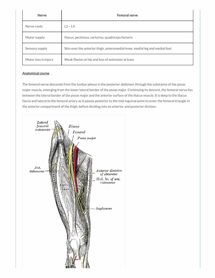
Anatomical course
The femoral nerve descends from the lumbar plexus in the posterior abdomen through the substance of the psoas
major muscle, emerging from the lower lateral border of the psoas major. Continuing its descent, the femoral nerve lies
between the lateral border of the psoas major and the anterior surface of the iliacus muscle. It is deep to the iliacus
fascia and lateral to the femoral artery as it passes posterior to the mid-inguinal point to enter the femoral triangle in
the anterior compartment of the thigh, before dividing into an anterior and posterior division.
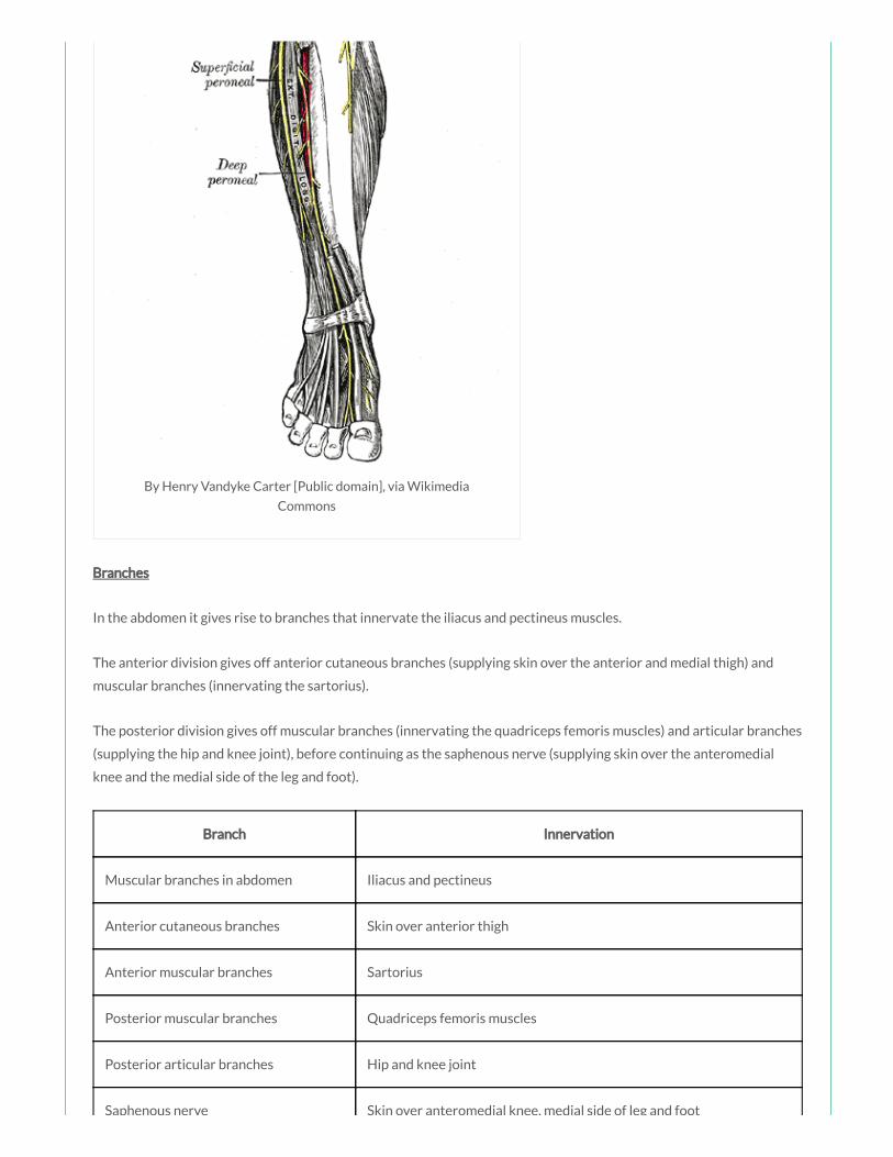
By Henry Vandyke Carter [Public domain], via Wikimedia
Commons
Branches
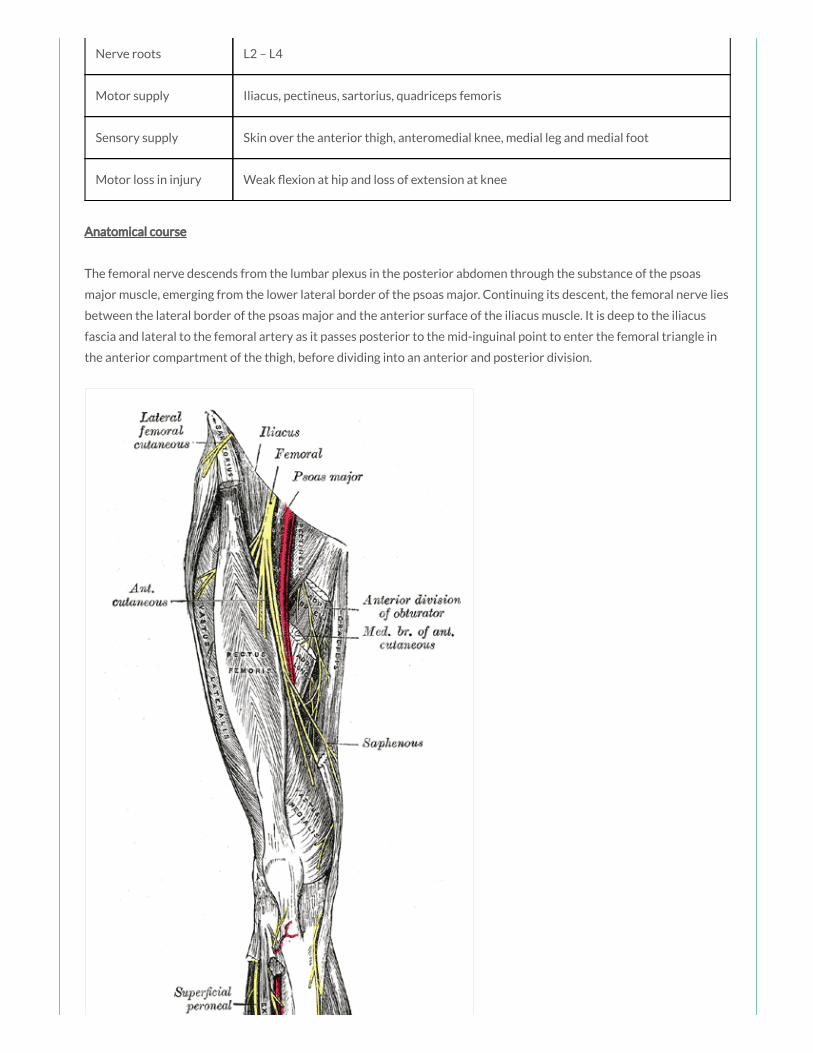
In the abdomen it gives rise to branches that innervate the iliacus and pectineus muscles.
The anterior division gives off anterior cutaneous branches (supplying skin over the anterior and medial thigh) and
muscular branches (innervating the sartorius).
The posterior division gives off muscular branches (innervating the quadriceps femoris muscles) and articular branches
(supplying the hip and knee joint), before continuing as the saphenous nerve (supplying skin over the anteromedial
knee and the medial side of the leg and foot).
Branch Innervation
Muscular branches in abdomen Iliacus and pectineus
Anterior cutaneous branches Skin over anterior thigh
Anterior muscular branches Sartorius
Posterior muscular branches Quadriceps femoris muscles
Posterior articular branches Hip and knee joint
Saphenous nerve Skin over anteromedial knee, medial side of leg and foot
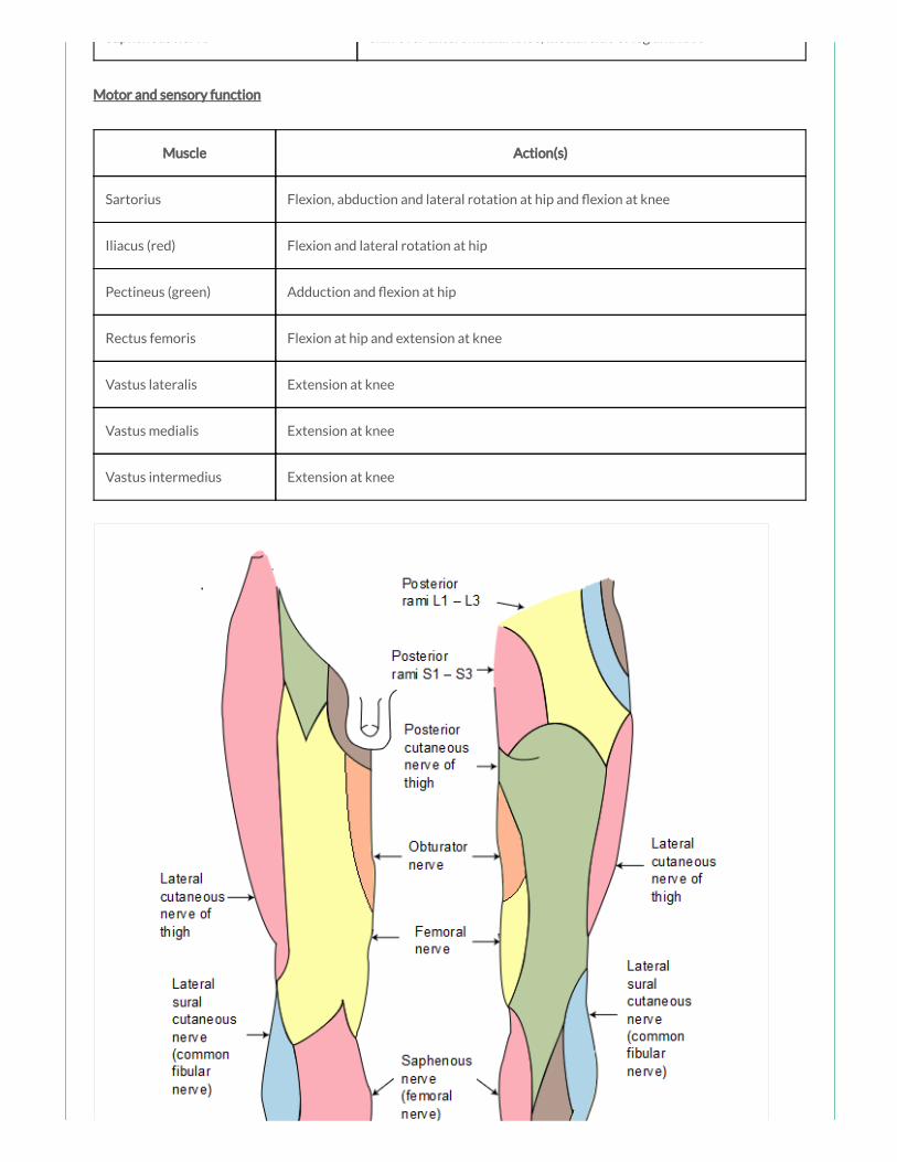
Motor and sensory function
Motor and sensory function
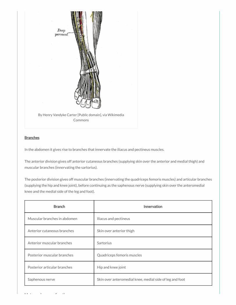
Muscle Action(s)
Sartorius Flexion, abduction and lateral rotation at hip and exion at knee
Iliacus (red) Flexion and lateral rotation at hip
Pectineus (green) Adduction and exion at hip
Rectus femoris Flexion at hip and extension at knee
Vastus lateralis Extension at knee
Vastus medialis Extension at knee
Vastus intermedius Extension at knee

Modi ed by FRCEM Success. Original by Henry Vandyke Carter [Public domain], via Wikimedia Commons
A 27 year old footballer presents with pain on exion and lateral rotation of the hip and
exion of the knee. You suspect a sartorius muscle pathology. The sartorius muscle is innervated by
which of the following nerves:
a) Femoral nerve
b) Obturator nerve
c) Sciatic nerve
d) Pudendal nerve
e) Inferior gluteal nerve
Answer
The sartorius is innervated by the femoral nerve (L2, L3).
Notes
The sartorius, iliopsoas, pectineus and rectus femoris are the primary exors of the thigh at the hip joint.
Muscle Actions Innervation
Sartorius (blue) Flexion, abduction and lateral rotation at hip and exion at
knee
Femoral nerve (L2, L3)
Iliacus (red) Flexion and lateral rotation at hip Femoral nerve (L2, L3)
Psoas major
(yellow)
Flexion and lateral rotation at hip Anterior rami L1 – L3
Pectineus (green) Adduction and exion at hip Femoral nerve (L2, L3)
Something wrong?

Pectineus (green) Adduction and exion at hip Femoral nerve (L2, L3)
Rectus femoris Flexion at hip and extension at knee Femoral nerve (L2 –
L4)
The sartorius (blue) is innervated by the femoral nerve (L2, L3). It acts to ex the thigh at the hip joint and ex the leg at
the knee joint. It also abducts the thigh and rotates it laterally, as when resting the foot on the opposite knee when
sitting.
The iliacus (red) and psoas major (yellow) muscles originate as separate muscles but insert by a common tendon onto
the femur and together are referred to as the iliopsoas muscle, which is a powerful exor of the thigh at the hip joint
and also contributes to lateral rotation of the thigh at the hip joint. The iliacus is innervated by the femoral nerve (L2,
L3). The psoas major is innervated by the anterior rami of spinal nerves L1 – L3.
The pectineus (green) is innervated by the femoral nerve (L2, L3). It acts to adduct and ex the thigh at the hip joint.

Modi ed by FRCEM Success. Original by Henry
Vandyke Carter [Public domain], via Wikimedia
Commons
A 63 year old woman with advanced ovarian malignancy presents to ED complaining of
weakness of her left leg while walking. Examination reveals weakness of adduction of the thigh at
the hip joint. Which of the following nerves is most likely being compressed to result in this
pattern:
a) Sciatic nerve
b) Femoral nerve
c) Obturator nerve
d) Superior gluteal nerve
e) Inferior gluteal nerve
Answer
Adduction at the hip is primarily produced by the adductor longus, brevis and magnus, all innervated by the obturator
nerve. The pectineus and gracilis muscles, innervated by the femoral nerve and obturator nerve respectively, assist in
this action.
Notes
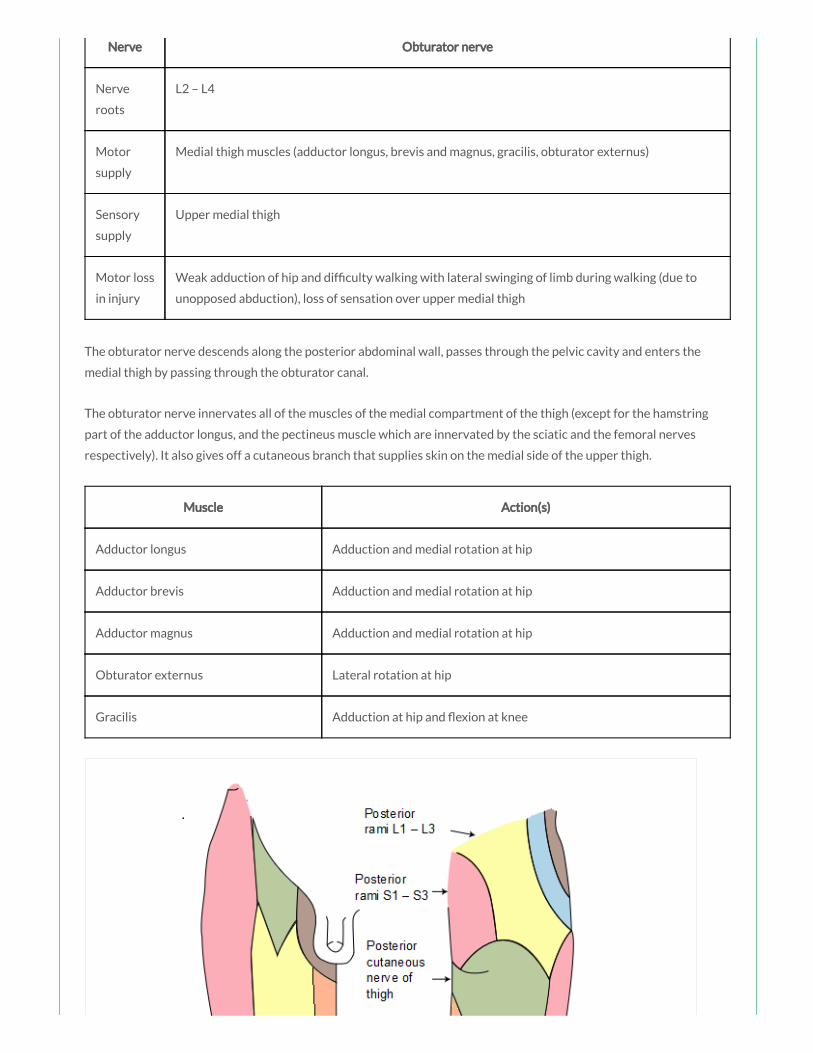
The obturator nerve arises from the lumbar plexus, formed from the anterior rami of L2 – L4.
Something wrong?
Nerve Obturator nerve
Nerve
roots
L2 – L4
Motor
supply
Medial thigh muscles (adductor longus, brevis and magnus, gracilis, obturator externus)
Sensory
supply
Upper medial thigh
Motor loss
in injury
Weak adduction of hip and dif culty walking with lateral swinging of limb during walking (due to
unopposed abduction), loss of sensation over upper medial thigh
The obturator nerve descends along the posterior abdominal wall, passes through the pelvic cavity and enters the
medial thigh by passing through the obturator canal.
The obturator nerve innervates all of the muscles of the medial compartment of the thigh (except for the hamstring
part of the adductor longus, and the pectineus muscle which are innervated by the sciatic and the femoral nerves
respectively). It also gives off a cutaneous branch that supplies skin on the medial side of the upper thigh.
Muscle Action(s)
Adductor longus Adduction and medial rotation at hip
Adductor brevis Adduction and medial rotation at hip
Adductor magnus Adduction and medial rotation at hip
Obturator externus Lateral rotation at hip
Gracilis Adduction at hip and exion at knee

Modi ed by FRCEM Success. Original by Henry Vandyke Carter [Public domain], via Wikimedia Commons
A 27 year old woman presents to the Emergency Department complaining of progressive
weakness in her lower limbs following a recent episode of food poisoning. You are asked to perform
a full neurological examination of the lower limbs, including testing sensation in all dermatomes.
The S3 dermatome is best tested at which of the following landmarks:
a) At the midpoint of the inguinal ligament
b) At the ischial tuberosity
c) At the pubic symphysis
d) At the popliteal fossa
e) At the medial femoral condyle
Answer
Something wrong?

The S3 dermatome is best tested over the ischial tuberosity or infragluteal fold (depending on the patient their skin can
move up, down or laterally over the ischii).
Notes
Dermatome Landmark
L1 Upper Anterior Thigh
L2 Mid Anterior Thigh
L3 Medial Femoral Condyle
L4 Medial Malleolus
L5 Dorsum 3rd MTP Joint
S1 Lateral Heel
S2 Popliteal Fossa
S3 Ischial Tuberosity
S5 Perianal Area
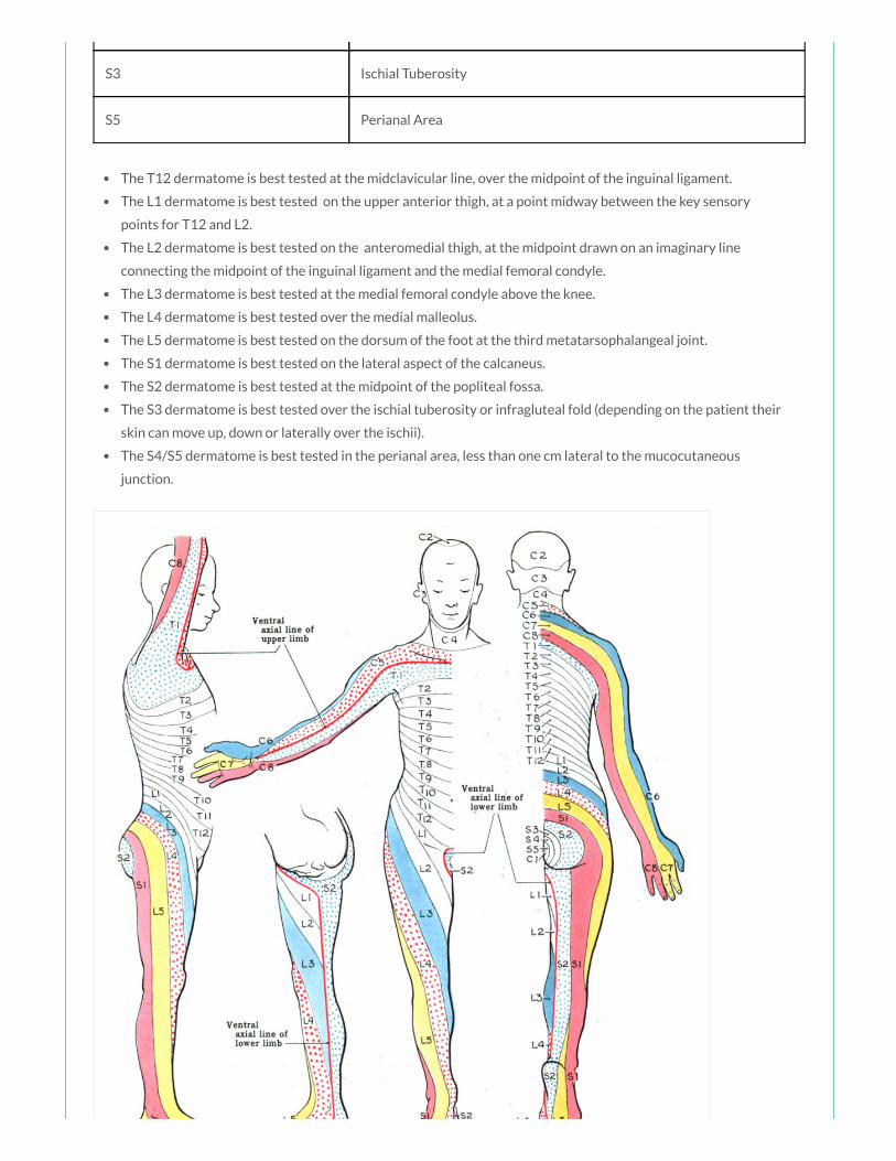
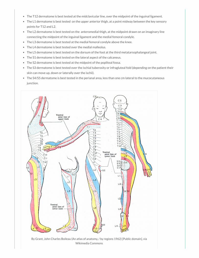
The T12 dermatome is best tested at the midclavicular line, over the midpoint of the inguinal ligament.
The L1 dermatome is best tested on the upper anterior thigh, at a point midway between the key sensory
points for T12 and L2.
The L2 dermatome is best tested on the anteromedial thigh, at the midpoint drawn on an imaginary line
connecting the midpoint of the inguinal ligament and the medial femoral condyle.
The L3 dermatome is best tested at the medial femoral condyle above the knee.
The L4 dermatome is best tested over the medial malleolus.
The L5 dermatome is best tested on the dorsum of the foot at the third metatarsophalangeal joint.
The S1 dermatome is best tested on the lateral aspect of the calcaneus.
The S2 dermatome is best tested at the midpoint of the popliteal fossa.
The S3 dermatome is best tested over the ischial tuberosity or infragluteal fold (depending on the patient their
skin can move up, down or laterally over the ischii).
The S4/S5 dermatome is best tested in the perianal area, less than one cm lateral to the mucocutaneous
junction.
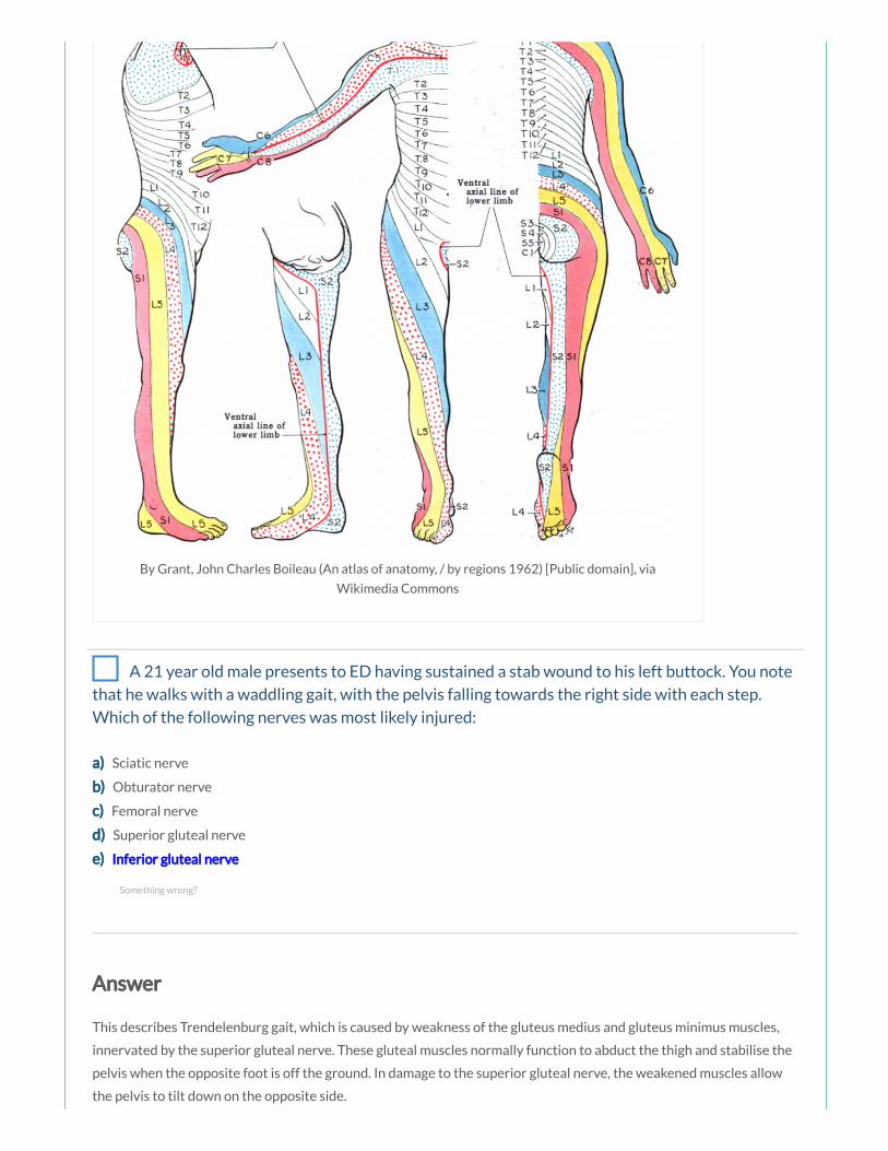
By Grant, John Charles Boileau (An atlas of anatomy, / by regions 1962) [Public domain], via
Wikimedia Commons
A 21 year old male presents to ED having sustained a stab wound to his left buttock. You note
that he walks with a waddling gait, with the pelvis falling towards the right side with each step.
Which of the following nerves was most likely injured:
a) Sciatic nerve
b) Obturator nerve
c) Femoral nerve
d) Superior gluteal nerve
e) Inferior gluteal nerve
Answer
This describes Trendelenburg gait, which is caused by weakness of the gluteus medius and gluteus minimus muscles,
innervated by the superior gluteal nerve. These gluteal muscles normally function to abduct the thigh and stabilise the
pelvis when the opposite foot is off the ground. In damage to the superior gluteal nerve, the weakened muscles allow
the pelvis to tilt down on the opposite side.
Something wrong?

Notes
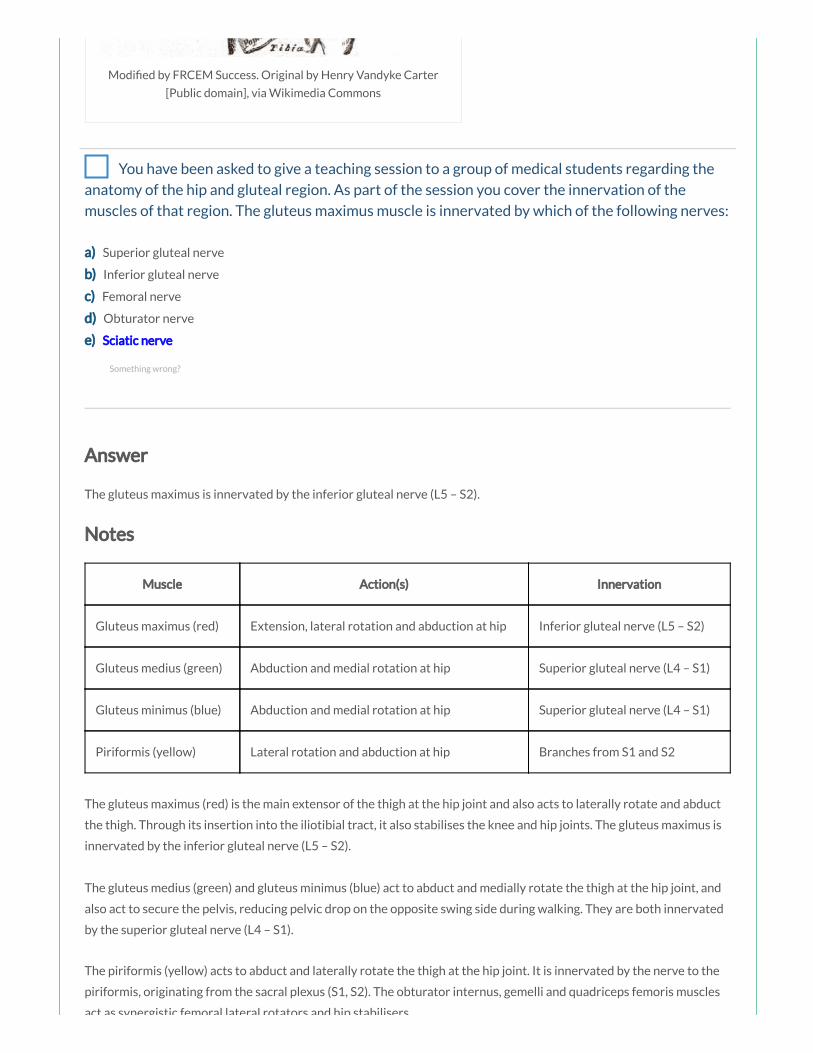
Muscle Action(s) Innervation
Gluteus maximus (red) Extension, lateral rotation and abduction at hip Inferior gluteal nerve (L5 – S2)
Gluteus medius (green) Abduction and medial rotation at hip Superior gluteal nerve (L4 – S1)
Gluteus minimus (blue) Abduction and medial rotation at hip Superior gluteal nerve (L4 – S1)
Piriformis (yellow) Lateral rotation and abduction at hip Branches from S1 and S2
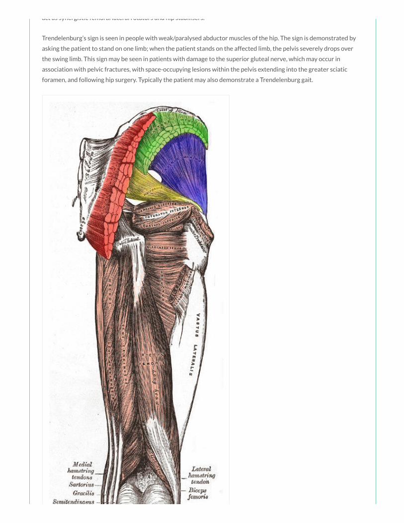
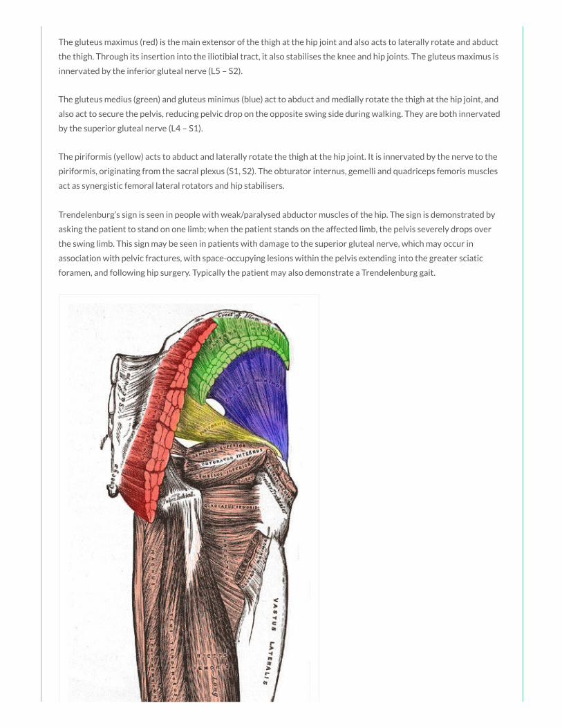
The gluteus maximus (red) is the main extensor of the thigh at the hip joint and also acts to laterally rotate and abduct
the thigh. Through its insertion into the iliotibial tract, it also stabilises the knee and hip joints. The gluteus maximus is
innervated by the inferior gluteal nerve (L5 – S2).
The gluteus medius (green) and gluteus minimus (blue) act to abduct and medially rotate the thigh at the hip joint, and
also act to secure the pelvis, reducing pelvic drop on the opposite swing side during walking. They are both innervated
by the superior gluteal nerve (L4 – S1).
The piriformis (yellow) acts to abduct and laterally rotate the thigh at the hip joint. It is innervated by the nerve to the
piriformis, originating from the sacral plexus (S1, S2). The obturator internus, gemelli and quadriceps femoris muscles
act as synergistic femoral lateral rotators and hip stabilisers.
Trendelenburg’s sign is seen in people with weak/paralysed abductor muscles of the hip. The sign is demonstrated by
asking the patient to stand on one limb; when the patient stands on the affected limb, the pelvis severely drops over
the swing limb. This sign may be seen in patients with damage to the superior gluteal nerve, which may occur in
association with pelvic fractures, with space-occupying lesions within the pelvis extending into the greater sciatic
foramen, and following hip surgery. Typically the patient may also demonstrate a Trendelenburg gait.

Modi ed by FRCEM Success. Original by Henry Vandyke Carter
[Public domain], via Wikimedia Commons
You are performing a full neurological examination on a patient who presented with weakness
of her left leg. The tendon of which of the following muscles is stretched during the patellar re ex:
a) Sartorius
b) Gracilis
c) Popliteus
d) Biceps femoris
e) Quadriceps femoris
Something wrong?
Answer
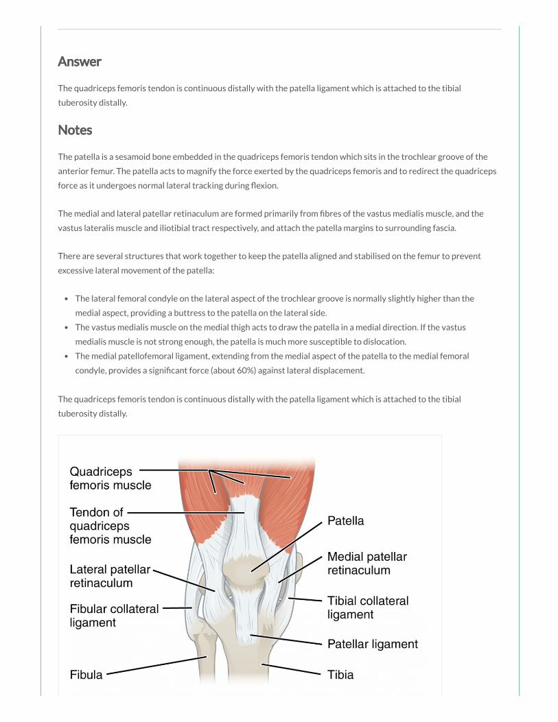
The quadriceps femoris tendon is continuous distally with the patella ligament which is attached to the tibial
tuberosity distally.
Notes
The patella is a sesamoid bone embedded in the quadriceps femoris tendon which sits in the trochlear groove of the
anterior femur. The patella acts to magnify the force exerted by the quadriceps femoris and to redirect the quadriceps
force as it undergoes normal lateral tracking during exion.
The medial and lateral patellar retinaculum are formed primarily from bres of the vastus medialis muscle, and the
vastus lateralis muscle and iliotibial tract respectively, and attach the patella margins to surrounding fascia.
There are several structures that work together to keep the patella aligned and stabilised on the femur to prevent
excessive lateral movement of the patella:
The lateral femoral condyle on the lateral aspect of the trochlear groove is normally slightly higher than the
medial aspect, providing a buttress to the patella on the lateral side.
The vastus medialis muscle on the medial thigh acts to draw the patella in a medial direction. If the vastus
medialis muscle is not strong enough, the patella is much more susceptible to dislocation.
The medial patellofemoral ligament, extending from the medial aspect of the patella to the medial femoral
condyle, provides a signi cant force (about 60%) against lateral displacement.
The quadriceps femoris tendon is continuous distally with the patella ligament which is attached to the tibial
tuberosity distally.

By OpenStax College [CC BY 3.0 (http://creativecommons.org/licenses/by/3.0)], via Wikimedia
Commons
A 21 year old man presents to the ED after sustaining multiple deep lacerations to the
posterior thigh during a ght. You suspect an injury to the hamstring muscles. The hamstring
muscles primarily act together to produce which of the following movements:
a) Flexion of the thigh and exion of the leg
b) Flexion of the thigh and extension of the leg
c) Extension of the thigh and exion of the leg
d) Extension of the thigh and extension of the leg
e) Extension and adduction of the thigh
Answer
The hamstrings act together to ex the leg at the knee joint and extend the thigh at the hip joint.
Notes
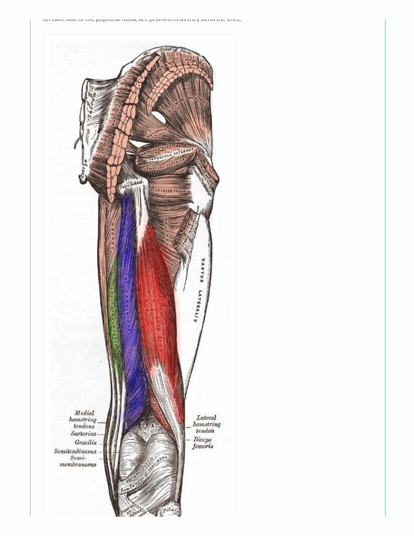
Muscle Action(s) Innervation
Biceps femoris (red) Flexion at knee, extension and lateral rotation at hip Sciatic nerve (L5 – S2)
Semitendinosus (blue) Flexion at knee, extension and medial rotation at hip Sciatic nerve (L5 – S2)
Semimembranosus (green) Flexion at knee, extension and medial rotation at hip Sciatic nerve (L5 – S2)
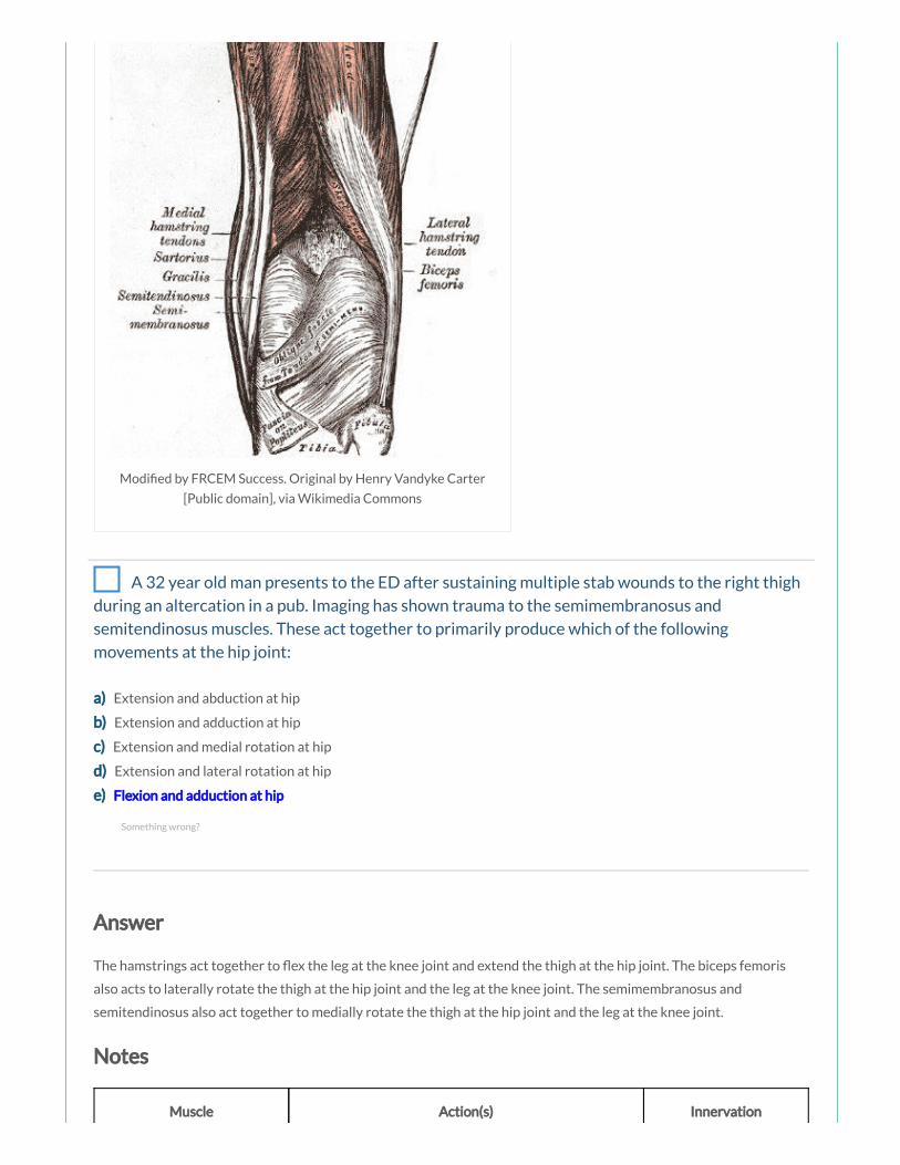
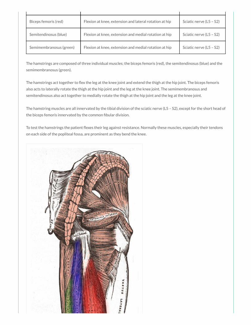
The hamstrings are composed of three individual muscles; the biceps femoris (red), the semitendinosus (blue) and the
semimembranosus (green).
The hamstrings act together to ex the leg at the knee joint and extend the thigh at the hip joint. The biceps femoris
also acts to laterally rotate the thigh at the hip joint and the leg at the knee joint. The semimembranosus and
semitendinosus also act together to medially rotate the thigh at the hip joint and the leg at the knee joint.
The hamstring muscles are all innervated by the tibial division of the sciatic nerve (L5 – S2), except for the short head of
the biceps femoris innervated by the common bular division.
To test the hamstrings the patient exes their leg against resistance. Normally these muscles, especially their tendons
on each side of the popliteal fossa, are prominent as they bend the knee.
Something wrong?
on each side of the popliteal fossa, are prominent as they bend the knee.
Modi ed by FRCEM Success. Original by Henry Vandyke Carter
[Public domain], via Wikimedia Commons
You have been asked to give a teaching session to a group of medical students regarding the
anatomy of the hip and gluteal region. As part of the session you cover the innervation of the
muscles of that region. The gluteus maximus muscle is innervated by which of the following nerves:
a) Superior gluteal nerve
b) Inferior gluteal nerve
c) Femoral nerve
d) Obturator nerve
e) Sciatic nerve
Answer
The gluteus maximus is innervated by the inferior gluteal nerve (L5 – S2).
Notes
Muscle Action(s) Innervation
Gluteus maximus (red) Extension, lateral rotation and abduction at hip Inferior gluteal nerve (L5 – S2)
Gluteus medius (green) Abduction and medial rotation at hip Superior gluteal nerve (L4 – S1)
Gluteus minimus (blue) Abduction and medial rotation at hip Superior gluteal nerve (L4 – S1)
Piriformis (yellow) Lateral rotation and abduction at hip Branches from S1 and S2
The gluteus maximus (red) is the main extensor of the thigh at the hip joint and also acts to laterally rotate and abduct
the thigh. Through its insertion into the iliotibial tract, it also stabilises the knee and hip joints. The gluteus maximus is
innervated by the inferior gluteal nerve (L5 – S2).
The gluteus medius (green) and gluteus minimus (blue) act to abduct and medially rotate the thigh at the hip joint, and
also act to secure the pelvis, reducing pelvic drop on the opposite swing side during walking. They are both innervated
by the superior gluteal nerve (L4 – S1).
The piriformis (yellow) acts to abduct and laterally rotate the thigh at the hip joint. It is innervated by the nerve to the
piriformis, originating from the sacral plexus (S1, S2). The obturator internus, gemelli and quadriceps femoris muscles
act as synergistic femoral lateral rotators and hip stabilisers.
Something wrong?
act as synergistic femoral lateral rotators and hip stabilisers.
Trendelenburg’s sign is seen in people with weak/paralysed abductor muscles of the hip. The sign is demonstrated by
asking the patient to stand on one limb; when the patient stands on the affected limb, the pelvis severely drops over
the swing limb. This sign may be seen in patients with damage to the superior gluteal nerve, which may occur in
association with pelvic fractures, with space-occupying lesions within the pelvis extending into the greater sciatic
foramen, and following hip surgery. Typically the patient may also demonstrate a Trendelenburg gait.

Modi ed by FRCEM Success. Original by Henry Vandyke Carter
[Public domain], via Wikimedia Commons
A 23 year old horse rider is brought to the ED following a fall during a show jumping
competition. She complains of pain in her lumbar spine. On examination you note loss of sensation
in the S5 dermatome. The S5 dermatome is best tested at which of the following landmarks:
a) Ischial tuberosity
b) Posterior superior iliac spine
c) Greater trochanter of the femur
d) Gluteal fold
e) Perianal area
Answer
The S4/S5 dermatome is best tested in the perianal area, less than one cm lateral to the mucocutaneous junction.
Notes
Dermatome Landmark
L1 Upper Anterior Thigh
L2 Mid Anterior Thigh
L3 Medial Femoral Condyle
L4 Medial Malleolus
L5 Dorsum 3rd MTP Joint
S1 Lateral Heel
S2 Popliteal Fossa
Something wrong?
S3 Ischial Tuberosity
S5 Perianal Area
The T12 dermatome is best tested at the midclavicular line, over the midpoint of the inguinal ligament.
The L1 dermatome is best tested on the upper anterior thigh, at a point midway between the key sensory
points for T12 and L2.
The L2 dermatome is best tested on the anteromedial thigh, at the midpoint drawn on an imaginary line
connecting the midpoint of the inguinal ligament and the medial femoral condyle.
The L3 dermatome is best tested at the medial femoral condyle above the knee.
The L4 dermatome is best tested over the medial malleolus.
The L5 dermatome is best tested on the dorsum of the foot at the third metatarsophalangeal joint.
The S1 dermatome is best tested on the lateral aspect of the calcaneus.
The S2 dermatome is best tested at the midpoint of the popliteal fossa.
The S3 dermatome is best tested over the ischial tuberosity or infragluteal fold (depending on the patient their
skin can move up, down or laterally over the ischii).
The S4/S5 dermatome is best tested in the perianal area, less than one cm lateral to the mucocutaneous
junction.
By Grant, John Charles Boileau (An atlas of anatomy, / by regions 1962) [Public domain], via
Wikimedia Commons
A 87 year old woman presents to the ED after her carers noted she was complaining of pain in
the right groin. On examination you note a tender swelling in keeping with a femoral hernia. The
femoral ring is bordered posteriorly by which of the following structures:
a) Inguinal ligament
b) Lacunar ligament
c) Pectineal ligament
d) Iliolumbar ligament
e) Femoral canal
Answer
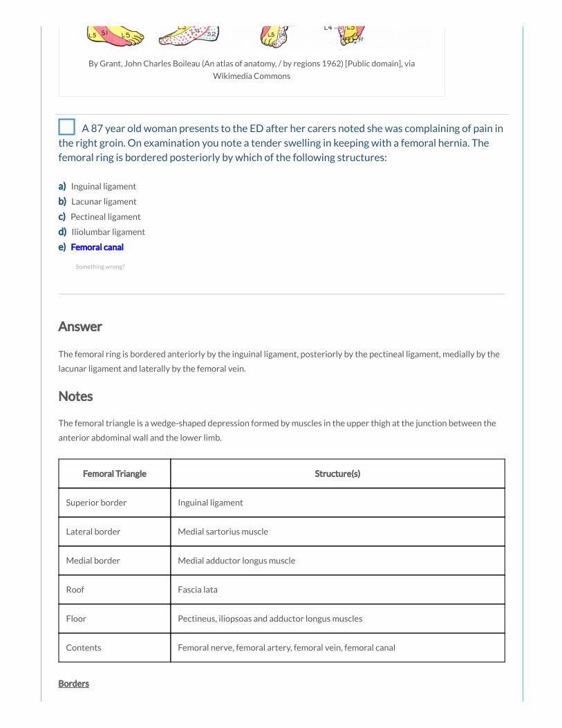
The femoral ring is bordered anteriorly by the inguinal ligament, posteriorly by the pectineal ligament, medially by the
lacunar ligament and laterally by the femoral vein.
Notes
The femoral triangle is a wedge-shaped depression formed by muscles in the upper thigh at the junction between the
anterior abdominal wall and the lower limb.
Femoral Triangle Structure(s)
Superior border Inguinal ligament
Lateral border Medial sartorius muscle
Medial border Medial adductor longus muscle
Roof Fascia lata
Floor Pectineus, iliopsoas and adductor longus muscles
Contents Femoral nerve, femoral artery, femoral vein, femoral canal
Borders
Something wrong?

By Henry Vandyke Carter [Public domain], via
Wikimedia Commons
By Henry Vandyke Carter [Public domain], via
Wikimedia Commons
The femoral triangle is bordered superiorly by the inguinal ligament, laterally by the medial border of the sartorius
muscle and medially by the medial border of the adductor longus muscle. The roof is formed by the fascia lata and the
oor is formed by the pectineus, iliopsoas and adductor longus muscles.
Contents
The femoral triangle contains (from lateral to medial):
The femoral nerve
The femoral artery
The femoral vein
The femoral canal
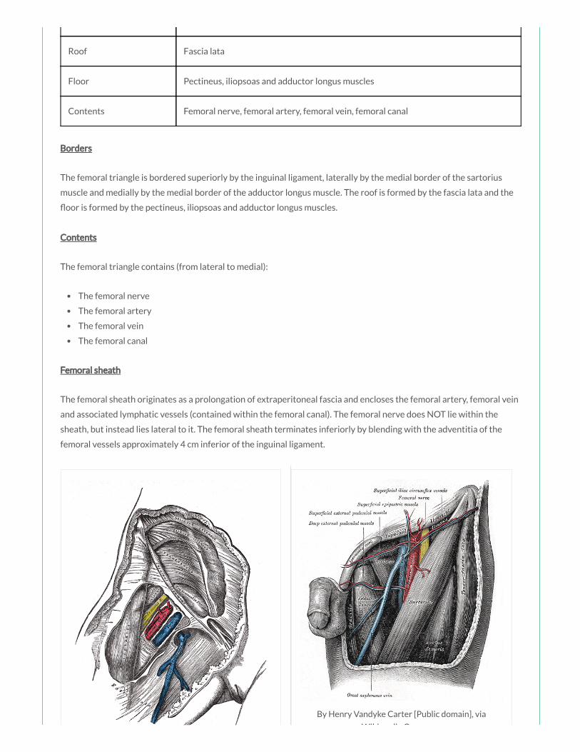
Femoral sheath
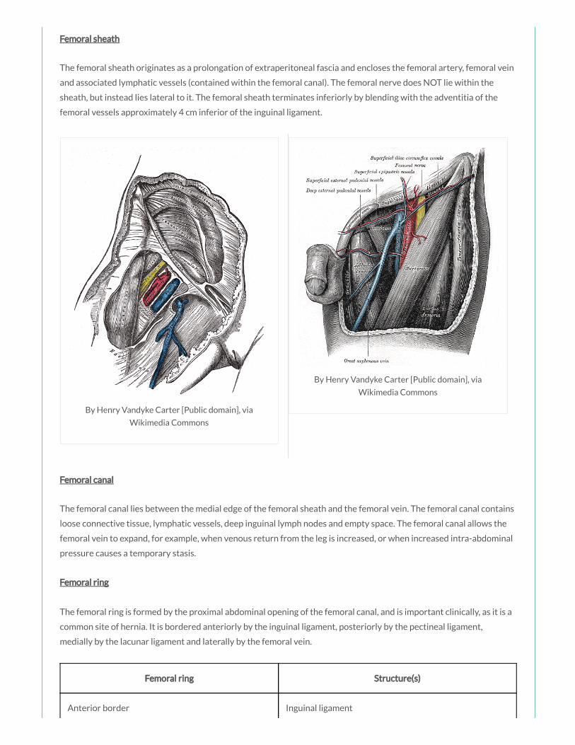
The femoral sheath originates as a prolongation of extraperitoneal fascia and encloses the femoral artery, femoral vein
and associated lymphatic vessels (contained within the femoral canal). The femoral nerve does NOT lie within the
sheath, but instead lies lateral to it. The femoral sheath terminates inferiorly by blending with the adventitia of the
femoral vessels approximately 4 cm inferior of the inguinal ligament.
Femoral canal
The femoral canal lies between the medial edge of the femoral sheath and the femoral vein. The femoral canal contains
loose connective tissue, lymphatic vessels, deep inguinal lymph nodes and empty space. The femoral canal allows the

loose connective tissue, lymphatic vessels, deep inguinal lymph nodes and empty space. The femoral canal allows the
femoral vein to expand, for example, when venous return from the leg is increased, or when increased intra-abdominal
pressure causes a temporary stasis.
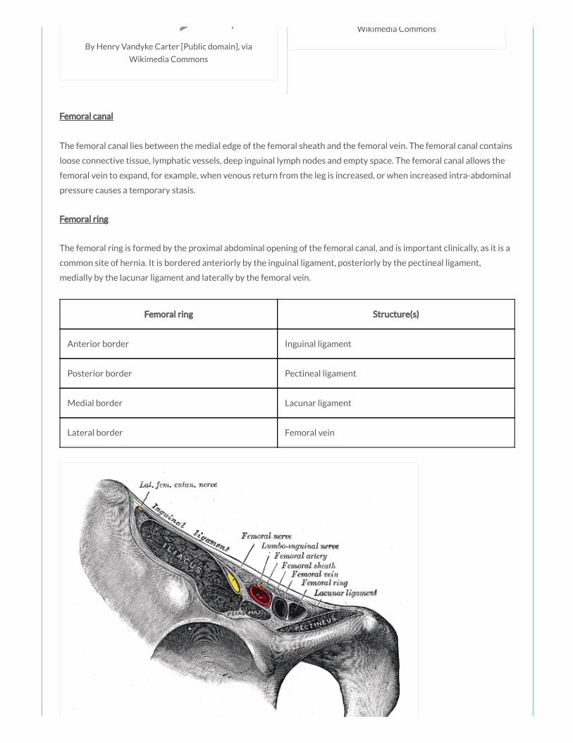
Femoral ring
The femoral ring is formed by the proximal abdominal opening of the femoral canal, and is important clinically, as it is a
common site of hernia. It is bordered anteriorly by the inguinal ligament, posteriorly by the pectineal ligament,
medially by the lacunar ligament and laterally by the femoral vein.
Femoral ring Structure(s)
Anterior border Inguinal ligament
Posterior border Pectineal ligament
Medial border Lacunar ligament
Lateral border Femoral vein
By Henry Vandyke Carter [Public domain], via Wikimedia Commons
You are teaching hip and thigh anatomy to a group of medical students. The femoral canal
contains which of the following structures:
a) Femoral vein
b) Super cial inguinal lymph nodes

b) Super cial inguinal lymph nodes
c) Deep inguinal lymph nodes
d) Femoral artery
e) Femoral nerve
Answer
The femoral canal contains loose connective tissue, lymphatic vessels, deep inguinal lymph nodes and empty space.
Notes
The femoral triangle is a wedge-shaped depression formed by muscles in the upper thigh at the junction between the
anterior abdominal wall and the lower limb.
Femoral Triangle Structure(s)
Superior border Inguinal ligament
Lateral border Medial sartorius muscle
Medial border Medial adductor longus muscle
Roof Fascia lata
Floor Pectineus, iliopsoas and adductor longus muscles
Contents Femoral nerve, femoral artery, femoral vein, femoral canal
Borders
The femoral triangle is bordered superiorly by the inguinal ligament, laterally by the medial border of the sartorius
muscle and medially by the medial border of the adductor longus muscle. The roof is formed by the fascia lata and the
oor is formed by the pectineus, iliopsoas and adductor longus muscles.
Contents
The femoral triangle contains (from lateral to medial):
The femoral nerve
The femoral artery
The femoral vein
The femoral canal
Something wrong?
By Henry Vandyke Carter [Public domain], via
Wikimedia Commons
By Henry Vandyke Carter [Public domain], via
Wikimedia Commons
Femoral sheath
The femoral sheath originates as a prolongation of extraperitoneal fascia and encloses the femoral artery, femoral vein
and associated lymphatic vessels (contained within the femoral canal). The femoral nerve does NOT lie within the
sheath, but instead lies lateral to it. The femoral sheath terminates inferiorly by blending with the adventitia of the
femoral vessels approximately 4 cm inferior of the inguinal ligament.
Femoral canal
The femoral canal lies between the medial edge of the femoral sheath and the femoral vein. The femoral canal contains
loose connective tissue, lymphatic vessels, deep inguinal lymph nodes and empty space. The femoral canal allows the
femoral vein to expand, for example, when venous return from the leg is increased, or when increased intra-abdominal
pressure causes a temporary stasis.
Femoral ring
The femoral ring is formed by the proximal abdominal opening of the femoral canal, and is important clinically, as it is a
common site of hernia. It is bordered anteriorly by the inguinal ligament, posteriorly by the pectineal ligament,
medially by the lacunar ligament and laterally by the femoral vein.
Femoral ring Structure(s)
Anterior border Inguinal ligament

Posterior border Pectineal ligament
Medial border Lacunar ligament
Lateral border Femoral vein
By Henry Vandyke Carter [Public domain], via Wikimedia Commons
A patient presents to ED for treatment having been informed by the GUM clinic that he has
tested positive for gonorrhoea. You administer intramuscular ceftriaxone. Intramuscular injections
in the buttocks should always be given in which of the following areas:
a) Upper lateral quadrant
b) Lower lateral quadrant
c) Upper medial quadrant
d) Lower medial quadrant
e) Lower medial or lateral quadrants
Answer
The sciatic nerve passes through the lower medial quadrant. Intramuscular injections in the buttocks should always be
given in the upper lateral quadrant of the gluteal region to avoid damage to the sciatic nerve and major vessels in the
Something wrong?

given in the upper lateral quadrant of the gluteal region to avoid damage to the sciatic nerve and major vessels in the
region.
Notes
The gluteal region can be divided into quadrants by 2 lines: one line descending vertically from the highest point of the
iliac crest, the other line passing horizontally through the rst line midway between the highest point of the iliac crest
and the ischial tuberosity. The sciatic nerve passes through the lower medial quadrant. Intramuscular injections in the
buttocks should always be given in the upper lateral quadrant of the gluteal region to avoid damage to the sciatic
nerve.
A 21 year old male is involved in a road traf c collision and sustains a pelvic fracture. In
rehabilitation it is noted that he has an inability to adduct his thigh. Which of the following nerves
has most likely been affected:
a) Obturator nerve
b) Femoral nerve
c) Inferior gluteal nerve
d) Superior gluteal nerve
e) Sciatic nerve
Answer
Adduction of the thigh is primarily produced by the adductor longus, magnus and brevis muscles and the gracilis
muscle, all innervated by the obturator nerve.
Notes
The obturator nerve arises from the lumbar plexus, formed from the anterior rami of L2 – L4.
Nerve Obturator nerve
Nerve
roots
L2 – L4
Motor
supply
Medial thigh muscles (adductor longus, brevis and magnus, gracilis, obturator externus)
Sensory
supply
Upper medial thigh
Motor loss
in injury
Weak adduction of hip and dif culty walking with lateral swinging of limb during walking (due to
unopposed abduction), loss of sensation over upper medial thigh
Something wrong?

in injury unopposed abduction), loss of sensation over upper medial thigh
The obturator nerve descends along the posterior abdominal wall, passes through the pelvic cavity and enters the
medial thigh by passing through the obturator canal.
The obturator nerve innervates all of the muscles of the medial compartment of the thigh (except for the hamstring
part of the adductor longus, and the pectineus muscle which are innervated by the sciatic and the femoral nerves
respectively). It also gives off a cutaneous branch that supplies skin on the medial side of the upper thigh.
Muscle Action(s)
Adductor longus Adduction and medial rotation at hip
Adductor brevis Adduction and medial rotation at hip
Adductor magnus Adduction and medial rotation at hip
Obturator externus Lateral rotation at hip
Gracilis Adduction at hip and exion at knee
Modi ed by FRCEM Success. Original by Henry Vandyke Carter [Public domain], via Wikimedia Commons
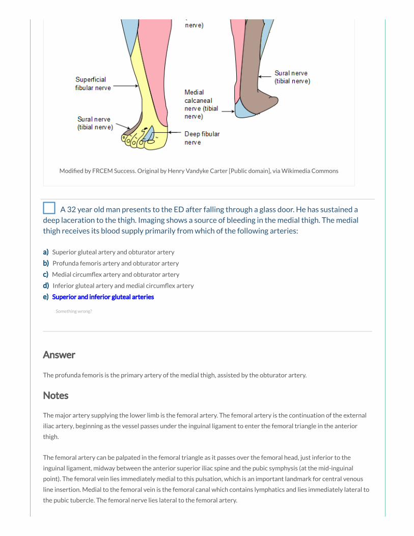
A 32 year old man presents to the ED after falling through a glass door. He has sustained a
deep laceration to the thigh. Imaging shows a source of bleeding in the medial thigh. The medial
thigh receives its blood supply primarily from which of the following arteries:
a) Superior gluteal artery and obturator artery
b) Profunda femoris artery and obturator artery
c) Medial circum ex artery and obturator artery
d) Inferior gluteal artery and medial circum ex artery
e) Superior and inferior gluteal arteries
Answer
The profunda femoris is the primary artery of the medial thigh, assisted by the obturator artery.
Notes
The major artery supplying the lower limb is the femoral artery. The femoral artery is the continuation of the external
iliac artery, beginning as the vessel passes under the inguinal ligament to enter the femoral triangle in the anterior
thigh.
The femoral artery can be palpated in the femoral triangle as it passes over the femoral head, just inferior to the
inguinal ligament, midway between the anterior superior iliac spine and the pubic symphysis (at the mid-inguinal
point). The femoral vein lies immediately medial to this pulsation, which is an important landmark for central venous
line insertion. Medial to the femoral vein is the femoral canal which contains lymphatics and lies immediately lateral to
the pubic tubercle. The femoral nerve lies lateral to the femoral artery.
Something wrong?
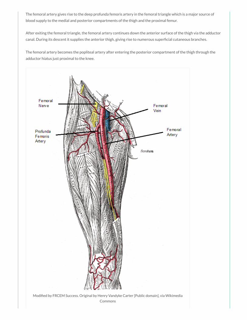
The femoral artery gives rise to the deep profunda femoris artery in the femoral triangle which is a major source of
blood supply to the medial and posterior compartments of the thigh and the proximal femur.
After exiting the femoral triangle, the femoral artery continues down the anterior surface of the thigh via the adductor
canal. During its descent it supplies the anterior thigh, giving rise to numerous super cial cutaneous branches.
The femoral artery becomes the popliteal artery after entering the posterior compartment of the thigh through the
adductor hiatus just proximal to the knee.
Modi ed by FRCEM Success. Original by Henry Vandyke Carter [Public domain], via Wikimedia
Commons

A 32 year old builder is brought to the ED after falling through a glass sky light onto the oor
below. A primary survey has been performed and the patient is stable enough to undergo imaging.
Imaging shows multiple injuries including a transection of the nerve supplying the adductor longus,
magnus and brevis muscles. Which nerve has been injured:
a) Sciatic nerve
b) Obturator nerve
c) Femoral nerve
d) Superior gluteal nerve
e) Inferior gluteal nerve
Answer
The adductor muscles are innervated by the obturator nerve (L2 – L4), except for the hamstrings portion of the
adductor magnus innervated by the tibial nerve (L4 – S3).
Notes
There are six muscles in the medial compartment of the thigh: the gracilis, the pectineus, the adductor longus, brevis
and magnus and the obturator externus. Other than the obturator externus, these muscles all act to adduct the thigh at
the hip joint.
Testing of the medial thigh muscles can be performed with the patient lying supine and the knee straight. The patient is
asked to adduct the thigh against resistance and the strength assessed (if the adductors are normal the proximal ends
of the gracilis and adductor longus can easily be palpated).
Muscle Action(s) Innervation
Adductor longus (red) Adduction and medial rotation at
hip
Obturator nerve (L2 – L4)
Adductor brevis (green) Adduction and medial rotation at
hip
Obturator nerve (L2, L3)
Adductor magnus (blue) Adduction and medial rotation at
hip
Obturator nerve (L2 – L4), tibial nerve (L4 –
S3)
Obturator externus
(yellow)
Lateral rotation at hip Obturator nerve (L3, L4)
Gracilis Adduction at hip and exion at Obturator nerve (L2, L3)
Something wrong?

knee
Pectineus Adduction and exion at hip Femoral nerve (L2, L3)
The adductor longus (red), brevis (green) and magnus (blue) are the prime adductors of the thigh at the hip joint and
also assist in medial rotation.
The adductor muscles are innervated by the obturator nerve (L2 – L4), except for the hamstrings portion of the
adductor magnus innervated by the tibial nerve (L4 – S3).
The obturator externus muscle (yellow) acts to laterally rotate the thigh at the hip joint. It is innervated by the
obturator nerve (L3, L4).
Modi ed by FRCEM Success. Original by Henry
Vandyke Carter [Public domain], via Wikimedia
Commons
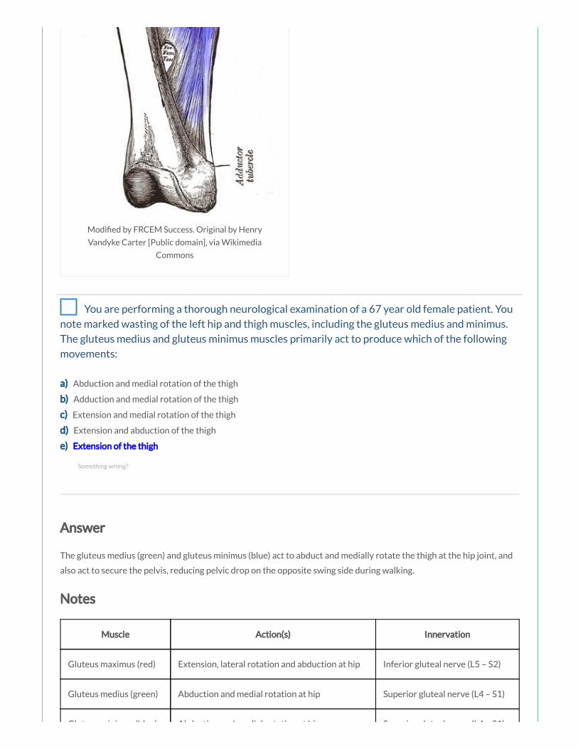
You are performing a thorough neurological examination of a 67 year old female patient. You
note marked wasting of the left hip and thigh muscles, including the gluteus medius and minimus.
The gluteus medius and gluteus minimus muscles primarily act to produce which of the following
movements:
a) Abduction and medial rotation of the thigh
b) Adduction and medial rotation of the thigh
c) Extension and medial rotation of the thigh
d) Extension and abduction of the thigh
e) Extension of the thigh
Answer
The gluteus medius (green) and gluteus minimus (blue) act to abduct and medially rotate the thigh at the hip joint, and
also act to secure the pelvis, reducing pelvic drop on the opposite swing side during walking.
Notes
Muscle Action(s) Innervation
Gluteus maximus (red) Extension, lateral rotation and abduction at hip Inferior gluteal nerve (L5 – S2)
Gluteus medius (green) Abduction and medial rotation at hip Superior gluteal nerve (L4 – S1)
Gluteus minimus (blue) Abduction and medial rotation at hip Superior gluteal nerve (L4 – S1)
Something wrong?

Gluteus minimus (blue) Abduction and medial rotation at hip Superior gluteal nerve (L4 – S1)
Piriformis (yellow) Lateral rotation and abduction at hip Branches from S1 and S2
The gluteus maximus (red) is the main extensor of the thigh at the hip joint and also acts to laterally rotate and abduct
the thigh. Through its insertion into the iliotibial tract, it also stabilises the knee and hip joints. The gluteus maximus is
innervated by the inferior gluteal nerve (L5 – S2).
The gluteus medius (green) and gluteus minimus (blue) act to abduct and medially rotate the thigh at the hip joint, and
also act to secure the pelvis, reducing pelvic drop on the opposite swing side during walking. They are both innervated
by the superior gluteal nerve (L4 – S1).
The piriformis (yellow) acts to abduct and laterally rotate the thigh at the hip joint. It is innervated by the nerve to the
piriformis, originating from the sacral plexus (S1, S2). The obturator internus, gemelli and quadriceps femoris muscles
act as synergistic femoral lateral rotators and hip stabilisers.
Trendelenburg’s sign is seen in people with weak/paralysed abductor muscles of the hip. The sign is demonstrated by
asking the patient to stand on one limb; when the patient stands on the affected limb, the pelvis severely drops over
the swing limb. This sign may be seen in patients with damage to the superior gluteal nerve, which may occur in
association with pelvic fractures, with space-occupying lesions within the pelvis extending into the greater sciatic
foramen, and following hip surgery. Typically the patient may also demonstrate a Trendelenburg gait.

Modi ed by FRCEM Success. Original by Henry Vandyke Carter
[Public domain], via Wikimedia Commons
You are examining the lower limbs of a 54 year old man who presented after falling from a
ladder at home. During your neurological assessment you note a weakness of hip extension.
Extension of the thigh at the hip joint is primarily produced by which of the following muscles:
a) Quadriceps femoris and gluteus maximus
b) Gluteus maximus and gluteus medius
c) Hamstrings and gluteus maximus
d) Quadriceps femoris, gluteus medius and gluteus minimus
e) Hamstrings, gluteus medius and gluteus minimus
Answer
Extension of the thigh at the hip joint is primarily produced by the hamstring muscles and the gluteus maximus,
innervated by the sciatic nerve and the inferior gluteal nerve respectively.
Notes
Something wrong?
Joint Hip joint
Type Synovial ball and socket joint
Articulations Head of femur with acetabulum of pelvis
Stabilising
factors
Acetabular labrum, thickened brous capsule, extracapsular ligaments (iliofemoral, ischiofemoral,
pubofemoral), medial rotator muscles (effectively ‘pull’ head of femur into acetabulum)
Movements Flexion/Extension, Abduction/Adduction, Medial/Lateral rotation, Circumduction
Blood supply Branches of obturator artery, medial and lateral circum ex branches of profunda femoris artery
and superior and inferior gluteal arteries
Innervation Femoral nerve, obturator nerve, superior gluteal nerve and nerve to the quadratus femoris
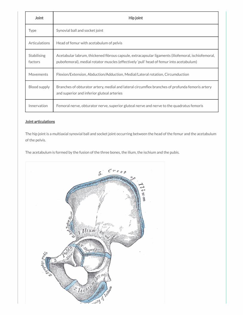
Joint articulations
The hip joint is a multiaxial synovial ball and socket joint occurring between the head of the femur and the acetabulum
of the pelvis.
The acetabulum is formed by the fusion of the three bones, the ilium, the ischium and the pubis.

By Henry Vandyke Carter [Public domain], via Wikimedia Commons
The rim of the acetabulum is raised slightly by the brocartilaginous acetabular labrum which increases its depth,
improving stability of the joint. The acetabular labrum continues inferiorly as the transverse acetabular ligament which
bridges the acetabular notch and converts the notch into a foramen for the passage of nutrient vessels and nerves.
Ligaments
The intracapsular ligament of the head of femur (ligamentum teres) runs from the fovea on the head of the femur at
one end to the acetabular fossa and the transverse acetabular ligament on the other. It carries a small acetabular
branch of the obturator artery.
By Henry Vandyke Carter [Public domain], via Wikimedia Commons
There are three main stabilising extracapsular ligaments; the iliofemoral (the largest and strongest ligament), the
pubofemoral and the ischiofemoral ligament.
The iliofemoral ligament is anterosuperior to the hip joint, attached proximally to the ilium just below the
anterior inferior iliac spine and distally to the intertrochanteric line of the femur. This ligament speci cally
prevents hyperextension and lateral rotation of the hip joint whilst standing.
The pubofemoral ligament is anteroinferior to the hip joint, attached to the pelvis at the iliopubic eminence

Modi ed by FRCEM Success. Original by Henry
Vandyke Carter [Public domain], via Wikimedia
Commons By Henry Vandyke Carter [Public domain], via
Wikimedia Commons
and adjacent bone and blending distally with the articular capsule. This ligament prevents excessive abduction
and extension of the femur at the hip joint.
The ischiofemoral ligament is posterior to the hip joint, attached medially to the ischium and laterally to the
greater trochanter. This ligament prevents excessive extension and medial rotation of the femur at the hip
joint.
Blood supply
The hip joint receives its blood supply primarily from branches of the obturator artery, the medial and lateral
circum ex arteries (branches of the profunda femoris artery) and the superior and inferior gluteal arteries. The
articular branches of these vessels form a network around the joint.
Innervation
The hip joint is innervated by articular branches from the femoral nerve (anteriorly), obturator nerve (inferiorly),
superior gluteal nerve (superiorly), and the nerve to the quadratus femoris (posteriorly).
Joint movements
The hip joint allows the movements of exion and extension, abduction and adduction, medial and lateral rotation and
circumduction.
Movement Muscles Involved

Flexion Sartorius, Iliacus, Psoas major, Pectineus, Rectus femoris
Extension Hamstrings, Gluteus maximus
Abduction Gluteus maximus, medius and minimus, Obturator internus, Gemelli, Piriformis, Sartorius
Adduction Adductor longus, magnus and brevis, Gracilis, Pectineus
Medial
rotation
Gluteus medius and minimus, Adductor longus, magnus and brevis, Semitendinosus and
Semimembranosus
Lateral
rotation
Obturator externus, Sartorius, Iliacus, Psoas major, Biceps femoris, Piriformis, Gluteus maximus,
Obturator internus, Gemelli, Quadriceps femoris
A 34 year old man presents to the ED after sustaining multiple deep stab wounds during an
altercation in a pub. Imagining has shown an injury to the femoral nerve. Which of the following
best describes the anatomical course of the femoral nerve:
a)
After emerging from the lower lateral border of the psoas major muscle, the femoral nerve descends between the
lateral border of the psoas major and the anterior surface of the iliacus muscle.
b)
After emerging from the lower border of the psoas major muscle, the femoral nerve descends posterior to the
iliacus muscle.
c)
After emerging from the lower medial border of the psoas major muscle, the femoral nerve descends adjacent to
the medial border of the psoas major muscle.
d) The femoral nerve emerges from the lumbar plexus within the substance of the iliacus muscle.
e) The femoral nerve descends in a groove between the psoas major and the psoas minor muscles.
Answer
The femoral nerve descends from the lumbar plexus in the posterior abdomen through the substance of the psoas
major muscle, emerging from the lower lateral border of the psoas major. Continuing its descent, the femoral nerve lies
between the lateral border of the psoas major and the anterior surface of the iliacus muscle. It is deep to the iliacus
fascia and lateral to the femoral artery as it passes posterior to the mid-inguinal point to enter the femoral triangle in
the anterior compartment of the thigh, before dividing into an anterior and posterior division.
Notes
The femoral nerve arises from the lumbar plexus, receiving bres from the anterior rami of L2 – L4.
Something wrong?
Nerve Femoral nerve
Nerve roots L2 – L4
Motor supply Iliacus, pectineus, sartorius, quadriceps femoris
Sensory supply Skin over the anterior thigh, anteromedial knee, medial leg and medial foot
Motor loss in injury Weak exion at hip and loss of extension at knee
Anatomical course
The femoral nerve descends from the lumbar plexus in the posterior abdomen through the substance of the psoas
major muscle, emerging from the lower lateral border of the psoas major. Continuing its descent, the femoral nerve lies
between the lateral border of the psoas major and the anterior surface of the iliacus muscle. It is deep to the iliacus
fascia and lateral to the femoral artery as it passes posterior to the mid-inguinal point to enter the femoral triangle in
the anterior compartment of the thigh, before dividing into an anterior and posterior division.
By Henry Vandyke Carter [Public domain], via Wikimedia
Commons
Branches
In the abdomen it gives rise to branches that innervate the iliacus and pectineus muscles.
The anterior division gives off anterior cutaneous branches (supplying skin over the anterior and medial thigh) and
muscular branches (innervating the sartorius).
The posterior division gives off muscular branches (innervating the quadriceps femoris muscles) and articular branches
(supplying the hip and knee joint), before continuing as the saphenous nerve (supplying skin over the anteromedial
knee and the medial side of the leg and foot).
Branch Innervation
Muscular branches in abdomen Iliacus and pectineus
Anterior cutaneous branches Skin over anterior thigh
Anterior muscular branches Sartorius
Posterior muscular branches Quadriceps femoris muscles
Posterior articular branches Hip and knee joint
Saphenous nerve Skin over anteromedial knee, medial side of leg and foot
Saphenous nerve Skin over anteromedial knee, medial side of leg and foot
Motor and sensory function
Muscle Action(s)
Sartorius Flexion, abduction and lateral rotation at hip and exion at knee
Iliacus (red) Flexion and lateral rotation at hip
Pectineus (green) Adduction and exion at hip
Rectus femoris Flexion at hip and extension at knee
Vastus lateralis Extension at knee
Vastus medialis Extension at knee
Vastus intermedius Extension at knee

Modi ed by FRCEM Success. Original by Henry Vandyke Carter [Public domain], via Wikimedia Commons
A 65 year old lady is brought to ED after falling off a ladder. The acetabulum is fractured at its
posterosuperior margin by dislocation of the hip joint. Which of the following bones is most likely
to be involved:
a) Ilium
b) Ischium
c) Pubis
d) Sacrum
e) Head of femur
Answer
The acetabulum is formed by the fusion of the three bones, the ilium superiorly, the ischium posteroinferiorly and the
pubis anteromedially.
Notes
Joint Hip joint
Type Synovial ball and socket joint
Articulations Head of femur with acetabulum of pelvis
Stabilising
factors
Acetabular labrum, thickened brous capsule, extracapsular ligaments (iliofemoral, ischiofemoral,
pubofemoral), medial rotator muscles (effectively ‘pull’ head of femur into acetabulum)
Movements Flexion/Extension, Abduction/Adduction, Medial/Lateral rotation, Circumduction
Something wrong?

Movements Flexion/Extension, Abduction/Adduction, Medial/Lateral rotation, Circumduction
Blood supply Branches of obturator artery, medial and lateral circum ex branches of profunda femoris artery
and superior and inferior gluteal arteries
Innervation Femoral nerve, obturator nerve, superior gluteal nerve and nerve to the quadratus femoris
Joint articulations
The hip joint is a multiaxial synovial ball and socket joint occurring between the head of the femur and the acetabulum
of the pelvis.
The acetabulum is formed by the fusion of the three bones, the ilium, the ischium and the pubis.
By Henry Vandyke Carter [Public domain], via Wikimedia Commons
The rim of the acetabulum is raised slightly by the brocartilaginous acetabular labrum which increases its depth,
improving stability of the joint. The acetabular labrum continues inferiorly as the transverse acetabular ligament which
bridges the acetabular notch and converts the notch into a foramen for the passage of nutrient vessels and nerves.
Ligaments

The intracapsular ligament of the head of femur (ligamentum teres) runs from the fovea on the head of the femur at
one end to the acetabular fossa and the transverse acetabular ligament on the other. It carries a small acetabular
branch of the obturator artery.
By Henry Vandyke Carter [Public domain], via Wikimedia Commons
There are three main stabilising extracapsular ligaments; the iliofemoral (the largest and strongest ligament), the
pubofemoral and the ischiofemoral ligament.
The iliofemoral ligament is anterosuperior to the hip joint, attached proximally to the ilium just below the
anterior inferior iliac spine and distally to the intertrochanteric line of the femur. This ligament speci cally
prevents hyperextension and lateral rotation of the hip joint whilst standing.
The pubofemoral ligament is anteroinferior to the hip joint, attached to the pelvis at the iliopubic eminence
and adjacent bone and blending distally with the articular capsule. This ligament prevents excessive abduction
and extension of the femur at the hip joint.
The ischiofemoral ligament is posterior to the hip joint, attached medially to the ischium and laterally to the
greater trochanter. This ligament prevents excessive extension and medial rotation of the femur at the hip
joint.

Modi ed by FRCEM Success. Original by Henry
Vandyke Carter [Public domain], via Wikimedia
Commons By Henry Vandyke Carter [Public domain], via
Wikimedia Commons
Blood supply
The hip joint receives its blood supply primarily from branches of the obturator artery, the medial and lateral
circum ex arteries (branches of the profunda femoris artery) and the superior and inferior gluteal arteries. The
articular branches of these vessels form a network around the joint.
Innervation
The hip joint is innervated by articular branches from the femoral nerve (anteriorly), obturator nerve (inferiorly),
superior gluteal nerve (superiorly), and the nerve to the quadratus femoris (posteriorly).
Joint movements
The hip joint allows the movements of exion and extension, abduction and adduction, medial and lateral rotation and
circumduction.
Movement Muscles Involved
Flexion Sartorius, Iliacus, Psoas major, Pectineus, Rectus femoris
Extension Hamstrings, Gluteus maximus
Abduction Gluteus maximus, medius and minimus, Obturator internus, Gemelli, Piriformis, Sartorius
Adduction Adductor longus, magnus and brevis, Gracilis, Pectineus
Medial
rotation
Gluteus medius and minimus, Adductor longus, magnus and brevis, Semitendinosus and
Semimembranosus

rotation Semimembranosus
Lateral
rotation
Obturator externus, Sartorius, Iliacus, Psoas major, Biceps femoris, Piriformis, Gluteus maximus,
Obturator internus, Gemelli, Quadriceps femoris
You are assessing a 76 year old woman who presents with weakness to the left hip following a
fall. She is pain free. On examination she has marked weakness of exion and lateral rotation of the
hip. You are aware that the iliacus performs these action. The iliacus muscle is innervated by which
of the following nerves:
a) Femoral nerve
b) Obturator nerve
c) Sciatic nerve
d) Pudendal nerve
e) Inferior gluteal nerve
Answer
The iliacus is innervated by the femoral nerve (L2, L3).
Notes
The sartorius, iliopsoas, pectineus and rectus femoris are the primary exors of the thigh at the hip joint.
Muscle Actions Innervation
Sartorius (blue) Flexion, abduction and lateral rotation at hip and exion at
knee
Femoral nerve (L2, L3)
Iliacus (red) Flexion and lateral rotation at hip Femoral nerve (L2, L3)
Psoas major
(yellow)
Flexion and lateral rotation at hip Anterior rami L1 – L3
Pectineus (green) Adduction and exion at hip Femoral nerve (L2, L3)
Rectus femoris Flexion at hip and extension at knee Femoral nerve (L2 –
L4)
The sartorius (blue) is innervated by the femoral nerve (L2, L3). It acts to ex the thigh at the hip joint and ex the leg at
the knee joint. It also abducts the thigh and rotates it laterally, as when resting the foot on the opposite knee when
sitting.
Something wrong?

The iliacus (red) and psoas major (yellow) muscles originate as separate muscles but insert by a common tendon onto
the femur and together are referred to as the iliopsoas muscle, which is a powerful exor of the thigh at the hip joint
and also contributes to lateral rotation of the thigh at the hip joint. The iliacus is innervated by the femoral nerve (L2,
L3). The psoas major is innervated by the anterior rami of spinal nerves L1 – L3.
The pectineus (green) is innervated by the femoral nerve (L2, L3). It acts to adduct and ex the thigh at the hip joint.

Modi ed by FRCEM Success. Original by Henry
Vandyke Carter [Public domain], via Wikimedia
Commons
A 28 year old man is brought into ED following a motorcyle accident where his left lower limb
was caught beneath the bike, and stabilised in the department. Later he is examined, and is noted
to demonstrate a waddling gait. When asked to stand with his weight supported by just his left
lower limb, the right side of his pelvis sags. Which of the following nerves has most likely been
damaged:
a) Femoral nerve
b) Sciatic nerve
c) Superior gluteal nerve
d) Inferior gluteal nerve
e) Obturator nerve
Answer
The patient demonstrates Trendelenburg’s sign which is seen in patients with weakened or paralysed abductor
muscles – primarily the gluteus medius and gluteus minimus, which are innervated by the superior gluteal nerve.
Notes
Muscle Action(s) Innervation
Gluteus maximus (red) Extension, lateral rotation and abduction at hip Inferior gluteal nerve (L5 – S2)
Gluteus medius (green) Abduction and medial rotation at hip Superior gluteal nerve (L4 – S1)
Gluteus minimus (blue) Abduction and medial rotation at hip Superior gluteal nerve (L4 – S1)
Piriformis (yellow) Lateral rotation and abduction at hip Branches from S1 and S2
Something wrong?
The gluteus maximus (red) is the main extensor of the thigh at the hip joint and also acts to laterally rotate and abduct
the thigh. Through its insertion into the iliotibial tract, it also stabilises the knee and hip joints. The gluteus maximus is
innervated by the inferior gluteal nerve (L5 – S2).
The gluteus medius (green) and gluteus minimus (blue) act to abduct and medially rotate the thigh at the hip joint, and
also act to secure the pelvis, reducing pelvic drop on the opposite swing side during walking. They are both innervated
by the superior gluteal nerve (L4 – S1).
The piriformis (yellow) acts to abduct and laterally rotate the thigh at the hip joint. It is innervated by the nerve to the
piriformis, originating from the sacral plexus (S1, S2). The obturator internus, gemelli and quadriceps femoris muscles
act as synergistic femoral lateral rotators and hip stabilisers.
Trendelenburg’s sign is seen in people with weak/paralysed abductor muscles of the hip. The sign is demonstrated by
asking the patient to stand on one limb; when the patient stands on the affected limb, the pelvis severely drops over
the swing limb. This sign may be seen in patients with damage to the superior gluteal nerve, which may occur in
association with pelvic fractures, with space-occupying lesions within the pelvis extending into the greater sciatic
foramen, and following hip surgery. Typically the patient may also demonstrate a Trendelenburg gait.
Modi ed by FRCEM Success. Original by Henry Vandyke Carter
[Public domain], via Wikimedia Commons
A 32 year old man presents to the ED after sustaining multiple stab wounds to the right thigh
during an altercation in a pub. Imaging has shown trauma to the semimembranosus and
semitendinosus muscles. These act together to primarily produce which of the following
movements at the hip joint:
a) Extension and abduction at hip
b) Extension and adduction at hip
c) Extension and medial rotation at hip
d) Extension and lateral rotation at hip
e) Flexion and adduction at hip
Answer
The hamstrings act together to ex the leg at the knee joint and extend the thigh at the hip joint. The biceps femoris
also acts to laterally rotate the thigh at the hip joint and the leg at the knee joint. The semimembranosus and
semitendinosus also act together to medially rotate the thigh at the hip joint and the leg at the knee joint.
Notes
Muscle Action(s) Innervation
Something wrong?
Biceps femoris (red) Flexion at knee, extension and lateral rotation at hip Sciatic nerve (L5 – S2)
Semitendinosus (blue) Flexion at knee, extension and medial rotation at hip Sciatic nerve (L5 – S2)
Semimembranosus (green) Flexion at knee, extension and medial rotation at hip Sciatic nerve (L5 – S2)
The hamstrings are composed of three individual muscles; the biceps femoris (red), the semitendinosus (blue) and the
semimembranosus (green).
The hamstrings act together to ex the leg at the knee joint and extend the thigh at the hip joint. The biceps femoris
also acts to laterally rotate the thigh at the hip joint and the leg at the knee joint. The semimembranosus and
semitendinosus also act together to medially rotate the thigh at the hip joint and the leg at the knee joint.
The hamstring muscles are all innervated by the tibial division of the sciatic nerve (L5 – S2), except for the short head of
the biceps femoris innervated by the common bular division.
To test the hamstrings the patient exes their leg against resistance. Normally these muscles, especially their tendons
on each side of the popliteal fossa, are prominent as they bend the knee.
Modi ed by FRCEM Success. Original by Henry Vandyke Carter
[Public domain], via Wikimedia Commons
A 45 year old man is brought to the ED after sustaining a knife wound to the left groin in a
ght. Your consultant is concerned about trauma in the region of the femoral triangle. Which of the
following structures is NOT found in the femoral triangle:
a) Femoral nerve
b) Femoral vein
c) Femoral artery
d) Femoral canal
e) Lateral femoral cutaneous nerve
Answer
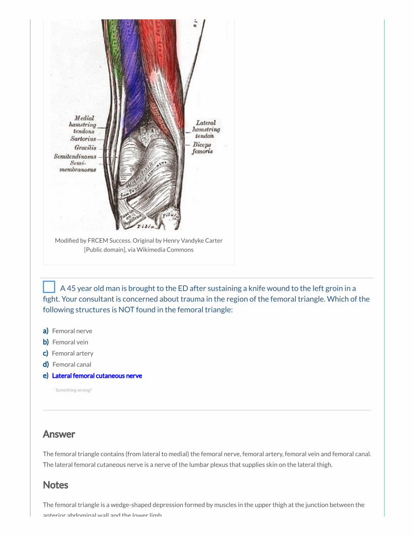
The femoral triangle contains (from lateral to medial) the femoral nerve, femoral artery, femoral vein and femoral canal.
The lateral femoral cutaneous nerve is a nerve of the lumbar plexus that supplies skin on the lateral thigh.
Notes
The femoral triangle is a wedge-shaped depression formed by muscles in the upper thigh at the junction between the
anterior abdominal wall and the lower limb.
Something wrong?

anterior abdominal wall and the lower limb.
Femoral Triangle Structure(s)
Superior border Inguinal ligament
Lateral border Medial sartorius muscle
Medial border Medial adductor longus muscle
Roof Fascia lata
Floor Pectineus, iliopsoas and adductor longus muscles
Contents Femoral nerve, femoral artery, femoral vein, femoral canal
Borders
The femoral triangle is bordered superiorly by the inguinal ligament, laterally by the medial border of the sartorius
muscle and medially by the medial border of the adductor longus muscle. The roof is formed by the fascia lata and the
oor is formed by the pectineus, iliopsoas and adductor longus muscles.
Contents
The femoral triangle contains (from lateral to medial):
The femoral nerve
The femoral artery
The femoral vein
The femoral canal
Femoral sheath
The femoral sheath originates as a prolongation of extraperitoneal fascia and encloses the femoral artery, femoral vein
and associated lymphatic vessels (contained within the femoral canal). The femoral nerve does NOT lie within the
sheath, but instead lies lateral to it. The femoral sheath terminates inferiorly by blending with the adventitia of the
femoral vessels approximately 4 cm inferior of the inguinal ligament.

By Henry Vandyke Carter [Public domain], via
Wikimedia Commons
By Henry Vandyke Carter [Public domain], via
Wikimedia Commons
Femoral canal
The femoral canal lies between the medial edge of the femoral sheath and the femoral vein. The femoral canal contains
loose connective tissue, lymphatic vessels, deep inguinal lymph nodes and empty space. The femoral canal allows the
femoral vein to expand, for example, when venous return from the leg is increased, or when increased intra-abdominal
pressure causes a temporary stasis.
Femoral ring
The femoral ring is formed by the proximal abdominal opening of the femoral canal, and is important clinically, as it is a
common site of hernia. It is bordered anteriorly by the inguinal ligament, posteriorly by the pectineal ligament,
medially by the lacunar ligament and laterally by the femoral vein.
Femoral ring Structure(s)
Anterior border Inguinal ligament
Posterior border Pectineal ligament
Medial border Lacunar ligament
Lateral border Femoral vein

By Henry Vandyke Carter [Public domain], via Wikimedia Commons
A 49 year old overweight female presents to ED complaining of a lump in her groin. You
examine the patient and diagnose a femoral hernia. Which of the following structures would be
found immediately lateral to the hernia:
a) Femoral artery
b) Femoral nerve
c) Femoral vein
d) Super cial epigastric vein
e) Ilioinguinal nerve
Answer
The femoral vein will be immediately lateral. Femoral hernias result from the herniation of abdominal structures
through the femoral ring, the proximal opening of the femoral canal, which is bordered medially by the lacunar
ligament and laterally by the femoral vein. Femoral hernias, more common in women, typically present as a lump
inferolaterally to the pubic tubercle, and are more prone to strangulation than inguinal hernias.
Notes
The femoral triangle is a wedge-shaped depression formed by muscles in the upper thigh at the junction between the
anterior abdominal wall and the lower limb.
Femoral Triangle Structure(s)
Superior border Inguinal ligament
Lateral border Medial sartorius muscle
Medial border Medial adductor longus muscle
Something wrong?
By Henry Vandyke Carter [Public domain], via
Wikimedia Commons
Roof Fascia lata
Floor Pectineus, iliopsoas and adductor longus muscles
Contents Femoral nerve, femoral artery, femoral vein, femoral canal
Borders
The femoral triangle is bordered superiorly by the inguinal ligament, laterally by the medial border of the sartorius
muscle and medially by the medial border of the adductor longus muscle. The roof is formed by the fascia lata and the
oor is formed by the pectineus, iliopsoas and adductor longus muscles.
Contents
The femoral triangle contains (from lateral to medial):
The femoral nerve
The femoral artery
The femoral vein
The femoral canal
Femoral sheath
The femoral sheath originates as a prolongation of extraperitoneal fascia and encloses the femoral artery, femoral vein
and associated lymphatic vessels (contained within the femoral canal). The femoral nerve does NOT lie within the
sheath, but instead lies lateral to it. The femoral sheath terminates inferiorly by blending with the adventitia of the
femoral vessels approximately 4 cm inferior of the inguinal ligament.
By Henry Vandyke Carter [Public domain], via
Wikimedia Commons
Wikimedia Commons
Femoral canal
The femoral canal lies between the medial edge of the femoral sheath and the femoral vein. The femoral canal contains
loose connective tissue, lymphatic vessels, deep inguinal lymph nodes and empty space. The femoral canal allows the
femoral vein to expand, for example, when venous return from the leg is increased, or when increased intra-abdominal
pressure causes a temporary stasis.
Femoral ring
The femoral ring is formed by the proximal abdominal opening of the femoral canal, and is important clinically, as it is a
common site of hernia. It is bordered anteriorly by the inguinal ligament, posteriorly by the pectineal ligament,
medially by the lacunar ligament and laterally by the femoral vein.
Femoral ring Structure(s)
Anterior border Inguinal ligament
Posterior border Pectineal ligament
Medial border Lacunar ligament
Lateral border Femoral vein

By Henry Vandyke Carter [Public domain], via Wikimedia Commons
You are examining a 38 year old woman who presents with lower back pain associated with
bilateral sciatica. On examination you note loss of sensation in the L1 dermatome. You are
concerned about a possible diagnosis of cauda equina. The L1 dermatome is best tested at which of
the following landmarks:
a) At the umbilicus in the midclavicular line
b) At the midpoint of the inguinal ligament
c) At a point on the posterolateral thigh
d) At a point on the upper anterior thigh
e) At the medial femoral condyle
Answer
The L1 dermatome is best tested on the upper anterior thigh, midway between the key sensory points for T12
(midpoint of the inguinal ligament) and L2 (mid anterior thigh).
Notes
Dermatome Landmark
L1 Upper Anterior Thigh
L2 Mid Anterior Thigh
L3 Medial Femoral Condyle
L4 Medial Malleolus
L5 Dorsum 3rd MTP Joint
S1 Lateral Heel
S2 Popliteal Fossa
S3 Ischial Tuberosity
S5 Perianal Area
Something wrong?
The T12 dermatome is best tested at the midclavicular line, over the midpoint of the inguinal ligament.
The L1 dermatome is best tested on the upper anterior thigh, at a point midway between the key sensory
points for T12 and L2.
The L2 dermatome is best tested on the anteromedial thigh, at the midpoint drawn on an imaginary line
connecting the midpoint of the inguinal ligament and the medial femoral condyle.
The L3 dermatome is best tested at the medial femoral condyle above the knee.
The L4 dermatome is best tested over the medial malleolus.
The L5 dermatome is best tested on the dorsum of the foot at the third metatarsophalangeal joint.
The S1 dermatome is best tested on the lateral aspect of the calcaneus.
The S2 dermatome is best tested at the midpoint of the popliteal fossa.
The S3 dermatome is best tested over the ischial tuberosity or infragluteal fold (depending on the patient their
skin can move up, down or laterally over the ischii).
The S4/S5 dermatome is best tested in the perianal area, less than one cm lateral to the mucocutaneous
junction.
By Grant, John Charles Boileau (An atlas of anatomy, / by regions 1962) [Public domain], via
Wikimedia Commons

A 87 year old woman presents to the ED after her carers noted she was complaining of pain in
the right groin. On examination you note a tender swelling in keeping with a femoral hernia. The
femoral ring is bordered laterally by which of the following structures:
a) Femoral artery
b) Femoral nerve
c) Femoral vein
d) Femoral sheath
e) Great saphenous vein
Answer
The femoral ring is bordered anteriorly by the inguinal ligament, posteriorly by the pectineal ligament, medially by the
lacunar ligament and laterally by the femoral vein.
Notes
The femoral triangle is a wedge-shaped depression formed by muscles in the upper thigh at the junction between the
anterior abdominal wall and the lower limb.
Femoral Triangle Structure(s)
Superior border Inguinal ligament
Lateral border Medial sartorius muscle
Medial border Medial adductor longus muscle
Roof Fascia lata
Floor Pectineus, iliopsoas and adductor longus muscles
Contents Femoral nerve, femoral artery, femoral vein, femoral canal
Borders
The femoral triangle is bordered superiorly by the inguinal ligament, laterally by the medial border of the sartorius
muscle and medially by the medial border of the adductor longus muscle. The roof is formed by the fascia lata and the
oor is formed by the pectineus, iliopsoas and adductor longus muscles.
Contents
Something wrong?

By Henry Vandyke Carter [Public domain], via
Wikimedia Commons
By Henry Vandyke Carter [Public domain], via
Wikimedia Commons
Contents
The femoral triangle contains (from lateral to medial):
The femoral nerve
The femoral artery
The femoral vein
The femoral canal
Femoral sheath
The femoral sheath originates as a prolongation of extraperitoneal fascia and encloses the femoral artery, femoral vein
and associated lymphatic vessels (contained within the femoral canal). The femoral nerve does NOT lie within the
sheath, but instead lies lateral to it. The femoral sheath terminates inferiorly by blending with the adventitia of the
femoral vessels approximately 4 cm inferior of the inguinal ligament.
Femoral canal
The femoral canal lies between the medial edge of the femoral sheath and the femoral vein. The femoral canal contains
loose connective tissue, lymphatic vessels, deep inguinal lymph nodes and empty space. The femoral canal allows the
femoral vein to expand, for example, when venous return from the leg is increased, or when increased intra-abdominal
pressure causes a temporary stasis.
Femoral ring

The femoral ring is formed by the proximal abdominal opening of the femoral canal, and is important clinically, as it is a
common site of hernia. It is bordered anteriorly by the inguinal ligament, posteriorly by the pectineal ligament,
medially by the lacunar ligament and laterally by the femoral vein.
Femoral ring Structure(s)
Anterior border Inguinal ligament
Posterior border Pectineal ligament
Medial border Lacunar ligament
Lateral border Femoral vein
By Henry Vandyke Carter [Public domain], via Wikimedia Commons
A patient with very poor peripheral access requires urgent intravenous uids. The femoral
vein in his groin is determined the most accessible vessel. Which of the following landmarks is the
most accurate to identify the femoral vein:
a) Just lateral to the femoral artery pulsation
b) Just medial to the femoral artery pulsation
c) Within the femoral canal
d) Just medial to the femoral nerve
e) Just lateral to the femoral nerve

e) Just lateral to the femoral nerve
Answer
The femoral artery can be palpated in the femoral triangle as it passes over the femoral head, just inferior to the
inguinal ligament, midway between the anterior superior iliac spine and the pubic symphysis (at the mid-inguinal
point). The femoral vein lies immediately medial to this pulsation.
Notes
The major artery supplying the lower limb is the femoral artery. The femoral artery is the continuation of the external
iliac artery, beginning as the vessel passes under the inguinal ligament to enter the femoral triangle in the anterior
thigh.
The femoral artery can be palpated in the femoral triangle as it passes over the femoral head, just inferior to the
inguinal ligament, midway between the anterior superior iliac spine and the pubic symphysis (at the mid-inguinal
point). The femoral vein lies immediately medial to this pulsation, which is an important landmark for central venous
line insertion. Medial to the femoral vein is the femoral canal which contains lymphatics and lies immediately lateral to
the pubic tubercle. The femoral nerve lies lateral to the femoral artery.
The femoral artery gives rise to the deep profunda femoris artery in the femoral triangle which is a major source of
blood supply to the medial and posterior compartments of the thigh and the proximal femur.
After exiting the femoral triangle, the femoral artery continues down the anterior surface of the thigh via the adductor
canal. During its descent it supplies the anterior thigh, giving rise to numerous super cial cutaneous branches.
The femoral artery becomes the popliteal artery after entering the posterior compartment of the thigh through the
adductor hiatus just proximal to the knee.
Something wrong?

Modi ed by FRCEM Success. Original by Henry Vandyke Carter [Public domain], via Wikimedia
Commons
You are examining the lower limbs of a 54 year old man who presented after falling from a
ladder at home. During your neurological assessment you note a weakness of hip extension. Which
of the following nerves are most important for extension of the thigh at the hip joint:
a) Superior and inferior gluteal nerves
b) Superior gluteal and sciatic nerve
c) Inferior gluteal and sciatic nerve
d) Sciatic and femoral nerve
e) Sciatic and obturator nerve
Answer
Extension of the thigh at the hip joint is primarily produced by the hamstring muscles, and the gluteus maximus,
innervated by the sciatic nerve and the inferior gluteal nerve respectively.
Notes
Joint Hip joint
Something wrong?

Type Synovial ball and socket joint
Articulations Head of femur with acetabulum of pelvis
Stabilising
factors
Acetabular labrum, thickened brous capsule, extracapsular ligaments (iliofemoral, ischiofemoral,
pubofemoral), medial rotator muscles (effectively ‘pull’ head of femur into acetabulum)
Movements Flexion/Extension, Abduction/Adduction, Medial/Lateral rotation, Circumduction
Blood supply Branches of obturator artery, medial and lateral circum ex branches of profunda femoris artery
and superior and inferior gluteal arteries
Innervation Femoral nerve, obturator nerve, superior gluteal nerve and nerve to the quadratus femoris
Joint articulations
The hip joint is a multiaxial synovial ball and socket joint occurring between the head of the femur and the acetabulum
of the pelvis.
The acetabulum is formed by the fusion of the three bones, the ilium, the ischium and the pubis.
By Henry Vandyke Carter [Public domain], via Wikimedia Commons

By Henry Vandyke Carter [Public domain], via Wikimedia Commons
The rim of the acetabulum is raised slightly by the brocartilaginous acetabular labrum which increases its depth,
improving stability of the joint. The acetabular labrum continues inferiorly as the transverse acetabular ligament which
bridges the acetabular notch and converts the notch into a foramen for the passage of nutrient vessels and nerves.
Ligaments
The intracapsular ligament of the head of femur (ligamentum teres) runs from the fovea on the head of the femur at
one end to the acetabular fossa and the transverse acetabular ligament on the other. It carries a small acetabular
branch of the obturator artery.
By Henry Vandyke Carter [Public domain], via Wikimedia Commons
There are three main stabilising extracapsular ligaments; the iliofemoral (the largest and strongest ligament), the
pubofemoral and the ischiofemoral ligament.
The iliofemoral ligament is anterosuperior to the hip joint, attached proximally to the ilium just below the
anterior inferior iliac spine and distally to the intertrochanteric line of the femur. This ligament speci cally
prevents hyperextension and lateral rotation of the hip joint whilst standing.
The pubofemoral ligament is anteroinferior to the hip joint, attached to the pelvis at the iliopubic eminence
and adjacent bone and blending distally with the articular capsule. This ligament prevents excessive abduction
and extension of the femur at the hip joint.

Modi ed by FRCEM Success. Original by Henry
Vandyke Carter [Public domain], via Wikimedia
Commons By Henry Vandyke Carter [Public domain], via
Wikimedia Commons
and extension of the femur at the hip joint.
The ischiofemoral ligament is posterior to the hip joint, attached medially to the ischium and laterally to the
greater trochanter. This ligament prevents excessive extension and medial rotation of the femur at the hip
joint.
Blood supply
The hip joint receives its blood supply primarily from branches of the obturator artery, the medial and lateral
circum ex arteries (branches of the profunda femoris artery) and the superior and inferior gluteal arteries. The
articular branches of these vessels form a network around the joint.
Innervation
The hip joint is innervated by articular branches from the femoral nerve (anteriorly), obturator nerve (inferiorly),
superior gluteal nerve (superiorly), and the nerve to the quadratus femoris (posteriorly).
Joint movements
The hip joint allows the movements of exion and extension, abduction and adduction, medial and lateral rotation and
circumduction.
Movement Muscles Involved
Flexion Sartorius, Iliacus, Psoas major, Pectineus, Rectus femoris

Extension Hamstrings, Gluteus maximus
Abduction Gluteus maximus, medius and minimus, Obturator internus, Gemelli, Piriformis, Sartorius
Adduction Adductor longus, magnus and brevis, Gracilis, Pectineus
Medial
rotation
Gluteus medius and minimus, Adductor longus, magnus and brevis, Semitendinosus and
Semimembranosus
Lateral
rotation
Obturator externus, Sartorius, Iliacus, Psoas major, Biceps femoris, Piriformis, Gluteus maximus,
Obturator internus, Gemelli, Quadriceps femoris
A 76 year old woman present to the ED after feeling in a “pop” in her right hip. She recently
underwent a right total hip replacement, you suspect the hip is now dislocated. Regarding the
extracapsular ligaments of the hip, which one of the following statements is INCORRECT:
a) The iliofemoral ligament is the strongest of the extracapsular ligaments.
b) There are three main extracapsular ligaments stabilising the hip joint.
c) The pubofemoral ligament is anteroinferior to the hip joint.
d) The ischiofemoral ligament is anterosuperior to the hip joint.
e) The extracapsular ligaments all act to prevent excessive extension of the hip joint.
Answer
There are three main stabilising extracapsular ligaments; the iliofemoral (the largest and strongest ligament), the
pubofemoral and the ischiofemoral ligament. The iliofemoral ligament is anterosuperior to the hip joint and speci cally
prevents hyperextension and lateral rotation of the hip joint whilst standing. The pubofemoral ligament is
anteroinferior to the hip joint and acts to prevent excessive extension and abduction of the hip joint. The ischiofemoral
ligament is posterior to the hip joint and acts to prevent excessive extension and medial rotation of the hip joint.
Notes
Joint Hip joint
Type Synovial ball and socket joint
Articulations Head of femur with acetabulum of pelvis
Stabilising
factors
Acetabular labrum, thickened brous capsule, extracapsular ligaments (iliofemoral, ischiofemoral,
pubofemoral), medial rotator muscles (effectively ‘pull’ head of femur into acetabulum)
Movements Flexion/Extension, Abduction/Adduction, Medial/Lateral rotation, Circumduction
Something wrong?
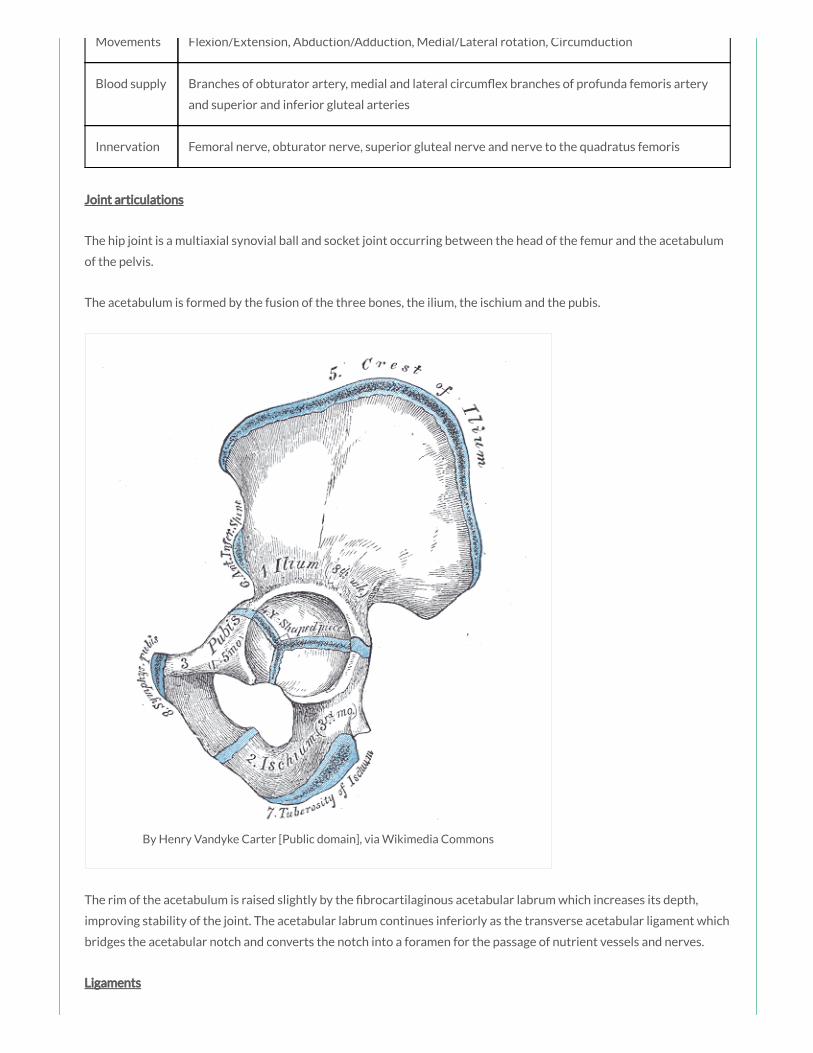
Movements Flexion/Extension, Abduction/Adduction, Medial/Lateral rotation, Circumduction
Blood supply Branches of obturator artery, medial and lateral circum ex branches of profunda femoris artery
and superior and inferior gluteal arteries
Innervation Femoral nerve, obturator nerve, superior gluteal nerve and nerve to the quadratus femoris
Joint articulations
The hip joint is a multiaxial synovial ball and socket joint occurring between the head of the femur and the acetabulum
of the pelvis.
The acetabulum is formed by the fusion of the three bones, the ilium, the ischium and the pubis.
By Henry Vandyke Carter [Public domain], via Wikimedia Commons
The rim of the acetabulum is raised slightly by the brocartilaginous acetabular labrum which increases its depth,
improving stability of the joint. The acetabular labrum continues inferiorly as the transverse acetabular ligament which
bridges the acetabular notch and converts the notch into a foramen for the passage of nutrient vessels and nerves.
Ligaments
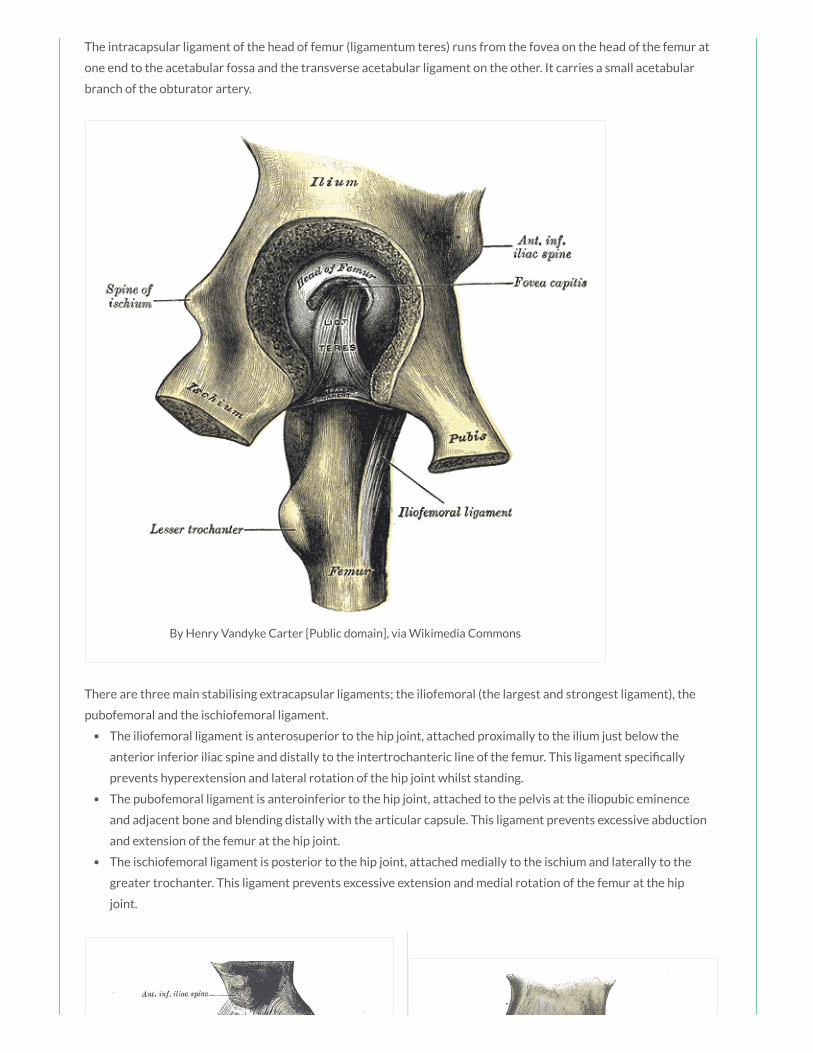
The intracapsular ligament of the head of femur (ligamentum teres) runs from the fovea on the head of the femur at
one end to the acetabular fossa and the transverse acetabular ligament on the other. It carries a small acetabular
branch of the obturator artery.
By Henry Vandyke Carter [Public domain], via Wikimedia Commons
There are three main stabilising extracapsular ligaments; the iliofemoral (the largest and strongest ligament), the
pubofemoral and the ischiofemoral ligament.
The iliofemoral ligament is anterosuperior to the hip joint, attached proximally to the ilium just below the
anterior inferior iliac spine and distally to the intertrochanteric line of the femur. This ligament speci cally
prevents hyperextension and lateral rotation of the hip joint whilst standing.
The pubofemoral ligament is anteroinferior to the hip joint, attached to the pelvis at the iliopubic eminence
and adjacent bone and blending distally with the articular capsule. This ligament prevents excessive abduction
and extension of the femur at the hip joint.
The ischiofemoral ligament is posterior to the hip joint, attached medially to the ischium and laterally to the
greater trochanter. This ligament prevents excessive extension and medial rotation of the femur at the hip
joint.

Modi ed by FRCEM Success. Original by Henry
Vandyke Carter [Public domain], via Wikimedia
Commons By Henry Vandyke Carter [Public domain], via
Wikimedia Commons
Blood supply
The hip joint receives its blood supply primarily from branches of the obturator artery, the medial and lateral
circum ex arteries (branches of the profunda femoris artery) and the superior and inferior gluteal arteries. The
articular branches of these vessels form a network around the joint.
Innervation
The hip joint is innervated by articular branches from the femoral nerve (anteriorly), obturator nerve (inferiorly),
superior gluteal nerve (superiorly), and the nerve to the quadratus femoris (posteriorly).
Joint movements
The hip joint allows the movements of exion and extension, abduction and adduction, medial and lateral rotation and
circumduction.
Movement Muscles Involved
Flexion Sartorius, Iliacus, Psoas major, Pectineus, Rectus femoris
Extension Hamstrings, Gluteus maximus
Abduction Gluteus maximus, medius and minimus, Obturator internus, Gemelli, Piriformis, Sartorius
Adduction Adductor longus, magnus and brevis, Gracilis, Pectineus
Medial
rotation
Gluteus medius and minimus, Adductor longus, magnus and brevis, Semitendinosus and
Semimembranosus
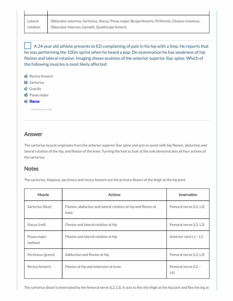
rotation Semimembranosus
Lateral
rotation
Obturator externus, Sartorius, Iliacus, Psoas major, Biceps femoris, Piriformis, Gluteus maximus,
Obturator internus, Gemelli, Quadriceps femoris
A 24 year old athlete presents to ED complaining of pain in his hip with a limp. He reports that
he was performing the 100m sprint when he heard a pop. On examination he has weakness of hip
exion and lateral rotation. Imaging shows avulsion of the anterior superior iliac spine. Which of
the following muscles is most likely affected:
a) Rectus femoris
b) Sartorius
c) Gracilis
d) Psoas major
e) Iliacus
Answer
The sartorius muscle originates from the anterior superior iliac spine and acts to assist with hip exion, abduction and
lateral rotation of the hip, and exion of the knee. Turning the foot to look at the sole demonstrates all four actions of
the sartorius.
Notes
The sartorius, iliopsoas, pectineus and rectus femoris are the primary exors of the thigh at the hip joint.
Muscle Actions Innervation
Sartorius (blue) Flexion, abduction and lateral rotation at hip and exion at
knee
Femoral nerve (L2, L3)
Iliacus (red) Flexion and lateral rotation at hip Femoral nerve (L2, L3)
Psoas major
(yellow)
Flexion and lateral rotation at hip Anterior rami L1 – L3
Pectineus (green) Adduction and exion at hip Femoral nerve (L2, L3)
Rectus femoris Flexion at hip and extension at knee Femoral nerve (L2 –
L4)
The sartorius (blue) is innervated by the femoral nerve (L2, L3). It acts to ex the thigh at the hip joint and ex the leg at
Something wrong?

the knee joint. It also abducts the thigh and rotates it laterally, as when resting the foot on the opposite knee when
sitting.
The iliacus (red) and psoas major (yellow) muscles originate as separate muscles but insert by a common tendon onto
the femur and together are referred to as the iliopsoas muscle, which is a powerful exor of the thigh at the hip joint
and also contributes to lateral rotation of the thigh at the hip joint. The iliacus is innervated by the femoral nerve (L2,
L3). The psoas major is innervated by the anterior rami of spinal nerves L1 – L3.
The pectineus (green) is innervated by the femoral nerve (L2, L3). It acts to adduct and ex the thigh at the hip joint.
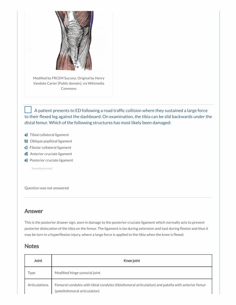
Modi ed by FRCEM Success. Original by Henry
Vandyke Carter [Public domain], via Wikimedia
Commons
A patient presents to ED following a road traf c collision where they sustained a large force
to their exed leg against the dashboard. On examination, the tibia can be slid backwards under the
distal femur. Which of the following structures has most likely been damaged:
a) Tibial collateral ligament
b) Oblique popliteal ligament
c) Fibular collateral ligament
d) Anterior cruciate ligament
e) Posterior cruciate ligament
Question was not answered
Answer
This is the posterior drawer sign, seen in damage to the posterior cruciate ligament which normally acts to prevent
posterior dislocation of the tibia on the femur. The ligament is lax during extension and taut during exion and thus it
may be torn in a hyper exion injury, where a large force is applied to the tibia when the knee is exed.
Notes
Joint Knee joint
Type Modi ed hinge synovial joint
Articulations Femoral condyles with tibial condyles (tibiofemoral articulation) and patella with anterior femur
(patellofemoral articulation)
Something wrong?
Stabilising
factors
Fibrous capsule, tibial spines, menisci, tibial/ bular collateral ligament, anterior/posterior
cruciate ligament, vastus medialis and lateralis muscles, oblique popliteal ligament, iliotibial tract,
muscle tendons (hamstrings, gastrocnemius, sartorius, gracilis)
Movements Flexion/Extension, Medial/Lateral rotation in exed position
Joint articulations
The knee joint is formed from two articulations:
the main weight bearing tibiofemoral articulation between the two femoral condyles and the adjacent surfaces
of the superior aspect of the tibial condyles
the patellofemoral articulation between the anterior femur and the patella which allows the pull of the
quadriceps femoris muscle to be directed anteriorly over the knee to the tibia without tendon wear
Joint movements
The knee joint is a modi ed hinge synovial joint, allowing mainly exion and extension, but also a small degree of
medial and lateral rotation.
Movement Muscles Involved
Flexion Hamstrings, Gracilis, Sartorius, Gastrocnemius, Plantaris
Extension Quadriceps femoris
When standing, the knee joint is ‘locked’ in position to reduce the amount of muscle work needed to maintain the
standing weight bearing position. This locking mechanism occurs partly due to the change in the shape/size of the
articulating femoral surfaces (in the exed position, the surfaces of the femoral condyles that articulate with the tibia
are curved/round, but in extension, the surfaces are at, and consequently the joint surfaces become larger and more
stable in extension) and partly due to medial rotation of the femur on the tibia in full extension; medial rotation and full
extension tightens all the associated ligaments (the screw home mechanism). Contraction of the popliteus muscle
‘unlocks’ the knee by initiating lateral rotation of the femur on the tibia, and allowing exion.
Joint capsule
The brous membrane of the knee joint is reinforced anteriorly by the tendinous expansions of the vastus lateralis and
vastus medialis muscles, anterolaterally by a brous extension from the iliotibial tract and posteromedially by the
oblique popliteal ligament, an extension from the tendon of the semimembranosus muscle (the oblique popliteal
ligament resists hyperextension and lateral rotation of the leg). The upper end of the popliteus muscle passes through
an opening in the posterolateral aspect of the brous membrane of the knee.
Menisci
The two menisci are C-shaped brocartilaginous structures that lie between the femoral condyles and the tibia,
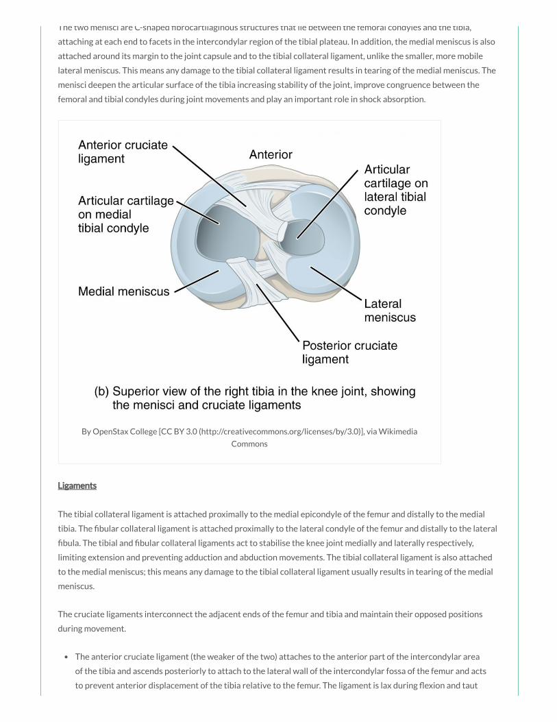
The two menisci are C-shaped brocartilaginous structures that lie between the femoral condyles and the tibia,
attaching at each end to facets in the intercondylar region of the tibial plateau. In addition, the medial meniscus is also
attached around its margin to the joint capsule and to the tibial collateral ligament, unlike the smaller, more mobile
lateral meniscus. This means any damage to the tibial collateral ligament results in tearing of the medial meniscus. The
menisci deepen the articular surface of the tibia increasing stability of the joint, improve congruence between the
femoral and tibial condyles during joint movements and play an important role in shock absorption.
By OpenStax College [CC BY 3.0 (http://creativecommons.org/licenses/by/3.0)], via Wikimedia
Commons
Ligaments
The tibial collateral ligament is attached proximally to the medial epicondyle of the femur and distally to the medial
tibia. The bular collateral ligament is attached proximally to the lateral condyle of the femur and distally to the lateral
bula. The tibial and bular collateral ligaments act to stabilise the knee joint medially and laterally respectively,
limiting extension and preventing adduction and abduction movements. The tibial collateral ligament is also attached
to the medial meniscus; this means any damage to the tibial collateral ligament usually results in tearing of the medial
meniscus.
The cruciate ligaments interconnect the adjacent ends of the femur and tibia and maintain their opposed positions
during movement.
The anterior cruciate ligament (the weaker of the two) attaches to the anterior part of the intercondylar area
of the tibia and ascends posteriorly to attach to the lateral wall of the intercondylar fossa of the femur and acts
to prevent anterior displacement of the tibia relative to the femur. The ligament is lax during exion and taut
during extension thus it may be torn when the knee is hyperextended (or by the application of a large force to

during extension thus it may be torn when the knee is hyperextended (or by the application of a large force to
the back of the knee with the joint partly exed). The anterior drawer sign may be seen where there is forward
sliding of the tibia on the femur.
The posterior cruciate ligament (the stronger of the two) attaches to the posterior part of the intercondylar
area of the tibia and ascends anteriorly to attach to the medial wall of the intercondylar fossa of the femur and
acts to prevent posterior dislocation of the tibia relative to the femur. The ligament is lax during extension
and taut during exion and thus it may be torn in a hyper exion injury, where a large force is applied to the
tibia when the knee is exed. The posterior drawer sign may be seen where there is backward sliding of the
tibia on the femur.
The ‘unhappy triad’ typically occurs due to a lateral force to an extended knee, e.g. in a football tackle. It refers to injury
of the anterior cruciate ligament (due to forward displacement of the tibia), the tibial collateral ligament (due to
excessive abduction) and the medial meniscus (due to its attachment on the tibial collateral ligament).
Bursae
The synovial membrane of the knee joint forms pouches in two locations to provide low-friction surfaces for the
movement of tendons associated with the joint:
The subpopliteal recess – extends posterolaterally and lies between the lateral meniscus and the tendon of the
popliteus muscle
The suprapatellar bursa extends superiorly between the distal end of the shaft of the femur and the quadriceps
femoris muscle and tendon
Other bursae associated with the knee, but not normally communicating with the synovial joint, include the
subcutaneous prepatellar bursa, the deep and subcutaneous infrapatellar bursae separated by the patella ligament,

and numerous other bursae associated with tendons and ligaments around the knee joint. Housemaid’s knee is
in ammation of the prepatellar bursa, and Clergyman’s knee is in ammation of the subcutaneous infrapatellar bursa.
A 28 year old man was involved in a road traf c accident, and subsequently complained of
weakness in extending his leg at the knee joint. Following examination, he was diagnosed with a
lesion of his femoral nerve. Which of the following clinical ndings would you most expect to see in
this patient:
a) Paralysis of the psoas major muscle
b) Loss of sensation over the lateral thigh
c) Paralysis of the rectus femoris muscle
d) Loss of sensation over the lateral leg
e) Paralysis of the gracilis muscle
Answer
The femoral nerve innervates the iliacus, pectineus, sartorius and quadriceps femoris muscles, and supplies skin on the
anterior thigh, anteromedial knee and medial leg.
Notes
The femoral nerve arises from the lumbar plexus, receiving bres from the anterior rami of L2 – L4.
Nerve Femoral nerve
Nerve roots L2 – L4
Motor supply Iliacus, pectineus, sartorius, quadriceps femoris
Sensory supply Skin over the anterior thigh, anteromedial knee, medial leg and medial foot
Motor loss in injury Weak exion at hip and loss of extension at knee
Anatomical course
The femoral nerve descends from the lumbar plexus in the posterior abdomen through the substance of the psoas
major muscle, emerging from the lower lateral border of the psoas major. Continuing its descent, the femoral nerve lies
between the lateral border of the psoas major and the anterior surface of the iliacus muscle. It is deep to the iliacus
fascia and lateral to the femoral artery as it passes posterior to the mid-inguinal point to enter the femoral triangle in
the anterior compartment of the thigh, before dividing into an anterior and posterior division.
Something wrong?
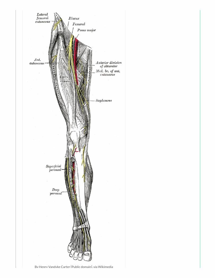
By Henry Vandyke Carter [Public domain], via Wikimedia
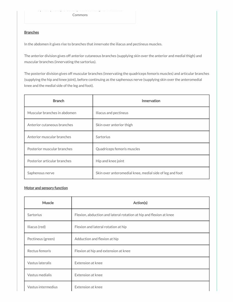
By Henry Vandyke Carter [Public domain], via Wikimedia
Commons
Branches
In the abdomen it gives rise to branches that innervate the iliacus and pectineus muscles.
The anterior division gives off anterior cutaneous branches (supplying skin over the anterior and medial thigh) and
muscular branches (innervating the sartorius).
The posterior division gives off muscular branches (innervating the quadriceps femoris muscles) and articular branches
(supplying the hip and knee joint), before continuing as the saphenous nerve (supplying skin over the anteromedial
knee and the medial side of the leg and foot).
Branch Innervation
Muscular branches in abdomen Iliacus and pectineus
Anterior cutaneous branches Skin over anterior thigh
Anterior muscular branches Sartorius
Posterior muscular branches Quadriceps femoris muscles
Posterior articular branches Hip and knee joint
Saphenous nerve Skin over anteromedial knee, medial side of leg and foot
Motor and sensory function
Muscle Action(s)
Sartorius Flexion, abduction and lateral rotation at hip and exion at knee
Iliacus (red) Flexion and lateral rotation at hip
Pectineus (green) Adduction and exion at hip
Rectus femoris Flexion at hip and extension at knee
Vastus lateralis Extension at knee
Vastus medialis Extension at knee
Vastus intermedius Extension at knee
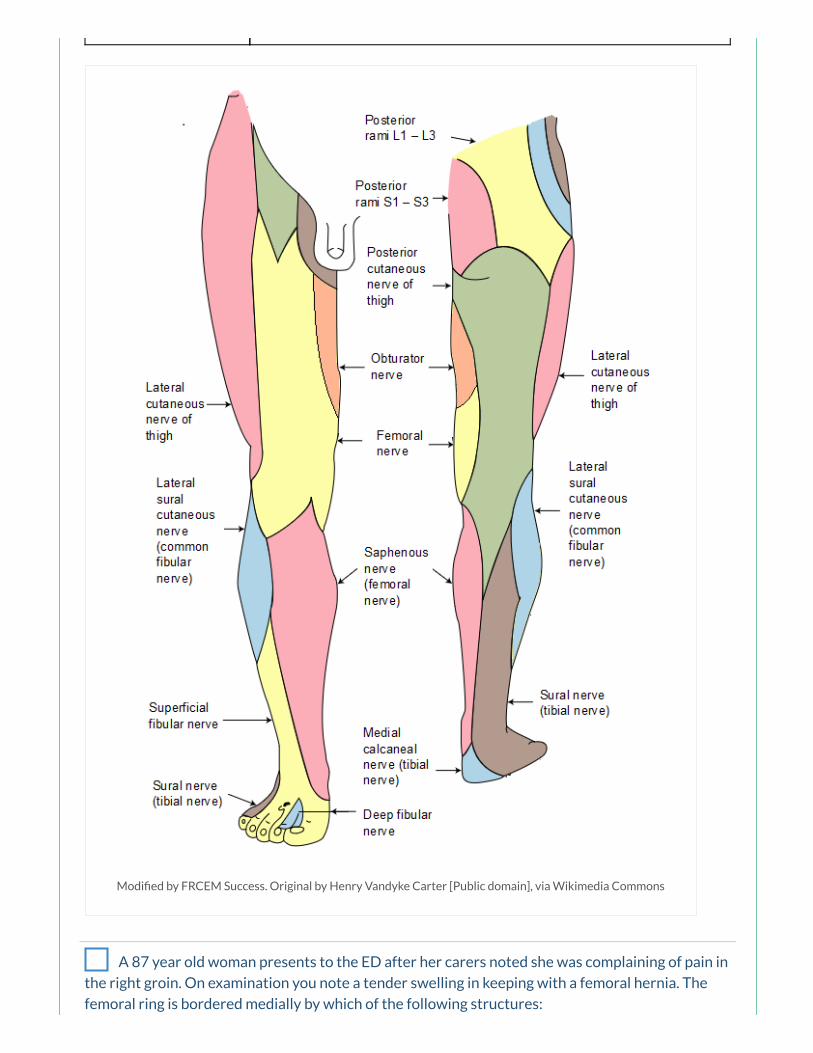
Modi ed by FRCEM Success. Original by Henry Vandyke Carter [Public domain], via Wikimedia Commons
A 87 year old woman presents to the ED after her carers noted she was complaining of pain in
the right groin. On examination you note a tender swelling in keeping with a femoral hernia. The
femoral ring is bordered medially by which of the following structures:
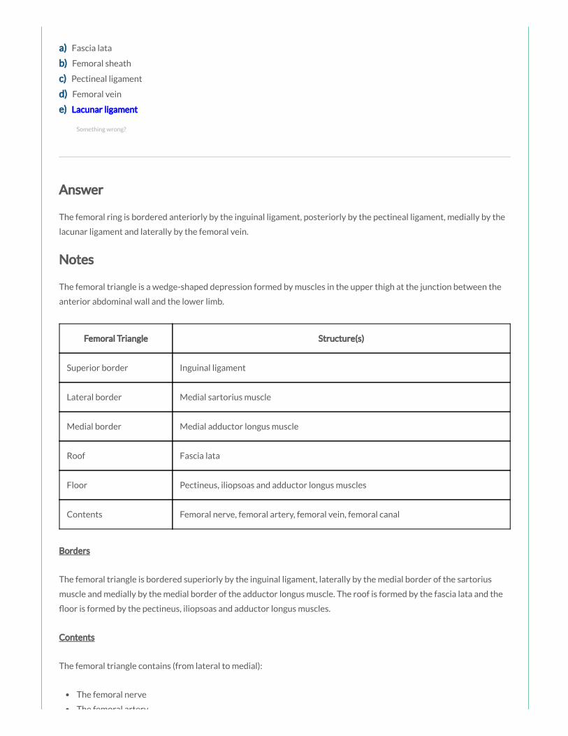
a) Fascia lata
b) Femoral sheath
c) Pectineal ligament
d) Femoral vein
e) Lacunar ligament
Answer
The femoral ring is bordered anteriorly by the inguinal ligament, posteriorly by the pectineal ligament, medially by the
lacunar ligament and laterally by the femoral vein.
Notes
The femoral triangle is a wedge-shaped depression formed by muscles in the upper thigh at the junction between the
anterior abdominal wall and the lower limb.
Femoral Triangle Structure(s)
Superior border Inguinal ligament
Lateral border Medial sartorius muscle
Medial border Medial adductor longus muscle
Roof Fascia lata
Floor Pectineus, iliopsoas and adductor longus muscles
Contents Femoral nerve, femoral artery, femoral vein, femoral canal
Borders
The femoral triangle is bordered superiorly by the inguinal ligament, laterally by the medial border of the sartorius
muscle and medially by the medial border of the adductor longus muscle. The roof is formed by the fascia lata and the
oor is formed by the pectineus, iliopsoas and adductor longus muscles.
Contents
The femoral triangle contains (from lateral to medial):
The femoral nerve
The femoral artery
Something wrong?

By Henry Vandyke Carter [Public domain], via
Wikimedia Commons
By Henry Vandyke Carter [Public domain], via
Wikimedia Commons
The femoral artery
The femoral vein
The femoral canal
Femoral sheath
The femoral sheath originates as a prolongation of extraperitoneal fascia and encloses the femoral artery, femoral vein
and associated lymphatic vessels (contained within the femoral canal). The femoral nerve does NOT lie within the
sheath, but instead lies lateral to it. The femoral sheath terminates inferiorly by blending with the adventitia of the
femoral vessels approximately 4 cm inferior of the inguinal ligament.
Femoral canal
The femoral canal lies between the medial edge of the femoral sheath and the femoral vein. The femoral canal contains
loose connective tissue, lymphatic vessels, deep inguinal lymph nodes and empty space. The femoral canal allows the
femoral vein to expand, for example, when venous return from the leg is increased, or when increased intra-abdominal
pressure causes a temporary stasis.
Femoral ring
The femoral ring is formed by the proximal abdominal opening of the femoral canal, and is important clinically, as it is a
common site of hernia. It is bordered anteriorly by the inguinal ligament, posteriorly by the pectineal ligament,
medially by the lacunar ligament and laterally by the femoral vein.

Femoral ring Structure(s)
Anterior border Inguinal ligament
Posterior border Pectineal ligament
Medial border Lacunar ligament
Lateral border Femoral vein
By Henry Vandyke Carter [Public domain], via Wikimedia Commons
A 76 year old woman presents with left ank pain and fever. A CT scan has shown a psoas
abscess. The psoas major muscle acts primarily to produce which of the following movements:
a) Flexion and lateral rotation of the thigh
b) Flexion of the thigh and exion of the leg
c) Flexion and abduction of the thigh
d) Flexion and adduction of the thigh
e) Flexion of the thigh and extension of the leg
Answer
Something wrong?

The iliacus (red) and psoas major (yellow) muscles originate as separate muscles but insert by a common tendon onto
the femur and together are referred to as the iliopsoas muscle, which is a powerful exor of the thigh at the hip joint
and also contributes to lateral rotation of the thigh at the hip joint.
Notes
The sartorius, iliopsoas, pectineus and rectus femoris are the primary exors of the thigh at the hip joint.
Muscle Actions Innervation
Sartorius (blue) Flexion, abduction and lateral rotation at hip and exion at
knee
Femoral nerve (L2, L3)
Iliacus (red) Flexion and lateral rotation at hip Femoral nerve (L2, L3)
Psoas major
(yellow)
Flexion and lateral rotation at hip Anterior rami L1 – L3
Pectineus (green) Adduction and exion at hip Femoral nerve (L2, L3)
Rectus femoris Flexion at hip and extension at knee Femoral nerve (L2 –
L4)
The sartorius (blue) is innervated by the femoral nerve (L2, L3). It acts to ex the thigh at the hip joint and ex the leg at
the knee joint. It also abducts the thigh and rotates it laterally, as when resting the foot on the opposite knee when
sitting.
The iliacus (red) and psoas major (yellow) muscles originate as separate muscles but insert by a common tendon onto
the femur and together are referred to as the iliopsoas muscle, which is a powerful exor of the thigh at the hip joint
and also contributes to lateral rotation of the thigh at the hip joint. The iliacus is innervated by the femoral nerve (L2,
L3). The psoas major is innervated by the anterior rami of spinal nerves L1 – L3.
The pectineus (green) is innervated by the femoral nerve (L2, L3). It acts to adduct and ex the thigh at the hip joint.
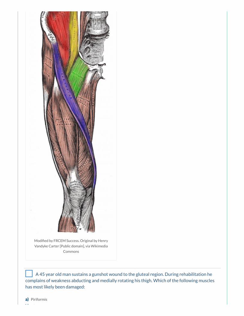
Modi ed by FRCEM Success. Original by Henry
Vandyke Carter [Public domain], via Wikimedia
Commons
A 45 year old man sustains a gunshot wound to the gluteal region. During rehabilitation he
complains of weakness abducting and medially rotating his thigh. Which of the following muscles
has most likely been damaged:
a) Piriformis
b) Gluteus maximus

b) Gluteus maximus
c) Gluteus minimus
d) Quadratus femoris
e) Obturator internus
Answer
The gluteus medius and gluteus minimus muscles act to abduct and medially rotate the thigh at the hip joint. The
gluteus maximus acts to extend, abduct and laterally rotate the thigh. The deep gluteal muscles (piriformis, obturator
internus, gemelli and quadratus femoris muscles) are all synergistic lateral rotators of the thigh at the hip joint.
Notes
Muscle Action(s) Innervation
Gluteus maximus (red) Extension, lateral rotation and abduction at hip Inferior gluteal nerve (L5 – S2)
Gluteus medius (green) Abduction and medial rotation at hip Superior gluteal nerve (L4 – S1)
Gluteus minimus (blue) Abduction and medial rotation at hip Superior gluteal nerve (L4 – S1)
Piriformis (yellow) Lateral rotation and abduction at hip Branches from S1 and S2
The gluteus maximus (red) is the main extensor of the thigh at the hip joint and also acts to laterally rotate and abduct
the thigh. Through its insertion into the iliotibial tract, it also stabilises the knee and hip joints. The gluteus maximus is
innervated by the inferior gluteal nerve (L5 – S2).
The gluteus medius (green) and gluteus minimus (blue) act to abduct and medially rotate the thigh at the hip joint, and
also act to secure the pelvis, reducing pelvic drop on the opposite swing side during walking. They are both innervated
by the superior gluteal nerve (L4 – S1).
The piriformis (yellow) acts to abduct and laterally rotate the thigh at the hip joint. It is innervated by the nerve to the
piriformis, originating from the sacral plexus (S1, S2). The obturator internus, gemelli and quadriceps femoris muscles
act as synergistic femoral lateral rotators and hip stabilisers.
Trendelenburg’s sign is seen in people with weak/paralysed abductor muscles of the hip. The sign is demonstrated by
asking the patient to stand on one limb; when the patient stands on the affected limb, the pelvis severely drops over
the swing limb. This sign may be seen in patients with damage to the superior gluteal nerve, which may occur in
association with pelvic fractures, with space-occupying lesions within the pelvis extending into the greater sciatic
foramen, and following hip surgery. Typically the patient may also demonstrate a Trendelenburg gait.
Something wrong?

Modi ed by FRCEM Success. Original by Henry Vandyke Carter
[Public domain], via Wikimedia Commons
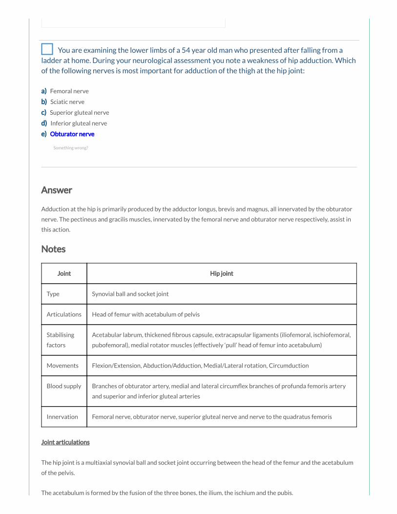
You are examining the lower limbs of a 54 year old man who presented after falling from a
ladder at home. During your neurological assessment you note a weakness of hip adduction. Which
of the following nerves is most important for adduction of the thigh at the hip joint:
a) Femoral nerve
b) Sciatic nerve
c) Superior gluteal nerve
d) Inferior gluteal nerve
e) Obturator nerve
Answer
Adduction at the hip is primarily produced by the adductor longus, brevis and magnus, all innervated by the obturator
nerve. The pectineus and gracilis muscles, innervated by the femoral nerve and obturator nerve respectively, assist in
this action.
Notes
Joint Hip joint
Type Synovial ball and socket joint
Articulations Head of femur with acetabulum of pelvis
Stabilising
factors
Acetabular labrum, thickened brous capsule, extracapsular ligaments (iliofemoral, ischiofemoral,
pubofemoral), medial rotator muscles (effectively ‘pull’ head of femur into acetabulum)
Movements Flexion/Extension, Abduction/Adduction, Medial/Lateral rotation, Circumduction
Blood supply Branches of obturator artery, medial and lateral circum ex branches of profunda femoris artery
and superior and inferior gluteal arteries
Innervation Femoral nerve, obturator nerve, superior gluteal nerve and nerve to the quadratus femoris
Joint articulations
The hip joint is a multiaxial synovial ball and socket joint occurring between the head of the femur and the acetabulum
of the pelvis.
The acetabulum is formed by the fusion of the three bones, the ilium, the ischium and the pubis.
Something wrong?
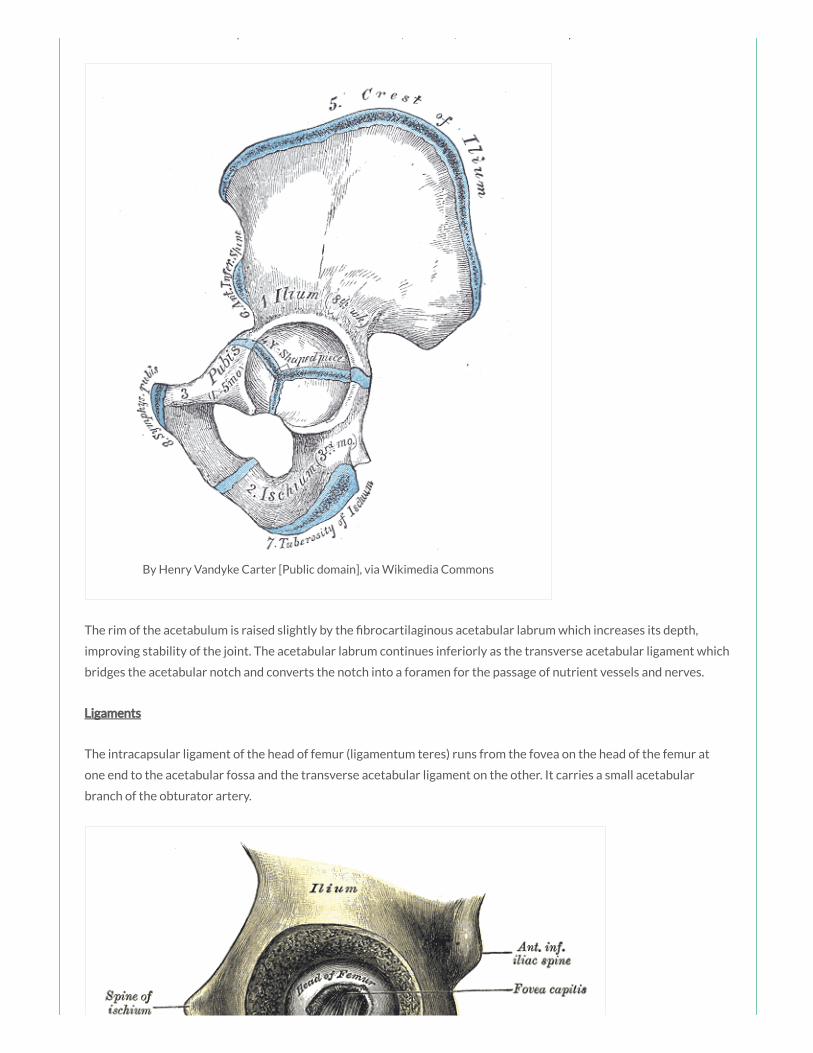
The acetabulum is formed by the fusion of the three bones, the ilium, the ischium and the pubis.
By Henry Vandyke Carter [Public domain], via Wikimedia Commons
The rim of the acetabulum is raised slightly by the brocartilaginous acetabular labrum which increases its depth,
improving stability of the joint. The acetabular labrum continues inferiorly as the transverse acetabular ligament which
bridges the acetabular notch and converts the notch into a foramen for the passage of nutrient vessels and nerves.
Ligaments
The intracapsular ligament of the head of femur (ligamentum teres) runs from the fovea on the head of the femur at
one end to the acetabular fossa and the transverse acetabular ligament on the other. It carries a small acetabular
branch of the obturator artery.
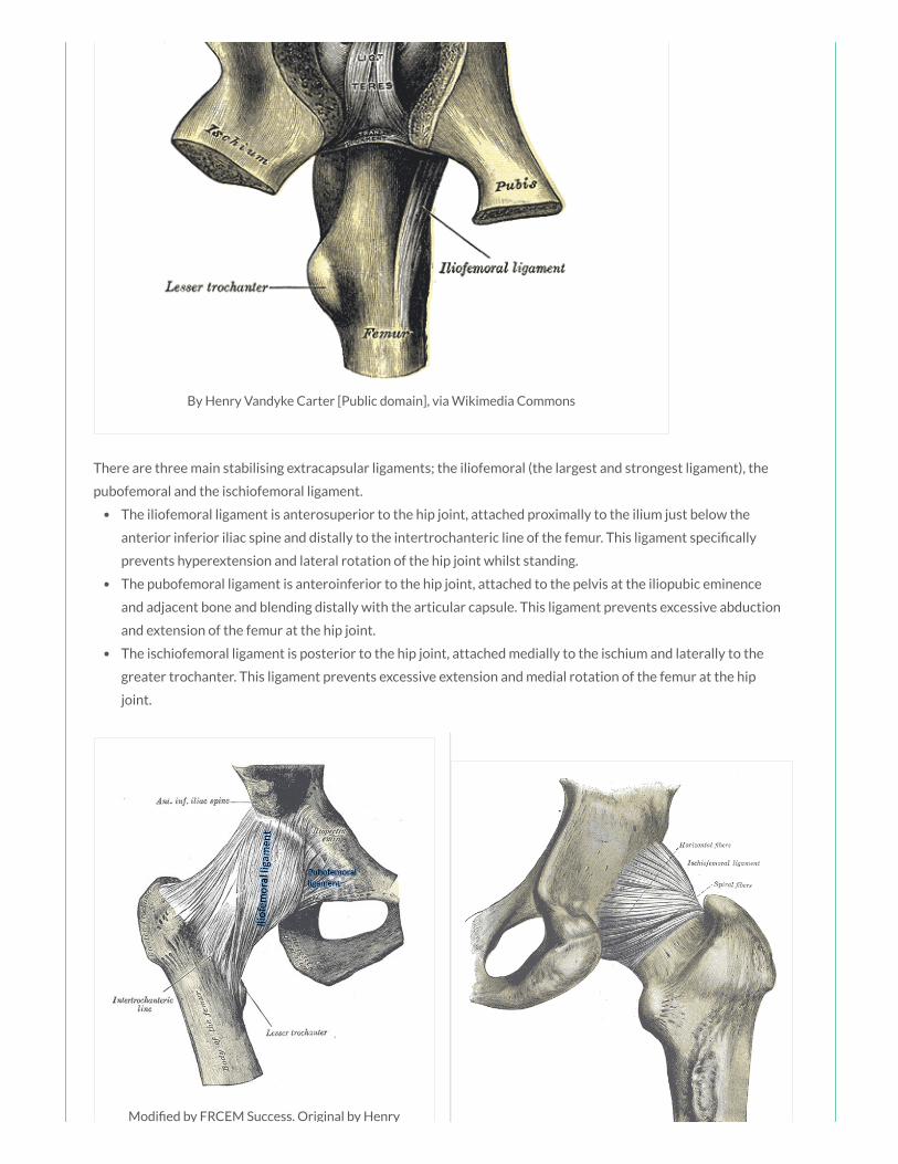
Modi ed by FRCEM Success. Original by Henry
By Henry Vandyke Carter [Public domain], via Wikimedia Commons
There are three main stabilising extracapsular ligaments; the iliofemoral (the largest and strongest ligament), the
pubofemoral and the ischiofemoral ligament.
The iliofemoral ligament is anterosuperior to the hip joint, attached proximally to the ilium just below the
anterior inferior iliac spine and distally to the intertrochanteric line of the femur. This ligament speci cally
prevents hyperextension and lateral rotation of the hip joint whilst standing.
The pubofemoral ligament is anteroinferior to the hip joint, attached to the pelvis at the iliopubic eminence
and adjacent bone and blending distally with the articular capsule. This ligament prevents excessive abduction
and extension of the femur at the hip joint.
The ischiofemoral ligament is posterior to the hip joint, attached medially to the ischium and laterally to the
greater trochanter. This ligament prevents excessive extension and medial rotation of the femur at the hip
joint.

Modi ed by FRCEM Success. Original by Henry
Vandyke Carter [Public domain], via Wikimedia
Commons By Henry Vandyke Carter [Public domain], via
Wikimedia Commons
Blood supply
The hip joint receives its blood supply primarily from branches of the obturator artery, the medial and lateral
circum ex arteries (branches of the profunda femoris artery) and the superior and inferior gluteal arteries. The
articular branches of these vessels form a network around the joint.
Innervation
The hip joint is innervated by articular branches from the femoral nerve (anteriorly), obturator nerve (inferiorly),
superior gluteal nerve (superiorly), and the nerve to the quadratus femoris (posteriorly).
Joint movements
The hip joint allows the movements of exion and extension, abduction and adduction, medial and lateral rotation and
circumduction.
Movement Muscles Involved
Flexion Sartorius, Iliacus, Psoas major, Pectineus, Rectus femoris
Extension Hamstrings, Gluteus maximus
Abduction Gluteus maximus, medius and minimus, Obturator internus, Gemelli, Piriformis, Sartorius
Adduction Adductor longus, magnus and brevis, Gracilis, Pectineus
Medial
rotation
Gluteus medius and minimus, Adductor longus, magnus and brevis, Semitendinosus and
Semimembranosus
Lateral
rotation
Obturator externus, Sartorius, Iliacus, Psoas major, Biceps femoris, Piriformis, Gluteus maximus,
Obturator internus, Gemelli, Quadriceps femoris
A 32 year old man presents to the ED after sustaining multiple stab wounds to the right thigh
during an altercation in a pub. Imaging has shown trauma to the biceps femoris muscle. The biceps
femoris acts primarily to produce which of the following movements at the hip:
a) Extension and medial rotation at hip
b) Extension and lateral rotation at hip
c) Extension and abduction at hip
d) Extension and adduction at hip

d) Extension and adduction at hip
e) Flexion and medial rotation at hip
Answer
The hamstrings act together to ex the leg at the knee joint and extend the thigh at the hip joint. The biceps femoris
also acts to laterally rotate the thigh at the hip joint and the leg at the knee joint. The semimembranosus and
semitendinosus also act together to medially rotate the thigh at the hip joint and the leg at the knee joint.
Notes
Muscle Action(s) Innervation
Biceps femoris (red) Flexion at knee, extension and lateral rotation at hip Sciatic nerve (L5 – S2)
Semitendinosus (blue) Flexion at knee, extension and medial rotation at hip Sciatic nerve (L5 – S2)
Semimembranosus (green) Flexion at knee, extension and medial rotation at hip Sciatic nerve (L5 – S2)
The hamstrings are composed of three individual muscles; the biceps femoris (red), the semitendinosus (blue) and the
semimembranosus (green).
The hamstrings act together to ex the leg at the knee joint and extend the thigh at the hip joint. The biceps femoris
also acts to laterally rotate the thigh at the hip joint and the leg at the knee joint. The semimembranosus and
semitendinosus also act together to medially rotate the thigh at the hip joint and the leg at the knee joint.
The hamstring muscles are all innervated by the tibial division of the sciatic nerve (L5 – S2), except for the short head of
the biceps femoris innervated by the common bular division.
To test the hamstrings the patient exes their leg against resistance. Normally these muscles, especially their tendons
on each side of the popliteal fossa, are prominent as they bend the knee.
Something wrong?
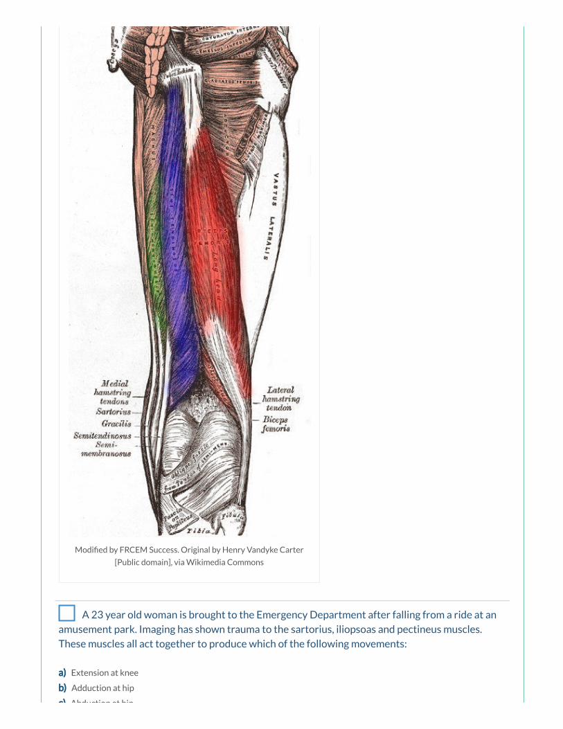
Modi ed by FRCEM Success. Original by Henry Vandyke Carter
[Public domain], via Wikimedia Commons
A 23 year old woman is brought to the Emergency Department after falling from a ride at an
amusement park. Imaging has shown trauma to the sartorius, iliopsoas and pectineus muscles.
These muscles all act together to produce which of the following movements:
a) Extension at knee
b) Adduction at hip
c) Abduction at hip
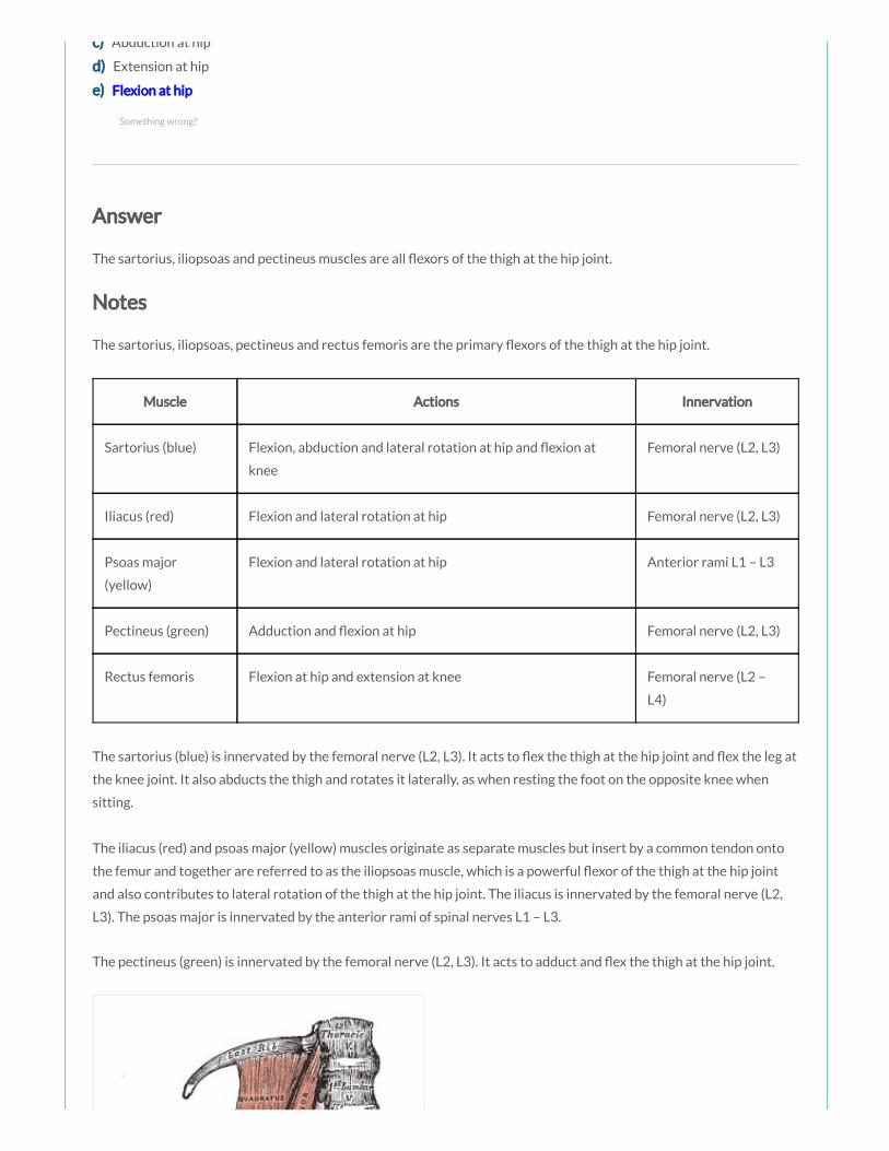
c) Abduction at hip
d) Extension at hip
e) Flexion at hip
Answer
The sartorius, iliopsoas and pectineus muscles are all exors of the thigh at the hip joint.
Notes
The sartorius, iliopsoas, pectineus and rectus femoris are the primary exors of the thigh at the hip joint.
Muscle Actions Innervation
Sartorius (blue) Flexion, abduction and lateral rotation at hip and exion at
knee
Femoral nerve (L2, L3)
Iliacus (red) Flexion and lateral rotation at hip Femoral nerve (L2, L3)
Psoas major
(yellow)
Flexion and lateral rotation at hip Anterior rami L1 – L3
Pectineus (green) Adduction and exion at hip Femoral nerve (L2, L3)
Rectus femoris Flexion at hip and extension at knee Femoral nerve (L2 –
L4)
The sartorius (blue) is innervated by the femoral nerve (L2, L3). It acts to ex the thigh at the hip joint and ex the leg at
the knee joint. It also abducts the thigh and rotates it laterally, as when resting the foot on the opposite knee when
sitting.
The iliacus (red) and psoas major (yellow) muscles originate as separate muscles but insert by a common tendon onto
the femur and together are referred to as the iliopsoas muscle, which is a powerful exor of the thigh at the hip joint
and also contributes to lateral rotation of the thigh at the hip joint. The iliacus is innervated by the femoral nerve (L2,
L3). The psoas major is innervated by the anterior rami of spinal nerves L1 – L3.
The pectineus (green) is innervated by the femoral nerve (L2, L3). It acts to adduct and ex the thigh at the hip joint.
Something wrong?

Modi ed by FRCEM Success. Original by Henry
Vandyke Carter [Public domain], via Wikimedia
Commons
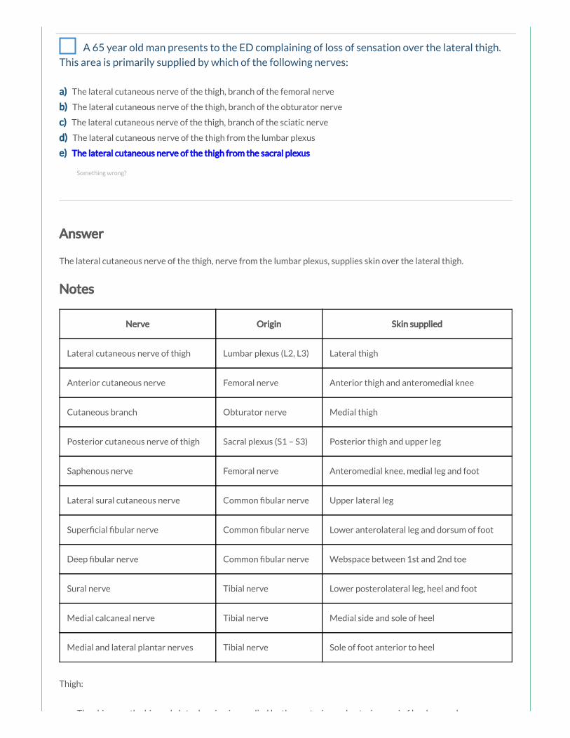
A 65 year old man presents to the ED complaining of loss of sensation over the lateral thigh.
This area is primarily supplied by which of the following nerves:
a) The lateral cutaneous nerve of the thigh, branch of the femoral nerve
b) The lateral cutaneous nerve of the thigh, branch of the obturator nerve
c) The lateral cutaneous nerve of the thigh, branch of the sciatic nerve
d) The lateral cutaneous nerve of the thigh from the lumbar plexus
e) The lateral cutaneous nerve of the thigh from the sacral plexus
Answer
The lateral cutaneous nerve of the thigh, nerve from the lumbar plexus, supplies skin over the lateral thigh.
Notes
Nerve Origin Skin supplied
Lateral cutaneous nerve of thigh Lumbar plexus (L2, L3) Lateral thigh
Anterior cutaneous nerve Femoral nerve Anterior thigh and anteromedial knee
Cutaneous branch Obturator nerve Medial thigh
Posterior cutaneous nerve of thigh Sacral plexus (S1 – S3) Posterior thigh and upper leg
Saphenous nerve Femoral nerve Anteromedial knee, medial leg and foot
Lateral sural cutaneous nerve Common bular nerve Upper lateral leg
Super cial bular nerve Common bular nerve Lower anterolateral leg and dorsum of foot
Deep bular nerve Common bular nerve Webspace between 1st and 2nd toe
Sural nerve Tibial nerve Lower posterolateral leg, heel and foot
Medial calcaneal nerve Tibial nerve Medial side and sole of heel
Medial and lateral plantar nerves Tibial nerve Sole of foot anterior to heel
Thigh:
The skin over the hip and gluteal region is supplied by the posterior and anterior rami of lumbosacral nerves
Something wrong?

The skin over the hip and gluteal region is supplied by the posterior and anterior rami of lumbosacral nerves
(L1 – S3).
The lateral cutaneous nerve of the thigh, nerve from the lumbar plexus, supplies skin over the lateral thigh.
The anterior cutaneous nerve, branch of the femoral nerve, supplies skin over the anterior thigh and
anteromedial knee.
The obturator nerve supplies skin over the upper medial thigh.
The posterior cutaneous nerve of the thigh, nerve from the sacral plexus, supplies skin over the posterior thigh
and upper leg.
Leg:
The saphenous nerve, branch of the femoral nerve, supplies the skin over the anteromedial knee, medial leg
and the medial side of the foot as far anteriorly as the head of the 1st metatarsal.
The lateral sural nerve, branch of the common bular nerve, supplies the skin over the upper lateral leg.
The super cial bular nerve supplies the area of skin over the lower anterolateral leg and the dorsum of the
foot (except for the webspace between the 1st and 2nd toe and the lateral side of the little toe).
The deep bular nerve supplies the skin over the webspace between the 1st and 2nd toe.
The sural nerve, branch of the tibial nerve, supplies the skin over the lower posterolateral leg, heel and foot.
The medial calcaneal nerve, branch of the tibial nerve, supplies the skin over the medial side and sole of the
heel.
The medial and lateral plantar nerves, branches of the tibial nerve, supply the skin over the sole of the foot
anterior of the heel.

Modi ed by FRCEM Success. Original by Henry Vandyke Carter [Public domain], via Wikimedia Commons
Modi ed by FRCEM Success. Original by Henry Vandyke Carter [Public
domain], via Wikimedia Commons
A 34 year old man presents to the ED following a 3 day history of fever. On examination you
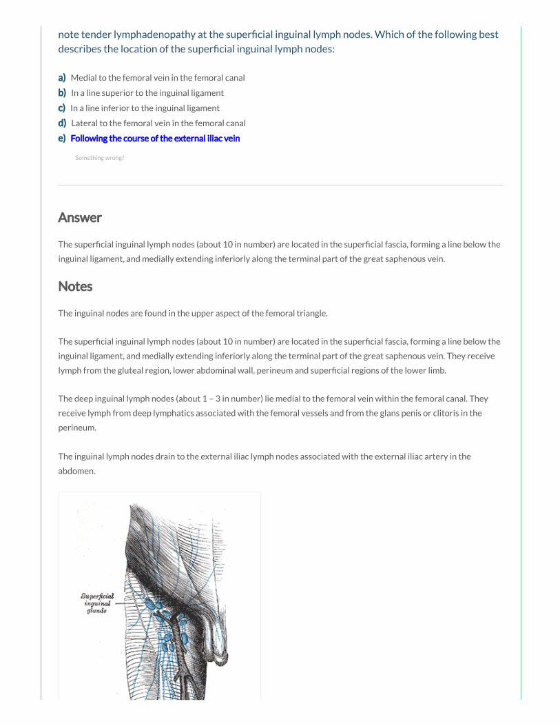
note tender lymphadenopathy at the super cial inguinal lymph nodes. Which of the following best
describes the location of the super cial inguinal lymph nodes:
a) Medial to the femoral vein in the femoral canal
b) In a line superior to the inguinal ligament
c) In a line inferior to the inguinal ligament
d) Lateral to the femoral vein in the femoral canal
e) Following the course of the external iliac vein
Answer
The super cial inguinal lymph nodes (about 10 in number) are located in the super cial fascia, forming a line below the
inguinal ligament, and medially extending inferiorly along the terminal part of the great saphenous vein.
Notes
The inguinal nodes are found in the upper aspect of the femoral triangle.
The super cial inguinal lymph nodes (about 10 in number) are located in the super cial fascia, forming a line below the
inguinal ligament, and medially extending inferiorly along the terminal part of the great saphenous vein. They receive
lymph from the gluteal region, lower abdominal wall, perineum and super cial regions of the lower limb.
The deep inguinal lymph nodes (about 1 – 3 in number) lie medial to the femoral vein within the femoral canal. They
receive lymph from deep lymphatics associated with the femoral vessels and from the glans penis or clitoris in the
perineum.
The inguinal lymph nodes drain to the external iliac lymph nodes associated with the external iliac artery in the
abdomen.
Something wrong?
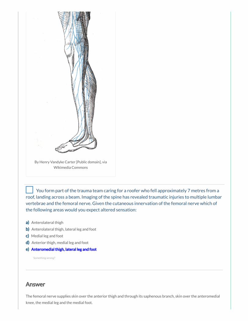
By Henry Vandyke Carter [Public domain], via
Wikimedia Commons
You form part of the trauma team caring for a roofer who fell approximately 7 metres from a
roof, landing across a beam. Imaging of the spine has revealed traumatic injuries to multiple lumbar
vertebrae and the femoral nerve. Given the cutaneous innervation of the femoral nerve which of
the following areas would you expect altered sensation:
a) Anterolateral thigh
b) Anterolateral thigh, lateral leg and foot
c) Medial leg and foot
d) Anterior thigh, medial leg and foot
e) Anteromedial thigh, lateral leg and foot
Answer
The femoral nerve supplies skin over the anterior thigh and through its saphenous branch, skin over the anteromedial
knee, the medial leg and the medial foot.
Something wrong?

Notes
The femoral nerve arises from the lumbar plexus, receiving bres from the anterior rami of L2 – L4.
Nerve Femoral nerve
Nerve roots L2 – L4
Motor supply Iliacus, pectineus, sartorius, quadriceps femoris
Sensory supply Skin over the anterior thigh, anteromedial knee, medial leg and medial foot
Motor loss in injury Weak exion at hip and loss of extension at knee
Anatomical course
The femoral nerve descends from the lumbar plexus in the posterior abdomen through the substance of the psoas
major muscle, emerging from the lower lateral border of the psoas major. Continuing its descent, the femoral nerve lies
between the lateral border of the psoas major and the anterior surface of the iliacus muscle. It is deep to the iliacus
fascia and lateral to the femoral artery as it passes posterior to the mid-inguinal point to enter the femoral triangle in
the anterior compartment of the thigh, before dividing into an anterior and posterior division.

By Henry Vandyke Carter [Public domain], via Wikimedia
Commons
Branches
In the abdomen it gives rise to branches that innervate the iliacus and pectineus muscles.
The anterior division gives off anterior cutaneous branches (supplying skin over the anterior and medial thigh) and
muscular branches (innervating the sartorius).
The posterior division gives off muscular branches (innervating the quadriceps femoris muscles) and articular branches
(supplying the hip and knee joint), before continuing as the saphenous nerve (supplying skin over the anteromedial
knee and the medial side of the leg and foot).
Branch Innervation
Muscular branches in abdomen Iliacus and pectineus
Anterior cutaneous branches Skin over anterior thigh
Anterior muscular branches Sartorius
Posterior muscular branches Quadriceps femoris muscles

Posterior muscular branches Quadriceps femoris muscles
Posterior articular branches Hip and knee joint
Saphenous nerve Skin over anteromedial knee, medial side of leg and foot
Motor and sensory function
Muscle Action(s)
Sartorius Flexion, abduction and lateral rotation at hip and exion at knee
Iliacus (red) Flexion and lateral rotation at hip
Pectineus (green) Adduction and exion at hip
Rectus femoris Flexion at hip and extension at knee
Vastus lateralis Extension at knee
Vastus medialis Extension at knee
Vastus intermedius Extension at knee
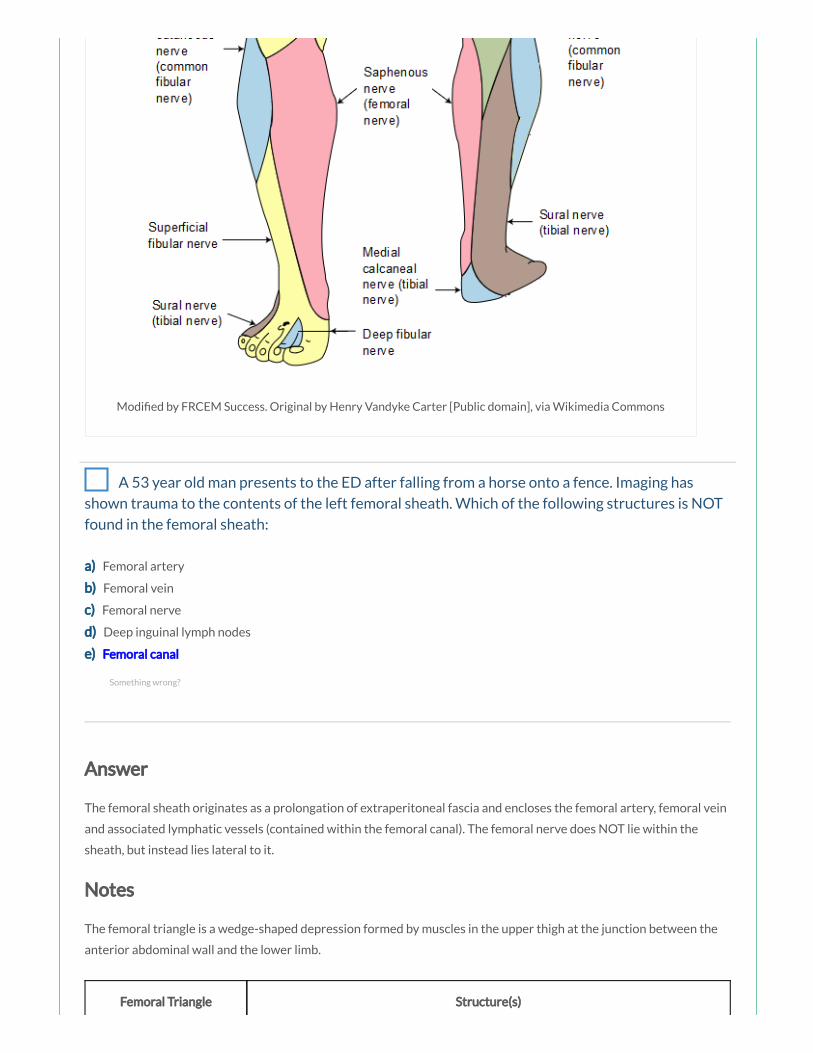
Modi ed by FRCEM Success. Original by Henry Vandyke Carter [Public domain], via Wikimedia Commons
A 53 year old man presents to the ED after falling from a horse onto a fence. Imaging has
shown trauma to the contents of the left femoral sheath. Which of the following structures is NOT
found in the femoral sheath:
a) Femoral artery
b) Femoral vein
c) Femoral nerve
d) Deep inguinal lymph nodes
e) Femoral canal
Answer
The femoral sheath originates as a prolongation of extraperitoneal fascia and encloses the femoral artery, femoral vein
and associated lymphatic vessels (contained within the femoral canal). The femoral nerve does NOT lie within the
sheath, but instead lies lateral to it.
Notes
The femoral triangle is a wedge-shaped depression formed by muscles in the upper thigh at the junction between the
anterior abdominal wall and the lower limb.
Femoral Triangle Structure(s)
Something wrong?

Superior border Inguinal ligament
Lateral border Medial sartorius muscle
Medial border Medial adductor longus muscle
Roof Fascia lata
Floor Pectineus, iliopsoas and adductor longus muscles
Contents Femoral nerve, femoral artery, femoral vein, femoral canal
Borders
The femoral triangle is bordered superiorly by the inguinal ligament, laterally by the medial border of the sartorius
muscle and medially by the medial border of the adductor longus muscle. The roof is formed by the fascia lata and the
oor is formed by the pectineus, iliopsoas and adductor longus muscles.
Contents
The femoral triangle contains (from lateral to medial):
The femoral nerve
The femoral artery
The femoral vein
The femoral canal
Femoral sheath
The femoral sheath originates as a prolongation of extraperitoneal fascia and encloses the femoral artery, femoral vein
and associated lymphatic vessels (contained within the femoral canal). The femoral nerve does NOT lie within the
sheath, but instead lies lateral to it. The femoral sheath terminates inferiorly by blending with the adventitia of the
femoral vessels approximately 4 cm inferior of the inguinal ligament.

By Henry Vandyke Carter [Public domain], via
Wikimedia Commons
By Henry Vandyke Carter [Public domain], via
Wikimedia Commons
Femoral canal
The femoral canal lies between the medial edge of the femoral sheath and the femoral vein. The femoral canal contains
loose connective tissue, lymphatic vessels, deep inguinal lymph nodes and empty space. The femoral canal allows the
femoral vein to expand, for example, when venous return from the leg is increased, or when increased intra-abdominal
pressure causes a temporary stasis.
Femoral ring
The femoral ring is formed by the proximal abdominal opening of the femoral canal, and is important clinically, as it is a
common site of hernia. It is bordered anteriorly by the inguinal ligament, posteriorly by the pectineal ligament,
medially by the lacunar ligament and laterally by the femoral vein.
Femoral ring Structure(s)
Anterior border Inguinal ligament
Posterior border Pectineal ligament
Medial border Lacunar ligament
Lateral border Femoral vein
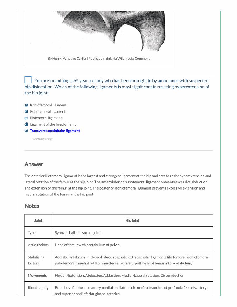
By Henry Vandyke Carter [Public domain], via Wikimedia Commons
You are examining a 65 year old lady who has been brought in by ambulance with suspected
hip dislocation. Which of the following ligaments is most signi cant in resisting hyperextension of
the hip joint:
a) Ischiofemoral ligament
b) Pubofemoral ligament
c) Iliofemoral ligament
d) Ligament of the head of femur
e) Transverse acetabular ligament
Answer
The anterior iliofemoral ligament is the largest and strongest ligament at the hip and acts to resist hyperextension and
lateral rotation of the femur at the hip joint. The anteroinferior pubofemoral ligament prevents excessive abduction
and extension of the femur at the hip joint. The posterior ischiofemoral ligament prevents excessive extension and
medial rotation of the femur at the hip joint.
Notes
Joint Hip joint
Type Synovial ball and socket joint
Articulations Head of femur with acetabulum of pelvis
Stabilising
factors
Acetabular labrum, thickened brous capsule, extracapsular ligaments (iliofemoral, ischiofemoral,
pubofemoral), medial rotator muscles (effectively ‘pull’ head of femur into acetabulum)
Movements Flexion/Extension, Abduction/Adduction, Medial/Lateral rotation, Circumduction
Blood supply Branches of obturator artery, medial and lateral circum ex branches of profunda femoris artery
and superior and inferior gluteal arteries
Something wrong?

Innervation Femoral nerve, obturator nerve, superior gluteal nerve and nerve to the quadratus femoris
Joint articulations
The hip joint is a multiaxial synovial ball and socket joint occurring between the head of the femur and the acetabulum
of the pelvis.
The acetabulum is formed by the fusion of the three bones, the ilium, the ischium and the pubis.
By Henry Vandyke Carter [Public domain], via Wikimedia Commons
The rim of the acetabulum is raised slightly by the brocartilaginous acetabular labrum which increases its depth,
improving stability of the joint. The acetabular labrum continues inferiorly as the transverse acetabular ligament which
bridges the acetabular notch and converts the notch into a foramen for the passage of nutrient vessels and nerves.
Ligaments
The intracapsular ligament of the head of femur (ligamentum teres) runs from the fovea on the head of the femur at
one end to the acetabular fossa and the transverse acetabular ligament on the other. It carries a small acetabular
branch of the obturator artery.

By Henry Vandyke Carter [Public domain], via Wikimedia Commons
There are three main stabilising extracapsular ligaments; the iliofemoral (the largest and strongest ligament), the
pubofemoral and the ischiofemoral ligament.
The iliofemoral ligament is anterosuperior to the hip joint, attached proximally to the ilium just below the
anterior inferior iliac spine and distally to the intertrochanteric line of the femur. This ligament speci cally
prevents hyperextension and lateral rotation of the hip joint whilst standing.
The pubofemoral ligament is anteroinferior to the hip joint, attached to the pelvis at the iliopubic eminence
and adjacent bone and blending distally with the articular capsule. This ligament prevents excessive abduction
and extension of the femur at the hip joint.
The ischiofemoral ligament is posterior to the hip joint, attached medially to the ischium and laterally to the
greater trochanter. This ligament prevents excessive extension and medial rotation of the femur at the hip
joint.

Modi ed by FRCEM Success. Original by Henry
Vandyke Carter [Public domain], via Wikimedia
Commons By Henry Vandyke Carter [Public domain], via
Wikimedia Commons
Blood supply
The hip joint receives its blood supply primarily from branches of the obturator artery, the medial and lateral
circum ex arteries (branches of the profunda femoris artery) and the superior and inferior gluteal arteries. The
articular branches of these vessels form a network around the joint.
Innervation
The hip joint is innervated by articular branches from the femoral nerve (anteriorly), obturator nerve (inferiorly),
superior gluteal nerve (superiorly), and the nerve to the quadratus femoris (posteriorly).
Joint movements
The hip joint allows the movements of exion and extension, abduction and adduction, medial and lateral rotation and
circumduction.
Movement Muscles Involved
Flexion Sartorius, Iliacus, Psoas major, Pectineus, Rectus femoris
Extension Hamstrings, Gluteus maximus
Abduction Gluteus maximus, medius and minimus, Obturator internus, Gemelli, Piriformis, Sartorius
Adduction Adductor longus, magnus and brevis, Gracilis, Pectineus
Medial
rotation
Gluteus medius and minimus, Adductor longus, magnus and brevis, Semitendinosus and
Semimembranosus
Lateral
rotation
Obturator externus, Sartorius, Iliacus, Psoas major, Biceps femoris, Piriformis, Gluteus maximus,
Obturator internus, Gemelli, Quadriceps femoris

A 87 year old woman presents to the ED after her carers noted she was complaining of pain in
the right groin. On examination you note a tender swelling in keeping with a femoral hernia. The
femoral ring is bordered anteriorly by which of the following structures:
a) Sartorius muscle
b) Inguinal ligament
c) Fascia lata
d) Femoral sheath
e) Femoral artery
Answer
The femoral ring is bordered anteriorly by the inguinal ligament, posteriorly by the pectineal ligament, medially by the
lacunar ligament and laterally by the femoral vein.
Notes
The femoral triangle is a wedge-shaped depression formed by muscles in the upper thigh at the junction between the
anterior abdominal wall and the lower limb.
Femoral Triangle Structure(s)
Superior border Inguinal ligament
Lateral border Medial sartorius muscle
Medial border Medial adductor longus muscle
Roof Fascia lata
Floor Pectineus, iliopsoas and adductor longus muscles
Contents Femoral nerve, femoral artery, femoral vein, femoral canal
Borders
The femoral triangle is bordered superiorly by the inguinal ligament, laterally by the medial border of the sartorius
muscle and medially by the medial border of the adductor longus muscle. The roof is formed by the fascia lata and the
oor is formed by the pectineus, iliopsoas and adductor longus muscles.
Contents
Something wrong?
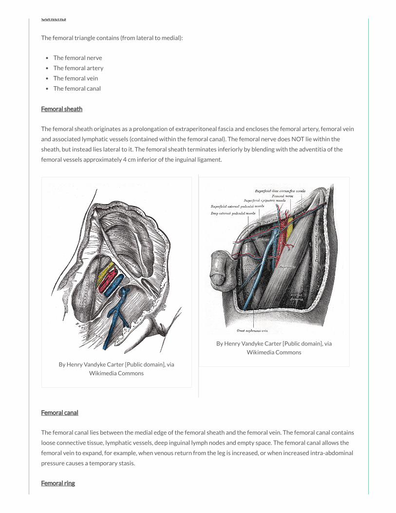
By Henry Vandyke Carter [Public domain], via
Wikimedia Commons
By Henry Vandyke Carter [Public domain], via
Wikimedia Commons
Contents
The femoral triangle contains (from lateral to medial):
The femoral nerve
The femoral artery
The femoral vein
The femoral canal
Femoral sheath
The femoral sheath originates as a prolongation of extraperitoneal fascia and encloses the femoral artery, femoral vein
and associated lymphatic vessels (contained within the femoral canal). The femoral nerve does NOT lie within the
sheath, but instead lies lateral to it. The femoral sheath terminates inferiorly by blending with the adventitia of the
femoral vessels approximately 4 cm inferior of the inguinal ligament.
Femoral canal
The femoral canal lies between the medial edge of the femoral sheath and the femoral vein. The femoral canal contains
loose connective tissue, lymphatic vessels, deep inguinal lymph nodes and empty space. The femoral canal allows the
femoral vein to expand, for example, when venous return from the leg is increased, or when increased intra-abdominal
pressure causes a temporary stasis.
Femoral ring

The femoral ring is formed by the proximal abdominal opening of the femoral canal, and is important clinically, as it is a
common site of hernia. It is bordered anteriorly by the inguinal ligament, posteriorly by the pectineal ligament,
medially by the lacunar ligament and laterally by the femoral vein.
Femoral ring Structure(s)
Anterior border Inguinal ligament
Posterior border Pectineal ligament
Medial border Lacunar ligament
Lateral border Femoral vein
By Henry Vandyke Carter [Public domain], via Wikimedia Commons
A 43 year old man presents to the ED complaining of weakness at the right knee. On
examination you note he is unable to extend the leg at the knee joint. Which of the following
muscles is the most important in producing extension of the leg at the knee joint:
a) Hamstrings
b) Quadriceps femoris
c) Gracilis
d) Sartorius
e) Gastrocnemius
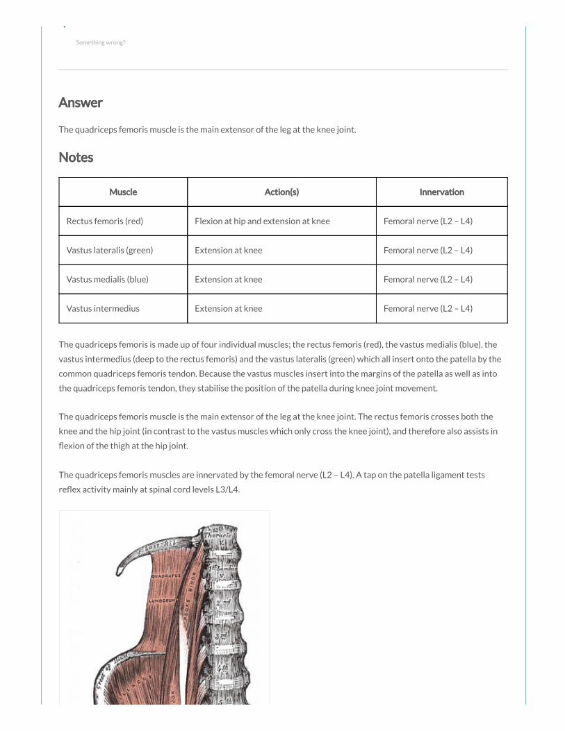
e) Gastrocnemius
Answer
The quadriceps femoris muscle is the main extensor of the leg at the knee joint.
Notes
Muscle Action(s) Innervation
Rectus femoris (red) Flexion at hip and extension at knee Femoral nerve (L2 – L4)
Vastus lateralis (green) Extension at knee Femoral nerve (L2 – L4)
Vastus medialis (blue) Extension at knee Femoral nerve (L2 – L4)
Vastus intermedius Extension at knee Femoral nerve (L2 – L4)
The quadriceps femoris is made up of four individual muscles; the rectus femoris (red), the vastus medialis (blue), the
vastus intermedius (deep to the rectus femoris) and the vastus lateralis (green) which all insert onto the patella by the
common quadriceps femoris tendon. Because the vastus muscles insert into the margins of the patella as well as into
the quadriceps femoris tendon, they stabilise the position of the patella during knee joint movement.
The quadriceps femoris muscle is the main extensor of the leg at the knee joint. The rectus femoris crosses both the
knee and the hip joint (in contrast to the vastus muscles which only cross the knee joint), and therefore also assists in
exion of the thigh at the hip joint.
The quadriceps femoris muscles are innervated by the femoral nerve (L2 – L4). A tap on the patella ligament tests
re ex activity mainly at spinal cord levels L3/L4.
Something wrong?

Modi ed by FRCEM Success. Original by Henry
Vandyke Carter [Public domain], via Wikimedia
Commons
You have been asked to perform an arterial blood gas collection from a patient’s femoral
artery. The femoral triangle is bordered medially by which of the following structures:
a) Adductor brevis muscle
b) Adductor longus muscle
c) Adductor magnus muscle
d) Pectineus muscle
e) Gracilis muscle
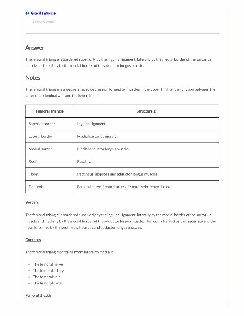
e) Gracilis muscle
Answer
The femoral triangle is bordered superiorly by the inguinal ligament, laterally by the medial border of the sartorius
muscle and medially by the medial border of the adductor longus muscle.
Notes
The femoral triangle is a wedge-shaped depression formed by muscles in the upper thigh at the junction between the
anterior abdominal wall and the lower limb.
Femoral Triangle Structure(s)
Superior border Inguinal ligament
Lateral border Medial sartorius muscle
Medial border Medial adductor longus muscle
Roof Fascia lata
Floor Pectineus, iliopsoas and adductor longus muscles
Contents Femoral nerve, femoral artery, femoral vein, femoral canal
Borders
The femoral triangle is bordered superiorly by the inguinal ligament, laterally by the medial border of the sartorius
muscle and medially by the medial border of the adductor longus muscle. The roof is formed by the fascia lata and the
oor is formed by the pectineus, iliopsoas and adductor longus muscles.
Contents
The femoral triangle contains (from lateral to medial):
The femoral nerve
The femoral artery
The femoral vein
The femoral canal
Femoral sheath
Something wrong?
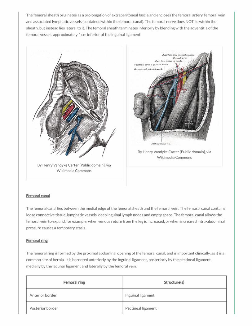
By Henry Vandyke Carter [Public domain], via
Wikimedia Commons
By Henry Vandyke Carter [Public domain], via
Wikimedia Commons
The femoral sheath originates as a prolongation of extraperitoneal fascia and encloses the femoral artery, femoral vein
and associated lymphatic vessels (contained within the femoral canal). The femoral nerve does NOT lie within the
sheath, but instead lies lateral to it. The femoral sheath terminates inferiorly by blending with the adventitia of the
femoral vessels approximately 4 cm inferior of the inguinal ligament.
Femoral canal
The femoral canal lies between the medial edge of the femoral sheath and the femoral vein. The femoral canal contains
loose connective tissue, lymphatic vessels, deep inguinal lymph nodes and empty space. The femoral canal allows the
femoral vein to expand, for example, when venous return from the leg is increased, or when increased intra-abdominal
pressure causes a temporary stasis.
Femoral ring
The femoral ring is formed by the proximal abdominal opening of the femoral canal, and is important clinically, as it is a
common site of hernia. It is bordered anteriorly by the inguinal ligament, posteriorly by the pectineal ligament,
medially by the lacunar ligament and laterally by the femoral vein.
Femoral ring Structure(s)
Anterior border Inguinal ligament
Posterior border Pectineal ligament

Medial border Lacunar ligament
Lateral border Femoral vein
By Henry Vandyke Carter [Public domain], via Wikimedia Commons
You are giving a teaching session to a group of nal year medical students regarding lower
limb neurology. Which of the following clinical features would be expected in an obturator nerve
palsy:
a) Weakness of hip extension
b) Weakness of hip abduction
c) Weakness of hip adduction
d) Loss of sensation over anterolateral thigh
e) Weakness of hip exion
Answer
Damage to the obturator nerve results in weak adduction of the hip with lateral swinging of the lmb during walking due
to unopposed abduction.
Notes
Something wrong?

Notes
The obturator nerve arises from the lumbar plexus, formed from the anterior rami of L2 – L4.
Nerve Obturator nerve
Nerve
roots
L2 – L4
Motor
supply
Medial thigh muscles (adductor longus, brevis and magnus, gracilis, obturator externus)
Sensory
supply
Upper medial thigh
Motor loss
in injury
Weak adduction of hip and dif culty walking with lateral swinging of limb during walking (due to
unopposed abduction), loss of sensation over upper medial thigh
The obturator nerve descends along the posterior abdominal wall, passes through the pelvic cavity and enters the
medial thigh by passing through the obturator canal.
The obturator nerve innervates all of the muscles of the medial compartment of the thigh (except for the hamstring
part of the adductor longus, and the pectineus muscle which are innervated by the sciatic and the femoral nerves
respectively). It also gives off a cutaneous branch that supplies skin on the medial side of the upper thigh.
Muscle Action(s)
Adductor longus Adduction and medial rotation at hip
Adductor brevis Adduction and medial rotation at hip
Adductor magnus Adduction and medial rotation at hip
Obturator externus Lateral rotation at hip
Gracilis Adduction at hip and exion at knee
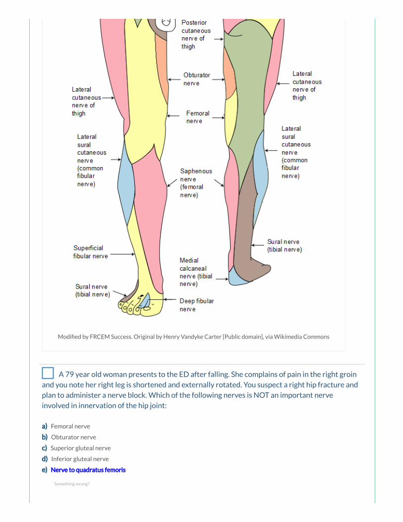
Modi ed by FRCEM Success. Original by Henry Vandyke Carter [Public domain], via Wikimedia Commons
A 79 year old woman presents to the ED after falling. She complains of pain in the right groin
and you note her right leg is shortened and externally rotated. You suspect a right hip fracture and
plan to administer a nerve block. Which of the following nerves is NOT an important nerve
involved in innervation of the hip joint:
a) Femoral nerve
b) Obturator nerve
c) Superior gluteal nerve
d) Inferior gluteal nerve
e) Nerve to quadratus femoris
Something wrong?
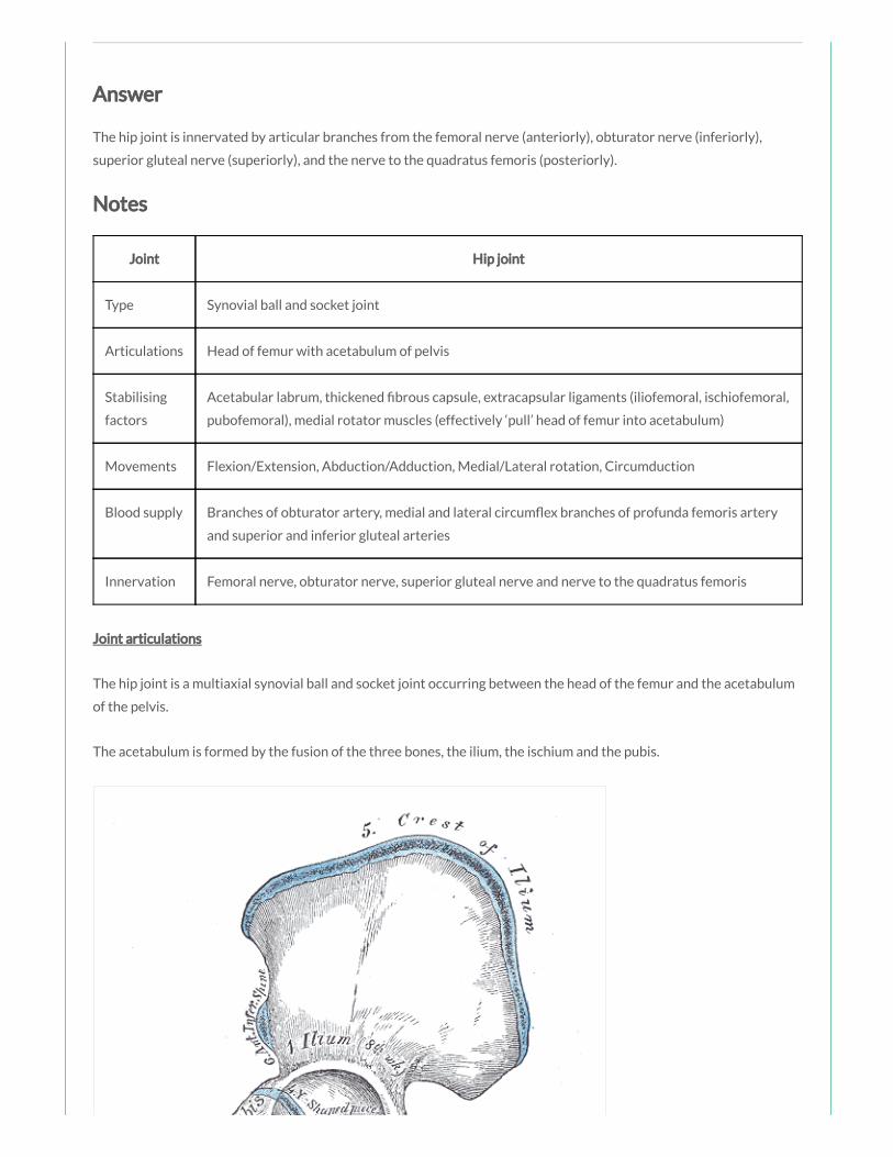
Answer
The hip joint is innervated by articular branches from the femoral nerve (anteriorly), obturator nerve (inferiorly),
superior gluteal nerve (superiorly), and the nerve to the quadratus femoris (posteriorly).
Notes
Joint Hip joint
Type Synovial ball and socket joint
Articulations Head of femur with acetabulum of pelvis
Stabilising
factors
Acetabular labrum, thickened brous capsule, extracapsular ligaments (iliofemoral, ischiofemoral,
pubofemoral), medial rotator muscles (effectively ‘pull’ head of femur into acetabulum)
Movements Flexion/Extension, Abduction/Adduction, Medial/Lateral rotation, Circumduction
Blood supply Branches of obturator artery, medial and lateral circum ex branches of profunda femoris artery
and superior and inferior gluteal arteries
Innervation Femoral nerve, obturator nerve, superior gluteal nerve and nerve to the quadratus femoris
Joint articulations
The hip joint is a multiaxial synovial ball and socket joint occurring between the head of the femur and the acetabulum
of the pelvis.
The acetabulum is formed by the fusion of the three bones, the ilium, the ischium and the pubis.
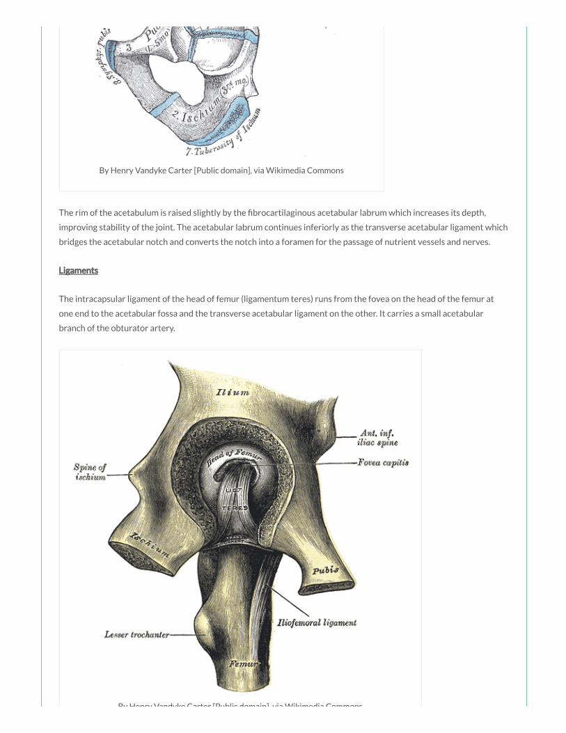
By Henry Vandyke Carter [Public domain], via Wikimedia Commons
The rim of the acetabulum is raised slightly by the brocartilaginous acetabular labrum which increases its depth,
improving stability of the joint. The acetabular labrum continues inferiorly as the transverse acetabular ligament which
bridges the acetabular notch and converts the notch into a foramen for the passage of nutrient vessels and nerves.
Ligaments
The intracapsular ligament of the head of femur (ligamentum teres) runs from the fovea on the head of the femur at
one end to the acetabular fossa and the transverse acetabular ligament on the other. It carries a small acetabular
branch of the obturator artery.
By Henry Vandyke Carter [Public domain], via Wikimedia Commons

Modi ed by FRCEM Success. Original by Henry
Vandyke Carter [Public domain], via Wikimedia
Commons By Henry Vandyke Carter [Public domain], via
Wikimedia Commons
By Henry Vandyke Carter [Public domain], via Wikimedia Commons
There are three main stabilising extracapsular ligaments; the iliofemoral (the largest and strongest ligament), the
pubofemoral and the ischiofemoral ligament.
The iliofemoral ligament is anterosuperior to the hip joint, attached proximally to the ilium just below the
anterior inferior iliac spine and distally to the intertrochanteric line of the femur. This ligament speci cally
prevents hyperextension and lateral rotation of the hip joint whilst standing.
The pubofemoral ligament is anteroinferior to the hip joint, attached to the pelvis at the iliopubic eminence
and adjacent bone and blending distally with the articular capsule. This ligament prevents excessive abduction
and extension of the femur at the hip joint.
The ischiofemoral ligament is posterior to the hip joint, attached medially to the ischium and laterally to the
greater trochanter. This ligament prevents excessive extension and medial rotation of the femur at the hip
joint.
Blood supply
The hip joint receives its blood supply primarily from branches of the obturator artery, the medial and lateral
circum ex arteries (branches of the profunda femoris artery) and the superior and inferior gluteal arteries. The
articular branches of these vessels form a network around the joint.
Innervation
The hip joint is innervated by articular branches from the femoral nerve (anteriorly), obturator nerve (inferiorly),
superior gluteal nerve (superiorly), and the nerve to the quadratus femoris (posteriorly).
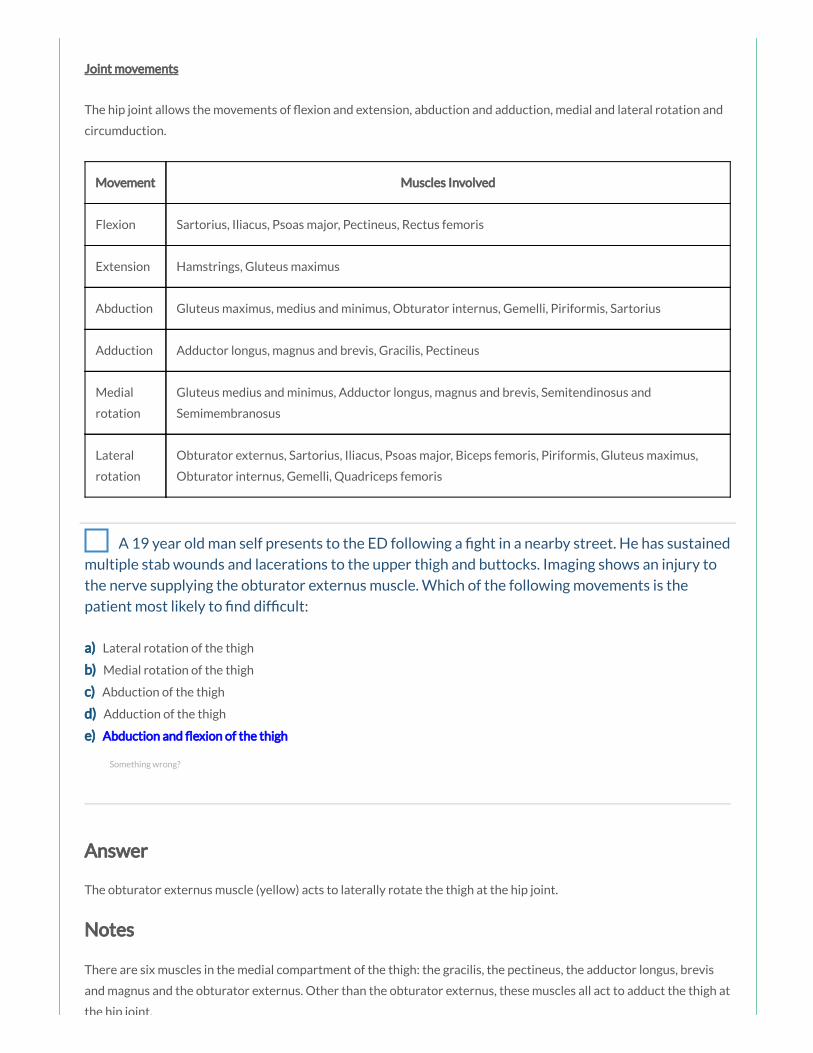
Joint movements
The hip joint allows the movements of exion and extension, abduction and adduction, medial and lateral rotation and
circumduction.
Movement Muscles Involved
Flexion Sartorius, Iliacus, Psoas major, Pectineus, Rectus femoris
Extension Hamstrings, Gluteus maximus
Abduction Gluteus maximus, medius and minimus, Obturator internus, Gemelli, Piriformis, Sartorius
Adduction Adductor longus, magnus and brevis, Gracilis, Pectineus
Medial
rotation
Gluteus medius and minimus, Adductor longus, magnus and brevis, Semitendinosus and
Semimembranosus
Lateral
rotation
Obturator externus, Sartorius, Iliacus, Psoas major, Biceps femoris, Piriformis, Gluteus maximus,
Obturator internus, Gemelli, Quadriceps femoris
A 19 year old man self presents to the ED following a ght in a nearby street. He has sustained
multiple stab wounds and lacerations to the upper thigh and buttocks. Imaging shows an injury to
the nerve supplying the obturator externus muscle. Which of the following movements is the
patient most likely to nd dif cult:
a) Lateral rotation of the thigh
b) Medial rotation of the thigh
c) Abduction of the thigh
d) Adduction of the thigh
e) Abduction and exion of the thigh
Answer
The obturator externus muscle (yellow) acts to laterally rotate the thigh at the hip joint.
Notes
There are six muscles in the medial compartment of the thigh: the gracilis, the pectineus, the adductor longus, brevis
and magnus and the obturator externus. Other than the obturator externus, these muscles all act to adduct the thigh at
the hip joint.
Something wrong?

the hip joint.
Testing of the medial thigh muscles can be performed with the patient lying supine and the knee straight. The patient is
asked to adduct the thigh against resistance and the strength assessed (if the adductors are normal the proximal ends
of the gracilis and adductor longus can easily be palpated).
Muscle Action(s) Innervation
Adductor longus (red) Adduction and medial rotation at
hip
Obturator nerve (L2 – L4)
Adductor brevis (green) Adduction and medial rotation at
hip
Obturator nerve (L2, L3)
Adductor magnus (blue) Adduction and medial rotation at
hip
Obturator nerve (L2 – L4), tibial nerve (L4 –
S3)
Obturator externus
(yellow)
Lateral rotation at hip Obturator nerve (L3, L4)
Gracilis Adduction at hip and exion at
knee
Obturator nerve (L2, L3)
Pectineus Adduction and exion at hip Femoral nerve (L2, L3)
The adductor longus (red), brevis (green) and magnus (blue) are the prime adductors of the thigh at the hip joint and
also assist in medial rotation.
The adductor muscles are innervated by the obturator nerve (L2 – L4), except for the hamstrings portion of the
adductor magnus innervated by the tibial nerve (L4 – S3).
The obturator externus muscle (yellow) acts to laterally rotate the thigh at the hip joint. It is innervated by the
obturator nerve (L3, L4).

Modi ed by FRCEM Success. Original by Henry
Vandyke Carter [Public domain], via Wikimedia
Commons
You form part of the trauma team caring for a roofer who fell approximately 7 metres from a
roof, landing across a beam. Imaging of the spine has revealed traumatic injuries to multiple lumbar
vertebrae. The femoral nerve receives nerve bres from which of the following nerve roots:
a) L1, L2
b) L1 – L4
c) L1 – L3
d) L2 – L4
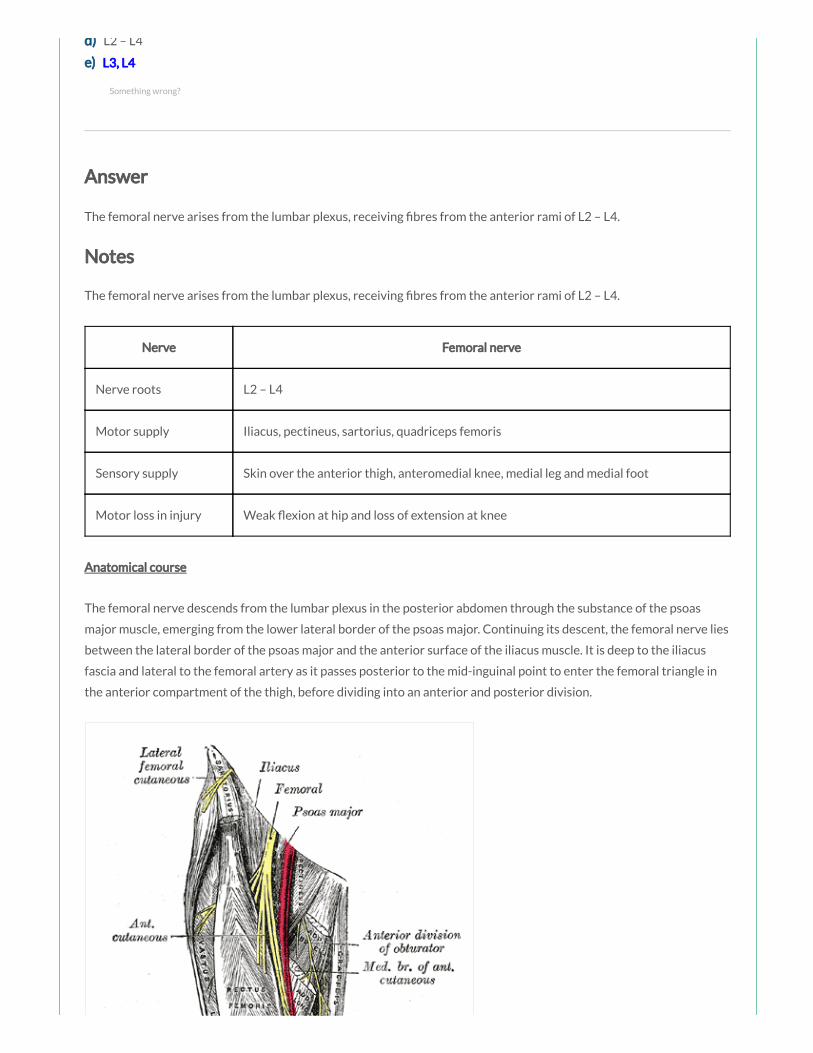
d) L2 – L4
e) L3, L4
Answer
The femoral nerve arises from the lumbar plexus, receiving bres from the anterior rami of L2 – L4.
Notes
The femoral nerve arises from the lumbar plexus, receiving bres from the anterior rami of L2 – L4.
Nerve Femoral nerve
Nerve roots L2 – L4
Motor supply Iliacus, pectineus, sartorius, quadriceps femoris
Sensory supply Skin over the anterior thigh, anteromedial knee, medial leg and medial foot
Motor loss in injury Weak exion at hip and loss of extension at knee
Anatomical course
The femoral nerve descends from the lumbar plexus in the posterior abdomen through the substance of the psoas
major muscle, emerging from the lower lateral border of the psoas major. Continuing its descent, the femoral nerve lies
between the lateral border of the psoas major and the anterior surface of the iliacus muscle. It is deep to the iliacus
fascia and lateral to the femoral artery as it passes posterior to the mid-inguinal point to enter the femoral triangle in
the anterior compartment of the thigh, before dividing into an anterior and posterior division.
Something wrong?

By Henry Vandyke Carter [Public domain], via Wikimedia
Commons
Branches
In the abdomen it gives rise to branches that innervate the iliacus and pectineus muscles.
The anterior division gives off anterior cutaneous branches (supplying skin over the anterior and medial thigh) and
muscular branches (innervating the sartorius).
The posterior division gives off muscular branches (innervating the quadriceps femoris muscles) and articular branches
(supplying the hip and knee joint), before continuing as the saphenous nerve (supplying skin over the anteromedial
knee and the medial side of the leg and foot).

Branch Innervation
Muscular branches in abdomen Iliacus and pectineus
Anterior cutaneous branches Skin over anterior thigh
Anterior muscular branches Sartorius
Posterior muscular branches Quadriceps femoris muscles
Posterior articular branches Hip and knee joint
Saphenous nerve Skin over anteromedial knee, medial side of leg and foot
Motor and sensory function
Muscle Action(s)
Sartorius Flexion, abduction and lateral rotation at hip and exion at knee
Iliacus (red) Flexion and lateral rotation at hip
Pectineus (green) Adduction and exion at hip
Rectus femoris Flexion at hip and extension at knee
Vastus lateralis Extension at knee
Vastus medialis Extension at knee
Vastus intermedius Extension at knee

Modi ed by FRCEM Success. Original by Henry Vandyke Carter [Public domain], via Wikimedia Commons
A 45 year old woman presents to the ED with a painful swelling in her right groin. She is
known to use intravenous drugs and last injected a few hours ago. Imaging has shown a femoral
artery pseudoaneurysm within the femoral sheath. The femoral sheath originates from which of
the following structures:
a) Femoral canal
b) Extraperitoneal fascia
c) Inguinal ligament
d) External oblique fascia
e) Internal oblique fascia
Answer
Something wrong?

The femoral sheath originates as a prolongation of extraperitoneal fascia and encloses the femoral artery, femoral vein
and associated lymphatic vessels (contained within the femoral canal).
Notes
The femoral triangle is a wedge-shaped depression formed by muscles in the upper thigh at the junction between the
anterior abdominal wall and the lower limb.
Femoral Triangle Structure(s)
Superior border Inguinal ligament
Lateral border Medial sartorius muscle
Medial border Medial adductor longus muscle
Roof Fascia lata
Floor Pectineus, iliopsoas and adductor longus muscles
Contents Femoral nerve, femoral artery, femoral vein, femoral canal
Borders
The femoral triangle is bordered superiorly by the inguinal ligament, laterally by the medial border of the sartorius
muscle and medially by the medial border of the adductor longus muscle. The roof is formed by the fascia lata and the
oor is formed by the pectineus, iliopsoas and adductor longus muscles.
Contents
The femoral triangle contains (from lateral to medial):
The femoral nerve
The femoral artery
The femoral vein
The femoral canal
Femoral sheath
The femoral sheath originates as a prolongation of extraperitoneal fascia and encloses the femoral artery, femoral vein
and associated lymphatic vessels (contained within the femoral canal). The femoral nerve does NOT lie within the
sheath, but instead lies lateral to it. The femoral sheath terminates inferiorly by blending with the adventitia of the
femoral vessels approximately 4 cm inferior of the inguinal ligament.
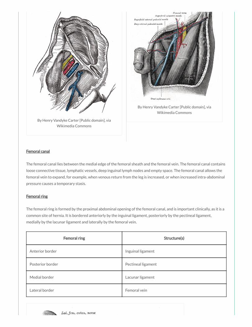
By Henry Vandyke Carter [Public domain], via
Wikimedia Commons
By Henry Vandyke Carter [Public domain], via
Wikimedia Commons
Femoral canal
The femoral canal lies between the medial edge of the femoral sheath and the femoral vein. The femoral canal contains
loose connective tissue, lymphatic vessels, deep inguinal lymph nodes and empty space. The femoral canal allows the
femoral vein to expand, for example, when venous return from the leg is increased, or when increased intra-abdominal
pressure causes a temporary stasis.
Femoral ring
The femoral ring is formed by the proximal abdominal opening of the femoral canal, and is important clinically, as it is a
common site of hernia. It is bordered anteriorly by the inguinal ligament, posteriorly by the pectineal ligament,
medially by the lacunar ligament and laterally by the femoral vein.
Femoral ring Structure(s)
Anterior border Inguinal ligament
Posterior border Pectineal ligament
Medial border Lacunar ligament
Lateral border Femoral vein

By Henry Vandyke Carter [Public domain], via Wikimedia Commons
A 67 year old man presents to the ED complaining of pain and swelling to the posterior knee.
On examination you note a tender swelling in the popliteal fossa. The popliteal fossa is bordered
inferiorly by which of the following muscles:
a) The plantaris and the soleus
b) The gastrocnemius and the plantaris
c) The gastrocnemius and the soleus
d) The gastrocnemius and the popliteus
e) The plantaris and the popliteus
Answer
The popliteal fossa is bordered inferomedially by the medial head of the gastrocnemius and inferolaterally by the
lateral head of the gastrocnemius and plantaris.
Notes
The popliteal fossa is a diamond-shaped space behind the knee joint.
Popliteal fossa Structure(s)
Superomedial border Semimembranosus
Something wrong?
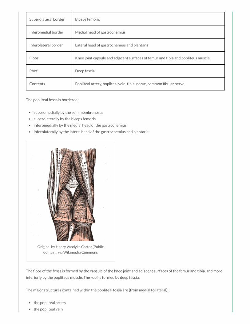
Superolateral border Biceps femoris
Inferomedial border Medial head of gastrocnemius
Inferolateral border Lateral head of gastrocnemius and plantaris
Floor Knee joint capsule and adjacent surfaces of femur and tibia and popliteus muscle
Roof Deep fascia
Contents Popliteal artery, popliteal vein, tibial nerve, common bular nerve
The popliteal fossa is bordered:
superomedially by the semimembranosus
superolaterally by the biceps femoris
inferomedially by the medial head of the gastrocnemius
inferolaterally by the lateral head of the gastrocnemius and plantaris
Original by Henry Vandyke Carter [Public
domain], via Wikimedia Commons
The oor of the fossa is formed by the capsule of the knee joint and adjacent surfaces of the femur and tibia, and more
inferiorly by the popliteus muscle. The roof is formed by deep fascia.
The major structures contained within the popliteal fossa are (from medial to lateral):
the popliteal artery
the popliteal vein
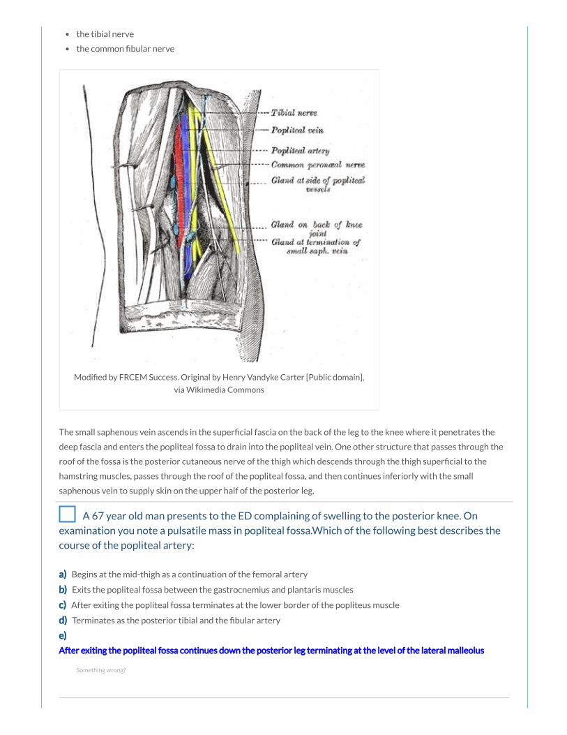
the tibial nerve
the common bular nerve
Modi ed by FRCEM Success. Original by Henry Vandyke Carter [Public domain],
via Wikimedia Commons
The small saphenous vein ascends in the super cial fascia on the back of the leg to the knee where it penetrates the
deep fascia and enters the popliteal fossa to drain into the popliteal vein. One other structure that passes through the
roof of the fossa is the posterior cutaneous nerve of the thigh which descends through the thigh super cial to the
hamstring muscles, passes through the roof of the popliteal fossa, and then continues inferiorly with the small
saphenous vein to supply skin on the upper half of the posterior leg.
A 67 year old man presents to the ED complaining of swelling to the posterior knee. On
examination you note a pulsatile mass in popliteal fossa.Which of the following best describes the
course of the popliteal artery:
a) Begins at the mid-thigh as a continuation of the femoral artery
b) Exits the popliteal fossa between the gastrocnemius and plantaris muscles
c) After exiting the popliteal fossa terminates at the lower border of the popliteus muscle
d) Terminates as the posterior tibial and the bular artery
e)
After exiting the popliteal fossa continues down the posterior leg terminating at the level of the lateral malleolus
Something wrong?

Answer
After exiting the popliteal fossa between the gastrocnemius and popliteus muscle, the popliteal artery bifurcates into
the anterior and posterior tibial arteries at the lower border of the popliteus
Notes
The femoral artery becomes the popliteal artery after entering the posterior compartment of the thigh through the
adductor hiatus just proximal to the knee.
The popliteal artery is the deepest neurovascular structure in the popliteal fossa. Palpation should be performed with
the person in the prone position with the knee exed to relax the popliteal fascia and hamstrings. The popliteal pulse is
best felt in the midline, in the inferior part of the fossa where the popliteal artery is related to the tibia.
After exiting the popliteal fossa between the gastrocnemius and popliteus muscle, the popliteal artery bifurcates into
the anterior and posterior tibial arteries at the lower border of the popliteus.

By Mikael Häggström, used with permission. (Image:Gray544.png) [Public domain
or Public domain], via Wikimedia Commons
A 65 year old man presents with weakness in the right hip, most marked on exion. You
consider the possibility of a psoas major pathology. The psoas major muscle is innervated by which
of the following nerves:
a) Femoral nerve
b) Obturator nerve
c) Anterior rami of spinal nerves L1 – L3
d) Pudendal nerve
e) Inferior gluteal nerve
Answer
The psoas major is innervated by the anterior rami of spinal nerves L1 – L3.
Notes
The sartorius, iliopsoas, pectineus and rectus femoris are the primary exors of the thigh at the hip joint.
Muscle Actions Innervation
Sartorius (blue) Flexion, abduction and lateral rotation at hip and exion at Femoral nerve (L2, L3)
Something wrong?

Sartorius (blue) Flexion, abduction and lateral rotation at hip and exion at
knee
Femoral nerve (L2, L3)
Iliacus (red) Flexion and lateral rotation at hip Femoral nerve (L2, L3)
Psoas major
(yellow)
Flexion and lateral rotation at hip Anterior rami L1 – L3
Pectineus (green) Adduction and exion at hip Femoral nerve (L2, L3)
Rectus femoris Flexion at hip and extension at knee Femoral nerve (L2 –
L4)
The sartorius (blue) is innervated by the femoral nerve (L2, L3). It acts to ex the thigh at the hip joint and ex the leg at
the knee joint. It also abducts the thigh and rotates it laterally, as when resting the foot on the opposite knee when
sitting.
The iliacus (red) and psoas major (yellow) muscles originate as separate muscles but insert by a common tendon onto
the femur and together are referred to as the iliopsoas muscle, which is a powerful exor of the thigh at the hip joint
and also contributes to lateral rotation of the thigh at the hip joint. The iliacus is innervated by the femoral nerve (L2,
L3). The psoas major is innervated by the anterior rami of spinal nerves L1 – L3.
The pectineus (green) is innervated by the femoral nerve (L2, L3). It acts to adduct and ex the thigh at the hip joint.

Modi ed by FRCEM Success. Original by Henry
Vandyke Carter [Public domain], via Wikimedia
Commons
A 43 year old window cleaner is brought to the ED after falling from a 5 metre platform.
Imaging has shown multiple vertebral fractures and an injury to the obturator nerve. Which of the
following best describes the cutaneous innervation of the obturator nerve:
a) Skin over anterior thigh
b) Skin over anterolateral thigh
c) Skin over posterior thigh
d) Skin over upper medial thigh
e) Skin over medial thigh and medial leg
Answer
The obturator nerve supplies skin over the medial upper thigh.
Something wrong?

Notes
The obturator nerve arises from the lumbar plexus, formed from the anterior rami of L2 – L4.
Nerve Obturator nerve
Nerve
roots
L2 – L4
Motor
supply
Medial thigh muscles (adductor longus, brevis and magnus, gracilis, obturator externus)
Sensory
supply
Upper medial thigh
Motor loss
in injury
Weak adduction of hip and dif culty walking with lateral swinging of limb during walking (due to
unopposed abduction), loss of sensation over upper medial thigh
The obturator nerve descends along the posterior abdominal wall, passes through the pelvic cavity and enters the
medial thigh by passing through the obturator canal.
The obturator nerve innervates all of the muscles of the medial compartment of the thigh (except for the hamstring
part of the adductor longus, and the pectineus muscle which are innervated by the sciatic and the femoral nerves
respectively). It also gives off a cutaneous branch that supplies skin on the medial side of the upper thigh.
Muscle Action(s)
Adductor longus Adduction and medial rotation at hip
Adductor brevis Adduction and medial rotation at hip
Adductor magnus Adduction and medial rotation at hip
Obturator externus Lateral rotation at hip
Gracilis Adduction at hip and exion at knee
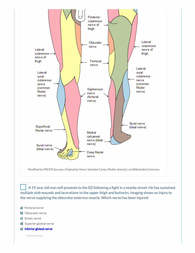
Modi ed by FRCEM Success. Original by Henry Vandyke Carter [Public domain], via Wikimedia Commons
A 19 year old man self presents to the ED following a ght in a nearby street. He has sustained
multiple stab wounds and lacerations to the upper thigh and buttocks. Imaging shows an injury to
the nerve supplying the obturator externus muscle. Which nerve has been injured:
a) Femoral nerve
b) Obturator nerve
c) Sciatic nerve
d) Superior gluteal nerve
e) Inferior gluteal nerve
Something wrong?
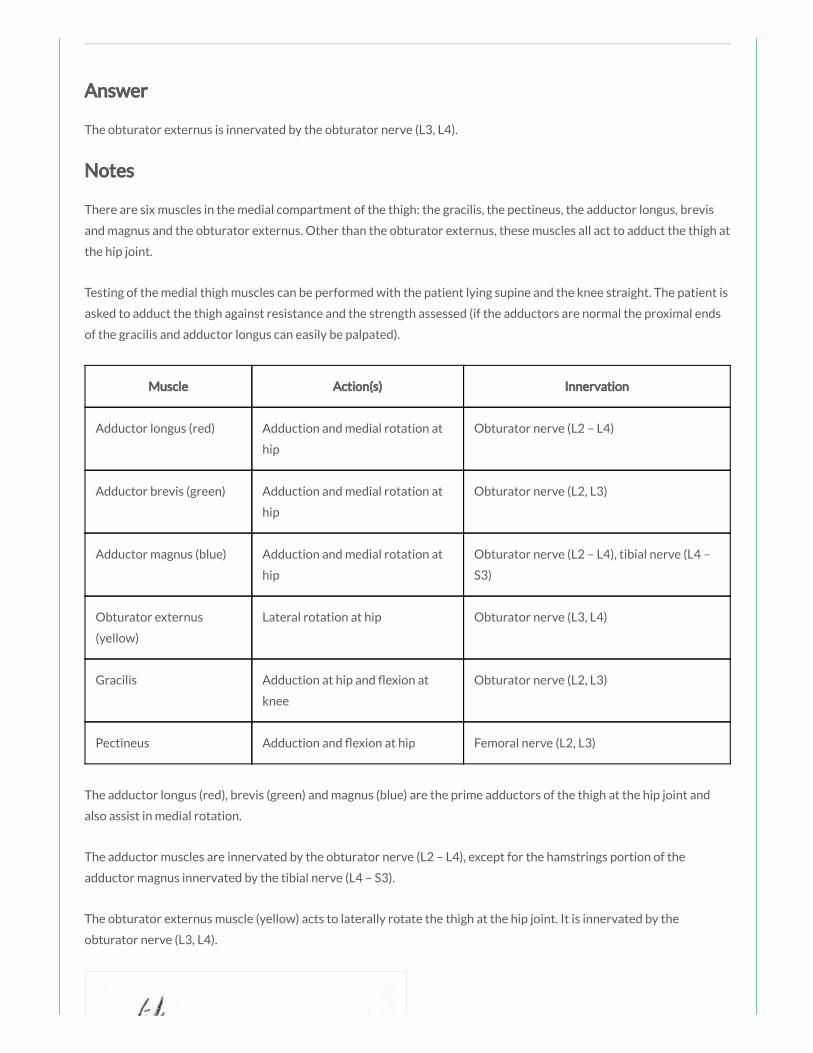
Answer
The obturator externus is innervated by the obturator nerve (L3, L4).
Notes
There are six muscles in the medial compartment of the thigh: the gracilis, the pectineus, the adductor longus, brevis
and magnus and the obturator externus. Other than the obturator externus, these muscles all act to adduct the thigh at
the hip joint.
Testing of the medial thigh muscles can be performed with the patient lying supine and the knee straight. The patient is
asked to adduct the thigh against resistance and the strength assessed (if the adductors are normal the proximal ends
of the gracilis and adductor longus can easily be palpated).
Muscle Action(s) Innervation
Adductor longus (red) Adduction and medial rotation at
hip
Obturator nerve (L2 – L4)
Adductor brevis (green) Adduction and medial rotation at
hip
Obturator nerve (L2, L3)
Adductor magnus (blue) Adduction and medial rotation at
hip
Obturator nerve (L2 – L4), tibial nerve (L4 –
S3)
Obturator externus
(yellow)
Lateral rotation at hip Obturator nerve (L3, L4)
Gracilis Adduction at hip and exion at
knee
Obturator nerve (L2, L3)
Pectineus Adduction and exion at hip Femoral nerve (L2, L3)
The adductor longus (red), brevis (green) and magnus (blue) are the prime adductors of the thigh at the hip joint and
also assist in medial rotation.
The adductor muscles are innervated by the obturator nerve (L2 – L4), except for the hamstrings portion of the
adductor magnus innervated by the tibial nerve (L4 – S3).
The obturator externus muscle (yellow) acts to laterally rotate the thigh at the hip joint. It is innervated by the
obturator nerve (L3, L4).

Modi ed by FRCEM Success. Original by Henry
Vandyke Carter [Public domain], via Wikimedia
Commons

Commons
Your Consultant is discussing a case she saw earlier today. A 34 year old carpet tter
presented with pain on certain movements of the right leg. She diagnosed a sartorius muscle strain.
Which of the following movements is he likely to have found painful:
a) Flexion of the hip and exion of the knee
b) Flexion of the hip and extension of the knee
c) Extension of the hip and exion of the knee
d) Flexion and medial rotation of the hip
e) Extension of the hip and extension of the knee
Answer
The sartorius acts to ex the thigh at the hip joint and ex the leg at the knee joint. It also abducts the thigh and rotates
it laterally, as when resting the foot on the opposite knee when sitting.
Notes
The sartorius, iliopsoas, pectineus and rectus femoris are the primary exors of the thigh at the hip joint.
Muscle Actions Innervation
Sartorius (blue) Flexion, abduction and lateral rotation at hip and exion at
knee
Femoral nerve (L2, L3)
Iliacus (red) Flexion and lateral rotation at hip Femoral nerve (L2, L3)
Psoas major
(yellow)
Flexion and lateral rotation at hip Anterior rami L1 – L3
Pectineus (green) Adduction and exion at hip Femoral nerve (L2, L3)
Rectus femoris Flexion at hip and extension at knee Femoral nerve (L2 –
L4)
The sartorius (blue) is innervated by the femoral nerve (L2, L3). It acts to ex the thigh at the hip joint and ex the leg at
the knee joint. It also abducts the thigh and rotates it laterally, as when resting the foot on the opposite knee when
sitting.
The iliacus (red) and psoas major (yellow) muscles originate as separate muscles but insert by a common tendon onto
the femur and together are referred to as the iliopsoas muscle, which is a powerful exor of the thigh at the hip joint
Something wrong?

the femur and together are referred to as the iliopsoas muscle, which is a powerful exor of the thigh at the hip joint
and also contributes to lateral rotation of the thigh at the hip joint. The iliacus is innervated by the femoral nerve (L2,
L3). The psoas major is innervated by the anterior rami of spinal nerves L1 – L3.
The pectineus (green) is innervated by the femoral nerve (L2, L3). It acts to adduct and ex the thigh at the hip joint.

Modi ed by FRCEM Success. Original by Henry
Vandyke Carter [Public domain], via Wikimedia
Commons
An 85 year old man is brought to the ED complaining of fever and pain in his right hip. You
suspect a septic arthritis of the right hip joint. Which of the following synovial joint types best
describes the hip joint:
a) Synovial modi ed hinge joint
b) Synovial pivot joint
c) Synovial saddle joint
d) Synovial ball and socket joint
e) Synovial condyloid joint
Answer
The hip joint is a multiaxial synovial ball and socket joint occurring between the head of the femur and the acetabulum
of the pelvis.
Notes
Joint Hip joint
Type Synovial ball and socket joint
Articulations Head of femur with acetabulum of pelvis
Stabilising
factors
Acetabular labrum, thickened brous capsule, extracapsular ligaments (iliofemoral, ischiofemoral,
pubofemoral), medial rotator muscles (effectively ‘pull’ head of femur into acetabulum)
Movements Flexion/Extension, Abduction/Adduction, Medial/Lateral rotation, Circumduction
Blood supply Branches of obturator artery, medial and lateral circum ex branches of profunda femoris artery
and superior and inferior gluteal arteries
Innervation Femoral nerve, obturator nerve, superior gluteal nerve and nerve to the quadratus femoris
Something wrong?

Innervation Femoral nerve, obturator nerve, superior gluteal nerve and nerve to the quadratus femoris
Joint articulations
The hip joint is a multiaxial synovial ball and socket joint occurring between the head of the femur and the acetabulum
of the pelvis.
The acetabulum is formed by the fusion of the three bones, the ilium, the ischium and the pubis.
By Henry Vandyke Carter [Public domain], via Wikimedia Commons
The rim of the acetabulum is raised slightly by the brocartilaginous acetabular labrum which increases its depth,
improving stability of the joint. The acetabular labrum continues inferiorly as the transverse acetabular ligament which
bridges the acetabular notch and converts the notch into a foramen for the passage of nutrient vessels and nerves.
Ligaments
The intracapsular ligament of the head of femur (ligamentum teres) runs from the fovea on the head of the femur at
one end to the acetabular fossa and the transverse acetabular ligament on the other. It carries a small acetabular
branch of the obturator artery.
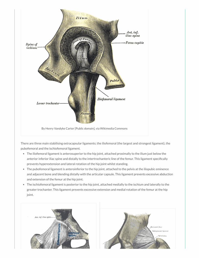
By Henry Vandyke Carter [Public domain], via Wikimedia Commons
There are three main stabilising extracapsular ligaments; the iliofemoral (the largest and strongest ligament), the
pubofemoral and the ischiofemoral ligament.
The iliofemoral ligament is anterosuperior to the hip joint, attached proximally to the ilium just below the
anterior inferior iliac spine and distally to the intertrochanteric line of the femur. This ligament speci cally
prevents hyperextension and lateral rotation of the hip joint whilst standing.
The pubofemoral ligament is anteroinferior to the hip joint, attached to the pelvis at the iliopubic eminence
and adjacent bone and blending distally with the articular capsule. This ligament prevents excessive abduction
and extension of the femur at the hip joint.
The ischiofemoral ligament is posterior to the hip joint, attached medially to the ischium and laterally to the
greater trochanter. This ligament prevents excessive extension and medial rotation of the femur at the hip
joint.
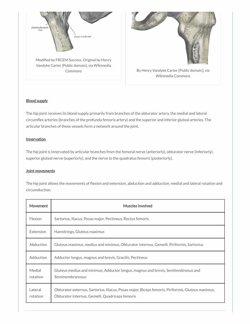
Modi ed by FRCEM Success. Original by Henry
Vandyke Carter [Public domain], via Wikimedia
Commons By Henry Vandyke Carter [Public domain], via
Wikimedia Commons
Blood supply
The hip joint receives its blood supply primarily from branches of the obturator artery, the medial and lateral
circum ex arteries (branches of the profunda femoris artery) and the superior and inferior gluteal arteries. The
articular branches of these vessels form a network around the joint.
Innervation
The hip joint is innervated by articular branches from the femoral nerve (anteriorly), obturator nerve (inferiorly),
superior gluteal nerve (superiorly), and the nerve to the quadratus femoris (posteriorly).
Joint movements
The hip joint allows the movements of exion and extension, abduction and adduction, medial and lateral rotation and
circumduction.
Movement Muscles Involved
Flexion Sartorius, Iliacus, Psoas major, Pectineus, Rectus femoris
Extension Hamstrings, Gluteus maximus
Abduction Gluteus maximus, medius and minimus, Obturator internus, Gemelli, Piriformis, Sartorius
Adduction Adductor longus, magnus and brevis, Gracilis, Pectineus
Medial
rotation
Gluteus medius and minimus, Adductor longus, magnus and brevis, Semitendinosus and
Semimembranosus
Lateral
rotation
Obturator externus, Sartorius, Iliacus, Psoas major, Biceps femoris, Piriformis, Gluteus maximus,
Obturator internus, Gemelli, Quadriceps femoris
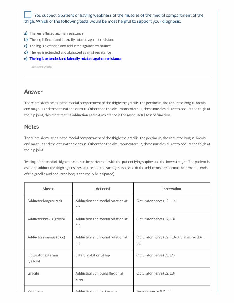
You suspect a patient of having weakness of the muscles of the medial compartment of the
thigh. Which of the following tests would be most helpful to support your diagnosis:
a) The leg is exed against resistance
b) The leg is exed and laterally rotated against resistance
c) The leg is extended and adducted against resistance
d) The leg is extended and abducted against resistance
e) The leg is extended and laterally rotated against resistance
Answer
There are six muscles in the medial compartment of the thigh: the gracilis, the pectineus, the adductor longus, brevis
and magnus and the obturator externus. Other than the obturator externus, these muscles all act to adduct the thigh at
the hip joint, therefore testing adduction against resistance is the most useful test of function.
Notes
There are six muscles in the medial compartment of the thigh: the gracilis, the pectineus, the adductor longus, brevis
and magnus and the obturator externus. Other than the obturator externus, these muscles all act to adduct the thigh at
the hip joint.
Testing of the medial thigh muscles can be performed with the patient lying supine and the knee straight. The patient is
asked to adduct the thigh against resistance and the strength assessed (if the adductors are normal the proximal ends
of the gracilis and adductor longus can easily be palpated).
Muscle Action(s) Innervation
Adductor longus (red) Adduction and medial rotation at
hip
Obturator nerve (L2 – L4)
Adductor brevis (green) Adduction and medial rotation at
hip
Obturator nerve (L2, L3)
Adductor magnus (blue) Adduction and medial rotation at
hip
Obturator nerve (L2 – L4), tibial nerve (L4 –
S3)
Obturator externus
(yellow)
Lateral rotation at hip Obturator nerve (L3, L4)
Gracilis Adduction at hip and exion at
knee
Obturator nerve (L2, L3)
Pectineus Adduction and exion at hip Femoral nerve (L2, L3)
Something wrong?
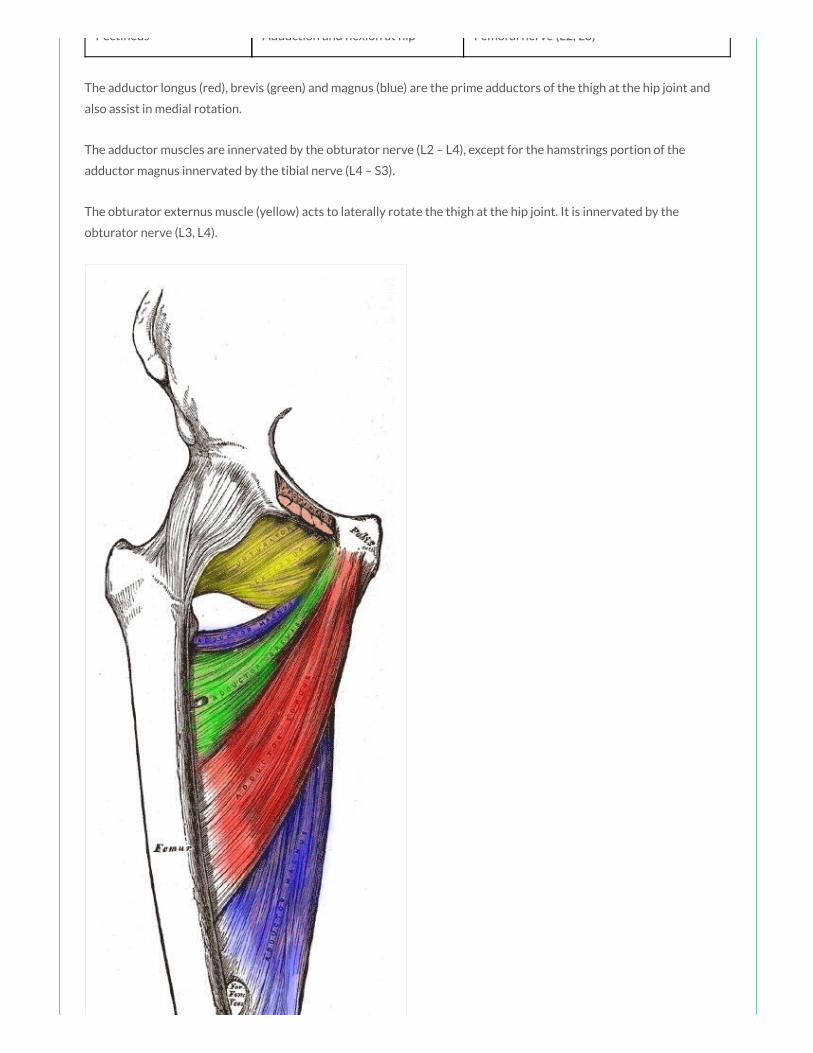
Pectineus Adduction and exion at hip Femoral nerve (L2, L3)
The adductor longus (red), brevis (green) and magnus (blue) are the prime adductors of the thigh at the hip joint and
also assist in medial rotation.
The adductor muscles are innervated by the obturator nerve (L2 – L4), except for the hamstrings portion of the
adductor magnus innervated by the tibial nerve (L4 – S3).
The obturator externus muscle (yellow) acts to laterally rotate the thigh at the hip joint. It is innervated by the
obturator nerve (L3, L4).

Modi ed by FRCEM Success. Original by Henry
Vandyke Carter [Public domain], via Wikimedia
Commons
A 23 year old rugby player presents to the ED complaining of ongoing thigh pain following a
match 3 days ago. Imaging has been arranged which shows a traumatic injury to the rectus femoris.
The rectus femoris muscle acts primarily to produce which of the following movements:
a) Flexion at hip and exion at knee
b) Flexion at hip and extension at knee
c) Extension at hip and exion at knee
d) Extension at hip and extension at knee
e) Flexion at knee
Answer
The quadriceps femoris muscle is the main extensor of the leg at the knee joint. The rectus femoris crosses both the
knee and the hip joint (in contrast to the vastus muscles which only cross the knee joint), and therefore also assists in
exion of the thigh at the hip joint.
Notes
Muscle Action(s) Innervation
Rectus femoris (red) Flexion at hip and extension at knee Femoral nerve (L2 – L4)
Vastus lateralis (green) Extension at knee Femoral nerve (L2 – L4)
Vastus medialis (blue) Extension at knee Femoral nerve (L2 – L4)
Vastus intermedius Extension at knee Femoral nerve (L2 – L4)
Something wrong?

The quadriceps femoris is made up of four individual muscles; the rectus femoris (red), the vastus medialis (blue), the
vastus intermedius (deep to the rectus femoris) and the vastus lateralis (green) which all insert onto the patella by the
common quadriceps femoris tendon. Because the vastus muscles insert into the margins of the patella as well as into
the quadriceps femoris tendon, they stabilise the position of the patella during knee joint movement.
The quadriceps femoris muscle is the main extensor of the leg at the knee joint. The rectus femoris crosses both the
knee and the hip joint (in contrast to the vastus muscles which only cross the knee joint), and therefore also assists in
exion of the thigh at the hip joint.
The quadriceps femoris muscles are innervated by the femoral nerve (L2 – L4). A tap on the patella ligament tests
re ex activity mainly at spinal cord levels L3/L4.

Modi ed by FRCEM Success. Original by Henry
Vandyke Carter [Public domain], via Wikimedia
Commons
A 32 year old woman is brought to the ED after falling from a horse. Imaging shows multiple
injuries to the pelvic region including transection of the nerve that supplies the piriformis muscle.
Which of the following nerves has been transected:
a) Obturator nerve
b) Superior gluteal nerve
c) Inferior gluteal nerve
d) Nerve to the piriformis, branch of the sacral plexus
e) Sciatic nerve
Answer
The piriformis is innervated by the nerve to the piriformis, originating from the sacral plexus (S1, S2).
Notes
Muscle Action(s) Innervation
Gluteus maximus (red) Extension, lateral rotation and abduction at hip Inferior gluteal nerve (L5 – S2)
Gluteus medius (green) Abduction and medial rotation at hip Superior gluteal nerve (L4 – S1)
Gluteus minimus (blue) Abduction and medial rotation at hip Superior gluteal nerve (L4 – S1)
Something wrong?

Piriformis (yellow) Lateral rotation and abduction at hip Branches from S1 and S2
The gluteus maximus (red) is the main extensor of the thigh at the hip joint and also acts to laterally rotate and abduct
the thigh. Through its insertion into the iliotibial tract, it also stabilises the knee and hip joints. The gluteus maximus is
innervated by the inferior gluteal nerve (L5 – S2).
The gluteus medius (green) and gluteus minimus (blue) act to abduct and medially rotate the thigh at the hip joint, and
also act to secure the pelvis, reducing pelvic drop on the opposite swing side during walking. They are both innervated
by the superior gluteal nerve (L4 – S1).
The piriformis (yellow) acts to abduct and laterally rotate the thigh at the hip joint. It is innervated by the nerve to the
piriformis, originating from the sacral plexus (S1, S2). The obturator internus, gemelli and quadriceps femoris muscles
act as synergistic femoral lateral rotators and hip stabilisers.
Trendelenburg’s sign is seen in people with weak/paralysed abductor muscles of the hip. The sign is demonstrated by
asking the patient to stand on one limb; when the patient stands on the affected limb, the pelvis severely drops over
the swing limb. This sign may be seen in patients with damage to the superior gluteal nerve, which may occur in
association with pelvic fractures, with space-occupying lesions within the pelvis extending into the greater sciatic
foramen, and following hip surgery. Typically the patient may also demonstrate a Trendelenburg gait.
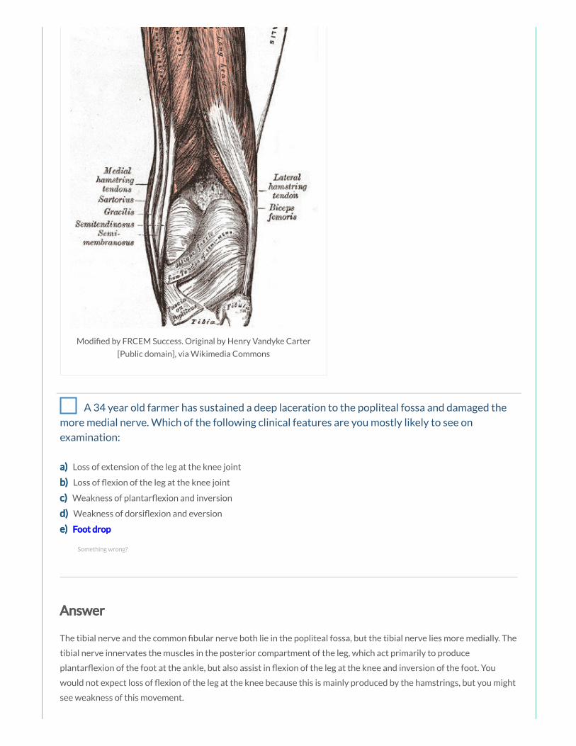
Modi ed by FRCEM Success. Original by Henry Vandyke Carter
[Public domain], via Wikimedia Commons
A 34 year old farmer has sustained a deep laceration to the popliteal fossa and damaged the
more medial nerve. Which of the following clinical features are you mostly likely to see on
examination:
a) Loss of extension of the leg at the knee joint
b) Loss of exion of the leg at the knee joint
c) Weakness of plantar exion and inversion
d) Weakness of dorsi exion and eversion
e) Foot drop
Answer
The tibial nerve and the common bular nerve both lie in the popliteal fossa, but the tibial nerve lies more medially. The
tibial nerve innervates the muscles in the posterior compartment of the leg, which act primarily to produce
plantar exion of the foot at the ankle, but also assist in exion of the leg at the knee and inversion of the foot. You
would not expect loss of exion of the leg at the knee because this is mainly produced by the hamstrings, but you might
see weakness of this movement.
Notes
Something wrong?

Notes
The tibial nerve is a branch of the sciatic nerve receiving nerve bres from L4 – S3.
Nerve Tibial nerve
Nerve
roots
L4 – S3
Motor
supply
All muscles in the posterior compartment of the leg and the intrinsic muscles in the sole of the foot
Sensory
supply
Skin on the posterolateral side of the lower leg, the lateral side of the ankle, foot and little toe, the
medial side of the heel and the sole of the heel, foot and toes
Injury Weakness of exion of knee, loss of plantar exion of ankle and exion of toes, weakness of foot
inversion and loss of sensation in distribution above
It arises at the apex of the popliteal fossa before descending in the leg to enter the popliteal fossa posterior to the
knee. The tibial nerve then passes under the tendinous arch formed by the two heads of the soleus muscle and then
descends through the deep region of the posterior compartment of the leg. The tibial nerve passes through the tarsal
tunnel, posterior to the medial malleolus to enter to foot.
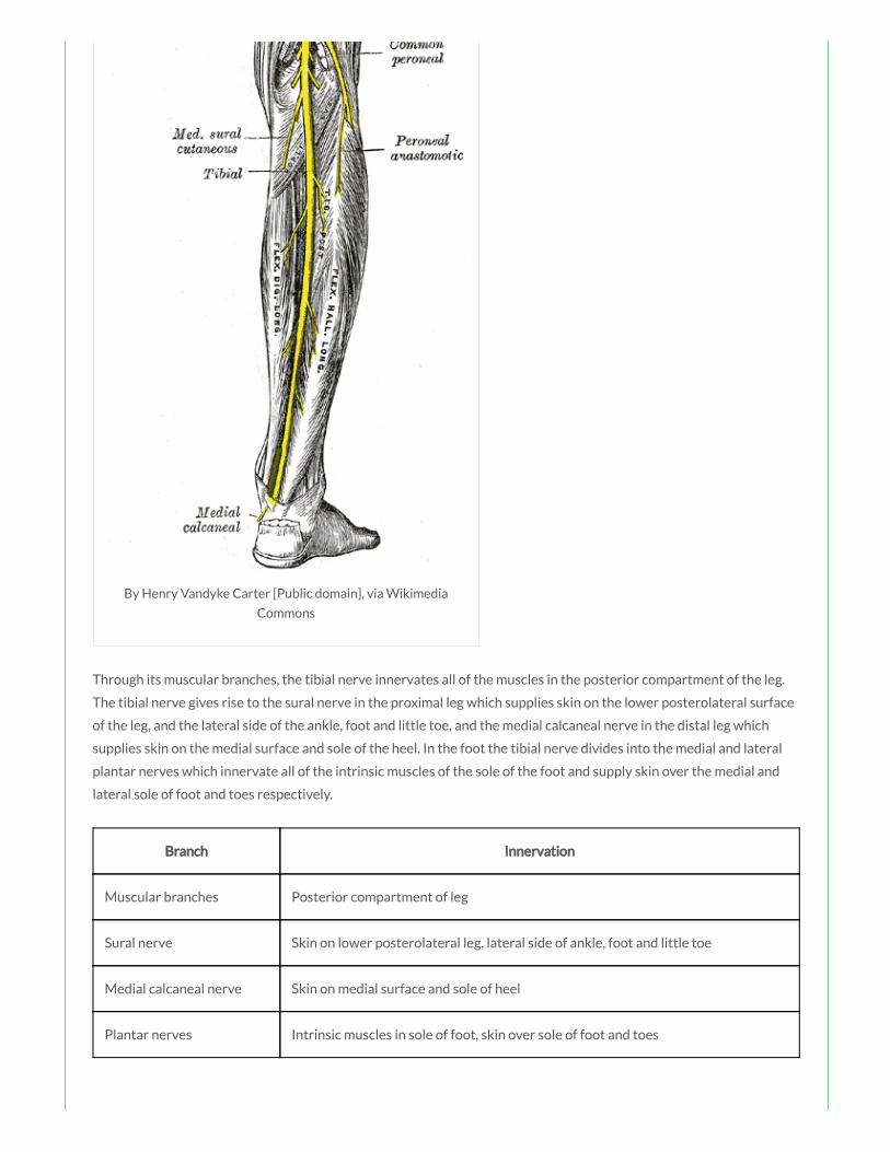
By Henry Vandyke Carter [Public domain], via Wikimedia
Commons
Through its muscular branches, the tibial nerve innervates all of the muscles in the posterior compartment of the leg.
The tibial nerve gives rise to the sural nerve in the proximal leg which supplies skin on the lower posterolateral surface
of the leg, and the lateral side of the ankle, foot and little toe, and the medial calcaneal nerve in the distal leg which
supplies skin on the medial surface and sole of the heel. In the foot the tibial nerve divides into the medial and lateral
plantar nerves which innervate all of the intrinsic muscles of the sole of the foot and supply skin over the medial and
lateral sole of foot and toes respectively.
Branch Innervation
Muscular branches Posterior compartment of leg
Sural nerve Skin on lower posterolateral leg, lateral side of ankle, foot and little toe
Medial calcaneal nerve Skin on medial surface and sole of heel
Plantar nerves Intrinsic muscles in sole of foot, skin over sole of foot and toes

Muscle Action(s)
Gastrocnemius Plantar exion of foot and exion of leg
Plantaris Plantar exion of foot and exion of leg
Soleus Plantar exion of foot
Flexor digitorum longus Flexion of lateral four toes
Flexor hallucis longus Flexion of great toe and plantar exion of foot
Tibialis posterior Plantar exion and inversion of foot, support of medial arch
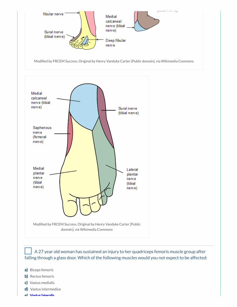
Modi ed by FRCEM Success. Original by Henry Vandyke Carter [Public domain], via Wikimedia Commons
Modi ed by FRCEM Success. Original by Henry Vandyke Carter [Public
domain], via Wikimedia Commons
A 27 year old woman has sustained an injury to her quadriceps femoris muscle group after
falling through a glass door. Which of the following muscles would you not expect to be affected:
a) Biceps femoris
b) Rectus femoris
c) Vastus medialis
d) Vastus intermedius
e) Vastus lateralis

e) Vastus lateralis
Answer
The quadriceps femoris is made up of four individual muscles; the rectus femoris (red), the vastus medialis (blue), the
vastus intermedius (deep to the rectus femoris) and the vastus lateralis (green) which all insert onto the patella by the
common quadriceps femoris tendon. The biceps femoris muscle is a hamstring muscle in the posterior thigh.
Notes
Muscle Action(s) Innervation
Rectus femoris (red) Flexion at hip and extension at knee Femoral nerve (L2 – L4)
Vastus lateralis (green) Extension at knee Femoral nerve (L2 – L4)
Vastus medialis (blue) Extension at knee Femoral nerve (L2 – L4)
Vastus intermedius Extension at knee Femoral nerve (L2 – L4)
The quadriceps femoris is made up of four individual muscles; the rectus femoris (red), the vastus medialis (blue), the
vastus intermedius (deep to the rectus femoris) and the vastus lateralis (green) which all insert onto the patella by the
common quadriceps femoris tendon. Because the vastus muscles insert into the margins of the patella as well as into
the quadriceps femoris tendon, they stabilise the position of the patella during knee joint movement.
The quadriceps femoris muscle is the main extensor of the leg at the knee joint. The rectus femoris crosses both the
knee and the hip joint (in contrast to the vastus muscles which only cross the knee joint), and therefore also assists in
exion of the thigh at the hip joint.
The quadriceps femoris muscles are innervated by the femoral nerve (L2 – L4). A tap on the patella ligament tests
re ex activity mainly at spinal cord levels L3/L4.
Something wrong?

Modi ed by FRCEM Success. Original by Henry
Vandyke Carter [Public domain], via Wikimedia
Commons
A 21 year old man presents to the ED after sustaining multiple deep lacerations to the
posterior thigh during a ght. You suspect an injury to the hamstring muscles. Which of the
following would best test the strength of the hamstring muscles:
a) The leg is exed against resistance
b) The leg is extended against resistance
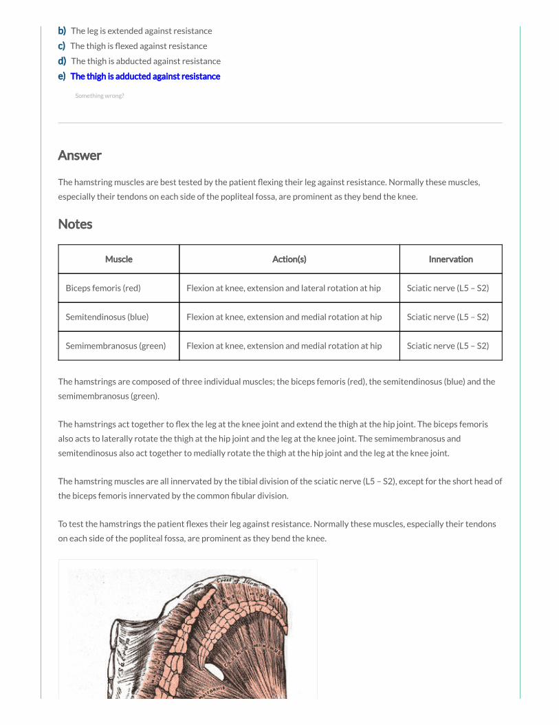
b) The leg is extended against resistance
c) The thigh is exed against resistance
d) The thigh is abducted against resistance
e) The thigh is adducted against resistance
Answer
The hamstring muscles are best tested by the patient exing their leg against resistance. Normally these muscles,
especially their tendons on each side of the popliteal fossa, are prominent as they bend the knee.
Notes
Muscle Action(s) Innervation
Biceps femoris (red) Flexion at knee, extension and lateral rotation at hip Sciatic nerve (L5 – S2)
Semitendinosus (blue) Flexion at knee, extension and medial rotation at hip Sciatic nerve (L5 – S2)
Semimembranosus (green) Flexion at knee, extension and medial rotation at hip Sciatic nerve (L5 – S2)
The hamstrings are composed of three individual muscles; the biceps femoris (red), the semitendinosus (blue) and the
semimembranosus (green).
The hamstrings act together to ex the leg at the knee joint and extend the thigh at the hip joint. The biceps femoris
also acts to laterally rotate the thigh at the hip joint and the leg at the knee joint. The semimembranosus and
semitendinosus also act together to medially rotate the thigh at the hip joint and the leg at the knee joint.
The hamstring muscles are all innervated by the tibial division of the sciatic nerve (L5 – S2), except for the short head of
the biceps femoris innervated by the common bular division.
To test the hamstrings the patient exes their leg against resistance. Normally these muscles, especially their tendons
on each side of the popliteal fossa, are prominent as they bend the knee.
Something wrong?

Modi ed by FRCEM Success. Original by Henry Vandyke Carter
[Public domain], via Wikimedia Commons
You are discussing approaches to analgesia in a patient with a fractured neck of femur. Your
consultant suggests a femoral nerve block. The femoral triangle is bordered laterally by which of
the following structures:
a) Femoral canal
b) Sartorius muscle
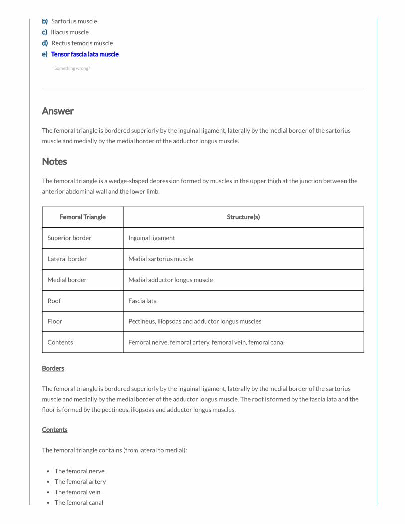
b) Sartorius muscle
c) Iliacus muscle
d) Rectus femoris muscle
e) Tensor fascia lata muscle
Answer
The femoral triangle is bordered superiorly by the inguinal ligament, laterally by the medial border of the sartorius
muscle and medially by the medial border of the adductor longus muscle.
Notes
The femoral triangle is a wedge-shaped depression formed by muscles in the upper thigh at the junction between the
anterior abdominal wall and the lower limb.
Femoral Triangle Structure(s)
Superior border Inguinal ligament
Lateral border Medial sartorius muscle
Medial border Medial adductor longus muscle
Roof Fascia lata
Floor Pectineus, iliopsoas and adductor longus muscles
Contents Femoral nerve, femoral artery, femoral vein, femoral canal
Borders
The femoral triangle is bordered superiorly by the inguinal ligament, laterally by the medial border of the sartorius
muscle and medially by the medial border of the adductor longus muscle. The roof is formed by the fascia lata and the
oor is formed by the pectineus, iliopsoas and adductor longus muscles.
Contents
The femoral triangle contains (from lateral to medial):
The femoral nerve
The femoral artery
The femoral vein
The femoral canal
Something wrong?
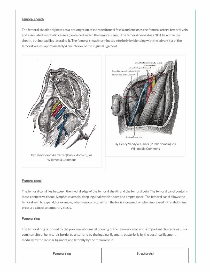
By Henry Vandyke Carter [Public domain], via
Wikimedia Commons
By Henry Vandyke Carter [Public domain], via
Wikimedia Commons
The femoral canal
Femoral sheath
The femoral sheath originates as a prolongation of extraperitoneal fascia and encloses the femoral artery, femoral vein
and associated lymphatic vessels (contained within the femoral canal). The femoral nerve does NOT lie within the
sheath, but instead lies lateral to it. The femoral sheath terminates inferiorly by blending with the adventitia of the
femoral vessels approximately 4 cm inferior of the inguinal ligament.
Femoral canal
The femoral canal lies between the medial edge of the femoral sheath and the femoral vein. The femoral canal contains
loose connective tissue, lymphatic vessels, deep inguinal lymph nodes and empty space. The femoral canal allows the
femoral vein to expand, for example, when venous return from the leg is increased, or when increased intra-abdominal
pressure causes a temporary stasis.
Femoral ring
The femoral ring is formed by the proximal abdominal opening of the femoral canal, and is important clinically, as it is a
common site of hernia. It is bordered anteriorly by the inguinal ligament, posteriorly by the pectineal ligament,
medially by the lacunar ligament and laterally by the femoral vein.
Femoral ring Structure(s)
Anterior border Inguinal ligament
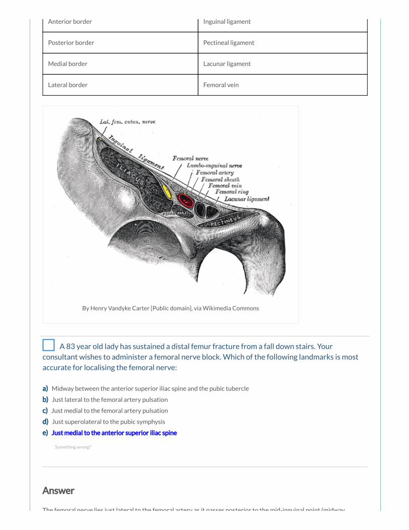
Anterior border Inguinal ligament
Posterior border Pectineal ligament
Medial border Lacunar ligament
Lateral border Femoral vein
By Henry Vandyke Carter [Public domain], via Wikimedia Commons
A 83 year old lady has sustained a distal femur fracture from a fall down stairs. Your
consultant wishes to administer a femoral nerve block. Which of the following landmarks is most
accurate for localising the femoral nerve:
a) Midway between the anterior superior iliac spine and the pubic tubercle
b) Just lateral to the femoral artery pulsation
c) Just medial to the femoral artery pulsation
d) Just superolateral to the pubic symphysis
e) Just medial to the anterior superior iliac spine
Answer
The femoral nerve lies just lateral to the femoral artery as it passes posterior to the mid-inguinal point (midway
Something wrong?
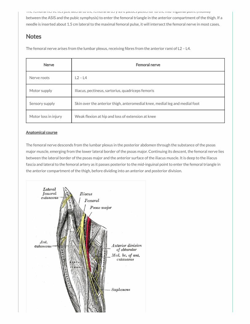
The femoral nerve lies just lateral to the femoral artery as it passes posterior to the mid-inguinal point (midway
between the ASIS and the pubic symphysis) to enter the femoral triangle in the anterior compartment of the thigh. If a
needle is inserted about 1.5 cm lateral to the maximal femoral pulse, it will intersect the femoral nerve in most cases.
Notes
The femoral nerve arises from the lumbar plexus, receiving bres from the anterior rami of L2 – L4.
Nerve Femoral nerve
Nerve roots L2 – L4
Motor supply Iliacus, pectineus, sartorius, quadriceps femoris
Sensory supply Skin over the anterior thigh, anteromedial knee, medial leg and medial foot
Motor loss in injury Weak exion at hip and loss of extension at knee
Anatomical course
The femoral nerve descends from the lumbar plexus in the posterior abdomen through the substance of the psoas
major muscle, emerging from the lower lateral border of the psoas major. Continuing its descent, the femoral nerve lies
between the lateral border of the psoas major and the anterior surface of the iliacus muscle. It is deep to the iliacus
fascia and lateral to the femoral artery as it passes posterior to the mid-inguinal point to enter the femoral triangle in
the anterior compartment of the thigh, before dividing into an anterior and posterior division.

By Henry Vandyke Carter [Public domain], via Wikimedia
Commons
Branches
In the abdomen it gives rise to branches that innervate the iliacus and pectineus muscles.
The anterior division gives off anterior cutaneous branches (supplying skin over the anterior and medial thigh) and
muscular branches (innervating the sartorius).
The posterior division gives off muscular branches (innervating the quadriceps femoris muscles) and articular branches
(supplying the hip and knee joint), before continuing as the saphenous nerve (supplying skin over the anteromedial
knee and the medial side of the leg and foot).
Branch Innervation
Muscular branches in abdomen Iliacus and pectineus
Anterior cutaneous branches Skin over anterior thigh

Anterior muscular branches Sartorius
Posterior muscular branches Quadriceps femoris muscles
Posterior articular branches Hip and knee joint
Saphenous nerve Skin over anteromedial knee, medial side of leg and foot
Motor and sensory function
Muscle Action(s)
Sartorius Flexion, abduction and lateral rotation at hip and exion at knee
Iliacus (red) Flexion and lateral rotation at hip
Pectineus (green) Adduction and exion at hip
Rectus femoris Flexion at hip and extension at knee
Vastus lateralis Extension at knee
Vastus medialis Extension at knee
Vastus intermedius Extension at knee
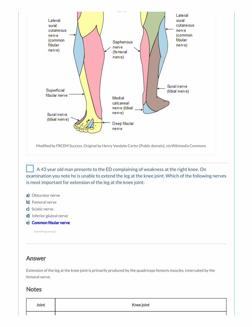
Modi ed by FRCEM Success. Original by Henry Vandyke Carter [Public domain], via Wikimedia Commons
A 43 year old man presents to the ED complaining of weakness at the right knee. On
examination you note he is unable to extend the leg at the knee joint. Which of the following nerves
is most important for extension of the leg at the knee joint:
a) Obturator nerve
b) Femoral nerve
c) Sciatic nerve
d) Inferior gluteal nerve
e) Common bular nerve
Answer
Extension of the leg at the knee joint is primarily produced by the quadriceps femoris muscles, innervated by the
femoral nerve.
Notes
Joint Knee joint
Something wrong?
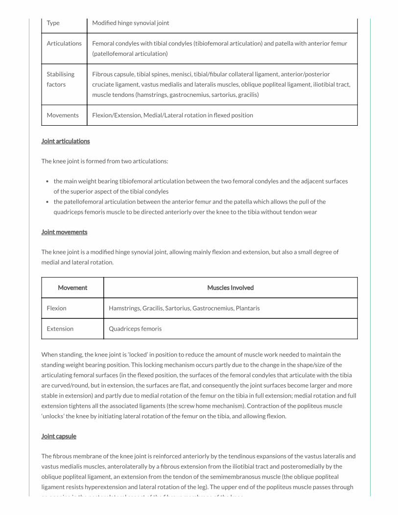
Type Modi ed hinge synovial joint
Articulations Femoral condyles with tibial condyles (tibiofemoral articulation) and patella with anterior femur
(patellofemoral articulation)
Stabilising
factors
Fibrous capsule, tibial spines, menisci, tibial/ bular collateral ligament, anterior/posterior
cruciate ligament, vastus medialis and lateralis muscles, oblique popliteal ligament, iliotibial tract,
muscle tendons (hamstrings, gastrocnemius, sartorius, gracilis)
Movements Flexion/Extension, Medial/Lateral rotation in exed position
Joint articulations
The knee joint is formed from two articulations:
the main weight bearing tibiofemoral articulation between the two femoral condyles and the adjacent surfaces
of the superior aspect of the tibial condyles
the patellofemoral articulation between the anterior femur and the patella which allows the pull of the
quadriceps femoris muscle to be directed anteriorly over the knee to the tibia without tendon wear
Joint movements
The knee joint is a modi ed hinge synovial joint, allowing mainly exion and extension, but also a small degree of
medial and lateral rotation.
Movement Muscles Involved
Flexion Hamstrings, Gracilis, Sartorius, Gastrocnemius, Plantaris
Extension Quadriceps femoris
When standing, the knee joint is ‘locked’ in position to reduce the amount of muscle work needed to maintain the
standing weight bearing position. This locking mechanism occurs partly due to the change in the shape/size of the
articulating femoral surfaces (in the exed position, the surfaces of the femoral condyles that articulate with the tibia
are curved/round, but in extension, the surfaces are at, and consequently the joint surfaces become larger and more
stable in extension) and partly due to medial rotation of the femur on the tibia in full extension; medial rotation and full
extension tightens all the associated ligaments (the screw home mechanism). Contraction of the popliteus muscle
‘unlocks’ the knee by initiating lateral rotation of the femur on the tibia, and allowing exion.
Joint capsule
The brous membrane of the knee joint is reinforced anteriorly by the tendinous expansions of the vastus lateralis and
vastus medialis muscles, anterolaterally by a brous extension from the iliotibial tract and posteromedially by the
oblique popliteal ligament, an extension from the tendon of the semimembranosus muscle (the oblique popliteal
ligament resists hyperextension and lateral rotation of the leg). The upper end of the popliteus muscle passes through
an opening in the posterolateral aspect of the brous membrane of the knee.
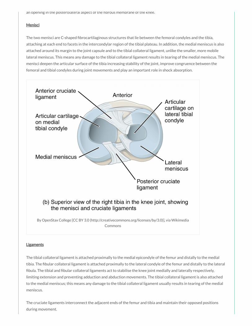
an opening in the posterolateral aspect of the brous membrane of the knee.
Menisci
The two menisci are C-shaped brocartilaginous structures that lie between the femoral condyles and the tibia,
attaching at each end to facets in the intercondylar region of the tibial plateau. In addition, the medial meniscus is also
attached around its margin to the joint capsule and to the tibial collateral ligament, unlike the smaller, more mobile
lateral meniscus. This means any damage to the tibial collateral ligament results in tearing of the medial meniscus. The
menisci deepen the articular surface of the tibia increasing stability of the joint, improve congruence between the
femoral and tibial condyles during joint movements and play an important role in shock absorption.
By OpenStax College [CC BY 3.0 (http://creativecommons.org/licenses/by/3.0)], via Wikimedia
Commons
Ligaments
The tibial collateral ligament is attached proximally to the medial epicondyle of the femur and distally to the medial
tibia. The bular collateral ligament is attached proximally to the lateral condyle of the femur and distally to the lateral
bula. The tibial and bular collateral ligaments act to stabilise the knee joint medially and laterally respectively,
limiting extension and preventing adduction and abduction movements. The tibial collateral ligament is also attached
to the medial meniscus; this means any damage to the tibial collateral ligament usually results in tearing of the medial
meniscus.
The cruciate ligaments interconnect the adjacent ends of the femur and tibia and maintain their opposed positions
during movement.
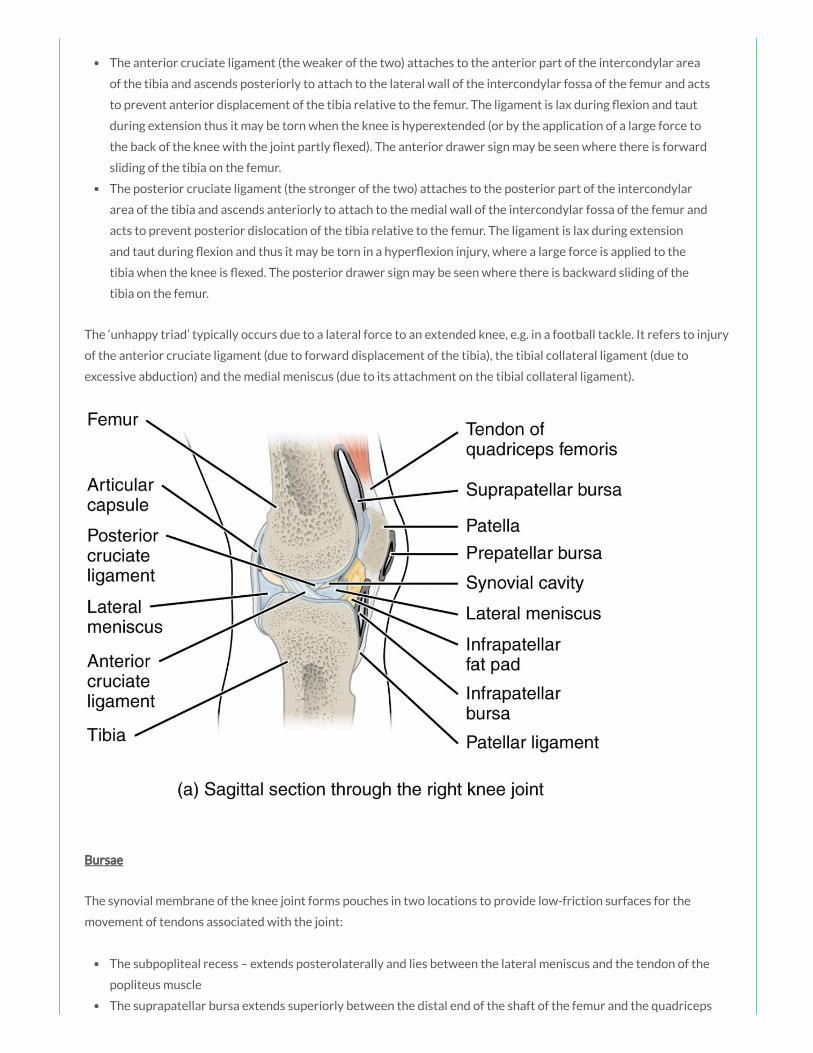
The anterior cruciate ligament (the weaker of the two) attaches to the anterior part of the intercondylar area
of the tibia and ascends posteriorly to attach to the lateral wall of the intercondylar fossa of the femur and acts
to prevent anterior displacement of the tibia relative to the femur. The ligament is lax during exion and taut
during extension thus it may be torn when the knee is hyperextended (or by the application of a large force to
the back of the knee with the joint partly exed). The anterior drawer sign may be seen where there is forward
sliding of the tibia on the femur.
The posterior cruciate ligament (the stronger of the two) attaches to the posterior part of the intercondylar
area of the tibia and ascends anteriorly to attach to the medial wall of the intercondylar fossa of the femur and
acts to prevent posterior dislocation of the tibia relative to the femur. The ligament is lax during extension
and taut during exion and thus it may be torn in a hyper exion injury, where a large force is applied to the
tibia when the knee is exed. The posterior drawer sign may be seen where there is backward sliding of the
tibia on the femur.
The ‘unhappy triad’ typically occurs due to a lateral force to an extended knee, e.g. in a football tackle. It refers to injury
of the anterior cruciate ligament (due to forward displacement of the tibia), the tibial collateral ligament (due to
excessive abduction) and the medial meniscus (due to its attachment on the tibial collateral ligament).
Bursae
The synovial membrane of the knee joint forms pouches in two locations to provide low-friction surfaces for the
movement of tendons associated with the joint:
The subpopliteal recess – extends posterolaterally and lies between the lateral meniscus and the tendon of the
popliteus muscle
The suprapatellar bursa extends superiorly between the distal end of the shaft of the femur and the quadriceps

femoris muscle and tendon
Other bursae associated with the knee, but not normally communicating with the synovial joint, include the
subcutaneous prepatellar bursa, the deep and subcutaneous infrapatellar bursae separated by the patella ligament,
and numerous other bursae associated with tendons and ligaments around the knee joint. Housemaid’s knee is
in ammation of the prepatellar bursa, and Clergyman’s knee is in ammation of the subcutaneous infrapatellar bursa.
A 45 year old tree surgeon is brought into the ED following a fall from approximately 5
metres. A full trauma primary survey has been performed – the trauma team leader is concerned
about a possible lumbar injury with resultant neurology. You have been asked to assess the
sensation in the lower limb dermatomes. The L2 dermatome is best tested at which of the following
landmarks:
a) At the midpoint of the inguinal ligament in the midclavicular line
b) At a point on the mid anterior thigh
c) At a point on the posterolateral thigh
d) At the medial femoral condyle
e) At the popliteal fossa
Answer
The L2 dermatome is best tested on the anteromedial thigh, at the midpoint drawn on an imaginary line connecting the
midpoint of the inguinal ligament and the medial femoral condyle.
Notes
Dermatome Landmark
L1 Upper Anterior Thigh
L2 Mid Anterior Thigh
L3 Medial Femoral Condyle
L4 Medial Malleolus
L5 Dorsum 3rd MTP Joint
S1 Lateral Heel
S2 Popliteal Fossa
Something wrong?

S3 Ischial Tuberosity
S5 Perianal Area
The T12 dermatome is best tested at the midclavicular line, over the midpoint of the inguinal ligament.
The L1 dermatome is best tested on the upper anterior thigh, at a point midway between the key sensory
points for T12 and L2.
The L2 dermatome is best tested on the anteromedial thigh, at the midpoint drawn on an imaginary line
connecting the midpoint of the inguinal ligament and the medial femoral condyle.
The L3 dermatome is best tested at the medial femoral condyle above the knee.
The L4 dermatome is best tested over the medial malleolus.
The L5 dermatome is best tested on the dorsum of the foot at the third metatarsophalangeal joint.
The S1 dermatome is best tested on the lateral aspect of the calcaneus.
The S2 dermatome is best tested at the midpoint of the popliteal fossa.
The S3 dermatome is best tested over the ischial tuberosity or infragluteal fold (depending on the patient their
skin can move up, down or laterally over the ischii).
The S4/S5 dermatome is best tested in the perianal area, less than one cm lateral to the mucocutaneous
junction.

By Grant, John Charles Boileau (An atlas of anatomy, / by regions 1962) [Public domain], via
Wikimedia Commons
You are examining a 65 year old man who presents with left leg weakness. You note a loss of
power to the left quadriceps femoris muscles. The quadriceps femoris muscles are innervated by
which of the following nerves:
a) Sciatic nerve
b) Pudendal nerve
c) Femoral nerve
d) Obturator nerve
e) Superior gluteal nerve
Answer
The quadriceps femoris muscles are innervated by the femoral nerve (L2 – L4).
Notes
Muscle Action(s) Innervation
Rectus femoris (red) Flexion at hip and extension at knee Femoral nerve (L2 – L4)
Vastus lateralis (green) Extension at knee Femoral nerve (L2 – L4)
Vastus medialis (blue) Extension at knee Femoral nerve (L2 – L4)
Vastus intermedius Extension at knee Femoral nerve (L2 – L4)
The quadriceps femoris is made up of four individual muscles; the rectus femoris (red), the vastus medialis (blue), the
vastus intermedius (deep to the rectus femoris) and the vastus lateralis (green) which all insert onto the patella by the
common quadriceps femoris tendon. Because the vastus muscles insert into the margins of the patella as well as into
the quadriceps femoris tendon, they stabilise the position of the patella during knee joint movement.
The quadriceps femoris muscle is the main extensor of the leg at the knee joint. The rectus femoris crosses both the
knee and the hip joint (in contrast to the vastus muscles which only cross the knee joint), and therefore also assists in
exion of the thigh at the hip joint.
The quadriceps femoris muscles are innervated by the femoral nerve (L2 – L4). A tap on the patella ligament tests
re ex activity mainly at spinal cord levels L3/L4.
Something wrong?

re ex activity mainly at spinal cord levels L3/L4.

Modi ed by FRCEM Success. Original by Henry
Vandyke Carter [Public domain], via Wikimedia
Commons
A 75 year old woman presents to the ED complaining of a loss of sensation to the skin over
the anterior thigh. This area is primarily supplied by which of the following nerves:
a) The medial cutaneous nerve of the thigh
b) Cutaneous branches from the femoral nerve
c) Cutaneous branches from the obturator nerve
d) Cutaneous branches from the sciatic nerve
e) Cutaneous branches from the lumbar plexus
Answer
The anterior cutaneous nerve, branch of the femoral nerve, supplies skin over the anterior thigh and anteromedial
knee.
Notes
Nerve Origin Skin supplied
Lateral cutaneous nerve of thigh Lumbar plexus (L2, L3) Lateral thigh
Anterior cutaneous nerve Femoral nerve Anterior thigh and anteromedial knee
Cutaneous branch Obturator nerve Medial thigh
Posterior cutaneous nerve of thigh Sacral plexus (S1 – S3) Posterior thigh and upper leg
Saphenous nerve Femoral nerve Anteromedial knee, medial leg and foot
Lateral sural cutaneous nerve Common bular nerve Upper lateral leg
Super cial bular nerve Common bular nerve Lower anterolateral leg and dorsum of foot
Deep bular nerve Common bular nerve Webspace between 1st and 2nd toe
Sural nerve Tibial nerve Lower posterolateral leg, heel and foot
Something wrong?
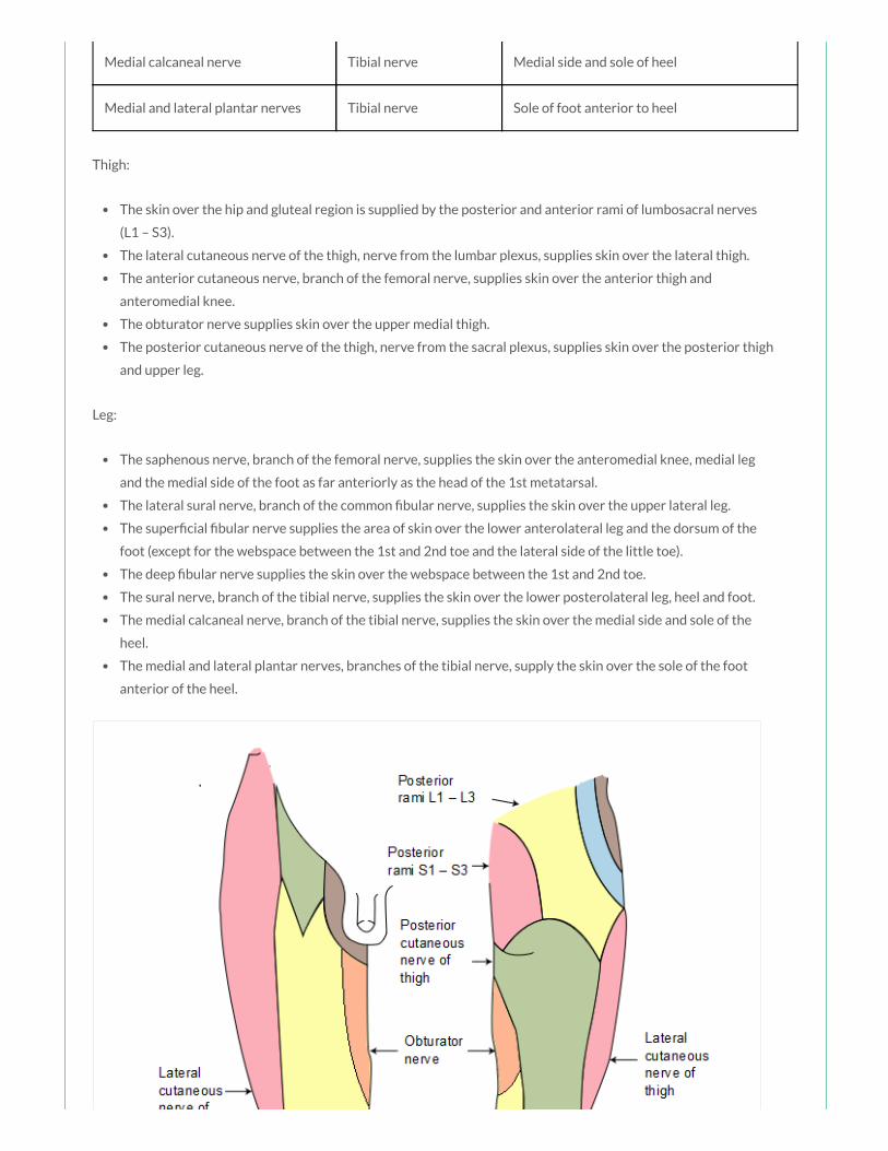
Medial calcaneal nerve Tibial nerve Medial side and sole of heel
Medial and lateral plantar nerves Tibial nerve Sole of foot anterior to heel
Thigh:
The skin over the hip and gluteal region is supplied by the posterior and anterior rami of lumbosacral nerves
(L1 – S3).
The lateral cutaneous nerve of the thigh, nerve from the lumbar plexus, supplies skin over the lateral thigh.
The anterior cutaneous nerve, branch of the femoral nerve, supplies skin over the anterior thigh and
anteromedial knee.
The obturator nerve supplies skin over the upper medial thigh.
The posterior cutaneous nerve of the thigh, nerve from the sacral plexus, supplies skin over the posterior thigh
and upper leg.
Leg:
The saphenous nerve, branch of the femoral nerve, supplies the skin over the anteromedial knee, medial leg
and the medial side of the foot as far anteriorly as the head of the 1st metatarsal.
The lateral sural nerve, branch of the common bular nerve, supplies the skin over the upper lateral leg.
The super cial bular nerve supplies the area of skin over the lower anterolateral leg and the dorsum of the
foot (except for the webspace between the 1st and 2nd toe and the lateral side of the little toe).
The deep bular nerve supplies the skin over the webspace between the 1st and 2nd toe.
The sural nerve, branch of the tibial nerve, supplies the skin over the lower posterolateral leg, heel and foot.
The medial calcaneal nerve, branch of the tibial nerve, supplies the skin over the medial side and sole of the
heel.
The medial and lateral plantar nerves, branches of the tibial nerve, supply the skin over the sole of the foot
anterior of the heel.
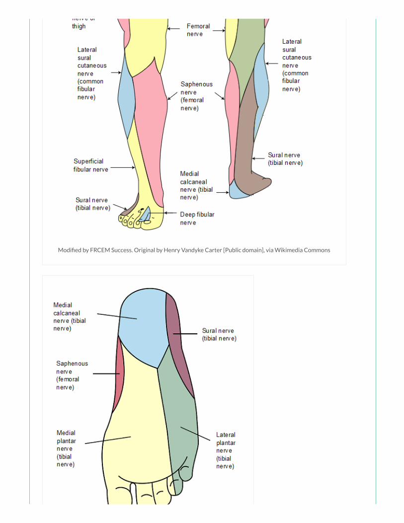
Modi ed by FRCEM Success. Original by Henry Vandyke Carter [Public domain], via Wikimedia Commons

Modi ed by FRCEM Success. Original by Henry Vandyke Carter [Public
domain], via Wikimedia Commons
You have been asked to give a tutorial to a group of medical students about the vascular
anatomy of the lower limb. The profunda femoris artery is a branch of which of the following
arteries:
a) Femoral artery
b) Obturator artery
c) External iliac artery
d) Internal iliac artery
e) Superior gluteal artery
Answer
The femoral artery gives rise to the deep profunda femoris artery in the femoral triangle which is a major source of
blood supply to the medial and posterior compartments of the thigh and the proximal femur.
Notes
The major artery supplying the lower limb is the femoral artery. The femoral artery is the continuation of the external
iliac artery, beginning as the vessel passes under the inguinal ligament to enter the femoral triangle in the anterior
thigh.
The femoral artery can be palpated in the femoral triangle as it passes over the femoral head, just inferior to the
inguinal ligament, midway between the anterior superior iliac spine and the pubic symphysis (at the mid-inguinal
point). The femoral vein lies immediately medial to this pulsation, which is an important landmark for central venous
line insertion. Medial to the femoral vein is the femoral canal which contains lymphatics and lies immediately lateral to
the pubic tubercle. The femoral nerve lies lateral to the femoral artery.
The femoral artery gives rise to the deep profunda femoris artery in the femoral triangle which is a major source of
blood supply to the medial and posterior compartments of the thigh and the proximal femur.
After exiting the femoral triangle, the femoral artery continues down the anterior surface of the thigh via the adductor
canal. During its descent it supplies the anterior thigh, giving rise to numerous super cial cutaneous branches.
The femoral artery becomes the popliteal artery after entering the posterior compartment of the thigh through the
adductor hiatus just proximal to the knee.
Something wrong?

Modi ed by FRCEM Success. Original by Henry Vandyke Carter [Public domain], via Wikimedia
Commons
A patient complains of pain in the hip region following a fall. Imaging has shown a traumatic
injury to the pectineus muscle. The pectineus muscle acts primarily to produce which of the
following movements:
a) Flexion and adduction of the thigh
b) Flexion and abduction of the thigh
c) Extension and medial rotation of the thigh
d) Flexion and lateral rotation of the thigh

e) Extension of the thigh
Answer
The pectineus acts to adduct and ex the thigh at the hip joint.
Notes
The sartorius, iliopsoas, pectineus and rectus femoris are the primary exors of the thigh at the hip joint.
Muscle Actions Innervation
Sartorius (blue) Flexion, abduction and lateral rotation at hip and exion at
knee
Femoral nerve (L2, L3)
Iliacus (red) Flexion and lateral rotation at hip Femoral nerve (L2, L3)
Psoas major
(yellow)
Flexion and lateral rotation at hip Anterior rami L1 – L3
Pectineus (green) Adduction and exion at hip Femoral nerve (L2, L3)
Rectus femoris Flexion at hip and extension at knee Femoral nerve (L2 –
L4)
The sartorius (blue) is innervated by the femoral nerve (L2, L3). It acts to ex the thigh at the hip joint and ex the leg at
the knee joint. It also abducts the thigh and rotates it laterally, as when resting the foot on the opposite knee when
sitting.
The iliacus (red) and psoas major (yellow) muscles originate as separate muscles but insert by a common tendon onto
the femur and together are referred to as the iliopsoas muscle, which is a powerful exor of the thigh at the hip joint
and also contributes to lateral rotation of the thigh at the hip joint. The iliacus is innervated by the femoral nerve (L2,
L3). The psoas major is innervated by the anterior rami of spinal nerves L1 – L3.
The pectineus (green) is innervated by the femoral nerve (L2, L3). It acts to adduct and ex the thigh at the hip joint.
Something wrong?

Modi ed by FRCEM Success. Original by Henry
Vandyke Carter [Public domain], via Wikimedia
Commons
A 21 year old man received a stab wound to the posterior part of his gluteal region. Following

A 21 year old man received a stab wound to the posterior part of his gluteal region. Following
resuscitation, he is found to have great dif culty rising to a standing position from the seated
position. Which of the following muscles was most likely affected by this injury:
a) Quadriceps femoris
b) Iliopsoas
c) Hamstrings
d) Gluteus maximus
e) Gluteus medius
Answer
The gluteus maximus is responsible for extension and lateral rotation at the hip and is the prime muscle that extends
the exed hip, and is used to rise from a seated position. It is innervated by the inferior gluteal nerve which may have
been injured in this case.
Notes
Muscle Action(s) Innervation
Gluteus maximus (red) Extension, lateral rotation and abduction at hip Inferior gluteal nerve (L5 – S2)
Gluteus medius (green) Abduction and medial rotation at hip Superior gluteal nerve (L4 – S1)
Gluteus minimus (blue) Abduction and medial rotation at hip Superior gluteal nerve (L4 – S1)
Piriformis (yellow) Lateral rotation and abduction at hip Branches from S1 and S2
The gluteus maximus (red) is the main extensor of the thigh at the hip joint and also acts to laterally rotate and abduct
the thigh. Through its insertion into the iliotibial tract, it also stabilises the knee and hip joints. The gluteus maximus is
innervated by the inferior gluteal nerve (L5 – S2).
The gluteus medius (green) and gluteus minimus (blue) act to abduct and medially rotate the thigh at the hip joint, and
also act to secure the pelvis, reducing pelvic drop on the opposite swing side during walking. They are both innervated
by the superior gluteal nerve (L4 – S1).
The piriformis (yellow) acts to abduct and laterally rotate the thigh at the hip joint. It is innervated by the nerve to the
piriformis, originating from the sacral plexus (S1, S2). The obturator internus, gemelli and quadriceps femoris muscles
act as synergistic femoral lateral rotators and hip stabilisers.
Trendelenburg’s sign is seen in people with weak/paralysed abductor muscles of the hip. The sign is demonstrated by
asking the patient to stand on one limb; when the patient stands on the affected limb, the pelvis severely drops over
the swing limb. This sign may be seen in patients with damage to the superior gluteal nerve, which may occur in
Something wrong?

association with pelvic fractures, with space-occupying lesions within the pelvis extending into the greater sciatic
foramen, and following hip surgery. Typically the patient may also demonstrate a Trendelenburg gait.

Modi ed by FRCEM Success. Original by Henry Vandyke Carter
[Public domain], via Wikimedia Commons
A 54 year old woman presents with unilateral swelling of the left leg. Her d-dimer is normal
and ultrasound has shown no demonstrable deep vein thrombosis. Your consultant has arranged
for a CT of the leg and pelvis which shows lymphadenopathy of the deep inguinal lymph nodes.
Which of the following best describes the location of the deep inguinal lymph nodes:
a) Medial to the femoral vein in the femoral canal
b) In a line superior to the inguinal ligament
c) In a line inferior to the inguinal ligament
d) Lateral to the femoral vein in the femoral canal
e) Following the course of the external iliac vein
Answer
The deep inguinal lymph nodes (about 1 – 3 in number) lie medial to the femoral vein within the femoral canal.
Notes
The inguinal nodes are found in the upper aspect of the femoral triangle.
The super cial inguinal lymph nodes (about 10 in number) are located in the super cial fascia, forming a line below the
inguinal ligament, and medially extending inferiorly along the terminal part of the great saphenous vein. They receive
lymph from the gluteal region, lower abdominal wall, perineum and super cial regions of the lower limb.
The deep inguinal lymph nodes (about 1 – 3 in number) lie medial to the femoral vein within the femoral canal. They
receive lymph from deep lymphatics associated with the femoral vessels and from the glans penis or clitoris in the
perineum.
The inguinal lymph nodes drain to the external iliac lymph nodes associated with the external iliac artery in the
abdomen.
Something wrong?

By Henry Vandyke Carter [Public domain], via
Wikimedia Commons
You are discussing clinically relevant lower limb anatomy with a group of medical students.
The femoral triangle is an important, clinically relevant concept. The femoral triangle is bordered
superiorly by which of the following structures:
a) Femoral sheath
b) Inguinal ligament
c) Femoral canal
d) Femoral artery
e) Pubic bone
Something wrong?
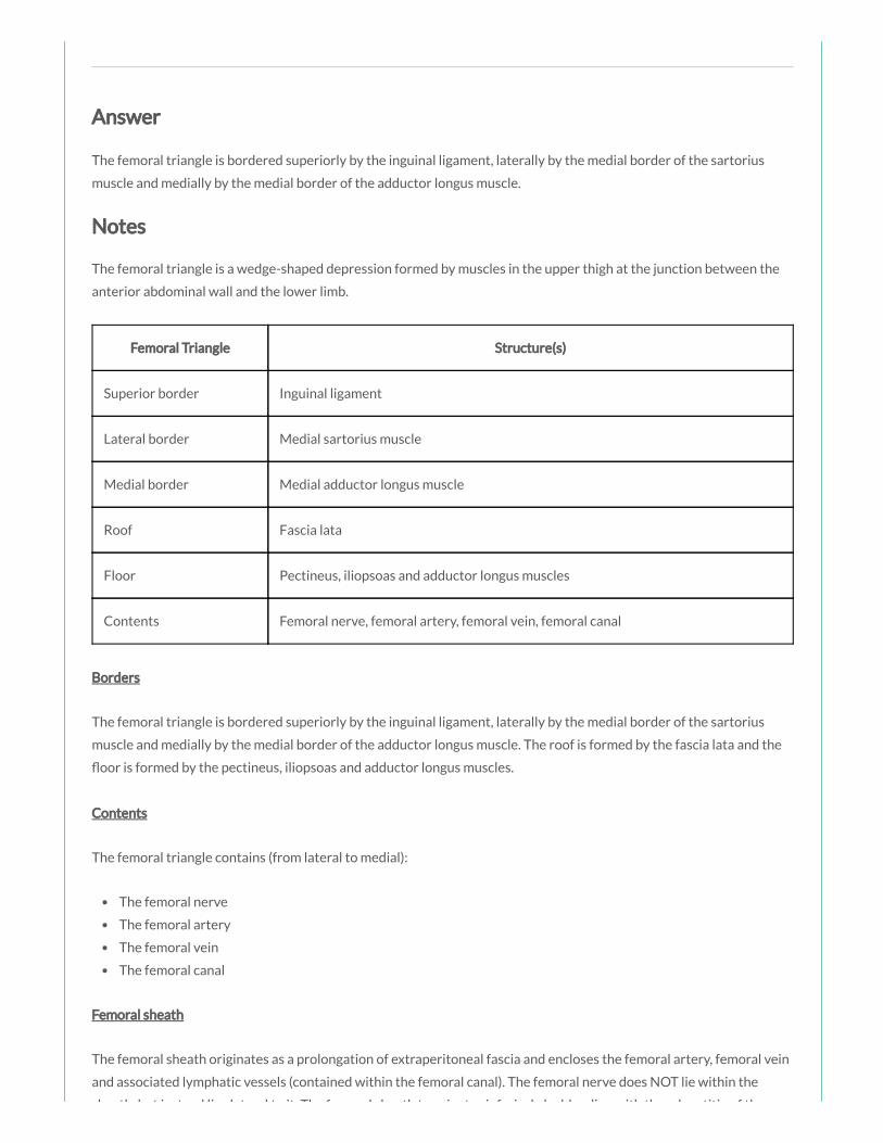
Answer
The femoral triangle is bordered superiorly by the inguinal ligament, laterally by the medial border of the sartorius
muscle and medially by the medial border of the adductor longus muscle.
Notes
The femoral triangle is a wedge-shaped depression formed by muscles in the upper thigh at the junction between the
anterior abdominal wall and the lower limb.
Femoral Triangle Structure(s)
Superior border Inguinal ligament
Lateral border Medial sartorius muscle
Medial border Medial adductor longus muscle
Roof Fascia lata
Floor Pectineus, iliopsoas and adductor longus muscles
Contents Femoral nerve, femoral artery, femoral vein, femoral canal
Borders
The femoral triangle is bordered superiorly by the inguinal ligament, laterally by the medial border of the sartorius
muscle and medially by the medial border of the adductor longus muscle. The roof is formed by the fascia lata and the
oor is formed by the pectineus, iliopsoas and adductor longus muscles.
Contents
The femoral triangle contains (from lateral to medial):
The femoral nerve
The femoral artery
The femoral vein
The femoral canal
Femoral sheath
The femoral sheath originates as a prolongation of extraperitoneal fascia and encloses the femoral artery, femoral vein
and associated lymphatic vessels (contained within the femoral canal). The femoral nerve does NOT lie within the
sheath, but instead lies lateral to it. The femoral sheath terminates inferiorly by blending with the adventitia of the

By Henry Vandyke Carter [Public domain], via
Wikimedia Commons
By Henry Vandyke Carter [Public domain], via
Wikimedia Commons
sheath, but instead lies lateral to it. The femoral sheath terminates inferiorly by blending with the adventitia of the
femoral vessels approximately 4 cm inferior of the inguinal ligament.
Femoral canal
The femoral canal lies between the medial edge of the femoral sheath and the femoral vein. The femoral canal contains
loose connective tissue, lymphatic vessels, deep inguinal lymph nodes and empty space. The femoral canal allows the
femoral vein to expand, for example, when venous return from the leg is increased, or when increased intra-abdominal
pressure causes a temporary stasis.
Femoral ring
The femoral ring is formed by the proximal abdominal opening of the femoral canal, and is important clinically, as it is a
common site of hernia. It is bordered anteriorly by the inguinal ligament, posteriorly by the pectineal ligament,
medially by the lacunar ligament and laterally by the femoral vein.
Femoral ring Structure(s)
Anterior border Inguinal ligament
Posterior border Pectineal ligament
Medial border Lacunar ligament
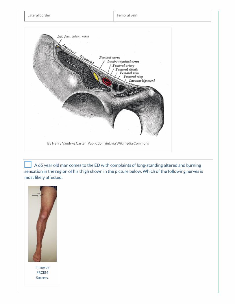
Lateral border Femoral vein
By Henry Vandyke Carter [Public domain], via Wikimedia Commons
A 65 year old man comes to the ED with complaints of long-standing altered and burning
sensation in the region of his thigh shown in the picture below. Which of the following nerves is
most likely affected:
Image by
FRCEM
Success.

a) Lateral cutaneous nerve of the sacral plexus
b) Posterior cutaneous nerve of the lumbar plexus
c) Posterior cutaneous nerve of the sacral plexus
d) Lateral femoral cutaneous nerve
e) Obturator nerve
Answer
The lateral cutaneous nerve of the thigh (lateral femoral cutaneous nerve), nerve from the lumbar plexus, supplies skin
over the lateral thigh.
Notes
Nerve Origin Skin supplied
Lateral cutaneous nerve of thigh Lumbar plexus (L2, L3) Lateral thigh
Anterior cutaneous nerve Femoral nerve Anterior thigh and anteromedial knee
Cutaneous branch Obturator nerve Medial thigh
Posterior cutaneous nerve of thigh Sacral plexus (S1 – S3) Posterior thigh and upper leg
Saphenous nerve Femoral nerve Anteromedial knee, medial leg and foot
Lateral sural cutaneous nerve Common bular nerve Upper lateral leg
Super cial bular nerve Common bular nerve Lower anterolateral leg and dorsum of foot
Deep bular nerve Common bular nerve Webspace between 1st and 2nd toe
Sural nerve Tibial nerve Lower posterolateral leg, heel and foot
Medial calcaneal nerve Tibial nerve Medial side and sole of heel
Medial and lateral plantar nerves Tibial nerve Sole of foot anterior to heel
Thigh:
The skin over the hip and gluteal region is supplied by the posterior and anterior rami of lumbosacral nerves
(L1 – S3).
The lateral cutaneous nerve of the thigh, nerve from the lumbar plexus, supplies skin over the lateral thigh.
Something wrong?
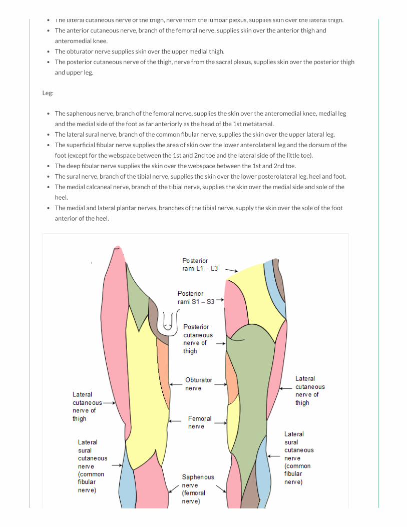
The lateral cutaneous nerve of the thigh, nerve from the lumbar plexus, supplies skin over the lateral thigh.
The anterior cutaneous nerve, branch of the femoral nerve, supplies skin over the anterior thigh and
anteromedial knee.
The obturator nerve supplies skin over the upper medial thigh.
The posterior cutaneous nerve of the thigh, nerve from the sacral plexus, supplies skin over the posterior thigh
and upper leg.
Leg:
The saphenous nerve, branch of the femoral nerve, supplies the skin over the anteromedial knee, medial leg
and the medial side of the foot as far anteriorly as the head of the 1st metatarsal.
The lateral sural nerve, branch of the common bular nerve, supplies the skin over the upper lateral leg.
The super cial bular nerve supplies the area of skin over the lower anterolateral leg and the dorsum of the
foot (except for the webspace between the 1st and 2nd toe and the lateral side of the little toe).
The deep bular nerve supplies the skin over the webspace between the 1st and 2nd toe.
The sural nerve, branch of the tibial nerve, supplies the skin over the lower posterolateral leg, heel and foot.
The medial calcaneal nerve, branch of the tibial nerve, supplies the skin over the medial side and sole of the
heel.
The medial and lateral plantar nerves, branches of the tibial nerve, supply the skin over the sole of the foot
anterior of the heel.

Modi ed by FRCEM Success. Original by Henry Vandyke Carter [Public domain], via Wikimedia Commons
Modi ed by FRCEM Success. Original by Henry Vandyke Carter [Public
domain], via Wikimedia Commons
You are asked to review a 54 year old man who presents complaining of lower back pain with
radiation down the right leg. You are concerned about sciatic nerve root compression. The sciatic
nerve receives nerve bres from which of the following nerve roots:

a) L5 – S2
b) L4 – S2
c) L4 – S3
d) L4 – S1
e) L3 – S2
Answer
The sciatic nerve is derived from the lumbosacral plexus and receives bres from L4 – S3.
Notes
The sciatic nerve is derived from the lumbosacral plexus and receives bres from L4 – S3.
Nerve Sciatic nerve
Nerve
roots
L4 – S3
Motor
supply
Posterior thigh muscles, hamstring portion of adductor magnus, all of the muscles in the leg and foot
Sensory
supply
Skin on the lateral leg and foot, the heel and the dorsum and sole of the foot
Motor loss
in injury
Weak extension of hip and exion of knee, loss of dorsi exion and plantar exion of ankle, loss of
inversion and eversion of foot, foot drop with high-stepping gait
The sciatic nerve leaves the pelvis and enters the gluteal region via the greater sciatic foramen inferior to the
piriformis muscle before descending through the gluteal region and entering the posterior thigh. In the posterior
compartment of the thigh, the sciatic nerve lies on the adductor magnus muscle and is crossed by the long head of the
biceps femoris muscle. The sciatic nerve terminates at the apex of the popliteal fossa by dividing into the tibial
(anterior divisions of L4 – S3) and the common bular (posterior divisions of L4 – S2) nerves.
Something wrong?
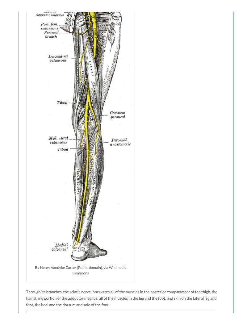
By Henry Vandyke Carter [Public domain], via Wikimedia
Commons
Through its branches, the sciatic nerve innervates all of the muscles in the posterior compartment of the thigh, the
hamstring portion of the adductor magnus, all of the muscles in the leg and the foot, and skin on the lateral leg and
foot, the heel and the dorsum and sole of the foot.

Modi ed by FRCEM Success. Original by Henry Vandyke Carter [Public domain], via Wikimedia Commons
Clinical implications
The gluteal region can be divided into quadrants by 2 lines: one line descending vertically from the highest point of the
iliac crest, the other line passing horizontally through the rst line midway between the highest point of the iliac crest
and the ischial tuberosity. The sciatic nerve passes through the lower medial quadrant. Intramuscular injections in the
buttocks should always be given in the upper lateral quadrant of the gluteal region to avoid damage to the sciatic nerve

buttocks should always be given in the upper lateral quadrant of the gluteal region to avoid damage to the sciatic nerve
and major vessels in the region.
An 18 year old man is brought into the ED after sustaining a deep wound to his left thigh after
an accident on a building site. Imaging has shown trauma to the quadriceps femoris muscles. The
quadriceps femoris muscles primarily act to produce which of the following movements:
a) Extension of the thigh
b) Extension of the leg
c) Flexion of the thigh
d) Flexion of the leg
e) Extension of the thigh and extension of the leg
Answer
The quadriceps femoris muscle is the main extensor of the leg at the knee joint. The rectus femoris crosses both the
knee and the hip joint (in contrast to the vastus muscles which only cross the knee joint), and therefore also assists in
exion of the thigh at the hip joint.
Notes
Muscle Action(s) Innervation
Rectus femoris (red) Flexion at hip and extension at knee Femoral nerve (L2 – L4)
Vastus lateralis (green) Extension at knee Femoral nerve (L2 – L4)
Vastus medialis (blue) Extension at knee Femoral nerve (L2 – L4)
Vastus intermedius Extension at knee Femoral nerve (L2 – L4)
The quadriceps femoris is made up of four individual muscles; the rectus femoris (red), the vastus medialis (blue), the
vastus intermedius (deep to the rectus femoris) and the vastus lateralis (green) which all insert onto the patella by the
common quadriceps femoris tendon. Because the vastus muscles insert into the margins of the patella as well as into
the quadriceps femoris tendon, they stabilise the position of the patella during knee joint movement.
The quadriceps femoris muscle is the main extensor of the leg at the knee joint. The rectus femoris crosses both the
knee and the hip joint (in contrast to the vastus muscles which only cross the knee joint), and therefore also assists in
exion of the thigh at the hip joint.
The quadriceps femoris muscles are innervated by the femoral nerve (L2 – L4). A tap on the patella ligament tests
re ex activity mainly at spinal cord levels L3/L4.
Something wrong?

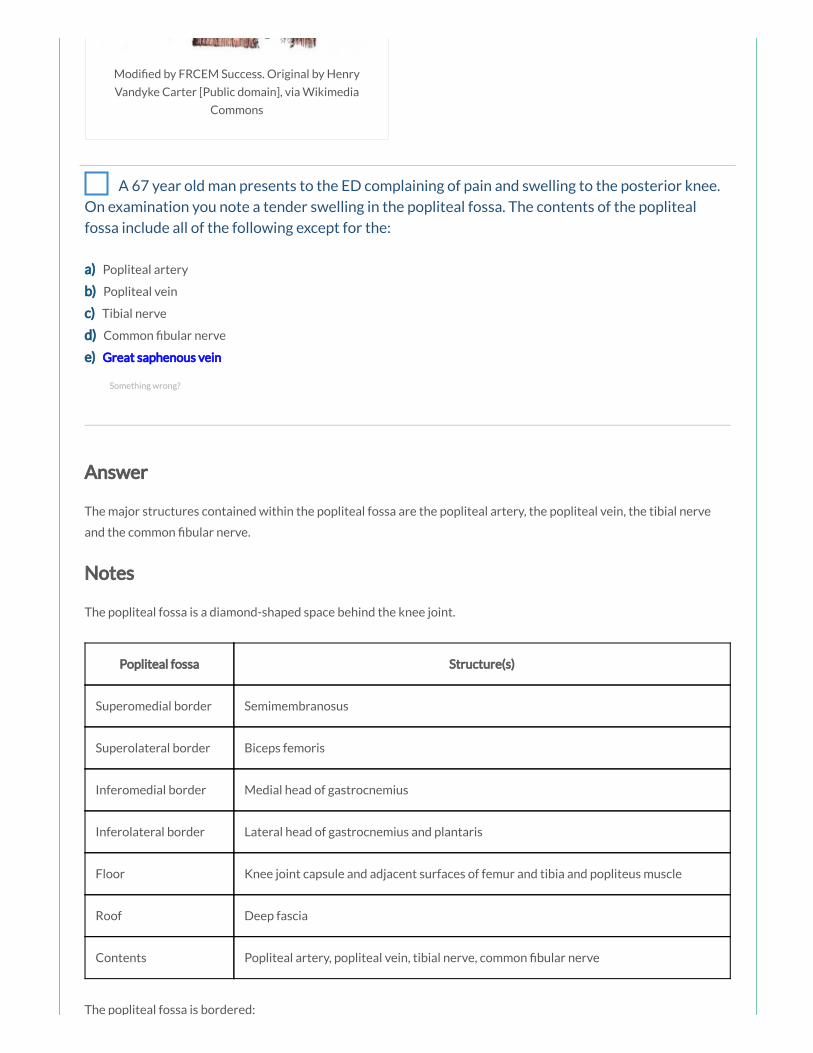
Modi ed by FRCEM Success. Original by Henry
Vandyke Carter [Public domain], via Wikimedia
Commons
A 67 year old man presents to the ED complaining of pain and swelling to the posterior knee.
On examination you note a tender swelling in the popliteal fossa. The contents of the popliteal
fossa include all of the following except for the:
a) Popliteal artery
b) Popliteal vein
c) Tibial nerve
d) Common bular nerve
e) Great saphenous vein
Answer
The major structures contained within the popliteal fossa are the popliteal artery, the popliteal vein, the tibial nerve
and the common bular nerve.
Notes
The popliteal fossa is a diamond-shaped space behind the knee joint.
Popliteal fossa Structure(s)
Superomedial border Semimembranosus
Superolateral border Biceps femoris
Inferomedial border Medial head of gastrocnemius
Inferolateral border Lateral head of gastrocnemius and plantaris
Floor Knee joint capsule and adjacent surfaces of femur and tibia and popliteus muscle
Roof Deep fascia
Contents Popliteal artery, popliteal vein, tibial nerve, common bular nerve
The popliteal fossa is bordered:
Something wrong?

The popliteal fossa is bordered:
superomedially by the semimembranosus
superolaterally by the biceps femoris
inferomedially by the medial head of the gastrocnemius
inferolaterally by the lateral head of the gastrocnemius and plantaris
Original by Henry Vandyke Carter [Public
domain], via Wikimedia Commons
The oor of the fossa is formed by the capsule of the knee joint and adjacent surfaces of the femur and tibia, and more
inferiorly by the popliteus muscle. The roof is formed by deep fascia.
The major structures contained within the popliteal fossa are (from medial to lateral):
the popliteal artery
the popliteal vein
the tibial nerve
the common bular nerve

Modi ed by FRCEM Success. Original by Henry Vandyke Carter [Public domain],
via Wikimedia Commons
The small saphenous vein ascends in the super cial fascia on the back of the leg to the knee where it penetrates the
deep fascia and enters the popliteal fossa to drain into the popliteal vein. One other structure that passes through the
roof of the fossa is the posterior cutaneous nerve of the thigh which descends through the thigh super cial to the
hamstring muscles, passes through the roof of the popliteal fossa, and then continues inferiorly with the small
saphenous vein to supply skin on the upper half of the posterior leg.
A 65 year old woman presents to the ED with a tender left leg. She has been assessed by your
Consultant colleague who feels she may have thrombophlebitis of the great saphenous vein.
Which of the following best describes the course of the great saphenous vein:
a) Passes anterior to medial malleolus and ascends medial side of leg, knee and thigh
b) Passes posterior to medial malleolus and ascends medial side of leg, knee and thigh
c) Passes anterior to lateral malleolus and ascends lateral side of leg, knee and thigh
d) Passes posterior to lateral malleolus and ascends lateral side of leg, knee and thigh
e) Passes posterior to lateral malleolus and ascends posterior leg, knee and thigh
Answer
The great saphenous vein passes anterior to the medial malleolus, travels up the medial side of the leg, knee and thigh
to pass through the saphenous opening in the deep fascia covering the femoral triangle, and join with the femoral vein
just below the inguinal ligament.
Notes
The great and small saphenous veins originate from the medial and lateral sides respectively of the dorsal venous arch
in the foot.
The great saphenous vein passes anterior to the medial malleolus, travels up the medial side of the leg, knee and thigh
to pass through the saphenous opening in the deep fascia covering the femoral triangle and join with the femoral vein
just below the inguinal ligament.
Something wrong?
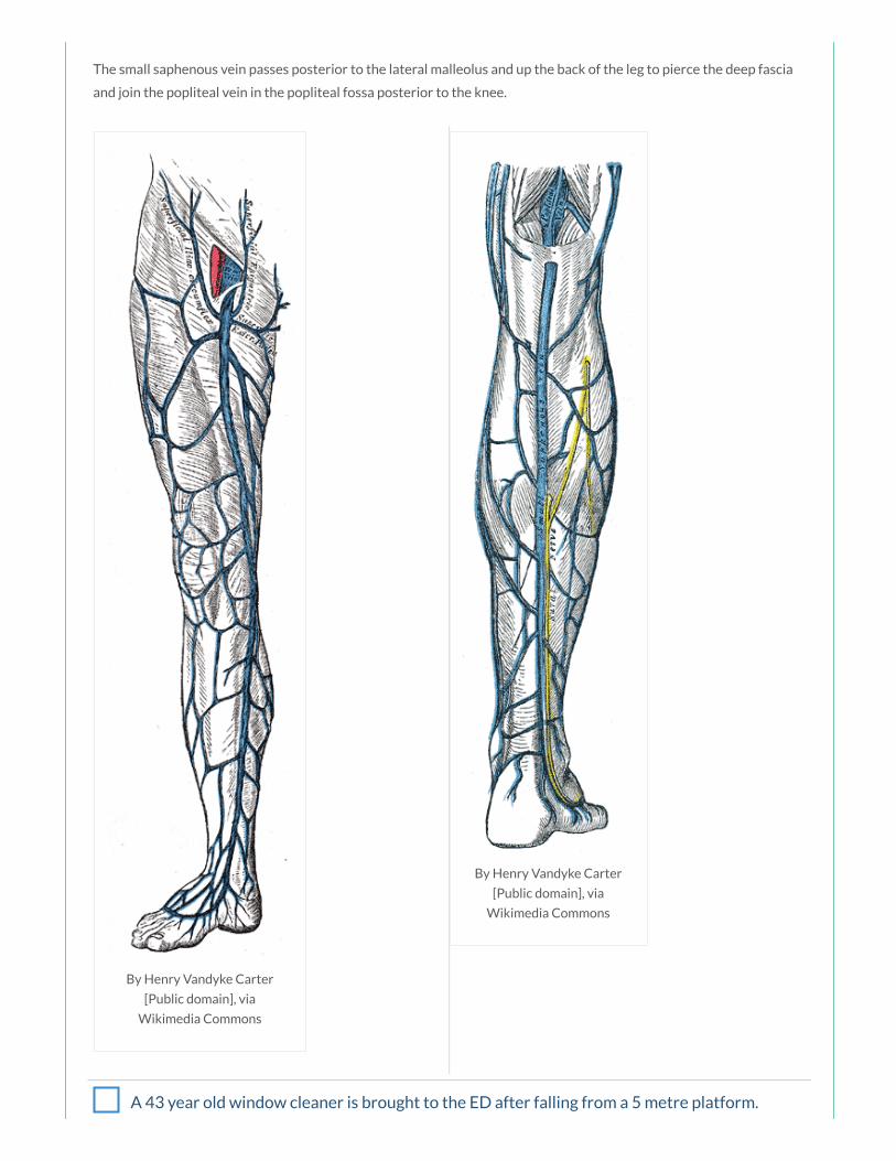
By Henry Vandyke Carter
[Public domain], via
Wikimedia Commons
By Henry Vandyke Carter
[Public domain], via
Wikimedia Commons
The small saphenous vein passes posterior to the lateral malleolus and up the back of the leg to pierce the deep fascia
and join the popliteal vein in the popliteal fossa posterior to the knee.
A 43 year old window cleaner is brought to the ED after falling from a 5 metre platform.
Imaging has shown multiple vertebral fractures and you are concerned about possible neurological
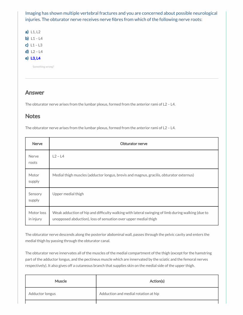
Imaging has shown multiple vertebral fractures and you are concerned about possible neurological
injuries. The obturator nerve receives nerve bres from which of the following nerve roots:
a) L1, L2
b) L1 – L4
c) L1 – L3
d) L2 – L4
e) L3, L4
Answer
The obturator nerve arises from the lumbar plexus, formed from the anterior rami of L2 – L4.
Notes
The obturator nerve arises from the lumbar plexus, formed from the anterior rami of L2 – L4.
Nerve Obturator nerve
Nerve
roots
L2 – L4
Motor
supply
Medial thigh muscles (adductor longus, brevis and magnus, gracilis, obturator externus)
Sensory
supply
Upper medial thigh
Motor loss
in injury
Weak adduction of hip and dif culty walking with lateral swinging of limb during walking (due to
unopposed abduction), loss of sensation over upper medial thigh
The obturator nerve descends along the posterior abdominal wall, passes through the pelvic cavity and enters the
medial thigh by passing through the obturator canal.
The obturator nerve innervates all of the muscles of the medial compartment of the thigh (except for the hamstring
part of the adductor longus, and the pectineus muscle which are innervated by the sciatic and the femoral nerves
respectively). It also gives off a cutaneous branch that supplies skin on the medial side of the upper thigh.
Muscle Action(s)
Adductor longus Adduction and medial rotation at hip
Something wrong?
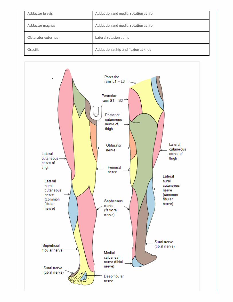
Adductor brevis Adduction and medial rotation at hip
Adductor magnus Adduction and medial rotation at hip
Obturator externus Lateral rotation at hip
Gracilis Adduction at hip and exion at knee

Modi ed by FRCEM Success. Original by Henry Vandyke Carter [Public domain], via Wikimedia Commons
A 25 year old man with haemophilia presents to the ED after falling onto his right hip. Plain x-
ray reveals no bony injury. Concerned by his pain your consultant arranges a CT which shows an
iliacus haematoma. The iliacus muscle primarily acts to perform which of the following movements:
a) Flexion and abduction of the thigh
b) Flexion of the thigh and exion of the leg
c) Flexion and medial rotation of the thigh
d) Flexion and lateral rotation of the thigh
e) Flexion and adduction of the thigh
Answer
The iliacus (red) and psoas major (yellow) muscles originate as separate muscles but insert by a common tendon onto
the femur and together are referred to as the iliopsoas muscle, which is a powerful exor of the thigh at the hip joint
and also contributes to lateral rotation of the thigh at the hip joint.
Notes
The sartorius, iliopsoas, pectineus and rectus femoris are the primary exors of the thigh at the hip joint.
Muscle Actions Innervation
Sartorius (blue) Flexion, abduction and lateral rotation at hip and exion at
knee
Femoral nerve (L2, L3)
Iliacus (red) Flexion and lateral rotation at hip Femoral nerve (L2, L3)
Psoas major
(yellow)
Flexion and lateral rotation at hip Anterior rami L1 – L3
Pectineus (green) Adduction and exion at hip Femoral nerve (L2, L3)
Rectus femoris Flexion at hip and extension at knee Femoral nerve (L2 –
L4)
The sartorius (blue) is innervated by the femoral nerve (L2, L3). It acts to ex the thigh at the hip joint and ex the leg at
the knee joint. It also abducts the thigh and rotates it laterally, as when resting the foot on the opposite knee when
sitting.
Something wrong?
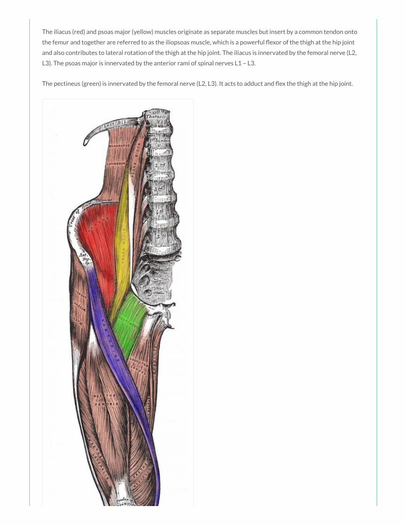
The iliacus (red) and psoas major (yellow) muscles originate as separate muscles but insert by a common tendon onto
the femur and together are referred to as the iliopsoas muscle, which is a powerful exor of the thigh at the hip joint
and also contributes to lateral rotation of the thigh at the hip joint. The iliacus is innervated by the femoral nerve (L2,
L3). The psoas major is innervated by the anterior rami of spinal nerves L1 – L3.
The pectineus (green) is innervated by the femoral nerve (L2, L3). It acts to adduct and ex the thigh at the hip joint.

Modi ed by FRCEM Success. Original by Henry
Vandyke Carter [Public domain], via Wikimedia
Commons
A 35 year old woman presents to the Emergency Department with a painful swelling in her
left groin. She is an intravenous drug user who regularly injects in the groins. On examination you
note a tender, discharging mass highly suspicious of an abscess. You discuss the case with the on-
call vascular team who are concerned about the possibility of a pseudoaneurysm of the femoral
artery. The femoral artery is a continuation of which of the following arteries:
a) Aorta
b) Common iliac artery
c) External iliac artery
d) Internal iliac artery
e) Obturator artery
Answer
The femoral artery is the continuation of the external iliac artery.
Notes
The major artery supplying the lower limb is the femoral artery. The femoral artery is the continuation of the external
iliac artery, beginning as the vessel passes under the inguinal ligament to enter the femoral triangle in the anterior
thigh.
The femoral artery can be palpated in the femoral triangle as it passes over the femoral head, just inferior to the
inguinal ligament, midway between the anterior superior iliac spine and the pubic symphysis (at the mid-inguinal
point). The femoral vein lies immediately medial to this pulsation, which is an important landmark for central venous
line insertion. Medial to the femoral vein is the femoral canal which contains lymphatics and lies immediately lateral to
the pubic tubercle. The femoral nerve lies lateral to the femoral artery.
The femoral artery gives rise to the deep profunda femoris artery in the femoral triangle which is a major source of
blood supply to the medial and posterior compartments of the thigh and the proximal femur.
Something wrong?
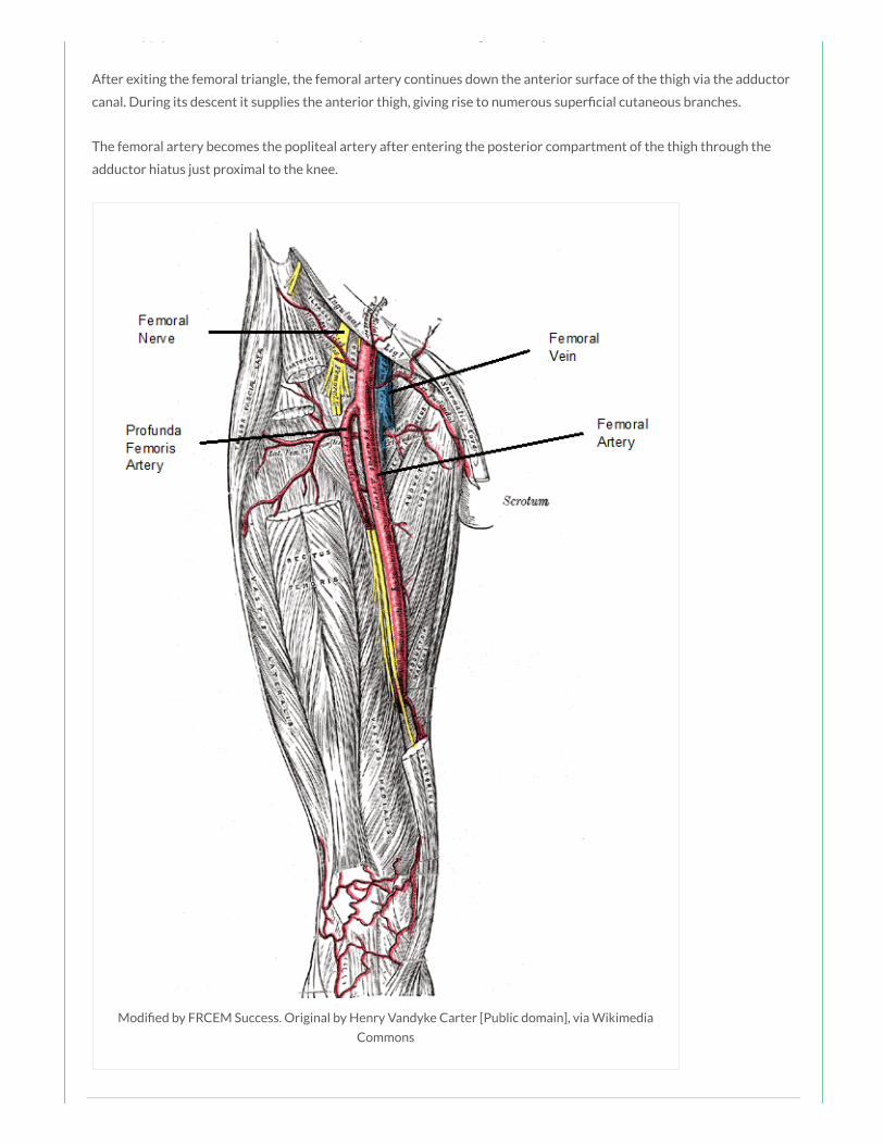
blood supply to the medial and posterior compartments of the thigh and the proximal femur.
After exiting the femoral triangle, the femoral artery continues down the anterior surface of the thigh via the adductor
canal. During its descent it supplies the anterior thigh, giving rise to numerous super cial cutaneous branches.
The femoral artery becomes the popliteal artery after entering the posterior compartment of the thigh through the
adductor hiatus just proximal to the knee.
Modi ed by FRCEM Success. Original by Henry Vandyke Carter [Public domain], via Wikimedia
Commons

An 83 year old woman is brought to ED following a fall in which she has sustained posterior
dislocation and fracture of the neck of femur. A tear of the ligament of the head of femur may result
in damage to a branch of which of the following arteries:
a) Femoral artery
b) Profunda femoris artery
c) Superior gluteal artery
d) Obturator artery
e) Medial circum ex artery
Answer
The obturator artery gives rise to an acetabular branch that runs in the round ligament of the head of the femur.
Notes
Joint Hip joint
Type Synovial ball and socket joint
Articulations Head of femur with acetabulum of pelvis
Stabilising
factors
Acetabular labrum, thickened brous capsule, extracapsular ligaments (iliofemoral, ischiofemoral,
pubofemoral), medial rotator muscles (effectively ‘pull’ head of femur into acetabulum)
Movements Flexion/Extension, Abduction/Adduction, Medial/Lateral rotation, Circumduction
Blood supply Branches of obturator artery, medial and lateral circum ex branches of profunda femoris artery
and superior and inferior gluteal arteries
Innervation Femoral nerve, obturator nerve, superior gluteal nerve and nerve to the quadratus femoris
Joint articulations
The hip joint is a multiaxial synovial ball and socket joint occurring between the head of the femur and the acetabulum
of the pelvis.
The acetabulum is formed by the fusion of the three bones, the ilium, the ischium and the pubis.
Something wrong?

By Henry Vandyke Carter [Public domain], via Wikimedia Commons
The rim of the acetabulum is raised slightly by the brocartilaginous acetabular labrum which increases its depth,
improving stability of the joint. The acetabular labrum continues inferiorly as the transverse acetabular ligament which
bridges the acetabular notch and converts the notch into a foramen for the passage of nutrient vessels and nerves.
Ligaments
The intracapsular ligament of the head of femur (ligamentum teres) runs from the fovea on the head of the femur at
one end to the acetabular fossa and the transverse acetabular ligament on the other. It carries a small acetabular
branch of the obturator artery.

Modi ed by FRCEM Success. Original by Henry
Vandyke Carter [Public domain], via Wikimedia
Commons By Henry Vandyke Carter [Public domain], via
Wikimedia Commons
By Henry Vandyke Carter [Public domain], via Wikimedia Commons
There are three main stabilising extracapsular ligaments; the iliofemoral (the largest and strongest ligament), the
pubofemoral and the ischiofemoral ligament.
The iliofemoral ligament is anterosuperior to the hip joint, attached proximally to the ilium just below the
anterior inferior iliac spine and distally to the intertrochanteric line of the femur. This ligament speci cally
prevents hyperextension and lateral rotation of the hip joint whilst standing.
The pubofemoral ligament is anteroinferior to the hip joint, attached to the pelvis at the iliopubic eminence
and adjacent bone and blending distally with the articular capsule. This ligament prevents excessive abduction
and extension of the femur at the hip joint.
The ischiofemoral ligament is posterior to the hip joint, attached medially to the ischium and laterally to the
greater trochanter. This ligament prevents excessive extension and medial rotation of the femur at the hip
joint.
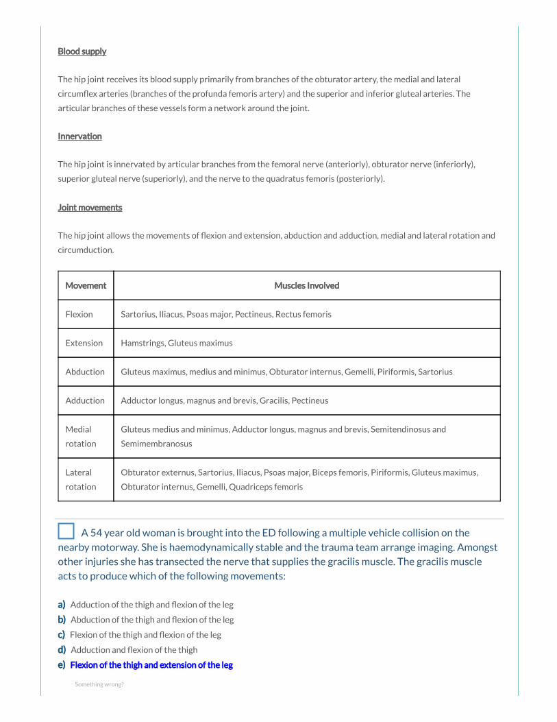
Blood supply
The hip joint receives its blood supply primarily from branches of the obturator artery, the medial and lateral
circum ex arteries (branches of the profunda femoris artery) and the superior and inferior gluteal arteries. The
articular branches of these vessels form a network around the joint.
Innervation
The hip joint is innervated by articular branches from the femoral nerve (anteriorly), obturator nerve (inferiorly),
superior gluteal nerve (superiorly), and the nerve to the quadratus femoris (posteriorly).
Joint movements
The hip joint allows the movements of exion and extension, abduction and adduction, medial and lateral rotation and
circumduction.
Movement Muscles Involved
Flexion Sartorius, Iliacus, Psoas major, Pectineus, Rectus femoris
Extension Hamstrings, Gluteus maximus
Abduction Gluteus maximus, medius and minimus, Obturator internus, Gemelli, Piriformis, Sartorius
Adduction Adductor longus, magnus and brevis, Gracilis, Pectineus
Medial
rotation
Gluteus medius and minimus, Adductor longus, magnus and brevis, Semitendinosus and
Semimembranosus
Lateral
rotation
Obturator externus, Sartorius, Iliacus, Psoas major, Biceps femoris, Piriformis, Gluteus maximus,
Obturator internus, Gemelli, Quadriceps femoris
A 54 year old woman is brought into the ED following a multiple vehicle collision on the
nearby motorway. She is haemodynamically stable and the trauma team arrange imaging. Amongst
other injuries she has transected the nerve that supplies the gracilis muscle. The gracilis muscle
acts to produce which of the following movements:
a) Adduction of the thigh and exion of the leg
b) Abduction of the thigh and exion of the leg
c) Flexion of the thigh and exion of the leg
d) Adduction and exion of the thigh
e) Flexion of the thigh and extension of the leg
Something wrong?
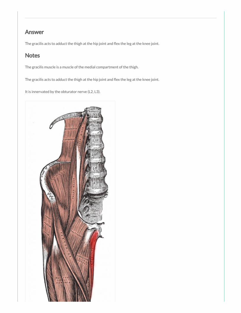
Answer
The gracilis acts to adduct the thigh at the hip joint and ex the leg at the knee joint.
Notes
The gracilis muscle is a muscle of the medial compartment of the thigh.
The gracilis acts to adduct the thigh at the hip joint and ex the leg at the knee joint.
It is innervated by the obturator nerve (L2, L3).
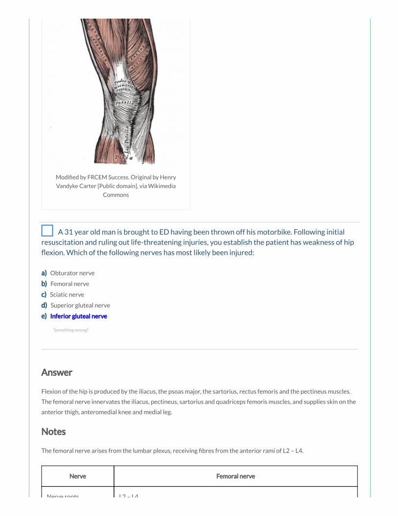
Modi ed by FRCEM Success. Original by Henry
Vandyke Carter [Public domain], via Wikimedia
Commons
A 31 year old man is brought to ED having been thrown off his motorbike. Following initial
resuscitation and ruling out life-threatening injuries, you establish the patient has weakness of hip
exion. Which of the following nerves has most likely been injured:
a) Obturator nerve
b) Femoral nerve
c) Sciatic nerve
d) Superior gluteal nerve
e) Inferior gluteal nerve
Answer
Flexion of the hip is produced by the iliacus, the psoas major, the sartorius, rectus femoris and the pectineus muscles.
The femoral nerve innervates the iliacus, pectineus, sartorius and quadriceps femoris muscles, and supplies skin on the
anterior thigh, anteromedial knee and medial leg.
Notes
The femoral nerve arises from the lumbar plexus, receiving bres from the anterior rami of L2 – L4.
Nerve Femoral nerve
Nerve roots L2 – L4
Something wrong?
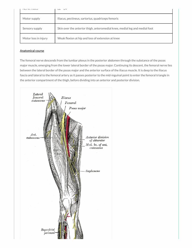
Nerve roots L2 – L4
Motor supply Iliacus, pectineus, sartorius, quadriceps femoris
Sensory supply Skin over the anterior thigh, anteromedial knee, medial leg and medial foot
Motor loss in injury Weak exion at hip and loss of extension at knee
Anatomical course
The femoral nerve descends from the lumbar plexus in the posterior abdomen through the substance of the psoas
major muscle, emerging from the lower lateral border of the psoas major. Continuing its descent, the femoral nerve lies
between the lateral border of the psoas major and the anterior surface of the iliacus muscle. It is deep to the iliacus
fascia and lateral to the femoral artery as it passes posterior to the mid-inguinal point to enter the femoral triangle in
the anterior compartment of the thigh, before dividing into an anterior and posterior division.

By Henry Vandyke Carter [Public domain], via Wikimedia
Commons
Branches
In the abdomen it gives rise to branches that innervate the iliacus and pectineus muscles.
The anterior division gives off anterior cutaneous branches (supplying skin over the anterior and medial thigh) and
muscular branches (innervating the sartorius).
The posterior division gives off muscular branches (innervating the quadriceps femoris muscles) and articular branches
(supplying the hip and knee joint), before continuing as the saphenous nerve (supplying skin over the anteromedial
knee and the medial side of the leg and foot).
Branch Innervation
Muscular branches in abdomen Iliacus and pectineus
Anterior cutaneous branches Skin over anterior thigh
Anterior muscular branches Sartorius
Posterior muscular branches Quadriceps femoris muscles
Posterior articular branches Hip and knee joint
Saphenous nerve Skin over anteromedial knee, medial side of leg and foot
Motor and sensory function

Muscle Action(s)
Sartorius Flexion, abduction and lateral rotation at hip and exion at knee
Iliacus (red) Flexion and lateral rotation at hip
Pectineus (green) Adduction and exion at hip
Rectus femoris Flexion at hip and extension at knee
Vastus lateralis Extension at knee
Vastus medialis Extension at knee
Vastus intermedius Extension at knee
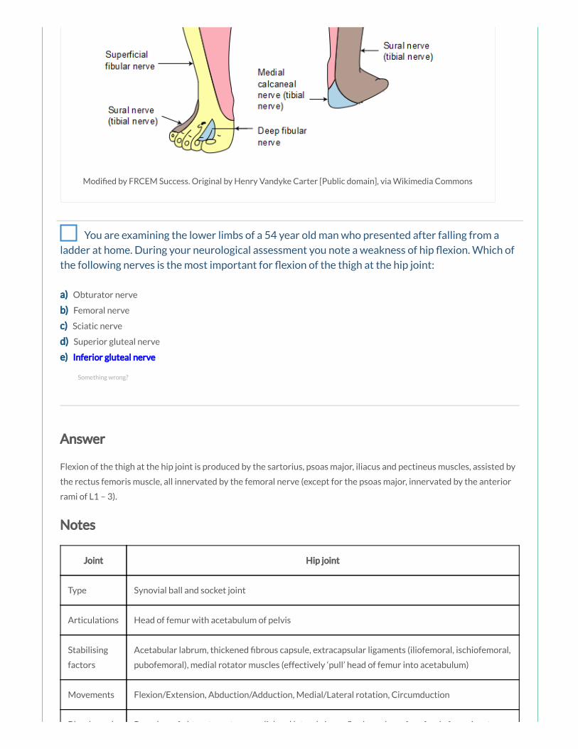
Modi ed by FRCEM Success. Original by Henry Vandyke Carter [Public domain], via Wikimedia Commons
You are examining the lower limbs of a 54 year old man who presented after falling from a
ladder at home. During your neurological assessment you note a weakness of hip exion. Which of
the following nerves is the most important for exion of the thigh at the hip joint:
a) Obturator nerve
b) Femoral nerve
c) Sciatic nerve
d) Superior gluteal nerve
e) Inferior gluteal nerve
Answer
Flexion of the thigh at the hip joint is produced by the sartorius, psoas major, iliacus and pectineus muscles, assisted by
the rectus femoris muscle, all innervated by the femoral nerve (except for the psoas major, innervated by the anterior
rami of L1 – 3).
Notes
Joint Hip joint
Type Synovial ball and socket joint
Articulations Head of femur with acetabulum of pelvis
Stabilising
factors
Acetabular labrum, thickened brous capsule, extracapsular ligaments (iliofemoral, ischiofemoral,
pubofemoral), medial rotator muscles (effectively ‘pull’ head of femur into acetabulum)
Movements Flexion/Extension, Abduction/Adduction, Medial/Lateral rotation, Circumduction
Blood supply Branches of obturator artery, medial and lateral circum ex branches of profunda femoris artery
Something wrong?

Blood supply Branches of obturator artery, medial and lateral circum ex branches of profunda femoris artery
and superior and inferior gluteal arteries
Innervation Femoral nerve, obturator nerve, superior gluteal nerve and nerve to the quadratus femoris
Joint articulations
The hip joint is a multiaxial synovial ball and socket joint occurring between the head of the femur and the acetabulum
of the pelvis.
The acetabulum is formed by the fusion of the three bones, the ilium, the ischium and the pubis.
By Henry Vandyke Carter [Public domain], via Wikimedia Commons
The rim of the acetabulum is raised slightly by the brocartilaginous acetabular labrum which increases its depth,
improving stability of the joint. The acetabular labrum continues inferiorly as the transverse acetabular ligament which
bridges the acetabular notch and converts the notch into a foramen for the passage of nutrient vessels and nerves.
Ligaments
The intracapsular ligament of the head of femur (ligamentum teres) runs from the fovea on the head of the femur at
one end to the acetabular fossa and the transverse acetabular ligament on the other. It carries a small acetabular

branch of the obturator artery.
By Henry Vandyke Carter [Public domain], via Wikimedia Commons
There are three main stabilising extracapsular ligaments; the iliofemoral (the largest and strongest ligament), the
pubofemoral and the ischiofemoral ligament.
The iliofemoral ligament is anterosuperior to the hip joint, attached proximally to the ilium just below the
anterior inferior iliac spine and distally to the intertrochanteric line of the femur. This ligament speci cally
prevents hyperextension and lateral rotation of the hip joint whilst standing.
The pubofemoral ligament is anteroinferior to the hip joint, attached to the pelvis at the iliopubic eminence
and adjacent bone and blending distally with the articular capsule. This ligament prevents excessive abduction
and extension of the femur at the hip joint.
The ischiofemoral ligament is posterior to the hip joint, attached medially to the ischium and laterally to the
greater trochanter. This ligament prevents excessive extension and medial rotation of the femur at the hip
joint.

Modi ed by FRCEM Success. Original by Henry
Vandyke Carter [Public domain], via Wikimedia
Commons By Henry Vandyke Carter [Public domain], via
Wikimedia Commons
Blood supply
The hip joint receives its blood supply primarily from branches of the obturator artery, the medial and lateral
circum ex arteries (branches of the profunda femoris artery) and the superior and inferior gluteal arteries. The
articular branches of these vessels form a network around the joint.
Innervation
The hip joint is innervated by articular branches from the femoral nerve (anteriorly), obturator nerve (inferiorly),
superior gluteal nerve (superiorly), and the nerve to the quadratus femoris (posteriorly).
Joint movements
The hip joint allows the movements of exion and extension, abduction and adduction, medial and lateral rotation and
circumduction.
Movement Muscles Involved
Flexion Sartorius, Iliacus, Psoas major, Pectineus, Rectus femoris
Extension Hamstrings, Gluteus maximus
Abduction Gluteus maximus, medius and minimus, Obturator internus, Gemelli, Piriformis, Sartorius
Adduction Adductor longus, magnus and brevis, Gracilis, Pectineus
Medial
rotation
Gluteus medius and minimus, Adductor longus, magnus and brevis, Semitendinosus and
Semimembranosus
Lateral Obturator externus, Sartorius, Iliacus, Psoas major, Biceps femoris, Piriformis, Gluteus maximus,
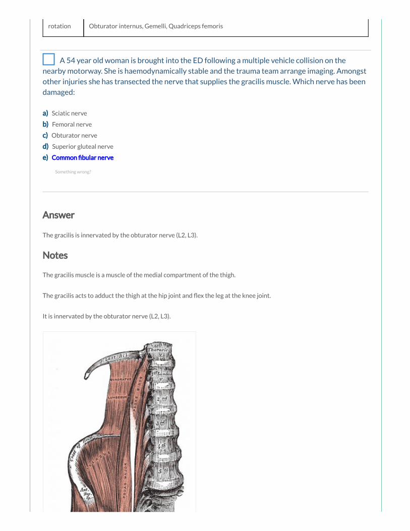
Lateral
rotation
Obturator externus, Sartorius, Iliacus, Psoas major, Biceps femoris, Piriformis, Gluteus maximus,
Obturator internus, Gemelli, Quadriceps femoris
A 54 year old woman is brought into the ED following a multiple vehicle collision on the
nearby motorway. She is haemodynamically stable and the trauma team arrange imaging. Amongst
other injuries she has transected the nerve that supplies the gracilis muscle. Which nerve has been
damaged:
a) Sciatic nerve
b) Femoral nerve
c) Obturator nerve
d) Superior gluteal nerve
e) Common bular nerve
Answer
The gracilis is innervated by the obturator nerve (L2, L3).
Notes
The gracilis muscle is a muscle of the medial compartment of the thigh.
The gracilis acts to adduct the thigh at the hip joint and ex the leg at the knee joint.
It is innervated by the obturator nerve (L2, L3).
Something wrong?

Modi ed by FRCEM Success. Original by Henry
Vandyke Carter [Public domain], via Wikimedia
Commons
A 32 year old woman is brought to the ED after falling from a horse. Imaging shows multiple
injuries to the pelvic region including transection of the nerve that supplies the piriformis muscle.
The piriformis muscle acts primarily to produce which of the following movements:
a) Abduction and lateral rotation of the thigh
b) Abduction and medial rotation of the thigh
c) Adduction and lateral rotation of the thigh
d) Adduction and medial rotation of the thigh
e) Abduction and exion of the thigh
Something wrong?

Answer
The piriformis (yellow) acts to abduct and laterally rotate the thigh at the hip joint.
Notes
Muscle Action(s) Innervation
Gluteus maximus (red) Extension, lateral rotation and abduction at hip Inferior gluteal nerve (L5 – S2)
Gluteus medius (green) Abduction and medial rotation at hip Superior gluteal nerve (L4 – S1)
Gluteus minimus (blue) Abduction and medial rotation at hip Superior gluteal nerve (L4 – S1)
Piriformis (yellow) Lateral rotation and abduction at hip Branches from S1 and S2
The gluteus maximus (red) is the main extensor of the thigh at the hip joint and also acts to laterally rotate and abduct
the thigh. Through its insertion into the iliotibial tract, it also stabilises the knee and hip joints. The gluteus maximus is
innervated by the inferior gluteal nerve (L5 – S2).
The gluteus medius (green) and gluteus minimus (blue) act to abduct and medially rotate the thigh at the hip joint, and
also act to secure the pelvis, reducing pelvic drop on the opposite swing side during walking. They are both innervated
by the superior gluteal nerve (L4 – S1).
The piriformis (yellow) acts to abduct and laterally rotate the thigh at the hip joint. It is innervated by the nerve to the
piriformis, originating from the sacral plexus (S1, S2). The obturator internus, gemelli and quadriceps femoris muscles
act as synergistic femoral lateral rotators and hip stabilisers.
Trendelenburg’s sign is seen in people with weak/paralysed abductor muscles of the hip. The sign is demonstrated by
asking the patient to stand on one limb; when the patient stands on the affected limb, the pelvis severely drops over
the swing limb. This sign may be seen in patients with damage to the superior gluteal nerve, which may occur in
association with pelvic fractures, with space-occupying lesions within the pelvis extending into the greater sciatic
foramen, and following hip surgery. Typically the patient may also demonstrate a Trendelenburg gait.

Modi ed by FRCEM Success. Original by Henry Vandyke Carter
[Public domain], via Wikimedia Commons
An unknown male is brought into the department after being struck by a lorry whilst crossing
the road. Imaging shows extensive injuries in the pelvic region including trauma to the gluteus
maximus. The gluteus maximus muscle primarily acts to produce which of the following
movements:
a) Extension, medial rotation and abduction of the thigh
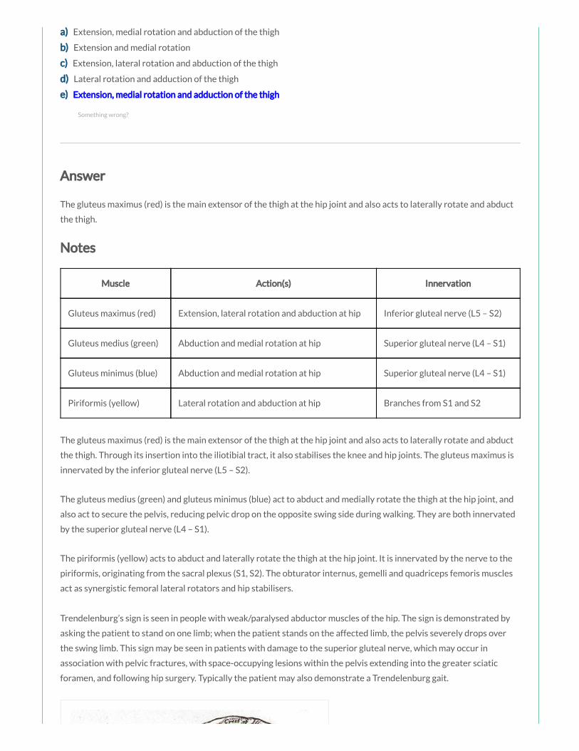
a) Extension, medial rotation and abduction of the thigh
b) Extension and medial rotation
c) Extension, lateral rotation and abduction of the thigh
d) Lateral rotation and adduction of the thigh
e) Extension, medial rotation and adduction of the thigh
Answer
The gluteus maximus (red) is the main extensor of the thigh at the hip joint and also acts to laterally rotate and abduct
the thigh.
Notes
Muscle Action(s) Innervation
Gluteus maximus (red) Extension, lateral rotation and abduction at hip Inferior gluteal nerve (L5 – S2)
Gluteus medius (green) Abduction and medial rotation at hip Superior gluteal nerve (L4 – S1)
Gluteus minimus (blue) Abduction and medial rotation at hip Superior gluteal nerve (L4 – S1)
Piriformis (yellow) Lateral rotation and abduction at hip Branches from S1 and S2
The gluteus maximus (red) is the main extensor of the thigh at the hip joint and also acts to laterally rotate and abduct
the thigh. Through its insertion into the iliotibial tract, it also stabilises the knee and hip joints. The gluteus maximus is
innervated by the inferior gluteal nerve (L5 – S2).
The gluteus medius (green) and gluteus minimus (blue) act to abduct and medially rotate the thigh at the hip joint, and
also act to secure the pelvis, reducing pelvic drop on the opposite swing side during walking. They are both innervated
by the superior gluteal nerve (L4 – S1).
The piriformis (yellow) acts to abduct and laterally rotate the thigh at the hip joint. It is innervated by the nerve to the
piriformis, originating from the sacral plexus (S1, S2). The obturator internus, gemelli and quadriceps femoris muscles
act as synergistic femoral lateral rotators and hip stabilisers.
Trendelenburg’s sign is seen in people with weak/paralysed abductor muscles of the hip. The sign is demonstrated by
asking the patient to stand on one limb; when the patient stands on the affected limb, the pelvis severely drops over
the swing limb. This sign may be seen in patients with damage to the superior gluteal nerve, which may occur in
association with pelvic fractures, with space-occupying lesions within the pelvis extending into the greater sciatic
foramen, and following hip surgery. Typically the patient may also demonstrate a Trendelenburg gait.
Something wrong?

Modi ed by FRCEM Success. Original by Henry Vandyke Carter
[Public domain], via Wikimedia Commons

A 75 year old lady slips on a wet bathroom oor and presents to ED with her left leg
shortened and externally rotated. Imaging demonstrates an intracapsular fracture of the neck of
femur. The patient is at risk of avascular necrosis of the femoral head. This is most likely to occur
due to lack of blood supply from which of the following arteries:
a) Medial circum ex artery
b) Obturator artery
c) Superior gluteal artery
d) Inferior gluteal artery
e) Lateral circum ex artery
Answer
In adults, the primary blood supply to the head of the femur is from branches of the medial femoral circum ex artery.
The lateral circum ex artery anastomoses with the medial femoral circum ex artery and assists in supplying the
femoral head. The obturator artery gives rise to the artery of the head of femur which runs in the ligamentum teres and
is usually insuf cient to supply the head of femur in adults (but is an important source of blood supply in children up to
about 8 years). The superior and inferior gluteal arteries supply the hip joint but not typically the head of femur.
Notes
Joint Hip joint
Type Synovial ball and socket joint
Articulations Head of femur with acetabulum of pelvis
Stabilising
factors
Acetabular labrum, thickened brous capsule, extracapsular ligaments (iliofemoral, ischiofemoral,
pubofemoral), medial rotator muscles (effectively ‘pull’ head of femur into acetabulum)
Movements Flexion/Extension, Abduction/Adduction, Medial/Lateral rotation, Circumduction
Blood supply Branches of obturator artery, medial and lateral circum ex branches of profunda femoris artery
and superior and inferior gluteal arteries
Innervation Femoral nerve, obturator nerve, superior gluteal nerve and nerve to the quadratus femoris
Joint articulations
The hip joint is a multiaxial synovial ball and socket joint occurring between the head of the femur and the acetabulum
Something wrong?
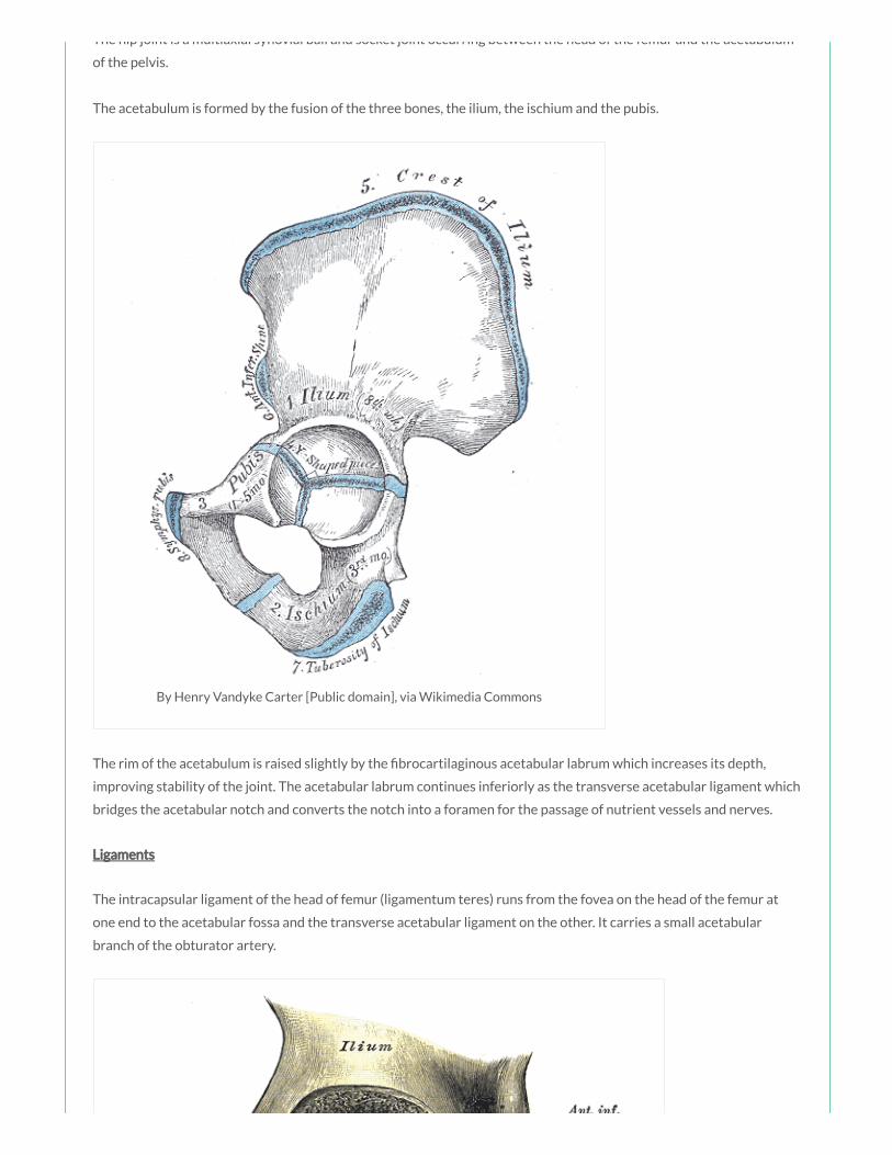
The hip joint is a multiaxial synovial ball and socket joint occurring between the head of the femur and the acetabulum
of the pelvis.
The acetabulum is formed by the fusion of the three bones, the ilium, the ischium and the pubis.
By Henry Vandyke Carter [Public domain], via Wikimedia Commons
The rim of the acetabulum is raised slightly by the brocartilaginous acetabular labrum which increases its depth,
improving stability of the joint. The acetabular labrum continues inferiorly as the transverse acetabular ligament which
bridges the acetabular notch and converts the notch into a foramen for the passage of nutrient vessels and nerves.
Ligaments
The intracapsular ligament of the head of femur (ligamentum teres) runs from the fovea on the head of the femur at
one end to the acetabular fossa and the transverse acetabular ligament on the other. It carries a small acetabular
branch of the obturator artery.

By Henry Vandyke Carter [Public domain], via Wikimedia Commons
There are three main stabilising extracapsular ligaments; the iliofemoral (the largest and strongest ligament), the
pubofemoral and the ischiofemoral ligament.
The iliofemoral ligament is anterosuperior to the hip joint, attached proximally to the ilium just below the
anterior inferior iliac spine and distally to the intertrochanteric line of the femur. This ligament speci cally
prevents hyperextension and lateral rotation of the hip joint whilst standing.
The pubofemoral ligament is anteroinferior to the hip joint, attached to the pelvis at the iliopubic eminence
and adjacent bone and blending distally with the articular capsule. This ligament prevents excessive abduction
and extension of the femur at the hip joint.
The ischiofemoral ligament is posterior to the hip joint, attached medially to the ischium and laterally to the
greater trochanter. This ligament prevents excessive extension and medial rotation of the femur at the hip
joint.

Modi ed by FRCEM Success. Original by Henry
Vandyke Carter [Public domain], via Wikimedia
Commons By Henry Vandyke Carter [Public domain], via
Wikimedia Commons
Blood supply
The hip joint receives its blood supply primarily from branches of the obturator artery, the medial and lateral
circum ex arteries (branches of the profunda femoris artery) and the superior and inferior gluteal arteries. The
articular branches of these vessels form a network around the joint.
Innervation
The hip joint is innervated by articular branches from the femoral nerve (anteriorly), obturator nerve (inferiorly),
superior gluteal nerve (superiorly), and the nerve to the quadratus femoris (posteriorly).
Joint movements
The hip joint allows the movements of exion and extension, abduction and adduction, medial and lateral rotation and
circumduction.
Movement Muscles Involved
Flexion Sartorius, Iliacus, Psoas major, Pectineus, Rectus femoris
Extension Hamstrings, Gluteus maximus
Abduction Gluteus maximus, medius and minimus, Obturator internus, Gemelli, Piriformis, Sartorius
Adduction Adductor longus, magnus and brevis, Gracilis, Pectineus
Medial
rotation
Gluteus medius and minimus, Adductor longus, magnus and brevis, Semitendinosus and
Semimembranosus
Lateral
rotation
Obturator externus, Sartorius, Iliacus, Psoas major, Biceps femoris, Piriformis, Gluteus maximus,
Obturator internus, Gemelli, Quadriceps femoris
You form part of the trauma team caring for a motorcyclist who was involved in a collision
with a van. You receive a phone call from the on-call radiologist informing you that the patient has
an injury to the nerve supplying the hamstring muscles. The hamstring muscles are innervated by
which of the following nerves:

a) Femoral nerve
b) Obturator nerve
c) Sciatic nerve
d) Superior gluteal nerve
e) Inferior gluteal nerve
Answer
The hamstring muscles are all innervated by the sciatic nerve (L5 – S2).
Notes
Muscle Action(s) Innervation
Biceps femoris (red) Flexion at knee, extension and lateral rotation at hip Sciatic nerve (L5 – S2)
Semitendinosus (blue) Flexion at knee, extension and medial rotation at hip Sciatic nerve (L5 – S2)
Semimembranosus (green) Flexion at knee, extension and medial rotation at hip Sciatic nerve (L5 – S2)
The hamstrings are composed of three individual muscles; the biceps femoris (red), the semitendinosus (blue) and the
semimembranosus (green).
The hamstrings act together to ex the leg at the knee joint and extend the thigh at the hip joint. The biceps femoris
also acts to laterally rotate the thigh at the hip joint and the leg at the knee joint. The semimembranosus and
semitendinosus also act together to medially rotate the thigh at the hip joint and the leg at the knee joint.
The hamstring muscles are all innervated by the tibial division of the sciatic nerve (L5 – S2), except for the short head of
the biceps femoris innervated by the common bular division.
To test the hamstrings the patient exes their leg against resistance. Normally these muscles, especially their tendons
on each side of the popliteal fossa, are prominent as they bend the knee.
Something wrong?
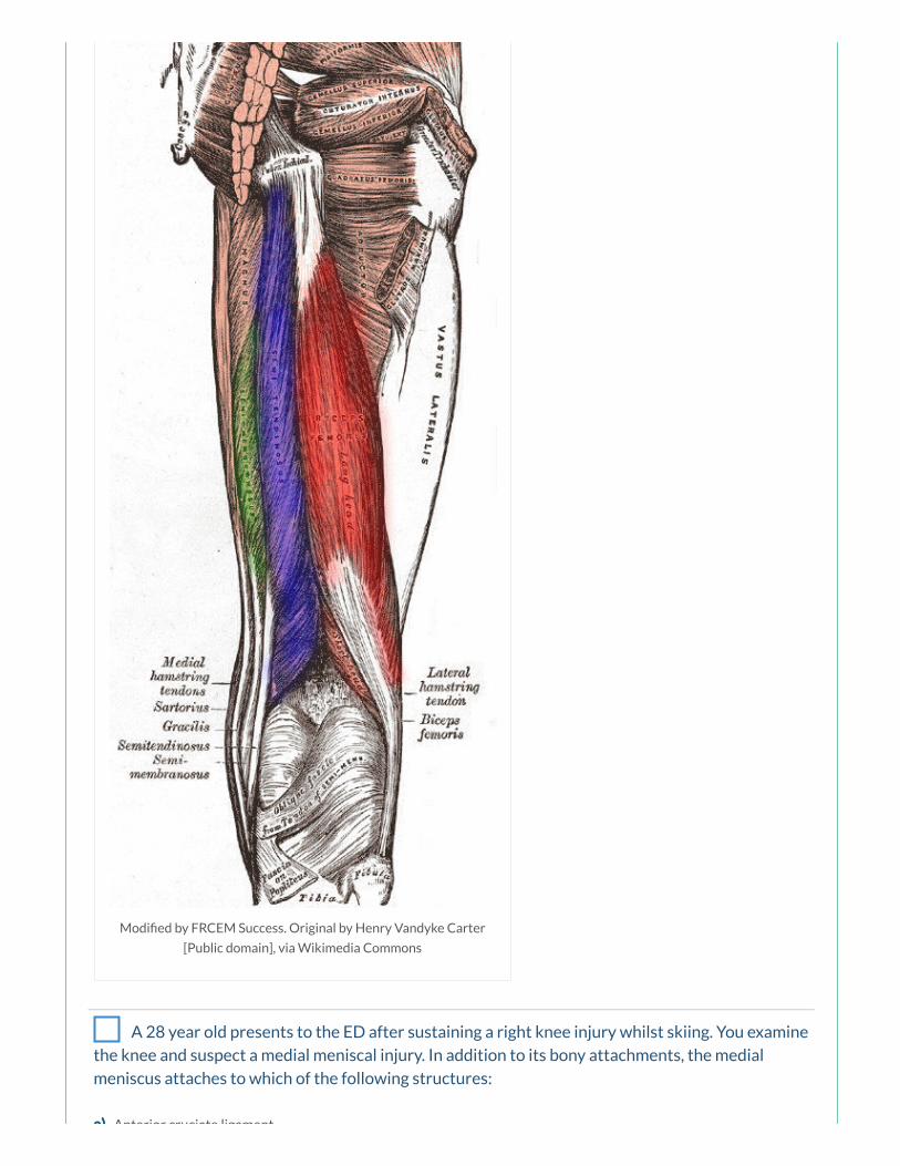
Modi ed by FRCEM Success. Original by Henry Vandyke Carter
[Public domain], via Wikimedia Commons
A 28 year old presents to the ED after sustaining a right knee injury whilst skiing. You examine
the knee and suspect a medial meniscal injury. In addition to its bony attachments, the medial
meniscus attaches to which of the following structures:
a) Anterior cruciate ligament
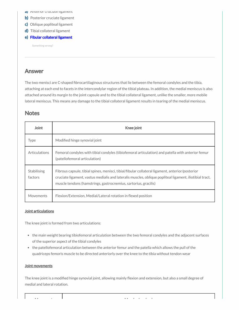
a) Anterior cruciate ligament
b) Posterior cruciate ligament
c) Oblique popliteal ligament
d) Tibial collateral ligament
e) Fibular collateral ligament
Answer
The two menisci are C-shaped brocartilaginous structures that lie between the femoral condyles and the tibia,
attaching at each end to facets in the intercondylar region of the tibial plateau. In addition, the medial meniscus is also
attached around its margin to the joint capsule and to the tibial collateral ligament, unlike the smaller, more mobile
lateral meniscus. This means any damage to the tibial collateral ligament results in tearing of the medial meniscus.
Notes
Joint Knee joint
Type Modi ed hinge synovial joint
Articulations Femoral condyles with tibial condyles (tibiofemoral articulation) and patella with anterior femur
(patellofemoral articulation)
Stabilising
factors
Fibrous capsule, tibial spines, menisci, tibial/ bular collateral ligament, anterior/posterior
cruciate ligament, vastus medialis and lateralis muscles, oblique popliteal ligament, iliotibial tract,
muscle tendons (hamstrings, gastrocnemius, sartorius, gracilis)
Movements Flexion/Extension, Medial/Lateral rotation in exed position
Joint articulations
The knee joint is formed from two articulations:
the main weight bearing tibiofemoral articulation between the two femoral condyles and the adjacent surfaces
of the superior aspect of the tibial condyles
the patellofemoral articulation between the anterior femur and the patella which allows the pull of the
quadriceps femoris muscle to be directed anteriorly over the knee to the tibia without tendon wear
Joint movements
The knee joint is a modi ed hinge synovial joint, allowing mainly exion and extension, but also a small degree of
medial and lateral rotation.
Movement Muscles Involved
Something wrong?
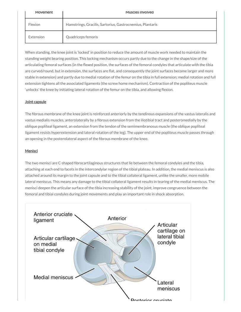
Movement Muscles Involved
Flexion Hamstrings, Gracilis, Sartorius, Gastrocnemius, Plantaris
Extension Quadriceps femoris
When standing, the knee joint is ‘locked’ in position to reduce the amount of muscle work needed to maintain the
standing weight bearing position. This locking mechanism occurs partly due to the change in the shape/size of the
articulating femoral surfaces (in the exed position, the surfaces of the femoral condyles that articulate with the tibia
are curved/round, but in extension, the surfaces are at, and consequently the joint surfaces become larger and more
stable in extension) and partly due to medial rotation of the femur on the tibia in full extension; medial rotation and full
extension tightens all the associated ligaments (the screw home mechanism). Contraction of the popliteus muscle
‘unlocks’ the knee by initiating lateral rotation of the femur on the tibia, and allowing exion.
Joint capsule
The brous membrane of the knee joint is reinforced anteriorly by the tendinous expansions of the vastus lateralis and
vastus medialis muscles, anterolaterally by a brous extension from the iliotibial tract and posteromedially by the
oblique popliteal ligament, an extension from the tendon of the semimembranosus muscle (the oblique popliteal
ligament resists hyperextension and lateral rotation of the leg). The upper end of the popliteus muscle passes through
an opening in the posterolateral aspect of the brous membrane of the knee.
Menisci
The two menisci are C-shaped brocartilaginous structures that lie between the femoral condyles and the tibia,
attaching at each end to facets in the intercondylar region of the tibial plateau. In addition, the medial meniscus is also
attached around its margin to the joint capsule and to the tibial collateral ligament, unlike the smaller, more mobile
lateral meniscus. This means any damage to the tibial collateral ligament results in tearing of the medial meniscus. The
menisci deepen the articular surface of the tibia increasing stability of the joint, improve congruence between the
femoral and tibial condyles during joint movements and play an important role in shock absorption.
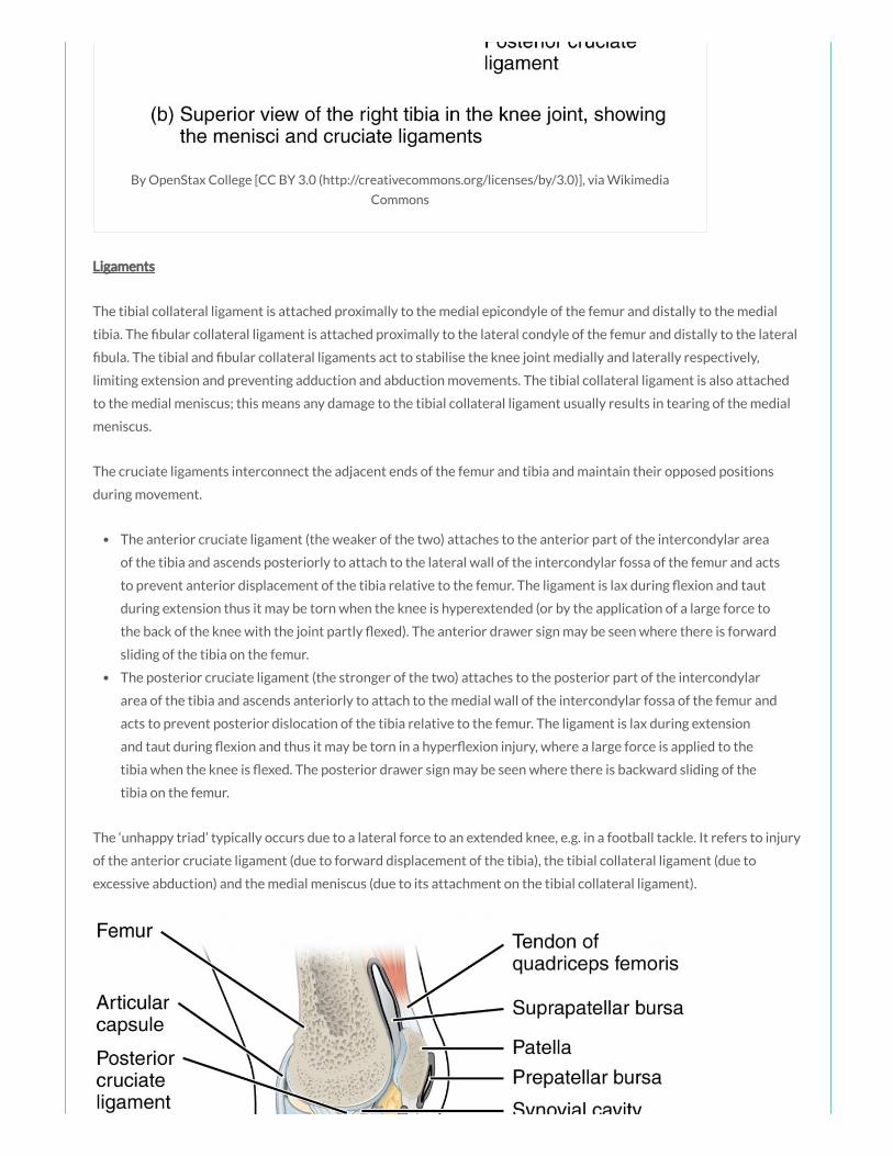
By OpenStax College [CC BY 3.0 (http://creativecommons.org/licenses/by/3.0)], via Wikimedia
Commons
Ligaments
The tibial collateral ligament is attached proximally to the medial epicondyle of the femur and distally to the medial
tibia. The bular collateral ligament is attached proximally to the lateral condyle of the femur and distally to the lateral
bula. The tibial and bular collateral ligaments act to stabilise the knee joint medially and laterally respectively,
limiting extension and preventing adduction and abduction movements. The tibial collateral ligament is also attached
to the medial meniscus; this means any damage to the tibial collateral ligament usually results in tearing of the medial
meniscus.
The cruciate ligaments interconnect the adjacent ends of the femur and tibia and maintain their opposed positions
during movement.
The anterior cruciate ligament (the weaker of the two) attaches to the anterior part of the intercondylar area
of the tibia and ascends posteriorly to attach to the lateral wall of the intercondylar fossa of the femur and acts
to prevent anterior displacement of the tibia relative to the femur. The ligament is lax during exion and taut
during extension thus it may be torn when the knee is hyperextended (or by the application of a large force to
the back of the knee with the joint partly exed). The anterior drawer sign may be seen where there is forward
sliding of the tibia on the femur.
The posterior cruciate ligament (the stronger of the two) attaches to the posterior part of the intercondylar
area of the tibia and ascends anteriorly to attach to the medial wall of the intercondylar fossa of the femur and
acts to prevent posterior dislocation of the tibia relative to the femur. The ligament is lax during extension
and taut during exion and thus it may be torn in a hyper exion injury, where a large force is applied to the
tibia when the knee is exed. The posterior drawer sign may be seen where there is backward sliding of the
tibia on the femur.
The ‘unhappy triad’ typically occurs due to a lateral force to an extended knee, e.g. in a football tackle. It refers to injury
of the anterior cruciate ligament (due to forward displacement of the tibia), the tibial collateral ligament (due to
excessive abduction) and the medial meniscus (due to its attachment on the tibial collateral ligament).
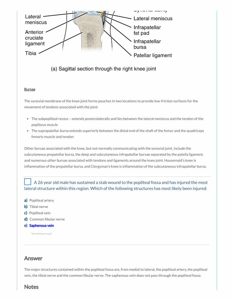
Bursae
The synovial membrane of the knee joint forms pouches in two locations to provide low-friction surfaces for the
movement of tendons associated with the joint:
The subpopliteal recess – extends posterolaterally and lies between the lateral meniscus and the tendon of the
popliteus muscle
The suprapatellar bursa extends superiorly between the distal end of the shaft of the femur and the quadriceps
femoris muscle and tendon
Other bursae associated with the knee, but not normally communicating with the synovial joint, include the
subcutaneous prepatellar bursa, the deep and subcutaneous infrapatellar bursae separated by the patella ligament,
and numerous other bursae associated with tendons and ligaments around the knee joint. Housemaid’s knee is
in ammation of the prepatellar bursa, and Clergyman’s knee is in ammation of the subcutaneous infrapatellar bursa.
A 26 year old male has sustained a stab wound to the popliteal fossa and has injured the most
lateral structure within this region. Which of the following structures has most likely been injured:
a) Popliteal artery
b) Tibial nerve
c) Popliteal vein
d) Common bular nerve
e) Saphenous vein
Answer
The major structures contained within the popliteal fossa are, from medial to lateral, the popliteal artery, the popliteal
vein, the tibial nerve and the common bular nerve. The saphenous vein does not pass through the popliteal fossa.
Notes
Something wrong?
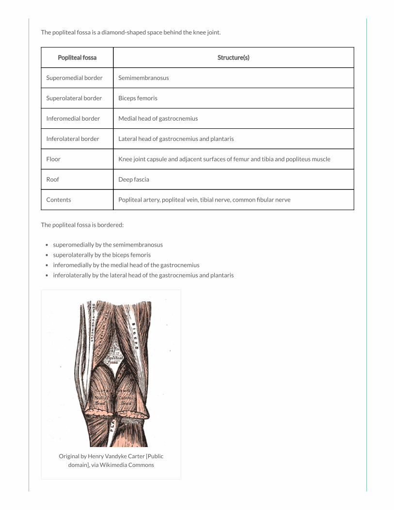
The popliteal fossa is a diamond-shaped space behind the knee joint.
Popliteal fossa Structure(s)
Superomedial border Semimembranosus
Superolateral border Biceps femoris
Inferomedial border Medial head of gastrocnemius
Inferolateral border Lateral head of gastrocnemius and plantaris
Floor Knee joint capsule and adjacent surfaces of femur and tibia and popliteus muscle
Roof Deep fascia
Contents Popliteal artery, popliteal vein, tibial nerve, common bular nerve
The popliteal fossa is bordered:
superomedially by the semimembranosus
superolaterally by the biceps femoris
inferomedially by the medial head of the gastrocnemius
inferolaterally by the lateral head of the gastrocnemius and plantaris
Original by Henry Vandyke Carter [Public
domain], via Wikimedia Commons
The oor of the fossa is formed by the capsule of the knee joint and adjacent surfaces of the femur and tibia, and more

The oor of the fossa is formed by the capsule of the knee joint and adjacent surfaces of the femur and tibia, and more
inferiorly by the popliteus muscle. The roof is formed by deep fascia.
The major structures contained within the popliteal fossa are (from medial to lateral):
the popliteal artery
the popliteal vein
the tibial nerve
the common bular nerve
Modi ed by FRCEM Success. Original by Henry Vandyke Carter [Public domain],
via Wikimedia Commons
The small saphenous vein ascends in the super cial fascia on the back of the leg to the knee where it penetrates the
deep fascia and enters the popliteal fossa to drain into the popliteal vein. One other structure that passes through the
roof of the fossa is the posterior cutaneous nerve of the thigh which descends through the thigh super cial to the
hamstring muscles, passes through the roof of the popliteal fossa, and then continues inferiorly with the small
saphenous vein to supply skin on the upper half of the posterior leg.
A 76 year old woman present to the ED after feeling in a “pop” in her right hip. She recently
underwent a right total hip replacement, you suspect the hip is now dislocated. Regarding the hip
joint, which of the following statements is INCORRECT:
a) The acetabulum is formed by the fusion of two bones, the ischium and pubis.
b) The transverse acetabular ligament is an extension of the acetabular labrum.
c) The ligament of the head of the femur extends from the fovea of the head of femur to the acetabular fossa.
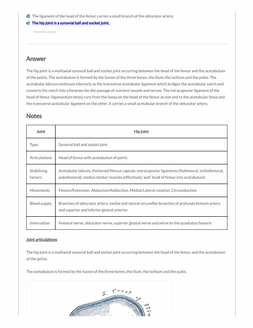
d) The ligament of the head of the femur carries a small branch of the obturator artery.
e) The hip joint is a synovial ball and socket joint.
Answer
The hip joint is a multiaxial synovial ball and socket joint occurring between the head of the femur and the acetabulum
of the pelvis. The acetabulum is formed by the fusion of the three bones, the ilium, the ischium and the pubis. The
acetabular labrum continues inferiorly as the transverse acetabular ligament which bridges the acetabular notch and
converts the notch into a foramen for the passage of nutrient vessels and nerves. The intracapsular ligament of the
head of femur (ligamentum teres) runs from the fovea on the head of the femur at one end to the acetabular fossa and
the transverse acetabular ligament on the other. It carries a small acetabular branch of the obturator artery.
Notes
Joint Hip joint
Type Synovial ball and socket joint
Articulations Head of femur with acetabulum of pelvis
Stabilising
factors
Acetabular labrum, thickened brous capsule, extracapsular ligaments (iliofemoral, ischiofemoral,
pubofemoral), medial rotator muscles (effectively ‘pull’ head of femur into acetabulum)
Movements Flexion/Extension, Abduction/Adduction, Medial/Lateral rotation, Circumduction
Blood supply Branches of obturator artery, medial and lateral circum ex branches of profunda femoris artery
and superior and inferior gluteal arteries
Innervation Femoral nerve, obturator nerve, superior gluteal nerve and nerve to the quadratus femoris
Joint articulations
The hip joint is a multiaxial synovial ball and socket joint occurring between the head of the femur and the acetabulum
of the pelvis.
The acetabulum is formed by the fusion of the three bones, the ilium, the ischium and the pubis.
Something wrong?

By Henry Vandyke Carter [Public domain], via Wikimedia Commons
The rim of the acetabulum is raised slightly by the brocartilaginous acetabular labrum which increases its depth,
improving stability of the joint. The acetabular labrum continues inferiorly as the transverse acetabular ligament which
bridges the acetabular notch and converts the notch into a foramen for the passage of nutrient vessels and nerves.
Ligaments
The intracapsular ligament of the head of femur (ligamentum teres) runs from the fovea on the head of the femur at
one end to the acetabular fossa and the transverse acetabular ligament on the other. It carries a small acetabular
branch of the obturator artery.

Modi ed by FRCEM Success. Original by Henry
Vandyke Carter [Public domain], via Wikimedia
Commons By Henry Vandyke Carter [Public domain], via
Wikimedia Commons
By Henry Vandyke Carter [Public domain], via Wikimedia Commons
There are three main stabilising extracapsular ligaments; the iliofemoral (the largest and strongest ligament), the
pubofemoral and the ischiofemoral ligament.
The iliofemoral ligament is anterosuperior to the hip joint, attached proximally to the ilium just below the
anterior inferior iliac spine and distally to the intertrochanteric line of the femur. This ligament speci cally
prevents hyperextension and lateral rotation of the hip joint whilst standing.
The pubofemoral ligament is anteroinferior to the hip joint, attached to the pelvis at the iliopubic eminence
and adjacent bone and blending distally with the articular capsule. This ligament prevents excessive abduction
and extension of the femur at the hip joint.
The ischiofemoral ligament is posterior to the hip joint, attached medially to the ischium and laterally to the
greater trochanter. This ligament prevents excessive extension and medial rotation of the femur at the hip
joint.
Blood supply

The hip joint receives its blood supply primarily from branches of the obturator artery, the medial and lateral
circum ex arteries (branches of the profunda femoris artery) and the superior and inferior gluteal arteries. The
articular branches of these vessels form a network around the joint.
Innervation
The hip joint is innervated by articular branches from the femoral nerve (anteriorly), obturator nerve (inferiorly),
superior gluteal nerve (superiorly), and the nerve to the quadratus femoris (posteriorly).
Joint movements
The hip joint allows the movements of exion and extension, abduction and adduction, medial and lateral rotation and
circumduction.
Movement Muscles Involved
Flexion Sartorius, Iliacus, Psoas major, Pectineus, Rectus femoris
Extension Hamstrings, Gluteus maximus
Abduction Gluteus maximus, medius and minimus, Obturator internus, Gemelli, Piriformis, Sartorius
Adduction Adductor longus, magnus and brevis, Gracilis, Pectineus
Medial
rotation
Gluteus medius and minimus, Adductor longus, magnus and brevis, Semitendinosus and
Semimembranosus
Lateral
rotation
Obturator externus, Sartorius, Iliacus, Psoas major, Biceps femoris, Piriformis, Gluteus maximus,
Obturator internus, Gemelli, Quadriceps femoris
A 67 year old man presents to the ED complaining of pain and swelling to the posterior knee.
On examination you note a tender swelling in the popliteal fossa. The popliteal fossa is bordered
superiorly by which of the following muscles:
a) The semimembranosus and the semitendinosus
b) The biceps femoris and the plantaris
c) The biceps femoris and the semimembranosus
d) The biceps femoris alone
e) The semitendinosus alone
Answer
Something wrong?
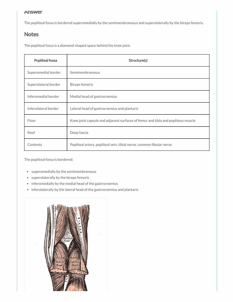
Answer
The popliteal fossa is bordered superomedially by the semimembranosus and superolaterally by the biceps femoris.
Notes
The popliteal fossa is a diamond-shaped space behind the knee joint.
Popliteal fossa Structure(s)
Superomedial border Semimembranosus
Superolateral border Biceps femoris
Inferomedial border Medial head of gastrocnemius
Inferolateral border Lateral head of gastrocnemius and plantaris
Floor Knee joint capsule and adjacent surfaces of femur and tibia and popliteus muscle
Roof Deep fascia
Contents Popliteal artery, popliteal vein, tibial nerve, common bular nerve
The popliteal fossa is bordered:
superomedially by the semimembranosus
superolaterally by the biceps femoris
inferomedially by the medial head of the gastrocnemius
inferolaterally by the lateral head of the gastrocnemius and plantaris
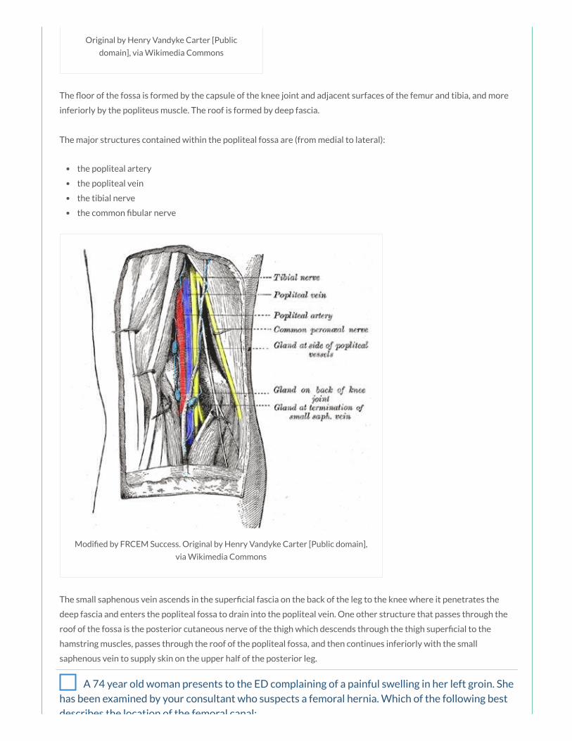
Original by Henry Vandyke Carter [Public
domain], via Wikimedia Commons
The oor of the fossa is formed by the capsule of the knee joint and adjacent surfaces of the femur and tibia, and more
inferiorly by the popliteus muscle. The roof is formed by deep fascia.
The major structures contained within the popliteal fossa are (from medial to lateral):
the popliteal artery
the popliteal vein
the tibial nerve
the common bular nerve
Modi ed by FRCEM Success. Original by Henry Vandyke Carter [Public domain],
via Wikimedia Commons
The small saphenous vein ascends in the super cial fascia on the back of the leg to the knee where it penetrates the
deep fascia and enters the popliteal fossa to drain into the popliteal vein. One other structure that passes through the
roof of the fossa is the posterior cutaneous nerve of the thigh which descends through the thigh super cial to the
hamstring muscles, passes through the roof of the popliteal fossa, and then continues inferiorly with the small
saphenous vein to supply skin on the upper half of the posterior leg.
A 74 year old woman presents to the ED complaining of a painful swelling in her left groin. She
has been examined by your consultant who suspects a femoral hernia. Which of the following best
describes the location of the femoral canal:
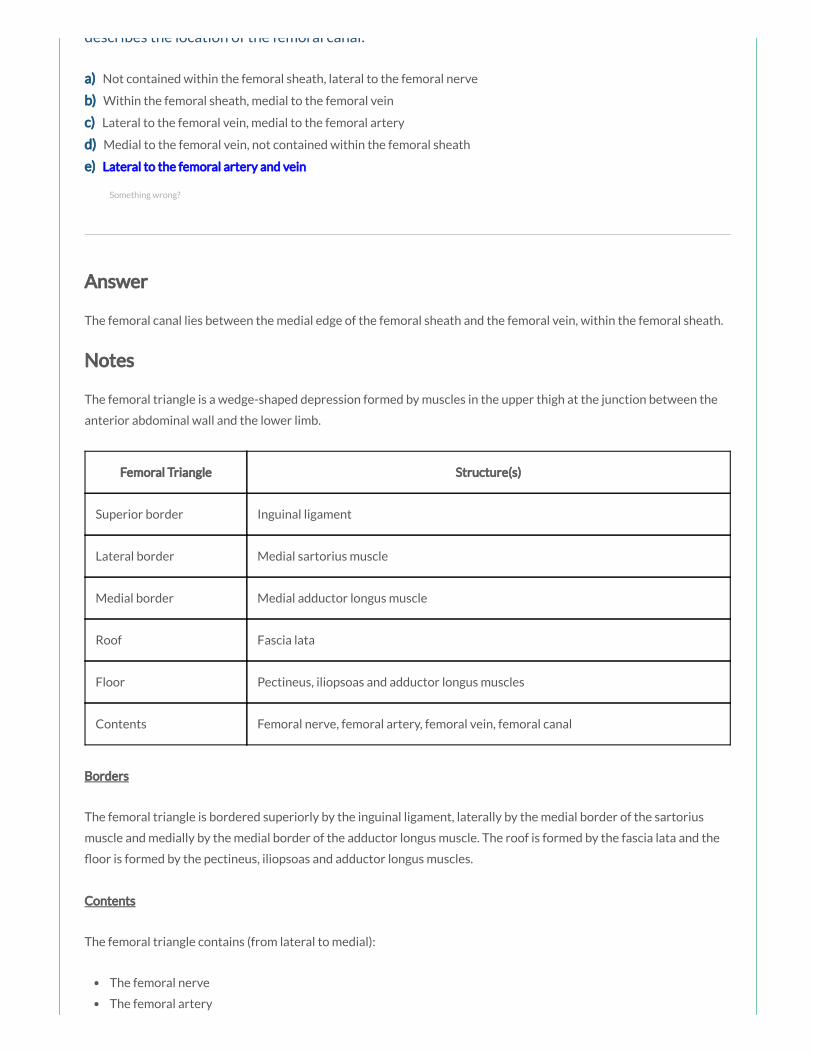
describes the location of the femoral canal:
a) Not contained within the femoral sheath, lateral to the femoral nerve
b) Within the femoral sheath, medial to the femoral vein
c) Lateral to the femoral vein, medial to the femoral artery
d) Medial to the femoral vein, not contained within the femoral sheath
e) Lateral to the femoral artery and vein
Answer
The femoral canal lies between the medial edge of the femoral sheath and the femoral vein, within the femoral sheath.
Notes
The femoral triangle is a wedge-shaped depression formed by muscles in the upper thigh at the junction between the
anterior abdominal wall and the lower limb.
Femoral Triangle Structure(s)
Superior border Inguinal ligament
Lateral border Medial sartorius muscle
Medial border Medial adductor longus muscle
Roof Fascia lata
Floor Pectineus, iliopsoas and adductor longus muscles
Contents Femoral nerve, femoral artery, femoral vein, femoral canal
Borders
The femoral triangle is bordered superiorly by the inguinal ligament, laterally by the medial border of the sartorius
muscle and medially by the medial border of the adductor longus muscle. The roof is formed by the fascia lata and the
oor is formed by the pectineus, iliopsoas and adductor longus muscles.
Contents
The femoral triangle contains (from lateral to medial):
The femoral nerve
The femoral artery
Something wrong?

By Henry Vandyke Carter [Public domain], via
Wikimedia Commons
By Henry Vandyke Carter [Public domain], via
Wikimedia Commons
The femoral vein
The femoral canal
Femoral sheath
The femoral sheath originates as a prolongation of extraperitoneal fascia and encloses the femoral artery, femoral vein
and associated lymphatic vessels (contained within the femoral canal). The femoral nerve does NOT lie within the
sheath, but instead lies lateral to it. The femoral sheath terminates inferiorly by blending with the adventitia of the
femoral vessels approximately 4 cm inferior of the inguinal ligament.
Femoral canal
The femoral canal lies between the medial edge of the femoral sheath and the femoral vein. The femoral canal contains
loose connective tissue, lymphatic vessels, deep inguinal lymph nodes and empty space. The femoral canal allows the
femoral vein to expand, for example, when venous return from the leg is increased, or when increased intra-abdominal
pressure causes a temporary stasis.
Femoral ring
The femoral ring is formed by the proximal abdominal opening of the femoral canal, and is important clinically, as it is a
common site of hernia. It is bordered anteriorly by the inguinal ligament, posteriorly by the pectineal ligament,
medially by the lacunar ligament and laterally by the femoral vein.
Femoral ring Structure(s)

Femoral ring Structure(s)
Anterior border Inguinal ligament
Posterior border Pectineal ligament
Medial border Lacunar ligament
Lateral border Femoral vein
By Henry Vandyke Carter [Public domain], via Wikimedia Commons
You are part of the team in the resuscitation area. You have been pre-alerted to the arrival of
a 35 year old man who has had a cardiac arrest at a football match. On arrival the team leader asks
you to perform a femoral pulse check. At which of the following sites is the femoral artery best
palpated:
a) Inferior to the inguinal ligament, just medial to the femoral vein
b) Inferior to the inguinal ligament, just medial to the femoral canal
c) Just inferior to the inguinal ligament at the mid-inguinal point
d) In the femoral triangle, just superior to the inguinal ligament
e) At the mid-inguinal point just lateral to the femoral nerve
Something wrong?

Answer
The femoral artery can be palpated in the femoral triangle as it passes over the femoral head, just inferior to the
inguinal ligament, midway between the anterior superior iliac spine and the pubic symphysis (at the mid-inguinal
point). Remember NAVY (from lateral to medial): femoral Nerve, femoral Artery, femoral Vein, Y-fronts (i.e. the
midline).
Notes
The major artery supplying the lower limb is the femoral artery. The femoral artery is the continuation of the external
iliac artery, beginning as the vessel passes under the inguinal ligament to enter the femoral triangle in the anterior
thigh.
The femoral artery can be palpated in the femoral triangle as it passes over the femoral head, just inferior to the
inguinal ligament, midway between the anterior superior iliac spine and the pubic symphysis (at the mid-inguinal
point). The femoral vein lies immediately medial to this pulsation, which is an important landmark for central venous
line insertion. Medial to the femoral vein is the femoral canal which contains lymphatics and lies immediately lateral to
the pubic tubercle. The femoral nerve lies lateral to the femoral artery.
The femoral artery gives rise to the deep profunda femoris artery in the femoral triangle which is a major source of
blood supply to the medial and posterior compartments of the thigh and the proximal femur.
After exiting the femoral triangle, the femoral artery continues down the anterior surface of the thigh via the adductor
canal. During its descent it supplies the anterior thigh, giving rise to numerous super cial cutaneous branches.
The femoral artery becomes the popliteal artery after entering the posterior compartment of the thigh through the
adductor hiatus just proximal to the knee.
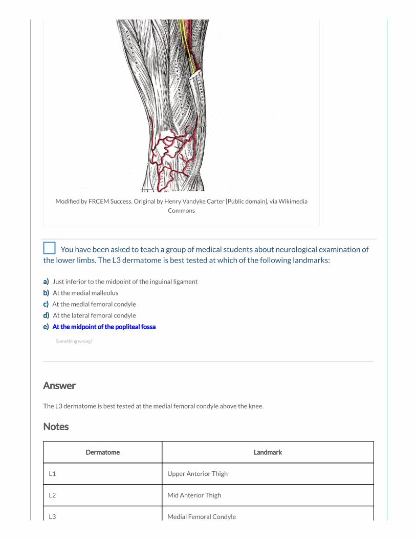
Modi ed by FRCEM Success. Original by Henry Vandyke Carter [Public domain], via Wikimedia
Commons
You have been asked to teach a group of medical students about neurological examination of
the lower limbs. The L3 dermatome is best tested at which of the following landmarks:
a) Just inferior to the midpoint of the inguinal ligament
b) At the medial malleolus
c) At the medial femoral condyle
d) At the lateral femoral condyle
e) At the midpoint of the popliteal fossa
Answer
The L3 dermatome is best tested at the medial femoral condyle above the knee.
Notes
Dermatome Landmark
L1 Upper Anterior Thigh
L2 Mid Anterior Thigh
L3 Medial Femoral Condyle
Something wrong?

L4 Medial Malleolus
L5 Dorsum 3rd MTP Joint
S1 Lateral Heel
S2 Popliteal Fossa
S3 Ischial Tuberosity
S5 Perianal Area
The T12 dermatome is best tested at the midclavicular line, over the midpoint of the inguinal ligament.
The L1 dermatome is best tested on the upper anterior thigh, at a point midway between the key sensory
points for T12 and L2.
The L2 dermatome is best tested on the anteromedial thigh, at the midpoint drawn on an imaginary line
connecting the midpoint of the inguinal ligament and the medial femoral condyle.
The L3 dermatome is best tested at the medial femoral condyle above the knee.
The L4 dermatome is best tested over the medial malleolus.
The L5 dermatome is best tested on the dorsum of the foot at the third metatarsophalangeal joint.
The S1 dermatome is best tested on the lateral aspect of the calcaneus.
The S2 dermatome is best tested at the midpoint of the popliteal fossa.
The S3 dermatome is best tested over the ischial tuberosity or infragluteal fold (depending on the patient their
skin can move up, down or laterally over the ischii).
The S4/S5 dermatome is best tested in the perianal area, less than one cm lateral to the mucocutaneous
junction.
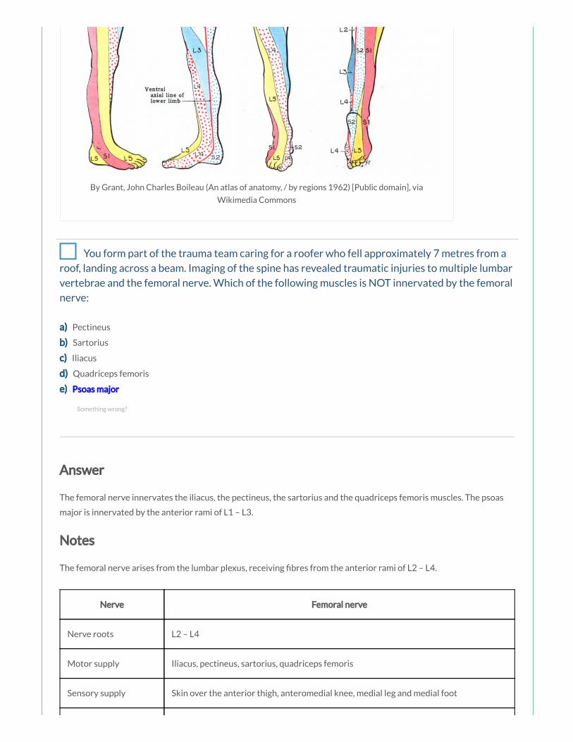
By Grant, John Charles Boileau (An atlas of anatomy, / by regions 1962) [Public domain], via
Wikimedia Commons
You form part of the trauma team caring for a roofer who fell approximately 7 metres from a
roof, landing across a beam. Imaging of the spine has revealed traumatic injuries to multiple lumbar
vertebrae and the femoral nerve. Which of the following muscles is NOT innervated by the femoral
nerve:
a) Pectineus
b) Sartorius
c) Iliacus
d) Quadriceps femoris
e) Psoas major
Answer
The femoral nerve innervates the iliacus, the pectineus, the sartorius and the quadriceps femoris muscles. The psoas
major is innervated by the anterior rami of L1 – L3.
Notes
The femoral nerve arises from the lumbar plexus, receiving bres from the anterior rami of L2 – L4.
Nerve Femoral nerve
Nerve roots L2 – L4
Motor supply Iliacus, pectineus, sartorius, quadriceps femoris
Sensory supply Skin over the anterior thigh, anteromedial knee, medial leg and medial foot
Something wrong?
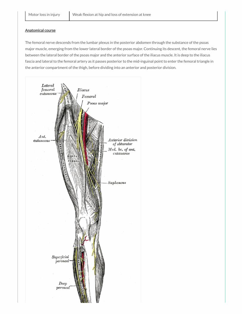
Motor loss in injury Weak exion at hip and loss of extension at knee
Anatomical course
The femoral nerve descends from the lumbar plexus in the posterior abdomen through the substance of the psoas
major muscle, emerging from the lower lateral border of the psoas major. Continuing its descent, the femoral nerve lies
between the lateral border of the psoas major and the anterior surface of the iliacus muscle. It is deep to the iliacus
fascia and lateral to the femoral artery as it passes posterior to the mid-inguinal point to enter the femoral triangle in
the anterior compartment of the thigh, before dividing into an anterior and posterior division.

By Henry Vandyke Carter [Public domain], via Wikimedia
Commons
Branches
In the abdomen it gives rise to branches that innervate the iliacus and pectineus muscles.
The anterior division gives off anterior cutaneous branches (supplying skin over the anterior and medial thigh) and
muscular branches (innervating the sartorius).
The posterior division gives off muscular branches (innervating the quadriceps femoris muscles) and articular branches
(supplying the hip and knee joint), before continuing as the saphenous nerve (supplying skin over the anteromedial
knee and the medial side of the leg and foot).
Branch Innervation
Muscular branches in abdomen Iliacus and pectineus
Anterior cutaneous branches Skin over anterior thigh
Anterior muscular branches Sartorius
Posterior muscular branches Quadriceps femoris muscles
Posterior articular branches Hip and knee joint
Saphenous nerve Skin over anteromedial knee, medial side of leg and foot
Motor and sensory function
Muscle Action(s)
Sartorius Flexion, abduction and lateral rotation at hip and exion at knee
Iliacus (red) Flexion and lateral rotation at hip
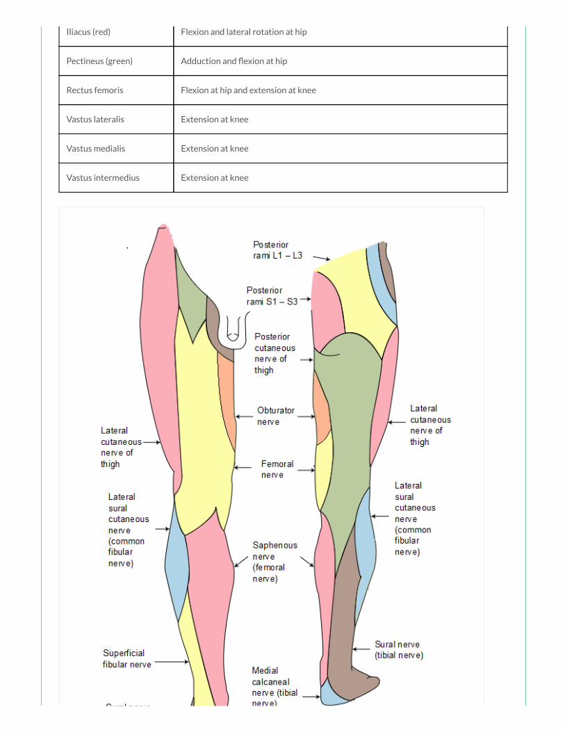
Iliacus (red) Flexion and lateral rotation at hip
Pectineus (green) Adduction and exion at hip
Rectus femoris Flexion at hip and extension at knee
Vastus lateralis Extension at knee
Vastus medialis Extension at knee
Vastus intermedius Extension at knee

Modi ed by FRCEM Success. Original by Henry Vandyke Carter [Public domain], via Wikimedia Commons
Imaging of a patient who has been involved in a car accident has shown damage to the nerve
supplying the pectineus muscle. Which nerve is involved:
a) Obturator nerve
b) Femoral nerve
c) Anterior rami of spinal nerves L1 – L3
d) Pudendal nerve
e) Inferior gluteal nerve
Answer
The pectineus (green) is innervated by the femoral nerve (L2, L3).
Notes
The sartorius, iliopsoas, pectineus and rectus femoris are the primary exors of the thigh at the hip joint.
Muscle Actions Innervation
Sartorius (blue) Flexion, abduction and lateral rotation at hip and exion at
knee
Femoral nerve (L2, L3)
Iliacus (red) Flexion and lateral rotation at hip Femoral nerve (L2, L3)
Psoas major
(yellow)
Flexion and lateral rotation at hip Anterior rami L1 – L3
Pectineus (green) Adduction and exion at hip Femoral nerve (L2, L3)
Rectus femoris Flexion at hip and extension at knee Femoral nerve (L2 –
L4)
The sartorius (blue) is innervated by the femoral nerve (L2, L3). It acts to ex the thigh at the hip joint and ex the leg at
the knee joint. It also abducts the thigh and rotates it laterally, as when resting the foot on the opposite knee when
sitting.
Something wrong?
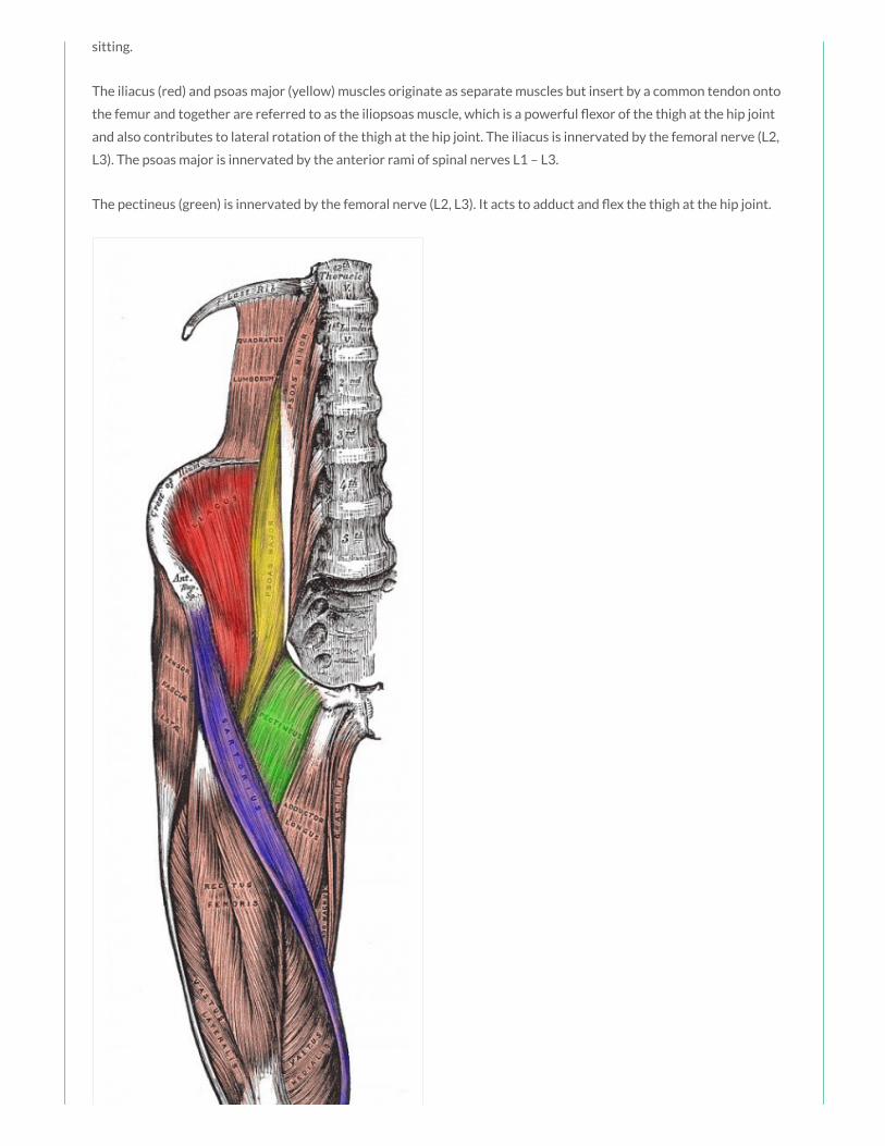
sitting.
The iliacus (red) and psoas major (yellow) muscles originate as separate muscles but insert by a common tendon onto
the femur and together are referred to as the iliopsoas muscle, which is a powerful exor of the thigh at the hip joint
and also contributes to lateral rotation of the thigh at the hip joint. The iliacus is innervated by the femoral nerve (L2,
L3). The psoas major is innervated by the anterior rami of spinal nerves L1 – L3.
The pectineus (green) is innervated by the femoral nerve (L2, L3). It acts to adduct and ex the thigh at the hip joint.

Modi ed by FRCEM Success. Original by Henry
Vandyke Carter [Public domain], via Wikimedia
Commons
A 43 year old window cleaner is brought to the ED after falling from a 5 metre platform.
Imaging has shown multiple vertebral fractures and you are concerned about possible neurological
injuries including the obturator nerve. Which of the following muscles is NOT innervated by the
obturator nerve:
a) Adductor longus
b) Adductor brevis
c) Adductor magnus
d) Obturator externus
e) Obturator internus
Answer
The obturator nerve innervates all of the muscles in the medial compartment of the thigh, except for the pectineus and
the hamstrings portion of the adductor magnus muscle. The obturator internus muscle is a deep gluteal muscle
innervated by the nerve to the obturator internus.
Notes
The obturator nerve arises from the lumbar plexus, formed from the anterior rami of L2 – L4.
Nerve Obturator nerve
Nerve
roots
L2 – L4
Motor
supply
Medial thigh muscles (adductor longus, brevis and magnus, gracilis, obturator externus)
Something wrong?

Sensory
supply
Upper medial thigh
Motor loss
in injury
Weak adduction of hip and dif culty walking with lateral swinging of limb during walking (due to
unopposed abduction), loss of sensation over upper medial thigh
The obturator nerve descends along the posterior abdominal wall, passes through the pelvic cavity and enters the
medial thigh by passing through the obturator canal.
The obturator nerve innervates all of the muscles of the medial compartment of the thigh (except for the hamstring
part of the adductor longus, and the pectineus muscle which are innervated by the sciatic and the femoral nerves
respectively). It also gives off a cutaneous branch that supplies skin on the medial side of the upper thigh.
Muscle Action(s)
Adductor longus Adduction and medial rotation at hip
Adductor brevis Adduction and medial rotation at hip
Adductor magnus Adduction and medial rotation at hip
Obturator externus Lateral rotation at hip
Gracilis Adduction at hip and exion at knee

Modi ed by FRCEM Success. Original by Henry Vandyke Carter [Public domain], via Wikimedia Commons
A 32 year old builder is brought to the ED after falling through a glass sky light onto the oor
below. A primary survey has been performed and the patient is stable enough to undergo imaging.
Imaging shows multiple injuries including a transection of the nerve supplying the adductor longus,
magnus and brevis muscles. Which of the following movements is the patient likely to nd dif cult:
a) Adduction and lateral rotation of the thigh
b) Adduction and medial rotation of the thigh
c) Abduction and exion of the thigh
d) Adduction and extension of the thigh
e) Adduction and abduction of the thigh
Answer
The adductor longus (red), brevis (green) and magnus (blue) are the prime adductors of the thigh at the hip joint and
also assist in medial rotation.
Notes
There are six muscles in the medial compartment of the thigh: the gracilis, the pectineus, the adductor longus, brevis
and magnus and the obturator externus. Other than the obturator externus, these muscles all act to adduct the thigh at
the hip joint.
Something wrong?
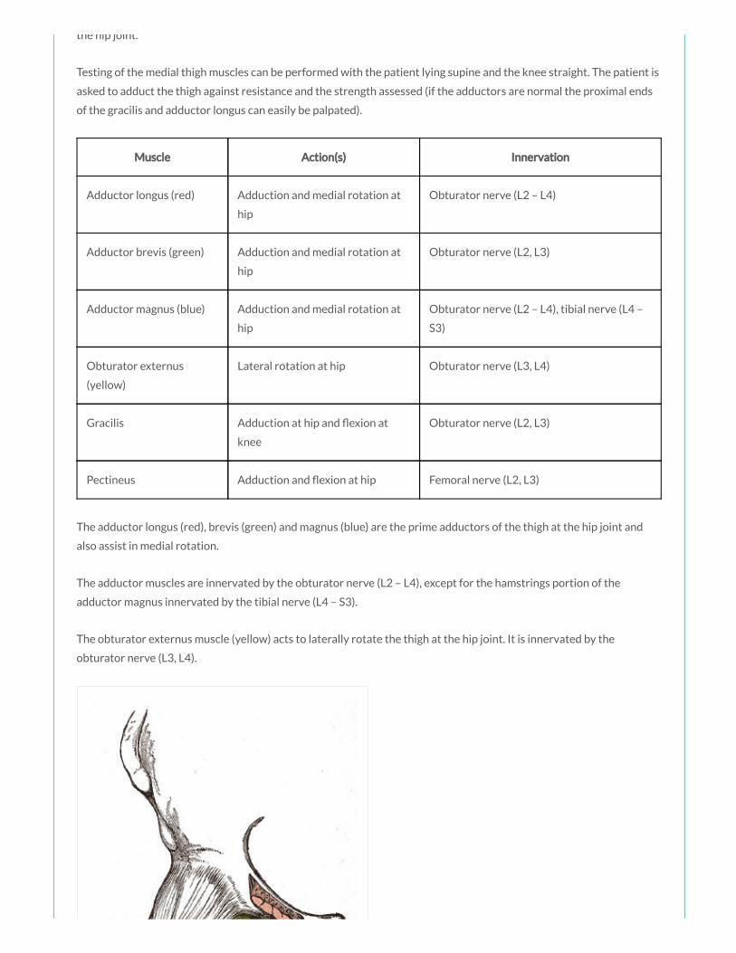
the hip joint.
Testing of the medial thigh muscles can be performed with the patient lying supine and the knee straight. The patient is
asked to adduct the thigh against resistance and the strength assessed (if the adductors are normal the proximal ends
of the gracilis and adductor longus can easily be palpated).
Muscle Action(s) Innervation
Adductor longus (red) Adduction and medial rotation at
hip
Obturator nerve (L2 – L4)
Adductor brevis (green) Adduction and medial rotation at
hip
Obturator nerve (L2, L3)
Adductor magnus (blue) Adduction and medial rotation at
hip
Obturator nerve (L2 – L4), tibial nerve (L4 –
S3)
Obturator externus
(yellow)
Lateral rotation at hip Obturator nerve (L3, L4)
Gracilis Adduction at hip and exion at
knee
Obturator nerve (L2, L3)
Pectineus Adduction and exion at hip Femoral nerve (L2, L3)
The adductor longus (red), brevis (green) and magnus (blue) are the prime adductors of the thigh at the hip joint and
also assist in medial rotation.
The adductor muscles are innervated by the obturator nerve (L2 – L4), except for the hamstrings portion of the
adductor magnus innervated by the tibial nerve (L4 – S3).
The obturator externus muscle (yellow) acts to laterally rotate the thigh at the hip joint. It is innervated by the
obturator nerve (L3, L4).
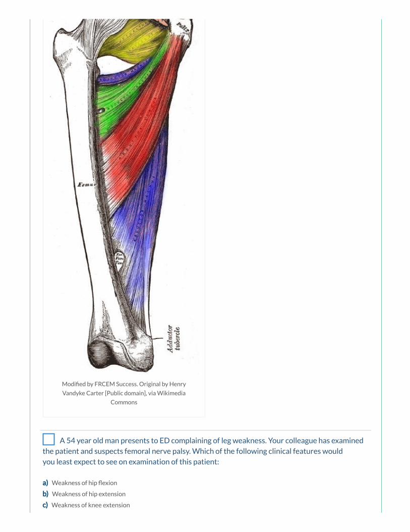
Modi ed by FRCEM Success. Original by Henry
Vandyke Carter [Public domain], via Wikimedia
Commons
A 54 year old man presents to ED complaining of leg weakness. Your colleague has examined
the patient and suspects femoral nerve palsy. Which of the following clinical features would
you least expect to see on examination of this patient:
a) Weakness of hip exion
b) Weakness of hip extension
c) Weakness of knee extension
d) Loss of sensation over anterior thigh
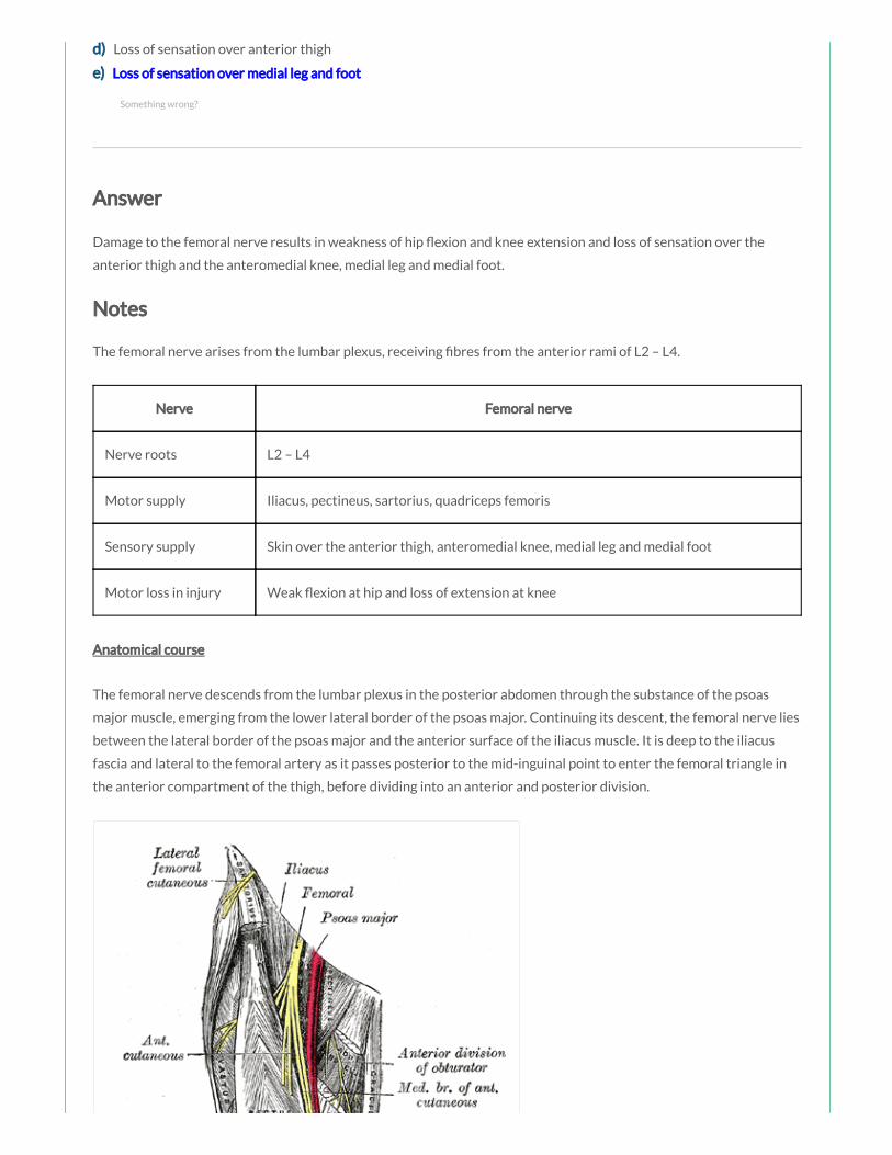
d) Loss of sensation over anterior thigh
e) Loss of sensation over medial leg and foot
Answer
Damage to the femoral nerve results in weakness of hip exion and knee extension and loss of sensation over the
anterior thigh and the anteromedial knee, medial leg and medial foot.
Notes
The femoral nerve arises from the lumbar plexus, receiving bres from the anterior rami of L2 – L4.
Nerve Femoral nerve
Nerve roots L2 – L4
Motor supply Iliacus, pectineus, sartorius, quadriceps femoris
Sensory supply Skin over the anterior thigh, anteromedial knee, medial leg and medial foot
Motor loss in injury Weak exion at hip and loss of extension at knee
Anatomical course
The femoral nerve descends from the lumbar plexus in the posterior abdomen through the substance of the psoas
major muscle, emerging from the lower lateral border of the psoas major. Continuing its descent, the femoral nerve lies
between the lateral border of the psoas major and the anterior surface of the iliacus muscle. It is deep to the iliacus
fascia and lateral to the femoral artery as it passes posterior to the mid-inguinal point to enter the femoral triangle in
the anterior compartment of the thigh, before dividing into an anterior and posterior division.
Something wrong?

By Henry Vandyke Carter [Public domain], via Wikimedia
Commons
Branches
In the abdomen it gives rise to branches that innervate the iliacus and pectineus muscles.
The anterior division gives off anterior cutaneous branches (supplying skin over the anterior and medial thigh) and
muscular branches (innervating the sartorius).
The posterior division gives off muscular branches (innervating the quadriceps femoris muscles) and articular branches
(supplying the hip and knee joint), before continuing as the saphenous nerve (supplying skin over the anteromedial
knee and the medial side of the leg and foot).
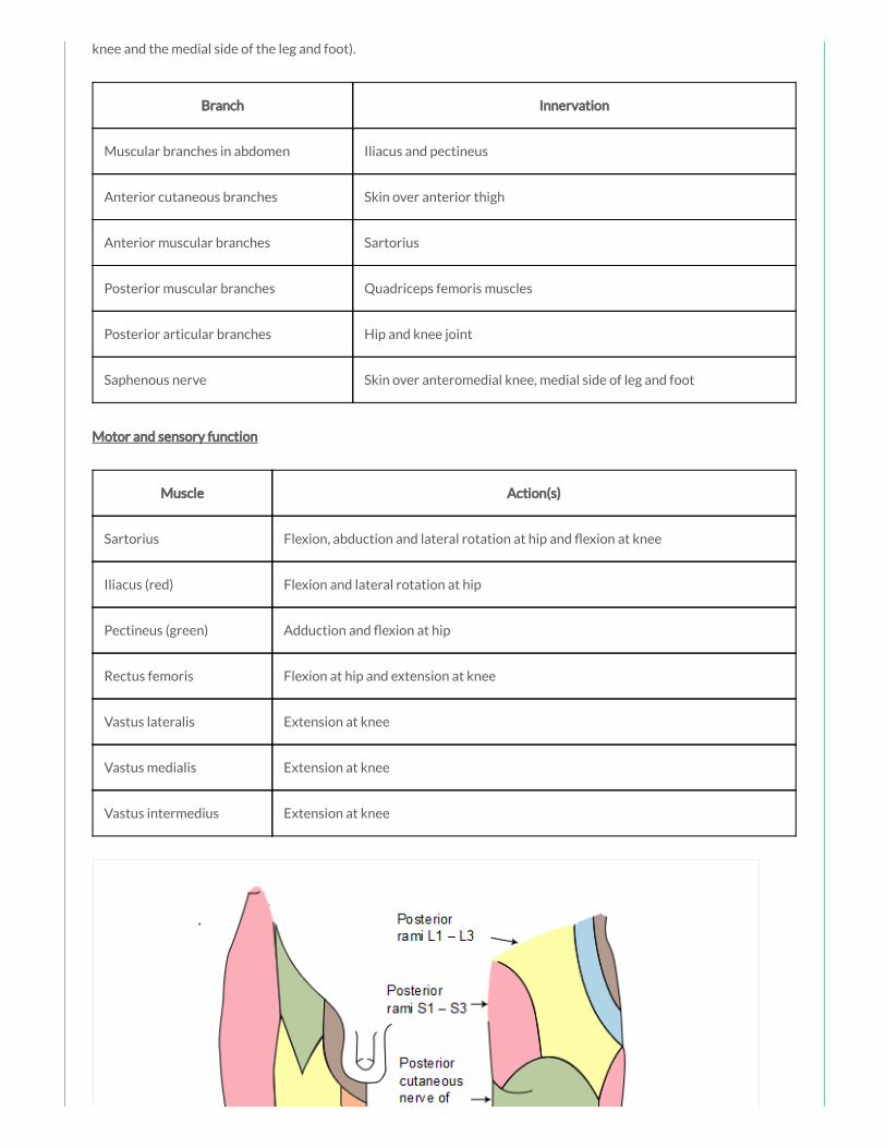
knee and the medial side of the leg and foot).
Branch Innervation
Muscular branches in abdomen Iliacus and pectineus
Anterior cutaneous branches Skin over anterior thigh
Anterior muscular branches Sartorius
Posterior muscular branches Quadriceps femoris muscles
Posterior articular branches Hip and knee joint
Saphenous nerve Skin over anteromedial knee, medial side of leg and foot
Motor and sensory function
Muscle Action(s)
Sartorius Flexion, abduction and lateral rotation at hip and exion at knee
Iliacus (red) Flexion and lateral rotation at hip
Pectineus (green) Adduction and exion at hip
Rectus femoris Flexion at hip and extension at knee
Vastus lateralis Extension at knee
Vastus medialis Extension at knee
Vastus intermedius Extension at knee
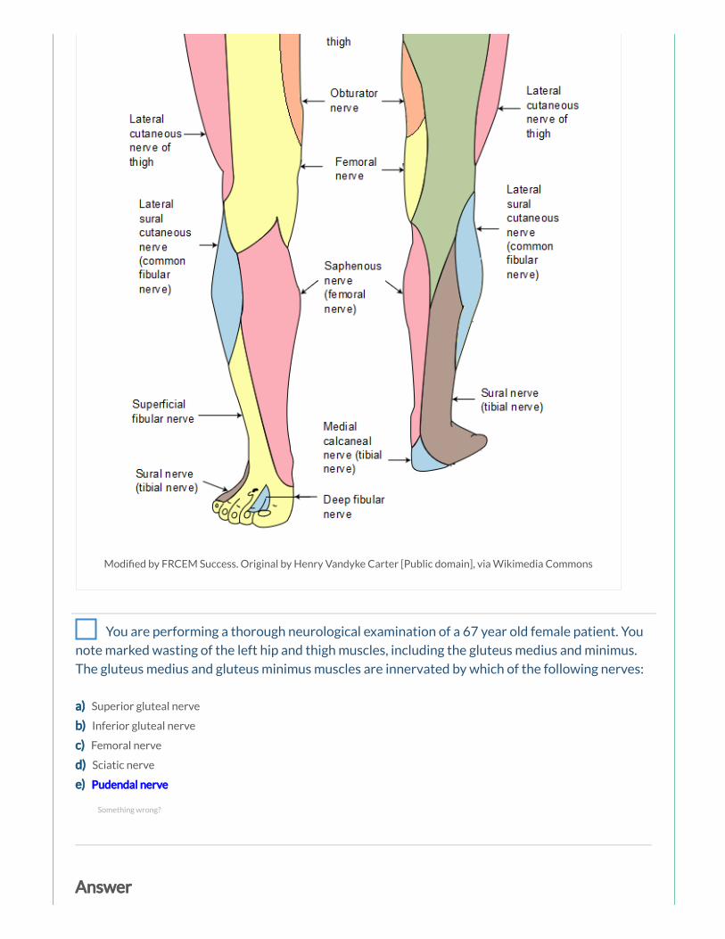
Modi ed by FRCEM Success. Original by Henry Vandyke Carter [Public domain], via Wikimedia Commons
You are performing a thorough neurological examination of a 67 year old female patient. You
note marked wasting of the left hip and thigh muscles, including the gluteus medius and minimus.
The gluteus medius and gluteus minimus muscles are innervated by which of the following nerves:
a) Superior gluteal nerve
b) Inferior gluteal nerve
c) Femoral nerve
d) Sciatic nerve
e) Pudendal nerve
Answer
Something wrong?
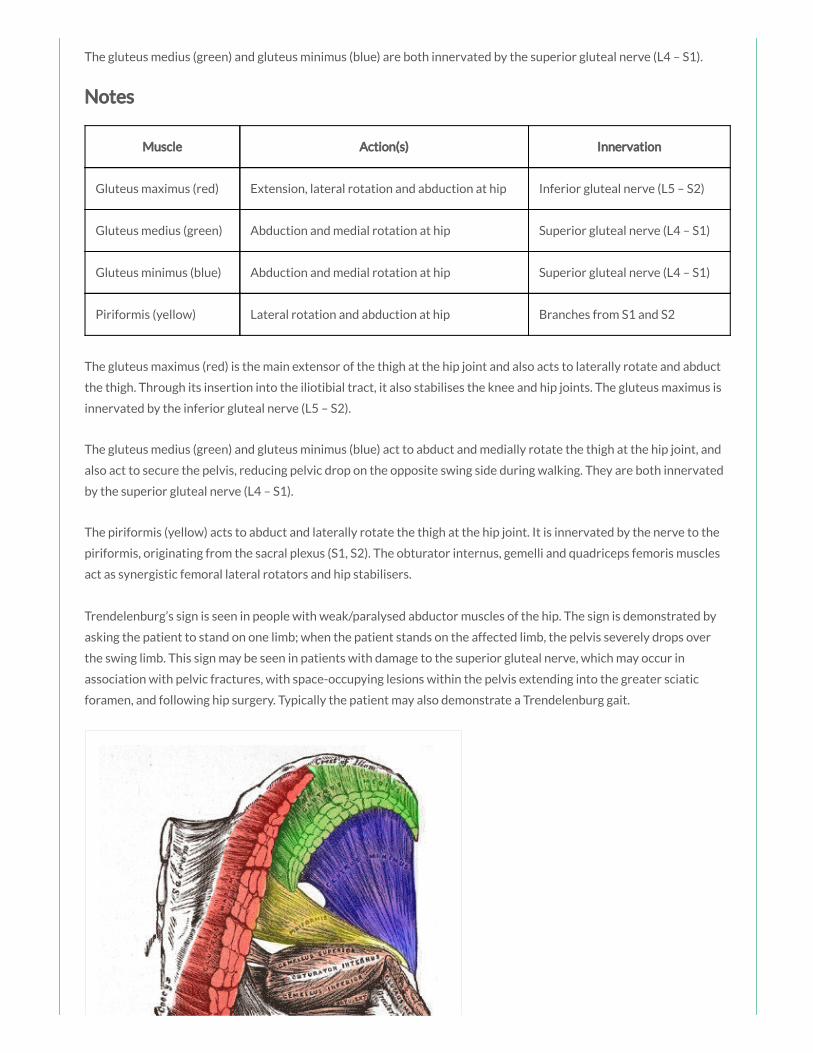
The gluteus medius (green) and gluteus minimus (blue) are both innervated by the superior gluteal nerve (L4 – S1).
Notes
Muscle Action(s) Innervation
Gluteus maximus (red) Extension, lateral rotation and abduction at hip Inferior gluteal nerve (L5 – S2)
Gluteus medius (green) Abduction and medial rotation at hip Superior gluteal nerve (L4 – S1)
Gluteus minimus (blue) Abduction and medial rotation at hip Superior gluteal nerve (L4 – S1)
Piriformis (yellow) Lateral rotation and abduction at hip Branches from S1 and S2
The gluteus maximus (red) is the main extensor of the thigh at the hip joint and also acts to laterally rotate and abduct
the thigh. Through its insertion into the iliotibial tract, it also stabilises the knee and hip joints. The gluteus maximus is
innervated by the inferior gluteal nerve (L5 – S2).
The gluteus medius (green) and gluteus minimus (blue) act to abduct and medially rotate the thigh at the hip joint, and
also act to secure the pelvis, reducing pelvic drop on the opposite swing side during walking. They are both innervated
by the superior gluteal nerve (L4 – S1).
The piriformis (yellow) acts to abduct and laterally rotate the thigh at the hip joint. It is innervated by the nerve to the
piriformis, originating from the sacral plexus (S1, S2). The obturator internus, gemelli and quadriceps femoris muscles
act as synergistic femoral lateral rotators and hip stabilisers.
Trendelenburg’s sign is seen in people with weak/paralysed abductor muscles of the hip. The sign is demonstrated by
asking the patient to stand on one limb; when the patient stands on the affected limb, the pelvis severely drops over
the swing limb. This sign may be seen in patients with damage to the superior gluteal nerve, which may occur in
association with pelvic fractures, with space-occupying lesions within the pelvis extending into the greater sciatic
foramen, and following hip surgery. Typically the patient may also demonstrate a Trendelenburg gait.

Modi ed by FRCEM Success. Original by Henry Vandyke Carter
[Public domain], via Wikimedia Commons
A 54 year old man presents to the ED complaining of a numbness over over the posterior
thigh. The skin in this area is primarily supplied by which of the following nerves:
a) The posterior cutaneous nerve of the thigh, branch of the sciatic nerve
b) The posterior cutaneous nerve of the thigh from the lumbar plexus
c) The posterior cutaneous nerve of the thigh from the sacral plexus
d) Posterior cutaneous branches from the femoral nerve
e) Posterior cutaneous branches from the obturator nerve
Something wrong?

Answer
The posterior cutaneous nerve of the thigh, nerve from the sacral plexus, supplies skin over the posterior thigh and
upper leg.
Notes
Nerve Origin Skin supplied
Lateral cutaneous nerve of thigh Lumbar plexus (L2, L3) Lateral thigh
Anterior cutaneous nerve Femoral nerve Anterior thigh and anteromedial knee
Cutaneous branch Obturator nerve Medial thigh
Posterior cutaneous nerve of thigh Sacral plexus (S1 – S3) Posterior thigh and upper leg
Saphenous nerve Femoral nerve Anteromedial knee, medial leg and foot
Lateral sural cutaneous nerve Common bular nerve Upper lateral leg
Super cial bular nerve Common bular nerve Lower anterolateral leg and dorsum of foot
Deep bular nerve Common bular nerve Webspace between 1st and 2nd toe
Sural nerve Tibial nerve Lower posterolateral leg, heel and foot
Medial calcaneal nerve Tibial nerve Medial side and sole of heel
Medial and lateral plantar nerves Tibial nerve Sole of foot anterior to heel
Thigh:
The skin over the hip and gluteal region is supplied by the posterior and anterior rami of lumbosacral nerves
(L1 – S3).
The lateral cutaneous nerve of the thigh, nerve from the lumbar plexus, supplies skin over the lateral thigh.
The anterior cutaneous nerve, branch of the femoral nerve, supplies skin over the anterior thigh and
anteromedial knee.
The obturator nerve supplies skin over the upper medial thigh.
The posterior cutaneous nerve of the thigh, nerve from the sacral plexus, supplies skin over the posterior thigh
and upper leg.
Leg:
Something wrong?
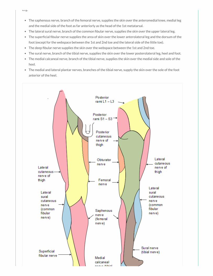
Leg:
The saphenous nerve, branch of the femoral nerve, supplies the skin over the anteromedial knee, medial leg
and the medial side of the foot as far anteriorly as the head of the 1st metatarsal.
The lateral sural nerve, branch of the common bular nerve, supplies the skin over the upper lateral leg.
The super cial bular nerve supplies the area of skin over the lower anterolateral leg and the dorsum of the
foot (except for the webspace between the 1st and 2nd toe and the lateral side of the little toe).
The deep bular nerve supplies the skin over the webspace between the 1st and 2nd toe.
The sural nerve, branch of the tibial nerve, supplies the skin over the lower posterolateral leg, heel and foot.
The medial calcaneal nerve, branch of the tibial nerve, supplies the skin over the medial side and sole of the
heel.
The medial and lateral plantar nerves, branches of the tibial nerve, supply the skin over the sole of the foot
anterior of the heel.

Modi ed by FRCEM Success. Original by Henry Vandyke Carter [Public domain], via Wikimedia Commons
Modi ed by FRCEM Success. Original by Henry Vandyke Carter [Public
domain], via Wikimedia Commons
FRCEM Success
We are an online revision
resource for FRCEM Primary
and Intermediate exam
preparation.
Resources
The Royal College of
Emergency Medicine
Irish Association for
Emergency Medicine
Advanced Trauma Life
Support
Advanced Life Support
Group
Emergency Medicine
Journal
Lifeinthefastlane
Instant Anatomy

©2014 - 2017 FRCEM Success | Website designed & hosted by Cyberfrog Design
Terms & Conditions
Get in Touch
Resuscitation Council (UK)
TeachMeAnatomy
Trauma.org
Radiopaedia
Patient.co.uk