Embed Size (px)
Citation preview

1
You have been booked for a
Ureteroscopy
Kidney/ureter
Stone Removal

2
WHAT IS A URETEROSCOPY?
Ureteroscpy is an examination or procedure using a
ureteroscope. A ureteroscope is an instrument for examining the
inside of the urinary tract. A ureteroscope is longer and thinner
than a cystoscope and is used to see beyond the bladder, into the
urteters, the tubes that carry urine from the kidney to the bladder.
Some ureteroscopies are flexible like a thin, long straw (flexible
uretersocope). Others are more rigid and firm (rigid uretroscope).
Ureteroscopy is used to remove kidney or ureteric stones
and/or to have a direct view of ureter and kidney to confirm or
rule out malignancies.
Rigid cystoscope and
Flexible ureteroscope

3
Ureteroscopy is a routine procedure performed by
urologists. The most common indication for ureteroscopy is to
treat upper urinary tract calculi, particularly those that are either
unsuitable for extracorporeal shockwave lithotripsy or are
refractory to that form of treatment.
Other common indications include evaluation of an
abnormal lesion revealed by less invasive imaging tools (eg,
intravenous urography (IVU), MRI, CT scanning) or localisation
of the source of positive urine culture or cytology results.
Thus, ureteroscopy is often an essential part of the
diagnostic algorithm and can also be used to treat the underlying
disorder.

4
The reasons for a ureteroscopy include the following
conditions:
- Frequent urinary tract infections
- Find the cause of symptoms such as blood in the urine
(haematuria).
- Unusual cells found in a urine sample, to confirm or rule
out malignancies.
- Urinary blockage caused by an abnormal narrowing to the
ureter.
- A kidney stone in the ureter or kidney
- An unusual growth, polyp, tumour or cancer in the ureter
or collecting system of the kidney.
- Remove tissue samples for biopsy
- Remove foreign objects.
WHAT ARE THE ALTERNATIVES?
Depending on your symptoms and circumstances, other
tests may be suitable for examining your upper urinary tract.
Other treatment options include:
-ESWL (extracorporeal shockwave lithotripsy): this is
suitable mostly for stones of a certain size in upper ureter and
kidney. It can be used for stones in the lower ureter near the
bladder; however, ureteroscopy tends to be the chosen modality
by many urologists.

5
-PCNL (percutaneous nephrolithotomy): this involves
making a small incision in the back and passing a tube through
the kidney to remove stones in the kidney and upper ureter. It is
more invasive than ureteroscopy.
-Laparoscopic or open surgery: This is as effective as
ureteroscopy, but involves making several incisions and needs a
longer hospital stay. There is a greater risk associated with this
modality, therefore this modality is considered as a last resort.
-Observation to allow spontaneous passage of the stone
when the stone is small, non obstructive and non infective and
the patient is asymptomatic.
CONTRAINDICATIONS TO URETEROSCOPY
Urinary tract infection, urosepsis,
Uncorrected bleeding diathesis.
Contraindications for lithotomy position.
WHAT SHOULD I EXPECT BEFORE THE
PROCEDURE?
You will usually be admitted on the same day as your
surgery, but often is as an emergency as a result of the stone
causing obstruction to the kidney.
You will normally receive an appointment for pre-
assessment before or the day of your admission to assess your
general fitness. Please, be sure to inform your Urologist and

6
Anaesthesist in advance of your surgery if you have any of the
following:
- an artificial heart valve
- a coronary stent
- an artificial joint
- an artificial blood vessel graft
- a neurosurgical shunt
- A previous or current MRSA
infection.
- a prescription for Warfarin, Aspiring, Clopidogrel
(Plavix), Prodaxa or any other blood thinner
Ureteroscopy is performed under general or spinal anaesthetic.
Patients are advised not to eat food for 6 hours before the
planned operation time.
Urine is tested by nurses to determine whether a urine
infection is present. Antibiotics are administered at the time of
the operation, but may be started a few days earlier if there is
concern about bacterial infection.
Imaging in the form of X-ray, ultrasound or CT scan may be
required just before the operation to confirm the exact
anatomical position of the stone. You should expect to be in
hospital for at least the day, but most of the times, an overnight
stay is required.

7
In some cases, a second or third procedure is required to
complete the treatment so be aware that this is unlikely to be the
only intervention. Because any medical procedure has small risk of injury,
patients must sign a consent form before the test once your
doctor explains to you complications and alternatives to the
procedure. In case you have any doubt, please contact with your
doctor or his office.
WHAT DOES THE PROCEDURE INVOLVE?
A telescope is inserted into the bladder through the water
pipe (Urethra). Under x-ray screening a flexible guidewire is
inserted into the affected ureter up to the kidney. A longer
telescope (either rigid or flexible) is then inserted into the ureter
and passed up to the kidney. The stone is disintegrated using a
mechanical probe or laser and the fragments extracted with
special retrieval devices. A ureteric stent may be left in place,
and rarely a bladder catheter will be left in. if a stent is left in,
you may need another operation, flexible cystoscopy under local
to remove it in 2 or 3 weeks. This is a minimally invasive
procedure that makes use of natural channels in the body.
Cystoscopy and Retrograde Pyelography
After cystoscopy a retrograde pyelography (injection of
contrast to visualize the upper urinary tract with the x-rays) is
8
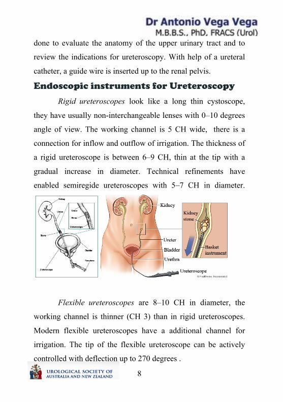
done to evaluate the anatomy of the upper urinary tract and to
review the indications for ureteroscopy. With help of a ureteral
catheter, a guide wire is inserted up to the renal pelvis.
Endoscopic instruments for Ureteroscopy
Rigid ureteroscopes look like a long thin cystoscope,
they have usually non-interchangeable lenses with 0–10 degrees
angle of view. The working channel is 5 CH wide, there is a
connection for inflow and outflow of irrigation. The thickness of
a rigid ureteroscope is between 6–9 CH, thin at the tip with a
gradual increase in diameter. Technical refinements have
enabled semiregide ureteroscopes with 5–7 CH in diameter.
Flexible ureteroscopes are 8–10 CH in diameter, the
working channel is thinner (CH 3) than in rigid ureteroscopes.
Modern flexible ureteroscopes have a additional channel for
irrigation. The tip of the flexible ureteroscope can be actively
controlled with deflection up to 270 degrees .

9
Pyeloscopy
Pyeloscopy is where a
thin fibre-optic telescope is
introduced into the kidney form the bladder via the urethra. The
diameter of the instrument is less than 3 mm and allows
visualization of the entire kidney drainage system due to the
flexible nature of the scope. It contains a
small instrument port which allows the
introduction of laser fibres (0.3 mm
dameter) to efficiently fragment stones, and
micro-baskets (less then 1 mm wide) to
retrieve stone fragments. Kidney stones up
to 2 cm in size can be treated using this approach.
What is the difference between rigid and flexible
ureteroscopy?
Rigid ureteroscopy is performed with a rigid telescope. As such,
it looks only in a straight line. Flexible ureteroscopy is
performed with a very thin and flexible telescope that can
perform almost a 180° turn and look back on itself. It is
sometimes known as flexible uretero-
renoscopy, because it is possible to look
into various parts of the inside of the
kidney. Using a laser, stones can be

10
vapourised and removed with a basket. Flexible uretero-
renoscopy tends to be used for stones in the kidney and near the
kidney in the upper ureter. Rigid ureteroscopy is mainly used for
stones in the lower and mid ureter closed to the bladder.
Ureteroscopic Treatment of Urolithiasis
Extraction of ureteral stones with grasping forceps:
Small ureteral stones can be easily extracted with
grasping forceps
Extraction of ureteral stones with dormia baskets:
Ureteral stones, which are small enough
to be extracted completely, can easily be
removed with Dormia Baskets
Lithotripsy of ureteral stones:
Larger ureteral stones cannot be extracted in toto, they
need a lithotripsy in the ureter. Afterwards, the fragments can be
removed. The following possibilities for ureteroscopic
lithotripsy exist:
• Electromechanical lithotripsy (e.g. Lithoclast)
• Electrohydraulic lithotripsy (EHL)
• Holmium laser lithotripsy
Laser lithotripsy has the advantage of minimizing the trauma
to the ureter and to reduce the stone dislocation during the
lithotripsy.

11
Ureteroscopic Treatment Options for
Strictures and Tumours
Ureteroscopic biopsy:
Biopsies should be taken from tumours or strictures of
unknown etiology. The amount of resulting tissue with
ureteroscopic biopsy is very small. Multiple biopsies are
necessary, to obtain a reliable diagnosis.
Ablation of ureteral tumours:
Endoscopic palliative/curative treatment of ureter or renal
pelvis tumours is possible with ureteroscopic resectoscopes.
Alternatively, tumour ablation is possible using laser coagulation.
Ureteroscoic incision of ureteral strictures.
After retrograde (or antegrade) pyelography and insertion of
a guide wire, the stricture is cut under endoscopic vision. A full
thickness cut through the ureteral wall is done until the
periureteral fat can be seen. Visualization is possible with
retrograde (URS) or antegrade (nephroscope) techniques.
Different technical solutions exist for the ureterotomy: cold
knife (without cauterization), laser fibers (holmium or Neodym:
YAG).
Stone treated with laser in the
ureter

12
Stenting of the ureter
After ureteroscopy, especially after longer manipulation,
the placement of a JJ ureteral stent (between the kidney and the
bladder) is necessary for 2–4 weeks. This is necessary because
the ureter swells and can obstruct the flow of urine from the
kidney to the bladder. Most of the times a JJ stet is left into the
ureter to decrease the pain and discomfort after the operation.
Sometimes a JJ stent is not used, but a special designed
thin catheter that passes all the way up to the kidney and passes
out through the urethra into a collecting bag. This is usually
present for a day or so, and is removed easily by pulling it out.
A catheter may be used to drain the bladder and this does
not usually need to stay more than 24 hours.
Important
If a JJ stent was inserted, it will
need to be removed usually in
outpatients with the aid of a flexible
cystoscope under local anaesthetic.
Occasionally, it is removed under
general anaesthetic, especially if a
contrast study is required to ensure
you are stone free.

13
What to expect post ureteroscopy?
The procedure is minimally invasive but may cause some
discomfort or pain immediately after surgery. You will be given
pain relief as needed.
For several hours after the procedure you may have a
burning feeling when you urinate. This feeling should go away
within a day. Drinking a lot of water can help reduce the burning.
Your doctor also may recommend you take medicine to numb
the burning (Ural Sachets)
Often the urine will appear red because there will be
blood present. This is normal and should not raise alarm, can last
for 2 or 3 days
Antibiotics will commonly be given after the procedure to
prevent infections.
When the catheter is removed, there may be some
symptoms if a JJ stent has been inserted. These may include:
-the need to pass urine more frequently
-discomfort felt in the bladder area and in the kidney area in the
back when passing urine.
-blood in the urine especially when physically activities.
Such symptoms are usually transient, but if are
bothersome may need review by a doctor especially if there is a
fever present.

14
At some point after the procedure, either a plain X-ray or
CT KUB may be requested. This is used to determine if the
stone is still present or not.
If the stone has been pushed up to the kidney, then
ESWL or flexible pyeloscopy is usually the next treatment to
fragment the stone.
What are the success rate of ureteroscopy?
The success rate of ureteroscopy
is over 95% for the majority of the
stones that are treated this way. Success
depends:
-whether there is 1 or more stones
present
-how long the stone has been stuck
- the size of the stone
-the location of the stone (where in the kidney or ureter)
-whether you have had previous surgery on the kidney.
-the experience of the urologist treating you.
It may not be possible to reach the stone on the first
attempt with the ureteroscope because of severe swelling that
occurs when a stone is present at the ureter. In that situation, a JJ
stent may be placed in the ureter. With a JJ stent in place, urine
can drain from the kidney to the bladder and the ureter expands

15
in size. As it becomes wider, it is easier to pass the ureteroscope
up to the stone and remove
Sometimes if the stone is very large, it may not be
possible to remove the stone in one session and a second
procedure may be necessary. On other occasions, small stone
fragments or the whole stone may pass up into the kidney. If a
flexible ureteroscope is available, this can be passed up into the
kidney and the fragments removed or broken with laser.
CONSENT AND RISKS
A consent form is a legal document, recognizing your
willingness to proceed with the intended treatment you are
required to sign a consent form for the operation once you fully
understand the reason for the operation and the risk involved.

16
All the operations have risks associated with them. All
risks should be discussed with your doctor. You should
understand the procedure and any available alternative treatment
discussed.
RISKS
Complications are uncommon with this test
Common (greater than 1 in 10)
Bleeding or mild burning when passing urine for a short
period after the operation
Temporary insertion of a bladder catheter
Insertion of a stent with a further procedure to remove it.
The stent may cause pain, frequency and bleeding in the
urine
Occasional (between 1 in 10 and 1 in 50)
Inability to retrieve the stone or movement of the stone
back into the kidney
Kidney damage or infection needing further treatment
Failure to pass the telescope if the ureter is narrow.
Failure to retrieve the stone, an alternative method could
be necessary.
Recurrence of stones.
Urinary infection requiring antibiotic treatment.

17
Rare (less than 1 in 50)
Damage of the ureter with need for open operation of tube
placed into the kidney directly form back to allow any leak to
heal.
Very rately, scarring or stricture of the ureter requiring
further procedures.
What should I expect when I get home?
When you leave hospital, you will be given a draft
discharge summary of your admission. If, in the first few weeks
after your discharge, you need to call your GP for any reason or
attend another hospital, please, take this summary with you to
allow the doctors to see the details of your treatment.
When you get home, you should drink twice as much
fluid as you would normally to flush your system through and
minimise any bleeding.
You may experience pain in the kidney over the first 24-
72 hours, due to the swelling caused by insertion of the
instrument or by the presence of a stent. Anti-inflammatory
painkillers will help this pain, which normally settles after 72
hours.
It will take at least 10 days to recover fully from the
operation. You should not expect to return to work within 7 days.
18
You may find that the ureteric stent, the lower end of
which sits in the bladder, caused some pain when you pass urine
and you may also see blood in the urine as a result of the stent.
The stent can also cause you to pass urine more frequently that
you would do normally. These symptoms will settle once the
stent has been removed.
Facts about ureteroscopy:
-typical operative time : 1 hour
-Usual hospital stay: overnight
-average number of days before going to work 7 days
-average number of days before feeling back to normal 15.6
days
(Data form study: Pearly and colleagues, Journal or Urology
2005)
Total recovery time
Driving can be resumed within 24 hours of a general anaesthetic,
but be wary of abrupt movement whilst driving
Normal daily activities may be resumed on discharge
Sexual activity, physical activities may resume when you are
comfortable
Normal diet may resume immediately after surgery.

19
DISCHARGE INFORMATION
- You may see blood in your urine; this is normal and
should clear after 1-2 days. It may burn or sting when you pass
urine afterwards, this should get better over the day.
-I f you develop a temperature, you urine is smelly/cloudy
and burns when you pass urine, you may have a urine infection.
You should contact your GP, as you may need a course of
antibiotics
- You can eat and drink normally. You should try to drink
at least 1.5-2 liters of fluid during the day after you cystoscopy,
unless you have been told by a doctor to restrict your fluid
intake. Drinking extra water can help to flush out your bladder
and reduce the risk of infection. If you continue to drink plenty
of fluid, this discomfort and bleeding will resolve rapidly.
- You can resume sexual activity as soon as you feel
comfortable to de so.
WHAT ELSE SHOULD I LOOK OUT FOR
- If you develop a fever, severe pain on passing urine,
inability to pass urine or worsening bleeding, you should contact
your GP immediately or go to your nearest Emergency
Department. Small blood clots or stone fragments may also pass
down the ureter from the kidney. Resulting in renal colic; you
should contact your GP immediately.

20
IN CASE OF PROBLEMS
Most people have no problems after a ureteroscopy, but you
should contact your GP or Emergency Department if you
develop any of the following symptoms
Persistent, severe pain with an inability to pass urine
A high temperature
Burning sensation on passing urine that gets worse or
starts again after any initial stinging has worn off
An unpleasant smell of your urine
Persistent and heavy blood and clots in your urine.
Unable to pass urine
NOTIFY THE UROLOGIST OR ATTEND TO THE
EMERGENCY DEPARTMENT


![Flexible Ureteroscopy and Laserlithotripsy for Kidney and ...1116854/FULLTEXT01.pdfcalcium phosphate. Calcium stones is usually a result from hypercalciuria [3]. Hypercalciuria is](https://img.pdfslide.us/doc/110x75/60e640fc51886875c210c19f/flexible-ureteroscopy-and-laserlithotripsy-for-kidney-and-1116854fulltext01pdf.jpg)