Embed Size (px)
Citation preview
YodL and YisK Possess Shape-Modifying Activities That AreSuppressed by Mutations in Bacillus subtilis mreB and mbl
Yi Duan, Anthony M. Sperber, Jennifer K. Herman
Department of Biochemistry and Biophysics, Texas A&M University, College Station, Texas, USA
ABSTRACT
Many bacteria utilize actin-like proteins to direct peptidoglycan (PG) synthesis. MreB andMreB-like proteins are thought to actas scaffolds, guiding the localization and activity of key PG-synthesizing proteins during cell elongation. Despite their criticalrole in viability and cell shape maintenance, very little is known about how the activity of MreB family proteins is regulated. Us-ing a Bacillus subtilismisexpression screen, we identified two genes, yodL and yisK, that when misexpressed lead to loss of cellwidth control and cell lysis. Expression analysis suggested that yodL and yisK are previously uncharacterized Spo0A-regulatedgenes, and consistent with these observations, a �yodL �yisKmutant exhibited reduced sporulation efficiency. Suppressors re-sistant to YodL’s killing activity occurred primarily inmreBmutants and resulted in amino acid substitutions at the interfacebetweenMreB and the highly conserved morphogenic protein RodZ, whereas suppressors resistant to YisK occurred primarilyinmblmutants andmapped toMbl’s predicted ATP-binding pocket. YodL’s shape-altering activity appears to require MreB, as a�mreBmutant was resistant to the effects of YodL but not YisK. Similarly, YisK appears to require Mbl, as a �mblmutant wasresistant to the cell-widening effects of YisK but not of YodL. Collectively, our results suggest that YodL and YisK likely modu-late MreB andMbl activity, possibly during the early stages of sporulation.
IMPORTANCE
The peptidoglycan (PG) component of the cell envelope confers structural rigidity to bacteria and protects them from osmoticpressure. MreB andMreB-like proteins are thought to act as scaffolds for PG synthesis and are essential in bacteria exhibitingnonpolar growth. Despite the critical role of MreB-like proteins, we lack mechanistic insight into how their activities are regu-lated. Here, we describe the discovery of two B. subtilis proteins, YodL and YisK, which modulate MreB andMbl activities. Ourdata suggest that YodL specifically targets MreB, whereas YisK targets Mbl. The apparent specificities with which YodL and YisKare able to differentially target MreB andMbl make them potentially powerful tools for probing the mechanics of cytoskeletalfunction in bacteria.
Bacterial cell growth requires that the machineries directingenlargement and division of the bacterial cell envelope be co-
ordinated in both time and space (1). The cell envelope is com-prised of membranes and a macromolecular mesh of peptidogly-can (PG) that possesses both rigid and elastic properties (2, 3). PGis highly cross-linked, allowing bacteria to maintain shapes andavoid lysis, even in the presence of several atmospheres of internalturgor pressure. PG rearrangements are required during the in-ward redirection of growth that occurs at the time of cell division,but they are also necessary when cells insert new PG and dynam-ically modify their morphologies in response to developmental orenvironmental signals (4, 5). To avoid lysis during PG rearrange-ments, bacteria must carefully regulate the making and breakingof glycan strands and peptide cross-links (3). In rod-shaped bac-teria, PG enlargement during steady-state growth is constrained inone dimension along the cell’s long axis and can either occurthrough polar growth, as is the case forAgrobacterium tumefaciensand Streptomyces coelicolor, or through incorporation of new cellwall material along the length of the cell cylinder, as observed withEscherichia coli, Bacillus subtilis, and Caulobacter crescentus (6).
To control cell diameter and create osmotically stable PG, bac-teria that exhibit nonpolar growth require the activity of the highlyconserved actin-like protein MreB. Biochemical, genetic, and cellbiological data suggest that MreB likely directs PG synthesis dur-ing cell elongation, and in some bacteria MreB may also functionduring cell division (7–9). MreB possesses ATPase activity and
polymerizes at sites along the cytoplasmic side of the inner mem-brane (10). ATP binding and hydrolysis are required for MreBpolymerization and activity (11), and two S-benzylisothiourea de-rivatives, A22 and MP265, target the ATPase domain of MreB inGram-negative organisms, possibly preventing nucleotide hydro-lysis and/or release (12–15). Depletion or inactivation of MreB islethal except in some conditional backgrounds (16, 17), so organ-isms sensitive to A22 and/or MP265 lose shape and eventually lyse(12–15).
MreB has been found to interact with several other proteinsinvolved in PG synthesis, including the bitopic membrane proteinRodZ (8, 16, 18–20). RodZ interacts directly with MreB through a
Received 25 February 2016 Accepted 18 May 2016
Accepted manuscript posted online 23 May 2016
Citation Duan Y, Sperber AM, Herman JK. 2016. YodL and YisK possess shape-modifying activities that are suppressed by mutations in Bacillus subtilis mreB andmbl. J Bacteriol 198:2074–2088. doi:10.1128/JB.00183-16.
Editor: T. M. Henkin, Ohio State University
Address correspondence to Jennifer K. Herman, [email protected].
Y.D. and A.M.S. contributed equally.
This work is dedicated to the memory of Arthur L. Koch.
Supplemental material for this article may be found at http://dx.doi.org/10.1128/JB.00183-16.
Copyright © 2016, American Society for Microbiology. All Rights Reserved.
crossmark
2074 jb.asm.org August 2016 Volume 198 Number 15Journal of Bacteriology
on July 13, 2016 by TEX
AS
A&
M U
NIV
http://jb.asm.org/
Dow
nloaded from
cytoplasmic helix-turn-helix motif located at its N terminus (18).A cocrystal structure of RodZ and MreB shows the N terminusof RodZ extending into a conserved hydrophobic pocket lo-cated in subdomain IIA of MreB (18). Depletion of RodZ alsoleads to loss of cell shape and cell death (21–23). However, invarious mutant backgrounds, rodZ can be deleted without lossof rod shape or viability, indicating that RodZ is not absolutelyrequired for MreB’s function in maintaining shape (24–26).Based on these observations and others, it has been proposedthat MreB-RodZ interactions may regulate some aspect ofMreB activity (10, 26).
Gram-positive bacteria often encode multiple paralogs (27). B.subtilis possesses three mreB family genes: mreB, mbl, and mreBH.mreB is distinguished from mbl and mreBH by its location withinthe highly conserved mreBCD operon. Although mreB, mbl, andmreBH are essential, it has been reported that each can be deletedunder conditions in which cells are provided sufficient magne-sium (28–30) or in strain backgrounds lacking ponA, the gene thatencodes penicillin binding protein 1 (PBP1) (20). In addition, allthree genes can be deleted in a single background with only minoreffects on cell shape if any one of the paralogs is artificially over-expressed in trans from an inducible promoter (31). The ability ofany one of the paralogs to compensate for the loss of the others,at least under some growth conditions, strongly suggests thatMreB, Mbl, and MreBH share significant functional redun-dancy (31, 32).
At the same time, several lines of evidence suggest that theparalogs possess nonoverlapping functions. The genes themselvesexhibit different patterns of transcriptional regulation, suggestingthat each likely possesses specialized activities that are importantin different growth contexts. For example,mreB andmbl are max-imally expressed at the end of exponential growth but expressionfalls off sharply during stationary phase (33), whereas mreBH ispart of the SigI heat shock regulon (34). There is also evidencesuggesting that each protein may possess specialized activities. Forexample, MreBH interacts with the lytic transglycosylase LytE andis required for LytE localization (35), whereas the lytic transglyco-sylase CwlO depends on Mbl for wild-type function (35). Morerecently, MreB (but not Mbl or MreBH) was shown to aid inescape from the competent cell state (33).
Aside from RodZ (10, 26), only a few proteins targeting MreBactivity in vivo have been identified. In E. coli, the YeeU-YeeVprophage toxin-antitoxin system is comprised of a negative regu-lator of MreB polymerization, CbtA (36), and a positive regulatorof MreB bundling, CbeA (37). Another E. coli prophage toxin,CptA, is also reported to inhibit MreB polymerization (38). TheMbiA protein of C. crescentus appears to regulate MreB in vivo;however, its physiological role is unknown (39). Given the impor-tance of PG synthesis to cell viability and in cell shape control, it islikely that many undiscovered factors exist that modulate the ac-tivity of MreB and its paralogs.
In the present work, we describe the identification of YodL andYisK, modulators of MreB and Mbl activity that are expressedduring early stages of B. subtilis sporulation. Misexpression of ei-ther yodL or yisK during vegetative growth results in loss of cellwidth control and cell death. Genetic evidence indicates that YodLtargets and inhibits MreB activity, whereas YisK targets and inhib-its Mbl. Our data also show that YisK activity affects cell lengthcontrol through an Mbl- and MreBH-independent pathway.
MATERIALS AND METHODSGeneral methods. All B. subtilis strains were derived from B. subtilis 168.E. coli and B. subtilis strains utilized in this study are listed in Table S2 inthe supplemental material. Plasmids are listed in Table S3 in the supple-mental material. Oligonucleotide primers are listed in Table S4 of thesupplemental material. Details on plasmid and strain construction can befound in the supplemental material. Escherichia coli DH5� was used forcloning. All E. coli strains were grown in LB-Lennox medium supple-mented with 100 �g/ml ampicillin. The following concentrations of anti-biotics were used for generating B. subtilis strains: 100 �g/ml spectinomy-cin, 7.5 �g/ml chloramphenicol, 0.8 mg/ml phleomycin, 10 �g/mltetracycline, 10 �g/ml kanamycin. To select for erythromycin resistance,plates were supplemented with 1 �g/ml erythromycin (ERM) and 25�g/ml lincomycin. B. subtilis transformations were carried out as de-scribed previously (40). When indicated, the LB in the B. subtilis micros-copy experiments was LB-Lennox broth. Sporulation by resuspension wascarried out at 37°C according to the Sterlini-Mandelstam method (41).Penassay broth (PAB) is composed of 5 g peptone, 1.5 g beef extract, 1.5 gyeast extract, 1.0 g D-glucose (dextrose), 3.5 g NaCl, 3.68 g dipotassiumphosphate, and 1.32 g monopotassium phosphate per liter of distilledwater. To make solid media, the relevant medium was supplemented with1.5% (wt/vol) Bacto agar.
Microscopy. For microscopy experiments, all strains were grown inthe indicated medium in volumes of 25 ml in 250-ml baffled flasks andplaced in a shaking water bath set at 37°C and 280 rpm. Unless statedotherwise, misexpression was performed by inducing samples with 1.0mM isopropyl-beta-D-thiogalactopyranoside (IPTG) and imaging ofsamples 90 min postinduction. Fluorescence microscopy was performedwith a Nikon Ti-E microscope equipped with a CFI Plan Apo lambda DM100� objective, Prior Scientific Lumen 200 illumination system, C-FLUV-2E/C 4=,6-diamidino-2-phenylindole and C-FL green fluorescentprotein (GFP) HC HISN Zero Shift filter cubes, and a CoolSNAP HQ2monochrome camera. Membranes were stained with TMA-DPH [1-(4-trimethylammoniumphenyl)-6-phenyl-1,3,5-hexatriene p-toluene sulfo-nate; 0.02 mM] and imaged with exposure times of 1 s with a neutraldensity filter in place to reduce cytoplasmic background. All GFP imageswere captured with a 1-s exposure time. All images were captured andprocessed with the NIS Elements Advanced Research program (version4.10) and ImageJ64 (42). Cells were mounted on glass slides with 1%agarose pads or polylysine-treated coverslips prior to imaging. To quan-titate cell lengths for the strains imaged in Fig. 6, the cell lengths for 500cells were determined for each population. The statistical significance ofcell length differences between populations was determined using an un-paired Student’s t test.
Plate growth assays. B. subtilis strains were streaked on LB-Lennoxplates containing 100 �g/ml spectinomycin and 1 mM IPTG. The plateswere supplemented with the indicated concentrations of MgCl2 whennecessary. Plates were incubated at 37°C overnight, and images were cap-tured on a ScanJet G4050 flatbed scanner (Hewlett Packard).
Heat kill. Spore formation was quantified by growing cells in Difcosporulation medium (DSM) (43). A freshly grown single colony of eachstrain was inoculated into 2 ml of DSM and placed in a roller drum at37°C, 60 rpm, for 36 h. To determine the number of CFU per milliliter, analiquot of each culture was serially diluted and plated on DSM agar plates.To enumerate the heat-resistant spores per milliliter, the serially dilutedcultures were subjected to a 20-min heat treatment at 80°C and plated onDSM agar plates. The plates were incubated at 37°C overnight, and thenext day colony counts were determined. The relative sporulation fre-quency compared to the wild type was determined by calculating thenumber of spores per CFU of each experimental and dividing it by thespores per CFU of the wild type. The reported statistical significance wasdetermined using an unpaired Student’s t test.
Transcriptional fusions.Transcriptional fusions were constructed byfusing an �200-bp region up to the start codon of either yodL or yisK togfp or lacZ and integrating the fusions into the B. subtilis chromosome at
Modulators of MreB and Mbl Activity
August 2016 Volume 198 Number 15 jb.asm.org 2075Journal of Bacteriology
on July 13, 2016 by TEX
AS
A&
M U
NIV
http://jb.asm.org/
Dow
nloaded from
the amyE locus (for more details, see the descriptions of strain construc-tions in the supplemental material). Microscopy was conducted on eachstrain over a time course in sporulation by resuspension media (see thesupplemental material) or in a nutrient exhaustion time course experi-ment in CH (41). Beta-galactosidase assays were performed as describedpreviously (44), except all samples were frozen at �80°C before process-ing. All experiments were performed on at least three independent bio-logical replicates.
Suppressor selections. Single colonies of BYD048 (harboring threecopies of Phy-yodL [3� Phy-yodL] and one copy of Phy-lacZ) or BYD076(3� Phy-yisK Phy-lacZ) were used to inoculate independent 5-ml LB-Len-nox cultures. Six independent cultures were grown for each strain. Thecultures were grown for 6 h at 37°C, and 0.3 �l of each culture was dilutedin 100 �l LB and plated on an LB-Lennox agar plate containing 100 �g/mlspectinomycin and 1 mM IPTG. After overnight growth, suppressors thatarose were patched on both LB-Lennox agar plates supplemented with100 �g/ml spectinomycin and LB-Lennox agar plates supplemented with100 �g/ml spectinomycin, 1.0 mM IPTG, and 40 �g/ml 5-bromo-4-chloro-3-indolyl-�-D-galactopyranoside and grown at 37°C overnight.Only blue colonies were selected for further analysis; this screen elimi-nated mutants unable to derepress Phy in the presence of IPTG. In addi-tion, each Phy-yodL or Phy-yisK construct was transformed into a wild-type background to ensure that the construct remained fully functionalwith respect to preventing cell growth on LB-Lennox agar plates supple-mented with the relevant antibiotic and 1 mM IPTG.
Whole-genome sequencing andanalysis.Genomic DNA was isolatedfrom six YodL-resistant suppressors obtained from independent culturesas well as the parent strain (BYD048) by inoculating a single colony in 6 mlLB-Lennox medium and growing at 37°C for 4 h in a roller drum. Cellswere collected by spinning at 21,130 � g for 2 min at room temperature,resuspending the pellets in lysis buffer (20 mM Tris-HCl [pH 7.5], 50 mMEDTA [pH 8], 100 mM NaCl, and 2 mg/ml lysozyme) and incubating at37°C for 30 min. Sarkosyl was added to a final concentration of 1% (wt/vol). Protein was removed by extracting with 600 �l phenol, centrifugingat 21,130 � g for 5 min at room temperature, and transferring the top(aqueous) layer to a new microcentrifuge tube. This was followed by anextraction with 600 �l phenol-saturated chloroform and centrifugation at21,130 � g for 5 min at room temperature. After transferring the aqueouslayer to a new microcentrifuge tube, a final extraction was performed with100% chloroform, followed by centrifugation at 21,130 � g for 5 min atroom temperature. The aqueous layer was transferred to a new microcen-trifuge tube, being careful to avoid the interphase material. To precipitatethe genomic DNA, a 1/10 volume of 3.0 M Na-acetate and 1 ml of 100%ethanol were added, and the tube was inverted multiple times. The samplewas centrifuged at 21,130 � g for 1 min at room temperature in a micro-centrifuge. The pellet was washed with 150 �l 70% ethanol and resus-pended in 500 �l TE (10 mM Tris [pH 7.5], 1 mM EDTA [pH 8.0]). Toeliminate potential RNA contamination, RNase was added to a final con-centration of 200 �g/ml and the sample was incubated at 55°C for 1 h. Toremove the RNase, the genomic DNA was repurified by phenol-chloro-form extraction and ethanol precipitation as described above. The finalpellet was resuspended in 100 �l TE. Bar-coded libraries were preparedfrom each genomic DNA sample by using a TruSeq DNA kit (Illumina)according to the manufacturer’s specifications, and the samples were sub-jected to Illumina-based whole-genome sequencing using a MiSeq 250paired-end run. CLC Genomics Workbench (Qiagen) was used to mapthe sequence reads against the Bs168 reference genome and to identifysingle nucleotide polymorphisms, insertions, and deletions. Mutationsassociated with the Phy integration constructs and those in which less than40% of the reads differed from the reference genome were excluded ascandidate changes responsible for suppression in our initial analysis (seeTable S1 in the supplemental material). The remaining suppressor muta-tions were identified by PCR amplification of mreB (using primer setOAS044 and OAS045) and mbl (using primer set OAS046 and OAS047)and sequencing with the same primers. To determine if the candidate
suppressor alleles identified were sufficient to confer resistance to theoriginal selective pressure, each was linked to a kanamycin resistance cas-sette and moved by transformation into a clean genetic background (seethe supplemental material for a further description of strain construc-tion).
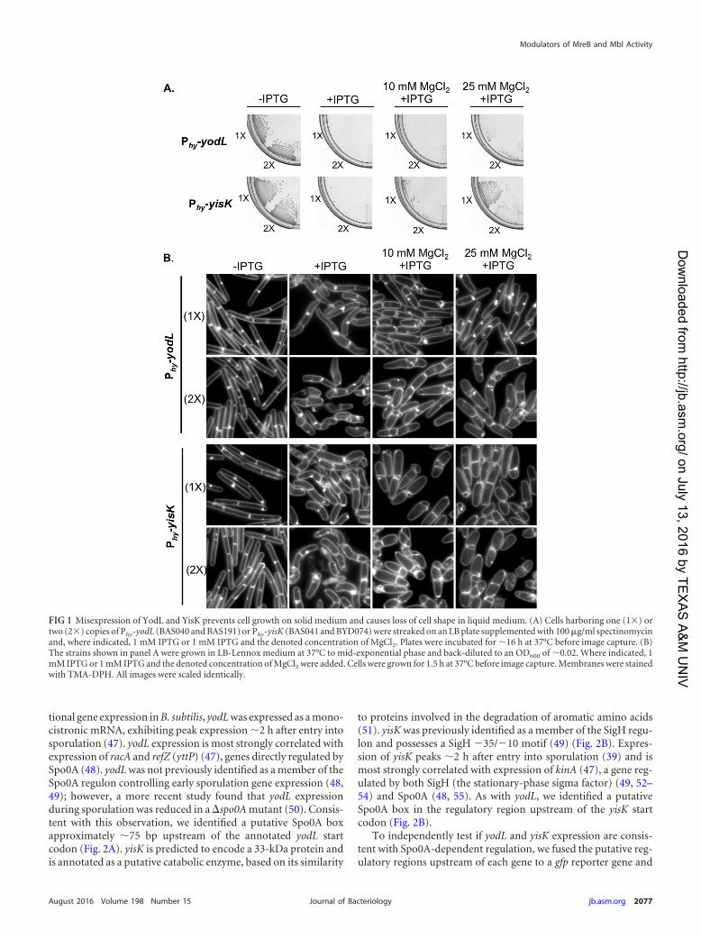
RESULTSYodL and YisK affect cell width. To identify novel factors in-volved in cellular morphogenesis, we created an ordered gene mis-expression library comprising over 800 previously uncharacter-ized genes fromB. subtilis. Each gene was placed under the controlof an IPTG-inducible promoter (Phy) and integrated in single copy(1�) at amyE, a nonessential locus in the B. subtilis chromosome.The library (called the BEIGEL, for Bacillus ectopic inducible geneexpression library) was screened for misexpression phenotypesthat perturbed growth on solid media and also resulted in obviousdefects in nucleoid morphology, changes in cell division fre-quency, and/or perturbations in overall cell shape in liquid cul-tures. Two strains, one harboring Phy-yodL and one harboringPhy-yisK, were unable to form colonies on plates containing in-ducer (Fig. 1A) and also produced wide, irregular cells withslightly tapered poles following misexpression in LB liquid me-dium (Fig. 1B). Cell lysis and aberrant cell divisions were alsoobserved. Introducing a second copy (2�) of each Phy misexpres-sion construct into the chromosome did not appreciably enhancecell widening at the 90-min postinduction time point, althoughcell lysis was more readily observed (Fig. 1B). Phy-yisK (2�) mis-expression also led to a drop in optical density over time (see Fig.S1A in the supplemental material), consistent with the cell lysisobserved microscopically. We concluded that the activities ofyodL and yisK target one or more processes integral to width con-trol during cell elongation.
The yodL and yisK misexpression phenotypes are similar tothose observed when proteins involved in cell elongation are per-turbed in B. subtilis (20, 31, 45). Since the addition of magnesiumwas previously reported to suppress the lethality and/or morpho-logical phenotypes associated with depletion or deletion of someproteins important for cell elongation in B. subtilis (16, 20, 29, 31,46), we assessed if the Phy-yodL and Phy-yisK misexpression phe-notypes could be rescued by growing cells with media supple-mented with two different concentrations of MgCl2. The YodL-producing cells failed to grow on any LB media containinginducer, regardless of MgCl2 concentration (Fig. 1A). In contrast,LB supplemented with 25 mM MgCl2 restored viability to thestrain producing YisK (Fig. 1A). Interestingly, even 25 mM MgCl2was not sufficient to suppress the cell-widening effect associatedwith YodL and YisK misexpression (Fig. 1B), although these cellsdid not lyse (see Fig. S1C in the supplemental material). Since PABmedium was often used in the prior studies that showed thatMgCl2 supplementation rescued cell shape (16, 20, 29, 31, 46), wealso assayed for growth on PAB following YodL and YisK expres-sion. PAB supplemented with 25 mM MgCl2 rescued growth onplates (see Fig. S2A in the supplemental material), but it still didnot rescue morphology in liquid culture (see Fig. S2B).
yodL and yisK expression. To better understand the possiblephysiological functions of the yodL and yisK gene products, weanalyzed the genes and their genetic contexts bioinformatically.yodL is predicted to encode a 12.5-kDa hypothetical proteinwhich, based on amino acid similarity, is conserved in the Bacillusgenus. In data from a global microarray study analyzing condi-
Duan et al.
2076 jb.asm.org August 2016 Volume 198 Number 15Journal of Bacteriology
on July 13, 2016 by TEX
AS
A&
M U
NIV
http://jb.asm.org/
Dow
nloaded from
tional gene expression inB. subtilis, yodLwas expressed as a mono-cistronic mRNA, exhibiting peak expression �2 h after entry intosporulation (47). yodL expression is most strongly correlated withexpression of racA and refZ (yttP) (47), genes directly regulated bySpo0A (48). yodLwas not previously identified as a member of theSpo0A regulon controlling early sporulation gene expression (48,49); however, a more recent study found that yodL expressionduring sporulation was reduced in a �spo0Amutant (50). Consis-tent with this observation, we identified a putative Spo0A boxapproximately �75 bp upstream of the annotated yodL startcodon (Fig. 2A). yisK is predicted to encode a 33-kDa protein andis annotated as a putative catabolic enzyme, based on its similarity
to proteins involved in the degradation of aromatic amino acids(51). yisK was previously identified as a member of the SigH regu-lon and possesses a SigH �35/�10 motif (49) (Fig. 2B). Expres-sion of yisK peaks �2 h after entry into sporulation (39) and ismost strongly correlated with expression of kinA (47), a gene reg-ulated by both SigH (the stationary-phase sigma factor) (49, 52–54) and Spo0A (48, 55). As with yodL, we identified a putativeSpo0A box in the regulatory region upstream of the yisK startcodon (Fig. 2B).
To independently test if yodL and yisK expression are consis-tent with Spo0A-dependent regulation, we fused the putative reg-ulatory regions upstream of each gene to a gfp reporter gene and
FIG 1 Misexpression of YodL and YisK prevents cell growth on solid medium and causes loss of cell shape in liquid medium. (A) Cells harboring one (1�) ortwo (2�) copies of Phy-yodL (BAS040 and BAS191) or Phy-yisK (BAS041 and BYD074) were streaked on an LB plate supplemented with 100 �g/ml spectinomycinand, where indicated, 1 mM IPTG or 1 mM IPTG and the denoted concentration of MgCl2. Plates were incubated for �16 h at 37°C before image capture. (B)The strains shown in panel A were grown in LB-Lennox medium at 37°C to mid-exponential phase and back-diluted to an OD600 of �0.02. Where indicated, 1mM IPTG or 1 mM IPTG and the denoted concentration of MgCl2 were added. Cells were grown for 1.5 h at 37°C before image capture. Membranes were stainedwith TMA-DPH. All images were scaled identically.
Modulators of MreB and Mbl Activity
August 2016 Volume 198 Number 15 jb.asm.org 2077Journal of Bacteriology
on July 13, 2016 by TEX
AS
A&
M U
NIV
http://jb.asm.org/
Dow
nloaded from

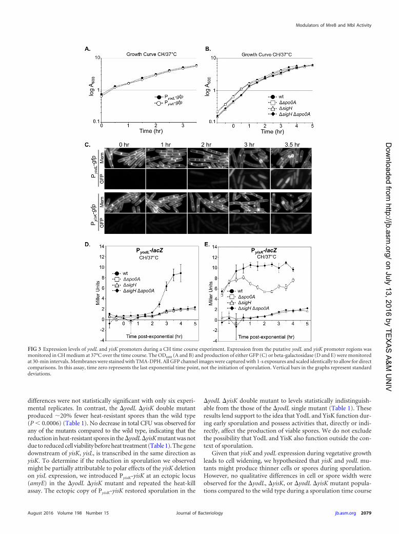
integrated the fusions into the amyE locus. We then followed ex-pression from the promoter fusions over a time course in CHliquid broth, a rich medium in which the cells first grow exponen-tially, transition to stationary phase, and finally gradually entersporulation (Fig. 3A to C). In this time course, the GFP signal fromPyisK-gfp increased dramatically from time zero (optical density at600 nm [OD600], �0.6) to time 1 h (OD600, �1.6) (Fig. 3C), con-sistent with yisK’s prior characterization as a SigH-regulated gene(49). In contrast, GFP fluorescence from PyodL-gfp became evidentat a later time point (120 min) and was more heterogeneous (Fig.3C), consistent with expression patterns previously observed forother Spo0A-P regulated genes (56, 57).
To quantitate expression from the promoters, we generatedPyodL-lacZ and PyisK-lacZ reporter strains and collected samplesover a CH time course beginning with early exponential phase(OD600, 0.2). Expression from PyodL-lacZ rose steadily, beginningabout 2 h after exit from exponential growth, and continued torise at least until the final time point (Fig. 3D). In contrast, expres-sion from PyisK-lacZ rose as cells transitioned from early to lateexponential growth, reached peak levels shortly after exit fromexponential growth, and remained steady for the remainder of thetime points (Fig. 3E). Wild-type expression from both PyodL-lacZand PyisK-lacZ required both SigH and Spo0A and was largelyeliminated in the absence of both regulators (Fig. 3D and E). Wedid not attempt to draw further conclusions from these data, sinceSpo0A and SigH each require the other for wild-type levels ofexpression (see Discussion).
We then followed expression from the promoter fusions over atime course following the sporulation by using the resuspensionmethod, which generates a more synchronous entry into sporula-tion (58). At time zero, neither the strain harboring PyodL-gfp northe strain harboring PyisK-gfp showed appreciable levels of fluores-cence (Fig. 4A) and appeared similar to a negative control harbor-ing gfp without a promoter (see Fig. S3 in the supplemental mate-rial). Between 0 and 40 min, both strains showed detectableincreases in fluorescence. At 60 min, when the first polar divisionscharacteristic of sporulation typically begin to manifest, bothstrains were more strongly fluorescent (Fig. 4A). GFP fluorescencefrom PyodL was qualitatively more intense than fluorescence pro-duced from PyisK (all images were captured and scaled with iden-
tical parameters to allow for direct comparisons). Moreover, theGFP signal continued to accumulate in the strain harboring PyodL-gfp for at least 2 h (Fig. 4A) and was heterogenous, consistent withactivation by Spo0A. In contrast, the fluorescence signal producedfrom PyisK-gfp was similar across the population and appearedsimilar at the 60- and 120-min time points (Fig. 4A), consistentwith SigH regulation.
To quantitate expression from the promoters during sporula-tion via a resuspension time course, we collected data from timepoints for strains harboring either the PyodL-lacZ or PyisK-lacZ re-porter constructs and performed beta-galactosidase assays. Ex-pression from PyodL-lacZ rose rapidly between the 40-min and100-min time points and steadily declined thereafter (Fig. 4B).The decline in signal was not observed for the GFP reporter, likelybecause GFP is stable once synthesized (59). In contrast, expres-sion from PyisK-lacZ was highest at the time of resuspension (T0)and declined until the final time point (Fig. 4C).
Collectively, the patterns of expression we observed for yodLare consistent with those observed for genes activated by high-threshold levels of Spo0A during sporulation, including racA,spoIIG, and spoIIA (60). In contrast, yisK’s expression pattern issimilar to that observed for kinA (47, 53, 61), with expressionincreasing in late exponential and stationary phases and early spo-rulation in a SigH-dependent manner (Fig. 3) but decreasing dur-ing sporulation in the resuspension time course experiment (Fig.4). We do not exclude the possibility that YodL and YisK alsofunction in other growth contexts.
A�yodL�yisKmutant is defective in sporulation. Since yodLand yisK expression levels correlate with those for other early spo-rulation genes, we next investigated if the gene products influ-enced the production of heat-resistant spores. To determine thenumber of heat-resistant spores in a sporulation culture, we quan-tified the number of CFU present in cultures before (total CFU)and after (heat-resistant CFU) a heat treatment that kills vegeta-tive cells. These values were normalized to display the sporulationefficiency of the mutants relative to wild type. Single mutants inwhich either yodL or yisK was deleted displayed only mild (97%and 94%, respectively) reductions in relative sporulation effi-ciency (Table 1). Although the single mutants always sporulatedless efficiently than wild type in each experimental replicate, the
FIG 2 DNA sequence upstream of yodL and yisK. (A) Putative Spo0A box (underlined) upstream of the yodL start codon. (B) SigH binding motifs (doubleunderline) and putative Spo0A box (underlined) upstream of the yisK start codon.
Duan et al.
2078 jb.asm.org August 2016 Volume 198 Number 15Journal of Bacteriology
on July 13, 2016 by TEX
AS
A&
M U
NIV
http://jb.asm.org/
Dow
nloaded from
differences were not statistically significant with only six experi-mental replicates. In contrast, the �yodL �yisK double mutantproduced �20% fewer heat-resistant spores than the wild type(P 0.0006) (Table 1). No decrease in total CFU was observed forany of the mutants compared to the wild type, indicating that thereduction in heat-resistant spores in the �yodL�yisKmutant was notdue to reduced cell viability before heat treatment (Table 1). The genedownstream of yisK, yisL, is transcribed in the same direction asyisK. To determine if the reduction in sporulation we observedmight be partially attributable to polar effects of the yisK deletionon yisL expression, we introduced PyisK-yisK at an ectopic locus(amyE) in the �yodL �yisK mutant and repeated the heat-killassay. The ectopic copy of PyisK-yisK restored sporulation in the
�yodL �yisK double mutant to levels statistically indistinguish-able from the those of the �yodL single mutant (Table 1). Theseresults lend support to the idea that YodL and YisK function dur-ing early sporulation and possess activities that, directly or indi-rectly, affect the production of viable spores. We do not excludethe possibility that YodL and YisK also function outside the con-text of sporulation.
Given that yisK and yodL expression during vegetative growthleads to cell widening, we hypothesized that yisK and yodL mu-tants might produce thinner cells or spores during sporulation.However, no qualitative differences in cell or spore width wereobserved for the �yodL, �yisK, or �yodL �yisK mutant popula-tions compared to the wild type during a sporulation time course
FIG 3 Expression levels of yodL and yisK promoters during a CH time course experiment. Expression from the putative yodL and yisK promoter regions wasmonitored in CH medium at 37°C over the time course. The OD600 (A and B) and production of either GFP (C) or beta-galactosidase (D and E) were monitoredat 30-min intervals. Membranes were stained with TMA-DPH. All GFP channel images were captured with 1-s exposures and scaled identically to allow for directcomparisons. In this assay, time zero represents the last exponential time point, not the initiation of sporulation. Vertical bars in the graphs represent standarddeviations.
Modulators of MreB and Mbl Activity
August 2016 Volume 198 Number 15 jb.asm.org 2079Journal of Bacteriology
on July 13, 2016 by TEX
AS
A&
M U
NIV
http://jb.asm.org/
Dow
nloaded from

assay (see Fig. S4 in the supplemental material). We also observedno qualitative differences in the shapes of germinating cells (datanot shown). Thus, although YodL and YisK contribute to the pro-duction of heat-resistant spores, they do not appear to be requiredto generate any of the major morphological changes required forspore production.
MreB andMbl are targets of YodL andYisK activity.To iden-tify genetic targets associated with YodL and YisK activity, we tookadvantage of the fact that misexpression of the proteins duringvegetative growth prevents colony formation on plates, and weperformed suppressor selection analysis. Strains harboring threecopies of each misexpression cassette were utilized to reduce the
chances of obtaining trivial suppressors in the misexpression cas-sette itself. In addition, Phy-lacZwas used as a reporter to eliminatesuppressors unable to release LacI repression following additionof inducer. In total, we obtained 14 suppressors resistant to YodLexpression and 13 suppressors resistant to YisK expression. Six ofthe suppressors resistant to YodL were subjected to whole-ge-nome sequencing. The results of the sequencing are shown inTable S1 in the supplemental material. All of the suppressors pos-sessed mutations in eithermreB ormbl, genes previously shown tobe important in regulating cell width (see Table S1). Using tar-geted sequencing, we determined that the remaining suppressorstrains resistant to YodL also harbored mutations in mreB or mbl.
FIG 4 Expression from yodL and yisK promoters following sporulation by resuspension. Expression from the putative yodL and yisK promoter regions wasmonitored in resuspension medium. The production of either GFP (A) or beta-galactosidase (B and C) was monitored at 20-min intervals. Membranes werestained with TMA-DPH. All GFP channel images were captured with 1-s exposures and scaled identically to allow for direct comparisons. Vertical bars in thegraphs represent standard deviations.
TABLE 1 Sporulation efficiencies of yodL and yisK mutantsa
Genotype Strain Total CFU Heat-resistant CFU% sporulationefficiency
% relativesporulation efficiency
Wild type B. subtilis 168 2.8 � 108 (4.7 � 107) 1.9 � 108 (4.5 � 107) 66.9 (5) 100�yodL BYD276 2.6 � 108 (3.9 � 107) 1.7 � 108 (2.8 � 107) 65.2 (7) 97�yisK BYD278 2.7 � 108 (4.6 � 107) 2.4 � 108 (2.7 � 107) 63.1 (6) 94�yodL �yisK BYD279 3.1 � 108 (6.5 � 107) 1.7 � 108 (4.1 � 107) 54.1 (4) 81�yodL �yisK PyisK-yisK BYD510 3.4 � 108 (3.3 � 107) 2.3 � 108 (4.1 � 107) 66.2 (7) 99a Sporulation efficiency was calculated as the number of spores per milliliter, divided by the total CFU per milliliter, times 100. Relative sporulation efficiency is the sporulationefficiency normalized to that of the wild type (times 100). Values are means, with standard deviations in parentheses. The data shown are the average results for six independentbiological replicates. The difference in sporulation efficiency between the wild type and the �yodL �yisK double mutant was statistically significant (P 0.0006).
Duan et al.
2080 jb.asm.org August 2016 Volume 198 Number 15Journal of Bacteriology
on July 13, 2016 by TEX
AS
A&
M U
NIV
http://jb.asm.org/
Dow
nloaded from
Since the phenotypes of YodL and YisK expression were similar,we also performed targeted sequencing of the mreB and mbl chro-mosomal regions in the YisK-resistant suppressors. All but one ofthe YisK-resistant suppressors possessed mutations in mbl; theremaining suppressor harbored a mutation in mreB.
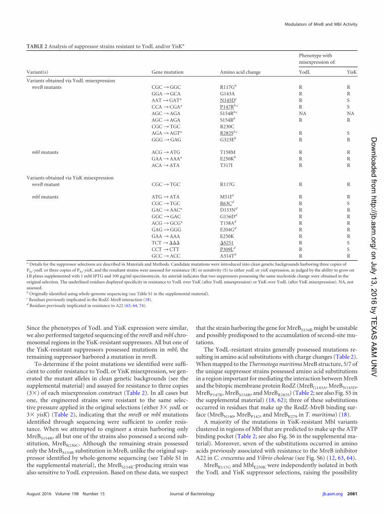
To determine if the point mutations we identified were suffi-cient to confer resistance to YodL or YisK misexpression, we gen-erated the mutant alleles in clean genetic backgrounds (see thesupplemental material) and assayed for resistance to three copies(3�) of each misexpression construct (Table 2). In all cases butone, the engineered strains were resistant to the same selec-tive pressure applied in the original selections (either 3� yodL or3� yisK) (Table 2), indicating that the mreB or mbl mutationsidentified through sequencing were sufficient to confer resis-tance. When we attempted to engineer a strain harboring onlyMreBS154R, all but one of the strains also possessed a second sub-stitution, MreBR230C. Although the remaining strain possessedonly the MreBS154R substitution in MreB, unlike the original sup-pressor identified by whole-genome sequencing (see Table S1 inthe supplemental material), the MreBS154R-producing strain wasalso sensitive to YodL expression. Based on these data, we suspect
that the strain harboring the gene for MreBS154R might be unstableand possibly predisposed to the accumulation of second-site mu-tations.
The YodL-resistant strains generally possessed mutations re-sulting in amino acid substitutions with charge changes (Table 2).When mapped to theThermotogamaritimaMreB structure, 5/7 ofthe unique suppressor strains possessed amino acid substitutionsin a region important for mediating the interaction between MreBand the bitopic membrane protein RodZ (MreBG143A, MreBN145D,MreBP147R, MreBS154R, and MreBR282S) (Table 2; see also Fig. S5 inthe supplemental material) (18, 62); three of these substitutionsoccurred in residues that make up the RodZ-MreB binding sur-face (MreBN140, MreBP142, and MreBR279 in T. maritima) (18).
A majority of the mutations in YisK-resistant Mbl variantsclustered in regions of Mbl that are predicted to make up the ATPbinding pocket (Table 2; see also Fig. S6 in the supplemental ma-terial). Moreover, seven of the substitutions occurred in aminoacids previously associated with resistance to the MreB inhibitorA22 in C. crescentus and Vibrio cholerae (see Fig. S6) (12, 63, 64).
MreBR117G and MblE250K were independently isolated in boththe YodL and YisK suppressor selections, raising the possibility
TABLE 2 Analysis of suppressor strains resistant to YodL and/or YisKa
Variant(s) Gene mutation Amino acid change
Phenotype withmisexpression of:
YodL YisK
Variants obtained via YodL misexpressionmreB mutants CGC ¡ GGC R117Gb R R
GGA ¡ GCA G143A R RAAT ¡ GAT* N145Dc R SCCA ¡ CGA* P147Rb,c R SAGC ¡ AGA S154Rb,c NA NAAGC ¡ AGA S154Rb R RCGC ¡ TGC R230CAGA ¡ AGT* R282Sb,c R SGGG ¡ GAG G323Eb R R
mbl mutants ACG ¡ ATG T158M R RGAA ¡ AAA* E250Kb R RACA ¡ ATA T317I R R
Variants obtained via YisK misexpressionmreB mutant CGC ¡ TGC R117G R R
mbl mutants ATG ¡ ATA M51Id R RCGC ¡ TGC R63Cd R SGAC ¡ AAC* D153Nd R RGGC ¡ GAC G156Dd R RACG ¡ GCG* T158Ad R RGAG ¡ GGG E204Gd R RGAA ¡ AAA E250K R RTCT ¡ ��� �S251 R SCCT ¡ CTT P309Ld R SGCC ¡ ACC A314Td R R
a Details for the suppressor selections are described in Materials and Methods. Candidate mutations were introduced into clean genetic backgrounds harboring three copies ofPhy-yodL or three copies of Phy-yisK, and the resultant strains were assessed for resistance (R) or sensitivity (S) to either yodL or yisK expression, as judged by the ability to grow onLB plates supplemented with 1 mM IPTG and 100 �g/ml spectinomycin. An asterisk indicates that two suppressors possessing the same nucleotide change were obtained in theoriginal selection. The underlined residues displayed specificity in resistance to YodL over YisK (after YodL misexpression) or YisK over YodL (after YisK misexpression). NA, notassessed.b Originally identified using whole-genome sequencing (see Table S1 in the supplemental material).c Residues previously implicated in the RodZ-MreB interaction (18).d Residues previously implicated in resistance to A22 (63, 64, 74).
Modulators of MreB and Mbl Activity
August 2016 Volume 198 Number 15 jb.asm.org 2081Journal of Bacteriology
on July 13, 2016 by TEX
AS
A&
M U
NIV
http://jb.asm.org/
Dow
nloaded from
that at least some of the other MreB and Mbl variants exhibitcross-resistance to YodL and YisK misexpression. To test forcross-resistance, we generated the mutant alleles in clean geneticbackgrounds and then introduced 3 copies of Phy-yisK into theYodL-resistant suppressors and 3 copies Phy-yodL into the YisK-resistant suppressors. We then assayed for the ability of the mis-expression strains to grow on medium in the presence of inducer.The results, summarized in Table 2, showed that several of thevariants exhibited resistance to both YodL and YisK. Three MreBvariants, MreBN145D, MreBP147R, and MreBR282S, exhibited speci-ficity in their resistance to YodL compared to YisK. Three Mblvariants, MblR63C, Mbl�S251, and MblP309L, showed specificity intheir resistance to YisK over YodL. These results suggest that thealleles exhibiting cross-resistance to both YisK and YodL are likelyto be general, possibly conferring gain of function to either MreBor Mbl activity.
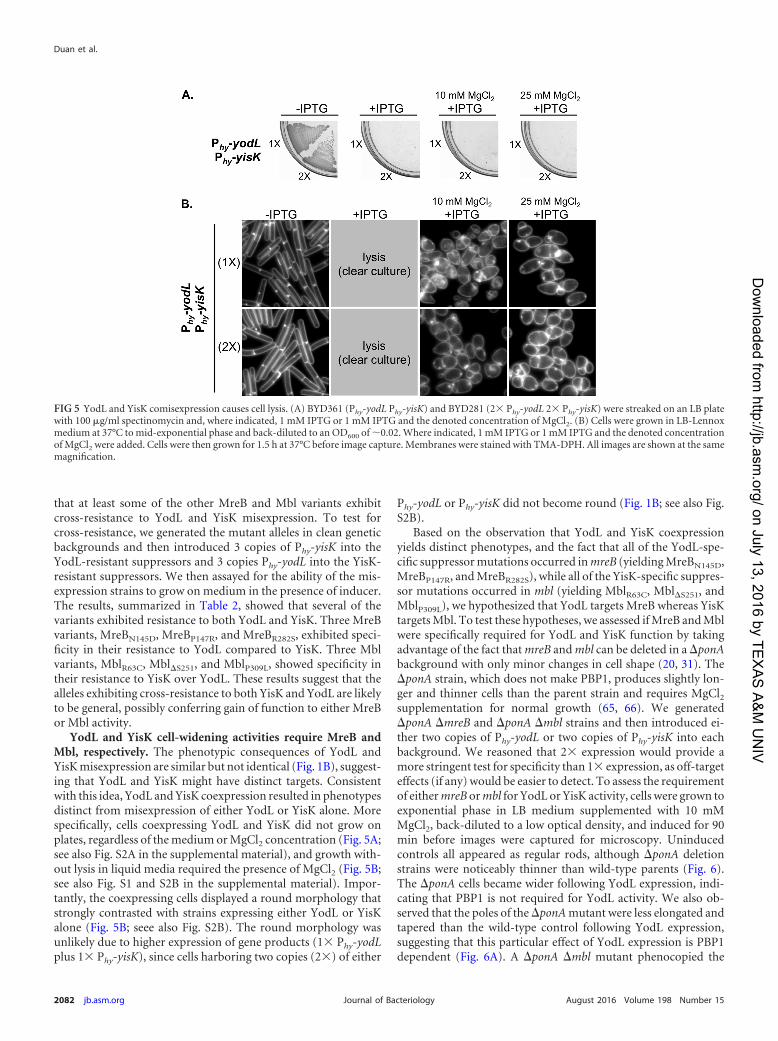
YodL and YisK cell-widening activities require MreB andMbl, respectively. The phenotypic consequences of YodL andYisK misexpression are similar but not identical (Fig. 1B), suggest-ing that YodL and YisK might have distinct targets. Consistentwith this idea, YodL and YisK coexpression resulted in phenotypesdistinct from misexpression of either YodL or YisK alone. Morespecifically, cells coexpressing YodL and YisK did not grow onplates, regardless of the medium or MgCl2 concentration (Fig. 5A;see also Fig. S2A in the supplemental material), and growth with-out lysis in liquid media required the presence of MgCl2 (Fig. 5B;see also Fig. S1 and S2B in the supplemental material). Impor-tantly, the coexpressing cells displayed a round morphology thatstrongly contrasted with strains expressing either YodL or YisKalone (Fig. 5B; seee also Fig. S2B). The round morphology wasunlikely due to higher expression of gene products (1� Phy-yodLplus 1� Phy-yisK), since cells harboring two copies (2�) of either
Phy-yodL or Phy-yisK did not become round (Fig. 1B; see also Fig.S2B).
Based on the observation that YodL and YisK coexpressionyields distinct phenotypes, and the fact that all of the YodL-spe-cific suppressor mutations occurred inmreB (yielding MreBN145D,MreBP147R, and MreBR282S), while all of the YisK-specific suppres-sor mutations occurred in mbl (yielding MblR63C, Mbl�S251, andMblP309L), we hypothesized that YodL targets MreB whereas YisKtargets Mbl. To test these hypotheses, we assessed if MreB and Mblwere specifically required for YodL and YisK function by takingadvantage of the fact that mreB and mbl can be deleted in a �ponAbackground with only minor changes in cell shape (20, 31). The�ponA strain, which does not make PBP1, produces slightly lon-ger and thinner cells than the parent strain and requires MgCl2supplementation for normal growth (65, 66). We generated�ponA �mreB and �ponA �mbl strains and then introduced ei-ther two copies of Phy-yodL or two copies of Phy-yisK into eachbackground. We reasoned that 2� expression would provide amore stringent test for specificity than 1� expression, as off-targeteffects (if any) would be easier to detect. To assess the requirementof eithermreB ormbl for YodL or YisK activity, cells were grown toexponential phase in LB medium supplemented with 10 mMMgCl2, back-diluted to a low optical density, and induced for 90min before images were captured for microscopy. Uninducedcontrols all appeared as regular rods, although �ponA deletionstrains were noticeably thinner than wild-type parents (Fig. 6).The �ponA cells became wider following YodL expression, indi-cating that PBP1 is not required for YodL activity. We also ob-served that the poles of the �ponAmutant were less elongated andtapered than the wild-type control following YodL expression,suggesting that this particular effect of YodL expression is PBP1dependent (Fig. 6A). A �ponA �mbl mutant phenocopied the
FIG 5 YodL and YisK comisexpression causes cell lysis. (A) BYD361 (Phy-yodL Phy-yisK) and BYD281 (2� Phy-yodL 2� Phy-yisK) were streaked on an LB platewith 100 �g/ml spectinomycin and, where indicated, 1 mM IPTG or 1 mM IPTG and the denoted concentration of MgCl2. (B) Cells were grown in LB-Lennoxmedium at 37°C to mid-exponential phase and back-diluted to an OD600 of �0.02. Where indicated, 1 mM IPTG or 1 mM IPTG and the denoted concentrationof MgCl2 were added. Cells were then grown for 1.5 h at 37°C before image capture. Membranes were stained with TMA-DPH. All images are shown at the samemagnification.
Duan et al.
2082 jb.asm.org August 2016 Volume 198 Number 15Journal of Bacteriology
on July 13, 2016 by TEX
AS
A&
M U
NIV
http://jb.asm.org/
Dow
nloaded from
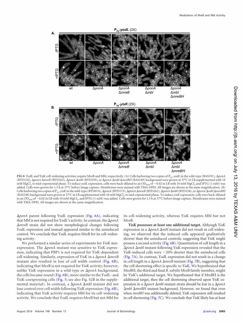
�ponA parent following YodL expression (Fig. 6A), indicatingthat Mbl is not required for YodL’s activity. In contrast, the �ponA�mreB strain did not show morphological changes followingYodL expression and instead appeared similar to the uninducedcontrol. We conclude that YodL requires MreB for its cell-widen-ing activity.
We performed a similar series of experiments for YisK mis-expression. The �ponA mutant was sensitive to YisK expres-sion, indicating that PBP1 is not required for YisK-dependentcell widening. Similarly, expression of YisK in a �ponA �mreBmutant also resulted in loss of cell width control (Fig. 6B),indicating that MreB is not required for YisK activity; however,unlike YisK expression in a wild-type or �ponA background,the cells became round (Fig. 6B), more similar to the YodL- andYisK-coexpressing cells (Fig. 5; see also Fig. S2B in the supple-mental material). In contrast, a �ponA �mbl mutant did notlose control over cell width following YisK expression (Fig. 6B),indicating that YisK activity requires Mbl for its cell-wideningactivity. We conclude that YodL requires MreB but not Mbl for
its cell-widening activity, whereas YisK requires Mbl but notMreB.
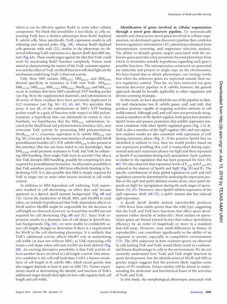
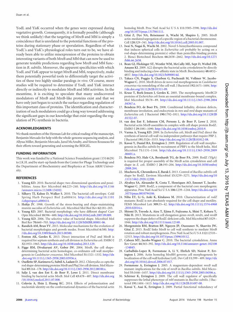
YisK possesses at least one additional target. Although YisKexpression in a �ponA �mbl mutant did not result in cell widen-ing, we observed that the induced cells appeared qualitativelyshorter than the uninduced controls, suggesting that YisK mightpossess a second activity (Fig. 6B). Quantitation of cell length in a�ponA �mbl mutant following YisK expression revealed that theYisK-induced cells were �20% shorter than the uninduced cells(Fig. 7A). In contrast, YodL expression did not result in a changein cell length in a �ponA �mreB mutant (Fig. 7B), suggesting thatthe cell shortening effect is specific to YisK. We hypothesized thatMreBH, the third and final B. subtilisMreB family member, mightbe YisK’s additional target. We hypothesized that if MreBH is theadditional target, then the cell shortening observed upon YisK ex-pression in a �ponA �mbl mutant strain should be lost in a �ponA�mbl �mreBH mutant background. However, we found that evenwhen mreBH was additionally deleted, YisK expression still resultedin cell shortening (Fig. 7C). We conclude that YisK likely has at least
FIG 6 YodL and YisK cell-widening activities require MreB and Mbl, respectively. (A) Cells harboring two copies of Phy-yodL in the wild-type (BAS191), �ponA(BYD176), �ponA�mreB (BYD263), �ponA�mbl (BYD259), or �ponA�mbl�mreBH (BAS249) background were grown at 37°C in LB supplemented with 10mM MgCl2 to mid-exponential phase. To induce yodL expression, cells were back-diluted to an OD600 of �0.02 in LB with 10 mM MgCl2 and IPTG (1 mM) wasadded. Cells were grown for 1.5 h at 37°C before image capture. Membranes were stained with TMA-DPH. All images are shown at the same magnification. (B)Cells harboring two copies of Phy-yisK in the wild-type (BYD074), �ponA (BYD175), �ponA�mreB (BYD262), �ponA�mbl (BYD258), or �ponA�mbl�mreBH(BAS248) background were grown at 37°C in LB supplemented with 10 mM MgCl2 to mid-exponential phase. To induce yisK expression, cells were back-dilutedto an OD600 of �0.02 in LB with 10 mM MgCl2, and IPTG (1 mM) was added. Cells were grown for 1.5 h at 37°C before image capture. Membranes were stainedwith TMA-DPH. All images are shown at the same magnification.
Modulators of MreB and Mbl Activity
August 2016 Volume 198 Number 15 jb.asm.org 2083Journal of Bacteriology
on July 13, 2016 by TEX
AS
A&
M U
NIV
http://jb.asm.org/
Dow
nloaded from
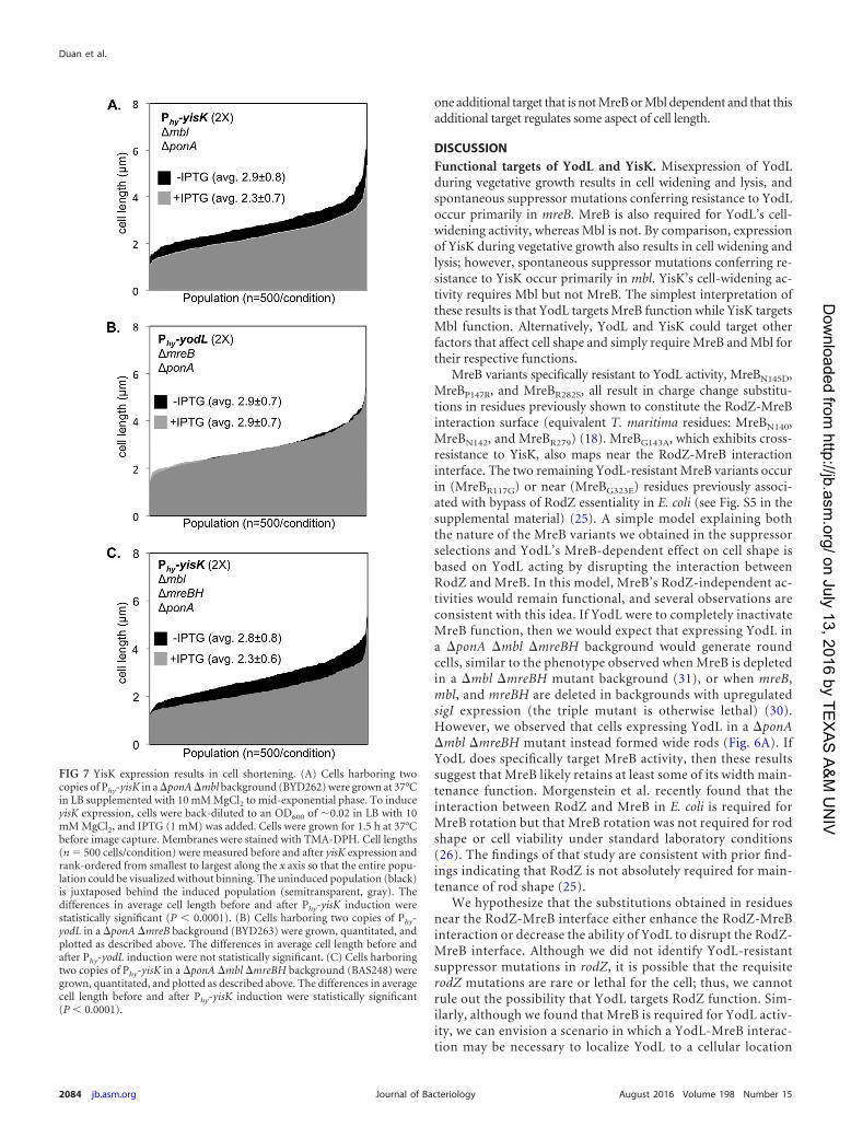
one additional target that is not MreB or Mbl dependent and that thisadditional target regulates some aspect of cell length.
DISCUSSIONFunctional targets of YodL and YisK. Misexpression of YodLduring vegetative growth results in cell widening and lysis, andspontaneous suppressor mutations conferring resistance to YodLoccur primarily in mreB. MreB is also required for YodL’s cell-widening activity, whereas Mbl is not. By comparison, expressionof YisK during vegetative growth also results in cell widening andlysis; however, spontaneous suppressor mutations conferring re-sistance to YisK occur primarily in mbl. YisK’s cell-widening ac-tivity requires Mbl but not MreB. The simplest interpretation ofthese results is that YodL targets MreB function while YisK targetsMbl function. Alternatively, YodL and YisK could target otherfactors that affect cell shape and simply require MreB and Mbl fortheir respective functions.
MreB variants specifically resistant to YodL activity, MreBN145D,MreBP147R, and MreBR282S, all result in charge change substitu-tions in residues previously shown to constitute the RodZ-MreBinteraction surface (equivalent T. maritima residues: MreBN140,MreBN142, and MreBR279) (18). MreBG143A, which exhibits cross-resistance to YisK, also maps near the RodZ-MreB interactioninterface. The two remaining YodL-resistant MreB variants occurin (MreBR117G) or near (MreBG323E) residues previously associ-ated with bypass of RodZ essentiality in E. coli (see Fig. S5 in thesupplemental material) (25). A simple model explaining boththe nature of the MreB variants we obtained in the suppressorselections and YodL’s MreB-dependent effect on cell shape isbased on YodL acting by disrupting the interaction betweenRodZ and MreB. In this model, MreB’s RodZ-independent ac-tivities would remain functional, and several observations areconsistent with this idea. If YodL were to completely inactivateMreB function, then we would expect that expressing YodL ina �ponA �mbl �mreBH background would generate roundcells, similar to the phenotype observed when MreB is depletedin a �mbl �mreBH mutant background (31), or when mreB,mbl, and mreBH are deleted in backgrounds with upregulatedsigI expression (the triple mutant is otherwise lethal) (30).However, we observed that cells expressing YodL in a �ponA�mbl �mreBH mutant instead formed wide rods (Fig. 6A). IfYodL does specifically target MreB activity, then these resultssuggest that MreB likely retains at least some of its width main-tenance function. Morgenstein et al. recently found that theinteraction between RodZ and MreB in E. coli is required forMreB rotation but that MreB rotation was not required for rodshape or cell viability under standard laboratory conditions(26). The findings of that study are consistent with prior find-ings indicating that RodZ is not absolutely required for main-tenance of rod shape (25).
We hypothesize that the substitutions obtained in residuesnear the RodZ-MreB interface either enhance the RodZ-MreBinteraction or decrease the ability of YodL to disrupt the RodZ-MreB interface. Although we did not identify YodL-resistantsuppressor mutations in rodZ, it is possible that the requisiterodZ mutations are rare or lethal for the cell; thus, we cannotrule out the possibility that YodL targets RodZ function. Sim-ilarly, although we found that MreB is required for YodL activ-ity, we can envision a scenario in which a YodL-MreB interac-tion may be necessary to localize YodL to a cellular location
FIG 7 YisK expression results in cell shortening. (A) Cells harboring twocopies of Phy-yisK in a �ponA�mbl background (BYD262) were grown at 37°Cin LB supplemented with 10 mM MgCl2 to mid-exponential phase. To induceyisK expression, cells were back-diluted to an OD600 of �0.02 in LB with 10mM MgCl2, and IPTG (1 mM) was added. Cells were grown for 1.5 h at 37°Cbefore image capture. Membranes were stained with TMA-DPH. Cell lengths(n 500 cells/condition) were measured before and after yisK expression andrank-ordered from smallest to largest along the x axis so that the entire popu-lation could be visualized without binning. The uninduced population (black)is juxtaposed behind the induced population (semitransparent, gray). Thedifferences in average cell length before and after Phy-yisK induction werestatistically significant (P 0.0001). (B) Cells harboring two copies of Phy-yodL in a �ponA �mreB background (BYD263) were grown, quantitated, andplotted as described above. The differences in average cell length before andafter Phy-yodL induction were not statistically significant. (C) Cells harboringtwo copies of Phy-yisK in a �ponA �mbl �mreBH background (BAS248) weregrown, quantitated, and plotted as described above. The differences in averagecell length before and after Phy-yisK induction were statistically significant(P 0.0001).
Duan et al.
2084 jb.asm.org August 2016 Volume 198 Number 15Journal of Bacteriology
on July 13, 2016 by TEX
AS
A&
M U
NIV
http://jb.asm.org/
Dow
nloaded from
where it can be effective against RodZ or some other cellularcomponent. We think this possibility is less likely, as cells ex-pressing YodL have a distinct phenotype from RodZ depletedB. subtilis cells. More specifically, YodL expression results in cellwidening and tapered poles (Fig. 1B), whereas RodZ-depletedcells generate wide rods (22), similar to the phenotype we ob-served following YodL expression in a �ponA�mbl�mreBH mu-tant (Fig. 6A). These results argue against the idea that YodL couldwork by inactivating RodZ function completely. Future workaimed at characterizing the nature of the YodL-resistant suppres-sors and the effect of YodL on MreB function will shed light on themechanism underlying YodL’s observed activity.
Only three Mbl variants MblR63C, Mbl�S251, and MblP309L
showed specificity in resistance to YisK over YodL. MblR63C,MblD153N, MblG156D, MblT158A, MblE204G, MreBP309L, and MblA314T
occur in residues that form Mbl’s predicted ATP binding pocket(see Fig. S6 in the supplemental material), and substitutions inall seven of these residues have been previously implicated inA22 resistance (see Fig. S6) (12, 63, 64). We speculate thatmost, if not all, of the substitutions in Mbl’s ATP bindingpocket result in a gain of function with respect to Mbl polym-erization, a hypothesis that can ultimately be tested in vitro.Similarly, we hypothesize that the MblM51I substitution, lo-cated at the MreB head-tail polymerization interface (62), mayovercome YisK activity by promoting Mbl polymerization.MreBE262 of C. crescentus, equivalent to B. subtilis MblE250 (seeFig. S6), is located at the interaction interface of antiparallel MreBprotofilament bundles (67). If B. subtilis MblE250 is also present atthis interface (this has not been tested to our knowledge), thenMblE250K could promote resistance to YodL and YisK by enhanc-ing Mbl bundling. How might YisK exert its activity? One idea isthat YisK disrupts Mbl bundling, possibly by competing for sitesrequired for protofilament formation. An alternative possibility isthat YisK somehow prevents Mbl from effectively binding or hy-drolyzing ATP. It is also possible that Mbl is simply required forYisK to target one or more other factors involved in cell widthcontrol.
In addition to Mbl-dependent cell widening, YisK expres-sion resulted in cell shortening, an effect that only becameapparent in a �ponA �mbl mutant background (Fig. 6B and7A). Given the similarities of MreB, Mbl, and MreBH to eachother, we initially hypothesized that YisK-dependent effects onMreB and/or MreBH might be responsible for the decrease incell length we observed; however, we found that mreBH was notrequired for cell shortening (Fig. 6B and 7C). Since YisK ex-pression results in a dramatic loss of cell shape in �mreB mu-tant backgrounds (Fig. 6A), we were unable to confidently as-sess cell length changes to determine if there is a requirementfor MreB in the cell-shortening phenotype. It is unlikely thatYisK’s additional activity affects MreB’s role in maintainingcell width (at least not without Mbl), as YisK-expressing cellsretain a rod shape when mbl and mreBH are both deleted (Fig.6B). An exciting alternative possibility is that YisK activity af-fects another factor involved in cell length control. One attrac-tive candidate is the cell wall hydrolase CwlO, a known modu-lator of cell length in B. subtilis (68), which recent genetic dataalso suggest depends at least in part on Mbl (35). Future experi-ments aimed at determining the identity and function of YisK’sadditional target should shed light on how cells regulate both celllength and cell width.
Identification of genes involved in cellular organizationthrough a novel gene discovery pipeline. To systematicallyidentify and characterize novel genes involved in cellular orga-nization, we developed a gene discovery pipeline that combinesknown regulatory information (47), phenotypes obtained frommisexpression screening, and suppressor selection analysis.The ability to identify genetic targets associated with the un-known genes provides a key parameter beyond phenotype fromwhich to formulate testable hypotheses regarding each gene’spossible function. The misexpression constructs we generatedare inducible and present in single copy on the chromosome.We have found that to obtain phenotypes, our strategy worksbest when the unknown genes are expressed outside their na-tive regulatory context. Thus far, we have restricted our genefunction discovery pipeline to B. subtilis; however, the generalapproach should be broadly applicable to other organisms anddiverse screening strategies.
In this work, we have described the use of the pipeline to iden-tify and characterize two B. subtilis genes, yodL and yisK, thatproduce proteins capable of targeting activities intrinsic to cellwidth control. Although yodL and yisKwere not previously recog-nized as members of the Spo0A regulon, both genes have putativeSpo0A boxes and possess promoters that exhibit expression pat-terns consistent with other Spo0A-regulated genes (Fig. 2 to 4).YisK is also a member of the SigH regulon (49), and our expres-sion analysis results are also consistent with expression of yisKduring stationary phase (Fig. 3). If the putative Spo0A box weidentified is utilized in vivo, then we would predict based onour expression profiling that yisK is transcribed during expo-nential and early stationary phases via SigH and then repressedas Spo0A-P accumulates during early sporulation. Such a patternis similar to the regulation that has been proposed for kinA (55,60). We also observed that expression levels of PyodL and PyisK arereduced in the absence of Spo0A and SigH (Fig. 3D and E). Thespecific contributions of these global regulators to yodL and yisKregulation cannot be determined by analyzing the expression pro-files of the sigH and spo0A deletion strains alone, since spo0A de-pends on SigH for upregulation during the early stages of sporu-lation (52, 55). Moreover, since Spo0A inhibits expression of thesigH repressor AbrB (69–72), a spo0A mutant also has reducedsigH expression.
A �yodL �yisK double mutant reproducibly produces�20% fewer heat-stable spores than the wild type, suggestingthat the YodL and YisK have functions that affect spore devel-opment (either directly or indirectly). Most studies on sporu-lation genes are biased toward factors that reduce sporulationefficiency by an order of magnitude or more in a standardheat-kill assay. However, even small differences in fitness (ifreproducible) can contribute significantly to the ability of anorganism to persist, especially in competitive environments(73). The 20% reduction in heat-resistant spores we observedin cells lacking YisK and YodL would likely result in a substan-tial fitness disadvantage to cells in the environment. We do notcurrently understand how YodL and YisK might function inspore development, but the identification of MreB and Mbl asgenetic targets suggests that the proteins likely regulate someaspect of PG synthesis. Future studies will be aimed at under-standing the molecular and biochemical bases of the activitiesof YodL and YisK.
In this study, the morphological phenotypes associated with
Modulators of MreB and Mbl Activity
August 2016 Volume 198 Number 15 jb.asm.org 2085Journal of Bacteriology
on July 13, 2016 by TEX
AS
A&
M U
NIV
http://jb.asm.org/
Dow
nloaded from
YodL and YisK occurred when the genes were expressed duringvegetative growth. Consequently, it is formally possible (althoughwe think unlikely) that the targeting of MreB and Mbl is simply acoincidence that is unrelated to the potential functions of the pro-teins during stationary phase or sporulation. Regardless of whatYodL’s and YisK’s physiological roles turn out to be, we have al-ready been able to utilize misexpression of the proteins to obtaininteresting variants of both MreB and Mbl that can now be used togenerate testable predictions regarding how MreB and Mbl func-tion in B. subtilis. Moreover, the apparent specificities with whichYodL and YisK appear to target MreB and Mbl, respectively, makethem potentially powerful tools to differentially target the activi-ties of these two highly similar paralogs in vivo. Of course, morestudies will be required to determine if YodL and YisK interactdirectly or indirectly to modulate MreB and Mbl activities. In themeantime, it is exciting to speculate that many undiscoveredmodulators of MreB and MreB-like proteins exist and that wehave only just begun to scratch the surface regarding regulation ofthis important class of proteins. The identification and character-ization of such modulators could go a long way toward addressingthe significant gaps in our knowledge that exist regarding the reg-ulation of PG synthesis in bacteria.
ACKNOWLEDGMENTS
We thank members of the Herman Lab for critical reading of the manuscript,Benjamin Mercado for help with the whole-genome sequencing analysis, andAllyssa Miller, Benjamin Mercado, Jared McAnulty, and Simon Rousseau fortheir efforts toward generating and screening the BEIGEL.
FUNDING INFORMATIONThis work was funded by a National Science Foundation grant (1514629)to J.K.H. and by start-up funds from the Center for Phage Technology andthe Department of Biochemistry and Biophysics at Texas A&M Univer-sity.
REFERENCES1. Young KD. 2010. Bacterial shape: two-dimensional questions and possi-
bilities. Annu Rev Microbiol 64:223–240. http://dx.doi.org/10.1146/annurev.micro.112408.134102.
2. Silhavy TJ, Kahne D, Walker S. 2010. The bacterial cell envelope. ColdSpring Harb Perspect Biol 2:a000414. http://dx.doi.org/10.1101/cshperspect.a000414.
3. Holtje JV. 1998. Growth of the stress-bearing and shape-maintainingmurein sacculus of Escherichia coli. Microbiol Mol Biol Rev 62:181–203.
4. Young KD. 2007. Bacterial morphology: why have different shapes? CurrOpin Microbiol 10:596–600. http://dx.doi.org/10.1016/j.mib.2007.09.009.
5. Young KD. 2006. The selective value of bacterial shape. Microbiol MolBiol Rev 70:660 –703. http://dx.doi.org/10.1128/MMBR.00001-06.
6. Randich AM, Brun YV. 2015. Molecular mechanisms for the evolution ofbacterial morphologies and growth modes. Front Microbiol 6:580. http://dx.doi.org/10.3389/fmicb.2015.00580.
7. Fenton AK, Gerdes K. 2013. Direct interaction of FtsZ and MreB isrequired for septum synthesis and cell division in Escherichia coli. EMBO J32:1953–1965. http://dx.doi.org/10.1038/emboj.2013.129.
8. Figge RM, Divakaruni AV, Gober JW. 2004. MreB, the cell shape-determining bacterial actin homologue, co-ordinates cell wall morpho-genesis in Caulobacter crescentus. Mol Microbiol 51:1321–1332. http://dx.doi.org/10.1111/j.1365-2958.2003.03936.x.
9. Ouellette SP, KarimovaG, Subtil A, Ladant D. 2012.Chlamydia co-opts therod shape-determining proteins MreB and Pbp2 for cell division. Mol Micro-biol 85:164–178. http://dx.doi.org/10.1111/j.1365-2958.2012.08100.x.
10. Salje J, van den Ent F, de Boer P, Lowe J. 2011. Direct membranebinding by bacterial actin MreB. Mol Cell 43:478 – 487. http://dx.doi.org/10.1016/j.molcel.2011.07.008.
11. Colavin A, Hsin J, Huang KC. 2014. Effects of polymerization andnucleotide identity on the conformational dynamics of the bacterial actin
homolog MreB. Proc Natl Acad Sci U S A 111:3585–3590. http://dx.doi.org/10.1073/pnas.1317061111.
12. Gitai Z, Dye NA, Reisenauer A, Wachi M, Shapiro L. 2005. MreBactin-mediated segregation of a specific region of a bacterial chromosome.Cell 120:329 –341. http://dx.doi.org/10.1016/j.cell.2005.01.007.
13. Iwai N, Nagai K, Wachi M. 2002. Novel S-benzylisothiourea compoundthat induces spherical cells in Escherichia coli probably by acting on arod-shape-determining protein(s) other than penicillin-binding protein2. Biosci Biotechnol Biochem 66:2658 –2662. http://dx.doi.org/10.1271/bbb.66.2658.
14. Bean GJ, Flickinger ST, Westler WM, McCully ME, Sept D, Weibel DB,Amann KJ. 2009. A22 disrupts the bacterial actin cytoskeleton by directlybinding and inducing a low-affinity state in MreB. Biochemistry 48:4852–4857. http://dx.doi.org/10.1021/bi900014d.
15. Takacs CN, Poggio S, Charbon G, Pucheault M, Vollmer W, Jacobs-Wagner C. 2010. MreB drives de novo rod morphogenesis in Caulobactercrescentus via remodeling of the cell wall. J Bacteriol 192:1671–1684. http://dx.doi.org/10.1128/JB.01311-09.
16. Kruse T, Bork-Jensen J, Gerdes K. 2005. The morphogenetic MreBCDproteins of Escherichia coli form an essential membrane-bound complex.Mol Microbiol 55:78 – 89. http://dx.doi.org/10.1111/j.1365-2598.2004.04367.x.
17. Bendezu FO, de Boer PA. 2008. Conditional lethality, division defects,membrane involution, and endocytosis in mre and mrd shape mutants ofEscherichia coli. J Bacteriol 190:1792–1811. http://dx.doi.org/10.1128/JB.01322-07.
18. van den Ent F, Johnson CM, Persons L, de Boer P, Lowe J. 2010.Bacterial actin MreB assembles in complex with cell shape protein RodZ.EMBO J 29:1081–1090. http://dx.doi.org/10.1038/emboj.2010.9.
19. Varma A, Young KD. 2009. In Escherichia coli, MreB and FtsZ direct thesynthesis of lateral cell wall via independent pathways that require PBP 2.J Bacteriol 191:3526 –3533. http://dx.doi.org/10.1128/JB.01812-08.
20. Kawai Y, Daniel RA, Errington J. 2009. Regulation of cell wall morpho-genesis in Bacillus subtilis by recruitment of PBP1 to the MreB helix. MolMicrobiol 71:1131–1144. http://dx.doi.org/10.1111/j.1365-2958.2009.06601.x.
21. Bendezu FO, Hale CA, Bernhardt TG, de Boer PA. 2009. RodZ (YfgA)is required for proper assembly of the MreB actin cytoskeleton and cellshape in E. coli. EMBO J 28:193–204. http://dx.doi.org/10.1038/emboj.2008.264.
22. Muchova K, Chromikova Z, Barak I. 2013. Control of Bacillus subtilis cellshape by RodZ. Environ Microbiol 15:3259 –3271. http://dx.doi.org/10.1111/1462-2920.12200.
23. Alyahya SA, Alexander R, Costa T, Henriques AO, Emonet T, Jacobs-Wagner C. 2009. RodZ, a component of the bacterial core morphogenicapparatus. Proc Natl Acad Sci U S A 106:1239 –1244. http://dx.doi.org/10.1073/pnas.0810794106.
24. Niba ET, Li G, Aoki K, Kitakawa M. 2010. Characterization of rodZmutants: RodZ is not absolutely required for the cell shape and motility.FEMS Microbiol Lett 309:35– 42. http://dx.doi.org/10.1111/j.1574-6968.2010.02014.x.
25. Shiomi D, Toyoda A, Aizu T, Ejima F, Fujiyama A, Shini T, Kohara Y,Niki H. 2013. Mutations in cell elongation genes mreB, mrdA, and mrdBsuppress the shape defect of RodZ-deficient cells. Mol Microbiol 87:1029 –1044. http://dx.doi.org/10.1111/mmi.12148.
26. Morgenstein RM, Bratton BP, Nguyen JP, Ouzounov N, Shaevitz JW,Gitai Z. 2015. RodZ links MreB to cell wall synthesis to mediate MreBrotation and robust morphogenesis. Proc Natl Acad Sci U S A 112:12510 –12515. http://dx.doi.org/10.1073/pnas.1509610112.
27. Cabeen MT, Jacobs-Wagner C. 2010. The bacterial cytoskeleton. AnnuRev Genet 44:365–392. http://dx.doi.org/10.1146/annurev-genet-102108-134845.
28. Carballido-Lopez R, Formstone A, Li Y, Ehrlich SD, Noirot P, Err-ington J. 2006. Actin homolog MreBH governs cell morphogenesis bylocalization of the cell wall hydrolase LytE. Dev Cell 11:399 – 409. http://dx.doi.org/10.1016/j.devcel.2006.07.017.
29. Formstone A, Errington J. 2005. A magnesium-dependent mreB nullmutant: implications for the role of mreB in Bacillus subtilis. Mol Micro-biol 55:1646 –1657. http://dx.doi.org/10.1111/j.1365-2958.2005.04506.x.
30. Schirner K, Errington J. 2009. The cell wall regulator �I specificallysuppresses the lethal phenotype of mbl mutants in Bacillus subtilis. J Bac-teriol 191:1404 –1413. http://dx.doi.org/10.1128/JB.01497-08.
31. Kawai Y, Asai K, Errington J. 2009. Partial functional redundancy of
Duan et al.
2086 jb.asm.org August 2016 Volume 198 Number 15Journal of Bacteriology
on July 13, 2016 by TEX
AS
A&
M U
NIV
http://jb.asm.org/
Dow
nloaded from
MreB isoforms, MreB, Mbl and MreBH, in cell morphogenesis of Bacillussubtilis. Mol Microbiol 73:719 –731. http://dx.doi.org/10.1111/j.1365-2958.2009.06805.x.
32. Defeu Soufo HJ, Graumann PL. 2006. Dynamic localization and inter-action with other Bacillus subtilis actin-like proteins are important for thefunction of MreB. Mol Microbiol 62:1340 –1356. http://dx.doi.org/10.1111/j.1365-2958.2006.05457.x.
33. Mirouze N, Ferret C, Yao Z, Chastanet A, Carballido-Lopez R. 2015.MreB-dependent inhibition of cell elongation during the escape fromcompetence in Bacillus subtilis. PLoS Genet 11:e1005299. http://dx.doi.org/10.1371/journal.pgen.1005299.
34. Tseng CL, Shaw GC. 2008. Genetic evidence for the actin homolog genemreBH and the bacitracin resistance gene bcrC as targets of the alternativesigma factor SigI of Bacillus subtilis. J Bacteriol 190:1561–1567. http://dx.doi.org/10.1128/JB.01497-07.
35. Dominguez-Cuevas P, Porcelli I, Daniel RA, Errington J. 2013. Differ-entiated roles for MreB-actin isologues and autolytic enzymes in Bacillussubtilis morphogenesis. Mol Microbiol 89:1084 –1098. http://dx.doi.org/10.1111/mmi.12335.
36. Masuda H, Tan Q, Awano N, Wu KP, Inouye M. 2012. YeeU enhancesthe bundling of cytoskeletal polymers of MreB and FtsZ, antagonizing theCbtA (YeeV) toxicity in Escherichia coli. Mol Microbiol 84:979 –989. http://dx.doi.org/10.1111/j.1365-2958.2012.08068.x.
37. Tan Q, Awano N, Inouye M. 2011. YeeV is an Escherichia coli toxin thatinhibits cell division by targeting the cytoskeleton proteins, FtsZ andMreB. Mol Microbiol 79:109 –118. http://dx.doi.org/10.1111/j.1365-2958.2010.07433.x.
38. Masuda H, Tan Q, Awano N, Yamaguchi Y, Inouye M. 2012. A novelmembrane-bound toxin for cell division, CptA (YgfX), inhibits polymer-ization of cytoskeleton proteins, FtsZ and MreB, in Escherichia coli. FEMSMicrobiol Lett 328:174 –181. http://dx.doi.org/10.1111/j.1574-6968.2012.02496.x.
39. Yakhnina AA, Gitai Z. 2012. The small protein MbiA interacts with MreBand modulates cell shape in Caulobacter crescentus. Mol Microbiol 85:1090 –1104. http://dx.doi.org/10.1111/j.1365-2958.2012.08159.x.
40. Ababneh QO, Herman JK. 2015. CodY regulates SigD levels and activityby binding to three sites in the fla/che operon. J Bacteriol 197:2999 –3006.http://dx.doi.org/10.1128/JB.00288-15.
41. Harwood CR, Cutting SM. 1990. Molecular biological methods for Ba-cillus. Wiley, New York, NY.
42. RasbandW. 2015. ImageJ. U.S. National Institutes of Health, Bethesda, MD.43. Schaeffer P, Millet J, Aubert JP. 1965. Catabolic repression of bacterial
sporulation. Proc Natl Acad Sci U S A 54:704 –711. http://dx.doi.org/10.1073/pnas.54.3.704.
44. Ababneh QO, Herman JK. 2015. RelA inhibits Bacillus subtilis motilityand chaining. J Bacteriol 197:128 –137. http://dx.doi.org/10.1128/JB.02063-14.
45. Abhayawardhane Y, Stewart GC. 1995. Bacillus subtilis possesses a seconddeterminant with extensive sequence similarity to theEscherichia colimreBmorphogene. J Bacteriol 177:765–773.
46. LeaverM, Errington J. 2005. Roles for MreC and MreD proteins in helicalgrowth of the cylindrical cell wall in Bacillus subtilis. Mol Microbiol 57:1196 –1209. http://dx.doi.org/10.1111/j.1365-2958.2005.04736.x.
47. Nicolas P, Mader U, Dervyn E, Rochat T, Leduc A, Pigeonneau N,Bidnenko E, Marchadier E, Hoebeke M, Aymerich S, Becher D, Bisic-chia P, Botella E, Delumeau O, Doherty G, Denham EL, Fogg MJ,Fromion V, Goelzer A, Hansen A, Hartig E, Harwood CR, Homuth G,Jarmer H, Jules M, Klipp E, Le Chat L, Lecointe F, Lewis P, Lieber-meister W, March A, Mars RA, Nannapaneni P, Noone D, Pohl S, RinnB, Rugheimer F, Sappa PK, Samson F, Schaffer M, Schwikowski B, SteilL, Stulke J, Wiegert T, Devine KM, Wilkinson AJ, van Dijl JM, HeckerM, Volker U, Bessieres P, Noirot P. 2012. Condition-dependent tran-scriptome reveals high-level regulatory architecture in Bacillus subtilis.Science 335:1103–1106. http://dx.doi.org/10.1126/science.1206848.
48. Molle V, Fujita M, Jensen ST, Eichenberger P, Gonzalez-Pastor JE, LiuJS, Losick R. 2003. The Spo0A regulon of Bacillus subtilis. Mol Microbiol50:1683–1701. http://dx.doi.org/10.1046/j.1365-2958.2003.03818.x.
49. Britton RA, Eichenberger P, Gonzalez-Pastor JE, Fawcett P, Monson R,Losick R, Grossman AD. 2002. Genome-wide analysis of the stationary-phase sigma factor (sigma-H) regulon of Bacillus subtilis. J Bacteriol 184:4881– 4890. http://dx.doi.org/10.1128/JB.184.17.4881-4890.2002.
50. Arrieta-Ortiz ML, Hafemeister C, Bate AR, Chu T, Greenfield A,Shuster B, Barry SN, Gallitto M, Liu B, Kacmarczyk T, Santoriello F,
Chen J, Rodrigues CD, Sato T, Rudner DZ, Driks A, Bonneau R,Eichenberger P. 2015. An experimentally supported model of the Bacillussubtilis global transcriptional regulatory network. Mol Syst Biol 11:839.http://dx.doi.org/10.15252/msb.20156236.
51. Caspi R, Altman T, Billington R, Dreher K, Foerster H, Fulcher CA,Holland TA, Keseler IM, Kothari A, Kubo A, Krummenacker M,Latendresse M, Mueller LA, Ong Q, Paley S, Subhraveti P, Weaver DS,Weerasinghe D, Zhang P, Karp PD. 2014. The MetaCyc database ofmetabolic pathways and enzymes and the BioCyc collection of pathway/genome databases. Nucleic Acids Res 42:D459 –D471. http://dx.doi.org/10.1093/nar/gkt1103.
52. Predich M, Nair G, Smith I. 1992. Bacillus subtilis early sporulation geneskinA, spo0F, and spo0A are transcribed by the RNA polymerase contain-ing sigma H. J Bacteriol 174:2771–2778.
53. Antoniewski C, Savelli B, Stragier P. 1990. The spoIIJ gene, whichregulates early developmental steps in Bacillus subtilis, belongs to a class ofenvironmentally responsive genes. J Bacteriol 172:86 –93.
54. FujitaM, Sadaie Y. 1998. Promoter selectivity of the Bacillus subtilis RNApolymerase sigmaA and sigmaH holoenzymes. J Biochem 124:89 –97.http://dx.doi.org/10.1093/oxfordjournals.jbchem.a022102.
55. Fujita M, Sadaie Y. 1998. Feedback loops involving Spo0A and AbrB in invitro transcription of the genes involved in the initiation of sporulation inBacillus subtilis. J Biochem 124:98 –104. http://dx.doi.org/10.1093/oxfordjournals.jbchem.a022103.
56. Chastanet A, Vitkup D, Yuan GC, Norman TM, Liu JS, Losick RM.2010. Broadly heterogeneous activation of the master regulator for sporu-lation in Bacillus subtilis. Proc Natl Acad Sci U S A 107:8486 – 8491. http://dx.doi.org/10.1073/pnas.1002499107.
57. de Jong IG, Veening JW, Kuipers OP. 2010. Heterochronic phosphorelaygene expression as a source of heterogeneity in Bacillus subtilis spore forma-tion. J Bacteriol 192:2053–2067. http://dx.doi.org/10.1128/JB.01484-09.
58. Sterlini JM, Mandelstam J. 1969. Commitment to sporulation in Bacillussubtilis and its relationship to development of actinomycin resistance.Biochem J 113:29 –37. http://dx.doi.org/10.1042/bj1130029.
59. Pan Q, Losick R. 2003. Unique degradation signal for ClpCP in Bacillussubtilis. J Bacteriol 185:5275–5278. http://dx.doi.org/10.1128/JB.185.17.5275-5278.2003.
60. Fujita M, Gonzalez-Pastor JE, Losick R. 2005. High- and low-thresholdgenes in the Spo0A regulon of Bacillus subtilis. J Bacteriol 187:1357–1368.http://dx.doi.org/10.1128/JB.187.4.1357-1368.2005.
61. Jiang M, Shao W, Perego M, Hoch JA. 2000. Multiple histidine kinasesregulate entry into stationary phase and sporulation in Bacillus subtilis.Mol Microbiol 38:535–542. http://dx.doi.org/10.1046/j.1365-2958.2000.02148.x.
62. van den Ent F, Amos LA, Lowe J. 2001. Prokaryotic origin of the actincytoskeleton. Nature 413:39 – 44. http://dx.doi.org/10.1038/35092500.
63. Dye NA, Pincus Z, Fisher IC, Shapiro L, Theriot JA. 2011. Mutations inthe nucleotide binding pocket of MreB can alter cell curvature and polarmorphology inCaulobacter. Mol Microbiol 81:368 –394. http://dx.doi.org/10.1111/j.1365-2958.2011.07698.x.
64. Srivastava P, Demarre G, Karpova TS, McNally J, Chattoraj DK. 2007.Changes in nucleoid morphology and origin localization upon inhibitionor alteration of the actin homolog, MreB, of Vibrio cholerae. J Bacteriol189:7450 –7463. http://dx.doi.org/10.1128/JB.00362-07.
65. Murray T, Popham DL, Setlow P. 1998. Bacillus subtilis cells lackingpenicillin-binding protein 1 require increased levels of divalent cations forgrowth. J Bacteriol 180:4555– 4563.
66. Popham DL, Setlow P. 1996. Phenotypes of Bacillus subtilis mutantslacking multiple class A high-molecular-weight penicillin-binding pro-teins. J Bacteriol 178:2079 –2085.
67. van den Ent F, Izore T, Bharat TA, Johnson CM, Lowe J. 2014. Bacterialactin MreB forms antiparallel double filaments. eLife 3:e02634. http://dx.doi.org/10.7554/eLife.02634.
68. Meisner J, Montero Llopis P, Sham LT, Garner E, Bernhardt TG,Rudner DZ. 2013. FtsEX is required for CwlO peptidoglycan hydrolaseactivity during cell wall elongation in Bacillus subtilis. Mol Microbiol 89:1069 –1083. http://dx.doi.org/10.1111/mmi.12330.
69. Dubnau EJ, Cabane K, Smith I. 1987. Regulation of spo0H, an earlysporulation gene in bacilli. J Bacteriol 169:1182–1191.
70. Weir J, Predich M, Dubnau E, Nair G, Smith I. 1991. Regulation ofspo0H, a gene coding for the Bacillus subtilis sigma H factor. J Bacteriol173:521–529.
71. Perego M, Spiegelman GB, Hoch JA. 1988. Structure of the gene for the
Modulators of MreB and Mbl Activity
August 2016 Volume 198 Number 15 jb.asm.org 2087Journal of Bacteriology
on July 13, 2016 by TEX
AS
A&
M U
NIV
http://jb.asm.org/
Dow
nloaded from

transition state regulator, abrB: regulator synthesis is controlled by thespo0A sporulation gene in Bacillus subtilis. Mol Microbiol 2:689 – 699.http://dx.doi.org/10.1111/j.1365-2958.1988.tb00079.x.
72. Strauch M, Webb V, Spiegelman G, Hoch JA. 1990. The SpoOA proteinof Bacillus subtilis is a repressor of the abrB gene. Proc Natl Acad Sci U S A87:1801–1805. http://dx.doi.org/10.1073/pnas.87.5.1801.
73. Wiser MJ, Lenski RE. 2015. A comparison of methods to measure fitnessin Escherichia coli. PLoS One 10:e0126210. http://dx.doi.org/10.1371/journal.pone.0126210.
74. Gitai Z, Dye N, Shapiro L. 2004. An actin-like gene can determine cellpolarity in bacteria. Proc Natl Acad Sci U S A 101:8643– 8648. http://dx.doi.org/10.1073/pnas.0402638101.
Duan et al.
2088 jb.asm.org August 2016 Volume 198 Number 15Journal of Bacteriology
on July 13, 2016 by TEX
AS
A&
M U
NIV
http://jb.asm.org/
Dow
nloaded from
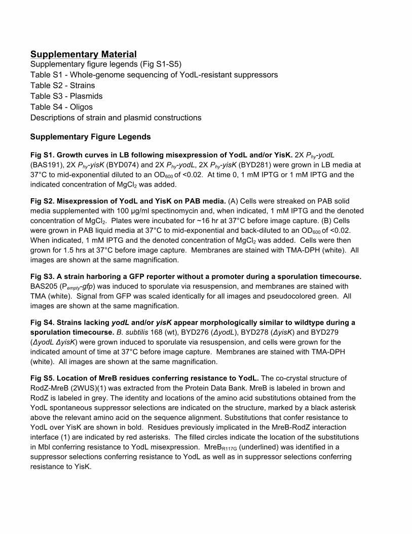
Supplementary Material Supplementary figure legends (Fig S1-S5) Table S1 - Whole-genome sequencing of YodL-resistant suppressors Table S2 - Strains Table S3 - Plasmids Table S4 - Oligos Descriptions of strain and plasmid constructions Supplementary Figure Legends Fig S1. Growth curves in LB following misexpression of YodL and/or YisK. 2X Phy-yodL (BAS191), 2X Phy-yisK (BYD074) and 2X Phy-yodL, 2X Phy-yisK (BYD281) were grown in LB media at 37°C to mid-exponential diluted to an OD600 of <0.02. At time 0, 1 mM IPTG or 1 mM IPTG and the indicated concentration of MgCl2 was added.
Fig S2. Misexpression of YodL and YisK on PAB media. (A) Cells were streaked on PAB solid media supplemented with 100 µg/ml spectinomycin and, when indicated, 1 mM IPTG and the denoted concentration of MgCl2. Plates were incubated for ~16 hr at 37°C before image capture. (B) Cells were grown in PAB liquid media at 37°C to mid-exponential and back-diluted to an OD600 of <0.02. When indicated, 1 mM IPTG and the denoted concentration of MgCl2 was added. Cells were then grown for 1.5 hrs at 37°C before image capture. Membranes are stained with TMA-DPH (white). All images are shown at the same magnification.
Fig S3. A strain harboring a GFP reporter without a promoter during a sporulation timecourse. BAS205 (Pempty-gfp) was induced to sporulate via resuspension, and membranes are stained with TMA (white). Signal from GFP was scaled identically for all images and pseudocolored green. All images are shown at the same magnification.
Fig S4. Strains lacking yodL and/or yisK appear morphologically similar to wildtype during a sporulation timecourse. B. subtilis 168 (wt), BYD276 (ΔyodL), BYD278 (ΔyisK) and BYD279 (ΔyodL ΔyisK) were grown induced to sporulate via resuspension, and cells were grown for the indicated amount of time at 37°C before image capture. Membranes are stained with TMA-DPH (white). All images are shown at the same magnification.
Fig S5. Location of MreB residues conferring resistance to YodL. The co-crystal structure of RodZ-MreB (2WUS)(1) was extracted from the Protein Data Bank. MreB is labeled in brown and RodZ is labeled in grey. The identity and locations of the amino acid substitutions obtained from the YodL spontaneous suppressor selections are indicated on the structure, marked by a black asterisk above the relevant amino acid on the sequence alignment. Substitutions that confer resistance to YodL over YisK are shown in bold. Residues previously implicated in the MreB-RodZ interaction interface (1) are indicated by red asterisks. The filled circles indicate the location of the substitutions in Mbl conferring resistance to YodL misexpression. MreBR117G (underlined) was identified in a suppressor selections conferring resistance to YodL as well as in suppressor selections conferring resistance to YisK.
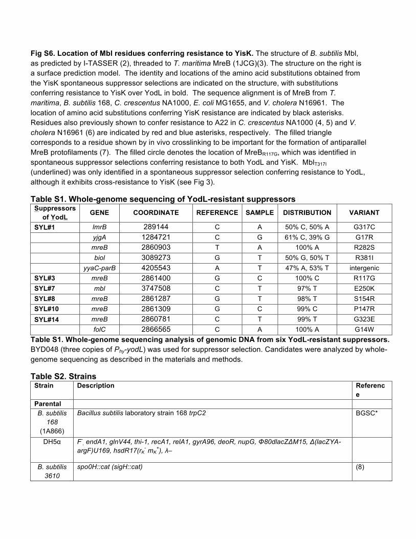
Fig S6. Location of Mbl residues conferring resistance to YisK. The structure of B. subtilis Mbl, as predicted by I-TASSER (2), threaded to T. maritima MreB (1JCG)(3). The structure on the right is a surface prediction model. The identity and locations of the amino acid substitutions obtained from the YisK spontaneous suppressor selections are indicated on the structure, with substitutions conferring resistance to YisK over YodL in bold. The sequence alignment is of MreB from T. maritima, B. subtilis 168, C. crescentus NA1000, E. coli MG1655, and V. cholera N16961. The location of amino acid substitutions conferring YisK resistance are indicated by black asterisks. Residues also previously shown to confer resistance to A22 in C. crescentus NA1000 (4, 5) and V. cholera N16961 (6) are indicated by red and blue asterisks, respectively. The filled triangle corresponds to a residue shown by in vivo crosslinking to be important for the formation of antiparallel MreB protofilaments (7). The filled circle denotes the location of MreBR117G, which was identified in spontaneous suppressor selections conferring resistance to both YodL and YisK. MblT317I (underlined) was only identified in a spontaneous suppressor selection conferring resistance to YodL, although it exhibits cross-resistance to YisK (see Fig 3).
Table S1. Whole-genome sequencing of YodL-resistant suppressors Suppressors
of YodL GENE COORDINATE REFERENCE SAMPLE DISTRIBUTION VARIANT
SYL#1 lmrB 289144 C A 50% C, 50% A G317C yjgA 1284721 C G 61% C, 39% G G17R mreB 2860903 T A 100% A R282S biol 3089273 G T 50% G, 50% T R381I yyaC-parB 4205543 A T 47% A, 53% T intergenic SYL#3 mreB 2861400 G C 100% C R117G SYL#7 mbl 3747508 C T 97% T E250K SYL#8 mreB 2861287 G T 98% T S154R SYL#10 mreB 2861309 G C 99% C P147R SYL#14 mreB 2860781 C T 99% T G323E
folC 2866565 C A 100% A G14W Table S1. Whole-genome sequencing analysis of genomic DNA from six YodL-resistant suppressors. BYD048 (three copies of Phy-yodL) was used for suppressor selection. Candidates were analyzed by whole-genome sequencing as described in the materials and methods.
Table S2. Strains Strain Description Referenc
e Parental B. subtilis
168 (1A866)
Bacillus subtilis laboratory strain 168 trpC2
BGSC*
DH5α F-, endA1, glnV44, thi-1, recA1, relA1, gyrA96, deoR, nupG, Φ80dlacZΔM15, Δ(lacZYA-
argF)U169, hsdR17(rK- mK
+), λ–
B. subtilis 3610
spo0H::cat (sigH::cat) (8)

B. subtilis PY79
spo0A::erm (RL891) (8)
B. subtilis 168
BAS040 amyE::Phy-yodL (spec) This study BAS041 amyE::Phy-yisK (spec) This study BAS146 ponA::erm, kanΩΔmreB This study BAS147 ponA::erm, kanΩΔmbl This study BAS170 amyE::PyodL-lacZ (spec) This study BAS171 amyE::PyodL-gfp (spec) This study BAS191 amyE::Phy-yodL (spec), yhdG::Phy-yodL (phleo) This study BAS192 amyE::PyisK-lacZ (spec) This study BAS193 amyE::PyisK-gfp (spec) This study BAS205 amyE::Pempty-gfp (spec) This study BAS248 ponA::erm, kanΩΔmbl, catΩΔmreBH, amyE::Phy-yisK (spec), yhdG::Phy-yisK (phleo) This study BAS249 ponA::erm, kanΩΔmbl, catΩΔmreBH, amyE::Phy-yodL (spec), yhdG::Phy-yodL (phleo) This study BAS265 spo0A::erm This study BAS266 amyE::PyodL-gfp (spec), spo0A::erm This study BAS267 amyE::PyisK-gfp (spec), spo0A::erm This study BAS282 sigH::cat This study BAS301 amyE::PyodL-lacZ (spec), spo0A::erm This study BAS302 amyE::PyisK-lacZ (spec), spo0A::erm This study BAS303 amyE::PyodL-lacZ (spec), sigH::cat This study BAS304 amyE::PyisK-lacZ (spec), sigH::cat This study BAS305 amyE::PyodL-lacZ (spec), spo0A::erm, sigH::cat This study BAS306 amyE::PyisK-lacZ (spec), spo0A::erm, sigH::cat This study BDR992 amyE::Phy-lacZ (spec) David Z.
Rudner BKE10750 yisK::erm BGSC* BKE19640 yodL::erm BGSC* BKE22320 ponA::erm BGSC* BYD048 amyE::Phy-yodL (spec), ycgO::Phy-yodL (tet), yhdG::Phy-yodL (phleo), sacA::Phy-lacZ (erm) This study BYD074 amyE::Phy-yisK (spec), yhdG::Phy-yisK (phleo) This study BYD076 amyE::Phy-yisK (spec), yhdG::Phy-yisK (phleo), yycR::Phy-yisK (cat), sacA::Phy-lacZ (erm) This study BYD175 ponA::erm, amyE:: Phy-yisK (spec), yhdG::Phy-yisK (phleo) This study BYD176 ponA::erm, amyE:: Phy-yodL (spec), yhdG::Phy-yodL (phleo) This study BYD177 kanΩmreBG323E, amyE::Phy-yisK (spec), yhdG::Phy-yisK (phleo), yycR::Phy-yisK (cat) This study BYD178 kanΩmreBP147R, amyE::Phy-yisK (spec), yhdG::Phy-yisK (phleo), yycR::Phy-yisK (cat) This study BYD179 kanΩmreBR282S, amyE::Phy-yisK (spec), yhdG::Phy-yisK (phleo), yycR::Phy-yisK (cat) This study BYD180 kanΩmreBG143A, amyE::Phy-yisK (spec), yhdG::Phy-yisK (phleo), yycR::Phy-yisK (cat) This study BYD184 kanΩmreBR117G, amyE::Phy-yisK (spec), yhdG::Phy-yisK (phleo), yycR::Phy-yisK (cat) This study BYD258 ponA::erm, kanΩΔmbl, amyE::Phy-yisK (spec), yhdG::Phy-yisK (phleo) This study BYD259 ponA::erm, kanΩΔmbl, amyE::Phy-yodL (spec), yhdG::Phy-yodL (phleo) This study BYD262 ponA::erm, kanΩΔmreB, amyE::Phy-yisK (spec), yhdG::Phy-yisK (phleo) This study BYD263 ponA::erm, kanΩΔmreB, amyE::Phy-yodL (spec), yhdG::Phy-yodL (phleo) This study BYD276 ΔyodL This study BYD278 ΔyisK This study
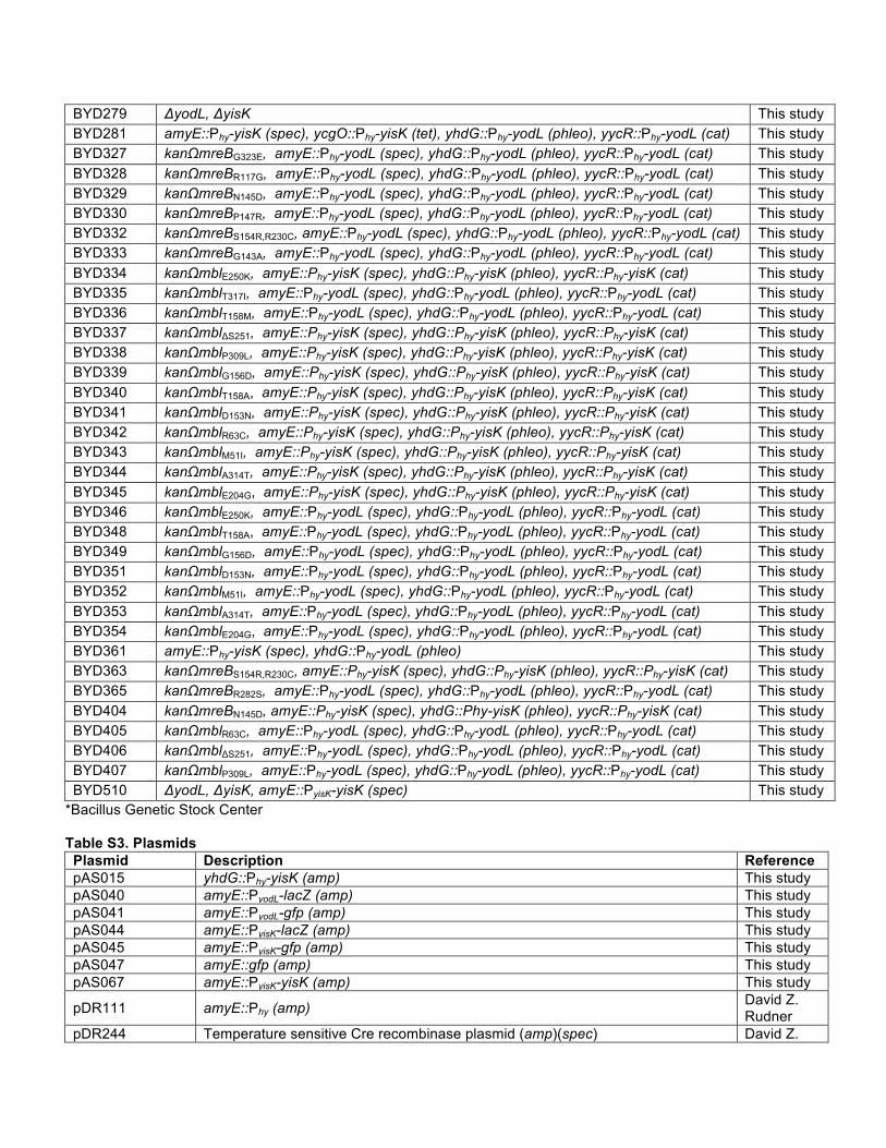
BYD279 ΔyodL, ΔyisK This study BYD281 amyE::Phy-yisK (spec), ycgO::Phy-yisK (tet), yhdG::Phy-yodL (phleo), yycR::Phy-yodL (cat) This study BYD327 kanΩmreBG323E, amyE::Phy-yodL (spec), yhdG::Phy-yodL (phleo), yycR::Phy-yodL (cat) This study BYD328 kanΩmreBR117G, amyE::Phy-yodL (spec), yhdG::Phy-yodL (phleo), yycR::Phy-yodL (cat) This study BYD329 kanΩmreBN145D, amyE::Phy-yodL (spec), yhdG::Phy-yodL (phleo), yycR::Phy-yodL (cat) This study BYD330 kanΩmreBP147R, amyE::Phy-yodL (spec), yhdG::Phy-yodL (phleo), yycR::Phy-yodL (cat) This study BYD332 kanΩmreBS154R,R230C, amyE::Phy-yodL (spec), yhdG::Phy-yodL (phleo), yycR::Phy-yodL (cat) This study BYD333 kanΩmreBG143A, amyE::Phy-yodL (spec), yhdG::Phy-yodL (phleo), yycR::Phy-yodL (cat) This study BYD334 kanΩmblE250K, amyE::Phy-yisK (spec), yhdG::Phy-yisK (phleo), yycR::Phy-yisK (cat) This study BYD335 kanΩmblT317I, amyE::Phy-yodL (spec), yhdG::Phy-yodL (phleo), yycR::Phy-yodL (cat) This study BYD336 kanΩmblT158M, amyE::Phy-yodL (spec), yhdG::Phy-yodL (phleo), yycR::Phy-yodL (cat) This study BYD337 kanΩmblΔS251, amyE::Phy-yisK (spec), yhdG::Phy-yisK (phleo), yycR::Phy-yisK (cat) This study BYD338 kanΩmblP309L, amyE::Phy-yisK (spec), yhdG::Phy-yisK (phleo), yycR::Phy-yisK (cat) This study BYD339 kanΩmblG156D, amyE::Phy-yisK (spec), yhdG::Phy-yisK (phleo), yycR::Phy-yisK (cat) This study BYD340 kanΩmblT158A, amyE::Phy-yisK (spec), yhdG::Phy-yisK (phleo), yycR::Phy-yisK (cat) This study BYD341 kanΩmblD153N, amyE::Phy-yisK (spec), yhdG::Phy-yisK (phleo), yycR::Phy-yisK (cat) This study BYD342 kanΩmblR63C, amyE::Phy-yisK (spec), yhdG::Phy-yisK (phleo), yycR::Phy-yisK (cat) This study BYD343 kanΩmblM51I, amyE::Phy-yisK (spec), yhdG::Phy-yisK (phleo), yycR::Phy-yisK (cat) This study BYD344 kanΩmblA314T, amyE::Phy-yisK (spec), yhdG::Phy-yisK (phleo), yycR::Phy-yisK (cat) This study BYD345 kanΩmblE204G, amyE::Phy-yisK (spec), yhdG::Phy-yisK (phleo), yycR::Phy-yisK (cat) This study BYD346 kanΩmblE250K, amyE::Phy-yodL (spec), yhdG::Phy-yodL (phleo), yycR::Phy-yodL (cat) This study BYD348 kanΩmblT158A, amyE::Phy-yodL (spec), yhdG::Phy-yodL (phleo), yycR::Phy-yodL (cat) This study BYD349 kanΩmblG156D, amyE::Phy-yodL (spec), yhdG::Phy-yodL (phleo), yycR::Phy-yodL (cat) This study BYD351 kanΩmblD153N, amyE::Phy-yodL (spec), yhdG::Phy-yodL (phleo), yycR::Phy-yodL (cat) This study BYD352 kanΩmblM51I, amyE::Phy-yodL (spec), yhdG::Phy-yodL (phleo), yycR::Phy-yodL (cat) This study BYD353 kanΩmblA314T, amyE::Phy-yodL (spec), yhdG::Phy-yodL (phleo), yycR::Phy-yodL (cat) This study BYD354 kanΩmblE204G, amyE::Phy-yodL (spec), yhdG::Phy-yodL (phleo), yycR::Phy-yodL (cat) This study BYD361 amyE::Phy-yisK (spec), yhdG::Phy-yodL (phleo) This study BYD363 kanΩmreBS154R,R230C, amyE::Phy-yisK (spec), yhdG::Phy-yisK (phleo), yycR::Phy-yisK (cat) This study BYD365 kanΩmreBR282S, amyE::Phy-yodL (spec), yhdG::Phy-yodL (phleo), yycR::Phy-yodL (cat) This study BYD404 kanΩmreBN145D, amyE::Phy-yisK (spec), yhdG::Phy-yisK (phleo), yycR::Phy-yisK (cat) This study BYD405 kanΩmblR63C, amyE::Phy-yodL (spec), yhdG::Phy-yodL (phleo), yycR::Phy-yodL (cat) This study BYD406 kanΩmblΔS251, amyE::Phy-yodL (spec), yhdG::Phy-yodL (phleo), yycR::Phy-yodL (cat) This study BYD407 kanΩmblP309L, amyE::Phy-yodL (spec), yhdG::Phy-yodL (phleo), yycR::Phy-yodL (cat) This study BYD510 ΔyodL, ΔyisK, amyE::PyisK-yisK (spec) This study
*Bacillus Genetic Stock Center
Table S3. Plasmids Plasmid Description Reference pAS015 yhdG::Phy-yisK (amp) This study pAS040 amyE::PyodL-lacZ (amp) This study pAS041 amyE::PyodL-gfp (amp) This study pAS044 amyE::PyisK-lacZ (amp) This study pAS045 amyE::PyisK-gfp (amp) This study pAS047 amyE::gfp (amp) This study pAS067 amyE::PyisK-yisK (amp) This study
pDR111 amyE::Phy (amp) David Z. Rudner
pDR244 Temperature sensitive Cre recombinase plasmid (amp)(spec) David Z.
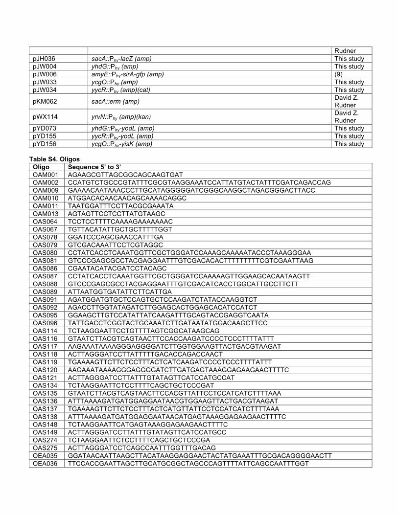
Rudner pJH036 sacA::Phy-lacZ (amp) This study pJW004 yhdG::Phy (amp) This study pJW006 amyE::Phy-sirA-gfp (amp) (9) pJW033 ycgO::Phy (amp) This study pJW034 yycR::Phy (amp)(cat) This study
pKM062 sacA::erm (amp) David Z. Rudner
pWX114 yrvN::Phy (amp)(kan) David Z. Rudner
pYD073 yhdG::Phy-yodL (amp) This study pYD155 yycR::Phy-yodL (amp) This study pYD156 ycgO::Phy-yisK (amp) This study
Table S4. Oligos Oligo Sequence 5’ to 3’ OAM001 AGAAGCGTTAGCGGCAGCAAGTGAT OAM002 CCATGTCTGCCCGTATTTCGCGTAAGGAAATCCATTATGTACTATTTCGATCAGACCAG OAM009 GAAAACAATAAACCCTTGCATAGGGGGATCGGGCAAGGCTAGACGGGACTTACC OAM010 ATGGACACAACAACAGCAAAACAGGC OAM011 TAATGGATTTCCTTACGCGAAATA OAM013 AGTAGTTCCTCCTTATGTAAGC OAS064 TCCTCCTTTTCAAAAGAAAAAAAC OAS067 TGTTACATATTGCTGCTTTTTGGT OAS078 GGATCCCAGCGAACCATTTGA OAS079 GTCGACAAATTCCTCGTAGGC OAS080 CCTATCACCTCAAATGGTTCGCTGGGATCCAAAGCAAAAATACCCTAAAGGGAA OAS081 GTCCCGAGCGCCTACGAGGAATTTGTCGACACACTTTTTTTTTCGTCGAATTAAG OAS086 CGAATACATACGATCCTACAGC OAS087 CCTATCACCTCAAATGGTTCGCTGGGATCCAAAAAGTTGGAAGCACAATAAGTT OAS088 GTCCCGAGCGCCTACGAGGAATTTGTCGACATCACCTGGCATTGCCTTCTT OAS089 ATTAATGGTGATATTCTTCATTGA OAS091 AGATGGATGTGCTCCAGTGCTCCAAGATCTATACCAAGGTCT OAS092 AGACCTTGGTATAGATCTTGGAGCACTGGAGCACATCCATCT OAS095 GGAAGCTTGTCCATATTATCAAGATTTGCAGTACCGAGGTCAATA OAS096 TATTGACCTCGGTACTGCAAATCTTGATAATATGGACAAGCTTCC OAS114 TCTAAGGAATTCCTGTTTTAGTCGGCATAAGCAG OAS116 GTAATCTTACGTCAGTAACTTCCACCAAGATCCCCTCCCTTTTATTT OAS117 AAGAAATAAAAGGGAGGGGATCTTGGTGGAAGTTACTGACGTAAGAT OAS118 ACTTAGGGATCCTTATTTTTGACACCAGACCAACT OAS119 TGAAAAGTTCTTCTCCTTTACTCATCAAGATCCCCTCCCTTTTATTT OAS120 AAGAAATAAAAGGGAGGGGATCTTGATGAGTAAAGGAGAAGAACTTTTC OAS121 ACTTAGGGATCCTTATTTGTATAGTTCATCCATGCCAT OAS134 TCTAAGGAATTCTCCTTTTCAGCTGCTCCCGAT OAS135 GTAATCTTACGTCAGTAACTTCCACGTTATTCCTCCATCATCTTTTAAA OAS136 ATTTAAAAGATGATGGAGGAATAACGTGGAAGTTACTGACGTAAGAT OAS137 TGAAAAGTTCTTCTCCTTTACTCATGTTATTCCTCCATCATCTTTTAAA OAS138 ATTTAAAAGATGATGGAGGAATAACATGAGTAAAGGAGAAGAACTTTTC OAS148 TCTAAGGAATTCATGAGTAAAGGAGAAGAACTTTTC OAS149 ACTTAGGGATCCTTATTTGTATAGTTCATCCATGCC OAS274 TCTAAGGAATTCTCCTTTTCAGCTGCTCCCGA OAS275 ACTTAGGGATCCTCAGCCAATTTGGTTTGACAG OEA035 GGATAACAATTAAGCTTACATAAGGAGGAACTACTATGAAATTTGCGACAGGGGAACTT OEA036 TTCCACCGAATTAGCTTGCATGCGGCTAGCCCAGTTTTATTCAGCCAATTTGGT
OEA275 GGATAACAATTAAGCTTACATAAGGAGGAACTACTATGATGTTATCCGTGTTTAAAAAG OEA276 TTCCACCGAATTAGCTTGCATGCGGCTAGCTTTCTTTTCATTATGTCGTTTGTA OJH159 CTGCAGGAATTCGACTCTCTA OJH160 TAGCTTGCATGCGGCTAGC OJH185 CAGGAATTCGACTCTCTAGC OJH186 CTCAGCTAGCTAACTCACATTAATTGCGTTGC
Strain Construction The Gibson enzymatic assembly method has been described in detail (10). Except where otherwise indicated, PCR amplifications are performed using B. subtilis 168 genomic DNA as template. To generate unmarked derivatives the BGSC strains (BKE22320, BKE19640, and BKE10750) each strain was transformed with pDR244, selecting for spectinomycin resistance at 30°C. Isolated colonies were restreaked on LB media and placed at 42°C. Isolated colonies from these plates were streaked again on spectinomycin, MLS, and LB plates. Clones that were sensitive to both spectinomycin and MLS were used to generate freezer stocks. Loss of the erm resistance cassette was confirmed by PCR. Moving suppressor mutations in mbl into clean genetic backgrounds To move the mutant mbl alleles obtained in the suppressor selections, we generated linear Gibson assembly products corresponding to a region of ~1,500 bp upstream of mbl (“UP”), a kanamycin resistance cassette (“KAN”), and each mutant mbl allele plus ~1,500 bp downstream of mbl (“DOWN”). This linear double stranded DNA product was then transformed directly into B. subtilis. Double cross-over recombination was selected for by plating on LB plates supplemented with 10 µg/ml kanamycin. To confirm the presence of the relevant mutations, and the absence of others, the entire mbl ORF was sequenced. The “UP” fragment comprised a region of homology to the B. subtilis chromosome upstream of mbl (including a region downstream of spoIIID), and was amplified from genomic DNA using oAS86 and oAS87. The “KAN” fragment was made by amplifying a kanamycin cassette from pWX114a using oAS78 and oAS79. The “DOWN” fragment corresponded to a region including the mbl promoter, mbl and 1500bp downstream of mbl and was amplified from genomic DNA of each corresponding suppressor strain using oAS88 and oAS89. For each allele of interest, the three products were combined in single Gibson assembly reaction and transformed directly into B. subtilis 168, selecting for kanamycin resistance. To confirm the presence of the relevant mutation(s) and the absence of others, the entire mbl ORF was sequenced. Moving suppressor mutations in mreB into clean genetic backgrounds To move the mutant mreB alleles obtained in the suppressor selections, we generated linear Gibson assembly products corresponding to a region of ~1,500 bp upstream of mreB (“UP”), a kanamycin resistance cassette (“KAN”), and each mutant mreB allele plus ~1,500 bp downstream of mreB (“DOWN”). This linear double stranded DNA product was then transformed directly into B. subtilis. Double cross-over recombination was selected for by plating on LB plates supplemented with 10 µg/ml kanamycin. To confirm the presence of the relevant mutations, and the absence of others, the entire mreB ORF was sequenced. The “UP” fragment comprised a region of homology to the B. subtilis chromosome upstream of mreB (including a region downstream of radC), and was amplified from genomic DNA using oAS64 and oAS80. The “KAN” fragment was made by amplifying a kanamycin cassette from pWX114a using oAS78 and oAS79. The “DOWN” fragment corresponded to a region including the mreB promoter, mreB and 1500bp downstream of mreB and was amplified from genomic DNA of each corresponding suppressor strain using oAS81 and oAS67. For each allele of interest, the three products were combined in single Gibson assembly reaction and transformed
directly into B. subtilis 168, selecting for kanamycin resistance. To confirm the presence of the relevant mutation(s) and the absence of others, the entire mreB ORF was sequenced.
BAS040 was generated by transforming a linear Gibson assembly product encoding a region upstream of amyE, a spectinomycin cassette, the Phy promoter, an optimized ribosome binding site, yodL, lacI, and a region downstream of amyE into B. subtilis 168, and selecting for spectinomycin resistance. The final strain was confirmed by PCR and sequencing. The region of upstream of amyE was PCR amplified from B. subtilis 168 genomic DNA using OAM009 and OAM010. The spectinomycin resistance cassette and the Phy promoter were PCR amplified from pDR111 using primers OAM10 and OAM13, and included a ribosome binding site (TAAGGAGG). The yodL ORF was PCR amplified using OEA275 and OEA276. lacI was PCR amplified from pDR111 using OAM011 and OAM012. The region downstream of amyE was PCR amplified from B. subtilis 168 genomic DNA using OAM001 and OAM002.
BAS041 was generated by transforming a linear Gibson assembly product encoding a region upstream of amyE, a spectinomycin cassette, the Phy promoter, an optimized ribosome binding site, yisK, lacI, and a region downstream of amyE into B. subtilis 168, and selecting for spectinomycin resistance. The final strain was confirmed by PCR and sequencing. The region of upstream of amyE was PCR amplified from B. subtilis 168 genomic DNA using OAM009 and OAM010. The spectinomycin resistance cassette and the Phy promoter were PCR amplified from pDR111 using primers OAM10 and OAM13, and included a ribosome binding site (TAAGGAGG). The yisK ORF was PCR amplified using OEA035 and OEA036. lacI was PCR amplified from pDR111 using OAM011 and OAM012. The region downstream of amyE was PCR amplified from B. subtilis 168 genomic DNA using OAM001 and OAM002. BAS147 was generated by directly transforming a Gibson assembly of a region upstream of mbl, a kanamycin resistance cassette, and an mbl knockout fragment (see below) directly into BKE22320, selecting for kanamycin resistance in the presence of 10 mM MgCl2. The region upstream of mbl was amplified using OAS86 and OAS87 (B. subtilis 168 genomic DNA template). The kanamycin resistance cassette was amplified from pWX114a using OAS78 and OAS79. The mbl knockout fragment was made by amplifying a DNA product that included the first 42 bp of mbl and its upstream region using oAS86 and oAS95 (B. subtilis 168 genomic DNA template), amplifying a DNA product that included the last 42 bp mbl and its downstream region using oAS96 and oAS89 (B. subtilis 168 genomic DNA template), and splicing the two fragments together using overlap extension PCR with OAS088 and OAS089. The kanamycin-linked mbl knockout was confirmed by PCR amplification. BAS170 was made by transforming B. subtilis 168 with pAS040 linearized with ScaI and selecting for spectinomycin resistance. Integrations were confirmed by starch test. BAS171 was made by transforming B. subtilis 168 with pAS041 linearized with ScaI and selecting for spectinomycin resistance. Integrations were confirmed by starch test. BAS192 was made by transforming B. subtilis 168 with pAS044 linearized with ScaI and selecting for spectinomycin resistance. Integrations were confirmed by starch test and sequencing. BAS193 was made by transforming B. subtilis 168 with pAS045 linearized with ScaI and selecting for spectinomycin resistance. Integrations were confirmed by starch test and sequencing. BAS265 was generated by transforming B. subtilis 168 with genomic DNA from RL891 and selecting for erythromycin resistance (erm). Recommend growth on 0.5 µg/ml erythromycin (erm) and 12.5 µg/ml lincomycin (0.5X normal antibiotic concentration).
BAS282 was generated by transforming B. subtilis 168 with genomic DNA from BJH154 and selecting on chloramphenicol. BYD258 was generated by directly transforming a Gibson assembly of a region upstream of mbl, a kanamycin resistance cassette, and an mbl knockout fragment (see below) directly into BYD175, selecting for kanamycin resistance in the presence of 10 mM MgCl2. The region upstream of mbl was amplified using OAS86 and OAS87 (B. subtilis 168 genomic DNA template). The kanamycin resistance cassette was amplified from pWX114a using OAS78 and OAS79. The mbl knockout fragment was made by amplifying a DNA product that included the first 42 bp of mbl and its upstream region using oAS86 and oAS95 (B. subtilis 168 genomic DNA template), amplifying a DNA product that included the last 42 bp mbl and its downstream region using oAS96 and oAS89 (B. subtilis 168 genomic DNA template), and splicing the two fragments together using overlap extension PCR with OAS088 and OAS089. The kanamycin-linked mbl knockout was confirmed by PCR amplification. BYD259 was generated as described for BYD258, but transforming the final Gibson assembly into BYD176. The kanamycin-linked mbl knockout was confirmed by PCR amplification. BYD262 was generated by directly transforming a Gibson assembly of a region upstream of mreB, a kanamycin resistance cassette, and an mreB knockout fragment (see below) directly into BYD175, selecting for kanamycin resistance in the presence of 10 mM MgCl2. The region upstream of mreB was amplified using OAS64 and OAS80 (B. subtilis 168 genomic DNA template) and was designed to include the putative radC terminator/mreB promoter region to avoid the chances of disrupting the terminator. The kanamycin resistance cassette was amplified from pWX114a using OAS78 and OAS79. The mreB knockout fragment was made by amplifying the mreBCD promoter region and the first 42bp of mreB using oAS64 and oAS91 (B. subtilis 168 genomic DNA template), amplifying the last 42bp of mreB along with region downstream of mreB using OAS92 and OAS67 (B. subtilis 168 genomic DNA template), and splicing the two fragments together using overlap extension PCR with oAS81 and oAS67. The kanamycin-linked mreB knockout was confirmed by PCR amplification. The final integration product encodes identical sequences before and after the kanamycin cassette, so loss of the kanamycin resistance cassette is possible in the absence of selective pressure. BYD263 was generated as described for BYD262, but transforming the final Gibson assembly into BYD176. The kanamycin-linked mreB knockout was confirmed by PCR amplification. Plasmid construction pAS015 was generated by cloning PCR product from OJH159 and OJH160 amplification of genomic DNA from BAS041 into pJW004 (EcoRI-NheI). pAS040 was generated with overlap extension PCR. The “UP” product encoding the yodL promoter region was amplified from B. subtilis 168 genomic DNA with primer pair OAS114/OAS116. The “DOWN” product encoding lacZ was amplified from genomic DNA from SYL#8 (Table S1) using primer pair OAS117/OAS118. The two PCR products were used as template for overlap extension PCR using primer pair OAS114/OAS118. The amplified fragment was cut with EcoRI and BamHI and cloned into pDR111 cut with the same enzymes.
pAS041 was generated with overlap extension PCR. The “UP” product was amplified from B. subtilis 168 genomic DNA with primer pair OAS114/OAS119. The “DOWN” product was amplified from pJW006 with primer pair OAS120/ OAS121. The two PCR products were used as template for overlap extension PCR with primer pair OAS114/OAS121. The amplified fragment was cut with EcoRI and BamHI and cloned into pDR111 cut with the same enzymes.
pAS044 was generated with overlap extension PCR. The “UP” product encoding the yisK promoter region was amplified from B. subtilis 168 genomic DNA template using primer pair OAS134/OAS135. The “DOWN” product encoding the lacZ gene was amplified from genomic DNA of SYL#8 (Table S1) with primer pair OAS136/OAS118. The two PCR products were used as template for overlap extension PCR using primer pair OAS134/OAS118. The amplified fragment was cut with EcoRI and BamHI and cloned into pDR111 cut with the same enzymes.
pAS045 was generated with overlap extension PCR. The “UP” product was amplified from B. subtilis 168 genomic DNA template using primer pair OAS134/OAS137. The “DOWN” product was amplified from pJW006 with primer pair OAS138/ OAS121. The two PCR products were used as template for overlap extension PCR with primer pair OAS134/OAS121. The amplified fragment was cut with EcoRI and BamHI and cloned into pDR111 cut with the same enzymes.
pAS047 was generated by cloning PCR product from an OAS148 and OAS149 amplification of pJW006 into pDR111 (EcoRI-BamHI). pAS067 was generated by cloning the PCR product obtained from an OAS274 and OAS275 amplification of B. subtilis 168 genomic DNA template (encoding 200bp upstream of yisK and yisK) into pDR111 (EcoRI-BamHI). pJH036 was generated by cloning the PCR product obtained from an OJH185 and OJH186 amplification of genomic DNA from BDR992 (encoding Phy-lacZ) into pKM062 (EcoRI-NheI). pJW004 was generated by cloning the ~1770 bp BamHI-EcoRI fragment from pDR111 (encoding the Phy promotor and lacI) into pBB280, an allelic exchange vector that integrates at the yhdG locus and confers phleomycin resistance. pJW033 was generated by cloning the ~1770 bp BamHI-EcoRI fragment from pDR111 (encoding the Phy promotor and lacI) into pKM086, an allelic integration vector that integrates at the ycgO locus and confers tetracycline resistance. pJW034 was was generated by cloning the ~1770 bp BamHI-EcoRI fragment from pDR111 (encoding the Phy promotor and lacI) into pNS037, an allelic exchange vector that integrates at the yycR locus and confers chloramphenicol resistance.
pYD073 was generated by cloning PCR product from OJH159 and OJH160 amplification using genomic DNA from BAS040 (amyE::Phy-yodL) and cloning into the into the EcoRI-NheI sites of pJW004. pYD155 was generated by cloning PCR product from OJH159 and OJH160 amplification using genomic DNA from BAS040 and cloning into the into the EcoRI-NheI sites of pJW034. pYD156 was generated by cloning PCR product from OJH159 and OJH160 amplification using genomic DNA from BAS040 and cloning into the into the EcoRI-NheI sites of pJW033.
REFERENCES
1. van den Ent F, Johnson CM, Persons L, de Boer P, Lowe J. 2010. Bacterial actin MreB assembles in complex with cell shape protein RodZ. EMBO J 29:1081-1090.
2. Yang J, Yan R, Roy A, Xu D, Poisson J, Zhang Y. 2015. The I-TASSER Suite: protein structure and function prediction. Nat Methods 12:7-8.
3. van den Ent F, Amos LA, Lowe J. 2001. Prokaryotic origin of the actin cytoskeleton. Nature 413:39-44.
4. Dye NA, Pincus Z, Fisher IC, Shapiro L, Theriot JA. 2011. Mutations in the nucleotide binding pocket of MreB can alter cell curvature and polar morphology in Caulobacter. Mol Microbiol 81:368-394.
5. Gitai Z, Dye NA, Reisenauer A, Wachi M, Shapiro L. 2005. MreB actin-mediated segregation of a specific region of a bacterial chromosome. Cell 120:329-341.
6. Srivastava P, Demarre G, Karpova TS, McNally J, Chattoraj DK. 2007. Changes in nucleoid morphology and origin localization upon inhibition or alteration of the actin homolog, MreB, of Vibrio cholerae. J Bacteriol 189:7450-7463.
7. van den Ent F, Izore T, Bharat TA, Johnson CM, Lowe J. 2014. Bacterial actin MreB forms antiparallel double filaments. Elife 3:e02634.
8. Branda SS, Gonzalez-Pastor JE, Ben-Yehuda S, Losick R, Kolter R. 2001. Fruiting body formation by Bacillus subtilis. Proc Natl Acad Sci U S A 98:11621-11626.
9. Wagner JK, Marquis KA, Rudner DZ. 2009. SirA enforces diploidy by inhibiting the replication initiator DnaA during spore formation in Bacillus subtilis. Mol Microbiol 73:963-974.
10. Gibson DG, Young L, Chuang RY, Venter JC, Hutchison CA, 3rd, Smith HO. 2009. Enzymatic assembly of DNA molecules up to several hundred kilobases. Nat Methods 6:343-345.
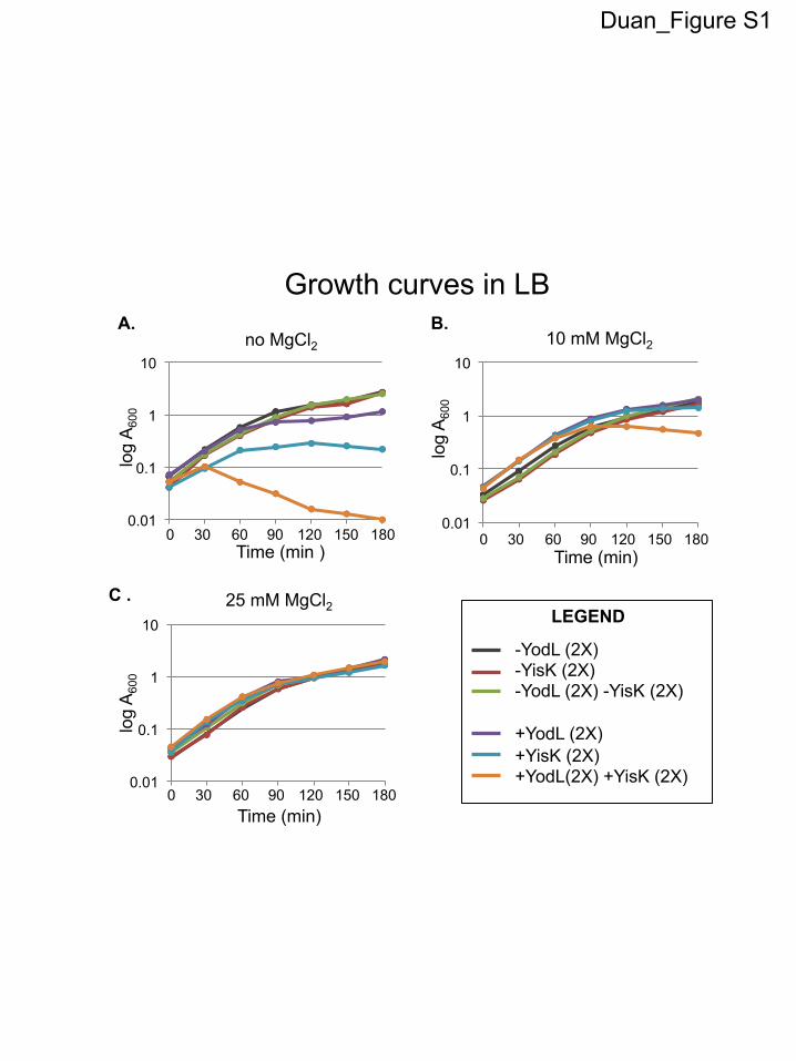
Duan_Figure S1
-YodL (2X) -YisK (2X) -YodL (2X) -YisK (2X) +YodL (2X) +YisK (2X) +YodL(2X) +YisK (2X)
LEGEND
log
A60
0
0.01
0.1
1
10 no MgCl2
Time (min )
0 30 60 90 120 150 180 0.01
0.1
1
10 10 mM MgCl2
log
A60
0
Time (min)
0 30 60 90 120 150 180
0.01
0.1
1
10 25 mM MgCl2
log
A60
0
0 30 60 90 120 150 180 Time (min)
Growth curves in LB A. B.
C .
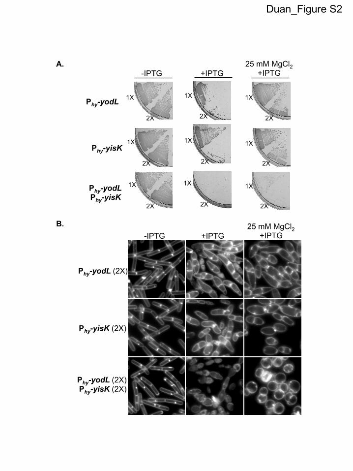
Duan_Figure S2
A.
B.
-IPTG
Phy-yodL
Phy-yisK
+IPTG 25 mM MgCl2
+IPTG
1X
2X
1X
2X
1X
2X
1X
2X
1X
2X
1X
2X
Phy-yodL Phy-yisK
1X
2X
1X
2X
1X
2X
Phy-yisK (2X)
Phy-yodL (2X)
-IPTG +IPTG 25 mM MgCl2
+IPTG
Phy-yodL (2X) Phy-yisK (2X)
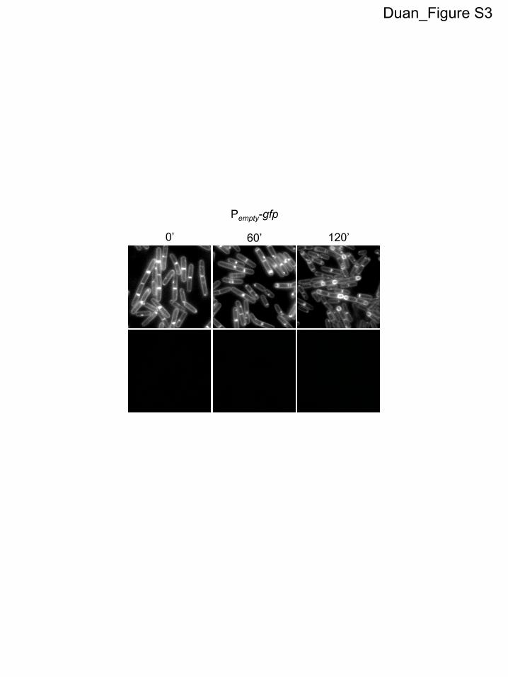
Duan_Figure S3
0’ 60’ 120’
Pempty-gfp
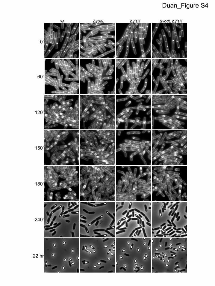
Duan_Figure S4
wt ΔyodL ΔyisK ΔyodL ΔyisK
0’
60’
120’
180’
240’
150’
22 hr
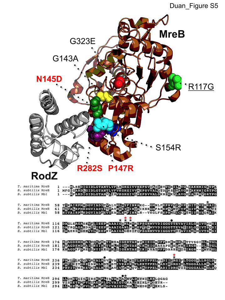
Duan_Figure S5
RodZ
R117G
G323E
N145D
P147R
S154R
G143A
R282S
MreB
1_Tmaritima_MreB 1 ---MLRKDIGIDLGTANTLVFLRGKGIVVNEPSVIAIDSTTGEILKVGLEAKNMIGKTPA2_Bsubtilis_MreB 1 MFGIGARDLGIDLGTANTLVFVKGKGIVVREPSVVALQTDTKSIVAVGNDAKNMIGRTPG3_Bsubtilis_Mbl 1 ---MFARDIGIDLGTANVLIHVKGKGIVLNEPSVVALDKNSGKVLAVGEEARRMVGRTPG
1_Tmaritima_MreB 58 TIKAIRPMRDGVIADYTVALVMLRYFINKAKG--GMNLFKPRVVIGVPIGITDVERRAIL2_Bsubtilis_MreB 61 NVVALRPMKDGVIADYETTATMMKYYINQAIKNKGMFARKPYVMVCVPSGITAVEERAVI3_Bsubtilis_Mbl 58 NIVAIRPLKDGVIADFEVTEAMLKHFINKLN--VKGLFSKPRMLICCPTNITSVEQKAIK
1_Tmaritima_MreB 116 DAGLEAGASKVFLIEEPMAAAIGSNLNVEEPSGNMVVDIGGGTTEVAVISLGSIVTWESI2_Bsubtilis_MreB 121 DATRQAGARDAYPIEEPFAAAIGANLPVWEPTGSMVVDIGGGTTEVAIISLGGIVTSQSI3_Bsubtilis_Mbl 116 EAAEKSGGKHVYLEEEPKVAAIGAGMEIFQPSGNMVVDIGGGTTDIAVISMGDIVTSSSI
1_Tmaritima_MreB 176 RIAGDEMDEAIVQYVRETYRVAIGERTAERVKIEIGNVFPSKENDELETTVSGIDLSTGL2_Bsubtilis_MreB 181 RVAGDEMDDAIINYIRKTYNLMIGDRTAEAIKMEIGSAEAPEESDNMEIRGR--DLLTGL3_Bsubtilis_Mbl 176 KMAGDKFDMEILNYIKREYKLLIGERTAEDIKIKVATVFPDARHEEISIRGR--DMVSGL
1_Tmaritima_MreB 236 PRKLTLKGGEVREALRSVVVAIVESVRTTLEKTPPELVSDIIERGIFLTGGGSLLRGLDT2_Bsubtilis_MreB 239 PKTIEITGKEISNALRDTVSTIVEAVKSTLEKTPPELAADIMDRGIVLTGGGALLRNLDK3_Bsubtilis_Mbl 234 PRTITVNSKEVEEALRESVAVIVQAAKQVLERTPPELSADIIDRGVIITGGGALLNGLDQ
1_Tmaritima_MreB 296 LLQKETGISVIRSEEPLTAVAKGAGMVLDKVNILKKLQGAG2_Bsubtilis_MreB 299 VISEETKMPVLIAEDPLDCVAIGTGKALEHIHLFKGKTR--3_Bsubtilis_Mbl 294 LLAEELKVPVLVAENPMDCVAIGTGVMLDNMDKLPKRKLS-
T. maritima MreBB. subtilis MreBB. subtilis Mbl
T. maritima MreBB. subtilis MreBB. subtilis Mbl
T. maritima MreBB. subtilis MreBB. subtilis Mbl
T. maritima MreBB. subtilis MreBB. subtilis Mbl
T. maritima MreBB. subtilis MreBB. subtilis Mbl
T. maritima MreBB. subtilis MreBB. subtilis Mbl
*
*
* * * *
* *
* * �
�
�
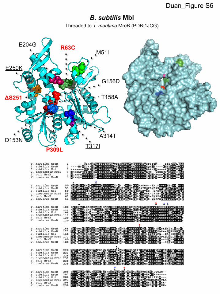
B. subtilis Mbl Threaded to T. maritima MreB (PDB:1JCG)
Duan_Figure S6
E204G R63C M51I
G156D
T158A
A314T
P309L D153N
ΔS251
T317I
E250K
Tmaritima_MreB 1 -------MLRKDIGIDLGTANTLVFLRGKGIVVNEPSVIAIDSTTG----EILKVGLEAKBsubtilis_MreB 1 ----MFGIGARDLGIDLGTANTLVFVKGKGIVVREPSVVALQTDT----KSIVAVGNDAKBsubtilis_Mbl 1 -------MFARDIGIDLGTANVLIHVKGKGIVLNEPSVVALDKNSG----KVLAVGEEARCcrescentus_MreB 1 MFSSLFGVISNDIAIDLGTANTLIYQKGKGIVLNEPSVVALRN--VGGRKVVHAVGIEAKEcoli 1 MLKKFRGMFSNDLSIDLGTANTLIYVKGQGIVLNEPSVVAIRQDRAGSPKSVAAVGHDAKVcholerae 1 MFKKLRGMFSNDLSIDLGTANTLIYVKGQGIVLDEPSVVAIRQDKGRGGKTVAAVGHAAK
Tmaritima_MreB 50 NMIGKTPATIKAIRPMRDGVIADYTVALVMLRYFINKAKGG--MNLFKPRVVIGVPIGITBsubtilis_MreB 53 NMIGRTPGNVVALRPMKDGVIADYETTATMMKYYINQAIKNKGMFARKPYVMVCVPSGITBsubtilis_Mbl 50 RMVGRTPGNIVAIRPLKDGVIADFEVTEAMLKHFINKLNVKG--LFSKPRMLICCPTNITCcrescentus_MreB 59 QMLGRTPGHMEAIRPMRDGVIADFEVAEEMIKYFIRKVHNRK-GFVN-PKVIVCVPSGATEcoli 61 QMLGRTPGNIAAIRPMKDGVIADFFVTEKMLQHFIKQVHSNS-FMRPSPRVLVCVPVGATVcholerae 61 QMLGRTPGNISAIRPMKDGVIADFYVTEKMLQHFIRQVHDNS-VLKPSPRVLVCVPCGST
Tmaritima_MreB 108 DVERRAILDAGLEAGASKVFLIEEPMAAAIGSNLNVEEPSGNMVVDIGGGTTEVAVISLGBsubtilis_MreB 113 AVEERAVIDATRQAGARDAYPIEEPFAAAIGANLPVWEPTGSMVVDIGGGTTEVAIISLGBsubtilis_Mbl 108 SVEQKAIKEAAEKSGGKHVYLEEEPKVAAIGAGMEIFQPSGNMVVDIGGGTTDIAVISMGCcrescentus_MreB 117 AVERRAINDSCLNAGARRVGLIDEPMAAAIGAGLPIHEPTGSMVVDIGGGTTEVAVLSLSEcoli 120 QVERRAIRESAQGAGAREVFLIEEPMAAAIGAGLPVSEATGSMVVDIGGGTTEVAVISLNVcholerae 120 QVERRAIRESALGAGAREVYLIDEPMAAAIGAGLRVSEPTGSMVIDIGGGTTEVAVISLN
Tmaritima_MreB 168 SIVTWESIRIAGDEMDEAIVQYVRETYRVAIGERTAERVKIEIGNVFPSKENDELETTVSBsubtilis_MreB 173 GIVTSQSIRVAGDEMDDAIINYIRKTYNLMIGDRTAEAIKMEIGSAEAP--EESDNMEIRBsubtilis_Mbl 168 DIVTSSSIKMAGDKFDMEILNYIKREYKLLIGERTAEDIKIKVATVFP--DARHEEISIRCcrescentus_MreB 177 GIVYSRSVRVGGDKMDEAIISYMRRHHNLLIGETTAERIKKEIGTARAPADGEGLSIDVKEcoli 180 GVVYSSSVRIGGDRFDEAIINYVRRNYGSLIGEATAERIKHEIGSAYP--GDEVREIEVRVcholerae 180 GVVYSSSVRIGGDRFDEAIINYVRRNYGSLIGEATAEKIKHEIGSAYP--GDDVQEIEVR
Tmaritima_MreB 228 GIDLSTGLPRKLTLKGGEVREALRSVVVAIVESVRTTLEKTPPELVSDIIERGIFLTGGGBsubtilis_MreB 231 GRDLLTGLPKTIEITGKEISNALRDTVSTIVEAVKSTLEKTPPELAADIMDRGIVLTGGGBsubtilis_Mbl 226 GRDMVSGLPRTITVNSKEVEEALRESVAVIVQAAKQVLERTPPELSADIIDRGVIITGGGCcrescentus_MreB 237 GRDLMQGVPREVRISEKQAADALAEPVGQIVEAVKVALEATPPELASDIADKGIMLTGGGEcoli 238 GRNLAEGVPRGFTLNSNEILEALQEPLTGIVSAVMVALEQCPPELASDISERGMVLTGGGVcholerae 238 GRNLAEGVPRSFTLNSNEILEALQEPLTGIVSAVMVALEQCPPELASDISENGMVLTGGG
Tmaritima_MreB 288 SLLRGLDTLLQKETGISVIRSEEPLTAVAKGAGMVLDKVNILKKLQGAG--Bsubtilis_MreB 291 ALLRNLDKVISEETKMPVLIAEDPLDCVAIGTGKALEHIHLFKGKTR----Bsubtilis_Mbl 286 ALLNGLDQLLAEELKVPVLVAENPMDCVAIGTGVMLDNMDKLPKRKLS---Ccrescentus_MreB 297 ALLRGLDAEIRDHTGLPVTVADDPLSCVALGCGKVLEHPKWMKGVLESTLAEcoli 298 ALLRNLDRLLMEETGIPVVVAEDPLTCVARGGGKALEMIDMHGGDLFSEE-Vcholerae 298 ALLKDLDRLLMEETGIPVVIADDPLTCVARGGGKALEMIDMHGGDLFSEE-
T. maritima MreB
C. crescentus MreBB. subtilis MblB. subtilis MreB
E. coli MreBV. cholerae MreB
T. maritima MreB
C. crescentus MreBB. subtilis MblB. subtilis MreB
E. coli MreBV. cholerae MreB
T. maritima MreB
C. crescentus MreBB. subtilis MblB. subtilis MreB
E. coli MreBV. cholerae MreB
T. maritima MreB
C. crescentus MreBB. subtilis MblB. subtilis MreB
E. coli MreBV. cholerae MreB
T. maritima MreB
C. crescentus MreBB. subtilis MblB. subtilis MreB
E. coli MreBV. cholerae MreB
T. maritima MreB
C. crescentus MreBB. subtilis MblB. subtilis MreB
E. coli MreBV. cholerae MreB
* * *
* * *
*
* *
* * * *
* * *
*
�